
HOW TO ACCESS ‘ACCESS SURGERY’ DATABASES AT LIBRARY PORTAL (lib.hukm.ukm.my)
Upload
jeya-sudhaharCategory
view
288download
3
Print | Close Window
Note: Large images and tables on this page may necessitate printing in landscape mode.
Maingot's Abdominal Operations > Chapter 4. Incisions, Closures, and Management of the AbdominalWound >
INCISIONSA well-calculated and well-performed incision is of paramount importance to abdominal surgery. Ofequal importance is a proper method of wound closure. Any error, such as a poorly chosen incision,unsatisfactory means to close, or unsuitable selection of suture material, may result in seriouscomplications including hematoma, hernia, wound infection, stitch abscess, unpleasant scar, and thedreaded wound dehiscence and evisceration.
In the most recent previous edition of this book, Ellis outlined three basic principles to guide selectionof the incision and closure of the wound. These are accessibility, flexibility, and security.1
Accessibility. The incision should provide direct and timely exposure to the diseased or injured anatomy andmust provide sufficient space for the procedure to be well performed. Exposure is greatly facilitated not onlyby a well-made incision, but also by the apt use of retractors and packs, correct posture of the patient onthe operating room table, and optimized lighting.
Flexibility. The incision should be amenable to extension if the complexity of the procedure demands greaterexposure than originally anticipated. It should, however, interfere as little as possible with the function ofthe abdominal wall, limiting sacrifice of nerve supply to the abdominal musculature, preferably the sacrificeof only a single segmental nerve trunk.
Security. Closure of the wound must be strong and reliable. Ideally, it should leave the abdominal wall withintegrity comparable to or superior to its preoperative state.
Types of IncisionsAbdominal incisions can be divided into four main anatomic categories.
Vertical. Vertical incisions may be midline or paramedian. They may be supraumbilical or infraumbilical andcan be extended superiorly or inferiorly in either direction. For optimal exposure of the entire abdominalcavity, as in the case of abdominal trauma, a midline vertical incision can be taken superiorly to the xiphoidprocess and inferiorly to the symphysis pubis.
Transverse and oblique. These incisions can be placed in any of the four quadrants of the abdomen.Common incisions include the Kocher subcostal incision for biliary surgery, the Pfannenstiel infraumbilicalincision for gynecologic surgery, the McBurney incision for appendectomy, and the transverse or obliquelateral incision for exposure of the colon.
Abdominothoracic. This incision provides superior exposure of the upper abdominal organs by joining theperitoneal cavity, pleural space, and mediastinum into a single operative field. It is particularly useful forextensive exposure of the liver and esophagogastric junction.
Retroperitoneal and extraperitoneal. These incisions are ideal for surgery of the kidney, adrenal gland,aorta, and for renal transplantation.
Choice of IncisionMany factors influence the choice of incision for abdominal surgery. These include the organ of interestand anticipated procedure, the body habitus of the patient and degree of obesity, the urgency of theoperation and whether speed is a pressing consideration, the presence of previous abdominal incisions,
AccessSurgery - Print file:///C:/Users/Jeyasudhahar/Desktop/II. Abdominal Wall/4. incisions, cl...
1 of 56 1/1/2011 9:41 PMCreate PDF files without this message by purchasing novaPDF printer (http://www.novapdf.com)

and the preference and experience of the operating surgeon.
Most surgeons prefer a midline or paramedian approach to the abdominal viscera. In emergencyoperations, the midline incision undoubtedly provides the most rapid access to the abdominal cavity,and if necessary can be extended swiftly to the whole length of the abdominal wall. Re-entry into theabdomen should be achieved through the previous incision, if possible. If the previous incision is weakor a site of incisional hernia, the abdominal wall can be repaired at this time. A new incision shouldnever be made closely parallel (within 5 cm) to the site of any previous incision without understandingthe distinct risk of ischemic necrosis of intervening skin and fascial bridges. Future surgical prospectsare considered and the incision should be placed so that it will not interfere with planned procedures,for example, to avoid or incorporate colostomies, fistulas, and the like. Similarly if the patient has thepotential for recurrent disease requiring re-exploration, consideration of location is important.
MIDLINE VERSUS TRANSVERSE INCISIONSIn thin patients with narrow subcostal angles, a transverse or oblique incision has little advantage;however, in obese patients with wide subcostal angles, a subcostal incision provides excellent exposureof the upper abdominal viscera, biliary tract, spleen, and pancreas. There are many occasions,however, in which either a vertical or transverse incision would provide appropriate an equal exposure.
Some authors feel that transverse incisions in abdominal surgery are based on better anatomic andsurgical principles than vertical incisions and should be preferred.2,3 Anatomically, the fascial fibers ofthe anterior abdominal wall lie in a transverse direction and are therefore divided by a vertical incision.Suture closure of a vertically placed wound places suture material between these fibers, as opposed tothe placement of suture material around these fibers as would be done in closing a transverse incision.Furthermore, tension lines lie mediolateral in vertically placed incisions and craniocaudal intransversely placed incisions. For these reasons, many surgeons believe that sutures placedperpendicularly to muscle fibers in transverse incisions are more secure and less liable to cut throughfascia.
A number of retrospective clinical studies and a meta-analysis have concluded that the transverseincision is superior to the vertical incision with regard to long-term and short-term outcomes, includingpostoperative pain, pulmonary complications, and frequencies of incisional hernia and burst abdomen.3
However, the vertical incision is still the most commonly performed incision in general surgery. Thisdiscrepancy is explained by a number of deficits in study design and analysis such as theunderpowering of clinical trials, lack of standardization of abdominal wall closure, nonblinded methods,and study inhomogeneities in meta-analysis.4 Clearly, only carefully designed randomized clinical trialscan compare one incision to another.
The only randomized controlled trial that has been performed that focused on the frequency of burstabdomen (i.e., evisceration) could find no benefit of transverse incision (0%) over midline incision(0.69%).5 Likewise, controlled clinical trials demonstrate no significant differences in incisional herniaswhen comparing transverse (0.85%) to midline incisions (3.85%),5 and transverse (14%) toparamedian incisions (14%).6 With regard to postoperative wound infections, there are no data tosupport a difference in infectious wound complications among these incisions.3 Data on early-onsetpulmonary complications are conflicting but in randomized trials they are equally distributed in eachgroup.7
MIDLINE VERSUS PARAMEDIAN INCISIONSThe vertical incision can be divided into three main subtypes: midline, medial paramedian, and lateralparamedian. The theoretical advantage of the paramedian incision over a midline incision is a
AccessSurgery - Print file:///C:/Users/Jeyasudhahar/Desktop/II. Abdominal Wall/4. incisions, cl...
2 of 56 1/1/2011 9:41 PMCreate PDF files without this message by purchasing novaPDF printer (http://www.novapdf.com)

diminished risk of wound dehiscence and incisional hernia. The paramedian incision enters theabdomen through dissection of the rectus muscle from its anterior and posterior sheaths. Upon closure,the rectus muscle should resume its original place and splint the defects in the anterior and posteriorrectus fascia. In actuality, however, when these incisions are reopened, the medial edge of the rectusmuscle is invariably noted to be adherent by scar to the posterior sheath incision and does noteffectively buttress the wound. The speculative advantages of the paramedian incision have beeninvestigated in prospective randomized trials demonstrating that the conventional (medial) paramedianincision offers no advantage in wound failure rates when compared to midline or transverse incisions.6
In 1980, Guillou described a modified paramedian incision, "the lateral paramedian incision," that iscreated several centimeters lateral to the traditional paramedian incision.8 Randomized prospectivedata show a statistically significantly decrease in incidence of incisional hernia in lateral paramedianincisions (0%) when compared to medial paramedian incisions (14.9%)8 and midline incisions (6.9%).9
The main disadvantage of the paramedian incision is the length of time needed to create the wound,which increases as the distance from the midline increases.
COLD BLADE VERSUS ELECTROCAUTERYAlthough it has become common practice for many surgeons to use electrocautery to create abdominalincisions, it is often taught that sharp dissection is superior to electrocautery for creation of theabdominal incision. These claims were supported by early animal data demonstrating higher woundinfection rates and poorer wound strength in incisions made with elecrocautery.10 More recentrandomized data have put this issue to rest. Six prospective randomized clinical trials have comparedcold blade scalpel to electrocautery for wound creation through skin and though the subcutaneouslayer.11–16 These trials differ with respect to incision (cholecystectomy, midline laparotomy, andthoracic incision), wound classification, and timing of surgery (elective or emergent). Their data havedemonstrated that incisions made with electrocautery are created significantly quicker, produce lessblood loss, allow for less postoperative pain, and produce no difference in short-term and long-termwound complications when compared to scalpel incisions.
Description of Individual IncisionsVERTICAL INCISIONS
Midline IncisionsThe midline incision is the fastest approach toward the peritoneal cavity and has a number ofadvantages. It offers adequate exposure to almost every region of the abdominal cavity andretroperitoneum. It is nearly bloodless and does not require division of muscle fibers or sectioning ofnerves. It is unsurpassed when speed is of the essence. The upper midline incision, or the epigastricmidline incision, provides exposure for most operations on the esophageal hiatus, abdominalesophagus and vagus nerves, stomach, duodenum, gallbladder, pancreas, and spleen (Fig 4–1). Thelower midline incision, or infraumbilical incision, is similar to the upper midline incision and mayextend superiorly to join it. It provides exposure for most operations on the lower abdominal and pelvicorgans.
Figure 4–1.
AccessSurgery - Print file:///C:/Users/Jeyasudhahar/Desktop/II. Abdominal Wall/4. incisions, cl...
3 of 56 1/1/2011 9:41 PMCreate PDF files without this message by purchasing novaPDF printer (http://www.novapdf.com)

Epigastric midline incision: surface markings.
The upper midline incision is made exactly in the midline of the abdomen and extends from the tip ofthe xiphoid process to approximately 1 cm above the umbilicus. The operating surgeon and assistantapply lateral opposing traction on the skin and a scalpel firmly sweeps along the course of the incisionwith the initial stroke cutting deeply well into the subcutaneous fat. Gauze pads are applied to providelateral opposing traction on the edges of the subcutaneous fat. Using either electocautery or a coldblade, the incision is carried down to the linea alba, identified by observing the decussation of fascialfibers in the upper abdomen. The linea alba, extraperitoneal fat, and peritoneum are then divided inseries. In the upper half of the incision, the extraperitoneal fat is abundant and vascular, requiringcoagulation of its vessels with diathermy. A subumbilical incision or a full-length midline laparotomyincision is made in a similar manner. When negotiating the umbilicus, the vertical incision is carriedaround it in a curvilinear manner. Alternatively, the skin may be held taught by an assistant towardshim- or herself, allowing the surgeon to carry the midline incision in a continuous and straightdirection while avoiding the retracted umbilicus and yet staying in the midline.
The peritoneum should be opened with the greatest care to avoid injury to underlying viscera,particularly when operating on patients with distended loops of bowel. The falciform ligament is bestavoided by entering the peritoneum well to the left, or preferably to the right of the midline, beneaththe belly of the rectus muscle. To avoid injuring the bladder, the peritoneum is entered in the upperportion of the incision. A safe method of entry involves picking up a fold of peritoneum with a pair oftoothed forceps, palpating the fold to ensure that there are no underlying viscera, and carefully andsharply incising the side of the raised fold, avoiding any loop of intestine that may be gripped by theforceps. The small opening is enlarged to admit two fingers that are then used to protect theunderlying viscera as the peritoneum is further divided along the length of the wound (Fig 4–2).Special attention must be given when operating on the patient with intestinal obstruction to avoiddistended bowel immediately beneath the line of incision. Likewise, when reopening the abdomenfollowing a previous surgery, it is critical to suspect and avoid loops of bowel adherant to theabdominal wall. If possible, the incision should start several centimeters beyond the previous scar so
AccessSurgery - Print file:///C:/Users/Jeyasudhahar/Desktop/II. Abdominal Wall/4. incisions, cl...
4 of 56 1/1/2011 9:41 PMCreate PDF files without this message by purchasing novaPDF printer (http://www.novapdf.com)

that the peritoneum can be opened where it is relatively free of adhesions. In difficult entries, it is wiseto attempt division of the peritoneum at the opposite end of the incision. Once the peritoneum hasbeen opened, the fascia and peritoneum can be held up with a hemostat and Kocher clamps so that theline of adhesions can be visualized and divided under direct vision. The falciform ligament can beclamped in two places, divided, and ligated if it interferes with exposure of the field or agility of thesurgeon.
Figure 4–2.
Vertical midline incision: the linea alba and peritoneum are divided.
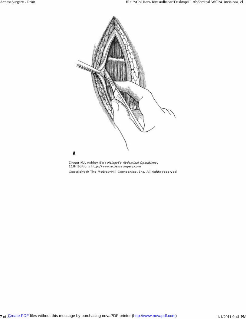
Paramedian IncisionsA paramedian incision is a vertical incision that is made 2.5–5 cm from the midline on either theright or left side of the abdomen. Like the midline incision, the paramedian incision avoids injury tonerves and limits trauma to the rectus muscle. It provides a secure, anatomic closure with goodrestoration of function. When necessary, it can also be extended from xiphisternum to pubis, allowingexcellent exposure of the abdomen. It is, however, considerably more time-consuming than a midlineincision.
Medial Paramedian IncisionAn upper paramedian incision is begun at the costal margin and carried to about 2–8 cm below the
AccessSurgery - Print file:///C:/Users/Jeyasudhahar/Desktop/II. Abdominal Wall/4. incisions, cl...
5 of 56 1/1/2011 9:41 PMCreate PDF files without this message by purchasing novaPDF printer (http://www.novapdf.com)

umbilicus. Additional access can be obtained by sloping the upper portion of the incision upward towardthe xiphoid (Fig 4–3). The lower paramedian incision is similar and indeed can be continuous with anupper paramedian incision, enabling exposure of the abdomen from the costal margin to the pubis.
Figure 4–3.
Upper paramedian incision: surface markings. Additional exposure can be obtained by sloping the upper portion ofthe incision upward toward the xiphoid process.
The anterior border of the rectus sheath is exposed and incised for the entire length of the wound. Themedial aspect of the anterior rectus fascia is then dissected off the rectus muscle to its medial edge(Fig 4–4). Particular care must be taken in the upper abdomen because of the tendinous inscriptionsthat attach the rectus muscle to the anterior fascia. These are located just inferior to the xiphoidprocess, at the umbilicus, and occasionally halfway between these two points. Segmental vessels willbe encountered at these three points and must be electrocoagulated or clamped and ligated. Once freeanteriorly and medially, the rectus muscle is easily retracted laterally because it is not adhered to theposterior rectus fascia. The posterior sheath and peritoneum are then incised vertically for the lengthof the skin incision.
Figure 4–4.
AccessSurgery - Print file:///C:/Users/Jeyasudhahar/Desktop/II. Abdominal Wall/4. incisions, cl...
6 of 56 1/1/2011 9:41 PMCreate PDF files without this message by purchasing novaPDF printer (http://www.novapdf.com)
AccessSurgery - Print file:///C:/Users/Jeyasudhahar/Desktop/II. Abdominal Wall/4. incisions, cl...
7 of 56 1/1/2011 9:41 PMCreate PDF files without this message by purchasing novaPDF printer (http://www.novapdf.com)

A. Paramedian incision: dissection of the rectus muscle from the anterior rectus sheath. B. Paramedian incision intransverse section.
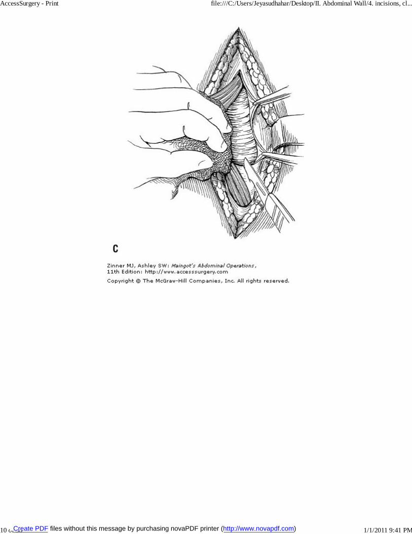
The lower paramedian incision differs in only two respects from the upper paramedian incision. Theinferior epigastric vessels will be encountered in dissection and should be divided and tied where theyrun across the inferior portion of the incision. Additionally, the posterior sheath of the rectus fascia isdeficient inferior to the semilunar fold of Douglas in this region (Fig 4–5).
Figure 4–5.
AccessSurgery - Print file:///C:/Users/Jeyasudhahar/Desktop/II. Abdominal Wall/4. incisions, cl...
8 of 56 1/1/2011 9:41 PMCreate PDF files without this message by purchasing novaPDF printer (http://www.novapdf.com)

AccessSurgery - Print file:///C:/Users/Jeyasudhahar/Desktop/II. Abdominal Wall/4. incisions, cl...
9 of 56 1/1/2011 9:41 PMCreate PDF files without this message by purchasing novaPDF printer (http://www.novapdf.com)
AccessSurgery - Print file:///C:/Users/Jeyasudhahar/Desktop/II. Abdominal Wall/4. incisions, cl...
10 of 56 1/1/2011 9:41 PMCreate PDF files without this message by purchasing novaPDF printer (http://www.novapdf.com)

AccessSurgery - Print file:///C:/Users/Jeyasudhahar/Desktop/II. Abdominal Wall/4. incisions, cl...
11 of 56 1/1/2011 9:41 PMCreate PDF files without this message by purchasing novaPDF printer (http://www.novapdf.com)
AccessSurgery - Print file:///C:/Users/Jeyasudhahar/Desktop/II. Abdominal Wall/4. incisions, cl...
12 of 56 1/1/2011 9:41 PMCreate PDF files without this message by purchasing novaPDF printer (http://www.novapdf.com)
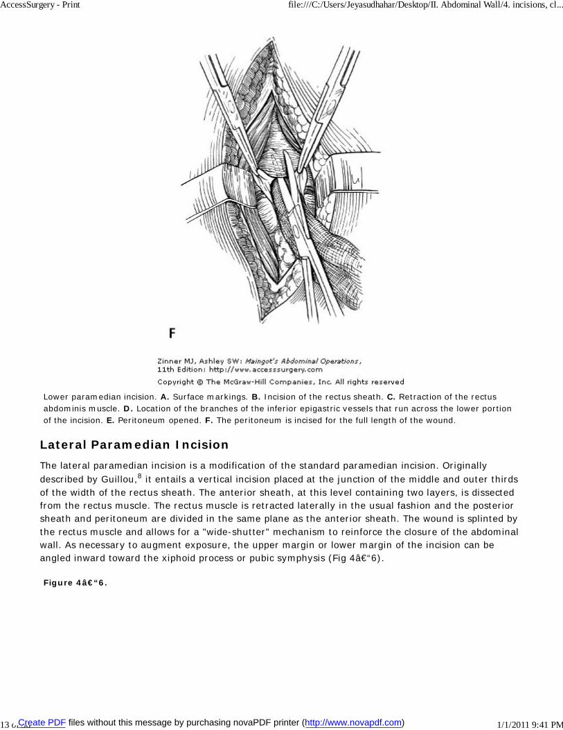
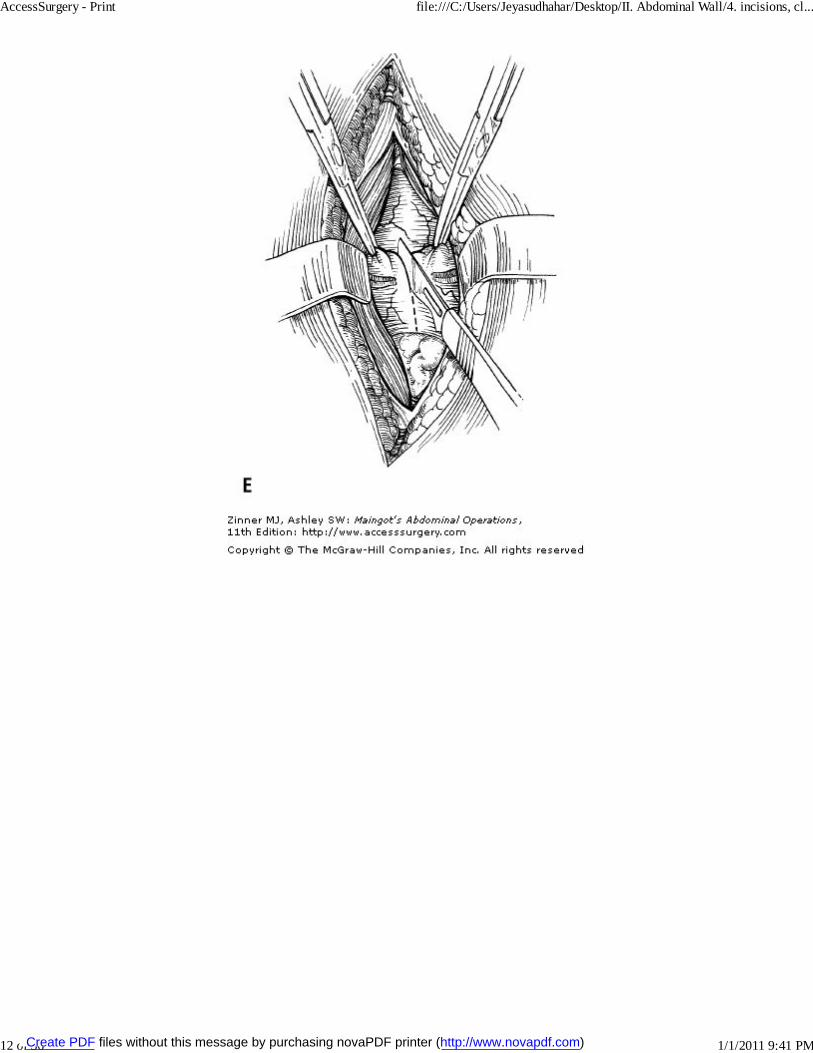
Lower paramedian incision. A. Surface markings. B. Incision of the rectus sheath. C. Retraction of the rectusabdominis muscle. D. Location of the branches of the inferior epigastric vessels that run across the lower portionof the incision. E. Peritoneum opened. F. The peritoneum is incised for the full length of the wound.
Lateral Paramedian IncisionThe lateral paramedian incision is a modification of the standard paramedian incision. Originallydescribed by Guillou,8 it entails a vertical incision placed at the junction of the middle and outer thirdsof the width of the rectus sheath. The anterior sheath, at this level containing two layers, is dissectedfrom the rectus muscle. The rectus muscle is retracted laterally in the usual fashion and the posteriorsheath and peritoneum are divided in the same plane as the anterior sheath. The wound is splinted bythe rectus muscle and allows for a "wide-shutter" mechanism to reinforce the closure of the abdominalwall. As necessary to augment exposure, the upper margin or lower margin of the incision can beangled inward toward the xiphoid process or pubic symphysis (Fig 4–6).
Figure 4–6.
AccessSurgery - Print file:///C:/Users/Jeyasudhahar/Desktop/II. Abdominal Wall/4. incisions, cl...
13 of 56 1/1/2011 9:41 PMCreate PDF files without this message by purchasing novaPDF printer (http://www.novapdf.com)

AccessSurgery - Print file:///C:/Users/Jeyasudhahar/Desktop/II. Abdominal Wall/4. incisions, cl...
14 of 56 1/1/2011 9:41 PMCreate PDF files without this message by purchasing novaPDF printer (http://www.novapdf.com)
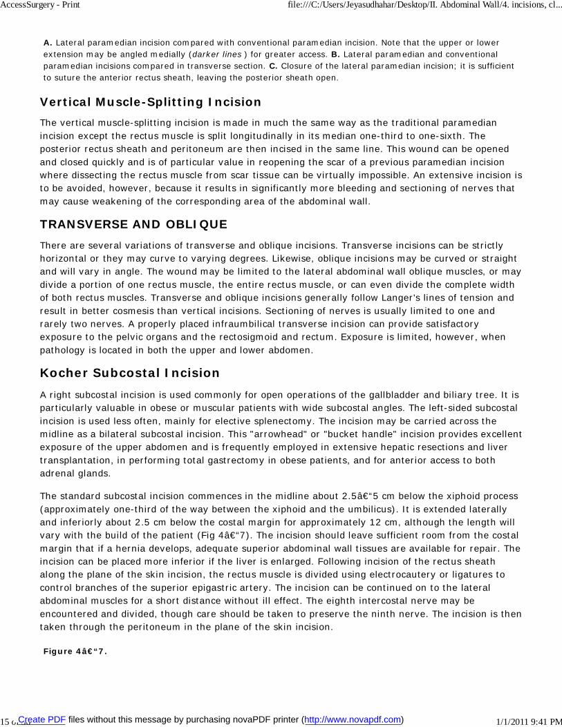
A. Lateral paramedian incision compared with conventional paramedian incision. Note that the upper or lowerextension may be angled medially (darker lines ) for greater access. B. Lateral paramedian and conventionalparamedian incisions compared in transverse section. C. Closure of the lateral paramedian incision; it is sufficientto suture the anterior rectus sheath, leaving the posterior sheath open.
Vertical Muscle-Splitting IncisionThe vertical muscle-splitting incision is made in much the same way as the traditional paramedianincision except the rectus muscle is split longitudinally in its median one-third to one-sixth. Theposterior rectus sheath and peritoneum are then incised in the same line. This wound can be openedand closed quickly and is of particular value in reopening the scar of a previous paramedian incisionwhere dissecting the rectus muscle from scar tissue can be virtually impossible. An extensive incision isto be avoided, however, because it results in significantly more bleeding and sectioning of nerves thatmay cause weakening of the corresponding area of the abdominal wall.
TRANSVERSE AND OBLIQUEThere are several variations of transverse and oblique incisions. Transverse incisions can be strictlyhorizontal or they may curve to varying degrees. Likewise, oblique incisions may be curved or straightand will vary in angle. The wound may be limited to the lateral abdominal wall oblique muscles, or maydivide a portion of one rectus muscle, the entire rectus muscle, or can even divide the complete widthof both rectus muscles. Transverse and oblique incisions generally follow Langer's lines of tension andresult in better cosmesis than vertical incisions. Sectioning of nerves is usually limited to one andrarely two nerves. A properly placed infraumbilical transverse incision can provide satisfactoryexposure to the pelvic organs and the rectosigmoid and rectum. Exposure is limited, however, whenpathology is located in both the upper and lower abdomen.
Kocher Subcostal IncisionA right subcostal incision is used commonly for open operations of the gallbladder and biliary tree. It isparticularly valuable in obese or muscular patients with wide subcostal angles. The left-sided subcostalincision is used less often, mainly for elective splenectomy. The incision may be carried across themidline as a bilateral subcostal incision. This "arrowhead" or "bucket handle" incision provides excellentexposure of the upper abdomen and is frequently employed in extensive hepatic resections and livertransplantation, in performing total gastrectomy in obese patients, and for anterior access to bothadrenal glands.
The standard subcostal incision commences in the midline about 2.5–5 cm below the xiphoid process(approximately one-third of the way between the xiphoid and the umbilicus). It is extended laterallyand inferiorly about 2.5 cm below the costal margin for approximately 12 cm, although the length willvary with the build of the patient (Fig 4–7). The incision should leave sufficient room from the costalmargin that if a hernia develops, adequate superior abdominal wall tissues are available for repair. Theincision can be placed more inferior if the liver is enlarged. Following incision of the rectus sheathalong the plane of the skin incision, the rectus muscle is divided using electrocautery or ligatures tocontrol branches of the superior epigastric artery. The incision can be continued on to the lateralabdominal muscles for a short distance without ill effect. The eighth intercostal nerve may beencountered and divided, though care should be taken to preserve the ninth nerve. The incision is thentaken through the peritoneum in the plane of the skin incision.
Figure 4–7.
AccessSurgery - Print file:///C:/Users/Jeyasudhahar/Desktop/II. Abdominal Wall/4. incisions, cl...
15 of 56 1/1/2011 9:41 PMCreate PDF files without this message by purchasing novaPDF printer (http://www.novapdf.com)

AccessSurgery - Print file:///C:/Users/Jeyasudhahar/Desktop/II. Abdominal Wall/4. incisions, cl...
16 of 56 1/1/2011 9:41 PMCreate PDF files without this message by purchasing novaPDF printer (http://www.novapdf.com)
Kocher incision. A. Surface markings. B. Division of the rectus and medial portions of the lateral abdominalmuscles.
The rectus muscle has a segmental nerve supply and a transverse or slightly oblique incision passesbetween adjacent nerves without injuring them. Provided its anterior and posterior sheaths are closed,the rectus muscle can be divided transversely without major weakness of the abdominal wall because itis not deprived of the distal part of its innervation. Healing of the incision results, effectively, in theformation of additional iatrogenic fibrous inscriptions of the muscle.
Mcburney Gridiron and Rockey-Davis Muscle Splitting Incisions
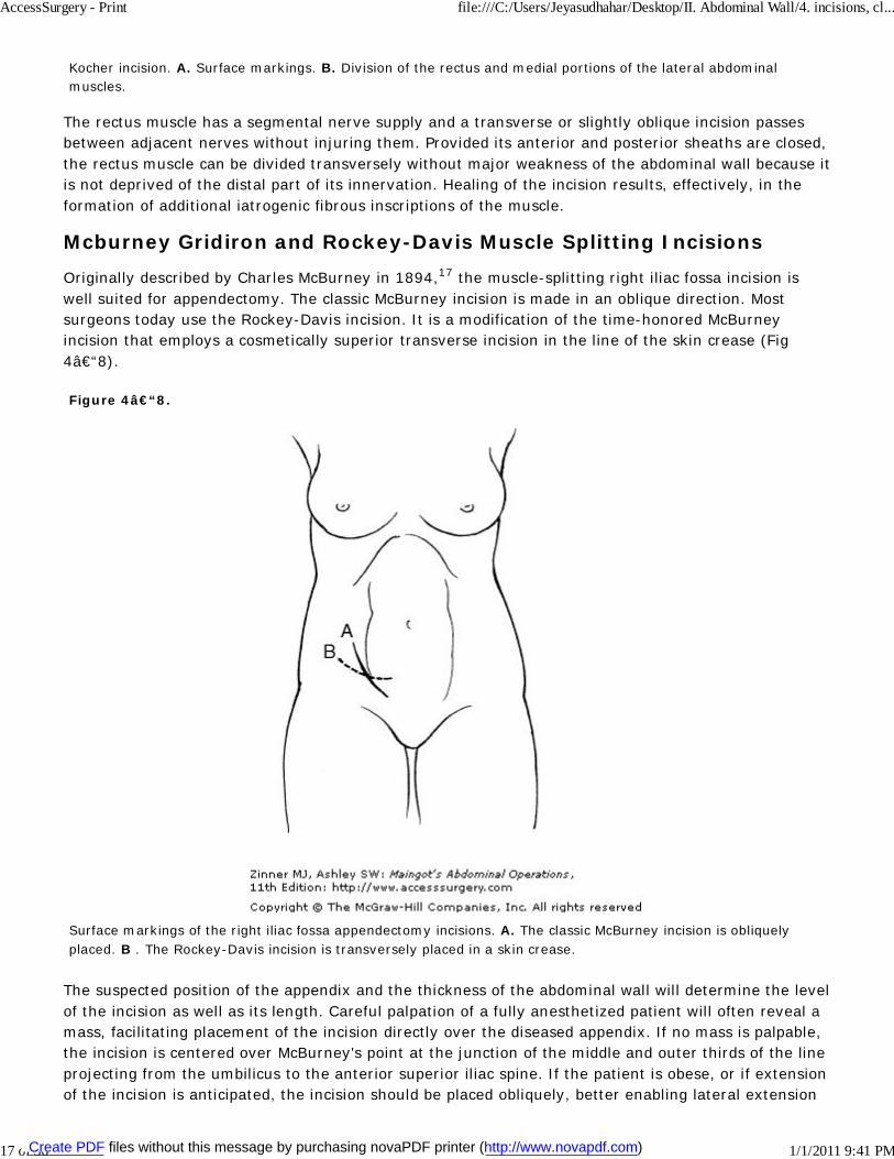
Originally described by Charles McBurney in 1894,17 the muscle-splitting right iliac fossa incision iswell suited for appendectomy. The classic McBurney incision is made in an oblique direction. Mostsurgeons today use the Rockey-Davis incision. It is a modification of the time-honored McBurneyincision that employs a cosmetically superior transverse incision in the line of the skin crease (Fig4–8).
Figure 4–8.
Surface markings of the right iliac fossa appendectomy incisions. A. The classic McBurney incision is obliquelyplaced. B . The Rockey-Davis incision is transversely placed in a skin crease.
The suspected position of the appendix and the thickness of the abdominal wall will determine the levelof the incision as well as its length. Careful palpation of a fully anesthetized patient will often reveal amass, facilitating placement of the incision directly over the diseased appendix. If no mass is palpable,the incision is centered over McBurney's point at the junction of the middle and outer thirds of the lineprojecting from the umbilicus to the anterior superior iliac spine. If the patient is obese, or if extensionof the incision is anticipated, the incision should be placed obliquely, better enabling lateral extension
AccessSurgery - Print file:///C:/Users/Jeyasudhahar/Desktop/II. Abdominal Wall/4. incisions, cl...
17 of 56 1/1/2011 9:41 PMCreate PDF files without this message by purchasing novaPDF printer (http://www.novapdf.com)

as a muscle-cutting incision.
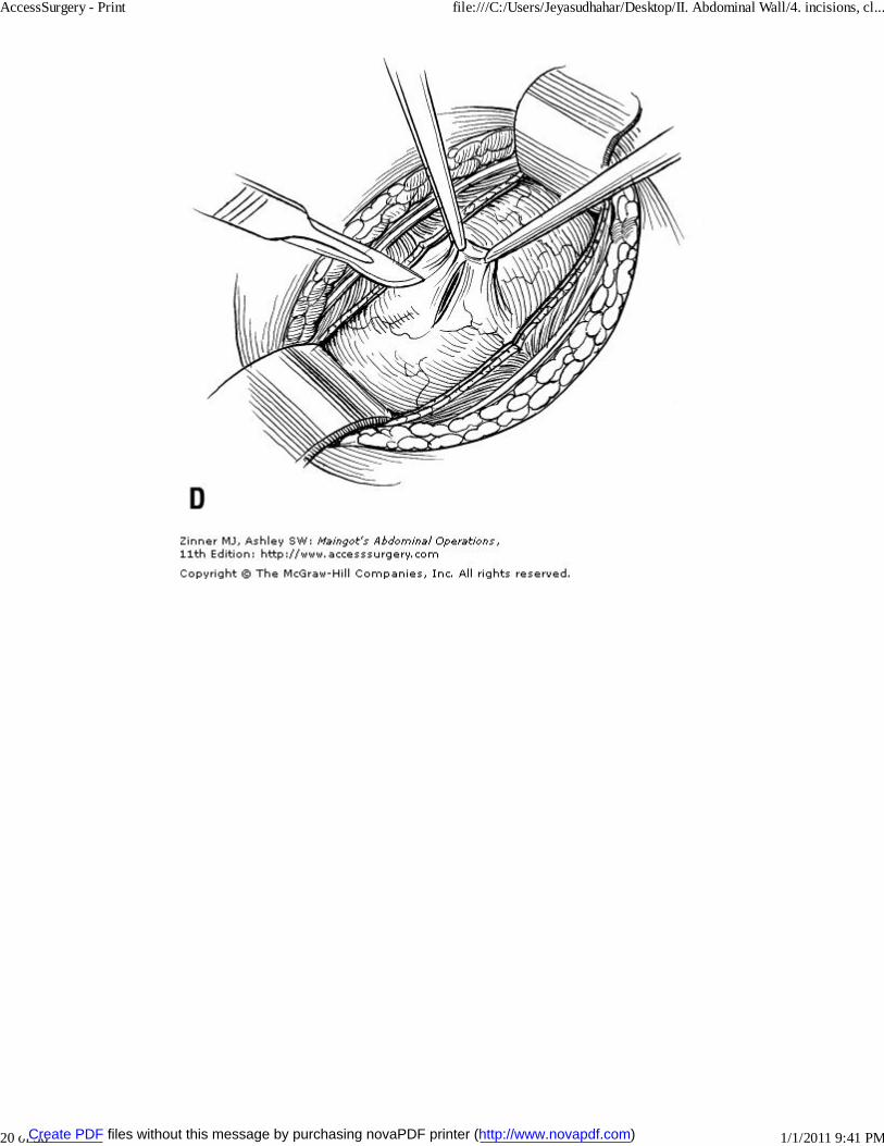
Each muscular layer of the abdominal wall is sequentially split in the direction of its muscle fibers. Thisis accomplished with the use of closed Kelly hemostats inserted in the center of each muscle andopened perpendicular to the direction of fibers. The surgeon's index finger can also be used to gingerlyopen the muscles. After skin and subcutaneous tissues are incised, the external oblique aponeurosis isexposed and divided in the "hands-in-pockets" direction of its fibers to reveal the underlying internaloblique muscle. At a point adjacent to the lateral border of the rectus sheath, a small incision is madein the internal oblique muscle which is then opened in the direction of its fibers. Once the underlyingtransversalis muscle is exposed, it is split in a similar manner to reveal the transversalis fascia andperitoneum. The peritoneum is entered by picking up a fold of its tissue and nicking it with a blade. Itis stretched with inserted index fingers and the appendix and cecum are exposed (Fig 4–9).
Figure 4–9.
AccessSurgery - Print file:///C:/Users/Jeyasudhahar/Desktop/II. Abdominal Wall/4. incisions, cl...
18 of 56 1/1/2011 9:41 PMCreate PDF files without this message by purchasing novaPDF printer (http://www.novapdf.com)

AccessSurgery - Print file:///C:/Users/Jeyasudhahar/Desktop/II. Abdominal Wall/4. incisions, cl...
19 of 56 1/1/2011 9:41 PMCreate PDF files without this message by purchasing novaPDF printer (http://www.novapdf.com)
AccessSurgery - Print file:///C:/Users/Jeyasudhahar/Desktop/II. Abdominal Wall/4. incisions, cl...
20 of 56 1/1/2011 9:41 PMCreate PDF files without this message by purchasing novaPDF printer (http://www.novapdf.com)

McBurney muscle-splitting incision. A. Division of the external oblique aponeurosis. B. The internal oblique andtransversus muscles are split. C. The index fingers of each hand enlarge the opening. D. Incision of theperitoneum. E. Exposure of the appendix.
If further exposure is necessary, the wound can be enlarged by dividing the anterior sheath in line withthe incision, retracting the rectus muscle medially, and extending the peritoneal defect medially intothe posterior rectus sheath. If the operation demands enlargement of the wound laterally (Weirextension), this can be accomplished by division of the oblique muscles superolaterally. This incisionprovides good access to the iliac fossa and can be exercised for a right- or left-sided hemicolectomy,cecostomy, or sigmoid colostomy. Medial and lateral extension of the McBurney incision bears thename the Rutherford-Morrison incision.
Pfannenstiel IncisionThe Pfannenstiel incision is used frequently for gynecologic operations and for access to the retropubicspace in the male for extraperitoneal retropubic prostatectomy. The skin incision is placed in thecurving interspinous crease that lies approximately 5 cm superior to the symphysis pubis. It usuallycarried out for about 12 cm in length. Both anterior rectus sheaths are exposed and dividedtransversely for the entire length of the wound. Hemostat clamps are applied to the superior andinferior leaflets of the divided sheath that are widely separated from the underlying rectus musclessuperiorly to the umbilicus and inferiorly to the pubic symphysis. The recti are retracted laterally andthe peritoneum is opened vertically in the midline. Care must be taken to protect the bladder at thelower end of the wound (Fig 4–10). Exposure provided by the Pfannenstiel incision is somewhatlimited and it should not be used when a procedure outside of the pelvis is anticipated. An advantage
AccessSurgery - Print file:///C:/Users/Jeyasudhahar/Desktop/II. Abdominal Wall/4. incisions, cl...
21 of 56 1/1/2011 9:41 PMCreate PDF files without this message by purchasing novaPDF printer (http://www.novapdf.com)
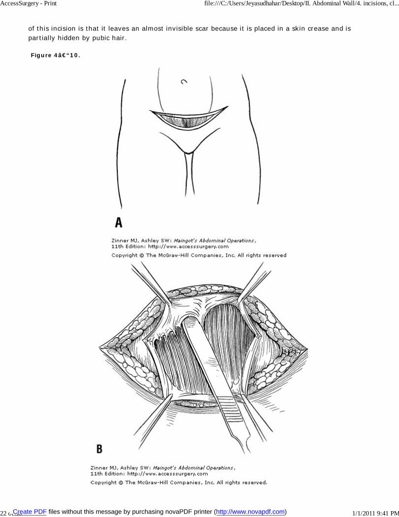
of this incision is that it leaves an almost invisible scar because it is placed in a skin crease and ispartially hidden by pubic hair.
Figure 4–10.
AccessSurgery - Print file:///C:/Users/Jeyasudhahar/Desktop/II. Abdominal Wall/4. incisions, cl...
22 of 56 1/1/2011 9:41 PMCreate PDF files without this message by purchasing novaPDF printer (http://www.novapdf.com)

AccessSurgery - Print file:///C:/Users/Jeyasudhahar/Desktop/II. Abdominal Wall/4. incisions, cl...
23 of 56 1/1/2011 9:41 PMCreate PDF files without this message by purchasing novaPDF printer (http://www.novapdf.com)

AccessSurgery - Print file:///C:/Users/Jeyasudhahar/Desktop/II. Abdominal Wall/4. incisions, cl...
24 of 56 1/1/2011 9:41 PMCreate PDF files without this message by purchasing novaPDF printer (http://www.novapdf.com)

Pfannenstiel incision. A. Skin incision. B. Horizontal division of the anterior rectus sheath and developing fascialflap. C. Dividing in the midline and entering the peritoneal cavity. D. Opening midline. E. Lateral retractors areplaced for exposure. F. Inferior retractors placed for exposure. G. Closure midline and inferior rectus.
ABDOMINOTHORACIC INCISIONSThe thoracoabdominal incision provides excellent exposure by converting the peritoneal and pleuralspaces into one common cavity. The left thoracoabdominal incision is particularly useful for access to
AccessSurgery - Print file:///C:/Users/Jeyasudhahar/Desktop/II. Abdominal Wall/4. incisions, cl...
25 of 56 1/1/2011 9:41 PMCreate PDF files without this message by purchasing novaPDF printer (http://www.novapdf.com)
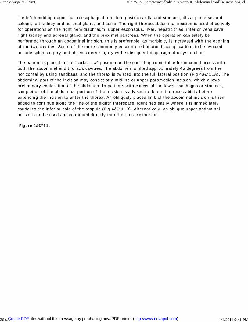
the left hemidiaphragm, gastroesophageal junction, gastric cardia and stomach, distal pancreas andspleen, left kidney and adrenal gland, and aorta. The right thoracoabdominal incision is used effectivelyfor operations on the right hemidiaphragm, upper esophagus, liver, hepatic triad, inferior vena cava,right kidney and adrenal gland, and the proximal pancreas. When the operation can safely beperformed through an abdominal incision, this is preferable, as morbidity is increased with the openingof the two cavities. Some of the more commonly encountered anatomic complications to be avoidedinclude splenic injury and phrenic nerve injury with subsequent diaphragmatic dysfunction.
The patient is placed in the "corkscrew" position on the operating room table for maximal access intoboth the abdominal and thoracic cavities. The abdomen is tilted approximately 45 degrees from thehorizontal by using sandbags, and the thorax is twisted into the full lateral position (Fig 4–11A). Theabdominal part of the incision may consist of a midline or upper paramedian incision, which allowspreliminary exploration of the abdomen. In patients with cancer of the lower esophagus or stomach,completion of the abdominal portion of the incision is advised to determine resectability beforeextending the incision to enter the thorax. An obliquely placed limb of the abdominal incision is thenadded to continue along the line of the eighth interspace, identified easily where it is immediatelycaudal to the inferior pole of the scapula (Fig 4–11B). Alternatively, an oblique upper abdominalincision can be used and continued directly into the thoracic incision.
Figure 4–11.
AccessSurgery - Print file:///C:/Users/Jeyasudhahar/Desktop/II. Abdominal Wall/4. incisions, cl...
26 of 56 1/1/2011 9:41 PMCreate PDF files without this message by purchasing novaPDF printer (http://www.novapdf.com)

Anterolateral thoracoabdominal incision to expose the distal esophagus and stomach, to resect tumors of theupper one-third of the stomach, to treat short esophagus with gastroesophageal reflux, or to expose suprarenalaortic aneurysms. A. The "corkscrew" position, with the thorax in the lateral position and the abdomen at 45degrees from the horizontal. Very careful positioning on the operating table is essential to prevent injury to thebrachial plexus or pressure on peripheral nerves and should be closely supervised by the surgeon. B. Theabdominal incision is ordinarily made first, to determine operability and be certain that the thoracic extension isneeded. This is usually done with a vertical midline incision that is extended into the chest through the eighthintercostal space. The abdomen has been opened and the pleural space is being entered. C. The diaphragm isusually opened in a radial fashion with an incision directed toward the esophageal or aortic hiatus. D. Thediaphragm can be opened with a hemielliptical incision 2–3 cm from the lateral chest wall; this incision is longer
AccessSurgery - Print file:///C:/Users/Jeyasudhahar/Desktop/II. Abdominal Wall/4. incisions, cl...
27 of 56 1/1/2011 9:41 PMCreate PDF files without this message by purchasing novaPDF printer (http://www.novapdf.com)

than a straight phrenicotomy but preserves phrenic nerve function, of importance in patients with chronicpulmonary disease or less than optimal pulmonary function. (Reproduced, with permission, from Penn I, BakerRJ. Abdominal wall incisions and repair. In: Baker RJ, Fischer JE (eds). Mastery of Surgery , 4th ed. Philadelphia,PA: Lippincott Williams & Wilkins; 2001:197.)
After the abdomen is explored, the chest incision is deepened through the latissimus dorsi and serratusanterior muscles, and then the external oblique muscle and aponeurosis. The intercostal muscles of theeighth interspace are divided to enter the pleural cavity and the incision is continued across the costalmargin, which is divided with a scalpel. It is often useful at this point to resect a short segment ofcostal cartilage to facilitate closure of the chest wall. A Finochietto self-retaining rib retractor isinserted and the intercostal space and is gently spread. Rib resection to gain exposure is rarelynecessary. The diaphragm is incised radially toward the esophageal or aortic hiatus, isolating and tyingbranches of the phrenic vessels before their division. If the operation does not require a radial incisionof the diaphragm to the esophageal hiatus, the diaphragm should be divided in a curvilinear fashion2–3 cm from its attachment to the chest wall. This hemielliptical incision will preserve phrenic nervefunction and is useful for patients with less than optimal pulmonary function.18
At completion of the operation, chest tubes placed in the pleural cavity are brought out of the thoraxthrough separate stab incisions. The diaphragm is repaired in two layers using nonabsorbable mattresssutures. Pericostal sutures are passed around the ribs and the chest muscles and abdominal wall areclosed in layers.
RETROPERITONEAL AND EXTRAPERITONEAL INCISIONSRetroperitoneal and extraperitoneal approaches to the abdomen have several advantages overintraperitoneal exposures. Manipulation and retraction of intra-abdominal viscera is limited andpostoperative ileus is reduced. Hemorrhage is more likely to be tamponaded in the retroperitoneumthan when it occurs in the peritoneal cavity. Infection and extravasation of urine are more frequentlylocalized here than within the peritoneal cavity and are more readily drainable. Retroperitoneal andextraperitoneal approaches can be used for operations on the kidney, ureter, adrenal gland, bladder,splenic artery and vein, groin hernias, vena cava, lumbar sympathetic chain, abdominal aorta, andcommon, internal, and external iliac vessels.
Retroperitoneal Approach to the Lumbar AreaThe retroperitoneal approach to the lumbar area is frequently used in aortic surgery, nephrectomy,lumbar symphathectomy, and ureterolithomy. The patient is positioned with the operative sideelevated 30–45 degrees with knee and hip flexed. The incision is carried from the level of theumbilicus at the lateral margin of the rectus sheath toward the twelfth rib for approximately 12–14cm (Fig 4–12). A portion of the twelfth rib is resected if further exposure is required, taking care notto injure the underlying pleura. The external oblique, internal oblique, and transversalis muscles areexposed, undermined, and opened in the direction of their fibers. The retroperitoneum is entered andthe peritoneum and retroperitoneal fat are moved anteriorly by blunt dissection, being mindful to notdissect behind the psoas muscle. The lower pole of the kidney, ureter, and sympathetic chain are easilyidentified. The vena cava is exposed on the right and the aorta on the left. If the peritoneum isunintentionally entered, it is closed immediately with continuous absorbable suture. At the conclusionof the procedure, the retroperitoneal fat and viscera fall back into place and the muscles of theabdominal wall are repaired in layers.
Figure 4–12.
AccessSurgery - Print file:///C:/Users/Jeyasudhahar/Desktop/II. Abdominal Wall/4. incisions, cl...
28 of 56 1/1/2011 9:41 PMCreate PDF files without this message by purchasing novaPDF printer (http://www.novapdf.com)

A. Left lumbar approach to the retroperitoneum, specifically for exposing the kidney, adrenal, and infrarenalabdominal aorta. B. The peritoneum has been bluntly dissected from the retroperitoneal structures with thepreperitoneal fat and soft tissue. Origins of the celiac, superior mesenteric, left renal, and inferior mesentericarteries are shown. (Reproduced, with permission, from Penn I, Baker RJ. Abdominal wall incisions and repair. In:Baker RJ, Fischer JE (eds). Mastery of Surgery , 4th ed. Philadelphia, PA: Lippincott Williams & Wilkins;2001:194.)
Posterior Approach to the Adrenal GlandsWith the posterior approach, dissection is performed entirely in the retroperitoneal space. Excellentexposure of the right adrenal vein and inferior vena cava is achieved. Inadvertent injury to the visceraor spleen is minimized and postoperative ileus is rare. Importantly, patients have decreased pain,fewer pulmonary complications, and shorter hospital stays than patients undergoing transabdominaladrenalectomy.
The patient is laid in the prone jackknife position. A curvilinear incision is made beginning on the tenthrib approximately three fingerbreadths lateral to the midline and carried inferiorly and laterally toward
AccessSurgery - Print file:///C:/Users/Jeyasudhahar/Desktop/II. Abdominal Wall/4. incisions, cl...
29 of 56 1/1/2011 9:41 PMCreate PDF files without this message by purchasing novaPDF printer (http://www.novapdf.com)

the iliac crest, ending approximately four fingerbreadths lateral to the midline (Fig 4–13). Thesubcutaneous tissues are divided to reveal the posterior layer of the lumbodorsal fascia. This fascia andthe fibers of the latissimus dorsi muscle which originate from it are divided. The erector spinae muscleis exposed and retracted medially to uncover the twelfth rib and the glistening middle layer of thelumbodorsal fascia. The attachments of the erector spinae to the twelfth rib are divided withelectrocautery, securing with clamps and ligating the vessels and nerves that penetrate the fascia toenter the erector spinae muscle. The twelfth rib is then resected periosteally, taking care not to injurethe underlying pleura. Gerota's fascia is exposed by incising the lumbodorsal fascia along the lateralmargin of the quadratus lumborum muscle. The intercostal neurovascular bundle should now becomevisible directly below the bed of the resected twelfth rib. The intercostal vessels are clamped, divided,and ligated and the intercostal nerve is gently retracted downward. The posterior fibers of thediaphragm where they insert on the periosteum of the twelfth rib are identified and divided. The lowermargin of the lung will enter the field with hyperinflation. The pleura is pushed gently away. If it isinadvertently entered, the resulting pneumothorax is handled at closure by insertion of a large-borerubber catheter into the pleural cavity and exiting through the wound. After closure of the fascial fibersaround the catheter, the lung is hyperinflated evacuating all air from the pleural space, and thecatheter is briskly removed.
Figure 4–13.
AccessSurgery - Print file:///C:/Users/Jeyasudhahar/Desktop/II. Abdominal Wall/4. incisions, cl...
30 of 56 1/1/2011 9:41 PMCreate PDF files without this message by purchasing novaPDF printer (http://www.novapdf.com)

The posterior approach to the kidney and adrenal. A. J-shaped incision over the tenth to twelfth ribs, extendinginferiorly 6–10 cm below the twelfth rib. B. Resection of the twelfth rib facilitates exposure. C. Thediaphragmatic attachment to the twelfth rib is taken down, with care taken not to enter the pleura. If the pleura isopened, the wound closure is done over a pleural suction catheter, which is removed with simultaneous positiveairway pressure by the anesthetist as the skin is being closed. (Reproduced, with permission, from Penn I, BakerRJ. Abdominal wall incisions and repair. In: Baker RJ, Fischer JE (eds). Mastery of Surgery , 4th ed. Philadelphia,PA: Lippincott Williams & Wilkins; 2001:195.)
Retroperitoneal Approach to the Iliac FossaThe retroperitoneal approach to the iliac fossa provides access to the bladder, distal ureter, andcommon, internal, and external iliac vessels. It is often employed for surgery on the iliac arteries andfor kidney transplantation into the iliac fossa. It is also used to drain psoas or retrocecal abscesses andto resect localized retroperitoneal tumors. The skin incision is an oblique incision extending fromapproximately 2 cm above the anterosuperior iliac spine to just lateral to the pubic symphysis (Fig4–14) and can be extended superiorly as far as the costal margin if necessary. The external oblique,internal oblique, and transverse abdominis muscles are divided in line with the skin incision. Theexternal and internal inguinal rings lie inferior to the lower edge of the incision and are not visualized.The retroperitoneal area is entered and the retroperitoneal fat and peritoneum are bluntly dissectedand retracted superomedially. If the peritoneum is inadvertently entered, it is closed immediately.Closure of each muscular and fascial layer is achieved with absorbable or nonabsorbable suture ineither continuous or interrupted fashion.
Figure 4–14.
Right lower quadrant extraperitoneal approach to the iliac vessels, ureter, and bladder, used for renal transplant,but also very useful to expose the iliac artery and vein, drain psoas or retrocecal abscesses, or resect localizedretroperitoneal tumors. A. The skin incision may be shorter than depicted in thinner patients or if an abscess is to
AccessSurgery - Print file:///C:/Users/Jeyasudhahar/Desktop/II. Abdominal Wall/4. incisions, cl...
31 of 56 1/1/2011 9:41 PMCreate PDF files without this message by purchasing novaPDF printer (http://www.novapdf.com)
be drained. B. Peritoneum is retracted medially by blunt dissection, which exposes the psoas muscle and gonadalartery and vein, shown anterior to the ureter. (Reproduced, with permission, from Penn I, Baker RJ. Abdominalwall incisions and repair. In: Baker RJ, Fischer JE (eds). Mastery of Surgery , 4th ed. Philadelphia, PA: LippincottWilliams & Wilkins; 2001:196.)
CLOSURE OF THE ABDOMINAL INCISIONClosure of the abdominal wall is a common denominator of all abdominal surgery. It is one of the firstthings that a surgeon is taught during his or her training. The methods of closure are often based onlocal traditions and the preferences of the teacher, and the surgeon is often reluctant to change thesemethods later on in his or her career. Abdominal closure is performed in a multitude of fashions andthere are an abundance of differently tailored studies on this matter.
The goal of wound closure is to restore function of the abdominal wall after a surgical procedure. Theoptimal method should be so technically simple that its results are as good for the hands of the traineeas they are for the experienced surgeon. It should leave the patient with a reasonably aesthetic scar,and most importantly, it should minimize the frequency of wound rupture, incisional hernia, woundinfection, and sinus formation.
Closure of the PeritoneumTraditional surgical dogma has taught that because all tissue layers of the abdominal wall are violatedwhen an abdominal incision is made, all layers should be approximated when the incision is closed.Closure of the peritoneum is based on the premise that normal anatomy will be restored, the risks ofinfection and wound herniation will be reduced, and adhesions will be minimized. Experience hasquestioned these surgical principles in the face of randomized controlled clinical trials. In 1977, Ellisand Heddle first reported their results of a randomized study comparing closure and nonclosure of theparietal peritoneum in a vertical laparotomy incision. No difference was found in the incidence ofwound dehiscence or hernia between the nonclosure arm (3.0% and 4.3%, respectively) and theclosure arm (2.5% and 4.3%, respectively).19 Similar results are found in randomized trials comparingclosure versus nonclosure of the peritoneum in open cholecystectomy incisions,20 lateral paramedianincisions,21 and gynecologic and obstetric incisions.22 It is concluded that closure of the peritoneum isunnecessary and not recommended. It is associated with a slightly longer operative time and morepostoperative pain, and there are some suggestions that it may cause increased formation ofadhesions.22
Closure of the FasciaClosure of the abdomen can be done in layers or en mass. A layered closure technique reconstructs theanterior and posterior aponeurotic sheaths in two different layers with the posterior layer generallyincorporating the peritoneum. Mass closure involves a single-layer closure of all musculofascial layersand may or may not include the peritoneum. Numerous clinical trials have compared layered to massabdominal closure. Some studies have shown an increased incidence of burst abdomen and incisionalhernia with layered closure,23–25 and some studies show no difference in these complications,26 butno studies demonstrate an advantage of layered over mass closure. Rates of wound sepsis and sinusformation have also been studied in randomized trials and do not depend on closure technique.
It has been claimed that a continuous, running suture will result in more secure wound closure than aseries of sutures placed in an interrupted fashion. The theoretical advantage of a continuous closure isthe distribution of tension differences across the suture line and the ability of the wound to adjust tothe stresses and strains of the postoperative period. This should minimize tissue strangulation andwound rupture from suture under strain cutting through fascia. The disadvantage of the continuous
AccessSurgery - Print file:///C:/Users/Jeyasudhahar/Desktop/II. Abdominal Wall/4. incisions, cl...
32 of 56 1/1/2011 9:41 PMCreate PDF files without this message by purchasing novaPDF printer (http://www.novapdf.com)
closure method is that a single thread holds the fascia together and its breakage jeopardizes the entirewound. Clinical evidence, however, demonstrates that continuous and interrupted closures of theabdomen are responsible for similar incidences of wound dehiscence, incisional hernia, woundinfection, wound pain, and suture fistula.27–31
The use of resorbable versus nonresorbable suture in closing the fascia has long been debated. Ratesof 17% for scar pain and 8% for suture fistula using permanent suture27 have stirred interest in theuse of resorbable sutures. Resorbable sutures, however, bear an intrinsic loss of tensile strengthduring the vulnerable postoperative period, and may result in an increase in wound disruption andventral hernia. The early use of the absorbable catgut suture has been shown to lead to a highincidence of wound rupture and incisional hernia due to its early degradation.26 To overcome thisproblem, synthetic absorbable sutures with delayed degradation were introduced to combine theadvantages of absorbability with strength comparable to nonabsorbable materials. There are conflictingreports in the literature about wound failure when nonabsorbable and absorbable suture are comparedin randomized clinical trials. The resorbable sutures polyglycolic acid (Dexon), polyglactic acid (Vicryl),polydioxanone (PDS), and polyglyconate (Maxon) have been shown to be equally as effective asnonabsorbable suture with respect to wound dehiscence and incisional hernia.32–35 Other studies,however, demonstrate that polydioxanone and polyglactic acid polymer absorbable suture may beassociated with an increased incidence of incisional hernia when weighed against nonabsorbablesuture.25,27
Another choice is monofilament versus multifilament suture. Multifilament suture is known to provide abetter growth environment for bacteria and is associated with a higher incidence of wound sepsis whencompared to monofilament suture.25,33 Bacteria are drawn into the fibers of multifilament suture bycapillary action and thrive there by escaping phagocytosis. Wound sepsis is a major risk factor forincisional hernia, but despite these considerations, multifilament suture has not been shown to resultin a greater incidence of wound failure over monofilament closure.23,30 Monofilament catgut suturealso deserves special consideration. It is a reactive material that causes a marked inflammatoryreaction and is associated with a higher incidence of wound infection than other monofilamentmaterials.36
Our experience and interpretation of the literature is that the optimal surgical method of closing theabdominal wound is a continuous mass closure. This technique appears to reduce the incidence ofwound rupture, is considerably less time consuming,28,37 is less expensive, and does not increase theincidence of incisional hernia, wound infection, or sinus formation. The choice of suture material ismore complex. We prefer to use a resorbable suture with delayed degradation, such as polydioxanone.Other resorbable materials are appropriate as well, but catgut should not be used. Amongnonresorbable sutures, monofilament suture is recommended.
METHOD OF MASS CLOSURE OF THE ABDOMENWhether the incision is vertical or transverse, the steps for closure are more or less the same. It is nowfully realized that healing of the wound takes place by formation of a dense fibrous scar that unites theopposing faces of the wound en mass. The purpose of the suture is to approximate the wound edgeswhile this dense fibrous scar deposits and matures.
For closure of the midline laparotomy incision, we employ two size 0 looped polydioxanone (PDS)sutures. One loop is used at the upper extremity and one at the lower extremity of the wound so thatonly one knot need be tied at the middle of the incision. The needle is passed securely through thevertex of the fascial incision and by passing the needle though the loop, the end of the suture line isanchored firmly to the abdominal wall. A medium-width metal ribbon is often placed into the peritoneal
AccessSurgery - Print file:///C:/Users/Jeyasudhahar/Desktop/II. Abdominal Wall/4. incisions, cl...
33 of 56 1/1/2011 9:41 PMCreate PDF files without this message by purchasing novaPDF printer (http://www.novapdf.com)

cavity to ensure a clear field for suturing and to avoid incorporating visceral structures into the sutureline. The suture is run in a continuous manner, taking full-thickness bites of the linea alba fasciaincorporating components of the anterior and posterior rectus aponeuroses (Fig 4–15). Theperitoneum need not be incorporated into the closure. Wide bites are taken a minimum of 1 cm fromthe wound edge and placed at 1-cm intervals. To reserve an adequate length of suture in the wound,the suture length should measure at least four times the length of the wound.38 This should preventthe cutting out of sutures that may occur during abdominal distention and dynamic tensile changesthat occur on the postoperative wound.
Figure 4–15.
Stages in the mass closure of the midline abdominal incision.
A similar technique is used for closure of the paramedian incision. The anterior and posterior rectussheaths are picked up and included in one bite (Fig 4–16). A transrectus incision will incorporate themedial sliver of rectus muscle into the suture loops. Mass closure of a lateral paramedian incision is notpossible. For this incision, the anterior and posterior rectus sheaths are closed separately. We prefer toclose the Kocher subcostal incision in two layers, closing the posterior rectus sheath with a continuousabsorbable suture and the anterior sheath with a separate, running nonabsorbable suture.Rockey-Davis muscle-splitting incisions are also closed in layers. The peritoneum can be closed withcontinuous absorbable suture to exclude the viscera from the field. The internal oblique andtransversalis muscles are closed as a single layer with interrupted absorbable suture and theaponeurosis of the external oblique is then closed with a continuous absorbable suture. The skin is left
AccessSurgery - Print file:///C:/Users/Jeyasudhahar/Desktop/II. Abdominal Wall/4. incisions, cl...
34 of 56 1/1/2011 9:41 PMCreate PDF files without this message by purchasing novaPDF printer (http://www.novapdf.com)

open if intraperitoneal purulence or a gangrenous appendix was present.
Figure 4–16.
Mass closure of the paramedian incision.
Subcutaneous Tissue ClosureWith the high prevalence of obesity in developed countries, treatment of the subcutaneous tissues inabdominal wound closure becomes increasingly important. The vascular supply to the subcutaneoustissue of the abdominal wall is poor, rendering it susceptible to soft-tissue infection. Likewise, if thislevel of the abdominal wall contains a potential space promoting accumulation of seroma, the risk ofinfection increases.
Only one prospective randomized trial has been conducted to determine the value of suturing thesubcutaneous fat. Using a subcostal incision for cholecystectomy, the authors demonstrated nosignificant differences in complications between closure and nonclosure of the subcutaneous tissues.39
Wound seepage, however, was reduced in incisions in which the subcutaneous layer was closed. Tworandomized controlled clinical trials of cesarean section incisions have produced different results.Suture closure of the subcutaneous layer resulted in a significant decrease in the rates of wounddisruption (14.5%) when compared to wounds in which the subcutaneous layer is left open (26.6%).40
A separate trial failed to demonstrate any benefit of suture closure in wounds with less than 2 cm ofsubcutaneous tissue, but confirm the reduction in wound disruption in wounds with greater than 2 cmof tissue.41
Some authors have speculated that reduction of serous fluid in the subcutaneous dead space byplacement of closed-system subcutaneous tissue drains would reduce the risk of wound complications.Prospective randomized clinical data from the general surgical literature do not support the use ofthese devices.42,43
We do not routinely close the subcutaneous layer of the wound. On some occasions, in obese patients,we will employ the use a series of simple, interrupted, absorbable polyglactic acid (vicryl) sutures to
AccessSurgery - Print file:///C:/Users/Jeyasudhahar/Desktop/II. Abdominal Wall/4. incisions, cl...
35 of 56 1/1/2011 9:41 PMCreate PDF files without this message by purchasing novaPDF printer (http://www.novapdf.com)
reapproximate the subcutaneous layer. These stitches are inverted to bury the knots within the wound.
Skin ClosureIf the surgical site is heavily contaminated (class III or class IV wound), the skin should be left open toheal by secondary intention or by delayed primary skin closure.44 A number of closure techniques forclean (class I) and clean-contaminated (class II) wounds are available for the skin. These includeinterrupted suture, subcuticular suture, surgical staples, surgical tape, and adhesive glues. Goals ofskin closure are tissue approximation, minimizing wound infection, acceptable cosmesis, andminimizing postoperative pain. These goals should be achieved with a simple, rapid, and cost-effectivemethod.
Three randomized controlled studies have compared skin staples to subcuticular sutures. In all studies,no difference in the rate of wound infection could be demonstrated.45–47 Two of these studiesrevealed less postoperative pain and less postoperative analgesia requirement in wounds closed withsubcuticular suture.45,46 Two of these studies also demonstrated a superior cosmetic result insubcuticular closures over surgical staples; however, this cosmetic difference narrowed over time andbecame insignificant by 6 months.46,47
Adhesive tapes are often used to reapproximate skin edges in simple lacerations. Following abdominalsurgery, adhesive tapes are useful to cover skin incisions closed by subcuticular suture, where theyserve to further reapproximate skin edges and to dress the wound. The use of adhesive tape withoutsuture closure was compared to interrupted silk skin suturing of abdominal wounds in one early trial.No difference in the rates of wound infection could be found. The tapes were significantly morecomfortable and patients preferred them over sutures, but wide scarring occurred more frequently withsurgical tapes.48
Synthetic glues are gaining popularity in skin closure of surgical wounds. When compared withtraditional skin-closing devices including sutures, staples, or adhesive tapes, some cyanoacrylate glueshave been found to be comparable in effectiveness and safety for repair of lacerations. They areapplied more rapidly and decrease the amount of required wound care by serving as their owndressings. In elective abdominal procedures with small and large (>4 cm) incisions, these glues havebeen shown in clinical trials to have similar outcomes with respect to wound durability when comparedto traditional techniques,49,50 although there are conflicting data on wound healing, cosmesis, andpostoperative pain.50,51
We prefer to close skin with a running, nonbraided, absorbable suture in a subcuticular technique.Adhesive tapes are placed over the closed incision without the use of skin glues.
Prophylactic Drainage of the AbdomenProphylactic operative drains are employed to remove intraperitoneal collections such as blood, bile,ascites, chyle, and pancreatic or intestinal juice. Drains are also placed to signal early complicationssuch as postoperative hemorrhage and leakage of intestinal suture lines. Prophylactic drainage of theabdomen is used routinely by gastrointestinal surgeons around the world. There are, however, goodrandomized controlled data to question their utility.
A recent systematic review and meta-analyses were performed to determine the evidence-based valueof prophylactic drainage in gastrointestinal surgery.52 There is evidence of level 1 quality thatoperatively-placed drains do not reduce complications after elective hepatic resection,52–55
cholecystectomy,56 pancreatic resection for cancer,57 elective colonic or rectal resection with primaryanastomosis,52 and appendectomy for any stage of appendicitis.52 One randomized controlled trial
AccessSurgery - Print file:///C:/Users/Jeyasudhahar/Desktop/II. Abdominal Wall/4. incisions, cl...
36 of 56 1/1/2011 9:41 PMCreate PDF files without this message by purchasing novaPDF printer (http://www.novapdf.com)
(evidence level 2b) has failed to show any benefit of prophylactic drainage after subtotal or totalgastrectomy with extended lymph node dissection for gastric cancer.58 However, in the absence ofmore complete prospective data on the use of abdominal drainage in upper gastrointestinal surgery,consensus opinion (evidence level 5) recommends that drains should be used in esophageal or gastricresections due to the potentially fatal outcome of anastomotic leak in these procedures.
When drains are used in abdominal surgery, they should be placed through stab wounds separate fromthe main incision. Drains placed through the operative wound increase the risk of incisional surgicalsite infection.44 Additionally, closed suction drains should be used to avoid the increase in surgical siteinfection risk of open drainage systems. The duration of drainage will vary with the purpose for whichthey have been inserted, and there are no evidence-based rules to guide timing of removal. Generally,drains that have been introduced to vent oozing or bleeding should be withdrawn after 24–48 hours,while drains placed where a localized abscess has been drained may need to remain in situ for morethan 3 days. Drains should be removed as soon as they have served their purpose.
Retention Closure of the AbdomenThe incidence of fascial disruption after major abdominal operations is 1–3% and is associated with amortality rate of 15–20%.59 Several patient-related factors are associated with an increased risk offascial dehiscence. These include advanced age, male gender, hypoproteinemia, malnutrition, anemia,malignant disease, jaundice, azotemia, and treatment with steroids. More important than systemicfactors in fascial disruption are local mechanical factors and a sound initial surgical closure.59 Bringingdrains or ostomies through the main incision clearly compromises fascial integrity. Wound sepsis is amajor predisposing factor for both fascial disruption as well as incisional hernia. Increased intra-abdominal pressure is almost uniformly incriminated as a cause of fascial dehiscence. It may besecondary to abdominal complications such as vomiting, ileus, or bowel obstruction, pulmonarycomplications such as atelectasis, pneumonia, or bronchitis, or the nature of the operation, as in repairof diaphragmatic hernia.
Most authors would agree on the use of retention sutures for repair of fascial dehiscence. However, theindications for prophylactic placement of retention sutures at initial operation have not been wellexamined prospectively and are not well agreed upon. The purpose of using retention sutures in thissetting is to relieve tension along the primary suture line to prevent wound disruption and allownormal relaxed wound healing. They are sometimes employed for initial laparotomy closure when poorwound healing is anticipated, as in obese, cirrhotic, and cachectic patients, those receivingcorticosteroids, or when increased intra-abdominal pressure is anticipated, as in postoperative ileus.
There has been only one randomized trial of full-thickness retention suture placement in midlinelaparotomy closure. In this trial, Hubbard and associates could not identify a benefit of retention sutureclosure over standard mass closure of the abdominal wall.60 The disadvantages of retention sutures,however, are well known and include the potential hazard of caught viscera, significant postoperativepain, a residual cross-hatched scar, and leakage of intraperitoneal fluid through the wound.61 For thesereasons, retention suture closure has largely fallen out of favor by many surgeons during primarylaparotomy closure. Others recommend primary closure with retention sutures in certaincircumstances. In a retrospective comparison of midline abdominal wound dehiscence, Makela andcolleagues identified several preoperative variables that are significantly associated with fascialdisruption: hypoalbuminemia, anemia, malnutrition, chronic pulmonary disease, and emergencyprocedure. For patients with three or more of these preoperative risk factors, this group recommendsinternal retention suture closure.62
The technique of placing retention sutures varies from surgeon to surgeon and many methods have
AccessSurgery - Print file:///C:/Users/Jeyasudhahar/Desktop/II. Abdominal Wall/4. incisions, cl...
37 of 56 1/1/2011 9:41 PMCreate PDF files without this message by purchasing novaPDF printer (http://www.novapdf.com)

been described. We employ 2-0 nylon simple, interrupted sutures to transfix all layers of theabdominal wall including skin and peritoneum. They are inserted via a long cutting needleapproximately 2.5 cm from the margin of the wound and approximately 2.5 cm apart. Various methodshave been devised in an attempt to protect the skin and subcutaneous tissue from damage. At the skinlevel, the sutures are threaded through 5-cm rubber tubing bolsters to prevent skin breakdown (Fig4–17). They are removed in approximately 14 days. If the skin is closed, it is done with staples.
Figure 4–17.
Retention sutures tied and held in position supported by rubber tubing.
Temporary Closure of the AbdomenSevere abdominal trauma with hemorrhagic shock and ongoing resuscitation can cause massive edemaof the bowel, abdominal wall, and retroperitoneum that may preclude a safe primary closure of theabdominal wound. Primary closure of the abdomen under tension leads to fascial necrosis anddehiscence, aggravates tissue injury, and promotes wound infection.63 In these instances and in thesetting of major abdominal wall tissue loss, alternative temporary abdominal closure devices areutilized to achieve a tension-free closure, facilitate "damage control," allow planned re-exploration, and
AccessSurgery - Print file:///C:/Users/Jeyasudhahar/Desktop/II. Abdominal Wall/4. incisions, cl...
38 of 56 1/1/2011 9:41 PMCreate PDF files without this message by purchasing novaPDF printer (http://www.novapdf.com)

prevent abdominal compartment syndrome.
The principles of management of temporary closure of the abdomen require a closure technique thatprotects and maintains the viscera within the abdomen while minimizing tension of the abdominal wall.Since the original description of temporary abdominal closure in Bogota, Columbia in 1984,64 variousclosure devices have been used including silos, towel-clip skin closures, various prosthetic meshes,retention sutures, and the vacuum-assisted closure (VAC) pack.65 There are no well designed trialscomparing the superiority of one technique over the others. Two techniques that have provenefficacious include absorbable mesh closure and the vacuum-assisted Bogota bag. Both of thesemethods provide effective temporary coverage of the abdominal wall with a low fistula rate and allowfor delayed definitive fascial closure.66,67
We commonly employ the vacuum-assisted Bogota bag dressing for difficult abdominal wall closures.This technique involves placement of a sterile saline bag beneath the fascia of the open abdomen. Asponge and suction tubing are placed over the subfascial bag and a second sterile saline bag is placedover this dressing and sutured to the skin. Subsequently, an occlusive dressing is placed over thesuperficial bag (Fig 4–18). This dressing is changed every 24–72 hours, either in the ICU or theoperating room, until the fascia can be closed. The VAC dressing allows for containment and protectionof the viscera, containment of fluid loss, prevention of wound contamination, prevention of abdominalcompartment syndrome, and maintenance of constant medial tension on the fascial edges, making latemedial mobilization of the fascia possible.
Figure 4–18.
AccessSurgery - Print file:///C:/Users/Jeyasudhahar/Desktop/II. Abdominal Wall/4. incisions, cl...
39 of 56 1/1/2011 9:41 PMCreate PDF files without this message by purchasing novaPDF printer (http://www.novapdf.com)

Vacuum-assisted Bogota bag dressing.
MANAGEMENT OF THE POSTOPERATIVE WOUND
Routine CareThe most common practice is to keep wounds covered postoperatively. Studies dating back to the1960s have documented that clean, surgically closed wounds, managed by a technique of earlyexposure on postoperative day two do not have an increased incidence of infection.68 By the secondpostoperative day, carefully approximated wound edges are sufficiently sealed by coagulum andepithelial regrowth to resist contamination. There are several advantages to early exposure of thewound: the healing wound remains clean and dry, daily inspection or palpation of the wound ispossible, and the patient does not have the annoyance of tape and bandages with the associated risk ofallergic skin reactions. This approach also eliminates the expense of replacing dressings.
The ideal dressing should be inexpensive, absorptive, nonadherent, and allow moist healing.Traditionally, wounds have been covered with a piece of dry sterile gauze applied with tape, anddespite the claims of manufacturers of commercially available dressings, none has proved superior. Theapplication of an occlusive nonporous strip of adhesive tape is sometimes indicated, in particular if anearby stoma or drain site may soak the gauze dressing. In the previous edition of this book, Ellisrefers to an unpublished study that was carried out in which abdominal wounds were one-half dressedwith conventional gauze and the other half covered with a completely waterproof occlusive dressing.The experiment was abandoned rapidly when a large number of stitch abscesses in the occludedportion of the wounds were seen. Others, however, have not found an increased incidence of woundinfection with such dressings and they may be used when indicated.69 Clean and dry wounds may evenbe left exposed immediately postoperatively. In a randomized study of patients undergoing eitheringuinal hernia repair or high saphenous ligation, there were no significant differences in terms ofwound infection, pain, or quality score when immediately-exposed wounds were compared to thosethat were covered with a dry gauze dressing or an occlusive film dressing.70
We routinely take down dressings on the morning of postoperative day two, after re-epithelializationhas taken place. Because the wound is water resistant at this time, patients are allowed to shower, andthe washing helps to reduce the bacterial count at the surgical site.
Wound Complications
SURGICAL SITE INFECTIONSSome degree of erythema at the surgical site is normal and reflects the inflammatory process thatleads to wound healing. In suspicious cases, wound erythema can be observed with the edgesdemarcated with ink. If the erythema is expanding, there is increased peri-incisional pain ortenderness, or purulent discharge from the wound is noted, the likelihood of an infective process isincreased and further intervention and treatment should be considered.
Surgical site infections (SSIs) are the most common nosocomial infections in surgical patients. It hasbeen estimated that each SSI results in 7.3 additional inpatient days and adds over $3000 to thehospital charges.44 One of the important risk factors in developing a wound infection is the bacterialcolony count at the surgical site. The threshold above which the risk is thought to increasesubstantially is greater than 105 colony counts per gram of tissue. In the presence of foreign bodies,however, a much lower count may result in development of an infection. Key goals are the use of goodsurgical technique to avoid tissue trauma and avoidance of excessive use of sutures whenever possible.Other risk factors for development of wound infections include advanced age, obesity, diabetes
AccessSurgery - Print file:///C:/Users/Jeyasudhahar/Desktop/II. Abdominal Wall/4. incisions, cl...
40 of 56 1/1/2011 9:41 PMCreate PDF files without this message by purchasing novaPDF printer (http://www.novapdf.com)
mellitus, smoking, malnutrition, altered immune response, preoperative hospitalization, presence ofinfection at a remote body site, length of operation, and use of surgical drains.44
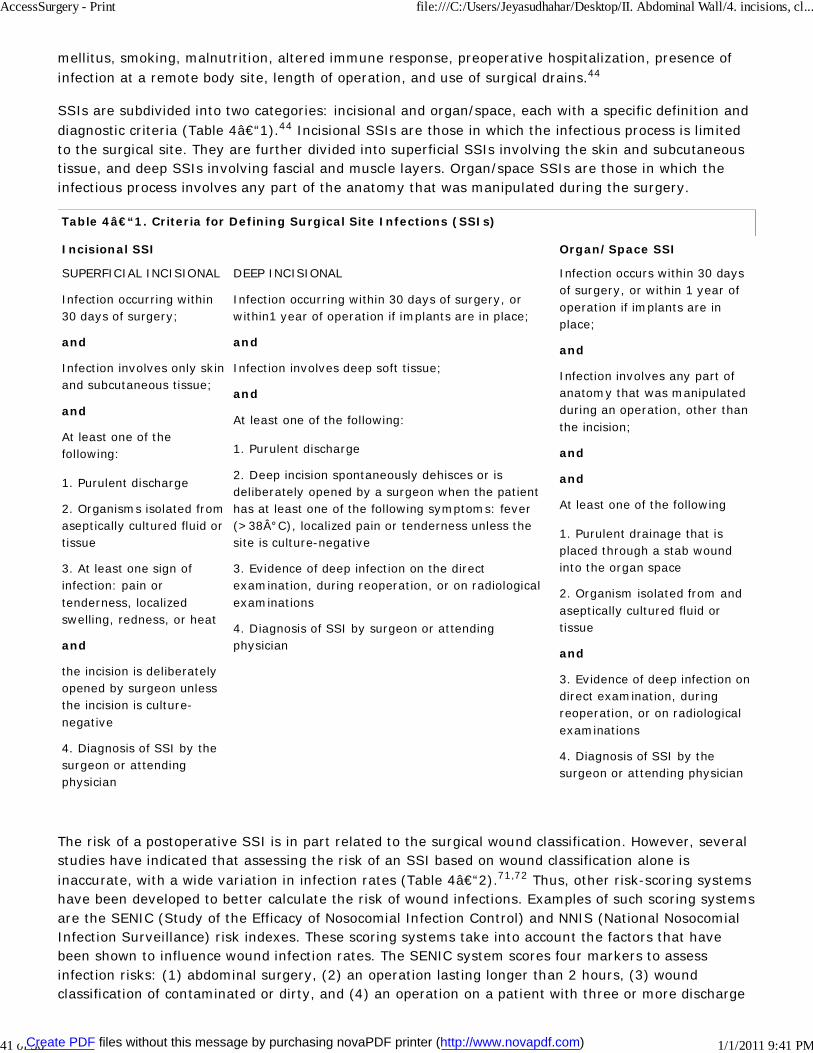
SSIs are subdivided into two categories: incisional and organ/space, each with a specific definition anddiagnostic criteria (Table 4–1).44 Incisional SSIs are those in which the infectious process is limitedto the surgical site. They are further divided into superficial SSIs involving the skin and subcutaneoustissue, and deep SSIs involving fascial and muscle layers. Organ/space SSIs are those in which theinfectious process involves any part of the anatomy that was manipulated during the surgery.
Table 4–1. Criteria for Defining Surgical Site Infections (SSIs)
Incisional SSI Organ/Space SSI
SUPERFICIAL INCISIONAL
Infection occurring within30 days of surgery;
and
Infection involves only skinand subcutaneous tissue;
and
At least one of thefollowing:
1. Purulent discharge
2. Organisms isolated fromaseptically cultured fluid ortissue
3. At least one sign ofinfection: pain ortenderness, localizedswelling, redness, or heat
and
the incision is deliberatelyopened by surgeon unlessthe incision is culture-negative
4. Diagnosis of SSI by thesurgeon or attendingphysician
DEEP INCISIONAL
Infection occurring within 30 days of surgery, orwithin1 year of operation if implants are in place;
and
Infection involves deep soft tissue;
and
At least one of the following:
1. Purulent discharge
2. Deep incision spontaneously dehisces or isdeliberately opened by a surgeon when the patienthas at least one of the following symptoms: fever(>38°C), localized pain or tenderness unless thesite is culture-negative
3. Evidence of deep infection on the directexamination, during reoperation, or on radiologicalexaminations
4. Diagnosis of SSI by surgeon or attendingphysician
Infection occurs within 30 daysof surgery, or within 1 year ofoperation if implants are inplace;
and
Infection involves any part ofanatomy that was manipulatedduring an operation, other thanthe incision;
and
and
At least one of the following
1. Purulent drainage that isplaced through a stab woundinto the organ space
2. Organism isolated from andaseptically cultured fluid ortissue
and
3. Evidence of deep infection ondirect examination, duringreoperation, or on radiologicalexaminations
4. Diagnosis of SSI by thesurgeon or attending physician
The risk of a postoperative SSI is in part related to the surgical wound classification. However, severalstudies have indicated that assessing the risk of an SSI based on wound classification alone isinaccurate, with a wide variation in infection rates (Table 4–2).71,72 Thus, other risk-scoring systemshave been developed to better calculate the risk of wound infections. Examples of such scoring systemsare the SENIC (Study of the Efficacy of Nosocomial Infection Control) and NNIS (National NosocomialInfection Surveillance) risk indexes. These scoring systems take into account the factors that havebeen shown to influence wound infection rates. The SENIC system scores four markers to assessinfection risks: (1) abdominal surgery, (2) an operation lasting longer than 2 hours, (3) woundclassification of contaminated or dirty, and (4) an operation on a patient with three or more discharge
AccessSurgery - Print file:///C:/Users/Jeyasudhahar/Desktop/II. Abdominal Wall/4. incisions, cl...
41 of 56 1/1/2011 9:41 PMCreate PDF files without this message by purchasing novaPDF printer (http://www.novapdf.com)

diagnoses.71 The NNIS system calculates risk of SSI by scoring three risk factors: (1) an AmericanSociety of Anesthesiologists preoperative assessment score of greater than 2, (2) a woundclassification of contaminated or dirty, and (3) increased duration of the operation.72 Using thesescoring systems, a more accurate preoperative forecast of the likelihood of an SSI can be made.High-risk wounds should be left open. These systems also allow a more precise comparison of outcomesamong surgeons and operative centers.
Table 4–2. Classification of Surgical Wounds
Type of Wound Definition Risk ofSSI
Class I: Clean An uninfected operative wound in which no inflammation is encountered andrespiratory, alimentary, genital, or uninfected urinary is not entered. They areprimarily closed, and if necessary drained with close drainage
1–5%
Class II: Clean-contaminated
An operative wound in which the respiratory, alimentary, genital, or urinary tractsare entered under controlled conditions and without unusual contamination. Inparticular, surgery involving the biliary tract, appendix, vagina, and oropharynx areincluded in this category provided no evidence of infection or a major break intechnique is encountered.
2–9%
Class III:Contaminated
Open fresh accidental wounds. In addition, surgery with major breaks in steriletechnique (e.g., open cardiac massage) or gross spillage from the gastrointestinaltract, and incisions in which acute, nonpurulent inflammation is encountered areincluded in this category
3–13%
Class IV: Dirty-infected
Old traumatic wounds with retained devitalized tissue and those that involve existingclinical infection or perforated viscera
3–13%
The organisms most commonly responsible for SSIs are Staphylococcus aureus and coagulase-negativestaphylococci. After abdominal surgery, enteric organisms (Escherichia coli and Enterobacter spp.) arethe most common pathogens. For most SSIs, the source of the responsible pathogen is the patient'sendogenous flora.
It is important that surgeons take measures to prevent SSIs. The Centers for Disease Control andPrevention recommendations for the prevention of SSIs are summarized in Table 4–3.44 The use ofpreoperative prophylactic antibiotics in all clean-contaminated, and clean cases with associated riskfactors is recommended. The antibiotic of choice for most upper gastrointestinal procedures is cefazolinor a comparable first-generation cephalosporin that is cheap and effective. For colorectal surgery,metronidazole is added to this regimen and mechanical bowel preparation should be undertaken. Theadministration of nonabsorbable oral antibiotics is also recommended prior to colorectal surgery,although this practice has been questioned and abandoned by some. Preoperative intravenousantibiotics should be administered 30–60 minutes before the incision is made to allow the agent toreach maximal tissue concentration. In obese patients, the antibiotic dose will need to be increasedaccordingly. For long procedures, it is important to repeat the dose of antibiotics after every twohalf-lives to maintain an effective serum concentration.
Table 4–3. CDC Recommendations to Prevent Surgical Site Infections
PREOPERATIVE FACTORS
Preparation of the patient:
1. Identify and treat all infections remote from the surgical site and postpone elective surgery until infection hasresolved
2. Do not remove hair unless it interferes with surgery
AccessSurgery - Print file:///C:/Users/Jeyasudhahar/Desktop/II. Abdominal Wall/4. incisions, cl...
42 of 56 1/1/2011 9:41 PMCreate PDF files without this message by purchasing novaPDF printer (http://www.novapdf.com)

3. If hair is to be removed, remove immediately preoperatively using clippers
4. Ensure good blood glucose control in diabetic patients and avoid hyperglycemia
5. Encourage cessation of tobacco use (at least for 30 days before surgery, if possible)
6. Do not withhold blood products, as transfusion does not affect rates of SSI
7. Require the patient to shower or bathe with an antiseptic solution the night before surgery
8. Remove gross contamination from the surgical site before performing antiseptic skin preparation
9. Use an appropriate antiseptic solution for skin preparation
10. Apply preoperative antiseptic skin solution for skin preparation in concentric circles moving outward towardthe periphery
11. Keep the preoperative hospital stay as short as possible
Hand/forearm antisepsis for surgical team:
1. Keep nails short and do not wear artificial nails
2. Perform a preoperative scrub for at least 2–5 minutes up to the elbows
3. After performing the surgical scrub, keep the hands up and away from the body (elbows flexed) so that thewater runs from the tips of fingers towards the elbows. Dry hands with a sterile towel and don a sterile gown andgloves
4. Clean underneath each fingernail
5. Do not wear hand or arm jewelry
Management of infected or colonized surgical personnel:
1. Educate and encourage surgical personnel who have signs and symptoms of a transmissible infectious illnessto report promptly to their supervisor and occupational health personnel
2. Develop well-defined policies concerning patient care responsibilities when personnel have potentiallytransmissible infectious conditions. These policies should govern (1) responsibility of personnel in using healthservices and reporting illness, (2) work restrictions, and (3) clearance to resume work after an illness thatrequired work restriction. The policies should also identify staff members that have the authority to removepersonnel from duty
3. Obtain appropriate cultures and exclude from duty surgical personnel who have draining skin lesions untilinfections has been ruled out, or until these personnel have received adequate therapy and infection has beenresolved
4. Do not routinely exclude surgical personnel who are colonized with organisms such as Staphylococcus aureusor group A streptococci, unless they have been linked epidemiologically to dissemination of the organism
Antibiotic prophylaxis:
1. Administer a prophylactic antimicrobial agent only when indicated, and select it based on its efficacy againstthe most common pathogens causing SSIs for a specific operation, and in accordance with publishedrecommendations
2. Administer by the IV route the initial dose of prophylactic antimicrobial agent, timed such that bactericidalconcentration of the drug is established in serum and tissue when the incision is made. Maintain therapeutic levelsof the agent in serum and tissues throughout the operation, and for a few hours after the incision has been closed
3. Before elective colorectal operations, in addition to the above measures, mechanically prepare the bowel byusing enemas and cathartic agents. Give nonabsorbable oral antimicrobial agents in divided doses on the daybefore the operation
4. For high-risk cesarean sections, administer the prophylactic antimicrobial agent immediately after theumbilical cord is clamped
5. Do not routinely use vancomycin for prophylaxis
INTRAOPERATIVE
AccessSurgery - Print file:///C:/Users/Jeyasudhahar/Desktop/II. Abdominal Wall/4. incisions, cl...
43 of 56 1/1/2011 9:41 PMCreate PDF files without this message by purchasing novaPDF printer (http://www.novapdf.com)

Ventilation:
1. Maintain positive pressure ventilation in the operating room with respect to the corridors and adjacent area
2. Maintain a minimum of 15 air changes per hour, of which at least 3 should be fresh air
3. Filter all air, recirculated and fresh, through the appropriate filters per the American Institute of Architects'recommendations
4. Introduce all air at the ceiling, and exhaust air near the floor
5. Do not use ultraviolet radiation in the operating room
6. Keep operating suite doors closed except as need for passage of equipment, personnel, or patients
7. Consider performing orthopedic implant operations in an operating suite supplied with ultraclean air
8. Limit the number of personnel entering the operating room
Cleaning and disinfection of environmental surfaces:
1. When visible soiling or contamination of surfaces or equipment with blood or other body fluids occurs during anoperation, use and EPA-approved hospital disinfectant to clean the affected areas before the next operation
2. Do not perform special cleaning (in addition to cleaning with routine Environmental Protection Agency-approved hospital disinfectant) or closing of operating rooms after contaminated or dirty operations
3. Do not use tacky mats at the entrance to the operating room suite or individual operating rooms for infectioncontrol
4. Wet vacuum the operating floor with an EPA-approved disinfectant after the last operation of the day or night
Microbiological sampling:
1. Do not perform routine environmental sampling of the operating room
Sterilization of surgical instruments:
1. Sterilize all surgical instruments according to published guidelines
2. Perform flash sterilization only for patient care items that will be used immediately. Do not flash sterilize forreasons of convenience or to save time
Surgical attire and drapes:
1. Wear a surgical mask that fully covers the mouth and nose when entering the operating room if an operationis about to begin or is underway, or if sterilized instruments are exposed. Wear the mask throughout theoperation
2. Wear a cap or hood to fully cover hair on the head and face
3. Do not wear shoe covers for prevention of SSIs
4. Wear sterile gloves if scrubbed as a surgical team member. Put on gloves after donning the sterile gown
5. Use surgical gowns and drapes that are effective barriers when wet
6. Change scrub suits that are visibly soiled, contaminated, and/or are penetrated by blood or other potentiallyinfectious material
Asepsis and surgical technique:
1. Adhere to principles of asepsis when placing intravascular devices, spinal or epidural anesthesia catheters, orwhen dispensing or administering IV drugs
2. Assemble sterile equipment and solutions immediately prior to use
3. Handle tissue gently, maintain effective hemostasis, minimize devitalized tissue and foreign bodies anderadicate dead space at the surgical site
4. Use delayed primary skin closure or leave an incision open if the surgeon considers the surgical site to beheavily contaminated
AccessSurgery - Print file:///C:/Users/Jeyasudhahar/Desktop/II. Abdominal Wall/4. incisions, cl...
44 of 56 1/1/2011 9:41 PMCreate PDF files without this message by purchasing novaPDF printer (http://www.novapdf.com)

5. If drain is necessary, use closed suction drain, and place it through a separate incision distant from theoperating incision. Remove the drain as soon as possible
POSTOPERATIVE INCISION CARE
1. Protect an incision that has been closed primarily with a sterile dressing for 24–48 hours postoperatively
2. Wash hands before and after dressing changes and before and after any contact with surgical site
3. When an incision dressing must be changed, use sterile technique
4. Educate the patient and family regarding proper incision care, symptoms of SSI, and the need to report suchsymptoms
SURVEILLANCE
1. Use CDC definitions of SSI without modification for identifying SSIs among surgical inpatients and outpatients
2. For inpatient cases, use direct prospective observation, indirect prospective detection, or a combination ofboth for the duration of the patient's hospitalization
3. When postdischarge surveillance is performed for detecting SSIs following certain operations, use a methodthat accommodates available resources and data needs
4. For outpatient cases, use a method that accommodates available resources and data needs
5. Assign a surgical wound classification upon completion of an operation. A surgical team member should makethe assignment
6. For a patient undergoing an operation chosen for surveillance, record those variables shown to be associatedwith increased risk of SSI
7. Periodically calculate operation-specific SSI rates stratified by variables shown to be associated with increasedrisk of SSI
8. Report appropriately stratified operation-specific SSI rates to surgical team members. The optimum frequencyand format of such rate computations will be determined by stratified case-load sizes and the objectives of local,continuous quality improvement initiatives
The treatment for incisional SSIs involves removal of several contiguous skin stitches or staples torelieve tension and drainage of any underlying collection. Antibiotics are indicated in the presence ofany surrounding cellulitis. The effective use of antibiotics depends on (1) knowledge of the organismsinvolved, (2) their sensitivities to the available antimicrobial agents, and (3) maintenance of anadequate tissue concentration of the drug. Cefazolin or an equivalent first- or second-generationcephalosporin is an appropriate agent if antimicrobial coverage is needed for an uncomplicatedincisional SSI. It must be stressed that purulence requires drainage and no antibiotic can substitute forthis fundamental surgical principle. Wound cultures are obtained only in the presence of purulence andare used to guide selection of appropriate antibiotics. Following abscess drainage, the wound is leftopen to provide further drainage.
Deep space SSIs also require drainage. This can be performed using carefully placed drains if theinfection is localized. Increasingly, such drains are placed percutaneously under CT or ultrasoundguidance. Deep space infections that are not safely amenable to interventional drainage will requireoperative drainage. Broad-spectrum antibiotics are indicated for intra-abdominal infections.
NECROTIZING WOUND INFECTIONSNecrotizing soft tissue infections are often lethal complications. Their incidence in postoperativewounds is low but they represent a serious infection that requires early identification and treatment.Although necrotizing soft tissue infections are a heterogenous group consisting of several specificsyndromes (Table 4–4),73 the treatment principles are essentially the same for all. Patients oftenpresent early within 48 hours postoperatively with incisional pain and often become toxic as the
AccessSurgery - Print file:///C:/Users/Jeyasudhahar/Desktop/II. Abdominal Wall/4. incisions, cl...
45 of 56 1/1/2011 9:41 PMCreate PDF files without this message by purchasing novaPDF printer (http://www.novapdf.com)

infection progresses. With these infections, the incision may initially appear benign but will usuallydemonstrate serous drainage. Disproportionate incisional pain and edema may be present. Theseinfections progress rapidly and result in deep tissue necrosis. In suspected cases, immediate surgicalexploration and extensive débridement is recommended. Débridement of affected tissues andsurrounding cellulitis is the most important single therapeutic factor. Necrotic tissue is resected untilhealthy bleeding tissue is encountered. Gram's stain and culture should be acquired from the drainageand from affected tissue. The wound is irrigated and antibiotics are administered. The infections areoften caused by Clostridium perfringens or group A beta-hemolytic streptococci but can be the result ofa mixed group of organisms. Initial therapy should include a broad spectrum of coverage. Arecommended regimen is penicillin, clindamycin, and an aminoglycoside. Following débridement, thewound is loosely dressed with gauze and is inspected at least daily. Any evidence of extension of thegangrenous tissue is an indication for further débridement.
Table 4–4. Types of Necrotizing Soft Tissue Infections
Predisposing Factors Microbiology Dominant Features
Necrotizing fasciitis type I(polymicrobial)
Surgery, trauma,diabetes mellitus
Anaerobes,gram-negative aerobicbacilli
Necrosis of fat and fascia; mayhave gas
Necrotizing fasciitis type II(group A streptococcal)
Surgery, minor trauma,Varicella
Streptococcus pyogenes
Rapidly progressive necrosis ofmultiple tissue layers; no gas;shock
Clostridial myonecrosis(gas gangrene)
Trauma, surgery,spontaneous (cancerpatient)
Clostridial species Myonecrosis; prominent gasformation
Although the initial management of these necrotizing infections is essentially the same, there areseveral specific disorders that deserve mentioning, as they may require specific therapy.
Gas GangreneWhen one considers how often abdominal wounds become contaminated with septic peritoneal fluid, itis surprising that gas gangrene infection of abdominal wounds is so rare. Gas gangrene infectionfollowing abdominal surgery results from liberation of clostridia contained within the alimentary tractor biliary system. Clostridia can almost invariably be recovered from normal stools, and they mayoccasionally be found when bile, gallbladder wall, or even gallstones are cultured anaerobically. Thepatients usually present with pain at the surgical wound site that is often severe and associated with ahigh temperature and rapid pulse rate, and it eventually progresses to profound shock. When suchwounds are examined in the early stages, the edges are found to be edematous, red, and acutelyinflamed. They later become dusky and necrotic. The patient looks ill. The wound is crepitant withpurulent discharge containing gas bubbles and an irritating brownish, watery fluid that has a peculiarfoul odor. Early surgical intervention is recommended in suspected cases with débridement of allinfected and nonviable tissue.
Although there have been no controlled clinical trials, there is strong evidence that hyperbaric oxygenis of considerable value in treating clostridial infection, and reduces the mortality rate by some reportsfrom 66–23%.74 The high oxygen pressure within the chamber improves tissue oxygenation,promotes healing, and increases free radical formation by neutrophils.75
Necrotizing FasciitisThis syndrome has been divided into two groups depending on the responsible organisms. Type I
AccessSurgery - Print file:///C:/Users/Jeyasudhahar/Desktop/II. Abdominal Wall/4. incisions, cl...
46 of 56 1/1/2011 9:41 PMCreate PDF files without this message by purchasing novaPDF printer (http://www.novapdf.com)

necrotizing fasciitis is a polymicrobial infective process with slower progress. Type II necrotizingfasciitis is caused by group A streptococci and has a more rapid progression.73,76
Type I (Polymicrobial)This type of necrotizing infection is a slowly progressive lesion that affects the total thickness of theskin, but does not involve the deep fascia. Pus formation is variable and it usually presents asnonspecific cellulitis around the wound and slowly extends over the next few days. Its central areabecomes purple and then develops all of the features of gangrene. When affecting the perineum, thedisease is referred to as Fournier's gangrene.
The organisms involved are usually a mixture of anaerobes, gram-negative rods, and enterococcus.Treatment is begun with a broad-spectrum regimen of antibiotics and then tailored pending the resultof microbiological cultures. Hyperbaric oxygen has also been shown to be effective in treatment ofFournier's gangrene.
Type II (Group A Streptococci)This infection presents as a progressive and rapidly necrotizing process affecting the subcutaneous fat,the superficial fascia, and the superior surface of the deep fascia. Initially, the skin is intact, but laterbecomes gangrenous secondary to interruption of its deep blood supply. The condition is readilydistinguished from gas gangrene because of the absence of crepitus, the absence of muscleinvolvement, and the failure to isolate clostridia from tissues or exudate. Necrotizing fasciitis occurspostoperatively, particularly after abdominal or perineal operations. Although CT or MRI imaging hasbeen proposed by some, early operative exploration is recommended in suspected cases. Frozensection can be sent intraoperatively and Gram's stain performed to confirm the diagnosis.
The organism involved is group A Streptococcus , which is usually very sensitive to penicillin. However,it has been suggested that the combination of penicillin and clindamycin is superior to the use ofpenicillin alone.76 Treatment principles are as highlighted above, namely high clinical suspicion, earlysurgical exploration with extensive débridement, and administration of antibiotics.
HematomaIt is difficult to accurately estimate the incidence of postoperative wound hematomas, but in cleanoperative wounds it is thought to be about 2%. The risk is increased in patients with a bleedingdiathesis, the most common cause of which is iatrogenic following the use of anticoagulants. With theever-increasing use of heparin as prophylaxis against thromboembolic complications, the prevalence ofwound hematomas has increased. Studies that have compared unfractionated heparin (UFH) tolow-molecular-weight heparin (LMWH) have shown a wound hematoma rate of around 4–8%following abdominal surgery. These studies have suggested that the use of LMWH is at least as safe asUFH with regard to wound hematomas and major bleeding complications.77,78 It has also been assumedthat the use of aspirin and nonsteroidal anti-inflammatory therapy prior to surgery increases the riskof postoperative hematoma formation, and many surgeons recommend discontinuing these agents 1week prior to surgery. However, there are few published data to defend this practice.
Hematomas are caused by faulty hemostasis of the layers of the abdominal wall, but they are notserious complications unless they become infected. They usually give rise to an aching pain in thewound that is accompanied by a slight rise in temperature. Small hematomas may be difficult to detectunless wounds are palpated carefully. Since these small hematomas usually resolve, they may be leftalone. If large, and particularly if soft, they should be aspirated with a wide-bore needle, or the edgesof the wound that overlies them should be separated with a probe and the contents evacuated. If thereis considerable extravasation of blood giving rise to a fluctuating mass, it is best to return the patient
AccessSurgery - Print file:///C:/Users/Jeyasudhahar/Desktop/II. Abdominal Wall/4. incisions, cl...
47 of 56 1/1/2011 9:41 PMCreate PDF files without this message by purchasing novaPDF printer (http://www.novapdf.com)
to the operating room. There the wound is opened, clot is evacuated, and any visible bleeding vesselsare ligated or coagulated with diathermy. The wound is then closed and consideration is given toplacing a suction drain.
Stitch AbscessStitch abscesses are usually seen about postoperative day 10, but may occur earlier or weeks after thewound has apparently healed. Stitch abscesses may be superficial or deep. When superficial, they mayappear as brown or mauve-colored fluctuating circumscribed blisters more or less in the line of theincision. They produce a certain amount of uneasiness and pain in the wound and are best evacuatedby incising the blistered area and expressing the contents. Antibiotic treatment rarely will benecessary. Such sinuses heal rapidly as soon as the offending stitch is removed, leaving only a slightscar.
When deep, stitch abscesses may be felt as rounded, indurated masses in the depths of the wound andare painful to touch. The use of nonabsorbable sutures such as polypropylene to close the abdominalwound has been associated with increasing incidence of deep stitch abscesses when compared toclosure with a slowly absorbing suture such as polydioxanone.79,80 When polypropylene has been used,treatment for persistent stitch sinuses requires removal of the offending suture.
Abdominal Wound Dehiscence and EviscerationDisruption of an abdominal wound may be limited to the deep fascia with the skin remaining intact.When it is complete, all the layers of the abdominal wall have burst apart with or without associatedprotrusion of a viscus (evisceration). Historically, dehiscence rates of up to 10% have been reported.With recent advances in suture material and the use of mass closure technique the rate of dehiscencehas generally been less than 1%,81 although a recent report from the Veterans Affairs national qualityprogram has documented a rate of 3.2%.82
The mean time to wound dehiscence is 8–10 postoperative days.80,83 There are several clinicalpresentations of wound disruption. The patient may have an abrupt rush of pink serosanguineousdischarge from the wound. This pink discharge is almost pathognomonic of dehiscence and when notedthe wound should be examined. A small portion of the skin incision is opened, and using a probe thedeeper layers are inspected to determine if fascial union is satisfactory. Sometimes this pink dischargeis associated with a large subcutaneous hematoma or a soft and tympanitic boggy swelling thatdistends the wound. Both these findings should be investigated in the operating room. If a largehematoma has formed, it should be evacuated and the depths of the wound examined to see if anyseparation has occurred. The soft boggy swelling generally indicates that a knuckle of bowel hasherniated through the abdominal wall and lies beneath the skin incision.
Another presentation of wound dehiscence is one in which the wound appears clean and dry, butfollowing some excessive strain the patient feels a sudden "give" in the wound, which when examinedwill be found to have dehisced with the gut eviscerated. It is surprising how painless this condition is,and how little if any shock results. Dehiscence can occur in infected wounds, in which case the onset isusually gradual. An abscess forms which is usually drained but results in gradual weakening andseparation of the deeper layers. Matted omentum and intestine are often adherent to the edges of themuscle, and evisceration in such circumstances is uncommon.
Abdominal dehiscence is a morbid complication. The mortality rate following wound dehiscence hasranged from 9–43%, with a recent review reporting a mortality rate of 16%.84 Prevention istherefore an important issue, and a cornerstone of this is meticulous surgical technique. Severaltechnical considerations are worth mentioning:
AccessSurgery - Print file:///C:/Users/Jeyasudhahar/Desktop/II. Abdominal Wall/4. incisions, cl...
48 of 56 1/1/2011 9:41 PMCreate PDF files without this message by purchasing novaPDF printer (http://www.novapdf.com)

1. Incisions . Although it had been suggested that midline incisions, particularly in the upper abdomen, are moreprone to disruption than paramedian incisions, several randomized studies have shown this to be an incorrectassumption.
2. Technique of laparotomy closure . Several randomized studies and meta-analyses have now shown that atechnique of mass closure with a running suture is the best method for closure of midline wounds. Several technicalfailures following laparotomy closure can lead to wound dehiscence. The suture material may rupture because it istoo weak for the tension placed upon it and thus sutures of appropriate strength should be used. This usually meansa suture of 0 or 1 caliber. The sutures may also cut through the tissues, either because the sutures are placed tooclose to the wound edge or because of excessive weakening of tissues. A general guideline is to place sutures 1 cmapart with 1-cm bites of fascia. Another frequent technical error is improper knot tying which may lead unraveling.
Several papers have specifically looked at the nontechnical risk factors for dehiscence in thepostoperative period. They form a heterogeneous group of studies using different techniques oflaparotomy closure. From these studies, several important risk factors have been revealed: age (>65years), hypoalbuminemia, wound infection (increases the risk by more than five times), ascites,obesity, steroid use, chronic obstructive pulmonary disease, current pneumonia, cerebrovascularaccident with residual deficit, anemia (hematocrit <30), prolonged ileus, coughing, emergencyoperation, operative time greater than 2.5 hours, and postgraduate year 4 resident as surgeon.62,82,84
The largest study among the group analyzed over 570 cases of dehiscence in the Veterans Affairspopulation. Data were subject to multivariate analysis and resulted in identification of severalindependent risk factors for this complication (Table 4–5). Together, these data were used to developan abdominal wound dehiscence risk index for prediction of development of postoperative wounddehiscence (Table 4–6).82 Whether placement of retention sutures in those at high risk is helpful inpreventing dehiscence has not been determined. Many surgeons place retention sutures at laparotomyclosure in those with several risk factors that put a patient at high risk for fascial dehiscence.
Table 4–5. Risk Factor Scores for Abdominal Wound Dehiscence
Risk Factor Score
Cerebrovascular accident/stroke without deficit 4
History of chronic obstructive pulmonary disease 4
Current pneumonia 4
Emergency procedure 6
Operative time >2.5 hours 2
Postgraduate year 4 as surgeon 3
Clean wound classification –3
Superficial wound infection 5
Deep wound infection 17
Failure to wean 6
One or more complications 7
Return to the operating room –11
Table 4–6. Risk Categories for Wound Dehiscence
Risk Category Total Score Predicted Rate of Dehiscence
Low 3 1.47%
Medium 4–10 2.70%
AccessSurgery - Print file:///C:/Users/Jeyasudhahar/Desktop/II. Abdominal Wall/4. incisions, cl...
49 of 56 1/1/2011 9:41 PMCreate PDF files without this message by purchasing novaPDF printer (http://www.novapdf.com)

Risk Category Total Score Predicted Rate of Dehiscence
High 11–14 4.53%
Very High >14 10.90%
The basic treatment principle for repair of the disrupted wound is re-suturing of wound edges. It isrecommended for the majority of cases, especially for those in whom the accident has occurred early inthe postoperative period in whom the edges of the wound, although they may be frayed and torn, arerelatively clean. The objective of surgery is to replace the eviscerated organs into the abdominalcavity, and to prevent recurrent dehiscence and later development of ventral hernias. As soon as thecondition is recognized, the wound and protruding viscera should be freely bathed with warm normalsaline solution and covered with large sterile dressing. When the patient has been moved to theoperating room, general anesthesia should be administered and a nasogastric tube placed todecompress the stomach. The edges of the abdominal wall are then lifted upward, and the prolapsedbowel is replaced below the level of the peritoneal edges. At this stage, fragments of suture materialare extracted, and the wound edges are freshened by snipping away necrotic tissue and edematousskin tags. It will be noted in many cases that the fascial edges are swollen and retracted outward,highlighting the need for immediate repair of the dehisced wound to prevent further retraction of thewound edges. If only a very small area of the wound has been disrupted, this portion alone should besutured. However, if more than half of the wound has been disrupted, the correct procedure is to openthe remaining portion of the wound and suture the whole wound afresh.
Resuturing should be performed using a strong monofilament nonabsorbable suture such aspolypropylene. There are several technical considerations when closing such a wound, such as whetherto use continuous or interrupted sutures, and whether or not to use retention sutures. Continuousclosure has the theoretical advantages of evenly distributing the tension along the length of the woundand being quicker to perform. However, the tissue at the site of dehiscence is often weakened, and ifone of the sutures cuts through the fascia the wound will open. There may therefore be a benefit inclosing the fascia with interrupted sutures. Two retrospective analyses, however, have shown nosignificant reduction in the incidence of late ventral hernia following interrupted closure of a dehiscedwound.83,85 Whether retention sutures need to be placed is another unresolved issue. Retrospectiveanalysis of data does not show any benefits in placement of retention sutures in terms of laterdevelopment of an incisional hernia. Some surgeons suggest that the incidence of evisceration wouldbe reduced and recommend the use of retention sutures. It has been shown, however, that placementof retention sutures results in increased pain and discomfort for the patient, and some have proposedabandoning their use altogether.61
Retention sutures, if placed, can be internal (wide bites of the fascia deep to the skin), or external(full-thickness bites of fascia and skin are taken together). One technique to place external retentionsutures consists of placing strong monofilament nylon stitches 2.5 cm from the margin of the woundand about 2.5 cm apart that transfix all the layers of the abdominal wall on both margins of the wound.As they are introduced, the free ends are clipped with hemostats. The fascia is then closed with arunning nonabsorbable suture before the retention sutures are threaded though 5-cm pieces ofprotective rubber tubing and firmly tied. These retention sutures are generally kept in place for 2–4weeks.
Despite these measures, repaired dehisced laparotomy wounds have a 69% hernia development riskover a 10-year period, the majority of which develops over the first 2 years. Dehisced woundsassociated with evisceration, or those in patients that had undergone abdominal aneurysm repair hadan even higher rate of hernia development.85 Because of such high hernia rates, there has been recentinterest in closure of dehisced wounds using nonabsorbable mesh. In a retrospective review, fascial
AccessSurgery - Print file:///C:/Users/Jeyasudhahar/Desktop/II. Abdominal Wall/4. incisions, cl...
50 of 56 1/1/2011 9:41 PMCreate PDF files without this message by purchasing novaPDF printer (http://www.novapdf.com)

dehiscence closed in this fashion had a trend towards lower incidence of ventral hernia development,although this did not reach statistical significance.83
If the size of defect is small, the patient is critically ill, and there is no evisceration of viscera, then thedehiscence can be dealt with conservatively. This will result in the development of an incisional herniathat can be repaired later, when the patient has recovered. This technique involves packing the openwound with a moist sterile dressing and generous strips of elastic adhesive tape placed transverselyacross the abdomen. An abdominal binder can be used for further support, and the patient shouldavoid excessive physical activity. The dressing should be changed at regular intervals until healingtakes place. In some cases, a secondary suture of the skin can be carried out at this stage, but in otherinstances one may elect to allow the wound to heal entirely by granulation. Recent introduction ofvacuum-assisted wound closure devices (VAC devices) has been helpful in such cases. By applying suchdressings the wounds are kept clean, and the controlled negative pressure helps to evacuate fluid andstimulate healing.86
Incisional HerniaAn incisional hernia is one that develops in the scar of a surgical incision. It may be small andinsignificant, or it may bulge through the wound to become a large, unsightly, and uncomfortablehernia. Only hernias with narrow necks and large sacs are at risk of strangulation; those with a wideneck are a nuisance but not usually a danger. The topic of incisional hernias has been coveredextensively in Chapter 5.
REFERENCES1. Ellis H. Maingot's Abdominal Operations. New York, NY: McGraw-Hill; 1997:395
2. Penn I, Baker R. Mastery of Surgery . New York, NY: Lippincott Williams & Wilkins; 2001
3. Grantcharov TP, Rosenberg J. Vertical compared with transverse incisions in abdominal surgery. EurJ Surg 2001;167:260 [PubMed: 11354317]
4. Reidel MA, Knaebel HP, Seiler CM, et al. Postsurgical pain outcome of vertical and transverseabdominal incision: design of a randomized controlled equivalence trial. BMC Surg 2003;3:9 [PubMed:14614782]
5. Greenall MJ, Evans M, Pollock AV. Midline or transverse laparotomy? A random controlled clinicaltrial. Part I: Influence on healing. Br J Surg 1980;67:188 [PubMed: 6988033]
6. Ellis H, Coleridge-Smith PD, Joyce AD. Abdominal incisions—vertical or transverse? Postgrad Med J1984;60:407 [PubMed: 6462980]
7. Greenall MJ, Evans M, Pollock AV. Midline or transverse laparotomy? A random controlled clinicaltrial. Part II: Influence on postoperative pulmonary complications. Br J Surg 1980;67:191 [PubMed:6988034]
8. Guillou PJ, Hall TJ, Donaldson DR, et al. Vertical abdominal incisions—a choice? Br J Surg1980;67:395 [PubMed: 6992914]
9. Cox PJ, Ausobsky JR, Ellis H, et al. Towards no incisional hernias: lateral paramedian versus midlineincisions. J R Soc Med 1986;79:711 [PubMed: 3543347]
10. Rappaport WD, Hunter GC, Allen R, et al. Effect of electrocautery on wound healing in midlinelaparotomy incisions. Am J Surg 1990;160:618 [PubMed: 2147542]
AccessSurgery - Print file:///C:/Users/Jeyasudhahar/Desktop/II. Abdominal Wall/4. incisions, cl...
51 of 56 1/1/2011 9:41 PMCreate PDF files without this message by purchasing novaPDF printer (http://www.novapdf.com)
11. Kearns SR, Connolly EM, McNally S, et al. Randomized clinical trial of diathermy versus scalpelincision in elective midline laparotomy. Br J Surg 2001;88:41 [PubMed: 11136307]
12. Hussain SA, Hussain S. Incisions with knife or diathermy and postoperative pain. Br J Surg1988;75:1179 [PubMed: 2976607]
13. Johnson CD, Serpell JW. Wound infection after abdominal incision with scalpel or diathermy. Br JSurg 1990;77:626 [PubMed: 2143426]
14. Pearlman NW, Stiegmann GV, Vance V, et al. A prospective study of incisional time, blood loss,pain, and healing with carbon dioxide laser, scalpel, and electrosurgery. Arch Surg 1991;126:1018[PubMed: 1907447]
15. Telfer JR, Canning G, Galloway DJ. Comparative study of abdominal incision techniques. Br J Surg1993;80:233 [PubMed: 8443666]
16. Groot G, Chappell EW. Electrocautery used to create incisions does not increase wound infectionrates. Am J Surg 1994;167:601 [PubMed: 8209936]
17. McBurney C. The incision made in the abdominal wall in cases of appendicitis, with a description ofa new method of operating. Ann Surg 1894;20:38
18. Lumsden AB, Colbourn GL, Sreeram S, et al. The surgical anatomy and technique of thethoracoabdominal incision. Surg Clin North Am 1993;73:633 [PubMed: 8378814]
19. Ellis H, Heddle R. Does the peritoneum need to be closed at laparotomy? Br J Surg 1977;64:733[PubMed: 336132]
20. Dorfman S, Rincon A, Shortt H. Cholecystectomy via Kocher incision without peritoneal closure.Invest Clin 1997;38:3 [PubMed: 9103704]
21. Gilbert JM, Ellis H, Foweraker S. Peritoneal closure after lateral paramedian incision. Br J Surg1987;74:113 [PubMed: 3545368]
22. Tulandi T, Al-Jaroudi D. Nonclosure of peritoneum: a reappraisal. Am J Obstet Gynecol2003;189:609 [PubMed: 14520243]
23. Wadstrom J, Gerdin B. Closure of the abdominal wall; how and why? Clinical review. Acta ChirScand 1990;156:75 [PubMed: 2181798]
24. Goligher JC, Irvin TT, Johnston D, et al. A controlled clinical trial of three methods of closure oflaparotomy wounds. Br J Surg 1975;62:823 [PubMed: 1104042]
25. Bucknall TE, Ellis H. Abdominal wound closure—a comparison of monofilament nylon andpolyglycolic acid. Surgery 1981;89:672 [PubMed: 6264641]
26. Leaper DJ, Pollock AV, Evans M. Abdominal wound closure: a trial of nylon, polyglycolic acid andsteel sutures. Br J Surg 1977;64:603 [PubMed: 329939]
27. Wissing J, van Vroonhoven TJ, Schattenkerk ME, et al. Fascia closure after midline laparotomy:results of a randomized trial. Br J Surg 1987;74:738 [PubMed: 3307992]
28. Trimbos JB, Smit IB, Holm JP, et al. A randomized clinical trial comparing two methods of fasciaclosure following midline laparotomy. Arch Surg 1992;127:1232 [PubMed: 1417492]
29. Richards PC, Balch CM, Aldrete JS. Abdominal wound closure. A randomized prospective study of571 patients comparing continuous vs. interrupted suture techniques. Ann Surg 1983;197:238[PubMed: 6297417]
AccessSurgery - Print file:///C:/Users/Jeyasudhahar/Desktop/II. Abdominal Wall/4. incisions, cl...
52 of 56 1/1/2011 9:41 PMCreate PDF files without this message by purchasing novaPDF printer (http://www.novapdf.com)
30. Larsen PN, Nielsen K, Schultz A, et al. Closure of the abdominal fascia after clean and clean-contaminated laparotomy. Acta Chir Scand 1989;155:461 [PubMed: 2531963]
31. Fagniez PL, Hay JM, Lacaine F, et al. Abdominal midline incision closure. A multicentric randomizedprospective trial of 3,135 patients, comparing continuous vs interrupted polyglycolic acid sutures. ArchSurg 1985;120:1351 [PubMed: 2998297]
32. Carlson MA, Condon RE. Polyglyconate (Maxon) versus nylon suture in midline abdominal incisionclosure: a prospective randomized trial. Am Surg 1995;61:980 [PubMed: 7486431]
33. Irvin TT, Koffman CG, Duthie HL. Layer closure of laparotomy wounds with absorbable andnon-absorbable suture materials. Br J Surg 1976;63:793 [PubMed: 791437]
34. Corman ML, Veidenheimer MC, Coller JA. Controlled clinical trial of three suture materials forabdominal wall closure after bowel operations. Am J Surg 1981;141:510 [PubMed: 6452825]
35. Cameron AE, Parker CJ, Field ES, et al. A randomised comparison of polydioxanone (PDS) andpolypropylene (Prolene) for abdominal wound closure. Ann R Coll Surg Engl 1987;69:113 [PubMed:3111339]
36. Murray DH, Blaisdell FW. Use of synthetic absorbable sutures for abdominal and chest woundclosure. Experience with 650 consecutive cases. Arch Surg 1978;113:477 [PubMed: 346006]
37. Martyak SN, Curtis LE. Abdominal incision and closure. A systems approach. Am J Surg1976;131:476 [PubMed: 773200]
38. Jenkins TP. The burst abdominal wound: a mechanical approach. Br J Surg 1976;63:873 [PubMed:137024]
39. Hussain SA. Closure of subcutaneous fat: a prospective randomized trial. Br J Surg 1990;77:107[PubMed: 2405931]
40. Naumann RW, Hauth JC, Owen J, et al. Subcutaneous tissue approximation in relation to wounddisruption after cesarean delivery in obese women. Obstet Gynecol 1995;85:412 [PubMed: 7862382]
41. Cetin A, Cetin M. Superficial wound disruption after cesarean delivery: effect of the depth andclosure of subcutaneous tissue. Int J Gynaecol Obstet 1997;57:17 [PubMed: 9175664]
42. Farnell MB, Worthington-Self S, Mucha P Jr, et al. Closure of abdominal incisions withsubcutaneous catheters. A prospective randomized trial. Arch Surg 1986;121:641 [PubMed: 2939815]
43. Shaffer D, Benotti PN, Bothe A Jr, et al. A prospective, randomized trial of abdominal wounddrainage in gastric bypass surgery. Ann Surg 1987;206:134 [PubMed: 3606238]
44. Mangram AJ, Horan TC, Pearson ML, et al. Guideline for Prevention of Surgical Site Infection,1999. Centers for Disease Control and Prevention (CDC) Hospital Infection Control Practices AdvisoryCommittee. Am J Infect Control 1999;27:97 [PubMed: 10196487]
45. Ranaboldo CJ, Rowe-Jones DC. Closure of laparotomy wounds: skin staples versus sutures. Br JSurg 1992;79:1172 [PubMed: 1467895]
46. Frishman GN, Schwartz T, Hogan JW. Closure of Pfannenstiel skin incisions. Staples vs.subcuticular suture. J Reprod Med 1997;42:627 [PubMed: 9350017]
47. Zwart HJ, de Ruiter P. Subcuticular, continuous and mechanical skin closure: cosmetic results of aprospective randomized trial. Neth J Surg 1989;41:57 [PubMed: 2501713]
AccessSurgery - Print file:///C:/Users/Jeyasudhahar/Desktop/II. Abdominal Wall/4. incisions, cl...
53 of 56 1/1/2011 9:41 PMCreate PDF files without this message by purchasing novaPDF printer (http://www.novapdf.com)
48. Webster DJ, Davis PW. Closure of abdominal wounds by adhesive strips: a clinical trial. Br Med J1975;20:696
49. Singer AJ, Quinn JV, Clark RE, Hollander JE. Closure of lacerations and incisions withoctylcyanoacrylate: a multicenter randomized trial. Surgery 2002;131:270 [PubMed: 11894031]
50. Blondeel PN, Murphy JW, Debrosse D, et al. Closure of long surgical incisions with a newformulation of 2-octylcyanoacrylate tissue adhesive versus commercially available methods. Am J Surg2004;188:307 [PubMed: 15450839]
51. Harold KL, Goldstein SL, Nelms CD, et al. Optimal closure method of five-millimeter trocar sites.Am J Surg 2004;187:24 [PubMed: 14706581]
52. Petrowsky H, Demartines N, Rousson V, et al. Evidence-based value of prophylactic drainage ingastrointestinal surgery: a systematic review and meta-analyses. Ann Surg 2004;240:1074 [PubMed:15570212]
53. Fong Y, Brennan MF, Brown K, et al. Drainage is unnecessary after elective liver resection. Am JSurg 1996;171:158 [PubMed: 8554132]
54. Belghiti J, Kabbej M, Sauvanet A, et al. Drainage after elective hepatic resection. A randomizedtrial. Ann Surg 1993;218:748 [PubMed: 8257225]
55. Liu CL, Fan ST, Lo CM, et al. Abdominal drainage after hepatic resection is contraindicated inpatients with chronic liver diseases. Ann Surg 2004;239:194 [PubMed: 14745327]
56. Lewis RT, Goodall RG, Marien B, et al. Simple elective cholecystectomy: to drain or not. Am J Surg1990;159:241 [PubMed: 2405730]
57. Conlon KC, Labow D, Leung D, et al. Prospective randomized clinical trial of the value ofintraperitoneal drainage after pancreatic resection. Ann Surg 2001;234:487 [PubMed: 11573042]
58. Kim J, Lee J, Hyung WJ, et al. Gastric cancer surgery without drains: a prospective randomizedtrial. J Gastrointest Surg 2004;8:727 [PubMed: 15358335]
59. Poole GV Jr. Mechanical factors in abdominal wound closure: the prevention of fascial dehiscence.Surgery 1985;97:631 [PubMed: 3890240]
60. Hubbard TB Jr, Rever WB Jr. Retention sutures in the closure of abdominal incisions. Am J Surg1972;124:378 [PubMed: 4262544]
61. Rink AD, Goldschmidt D, Dietrich J, et al. Negative side-effects of retention sutures for abdominalwound closure. A prospective randomised study. Eur J Surg 2000;166:932 [PubMed: 11152253]
62. Makela JT, Kiviniemi H, Juvonen T, et al. Factors influencing wound dehiscence after midlinelaparotomy. Am J Surg 1995;170:387 [PubMed: 7573734]
63. Saxe JM, Ledgerwood AM, Lucas CE. Management of the difficult abdominal closure. Surg ClinNorth Am 1993;73:243 [PubMed: 8456355]
64. Burch JM, Ortiz VB, Richardson RJ, et al. Abbreviated laparotomy and planned reoperation forcritically injured patients. Ann Surg 1992;215:476 [PubMed: 1616384]
65. Brock WB, Barker DE, Burns RP. Temporary closure of open abdominal wounds: the vacuum pack.Am Surg 1995;61:30 [PubMed: 7832378]
66. Stone PA, Hass SM, Flaherty SK, et al. Vacuum-assisted fascial closure for patients with abdominaltrauma. J Trauma 2004;57:1082 [PubMed: 15580036]
AccessSurgery - Print file:///C:/Users/Jeyasudhahar/Desktop/II. Abdominal Wall/4. incisions, cl...
54 of 56 1/1/2011 9:41 PMCreate PDF files without this message by purchasing novaPDF printer (http://www.novapdf.com)

67. Jernigan TW, Fabian TC, Croce MA, et al. Staged management of giant abdominal wall defects:acute and long-term results. Ann Surg 2003;238:349 [PubMed: 14501501]
68. Hermann RE, Flowers RE, Wasylenki EW. Early exposure in the management of the postoperativewound. Surg Gynecol Obstet 1965;120:503 [PubMed: 14262048]
69. Hulten L. Dressings for surgical wounds. Am J Surg 1994;167:42S
70. Law N, Ellis HE. Exposure of the wound—a safe economy in the NHS. Postgrad Med J 1987;63:27[PubMed: 3671224]
71. Haley RW, Morgan WM, Culver DH, et al. Update from the SENIC project. Hospital infection control:recent progress and opportunities under prospective payment. Am J Infect Control 1985;13:97[PubMed: 3849273]
72. Culver DH, Horan TC, Gaynes RP, et al. Surgical wound infection rates by wound class, operativeprocedure, and patient risk index. National Nosocomial Infections Surveillance System. Am J Med1991;91:152S
73. Urschel J. Necrotizing soft tissue infections. Postgrad Med J 1999;75:645 [PubMed: 10621873]
74. Riseman JA, Zamboni WA, Curtis A, et al. Hyperbaric oxygen therapy for necrotizing fasciitisreduces mortality and the need for debridements. Surgery 1990;108:847 [PubMed: 2237764]
75. Clark L, Moon R. Hyperbaric oxygen in the treatment of life-threatening soft-tissue infections.Respir Care Clin N Am 1999;5:203 [PubMed: 10333449]
76. Bisno A, Stevens D. Streptococcal infections of skin and soft tissues. N Engl J Med 1996;334:240[PubMed: 8532002]
77. Nurmohamed MT, Verhaeghe R, Haas S, et al. A comparative trial of a low molecular weightheparin (enoxaparin) versus standard heparin for the prophylaxis of postoperative deep veinthrombosis in general surgery. Am J Surg 1995;169:567 [PubMed: 7771617]
78. Kakkar VV, Boeckl O, Boneu B, et al. Efficacy and safety of a low-molecular-weight heparin andstandard unfractionated heparin for prophylaxis of postoperative venous thromboembolism: Europeanmulticenter trial. World J Surg 1997;21:2 [PubMed: 8943170]
79. Hodgson NC, Malthaner RA, Ostbye T. The search for an ideal method of abdominal fascial closure:a meta-analysis. Ann Surg 2000;231:436 [PubMed: 10714638]
80. van't Riet M, Steyerberg EW, Nellensteyn J, et al. Meta-analysis of techniques for closure ofmidline abdominal incisions. Br J Surg 2002;89:1350 [PubMed: 17216342]
81. Bucknell TE, Cox PJ, Ellis H. Burst abdomen and incisional hernia: a prospective study of 1129major laparotomies. BMJ 1982;284:931
82. Webster C, Neumayer L, Smout R, et al. Prognostic models of abdominal wound dehiscence afterlaparotomy. J Surg Res 2003;109:130 [PubMed: 12643854]
83. Gislason H, Viste A. Closure of burst abdomen after major gastrointestinaloperations—comparison of different surgical techniques and later development of incisional hernia.Eur J Surg 1999;165:958 [PubMed: 10574104]
84. Pavlidis TE, Galatianos IN, Papaziogas BT, et al. Complete dehiscence of the abdominal wound andincriminating factors. Eur J Surg 2001;167:351 [PubMed: 11419550]
AccessSurgery - Print file:///C:/Users/Jeyasudhahar/Desktop/II. Abdominal Wall/4. incisions, cl...
55 of 56 1/1/2011 9:41 PMCreate PDF files without this message by purchasing novaPDF printer (http://www.novapdf.com)

85. van't RM, De Vos Van Steenwijk PJ, Bonjer HJ, et al. Incisional hernia after repair of wounddehiscence: incidence and risk factors. Am Surg 2004;70:281 [PubMed: 17216342]
86. Schimp VL, Worley C, Brunello S, et al. Vacuum-assisted closure in the treatment of gynecologiconcology wound failures. Gynecol Oncol 2004;92:586 [PubMed: 14766251]
Copyright ©2007 The McGraw-Hill Companies. All rights reserved.Privacy Notice . Any use is subject to the Terms of Use and Notice . Additional Credits and Copyright Information .
AccessSurgery - Print file:///C:/Users/Jeyasudhahar/Desktop/II. Abdominal Wall/4. incisions, cl...
56 of 56 1/1/2011 9:41 PMCreate PDF files without this message by purchasing novaPDF printer (http://www.novapdf.com)


![[Print] - eMedicine Orthopedic Surgery](https://static.fdocuments.net/doc/165x107/5514008a4a7959df028b4dd8/print-emedicine-orthopedic-surgery.jpg)

