Accelerated deactivation studies of the natural-gas ... · Two mechanisms for sintering of catalyst...
26
Tampere University of Technology Accelerated deactivation studies of the natural-gas oxidation catalyst-Verifying the role of sulfur and elevated temperature in catalyst aging Citation Honkanen, M., Kärkkäinen, M., Kolli, T., Heikkinen, O., Viitanen, V., Zeng, L., ... Vippola, M. (2016). Accelerated deactivation studies of the natural-gas oxidation catalyst-Verifying the role of sulfur and elevated temperature in catalyst aging. Applied Catalysis B-Environmental, 439-448. DOI: 10.1016/j.apcatb.2015.09.054 Year 2016 Version Peer reviewed version (post-print) Link to publication TUTCRIS Portal (http://www.tut.fi/tutcris) Published in Applied Catalysis B-Environmental DOI 10.1016/j.apcatb.2015.09.054 Copyright © 2015. This manuscript version is made available under the CC-BY-NC-ND 4.0 license http://creativecommons.org/licenses/by-nc-nd/4.0/ Take down policy If you believe that this document breaches copyright, please contact [email protected], and we will remove access to the work immediately and investigate your claim. Download date:26.08.2018
Transcript of Accelerated deactivation studies of the natural-gas ... · Two mechanisms for sintering of catalyst...

Tampere University of Technology
Accelerated deactivation studies of the natural-gas oxidation catalyst-Verifying the roleof sulfur and elevated temperature in catalyst aging
CitationHonkanen, M., Kärkkäinen, M., Kolli, T., Heikkinen, O., Viitanen, V., Zeng, L., ... Vippola, M. (2016). Accelerateddeactivation studies of the natural-gas oxidation catalyst-Verifying the role of sulfur and elevated temperature incatalyst aging. Applied Catalysis B-Environmental, 439-448. DOI: 10.1016/j.apcatb.2015.09.054Year2016
VersionPeer reviewed version (post-print)
Link to publicationTUTCRIS Portal (http://www.tut.fi/tutcris)
Published inApplied Catalysis B-Environmental
DOI10.1016/j.apcatb.2015.09.054
Copyright© 2015. This manuscript version is made available under the CC-BY-NC-ND 4.0 licensehttp://creativecommons.org/licenses/by-nc-nd/4.0/
Take down policyIf you believe that this document breaches copyright, please contact [email protected], and we will remove access tothe work immediately and investigate your claim.
Download date:26.08.2018

1
Accelerated Deactivation Studies of the Natural-Gas Oxidation Catalyst - Verifying the Role
of Sulfur and Elevated Temperature in Catalyst Aging
Mari Honkanen(a),(*), Marja Kärkkäinen(b), Tanja Kolli(b), Olli Heikkinen(c), Ville Viitanen(c), Lunjie
Zeng(d), Hua Jiang(c), Kauko Kallinen(e), Mika Huuhtanen(b), Riitta L. Keiski(b), Jouko Lahtinen(c),
Eva Olsson(d), Minnamari Vippola(a)
(a) Department of Materials Science, Tampere University of Technology, P.O. Box 589, 33101
Tampere, Finland(b) Environmental and Chemical Engineering, Faculty of Technology, University of Oulu,
P.O. Box 4300, 90014 Oulu, Finland(c) Department of Applied Physics, Aalto University, P.O. Box 14100, 00076 Aalto, Finland(d) Department of Applied Physics, Chalmers University of Technology, 41296 Gothenburg,
Sweden(e)Dinex Ecocat Oy, Typpitie 1, 90620 Oulu, Finland
(*) Corresponding author, e-mail: [email protected], telephone: +358408490133
Co-authors’ e-mails: Marja Kärkkäinen, [email protected] Kolli, [email protected] Heikkinen, [email protected] Viitanen, [email protected] Zeng, [email protected] Jiang, [email protected] Kallinen, [email protected] Huuhtanen, [email protected] L. Keiski, [email protected] Lahtinen, [email protected] Olsson, [email protected] Vippola, [email protected]
2
Graphical abstract:
Highlights:
· In thermal aging, grown particles with Pt/Pd core and PdO shell formed
· Al2(SO4)3 formed near small PtPd particles during S-treatment
· With grown particles, Pt/Pd core + PdO shell, Al2(SO4)3 formed randomly in catalyst
· Combined morphological changes and Al2(SO4)3 formation deactivated catalyst

3
Abstract
Accelerated deactivation, caused by thermal aging (TA) and/or sulfur+water poisoning (SW), of the
PtPd/g-Al2O3 natural-gas oxidation catalyst was studied. Thermal aging and poisoning treatments
were performed separately and with varied combinations and comprehensive characterization of the
catalyst was carried out after each step. The fresh catalyst has small, oxidized PtPd particles (<5
nm) uniformly distributed in the g-alumina washcoat. After the SW-treatment, a small amount of
bulk aluminum sulfate was observed near the slightly grown noble metal particles. During the
thermal aging, g-alumina changed to d-/q- and a-alumina. In addition, total decomposition of
oxidized Pt and partly decomposition of oxidized Pd occurred resulting in the formation of the
grown noble metal particles with a bimetallic PtPd core and a polycrystalline PdO shell. Also few,
small (~5 nm) bimetallic PtPd particles were still detected. In the TA+SW-treated catalyst with
grown noble metal particles, a small amount of bulk aluminum sulfate was detected and it was
randomly distributed over the noble metal particles and washcoat. The activity in the terms of
methane conversion over the TA-, SW-, and SW+TA-treated catalysts was similar but it was
decreased compared to the fresh catalyst. The activity of the TA+SW-treated catalyst was
drastically decreased compared to the fresh catalyst due to significant morphological changes and
aluminum sulfate formation.
Keywords: palladium, platinum, deactivation; thermal aging; sulfur poisoning

4
1 INTRODUCTION
Natural gas (NG) is a potential sustainable energy source for vehicles and engines as its
environmental impacts are smaller than those of e.g. crude oil-based fuels. For example exhaust
emissions of the natural gas vehicles (NGVs) are much lower than those in the diesel vehicles.
However, exhaust gases of NGVs contain unburned methane and carbon monoxide, a potential
greenhouse gases, and thus catalytic oxidation converters are needed [1,2]. Supported palladium
catalysts are widely known to be active for methane combustion and it is agreed that PdO is the
active phase while metallic palladium is much less active. Pd«PdO transformation is reversible
and according to Farrauto et al. [3], decomposition temperature of PdO to Pd in the PdO/Al2O3
catalyst is ~800°C and reformation temperature of Pd to PdO during cooling is ~600°C. Hysteresis
in the reformation is due to strongly bound oxygen on the Pd surface inhibiting bulk oxidation [4].
Many other factors, such as gas phase composition and pressure, type of support, additives and
contaminants, and pretreatment conditions, have also effects on the Pd«PdO transformation [5]. In
addition, several studies have reported that the activity and stability of Pd catalysts in methane
combustion can be improved by adding Pt into the catalyst to decelerate sintering of Pd and PdO,
e.g. [6–9].
Deactivation of the catalyst, caused by poisoning and thermal aging, is a problem also in the natural
gas applications. Poisoning is due to adsorption of impurities, e.g. S, P, Zn, Ca, and Mg, present in
the exhaust gases, on the catalytic active sites. Poisons can also react with the active sites followed
by formation of non-active compounds [10]. Sulfur is known to be one of the most important
components for deactivation of the Pd catalysts for methane oxidation [11] and only a small amount
of SO2 significantly decreases catalyst activity by blocking the active noble metal sites by sulfur
compounds [12]. It is known that sulfur in the NGV exhaust gas originates from odorants and
lubricating oils and from gas itself. Under the typical natural-gas engine conditions, with large
excess of air and with an oxidation catalyst, sulfur will be oxidized to SO2 and SO3. Above 300°C, a
catalyst has enough activity for the oxidation of SO2 to SO3 [10]. Metallic Pt on the PtPd catalysts is
found to be active to adsorb sulfur compounds. Therefore, the presence of metallic Pt prevents
adsorption of sulfur on the Pd surface sites which are then available for methane oxidation [13].
Also the catalyst washcoat affects sulfur poisoning. For example Pd/Al2O3 catalysts deactivate less
than Pd/SiO2 catalysts due to adsorption of SOx also on the Al2O3 washcoat material [11].
According to Mowery et al. [14], when SO2 and water exist, the sulfation of the PdO/Al2O3 catalyst
happens by oxidation of SO2 to SO3 on PdO and further SO3 over PdO can form PdSO4 or can
migrate to Al2O3 forming Al2(SO4)3. The sulfation is reversible and SOx from the catalyst surface

5
easily desorbs already below 400°C and more stable washcoat sulfates decompose above 700°C
[15]. SO2 can affect also the acidity of the Al2O3 support. Konsolakis et al. [16] noticed that the
fresh Pd/g-Al2O3 catalyst has only Lewis acid sites but SO2 treatment increases significantly
Brönsted acid sites on the g-Al2O3 support. According to Wischert et al. [17], the reactivity of g-
Al2O3 towards methane is due to the highly reactive Lewis acid-base pairs (Al,O) enabling low-
energy pathways for the dissociation of C-H bond in methane.
Thermal deactivation of a catalyst is reported to be caused by: a) loss of active surface area due to
crystallite growth of the noble metal particles and due to pore collapse on crystallites of the active
phase, b) loss of support area due to collapse of the washcoat and/or c) chemical transformations of
active catalytic phases to non-active phases. In general, sintering occurs at elevated temperatures
(>500°C) and is accelerated by water vapor [18]; the driving force for sintering is to minimize
surface energy and it is reduced with the transport, growth, and coalescence of the particles [10].
Two mechanisms for sintering of catalyst nanoparticles have been proposed: particle migration
followed by coalescence (PMC) and Ostwald ripening (OR). In the particle migration, sintering
occurs by migration of the whole crystallites along the support surface and coalescence of the
crystallites forms larger particles. OR is migration of single metal atoms or molecular species from
small particles to large ones; larger particles grow at the expense of smaller particles [10,18–21].
Hansen et al. [19] studied the mechanism of the catalyst nanoparticle sintering mainly by in situ
transmission electron microscopy. They noticed that the early stage of the sintering is dominated by
OR and surface area increases rapidly caused by the disappearance of the smallest particles. In the
further stage, grown particles coalesce when they are close to each other. Sintering becomes slower
when particles grow and the distance between them increases [19]. Many factors e.g. temperature,
atmosphere, metal type, support, and impurities affect the rate of noble metal sintering. The
sintering rate increases exponentially with elevated temperature and metals sinter faster in oxygen
than in hydrogen atmosphere [18]. Generally, sintering of the noble metal clusters is accelerated by
the presence of gases; Parkinson et al. [22] reported that existence of carbon monoxide induced
coalescence of Pd atoms in the Pd/Fe3O4 system and that formed Pd-carbonyl species were
responsible for increased Pd mobility. Water vapor has also been found to accelerate sintering. In
reducing atmosphere, the stability of the metal crystallites typically decreases with decreasing noble
metal melting temperature. In oxidizing atmosphere, stability depends on the volatility of metal
oxides and on the noble metal-support interactions [18]. In addition, the position of the noble metal
particles affects the rate of sintering; a valley position is much more stable than an on-top position
6
[21]. Pores in the support hinder the mobility of the metal crystallites; especially if pore diameters
are about same size as the crystallites [18]. Impurities affect the sintering rate, for example sulfur
increases mobility of the metal atoms [18,23,24] while for example calcium is found to decrease it
[18].
In the case of the catalysts with an alumina washcoat, phase transformations of the support may
occur; g-alumina with high specific surface area can change to d-alumina and finally via q-alumina
to stable a-alumina [10]. Phase transformation results in sintering or grain growth followed by a
decrease in the surface area [25]. According to Wischert et al. [17], the phase transformation of g-
Al2O3 to q-Al2O3 causes the reconstruction of the surface resulting in decrease of highly reactive
sites for methane.
In the real vehicle aging conditions, deactivation process is a complex phenomenon and e.g. both
thermal aging and poisoning exist. In our earlier study [26], the natural-gas heavy-duty-vehicle-
aged (160000 km) PtPd/g-Al2O3-based catalyst was studied. Significant morphological changes and
chemical poisoning were detected compared to the fresh catalyst and this has led to drastic
deactivation in methane conversion. It was difficult to conclude which changes were caused by
thermal aging and which by chemical poisoning. Thus, detailed studying of deactivation phenomena
in the vehicle-aged catalysts was found very challenging. Sulfur poisoning of the oxidation catalysts
is widely studied, e.g. [10,11,13–15,18]. In this study, sulfur poisoning and thermal aging were
studied separately and with varied combinations enabling detailed analysis of the role of sulfur in
the catalyst and effects of thermal aging before or after sulfur poisoning. This kind of in-depth
knowledge gained is crucial in the development of efficient exhaust emission reduction systems for
NGVs.
2 EXPERIMENTAL
2.1 Catalyst material
The studied material was a PtPd (1:4 wt-%) catalyst supported on g-alumina washcoat on the
metallic monolith. The studied catalyst was manufactured and designed by Dinex Ecocat Oy for
lean-burn natural gas applications. Total metal loading in the catalyst was 8.8 g/dm3 and the catalyst

7
was calcined at 550°C for 4 hours. The catalyst was studied as fresh and after various chemical and
thermal laboratory-scale accelerated deactivation treatments.
2.2 Laboratory-scale accelerated deactivation treatments
Laboratory-scale sulfur + water treatment (SW) and thermal aging (TA) treatments were carried out
to find out detailed knowledge about deactivation phenomena of the PtPd/g-Al2O3 oxidation catalyst
for natural-gas applications. SW-treatment was performed in the following conditions: 100 ppm
SO2, 10 vol-% H2O, 10 vol-% air, balanced with N2. The quartz tube reactor was heated from room
temperature to 400°C in a nitrogen and air flow with the heating rate of 10°C/min. After 5 hours of
the SW-treatment, the reactor was cooled down to the room temperature in nitrogen and air flow.
The gas hourly space velocity (GHSV) was 20 000 h-1during the treatment. Thermal aging was
carried out in the tube reactor under synthetic air (80% N2 + 20% O2) at 1000°C for 5 hours. The
used temperature was above the normal operation temperature of the catalysts but it was chosen to
mimic an accelerated thermal aging and thus the long-term behavior of the catalyst. Temperature
for the TA-treatment was chosen based on our tentative study about laboratory-scale thermal aging
treatments for natural gas oxidation catalyst (various temperatures between 400°C and 1100°C);
with this thermal aging temperature, the catalyst structure changes similar to a vehicle-aged catalyst
were achieved [26]. The catalyst samples were set into the tubular ceramic furnace when the
treatment temperature was reached, then the gas flow was switched on. After the treatment period
the gas flow was switched off and the catalyst samples were removed from the furnace and let to
cool down to room temperature. TA- and SW-treatments were performed separately and with varied
combinations (Table 1). Detailed characterization of the catalyst was carried out after each step.
Table 1. Laboratory-scale treatments and corresponding markings.Marking TreatmentFresh As-received from Dinex Ecocat OySW Sulfur + water poisoning (100 ppm SO2 + 10 vol-% H2O + 10 vol-% air + N2 (bal.), 400°C/5h)TA Thermal aging at 1000°C/5h (80 vol-% N2 + 20 vol-% O2)TA+SW Thermal aging followed by sulfur + water poisoningSW+TA Sulfur + water poisoning followed by thermal aging (1000°C/5h)
2.3 Characterizations
The structure of the fresh and treated catalysts was studied by scanning electron microscopy (SEM),
transmission electron microscopy (TEM), and X-ray diffractometry (XRD). A field-emission SEM

8
(FESEM, Zeiss ULTRAplus) is equipped with an energy dispersive spectrometer (EDS, INCAx-act
silicon-drift detector (SDD), Oxford Instruments). Cross-sectional FESEM samples were prepared
with a conventional metallographic sample preparation technique by molding the catalyst samples
into resin followed by grinding and polishing and carbon coating to avoid sample charging during
the FESEM studies. All the presented cross-sectional FESEM images were taken with an angular
selective backscatter (AsB) detector to maximize Z-contrast. Three different TEMs were used to
characterize the samples. A TEM (Jeol JEM-2010) equipped with an EDS (Noran Vantage Si(Li)
detector, Thermo Scientific) was used for imaging and elemental analysis. A high resolution TEM
(HRTEM, Jeol 2200FS) with two aberration correctors (CEOS GmbH) equipped with EDS (Jeol
Si(Li) detector) was used for high resolution imaging and elemental analysis. A high resolution
TEM/scanning TEM (STEM, FEI Titan 80-300) with a probe Cs corrector, a Gatan Imaging Filter
(GIF, Tridium), and an EDS (INCAx-sight Si(Li) detector) was used for high resolution imaging,
electron energy loss spectroscopy (EELS), and elemental analysis. Annular dark field (ADF) STEM
images were acquired using a 17.5 mrad beam convergence angle and ~54-270 mrad detector
collection angle. The collection angle for EELS is ~24 mrad. Cross-sectional TEM samples from
the fresh catalyst and from the TA+SW-treated catalyst were prepared as follows. Small pieces of
the catalyst monolith were attached the washcoats face-to-face to a titanium grid by carbon glue.
The grid was pre-thinned by hand to the thickness of ~70 µm and then with a dimple grinder
(Model 565, Gatan Inc.) to the thickness of ~20 µm. The final thinning for electron transparency
was made with a precision ion polishing system (PIPS, Model 691, Gatan Inc.). Powdered TEM
samples from all studied catalysts were prepared by crushing the scraped catalyst powder between
two laboratory glass slides and dispersing the crushed powder with ethanol onto a copper grid with
a holey carbon film. Scraped catalyst powders were used also for XRD studies (Empyrean with the
PIXcel3D detector, PANanalytical, using Cu Ka radiation). Crystallite sizes were determined from
the XRD patterns with the aid of the HighScore plus software based on the Scherrer equation (shape
factor 0.9) and phases were identified by using the database (PDF-4+ 2014 ) from International
Centre for Diffraction Data (ICDD).
The chemical state and composition of the fresh and treated catalysts were studied by X-ray
photoelectron spectroscopy (XPS, SSX-100, Surface Science Instruments, using monochromatic Al
Ka radiation). For XPS measurements, a small amount of the scraped catalyst powder was pressed
into a piece of indium and the samples were pre-treated in high vacuum for a few hours before
measurements. The binding energy values in the acquired spectra were calibrated by setting carbon
1s line at 284.6 eV. Carbon and indium were excluded from the compositional analysis. In
quantitative analysis, Shirley background subtraction was applied [27].

9
Specific surface areas, pore sizes, and pore volumes of the fresh and treated catalysts were
determined using the Micrometrics ASAP 2020 device. Specific surface areas were measured from
the N2 adsorption isotherms at -196°C according to the standard BET (Brunauer-Emmett-Teller)
method. Pore size and pore volume distributions of catalysts were calculated from N2 desorption
isotherms by the BJH (Barrett–Joyner–Halenda) method.
A Fourier transform infrared (FT-IR) spectrometer (Bruker Vertex V80) equipped with a diffuse
reflectance infrared Fourier transform (DRIFT) unit and a liquid nitrogen-cooled mercury cadmium
telluride (MCT) detector was utilized to find the information about the bonding of the compounds
on the scraped catalyst powder. The DRIFT analyses were performed at room temperature under
normal atmosphere conditions. The mirror was used as a background spectrum. Spectra were
recorded by using a resolution of 4 cm-1.
Laboratory scale light-off tests were used to define catalyst activity before and after SW-, TA-,
TA+SW-, and SW+TA-treatments. Catalytic activities were determined in lean reaction conditions
using the following gas mixture: 600 ppm CH4, 500 ppm CO, 10 vol-% CO2, 12 vol-% O2, 10 vol-
% H2O, and N2 as balance gas. The total gas flow was 1 dm3/min resulting in a GHSV of 31000 h-1.
The measurements were carried out at atmospheric pressure in a horizontally aligned tubular quartz
reactor. The temperature of the catalyst bed was increased from room temperature up to 600°C with
a linear heating rate of 10°C/min. H2O was added at 110°C with a peristaltic pump. The catalyst
was kept at steady state for 15 min at 600°C and after that the furnace was cooled down to room
temperature under the N2 flow. The procedure was repeated (run 1 and run 2). If not specifically
mentioned, the data of the catalyst activities was taken from the second run. Gas flow rates were
controlled by using mass flow controllers (Brooks 5280S). The outlet gas composition was
analyzed as a function of temperature by a GasmetTM FT-IR gas analyzer. Oxygen concentration
was determined by using a paramagnetic oxygen analyzer (ABB Advanced Optima).
3 RESULTS AND DISCUSSION
3.1 Structural characteristics of the catalyst
Structural characteristics of the fresh and treated catalysts were studied by several methods. Based
on the FESEM studies, the structure of the fresh and SW-treated catalyst was similar. Moreover, the
structures of the TA-, TA+SW-, and SW+TA-treated catalysts were observed to be analogous.

10
Cross-sectional AsB images of the fresh and TA+SW-treated catalyst are presented in Figs. 1 (a)
and (b), respectively. In the fresh and SW-treated catalysts, the noble metal particles were too small
to be detected with FESEM. In the TA-, TA+SW-, and SW+TA-treated catalysts, the particle size
of the noble metals was increased significantly through the catalysts (white spots in Fig. 1 (b)).
Noble metals were still well distributed in the cross-sections of the TA-, TA+SW-, and SW+TA-
treated catalysts.
Figure 1. Cross-sectional FESEM (AsB) images, higher magnifications as insets, (a) the freshcatalyst and (b) the TA+SW-treated catalyst, white spots represent the grown noble metal particles.
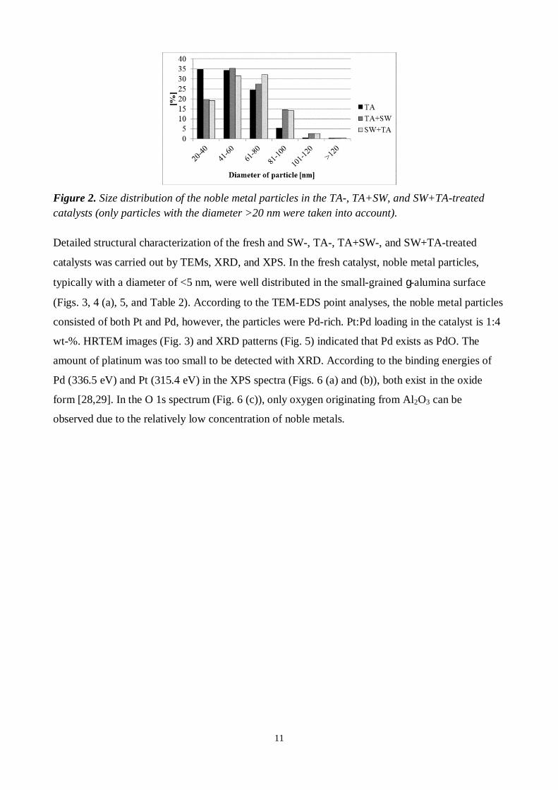
Average noble metal particle sizes (diameters) of the TA-, SW+TA-, and TA+SW-treated catalysts
were 50, 60, and 60 nm, respectively. Particle size distribution is presented in Fig. 2. It is important
to notice that according to TEM studies, particles with the size <20 nm also existed in the catalyst
samples. However, only the particles >20 nm were taken into account in the particle size analyses
from the FESEM images because it was the minimum reliable detection limit. Particles were
measured from the cross-sectional FESEM images with the aid of ImageJ-software (>500 particles
with size >20 nm were measured). Thus, in the results there is a bias towards the bigger particle
sizes. However, the results can be used in the comparison of the large, agglomerated particles in all
TA-treated catalysts. Based on the results, sizes of the formed large particles were similar after TA-,
TA+SW-, and SW+TA-treatments.
11
Figure 2. Size distribution of the noble metal particles in the TA-, TA+SW, and SW+TA-treatedcatalysts (only particles with the diameter >20 nm were taken into account).
Detailed structural characterization of the fresh and SW-, TA-, TA+SW-, and SW+TA-treated
catalysts was carried out by TEMs, XRD, and XPS. In the fresh catalyst, noble metal particles,
typically with a diameter of <5 nm, were well distributed in the small-grained g-alumina surface
(Figs. 3, 4 (a), 5, and Table 2). According to the TEM-EDS point analyses, the noble metal particles
consisted of both Pt and Pd, however, the particles were Pd-rich. Pt:Pd loading in the catalyst is 1:4
wt-%. HRTEM images (Fig. 3) and XRD patterns (Fig. 5) indicated that Pd exists as PdO. The
amount of platinum was too small to be detected with XRD. According to the binding energies of
Pd (336.5 eV) and Pt (315.4 eV) in the XPS spectra (Figs. 6 (a) and (b)), both exist in the oxide
form [28,29]. In the O 1s spectrum (Fig. 6 (c)), only oxygen originating from Al2O3 can be
observed due to the relatively low concentration of noble metals.

12
Figure 3. TEM images of the fresh catalyst, (a) the cross-sectional sample, the inset with highermagnification and (b)-(d) the powdered sample with different magnifications.
Figure 4. STEM ADF images of the (a) fresh catalyst, (b) SW-treated catalyst, and (c) TA+SW-treated catalyst. Notice the different scale bar in (a) and (b) versus (c).

13
Figure 5. XRD patterns of the fresh, sulfur-poisoned (SW), thermal-aged (TA), thermal-aged +sulfur-poisoned (TA+SW), thermal-aged + sulfur-poisoned after activity measurement(TA+SW+act.), and sulfur-poisoned + thermal-aged (SW+TA) catalysts (g = g-alumina, d = d-alumina, q = q-alumina, and a = a-alumina).
Table 2. Crystallite sizes of PdO and PtPd in the fresh, SW-, TA-, TA+SW-, and SW+TA-treatedcatalysts determined from the XRD spectra.
Size [nm] Fresh SW TA TA+SW TA+SW+act SW+TAPdO(a)
lattice parameters:a=b=3.0Å, c=5.3Å
3 6 9 914
8
PtPd(b)
lattice parameters:a=b=c=3.9 Å
- - 26 3158
27
(a)determined from PdO-peak at 2q=33.9° with the Scherrer equation (shape factor 0.9)(b) determined from Pd/Pt-peak at 2q=40.1° with the Scherrer equation (shape factor 0.9)

14
Figure 6. XPS spectra and binding energies, (a) Pd 3d, (b) Pt 4d, and (c) O 1s spectra for the fresh,SW-, TA-, TA+SW, SW-TA- treated catalysts and TA+SW-treated catalyst after activitymeasurements.
In the SW-treated catalyst, the size of the noble metal particles seemed to be slightly increased
(Figs. 4 (b) and 7) compared to the fresh catalyst. A slight increase in the crystallite size of PdO was
observed also by XRD (Table 2); the amount of platinum was too small to be detected with XRD.
According to our tentative study, no changes in the PtPd particles of the PtPd/g-Al2O3 catalyst were
observed after 400°C for 5 hours treatment in synthetic air (same temperature and exposure time
than in the SW-treatment). Thus, a slight growth of the Pt/Pd particles seemed to be induced by
presence of SO2 + H2O. According to the literature, sulfur and water vapor increase the mobility of
the noble metal atoms, e.g. [18,23,24]. Based on the HRTEM studies, mainly PdO and PtO existed
in the noble metal particles which agrees with XPS results (Fig. 6); despite the change in the
binding energy of O 1s line, it can still be attributed to alumina [30]. TEM-EDS point analyses
indicated both Pd and Pt in the noble metal particles as in the fresh catalyst. The specific surface
area of the SW-treated catalyst was slightly decreased compared to the fresh catalyst (Table 3). This
may be caused due to slight growth of the noble metal crystallites and sulfur species blocking some
pore openings in the catalyst.

15
Figure 7. TEM images of the SW-treated catalyst (the powdered sample), (a), (b), and (c) differentmagnifications.
Table 3. Specific surface area, average pore size, and total pore volume of the fresh, SW-, TA-,TA+SW-, and SW+TA-treated catalysts.
Fresh SW TA TA+SW SW+TASpecific surface area [m2/g] 154 147 96 96 92
Average pore size [nm] 10 10 14 14 15Total pore volume [cm3/g] 0.4 0.4 0.3 0.3 0.4
Significant morphological changes, due to the TA-treatments, were detected in them compared to
the fresh and SW-treated catalysts. According to the TEM studies, morphology of the TA-,
TA+SW-, and SW+TA-treated catalysts was similar. Based on the XRD results, in all thermally-
treated catalysts g-alumina changed to d- and/or q-alumina and to a-alumina (Fig. 5). Due to the
phase transformation of alumina, the particle size of d/q-alumina and especially a-alumina
increased compared to g-alumina in the fresh catalyst. In addition, the size of the noble metal
particles grew significantly under the thermal aging treatment (Figs. 4 (c) and 8) as already
observed with FESEM indicating noble metal particles with a diameter of 50-60 nm. According to
the HRTEM images (Fig. 8 (c)), the grown noble metal particles consisted of several crystals. The
particle had a bimetallic PtPd-alloy core surrounded by several PdO crystals. Based on the TEM-
EDS results, cores consist of both Pt and Pd and the shells are Pd-rich. After the TA-, TA+SW- and
SW+TA-treatments, both PdO and bimetallic Pt/Pd alloy were detected by XRD (Fig. 5). In the
TA-, TA+SW-, and SW+TA-treated catalysts, the crystallite size of PdO was ~10 nm and that of
PtPd was ~30 nm (Table 2) which agrees very well with TEM results. Pd was observed as an oxide
[28], but metallic Pt and Pd are invisible in the XPS spectra (Figs. 6 (a) and (b)) due to the PdO
shell and the surface sensitivity of the technique. Similar grown noble metal particles were detected
also in our earlier study concerning the lean-burn natural-gas-heavy-duty-vehicle-aged catalyst
(160000 km) [26]. Persson et al. reported that in thermal decomposition of PdO, Pd is incorporated
into the Pt/Pd alloy in the Pd-Pt/Al2O3 catalyst [7]. Carrillo et al. reported that in the thermal aging
in air at even 650°C, small Pt particles formed mobile species which migrated and were trapped by

16
PdO particles resulting in formation of PtPd particles [31]. Generally, decomposition of PtO to Pt
happens above 350°C and PdO to Pd at ~800°C [3,32]. Johns et al. reported that after thermal aging
in air at 750°C for 10 hours, Pt was fully reduced but almost 30 % of the Pd species were still in the
oxide form [33]. In the thermally aged catalysts, in addition to the grown noble metal particles there
were still also few, small noble metal particles with a diameter of ~5 nm (Figs. 4 (c) and 8 (d)).
According to the TEM-EDS analyses, they contain both Pd and Pt and based on HRTEM images
(Fig. 8 (d)), they seemed to be single crystals in the metallic form indicating likely bimetallic PtPd
particles. Small, likely bimetallic PtPd particles were hard to detect by XPS (Fig. 6 (a) and (b))
because only few these kind of particles were observed by TEM. According to the literature [22],
the position of the noble metal particles affects their sintering so that a valley position is much more
stable than an on-top position. As a result, some particles at the valley position do not grow even at
elevated temperature. Therefore, few small particles still existed after thermal treatment at 1000°C;
however, particles seemed to be reduced and formed bimetallic PtPd crystallites. The specific
surface area decreased significantly (~ 40 %) and the average pore size increased (~40 %) during
the TA-, TA+SW, and SW+TA-treatments compared to the fresh catalyst (Table 3) due to the
significant growing of the noble metal particles and phase transformations of the washcoat followed
by the crystal growth. SW-treatment after TA-treatment has no further effect on the specific surface
area. It can be concluded that in the thermal treatment used in this study, the oxidized Pt totally
reduces and partial decomposition of the oxidized Pd occurs followed by formation of the grown
particles with a bimetallic PtPd core and a polycrystalline PdO shell. In addition there are still also
few, small bimetallic PtPd particles.

17
Figure 8. TEM images of the TA+SW-treated catalyst, (a) the cross-sectional sample, (b), (c), and(d) the powdered sample with different magnifications.
3.2 Sulfur species on the catalyst
According to the FESEM-EDS and TEM-EDS analyses, in the SW- and TA+SW-treated catalysts
sulfur existed uniformly through the whole catalyst structure from the inlet part to the outlet part
and from the surface to the metallic monolith; the amount of sulfur was ~3 wt-% which agrees well
with XPS measurements. In the SW+TA-treated catalyst, no sulfur was detected. According to the
literature, the sulfation process is reversible and SOx from the catalyst surface easily desorbs and
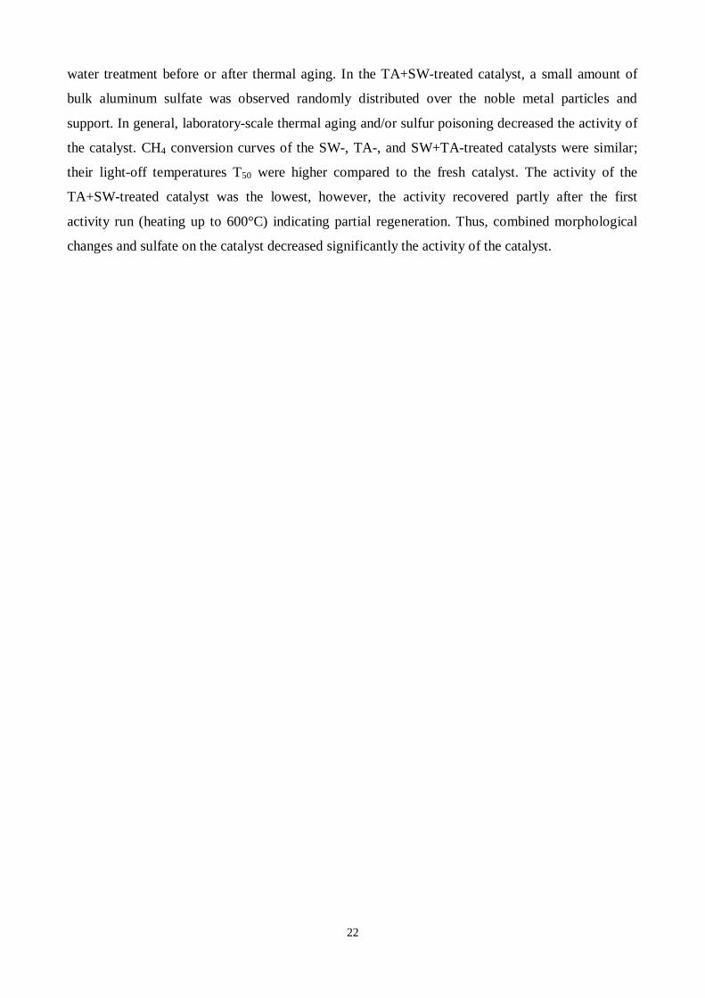
decomposes at elevated temperatures [15]. Based on the XPS S 2p spectrum (Fig. 9), sulfur is as
sulfates [30]. In the O 1s spectra, the contribution of SO4 species cannot be seen due to the
domination of alumina and the possible overlapping of the binding energies of these two [30]. The
DRIFT spectra of the SW- and TA+SW-treated catalysts (Fig. 10) have a broad band at around
1200-1100 cm-1 indicating bulk aluminum sulfate species [34,35]. The band around 1435 cm-1
attributed to νS=O of stable Pd- or Pt-SO4 species [34,36] is missing. Furthermore, any Pd 3d5/2
lines above 338 eV, which could be attributed to PdSO4 [14], were not detected either by XPS.
Bands around 1390-1300cm-1 attributed to surface sulfate species on Al2O3 [16,34] or surface
Al2(SO3)3 [34] were not observed in the DRIFT spectra. According to Mowery et al. [14], sulfation
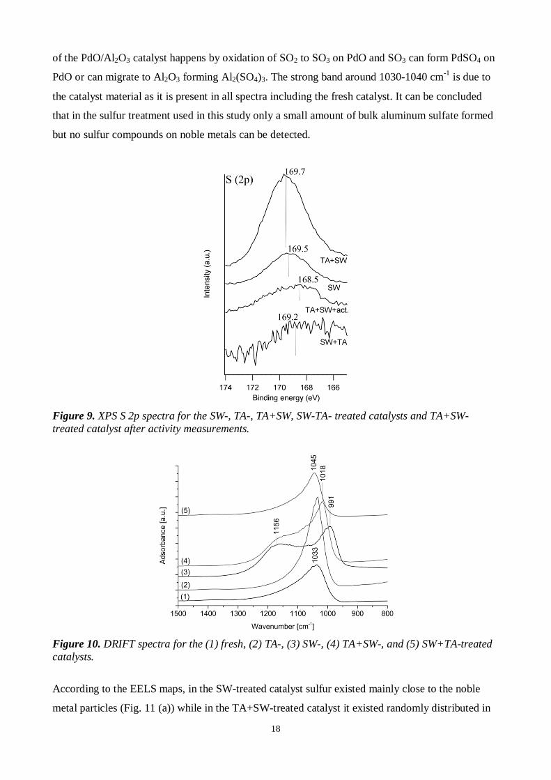
18
of the PdO/Al2O3 catalyst happens by oxidation of SO2 to SO3 on PdO and SO3 can form PdSO4 on
PdO or can migrate to Al2O3 forming Al2(SO4)3. The strong band around 1030-1040 cm-1 is due to
the catalyst material as it is present in all spectra including the fresh catalyst. It can be concluded
that in the sulfur treatment used in this study only a small amount of bulk aluminum sulfate formed
but no sulfur compounds on noble metals can be detected.
Figure 9. XPS S 2p spectra for the SW-, TA-, TA+SW, SW-TA- treated catalysts and TA+SW-treated catalyst after activity measurements.
Figure 10. DRIFT spectra for the (1) fresh, (2) TA-, (3) SW-, (4) TA+SW-, and (5) SW+TA-treatedcatalysts.
According to the EELS maps, in the SW-treated catalyst sulfur existed mainly close to the noble
metal particles (Fig. 11 (a)) while in the TA+SW-treated catalyst it existed randomly distributed in

19
the catalyst (Fig. 11 (b)). Probably, small noble metal particles are very active to adsorb and oxidize
sulfur species and Al2(SO4)3 forms near the noble metal particles. While, the grown noble metal
particles may be not so active to adsorb and oxidize sulfur species resulting in that Al2(SO4)3 forms
randomly over the noble metal particles and support. According to Smirnov et al. [37], at
temperatures above 300°C sulfate species form on the Al2O3 thin film without noble metal particles.
Thus, it can be assumed that Al2(SO4)3 can form also straight with the alumina support. In addition,
due to the thermal aging treatments g-alumina changed to d-,q-, and a-alumina which may affect
the sulfation phenomenon of the support in the TA+SW-treated catalyst.
Figure 11. The STEM ADF image, the enlarged STEM ADF image, and the STEM EELS sulfurmap from the selected area (brighter color indicates higher amount) for (a) the SW-treated catalystand (b) the TA+SW-treated catalyst. Notice the different scale bar in (a) and (b).
3.3 Activity of the catalyst
Conversions of CH4 as a function of temperature over the fresh catalyst and SW-, TA-, TA+SW-
and SW+TA-treated catalysts are presented in Fig. 12 and the light-off temperatures (T50 and T90)
are collected into Table 4. In general, laboratory-scale thermal treatment and/or sulfur exposure
decreased the activity of the catalyst. Methane oxidation over the treated samples started at least
100°C higher temperatures compared to the fresh catalyst and the light-off temperatures (T50) were
observed to increase at least by 50°C compared to the fresh catalyst. The activities of the TA-, SW-,
and SW+TA-treated catalysts were similar and the activity of the TA+SW-treated catalyst was
notably lower than that of the other catalysts. The reason why in all the TA-treated catalyst the
activity is decreased is due to the significant morphological changes compared to the fresh catalyst.
Due to the growing of the noble metal particles, the amount of active sites decreased. In addition,
phase transformation of g-alumina to d/q- and a-alumina significantly decreased specific surface
area of the TA-treated catalyst (Table 3). According to the literature [17], phase transformation of g-

20
Al2O3 to q-Al2O3 causes the reconstruction of the surface decreasing reactive sites of the support. In
the SW-treated catalyst, slight increasing in the size of the noble metal particles and formation of
Al2(SO3)4 species close to the noble metal particles was detected which are probably blocking
active sites followed by decreased conversion of methane. According to the literature [16], presence
of SO2 creates Brönsted acid sites on the g-Al2O3 support while the fresh Pd/Al2O3 catalyst has only
more reactive Lewis acid sites. Probably, this kind of changing in the acidity of the g-Al2O3 support
happens also in our SW-treatment causing further decreased activity of the SW-treated catalyst. In
the TA+SW-treated catalyst, these above mentioned changes in the TA-treated and SW-treated
catalysts are combined resulting in dramatic decreasing in the methane conversion. In the SW+TA-
treated catalyst, formed Al2(SO3)4 decomposed during TA-treatment resulting in similar catalyst
activity to the TA-treated catalyst. Thus, formation of aluminum sulfate randomly over the
TA+SW-treated catalyst has a significant effect on the catalyst deactivation.
The catalyst activity tests were done twice (run 1 and run 2); notable differences between run 1 and
run 2 were observed only with the TA+SW-treated catalyst. Its activity was higher in the latter run
indicating partial regeneration, i.e. decomposition of sulfate, of the catalyst. Thus, a short time (15
minutes) at 600°C is enough to remove some sulfates from the catalyst. According to the FESEM-
EDS analyses and XPS measurements, during the activity tests the amount of sulfur in the TA+SW-
treated catalyst decreased from ~3 wt-% to <1 wt-%. In addition based on the XPS results (Fig. 6
(a)), PdO reduced during the activity tests of the TA+SW-treated catalyst. Also a shoulder,
indicating another chemical state, was observed in its O 1s spectrum (Fig. 6 (c)). A possible origin
for this is by-products of methane combustion, such as hydroxides or carbon compounds. The
reduction of PdO was also detected by XRD (Fig. 5). According to the literature, e.g. [38,39], PdO
is reduced by CH4. However, no significant reduction of PdO was observed by XRD in the SW- or
SW+TA-treated catalysts during the activity tests. In the future, more characterization will be
needed for activity-tested samples to study comprehensively their structure in the working state.

21
Figure 12. CH4 conversions as a function of temperature for the fresh, SW-, TA-, SW+TA-, andTA+SW-treated catalysts. For the TA+SW-treated catalyst two runs are presented.
Table 4. Light-off temperatures (T50 and T90) for the fresh, SW-, TA-, SW+TA-, and TA+SW-treatedcatalysts. For TA+SW-treated catalyst values are presented after the first and the second run.
Catalyst T50 [°C] T90 [°C]Fresh 390 488SW 437 550TA 445 570
SW+TA 441 552TA+SW (first run) 564 -
TA+SW (second run) 498 -
4 CONCLUSIONS
Accelerated deactivation phenomena in the PtPd/g-Al2O3 natural-gas oxidation catalyst caused by
thermal aging and/or sulfur + water poisoning were studied. Thermal aging and sulfur + water
poisoning treatments were performed separately and with varied combinations. Comprehensive
characterization of the catalyst was carried out after each step to get knowledge about existing of
sulfur and influence of thermal aging before or after sulfur poisoning. The fresh catalyst contains
oxidized Pt and Pd particles (<5 nm) uniformly distributed in the g-alumina surface. In the SW-
treated catalyst, a slight growing of the noble metal particles was observed. In addition, a small
amount of bulk aluminum sulfate was detected near the noble metal particles. In the thermal aging,
g-alumina changed to d- and/or q- and a-alumina. In addition, total decomposition of oxidized Pt
and partly decomposition of oxidized Pd occurred resulting in the formation of grown noble metal
particles (50-60 nm) with a bimetallic PtPd core (~30 nm) and a polycrystalline PdO (~10 nm)
shell. In addition, few, small (~5 nm) and likely bimetallic PtPd particles were present in the
catalyst as well. The noble metal particles and washcoat remained similar regardless of sulfur +
22
water treatment before or after thermal aging. In the TA+SW-treated catalyst, a small amount of
bulk aluminum sulfate was observed randomly distributed over the noble metal particles and
support. In general, laboratory-scale thermal aging and/or sulfur poisoning decreased the activity of
the catalyst. CH4 conversion curves of the SW-, TA-, and SW+TA-treated catalysts were similar;
their light-off temperatures T50 were higher compared to the fresh catalyst. The activity of the
TA+SW-treated catalyst was the lowest, however, the activity recovered partly after the first
activity run (heating up to 600°C) indicating partial regeneration. Thus, combined morphological
changes and sulfate on the catalyst decreased significantly the activity of the catalyst.

23
Acknowledgements:
The Academy of Finland is thanked for funding (Decision numbers 138798 and 139187). The
research leading to these results has received funding also from the European Union Seventh
Framework Programme under Grant Agreement 312483 - ESTEEM2 (Integrated Infrastructure
Initiative–I3).
24
References:
[1] P. Gélin, M. Primet, Appl. Catal. B Environ. 39 (2002) 1–37.
[2] E.M. Holmgreen, M.M. Yung, U.S. Ozkan, Appl. Catal. B Environ. 74 (2007) 73–82.
[3] R.J. Farrauto, J.K. Lampert, M.C. Hobson, E.M. Waterman, Appl. Catal. B Environ. 6 (1995)263–270.
[4] A.K. Datye, J. Bravo, T.R. Nelson, P. Atanasova, M. Lyubovsky, L. Pfefferle, Appl. Catal. AGen. 198 (2000) 179–196.
[5] M. Lyubovsky, L. Pfefferle, Catal. Today 47 (1999) 29–44.
[6] Y. Ozawa, Y. Tochihara, A. Watanabe, M. Nagai, S. Omi, Appl. Catal. A Gen. 259 (2004)1–7.
[7] K. Persson, L.D. Pfefferle, W. Schwartz, A. Ersson, S.G. Järås, Appl. Catal. B Environ. 74(2007) 242–250.
[8] K. Persson, K. Jansson, S. Jarås, J. Catal. 245 (2007) 401–414.
[9] N.M. Kinnunen, J.T. Hirvi, M. Suvanto, T.A. Pakkanen, J. Mol. Catal. A Chem. 356 (2012)20–28.
[10] A.K. Neyestanaki, F. Klingstedt, T. Salmi, D.Y. Murzin, Fuel. 83 (2004) 395–408.
[11] J.K. Lampert, M. Shahjahan, R.J. Farrauto, Appl. Catal. B Environ. 14 (1997) 211-223.
[12] A.T. Gremminger, H.W. Pereira de Carvalho, R. Popescu, J.-D. Grunwaldt, O. Deutschmann,Catal. Today. (2015) 1–11.
[13] G. Corro, C. Cano, J.L.G. Fierro, J. Mol. Catal. A Chem. 315 (2010) 35–42.
[14] D.L. Mowery, R.L. McCormick, Appl. Catal. B Environ. 34 (2001) 287–297.
[15] O. Kröcher, M. Widmer, M. Elsener, D. Rothe, Ind. Eng. Chem. Res. 48 (2009) 9847–9857.
[16] M. Konsolakis, I. V. Yentekakis, G. Pekridis, N. Kaklidis, a. C. Psarras, G.E. Marnellos,Appl. Catal. B Environ. 138-139 (2013) 191–198.
[17] R. Wischert, C. Copéret, F. Delbecq, P. Sautet, Angew. Chemie - Int. Ed. 50 (2011) 3202–3205.
[18] C.H. Bartholomew, Appl. Catal. A Gen. 212 (2001) 17–60.
[19] T.W. Hansen, A.T. Delariva, S.R. Challa, A.K. Datye, Acc. Chem. Res. 46 (2013) 1720–1730.
[20] P.J.F. Harris, Int. Mater. Rev. 40 (1995) 97–115.
[21] J.A. Moulijn, A.E. Van Diepen, F. Kapteijn, Appl. Catal. A Gen. 212 (2001) 3–16.

25
[22] G.S. Parkinson, Z. Novotny, G. Argentero, M. Schmid, J. Pavelec, R. Kosak, P. Blaha, U.Diebold, Nat. Mater. 12 (2013) 724–728.
[23] A. Martínez, M.A. Arribas, M. Derewinski, A. Burkat-Dulak, Appl. Catal. A Gen. 379(2010) 188–197.
[24] A.M. Azad, M.J. Duran, A.K. McCoy, M.A. Abraham, Appl. Catal. A Gen. 332 (2007) 225–236.
[25] H. Arai, M. Machida, Appl. Catal. A Gen. 138 (1996) 161–176.
[26] M. Honkanen, M. Kärkkäinen, V. Viitanen, H. Jiang, K. Kallinen, M. Huuhtanen, M.Vippola, J. Lahtinen, R. Keiski, T. Lepistö, Top. Catal. 56 (2013) 576–585.
[27] M. Aronniemi, J. Sainio, J. Lahtinen, Surf. Sci. 578 (2005) 108–123.
[28] M. Brun, A. Berthet, J.C. Bertolini, J. Electron. Spectrosc. Relat. Phenom. 104 (1999) 55–60.
[29] J.Z. Shyu, K. Otto, Appl. Surf. Sci. 32 (1988) 246-252.
[30] J.F. Moulder, W.F. Stickle, P.E. Sobol, K.D. Bomben, Handbook of X-ray PhotoelectronSpectroscopy, Physical Electronics, Inc., Eden Prairie, 1995.
[31] C. Carrillo, T.R. Johns, H. Xiong, A. DeLaRiva, S.R. Challa, R.S. Goeke, K. Artyushkova,W. Lei, C.H. Kim, A.K. Datye, J. Phys. Chem. Lett. 5 (2014) 2089-2093.
[32] K. Hauff, U. Tuttlies, G. Eigenberger, U. Nieken, Appl. Catal. B Environ. 123-124 (2012)107–116.
[33] T.R. Johns, J.R. Gaudet, E.J. Peterson, J.T. Miller, E.A. Stach, C.H. Kim, M.P. Balogh, A.K.Datye, ChemCatChem 5 (2013) 2636-2645.
[34] H. Abdulhamid, E. Fridell, J. Dawody, M. Skoglundh, J. Catal. 241 (2006) 200–210.
[35] H. Mahzoul, L. Limousy, J.F. Brilhac, P. Gilot, J. Anal. Appl. Pyrolysis. 56 (2000) 179–193.
[36] S. Colussi, A. Trovarelli, E. Vesselli, A. Baraldi, G. Comelli, G. Groppi, J. Llorca, Appl.Catal. A Gen. 390 (2010) 1–10.
[37] M.Y. Smirnov, A. V Kalinkin, A. V Pashis, A.M. Sorokin, A.S. Noskov, K.C. Kharas, V.I.Bukhtiyarov, J. Phys. Chem. B 109 (2005) 11712–11719.
[38] P. Castellazzi, G. Groppi, P. Forzatti, Appl. Catal. B Environ. 95 (2010) 303–311.
[39] Z. Li, G.B. Hoflund, J. Nat. Gas Chem. 12 (2003) 153–160.