ACC/AHA Practice Guidelines · ACC/AHA Guidelines for the Management of Patients With ST-Elevation...
49
ACC/AHA Guidelines for the Management of Patients With ST-Elevation Myocardial Infarction—Executive Summary A Report of the American College of Cardiology/American Heart Association Task Force on Practice Guidelines (Writing Committee to Revise the 1999 Guidelines for the Management of Patients With Acute Myocardial Infarction) Developed in Collaboration With the Canadian Cardiovascular Society WRITING COMMITTEE MEMBERS Elliott M. Antman, MD, FACC, FAHA, Chair; Daniel T. Anbe, MD, FACC, FAHA; Paul Wayne Armstrong, MD, FACC, FAHA; Eric R. Bates, MD, FACC, FAHA; Lee A. Green, MD, MPH; Mary Hand, MSPH, RN, FAHA; Judith S. Hochman, MD, FACC, FAHA; Harlan M. Krumholz, MD, FACC, FAHA; Frederick G. Kushner, MD, FACC, FAHA; Gervasio A. Lamas, MD, FACC; Charles J. Mullany, MB, MS, FACC; Joseph P. Ornato, MD, FACC, FAHA; David L. Pearle, MD, FACC, FAHA; Michael A. Sloan, MD, FACC; Sidney C. Smith, Jr, MD, FACC, FAHA TASK FORCE MEMBERS Elliott M. Antman, MD, FACC, FAHA, Chair; Sidney C. Smith, Jr, MD, FACC, FAHA, Vice-chair; Joseph S. Alpert, MD, FACC, FAHA*; Jeffrey L. Anderson, MD, FACC, FAHA; David P. Faxon, MD, FACC, FAHA; Valentin Fuster, MD, PhD, FACC, FAHA; Raymond J. Gibbons, MD, FACC, FAHA*†; Gabriel Gregoratos, MD, FACC, FAHA*; Jonathan L. Halperin, MD, FACC, FAHA; Loren F. Hiratzka, MD, FACC, FAHA; Sharon Ann Hunt, MD, FACC, FAHA; Alice K. Jacobs, MD, FACC, FAHA; Joseph P. Ornato, MD, FACC, FAHA This document was approved by the American College of Cardiology Foundation Board of Trustees on May 7, 2004 and by the American Heart Association Science Advisory and Coordinating Committee on May 5, 2004. The ACC/AHA Task Force on Practice Guidelines makes every effort to avoid any actual or potential conflicts of interest that might arise as a result of an outside relationship or personal interest of a member of the writing panel. Specifically, all members of the writing panel are asked to provide disclosure statements of all such relationships that might be perceived as real or potential conflicts of interest. These statements are reviewed by the parent task force, reported orally to all members of the writing panel at the first meeting, and updated as changes occur. The relationship with industry information for the writing committee members is posted on the ACC and AHA World Wide Web sites with the full-length version of the update, along with the names and relationships with industry of the peer reviewers. When citing this document, the American College of Cardiology Foundation and the American Heart Association would appreciate the following citation format: Antman EM, Anbe DT, Armstrong PW, Bates ER, Green LA, Hand M, Hochman JS, Krumholz HM, Kushner FG, Lamas GA, Mullany CJ, Ornato JP, Pearle DL, Sloan MA, Smith SC Jr. ACC/AHA guidelines for the management of patients with ST-elevation myocardial infarction: executive summary: a report of the ACC/AHA Task Force on Practice Guidelines (Committee to Revise the 1999 Guidelines on the Management of Patients With Acute Myocardial Infarction). J Am Coll Cardiol 2004;44:671–719. Copies: This document and the full-text guideline are available on the World Wide Web sites of the American College of Cardiology (www.acc.org), the American Heart Association (www.americanheart.org), and the Canadian Cardiovascular Society (www.ccs.ca). Single copies of this executive summary, published in the August 4, 2004 issue of the Journal of the American College of Cardiology or the August 3, 2004 issue of Circulation or the companion full-text guideline are available for $10.00 each by calling 1-800-253-4636 or writing to the American College of Cardiology Foundation, Resource Center, 9111 Old Georgetown Road, Bethesda, MD 20814-1699. To purchase bulk reprints (specify version and reprint number: 71-0294 for the executive summary; 71-0293 for the full-text guideline): up to 999 copies, call 1-800-611-6083 (US only) or fax 413-665-2671; 1000 or more copies, call 214-706-1789, fax 214-691-6342, or e-mail [email protected]. Permissions: Multiple copies, modification, alteration, enhancement, and/or distribution of this document are not permitted without the express permission of the American College of Cardiology Foundation. Please direct requests to [email protected]. *Former Task Force member. †Immediate Past Chair. (J Am Coll Cardiol 2004;44:671–719.) ©2004 by the American College of Cardiology Foundation and the American Heart Association, Inc. doi:10.1016/j.jacc.2004.07.002 ACC/AHA Practice Guidelines
Transcript of ACC/AHA Practice Guidelines · ACC/AHA Guidelines for the Management of Patients With ST-Elevation...

ACC/AHA Guidelines for the Management of Patients WithST-Elevation Myocardial Infarction—Executive Summary
A Report of the American College of Cardiology/American HeartAssociation Task Force on Practice Guidelines (Writing Committee to
Revise the 1999 Guidelines for the Management of Patients With AcuteMyocardial Infarction)
Developed in Collaboration With the Canadian Cardiovascular Society
WRITING COMMITTEE MEMBERS
Elliott M. Antman, MD, FACC, FAHA, Chair; Daniel T. Anbe, MD, FACC, FAHA;Paul Wayne Armstrong, MD, FACC, FAHA; Eric R. Bates, MD, FACC, FAHA;
Lee A. Green, MD, MPH; Mary Hand, MSPH, RN, FAHA; Judith S. Hochman, MD, FACC, FAHA;Harlan M. Krumholz, MD, FACC, FAHA; Frederick G. Kushner, MD, FACC, FAHA;
Gervasio A. Lamas, MD, FACC; Charles J. Mullany, MB, MS, FACC;Joseph P. Ornato, MD, FACC, FAHA; David L. Pearle, MD, FACC, FAHA;
Michael A. Sloan, MD, FACC; Sidney C. Smith, Jr, MD, FACC, FAHA
TASK FORCE MEMBERS
Elliott M. Antman, MD, FACC, FAHA, Chair; Sidney C. Smith, Jr, MD, FACC, FAHA, Vice-chair;Joseph S. Alpert, MD, FACC, FAHA*; Jeffrey L. Anderson, MD, FACC, FAHA;David P. Faxon, MD, FACC, FAHA; Valentin Fuster, MD, PhD, FACC, FAHA;
Raymond J. Gibbons, MD, FACC, FAHA*†;Gabriel Gregoratos, MD, FACC, FAHA*; Jonathan L. Halperin, MD, FACC, FAHA;
Loren F. Hiratzka, MD, FACC, FAHA; Sharon Ann Hunt, MD, FACC, FAHA;Alice K. Jacobs, MD, FACC, FAHA; Joseph P. Ornato, MD, FACC, FAHA
This document was approved by the American College of Cardiology Foundation Board of Trustees on May 7, 2004 and by the American HeartAssociation Science Advisory and Coordinating Committee on May 5, 2004.
The ACC/AHA Task Force on Practice Guidelines makes every effort to avoid any actual or potential conflicts of interest that might arise as a resultof an outside relationship or personal interest of a member of the writing panel. Specifically, all members of the writing panel are asked to providedisclosure statements of all such relationships that might be perceived as real or potential conflicts of interest. These statements are reviewed by the parenttask force, reported orally to all members of the writing panel at the first meeting, and updated as changes occur. The relationship with industryinformation for the writing committee members is posted on the ACC and AHA World Wide Web sites with the full-length version of the update, alongwith the names and relationships with industry of the peer reviewers.
When citing this document, the American College of Cardiology Foundation and the American Heart Association would appreciate the followingcitation format: Antman EM, Anbe DT, Armstrong PW, Bates ER, Green LA, Hand M, Hochman JS, Krumholz HM, Kushner FG, Lamas GA, MullanyCJ, Ornato JP, Pearle DL, Sloan MA, Smith SC Jr. ACC/AHA guidelines for the management of patients with ST-elevation myocardial infarction:executive summary: a report of the ACC/AHA Task Force on Practice Guidelines (Committee to Revise the 1999 Guidelines on the Management ofPatients With Acute Myocardial Infarction). J Am Coll Cardiol 2004;44:671–719.
Copies: This document and the full-text guideline are available on the World Wide Web sites of the American College of Cardiology (www.acc.org),the American Heart Association (www.americanheart.org), and the Canadian Cardiovascular Society (www.ccs.ca). Single copies of this executivesummary, published in the August 4, 2004 issue of the Journal of the American College of Cardiology or the August 3, 2004 issue of Circulation or thecompanion full-text guideline are available for $10.00 each by calling 1-800-253-4636 or writing to the American College of Cardiology Foundation,Resource Center, 9111 Old Georgetown Road, Bethesda, MD 20814-1699. To purchase bulk reprints (specify version and reprint number: 71-0294 forthe executive summary; 71-0293 for the full-text guideline): up to 999 copies, call 1-800-611-6083 (US only) or fax 413-665-2671; 1000 or more copies,call 214-706-1789, fax 214-691-6342, or e-mail [email protected].
Permissions: Multiple copies, modification, alteration, enhancement, and/or distribution of this document are not permitted without the expresspermission of the American College of Cardiology Foundation. Please direct requests to [email protected].
*Former Task Force member.†Immediate Past Chair.(J Am Coll Cardiol 2004;44:671–719.)©2004 by the American College of Cardiology Foundation and the American Heart Association, Inc.
doi:10.1016/j.jacc.2004.07.002
ACC/AHA Practice Guidelines

Table of Contents
I. Introduction....................................................................673II. Pathology ......................................................................673
A. Epidemiology ...........................................................673III. Management Before STEMI.......................................673
A. Identification of Patients at Risk of STEMI ...........673B. Patient Education for Early Recognition and
Response to STEMI .................................................673IV. Onset of STEMI .........................................................675
A. Out-of-Hospital Cardiac Arrest ...............................675V. Prehospital Issues .........................................................675
A. Emergency Medical Services Systems....................675B. Prehospital Chest Pain Evaluation and Treatment ..675C. Prehospital Fibrinolysis ............................................675D. Prehospital Destination Protocols ............................677
VI. Initial Recognition and Management in theEmergency Department ..............................................677
A. Optimal Strategies for Emergency DepartmentTriage........................................................................677
B. Initial Patient Evaluation..........................................6771. History ...................................................................6782. Physical Examination............................................6783. Electrocardiogram .................................................6784. Laboratory Examinations ......................................6785. Biomarkers of Cardiac Damage ...........................678
a. Bedside Testing for SerumCardiac Biomarkers...........................................679
6. Imaging..................................................................679C. Management .............................................................679
1. Routine Measures..................................................679a. Oxygen...............................................................679b. Nitroglycerin .....................................................679c. Analgesia ...........................................................679d. Aspirin ...............................................................680e. Beta-Blockers ....................................................680f. Reperfusion ........................................................680
• General Concepts............................................680• Selection of Reperfusion
Strategy ...........................................................680• Pharmacological Reperfusion.........................682• Percutaneous Coronary Intervention ..............684• Acute Surgical Reperfusion............................688• Patients With STEMI Not Receiving
Reperfusion .....................................................688• Assessment of Reperfusion ............................688• Ancillary Therapy...........................................688• Other Pharmacological Measures...................690
VII. Hospital Management................................................691A. Location....................................................................691
1. Coronary Care Unit...........................................6912. Stepdown Unit...................................................691
B. Early, General Measures ..........................................6921. Level of Activity ...............................................6922. Diet.....................................................................6923. Patient Education in the Hospital Setting.........6924. Analgesia/Anxiolytics ........................................692
C. Risk Stratification During Early HospitalCourse .......................................................................692
D. Medication Assessment............................................6931. Beta-Blockers.....................................................6932. Nitroglycerin ......................................................6933. Inhibition of the Renin-Angiotensin-
Aldosterone System ..........................................6934. Antiplatelets .......................................................6945. Antithrombotics .................................................6946. Oxygen ...............................................................694
E. Estimation of Infarct Size ....................................6941. Electrocardiographic Techniques ......................6942. Cardiac Biomarker Methods .............................6943. Radionuclide Imaging........................................6944. Echocardiography ..............................................6945. Magnetic Resonance Imaging ...........................694
F. Hemodynamic Disturbances .................................6941. Hemodynamic Assessment ................................6942. Hypotension .......................................................6953. Low-Output State...............................................6954. Pulmonary Congestion.......................................6955. Cardiogenic Shock.............................................6966. Right Ventricular Infarction ..............................6977. Mechanical Causes of Heart Failure/Low-
Output Syndrome ..............................................697a. Diagnosis .......................................................697b. Mitral Valve Regurgitation...........................697c. Ventricular Septal Rupture After
STEMI ...........................................................698d. Left Ventricular Free-Wall Rupture .............698e. Left Ventricular Aneurysm ...........................698f. Mechanical Support of the Failing Heart .....698
• Intra-Aortic BalloonCounterpulsation .........................................698
G. Arrhythmias After STEMI...................................6981. Ventricular Arrhythmias ...................................698
a. Ventricular Fibrillation..................................698b. Ventricular Tachycardia................................699c. Ventricular Premature Beats .........................699d. Accelerated Idioventricular Rhythms and
Accelerated Junctional Rhythms ..................699e. ICD Implantation in Patients After
STEMI ...........................................................7002. Supraventricular Arrhythmias/Atrial
Fibrillation .........................................................7003. Bradyarrhythmias ..............................................701
a. Acute Treatment of Conduction Disturbancesand Bradyarrhythmias ...................................701• Ventricular Asystole ...................................701
b. Use of Permanent Pacemakers .....................701• Permanent Pacing for Bradycardia or
Conduction Blocks Associated WithSTEMI.........................................................701
• Sinus Node Dysfunction AfterSTEMI.........................................................701
• Pacing Mode Selection in PatientsWith STEMI ...............................................701
H. Recurrent Chest Pain After STEMI ....................7011. Pericarditis.........................................................7012. Recurrent Ischemia/Infarction...........................703
I. Other Complications ..............................................704
672 Antman et al. JACC Vol. 44, No. 3, 2004Management of Patients With STEMI: Executive Summary August 4, 2004:671–719

1. Ischemic Stroke.................................................7042. DVT and Pulmonary Embolism .......................704
J. Coronary Artery Bypass Graft Surgery AfterSTEMI ...................................................................7041. Timing of Surgery.............................................7042. Arterial Grafting................................................7043. CABG for Recurrent Ischemia After STEMI..7044. Elective CABG Surgery After STEMI in
Patients With Angina ........................................7055. CABG Surgery After STEMI and Antiplatelet
Agents................................................................705K. Convalescence, Discharge, and Post-Myocardial
Infarction Care .....................................................7051. Risk Stratification at Hospital Discharge.........705
a. Role of Exercise Testing...............................705b. Role of Echocardiography ............................705c. Exercise Myocardial Perfusion
Imaging..........................................................707d. Left Ventricular Function .............................707e. Invasive Evaluation .......................................707f. Assessment of Ventricular
Arrhythmias ...................................................707L. Secondary Prevention ...........................................708
1. Patient Education Before Discharge.................7082. Lipid Management ............................................7083. Weight Management .........................................7084. Smoking Cessation............................................7105. Antiplatelet Therapy .........................................7106. Inhibition of Renin-Angiotensin-
Aldosterone-System ..........................................7107. Beta-Blockers ....................................................7118. Blood Pressure Control.....................................7119. Diabetes Management.......................................71210. Hormone Therapy ...........................................71211. Warfarin Therapy............................................71212. Physical Activity .............................................71213. Antioxidants ....................................................712
VIII. Long-Term Management .........................................713A. Psychosocial Impact of STEMI...........................713B. Cardiac Rehabilitation ..........................................713C. Follow-Up Visit With Medical Provider.............713
References ..........................................................................714
I. IntroductionAlthough considerable improvement has occurred in the processof care for patients with ST-elevation myocardial infarction(STEMI), room for improvement exists (1–3). The purpose ofthe present guideline is to focus on the numerous advances in thediagnosis and management of patients with STEMI since 1999.This is reflected in the changed name of the guideline: “ACC/AHA Guidelines for the Management of Patients With ST-Elevation Myocardial Infarction.” The final recommendationsfor indications for a diagnostic procedure, a particular therapy, oran intervention in patients with STEMI summarize both clinicalevidence and expert opinion (Table 1). To provide clinicianswith a set of recommendations that can easily be translated intothe practice of caring for patients with STEMI, this guideline isorganized around the chronology of the interface between the
patient and the clinician. The full guideline is available athttp://www.acc.org/clinical/guidelines/stemi/index.htm.
II. PathologyA. EpidemiologySTEMI continues to be a significant public health problem inindustrialized countries and is becoming an increasinglysignificant problem in developing countries (4). Although theexact incidence is difficult to ascertain, using first-listed andsecondary hospital discharge data, there were 1 680 000unique discharges for ACS in 2001 (5). Applying the conser-vative estimate of 30% of the ACS patients who have STEMIfrom the National Registry of Myocardial Infarction-4[NRMI-4] (5a), we estimate 500 000 STEMI events per yearin the U.S. This writing committee strongly endorses severalpublic health campaigns that are likely to contribute to areduction in the incidence of and fatality from STEMI in thefuture and additional research of new strategies for themanagement of STEMI patients in the community (6–13).
III. Management Before STEMIA. Identification of Patients at Risk of STEMI
Class I1. Primary care providers should evaluate the presence and
status of control of major risk factors for coronary heartdisease (CHD) for all patients at regular intervals (ap-proximately every 3 to 5 years). (Level of Evidence: C)
2. Ten-year risk (National Cholesterol Education Program[NCEP] global risk) of developing symptomatic CHDshould be calculated for all patients who have 2 or moremajor risk factors to assess the need for primary preven-tion strategies (14) (Level of Evidence: B).
3. Patients with established CHD should be identified forsecondary prevention, and patients with a CHD riskequivalent (eg, diabetes mellitus, chronic kidney disease,or 10-year risk greater than 20% as calculated by Fra-mingham equations) should receive equally intensive riskfactor intervention as those with clinically apparent CHD.(Level of Evidence: A)
B. Patient Education for Early Recognition andResponse to STEMI
Class I1. Patients with symptoms of STEMI (chest discomfort
with or without radiation to the arms[s], back, neck,jaw, or epigastrium; shortness of breath; weakness;diaphoresis; nausea; lightheadedness) should be trans-ported to the hospital by ambulance rather than byfriends or relatives. (Level of Evidence: B)
2. Healthcare providers should actively address the fol-lowing issues regarding STEMI with patients and theirfamilies:
a. The patient’s heart attack risk (Level ofEvidence: C)
b. How to recognize symptoms of STEMI (Level ofEvidence: C)
c. The advisability of calling 9-1-1 if symptoms areunimproved or worsening after 5 minutes, despitefeelings of uncertainty about the symptoms and
673JACC Vol. 44, No. 3, 2004 Antman et al.August 4, 2004:671–719 Management of Patients With STEMI: Executive Summary

fear of potential embarrassment (Level of Evi-dence: C)
d. A plan for appropriate recognition and re-sponse to a potential acute cardiac event thatincludes the phone number to access emergencymedical services (EMS), generally 9-1-1 (15). (Levelof Evidence: C)
3. Healthcare providers should instruct patients forwhom nitroglycerin has been prescribed previously totake ONE nitroglycerin dose sublingually in responseto chest discomfort/pain. If chest discomfort/pain isunimproved or worsening 5 minutes after 1 sublingualnitroglycerin dose has been taken, it is recommended
that the patient or family member/friend call 9-1-1immediately to access EMS. (Level of Evidence: C)
Morbidity and mortality due to STEMI can be reducedsignificantly if patients and bystanders recognize symp-toms early, activate the EMS system, and thereby shortenthe time to definitive treatment. Patients with possiblesymptoms of STEMI should be transported to the hospitalby ambulance rather than by friends or relatives becausethere is a significant association between arrival at theemergency department (ED) by ambulance and earlyreperfusion therapy (16 –19). Although the traditionalrecommendation is for patients to take 1 nitroglycerin dosesublingually, 5 minutes apart, for up to 3 doses before
TABLE 1. Applying Classification of Recommendations and Level of Evidence
“Size of Treatment Effect”
Class I Class IIa Class IIb Class III
Benefit ��� RiskProcedure/Treatment SHOULD beperformed/administered
Benefit �� RiskAdditional studies withfocused objectives neededIT IS REASONABLE toperformprocedure/administertreatment
Benefit � RiskAdditional studies withbroad objectives needed;additional registry datawould be helpfulProcedure/TreatmentMAY BE CONSIDERED
Risk � BenefitNo additional studies neededProcedure/Treatment shouldNOT beperformed/administered SINCEIT IS NOT HELPFUL AND MAYBE HARMFUL
Level AMultiple (3–5)population risk strataevaluated *General consistency ofdirection and magnitudeof effect
• Recommendation thatprocedure or treatment isuseful/effective
• Sufficient evidence frommultiple randomized trials ormeta-analyses
• Recommendation in favorof treatment or procedurebeing useful/effective
• Some conflicting evidencefrom multiple randomizedtrials or meta-analyses
• Recommendation’susefulness/efficacyless well established
• Greater conflictingevidence frommultiple randomizedtrials or meta-analyses
• Recommendation thatprocedure or treatment isnot useful/effective andmay be harmful
• Sufficient evidence frommultiple randomized trialsor meta-analyses
Level BLimited (2–3) populationrisk strata evaluated*
• Recommendation thatprocedure or treatment isuseful/effective
• Limited evidence from singlerandomized trial ornonrandomized studies
• Recommendation in favorof treatment or procedurebeing useful/effective
• Some conflicting evidencefrom single randomizedtrial or nonrandomizedstudies
• Recommendation’susefulness/efficacyless well established
• Greater conflictingevidence from singlerandomized trial ornonrandomizedstudies
• Recommendation thatprocedure or treatment isnot useful/effective andmay be harmful
• Limited evidence fromsingle randomized trial ornonrandomized studies
Level CVery limited (1–2)population risk strataevaluated *
• Recommendation thatprocedure or treatment isuseful/effective
• Only expert opinion, casestudies, or standard-of-care
• Recommendation in favorof treatment or procedurebeing useful/effective
• Only diverging expertopinion, case studies, orstandard-of-care
• Recommendation’susefulness/efficacyless well established
• Only diverging expertopinion, case studies,or standard-of-care
• Recommendation thatprocedure or treatment isnot useful/effective andmay be harmful
• Only expert opinion, casestudies, or standard-of-care
Suggested phrases forwritingrecommendations†
shouldis recommendedis indicatedis useful/effective/beneficial
is reasonablecan be useful/effective/
beneficialis probably recommended or
indicated
may/might be consideredmay/might be reasonableusefulness/effectiveness
is unknown/unclear/uncertain or notwell established
is not recommendedis not indicatedshould notis notuseful/effective/beneficialmay be harmful
*Data available from clinical trials or registries about the usefulness/efficacy in different subpopulations, such as gender, age, history of diabetes, history of priorMI, history of heart failure, and prior aspirin use.
†The ACC/AHA Task Force on Practice Guidelines recently provided a list of suggested phrases to use when writing recommendations. All recommendations in theSTEMI guideline have been written in full sentences that express a complete thought, such that a recommendation, even if separated and presented apart from therest of the document (including headings above sets of recommendations), would still convey the full intent of the recommendation. It is hoped that this will increasereaders’ comprehension of the guidelines and will allow queries at the individual recommendation level.
“Est
imat
eof
Cert
aint
y(P
reci
sion
)of
Trea
tmen
tof
Effe
ct”
674 Antman et al. JACC Vol. 44, No. 3, 2004Management of Patients With STEMI: Executive Summary August 4, 2004:671–719

calling for emergency evaluation, this recommendation hasbeen modified by the writing committee to encourageearlier contacting of EMS by patients with symptomssuggestive of STEMI (20,21).
IV. Onset of STEMIA. Out-of-Hospital Cardiac Arrest
Class I1. All communities should create and maintain a strong
“Chain of Survival” for out-of-hospital cardiac arrestthat includes early access (recognition of the problemand activation of the EMS system by a bystander),early cardiopulmonary resuscitation (CPR), early de-fibrillation for patients who need it, and early ad-vanced cardiac life support (ACLS). (Level of Evi-dence: C)
2. Family members of patients experiencing STEMIshould be advised to take CPR training and familiarizethemselves with the use of an automated externaldefibrillator (AED). In addition, they should be re-ferred to a CPR training program that has a socialsupport component for family members of post-STEMIpatients. (Level of Evidence: B)
The links in the chain include early access (recognition ofthe problem and activation of the EMS system by a by-stander), early CPR, early defibrillation for patients who needit, and early ACLS.
V. Prehospital IssuesA. Emergency Medical Services Systems
Class I1. All EMS first responders who respond to patients with
chest pain and/or suspected cardiac arrest should betrained and equipped to provide early defibrillation.(Level of Evidence: A)
2. All public safety first responders who respond to patientswith chest pain and/or suspected cardiac arrest should betrained and equipped to provide early defibrillation withAEDs. (Provision of early defibrillation with AEDs bynonpublic safety first responders is a promising newstrategy, but further study is needed to determine itssafety and efficacy.) (Level of Evidence: B)
3. Dispatchers staffing 9-1-1 center emergency medicalcalls should have medical training, should use nation-ally developed and maintained protocols, and shouldhave a quality-improvement system in place to ensurecompliance with protocols. (Level of Evidence: C)
Early access to EMS is promoted by a 9-1-1 systemcurrently available to more than 90% of the US population.To minimize time to treatment, particularly for cardiopulmo-nary arrest, many communities allow volunteer and/or paidfirefighters and other first-aid providers to function as firstresponders, providing CPR and, increasingly, early defibril-lation using automated external defibrillators (AEDs) untilemergency medical technicians and paramedics arrive. Mostcities and larger suburban areas provide EMS ambulance
services with providers from the fire department, a privateambulance company, and/or volunteers.
B. Prehospital Chest Pain Evaluationand Treatment
Class I1. Prehospital EMS providers should administer 162 to 325
mg of aspirin (chewed) to chest pain patients suspected ofhaving STEMI unless contraindicated or already takenby patient. Although some trials have used enteric-coatedaspirin for initial dosing, more rapid buccal absorptionoccurs with non–enteric-coated formulations. (Level ofEvidence: C)
Class IIa1. It is reasonable for all 9-1-1 dispatchers to advise
patients without a history of aspirin allergy who havesymptoms of STEMI to chew aspirin (162 to 325 mg)while awaiting arrival of prehospital EMS providers.Although some trials have used enteric-coated aspirinfor initial dosing, more rapid buccal absorption occurswith non–enteric-coated formulations. (Level of Evi-dence: C)
2. It is reasonable that all ACLS providers perform andevaluate 12-lead electrocardiograms (ECGs) routinelyon chest pain patients suspected of STEMI. (Level ofEvidence: B)
3. If the ECG shows evidence of STEMI, it is reasonablethat prehospital ACLS providers review a reperfusion“checklist” and relay the ECG and checklist findings toa predetermined medical control facility and/or receiv-ing hospital. (Level of Evidence: C)
It is reasonable for physicians to encourage the prehospitaladministration of aspirin via EMS personnel (ie, EMS dis-patchers and providers) in patients with symptoms suggestiveof STEMI unless its use is contraindicated (22). For patientswho have ECG evidence of STEMI, it is reasonable thatparamedics review a reperfusion checklist and relay the ECGand checklist findings to a predetermined medical controlfacility and/or receiving hospital.
C. Prehospital Fibrinolysis
Class IIa1. Establishment of a prehospital fibrinolysis protocol is
reasonable in 1) settings in which physicians are presentin the ambulance or in 2) well-organized EMS systemswith full-time paramedics who have 12-lead ECGs in thefield with transmission capability, paramedic initial andongoing training in ECG interpretation and STEMItreatment, online medical command, a medical directorwith training/experience in STEMI management, andan ongoing continuous quality-improvement program.(Level of Evidence: B)
Randomized controlled trials of fibrinolytic therapy havedemonstrated the benefit of initiating fibrinolytic therapy asearly as possible after onset of ischemic-type chest discom-fort (Figure 1) (23–25). It appears reasonable to expect that if
675JACC Vol. 44, No. 3, 2004 Antman et al.August 4, 2004:671–719 Management of Patients With STEMI: Executive Summary

Figure 1. Options for transportation of STEMI patients and initial reperfusion treatment. Panel A, Patient transported by EMS after calling9-1-1: Reperfusion in patients with STEMI can be accomplished by the pharmacological (fibrinolysis) or catheter-based (primary PCI)approaches. Implementation of these strategies varies based on the mode of transportation of the patient and capabilities at the receivinghospital. Transport time to the hospital is variable from case to case, but the goal is to keep total ischemic time within 120 minutes. There are3 possibilities: (1) If EMS has fibrinolytic capability and the patient qualifies for therapy, prehospital fibrinolysis should be started within 30minutes of EMS arrival on scene. (2) If EMS is not capable of administering prehospital fibrinolysis and the patient is transported to a non–PCI-capable hospital, the hospital door-to-needle time should be within 30 minutes for patients in whom fibrinolysis is indicated. (3) If EMS isnot capable of administering prehospital fibrinolysis and the patient is transported to a PCI-capable hospital, the hospital door-to-balloontime should be within 90 minutes. Interhospital transfer: It is also appropriate to consider emergency interhospital transfer of the patient to aPCI-capable hospital for mechanical revascularization if (1) there is a contraindication to fibrinolysis; (2) PCI can be initiated promptly
676 Antman et al. JACC Vol. 44, No. 3, 2004Management of Patients With STEMI: Executive Summary August 4, 2004:671–719

fibrinolytic therapy could be started at the time of prehospitalevaluation, a greater number of lives could be saved. Prehos-pital fibrinolysis is reasonable in those settings in whichphysicians are present in the ambulance or prehospital trans-port times are more than 60 minutes in high-volume (morethan 25,000 runs per year) EMS systems (26). Other consid-erations for implementing a prehospital fibrinolytic serviceinclude the ability to transmit ECGs, paramedic initial andongoing training in ECG interpretation and myocardial in-farction (MI) treatment, online medical command, a medicaldirector with training/experience in management of STEMI,and full-time paramedics (27).
D. Prehospital Destination Protocols
Class I1. Patients with STEMI who have cardiogenic shock and
are less than 75 years of age should be broughtimmediately or secondarily transferred to facilitiescapable of cardiac catheterization and rapid revascu-larization (percutaneous coronary intervention [PCI]or coronary artery bypass graft surgery [CABG]) if itcan be performed within 18 hours of onset of shock.(Level of Evidence: A)
2. Patients with STEMI who have contraindications tofibrinolytic therapy should be brought immediately orsecondarily transferred promptly (ie, primary-receiving hospital door-to-departure time less than 30minutes) to facilities capable of cardiac catheterizationand rapid revascularization (PCI or CABG). (Level ofEvidence: B)
3. Every community should have a written protocol thatguides EMS system personnel in determining where to
take patients with suspected or confirmed STEMI.(Level of Evidence: C)
Class IIa1. It is reasonable that patients with STEMI who have
cardiogenic shock and are 75 years of age or older beconsidered for immediate or prompt secondary trans-fer to facilities capable of cardiac catheterization andrapid revascularization (PCI or CABG) if it can beperformed within 18 hours of onset of shock. (Level ofEvidence: B)
2. It is reasonable that patients with STEMI who are atespecially high risk of dying, including those withsevere congestive heart failure (CHF), be consideredfor immediate or prompt secondary transfer (ie,primary-receiving hospital door-to-departure time lessthan 30 minutes) to facilities capable of cardiac cath-eterization and rapid revascularization (PCI orCABG). (Level of Evidence: B)
Every community should have a written protocol thatguides EMS system personnel in determining where to takepatients with suspected or confirmed STEMI. Active involve-ment of local healthcare providers, particularly cardiologistsand emergency physicians, is needed to formulate local EMSdestination protocols for these patients. In general, patientswith suspected STEMI should be taken to the nearest appro-priate hospital. However, patients with STEMI and shock arean exception to this general rule. Whenever possible, STEMIpatients less than 75 years of age with shock should betransferred to facilities capable of cardiac catheterization andrapid revascularization (PCI or CABG). On the basis ofobservations in the SHOCK Trial Registry and other regis-tries, it is reasonable to extend such considerations of transferto invasive centers for elderly patients with shock (seeVII.F.5 and Section 7.6.5 of the full-text guidelines). Patientswith STEMI who have contraindications to fibrinolytic ther-apy should be brought immediately or secondarily transferredpromptly (ie, primary-receiving hospital door-to-departuretime less than 30 minutes) to facilities capable of cardiaccatheterization and rapid revascularization (PCI or CABG).
VI. Initial Recognition and Management inthe Emergency Department
A. Optimal Strategies for EmergencyDepartment Triage
Class I1. Hospitals should establish multidisciplinary teams (in-
cluding primary care physicians, emergency medicinephysicians, cardiologists, nurses, and laboratorians) todevelop guideline-based, institution-specific writtenprotocols for triaging and managing patients who areseen in the prehospital setting or present to the EDwith symptoms suggestive of STEMI. (Level of Evi-dence: B)
B. Initial Patient Evaluation
Class I1. The delay from patient contact with the healthcare
system (typically, arrival at the ED or contact with
Figure 1 (continued). (within 90 minutes after the patient pre-sented to the initial receiving hospital or within 60 minutes com-pared to when fibrinolysis with a fibrin-specific agent could beinitiated at the initial receiving hospital); or (3) fibrinolysis isadministered and is unsuccessful (ie, “rescue PCI”). Secondarynonemergency interhospital transfer can be considered forrecurrent ischemia. Patient self-transport: Patient self-transportation is discouraged. If the patient arrives at a non–PCI-capable hospital, the door-to-needle time should be within30 minutes. If the patient arrives at a PCI-capable hospital, thedoor-to-balloon time should be within 90 minutes. The treatmentoptions and time recommendations after first hospital arrival arethe same. Panel B, For patients who receive fibrinolysis, nonin-vasive risk stratification is recommended to identify the need forrescue PCI (failed fibrinolysis) or ischemia-driven PCI. See Sec-tions 6.3.1.6.4.5. and 6.3.1.6.7. in the full-text guidelines.Regardless of the initial method of reperfusion treatment, allpatients should receive late hospital care and secondary pre-vention of STEMI. EMS indicates Emergency Medical System;PCI, percutaneous coronary intervention; CABG, coronary arterybypass graft surgery; Hosp, hospital; Noninv., Noninvasive. *Golden hour � First 60 minutes;† The medical system goal is tofacilitate rapid recognition and treatment of patients with STEMIsuch that door-to-needle (or medical contact–to-needle) time forinitiation of fibrinolytic therapy is within 30 minutes or that door-to-balloon (or medical contact–to-balloon) time for PCI is within90 minutes. These goals should not be understood as idealtimes but rather as the longest times that should be consideredacceptable for a given system. Systems that are able to achieveeven more rapid times for treatment of patients with STEMIshould be encouraged. Modified with permission fromArmstrong et al. Circulation. 2003;107:2533–7 (25).
677JACC Vol. 44, No. 3, 2004 Antman et al.August 4, 2004:671–719 Management of Patients With STEMI: Executive Summary

paramedics) to initiation of fibrinolytic therapy shouldbe less than 30 minutes. Alternatively, if PCI is chosen,the delay from patient contact with the healthcaresystem (typically, arrival at the ED or contact withparamedics) to balloon inflation should be less than 90minutes. (Level of Evidence: B)
2. The choice of initial STEMI treatment should be madeby the emergency medicine physician on duty based ona predetermined, institution-specific, written protocolthat is a collaborative effort of cardiologists (both thoseinvolved in coronary care unit management and inter-ventionalists), emergency physicians, primary carephysicians, nurses, and other appropriate personnel.For cases in which the initial diagnosis and treatmentplan is unclear to the emergency physician or is notcovered directly by the agreed-on protocol, immediatecardiology consultation is advisable. (Level of Evi-dence: C)
Regardless of the approach used, all patients presenting tothe ED with chest discomfort or other symptoms suggestiveof STEMI or unstable angina should be considered high-priority triage cases and should be evaluated and treatedbased on a predetermined, institution-specific chest painprotocol. The goal for patients with STEMI should be toachieve a door-to-needle time within 30 minutes and adoor-to-balloon time within 90 minutes (Figure 1) (25).
1. History
Class I1. The targeted history of STEMI patients taken in the
ED should ascertain whether the patient has had priorepisodes of myocardial ischemia such as stable orunstable angina, MI, CABG, or PCI. Evaluation of thepatient’s complaints should focus on chest discomfort,associated symptoms, sex- and age-related differencesin presentation, hypertension, diabetes mellitus, possi-bility of aortic dissection, risk of bleeding, and clinicalcerebrovascular disease (amaurosis fugax, face/limbweakness or clumsiness, face/limb numbness or sen-sory loss, ataxia, or vertigo). (Level of Evidence: C)
2. Physical Examination
Class I1. A physical examination should be performed to aid
in the diagnosis and assessment of the extent, loca-tion, and presence of complications of STEMI. (Levelof Evidence: C)
2. A brief, focused, and limited neurological examinationto look for evidence of prior stroke or cognitive deficitsshould be performed on STEMI patients before admin-istration of fibrinolytic therapy. (Level of Evidence: C)
A brief physical examination may promote rapid triage,whereas a more detailed physical examination aids in thedifferential diagnosis and is useful for assessing the extent,location, and presence of complications of STEMI.
3. Electrocardiogram
Class I1. A 12-lead ECG should be performed and shown to an
experienced emergency physician within 10 minutes ofED arrival for all patients with chest discomfort (oranginal equivalent) or other symptoms suggestive ofSTEMI. (Level of Evidence: C)
2. If the initial ECG is not diagnostic of STEMI but thepatient remains symptomatic, and there is a highclinical suspicion for STEMI, serial ECGs at 5- to10-minute intervals or continuous 12-lead ST-segmentmonitoring should be performed to detect the potentialdevelopment of ST elevation. (Level of Evidence: C)
3. In patients with inferior STEMI, right-sided ECGleads should be obtained to screen for ST elevationsuggestive of right ventricular (RV) infarction. (SeeSection 7.6.6 of the full-text guidelines and the ACC/AHA/ASE 2003 Guideline Update for the Clinical Appli-cation of Echocardiography.) (Level of Evidence: B)
The 12-lead ECG in the ED is at the center of thetherapeutic decision pathway because of the strong evidencethat ST-segment elevation identifies patients who benefitfrom reperfusion therapy (28).
4. Laboratory Examinations
Class I1. Laboratory examinations should be performed as part
of the management of STEMI patients but should notdelay the implementation of reperfusion therapy.(Level of Evidence: C)
In addition to serum cardiac biomarkers for cardiac damage,several routine evaluations have important implications formanagement of patients with STEMI. Although these studiesshould be ordered when the patient is first seen, therapeuticdecisions should not be delayed until results are obtainedbecause of the crucial role of time to therapy in STEMI.
5. Biomarkers of Cardiac Damage
Class I1. Cardiac-specific troponins should be used as the opti-
mum biomarkers for the evaluation of patients withSTEMI who have coexistent skeletal muscle injury.(Level of Evidence: C)
2. For patients with ST elevation on the 12-lead ECG andsymptoms of STEMI, reperfusion therapy should beinitiated as soon as possible and is not contingent on abiomarker assay. (Level of Evidence: C)
Class IIa1. Serial biomarker measurements can be useful to pro-
vide supportive noninvasive evidence of reperfusion ofthe infarct artery after fibrinolytic therapy in patientsnot undergoing angiography within the first 24 hoursafter fibrinolytic therapy. (Level of Evidence: B)
678 Antman et al. JACC Vol. 44, No. 3, 2004Management of Patients With STEMI: Executive Summary August 4, 2004:671–719

Class III1. Serial biomarker measurements should not be relied
on to diagnose reinfarction within the first 18 hoursafter the onset of STEMI. (Level of Evidence: C)
For patients with ST-segment elevation, the diagnosis ofSTEMI is secure; initiation of reperfusion therapy should not bedelayed to wait for the results of a cardiac biomarker assay (29).Quantitative analysis of cardiac biomarker measurements pro-vides prognostic information and a noninvasive assessment ofthe likelihood that the patient has undergone successful reperfu-sion when fibrinolytic therapy is administered.
a. Bedside Testing for Serum Cardiac Biomarkers
Class I1. Although handheld bedside (point-of-care) assays may be
used for a qualitative assessment of the presence of anelevated level of a serum cardiac biomarker, subsequentmeasurements of cardiac biomarker levels should beperformed with a quantitative test. (Level of Evidence: B)
2. For patients with ST elevation on the 12-lead ECG andsymptoms of STEMI, reperfusion therapy should beinitiated as soon as possible and is not contingent on abedside biomarker assay. (Level of Evidence: C)
A positive bedside test should be confirmed by a conven-tional quantitative test. However, reperfusion therapy shouldnot be delayed to wait for the results of a quantitative assay.
6. Imaging
Class I1. Patients with STEMI should have a portable chest X-ray,
but this should not delay implementation of reperfusiontherapy (unless a potential contraindication, such asaortic dissection, is suspected). (Level of Evidence: C)
2. Imaging studies such as a high-quality portable chestX-ray, transthoracic and/or transesophageal echocar-diography, and a contrast chest computed tomo-graphic scan or a MRI scan should be used to differ-entiate STEMI from aortic dissection in patients forwhom this distinction is initially unclear. (Level ofEvidence: B)
Class IIa1. Portable echocardiography is reasonable to clarify the
diagnosis of STEMI and allow risk stratification of pa-tients with chest pain on arrival at the ED, especially ifthe diagnosis of STEMI is confounded by left bundle-branch block (LBBB) or pacing, or there is suspicion ofposterior STEMI with anterior ST depressions. (SeeSection 7.6.7 Mechanical Causes of Heart Failure/LowOutput Syndrome of the full-text guidelines.) (Level ofEvidence: B)
Class III1. Single-photon emission computed tomography
(SPECT) radionuclide imaging should not be per-formed to diagnose STEMI in patients for whom thediagnosis of STEMI is evident on the ECG. (Level ofEvidence: B)
C. Management
1. Routine Measures
a. Oxygen
Class I1. Supplemental oxygen should be administered to pa-
tients with arterial oxygen desaturation (SaO2 less than90%). (Level of Evidence: B)
Class IIa1. It is reasonable to administer supplemental oxygen to
all patients with uncomplicated STEMI during the first6 hours. (Level of Evidence: C)
b. Nitroglycerin
Class I1. Patients with ongoing ischemic discomfort should re-
ceive sublingual nitroglycerin (0.4 mg) every 5 minutesfor a total of 3 doses, after which an assessment shouldbe made about the need for intravenous nitroglycerin.(Level of Evidence: C)
2. Intravenous nitroglycerin is indicated for relief ofongoing ischemic discomfort, control of hypertension,or management of pulmonary congestion. (Level ofEvidence: C)
Class III1. Nitrates should not be administered to patients with
systolic blood pressure less than 90 mm Hg or greaterthan or equal to 30 mm Hg below baseline, severebradycardia (less than 50 bpm), tachycardia (morethan 100 bpm), or suspected RV infarction. (Level ofEvidence: C)
2. Nitrates should not be administered to patients whohave received a phosphodiesterase inhibitor for erectiledysfunction within the last 24 hours (48 hours fortadalafil). (Level of Evidence: B)
Nitroglycerin may be administered to relieve ischemic painand is clearly indicated as a vasodilator in patients withSTEMI associated with left ventricular (LV) failure. Nitratesin all forms should be avoided in patients with initial systolicblood pressures less than 90 mm Hg or greater than or equalto 30 mm Hg below baseline, in patients with markedbradycardia or tachycardia (30), and in patients with knownor suspected RV infarction. In view of their marginal treat-ment benefits, nitrates should not be used if hypotensionlimits the administration of beta-blockers, which have morepowerful salutary effects.
c. Analgesia
Class I1. Morphine sulfate (2 to 4 mg IV with increments of 2 to
8 mg IV repeated at 5- to 15-minute intervals) is theanalgesic of choice for management of pain associatedwith STEMI. (Level of Evidence: C)
679JACC Vol. 44, No. 3, 2004 Antman et al.August 4, 2004:671–719 Management of Patients With STEMI: Executive Summary

d. Aspirin
Class I1. Aspirin should be chewed by patients who have not taken
aspirin before presentation with STEMI. The initial doseshould be 162 mg (Level of Evidence: A) to 325 mg (Level ofEvidence: C). Although some trials have used enteric-coatedaspirin for initial dosing, more rapid buccal absorptionoccurs with non–enteric-coated aspirin formulations.
In a dose of 162 mg or more, aspirin produces a rapid clinicalantithrombotic effect caused by immediate and near-total inhi-bition of thromboxane A2 production. Aspirin now forms part ofthe early management of all patients with suspected STEMI andshould be given promptly, and certainly within the first 24 hours,at a dose between 162 and 325 mg and continued indefinitely ata daily dose of 75 to 162 mg (31). Although some trials haveused enteric-coated aspirin for initial dosing, more rapid buccalabsorption occurs with non–enteric-coated formulations (32).
e. Beta-Blockers
Class I1. Oral beta-blocker therapy should be administered
promptly to those patients without a contraindication,irrespective of concomitant fibrinolytic therapy orperformance of primary PCI. (Level of Evidence: A)
Class IIa1. It is reasonable to administer IV beta-blockers
promptly to STEMI patients without contraindica-tions, especially if a tachyarrhythmia or hypertensionis present. (Level of Evidence: B)
Immediate beta-blocker therapy appears to reduce the mag-nitude of infarction and incidence of associated complications insubjects not receiving concomitant fibrinolytic therapy, the rateof reinfarction in patients receiving fibrinolytic therapy, and thefrequency of life-threatening ventricular tachyarrhythmias.
f. Reperfusion
GENERAL CONCEPTS.
Class I1. All STEMI patients should undergo rapid evaluation
for reperfusion therapy and have a reperfusion strat-egy implemented promptly after contact with the med-ical system. (Level of Evidence: A)
Evidence exists that expeditious restoration of flow in theobstructed infarct artery after the onset of symptoms in STEMIpatients is a key determinant of short- and long-term outcomesregardless of whether reperfusion is accomplished by fibrinolysis orPCI (33–35). As discussed previously (also see Section 4.1 of thefull- text guidelines), efforts should be made to shorten the timefrom recognition of symptoms by the patient to contact with themedical system. All healthcare providers caring for STEMI patientsfrom the point of entry into the medical system must recognize theneed for rapid triage and implementation of care in a fashionanalogous to the handling of trauma patients. When consideringrecommendations for timely reperfusion of STEMI patients, the
Writing Committee reviewed data from clinical trials, focusingparticular attention on enrollment criteria for selection of patients forrandomization, actual times reported in the trial report rather thansimply the allowable window specified in the trial protocol, treat-ment effect of the reperfusion strategy on individual components ofa composite primary end point (eg, mortality, recurrent nonfatalinfarction), ancillary therapies (eg, antithrombin and antiplateletagents), and the interface between fibrinolysis and referral forangiography and revascularization. When available, data fromregistries were also reviewed to assess the generalizability ofobservations from clinical trials of reperfusion to routine practice.Despite the wealth of reports on reperfusion for STEMI, it is notpossible to produce a simple algorithm, given the heterogeneity ofpatient profiles and availability of resources in various clinicalsettings at various times of day. This section introduces the recom-mendations for an aggressive attempt to minimize the time fromentry into the medical system to implementation of a reperfusionstrategy using the concept of medical system goals. More detaileddiscussion of these goals and the issues to be considered in selectingthe type of reperfusion therapy are discussed in the Selection ofReperfusion Therapy section of VI.C.1.f (Section 6.3.1.6.2 of thefull-text guidelines), followed by a discussion of available resources.
The medical system goal is to facilitate rapid recognition andtreatment of patients with STEMI such that door-to-needle (ormedical contact–to-needle) time for initiation of fibrinolytictherapy can be achieved within 30 minutes or that door-to-balloon (or medical contact–to-balloon) time for PCI can be keptunder 90 minutes. These goals may not be relevant for thepatients with an appropriate reason for delay, such as uncertaintyabout the diagnosis (particularly for the use of fibrinolytictherapy), need for the evaluation and treatment of other life-threatening conditions (eg, respiratory failure), or delays associ-ated with the patient’s informed choice to have more time toconsider the decision. In the absence of such types of circum-stances, the emphasis is on having a system in place such thatwhen a patient with STEMI presents for medical care, reperfu-sion therapy is able to be provided as soon as possible withinthese time periods. Because there is not considered to be athreshold effect for the benefit of shorter times to reper-fusion, these goals should not be understood as “ideal” times butthe longest times that should be considered acceptable. Systems thatare able to achieve even more rapid times for patients should beencouraged. Also, this goal should not be perceived as an averageperformance standard but a goal of an early treatment system thatevery hospital should seek for every appropriate patient.
SELECTION OF REPERFUSION STRATEGY. Several issues shouldbe considered in selecting the type of reperfusion therapy:
● Time From Onset of Symptoms. Time from onset ofsymptoms to fibrinolytic therapy is an important predictorof MI size and patient outcome (36). The efficacy offibrinolytic agents in lysing thrombus diminishes with thepassage of time (37). Fibrinolytic therapy administeredwithin the first 2 hours (especially the first hour) canoccasionally abort MI and dramatically reduce mortality(23,38).
In contrast, the ability to produce a patent infarct arteryis much less dependent on symptom duration in patientsundergoing PCI. Several reports claim no influence of timedelay on mortality rates when PCI is performed after 2 to
680 Antman et al. JACC Vol. 44, No. 3, 2004Management of Patients With STEMI: Executive Summary August 4, 2004:671–719

3 hours of symptom duration (39,40). Importantly, afteradjustment for baseline characteristics, time from symptomonset to balloon inflation is significantly correlated with1-year mortality in patients undergoing primary PCI forSTEMI (41). The Task Force on the Management of AcuteMyocardial Infarction of the European Society of Cardiol-ogy (42) and this Committee both recommend a target ofmedical contact–to-balloon or door-to-balloon time within90 minutes.
● Risk of STEMI. Several models have been developed thatassist clinicians in estimating the risk of mortality inpatients with STEMI (43–47). Although these models varysomewhat in the factors loaded into the risk-prediction tooland also vary with respect to statistical measures of theirdiscriminative power (eg, C statistic), all the modelsprovide clinicians with a means to assess the continuum ofrisk from STEMI. When the estimated mortality withfibrinolysis is extremely high, as is the case in patients withcardiogenic shock, compelling evidence exists that favors aPCI strategy.
● Risk of Bleeding. Choice of reperfusion therapy is alsoaffected by the patient’s risk of bleeding. When both typesof reperfusion are available, the higher the patient’s risk ofbleeding with fibrinolytic therapy, the more strongly thedecision should favor PCI. If PCI is unavailable, then thebenefit of pharmacological reperfusion therapy should bebalanced against the risk.
● Time Required for Transport to a Skilled PCI Laboratory.The availability of interventional cardiology facilities is akey determinant of whether PCI can be provided. Forfacilities that can offer PCI, the literature suggests that thisapproach is superior to pharmacological reperfusion (48).The trials comparing pharmacological and PCI strategies,however, were conducted before the advent of more recentpharmacological and PCI strategies. When a composite endpoint of death, nonfatal recurrent MI, or stroke is analyzed,much of the superiority of a PCI strategy is driven by areduction in the rate of nonfatal recurrent MI (Figure 2)(36). The rate of nonfatal recurrent MI can be influenced
Figure 2. PCI vs fibrinolysis for STEMI. Short-term (4 to 6 weeks; top left) and long-term (top right) outcomes for various end points shownare plotted for STEMI patients randomized to PCI or fibrinolysis for reperfusion in 23 trials (n�7739). Given the frequency of events for eachend point in the 2 treatment groups, the number needed to treat (NNT) or number needed to harm (NNH) is shown for the short-term (bottomleft) and long-term (bottom right) outcomes. The magnitude of treatment differences for death, nonfatal reinfarction, and stroke variesdepending on whether PCI is compared with streptokinase or a fibrin-specific lytic. For example, when primary PCI is compared with alte-plase and the SHOCK trial is excluded, the mortality rate is 5.5% vs 6.7% (odds ratio 0.81, 95% confidence interval 0.64 to 1.03, P�0.081)(76a). See references 76 and 76a for additional discussion. Modified with permission from Elsevier (Keeley et al. The Lancet. 2003;361:13–20)(76) ReMI indicates recurrent MI; Rec. Isch, recurrent ischemia; Hem. Stroke, hemorrhagic stroke; and CVA, cerebrovascular accident.
681JACC Vol. 44, No. 3, 2004 Antman et al.August 4, 2004:671–719 Management of Patients With STEMI: Executive Summary

both by the adjunctive therapy used and by the proportionof patients who are referred for PCI when the initial attemptat fibrinolysis fails or myocardial ischemia recurs afterinitially successful pharmacological reperfusion.
The experience and location of the PCI laboratory also playsa role in the choice of therapy. Not all laboratories can provideprompt, high-quality primary PCI. Even centers with interven-tional cardiology facilities may not be able to provide thestaffing required for 24-hour coverage of the catheterizationlaboratory. Despite staffing availability, the volume of cases inthe laboratory may be insufficient for the team to acquire andmaintain skills required for rapid PCI reperfusion strategies.
A decision must be made when a STEMI patient presents toa center without interventional cardiology facilities. Fibrinolytictherapy can generally be provided sooner than primary PCI. Asthe time delay for performing PCI increases, the mortalitybenefit associated with expeditiously performed primary PCIover fibrinolysis decreases (49). Compared with a fibrin-specificlytic agent, a PCI strategy may not reduce mortality when adelay greater than 60 minutes is anticipated versus immediateadministration of a lytic.
Given the current literature, it is not possible to say defini-tively that a particular reperfusion approach is superior for allpatients, in all clinical settings, at all times of day (Danchin N;oral presentation at American Heart Association Scientific Ses-sions 2003, Orlando, FL, November 2003) (50–52). The main
point is that some type of reperfusion therapy should be selectedfor all appropriate patients with suspected STEMI. The appro-priate and timely use of some reperfusion therapy is likely moreimportant than the choice of therapy, given the current literatureand the expanding array of options. Clinical circumstances inwhich fibrinolytic therapy is generally preferred or an invasivestrategy is generally preferred are shown in Figure 3.
Available Resources
Class I1. STEMI patients presenting to a facility without the
capability for expert, prompt intervention with pri-mary PCI within 90 minutes of first medical contactshould undergo fibrinolysis unless contraindicated.(Level of Evidence: A)
PHARMACOLOGICAL REPERFUSION.
Indications for Fibrinolytic Therapy
Class I1. In the absence of contraindications, fibrinolytic ther-
apy should be administered to STEMI patients withsymptom onset within the prior 12 hours and STelevation greater than 0.1 mV in at least 2 contiguousprecordial leads or at least 2 adjacent limb leads.(Level of Evidence: A)
2. In the absence of contraindications, fibrinolytic ther-apy should be administered to STEMI patients with
Figure 3. Assessment of reperfusion options for patients with STEMI. STEMI indicates ST-elevation myocardial infarction; PCI, percuta-neous coronary intervention; ICH, intracranial hemorrhage. *Applies to fibrin-specific agents (see Figure 15 in the full-text STEMI guide-lines). †Operator experience greater than a total of 75 primary PCI cases per year. ‡Team experience greater than a total of 36 primaryPCI cases per year. §This calculation implies that the estimated delay to the implementation of the invasive strategy is greater than 1hour vs initiation of fibrinolytic therapy immediately with a fibrin-specific agent.
682 Antman et al. JACC Vol. 44, No. 3, 2004Management of Patients With STEMI: Executive Summary August 4, 2004:671–719

symptom onset within the prior 12 hours and new orpresumably new LBBB. (Level of Evidence: A)
Class IIa1. In the absence of contraindications, it is reasonable to
administer fibrinolytic therapy to STEMI patients withsymptom onset within the prior 12 hours and 12-lead ECGfindings consistent with a true posterior MI. (Level ofEvidence: C)
2. In the absence of contraindications, it is reasonableto administer fibrinolytic therapy to patients withsymptoms of STEMI beginning within the prior 12to 24 hours who have continuing ischemic symptomsand ST elevation greater than 0.1 mV in at least 2contiguous precordial leads or at least 2 adjacent limbleads. (Level of Evidence: B)
Class III1. Fibrinolytic therapy should not be administered to
asymptomatic patients whose initial symptoms ofSTEMI began more than 24 hours earlier. (Level ofEvidence: C)
2. Fibrinolytic therapy should not be administered to pa-tients whose 12-lead ECG shows only ST-segment depres-sion except if a true posterior MI is suspected. (Level ofEvidence: A)
Because the benefit of fibrinolytic therapy is directlyrelated to the time from symptom onset, treatment benefitis maximized by the earliest possible application of ther-apy. The constellation of clinical features that must bepresent (although not necessarily at the same time) to serveas an indication for fibrinolysis includes symptoms ofmyocardial ischemia and ST elevation greater than 0.1mV, in at least 2 contiguous leads, or new or presumablynew LBBB on the presenting ECG (23,54).
Contraindications/Cautions
Class I1. Healthcare providers should ascertain whether the pa-
tient has neurological contraindications to fibrinolytictherapy, including any history of intracranial hemor-rhage (ICH), significant closed head or facial traumawithin the past 3 months, uncontrolled hypertension, orischemic stroke within the past 3 months. (See Table 2 fora comprehensive list.) (Level of Evidence: A)
2. STEMI patients at substantial (greater than or equal to4%) risk of ICH should be treated with PCI rather thanwith fibrinolytic therapy. (See Figure 3 for further man-agement considerations.) (Level of Evidence: A)
A detailed list of contraindications and cautions for the useof fibrinolytic therapy is shown in Table 2.
Complications of Fibrinolytic Therapy: Neurological andOther
Class I1. The occurrence of a change in neurological status
during or after reperfusion therapy, particularlywithin the first 24 hours after initiation of treatment, is
considered to be due to ICH until proven otherwise.Fibrinolytic, antiplatelet, and anticoagulant therapiesshould be discontinued until brain imaging scan showsno evidence of ICH. (Level of Evidence: A)
2. Neurology and/or neurosurgery or hematology consulta-tions should be obtained for STEMI patients who haveICH as dictated by clinical circumstances. (Level ofEvidence: C)
3. In patients with ICH, infusions of cryoprecipitate,fresh frozen plasma, protamine, and platelets shouldbe given, as dictated by clinical circumstances.(Level of Evidence: C)
Class IIa1. In patients with ICH, it is reasonable to:
a. Optimize blood pressure and blood glucose levels.(Level of Evidence: C)
b. Reduce intracranial pressure with an infusion ofmannitol, endotracheal intubation, and hyperven-tilation. (Level of Evidence: C)
c. Consider neurosurgical evacuation of ICH. (Levelof Evidence: C)
TABLE 2. Contraindications and Cautions for Fibrinolysis Usein ST-Elevation Myocardial Infarction*
Absolute contraindications
● Any prior ICH
● Known structural cerebral vascular lesion (eg, AVM)
● Known malignant intracranial neoplasm (primary or metastatic)
● Ischemic stroke within 3 months EXCEPT acute ischemic stroke within 3hours
● Suspected aortic dissection
● Active bleeding or bleeding diathesis (excluding menses)
● Significant closed head or facial trauma within 3 months
Relative contraindications
● History of chronic severe, poorly controlled hypertension
● Severe uncontrolled hypertension on presentation (SBP greater than180 mm Hg or DBP greater than 110 mm Hg)†
● History of prior ischemic stroke greater than 3 months, dementia, orknown intracranial pathology not covered in contraindications
● Traumatic or prolonged (greater than 10 minutes) CPR or major surgery(less than 3 weeks)
● Recent (within 2 to 4 weeks) internal bleeding
● Noncompressible vascular punctures
● For streptokinase/anistreplase: prior exposure (more than 5 days ago) orprior allergic reaction to these agents
● Pregnancy
● Active peptic ulcer
● Current use of anticoagulants: the higher the INR, the higher the risk ofbleeding
AVM indicates arteriovenous malformation; SBP, systolic blood pressure;DBP, diastolic blood pressure; ICH, intracranial hemorrhage; CPR, cardiopul-monary resuscitation.
*Viewed as advisory for clinical decision making and may not be all-inclusiveor definitive.
†Could be an absolute contraindication in low-risk patients with ST-elevationmyocardial infarction (see Section 6.3.1.6.3.2 of the full-text guidelines).
683JACC Vol. 44, No. 3, 2004 Antman et al.August 4, 2004:671–719 Management of Patients With STEMI: Executive Summary

Combination Therapy With Glycoprotein IIb/IIIa Inhibitors
Class IIb
1. Combination pharmacological reperfusion with abcix-imab and half-dose reteplase or tenecteplase may beconsidered for prevention of reinfarction (Level of Evi-dence: A) and other complications of STEMI in selectedpatients: anterior location of MI, age less than 75 years,and no risk factors for bleeding. In two clinical trials ofcombination reperfusion, the prevention of reinfarctiondid not translate into a survival benefit at either 30 daysor 1 year (54a). (Level of Evidence: B)
2. Combination pharmacological reperfusion with abcix-imab and half-dose reteplase or tenecteplase may beconsidered for prevention of reinfarction and othercomplications of STEMI in selected patients (anteriorlocation of MI, age less than 75 years, and no riskfactors for bleeding) in whom an early referral forangiography and PCI (ie, facilitated PCI) is planned.(Level of Evidence: C)
Class III1. Combination pharmacological reperfusion with abcix-
imab and half-dose reteplase or tenecteplase should notbe given to patients aged greater than 75 years becauseof an increased risk of ICH. (Level of Evidence: B)
PERCUTANEOUS CORONARY INTERVENTION
Coronary Angiography
Class I
1. Diagnostic coronary angiography should beperformed:
a. In candidates for primary or rescue PCI. (Level ofEvidence: A)
b. In patients with cardiogenic shock who are candi-dates for revascularization. (Level of Evidence: A)
c. In candidates for surgical repair of ventricularseptal rupture or severe mitral regurgitation(MR). (Level of Evidence: B)
d. In patients with persistent hemodynamic and/orelectrical instability. (Level of Evidence: C)
Class III1. Coronary angiography should not be performed in
patients with extensive comorbidities in whom the risksof revascularization are likely to outweigh the benefits.(Level of Evidence: C)
Primary PCI
Class I
1. General considerations: If immediately available, pri-mary PCI should be performed in patients withSTEMI (including true posterior MI) or MI with newor presumably new LBBB who can undergo PCI of theinfarct artery within 12 hours of symptom onset, ifperformed in a timely fashion (balloon inflation within90 minutes of presentation) by persons skilled in theprocedure (individuals who perform more than 75 PCIprocedures per year). The procedure should be sup-
ported by experienced personnel in an appropriatelaboratory environment (performs more than 200 PCIprocedures per year, of which at least 36 are primaryPCI for STEMI, and has cardiac surgery capability).(Level of Evidence: A)
2. Specific considerations:a. Primary PCI should be performed as quickly as
possible, with a goal of a medical contact–to-balloon or door-to-balloon time of within 90 min-utes. (Level of Evidence: B)
b. If the symptom duration is within 3 hours and theexpected door-to-balloon time minus the expecteddoor-to-needle time is:i) within 1 hour, primary PCI is generally pre-
ferred. (Level of Evidence: B)ii) greater than 1 hour, fibrinolytic therapy
(fibrin-specific agents) is generally preferred.(Level of Evidence: B)
c. If symptom duration is greater than 3 hours,primary PCI is generally preferred and should beperformed with a medical contact–to-balloon ordoor-to-balloon time as brief as possible, with agoal of within 90 minutes. (Level of Evidence: B)
d. Primary PCI should be performed for patientsyounger than 75 years old with ST elevation orLBBB who develop shock within 36 hours of MIand are suitable for revascularization that can beperformed within 18 hours of shock, unless fur-ther support is futile because of the patient’swishes or contraindications/unsuitability for fur-ther invasive care. (Level of Evidence: A)
e. Primary PCI should be performed in patients withsevere CHF and/or pulmonary edema (Killip class3) and onset of symptoms within 12 hours. Themedical contact–to-balloon or door-to-balloontime should be as short as possible (ie, goal within90 min). (Level of Evidence: B)
Class IIa
1. Primary PCI is reasonable for selected patients 75 yearsor older with ST elevation or LBBB or who develop shockwithin 36 hours of MI and are suitable for revasculariza-tion that can be performed within 18 hours of shock.Patients with good prior functional status who are suit-able for revascularization and agree to invasive care maybe selected for such an invasive strategy. (Level of Evi-dence: B)
2. It is reasonable to perform primary PCI for patients withonset of symptoms within the prior 12 to 24 hours and 1or more of the following:
a. Severe CHF (Level of Evidence: C)b. Hemodynamic or electrical instability (Level of
Evidence: C)c. Persistent ischemic symptoms. (Level of Evidence: C)
Class IIb1. The benefit of primary PCI for STEMI patients eligi-
ble for fibrinolysis is not well established when per-
684 Antman et al. JACC Vol. 44, No. 3, 2004Management of Patients With STEMI: Executive Summary August 4, 2004:671–719
formed by an operator who performs fewer than 75PCI procedures per year. (Level of Evidence: C)
Class III
1. PCI should not be performed in a noninfarct artery atthe time of primary PCI in patients without hemody-namic compromise. (Level of Evidence: C)
2. Primary PCI should not be performed in asymptom-atic patients more than 12 hours after onset of STEMIif they are hemodynamically and electrically stable.(Level of Evidence: C)
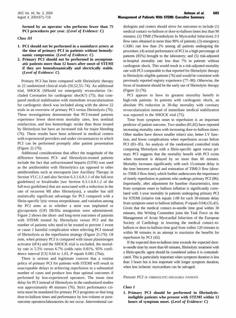
Primary PCI has been compared with fibrinolytic therapyin 22 randomized clinical trials (50,52,55–74). An additionaltrial, SHOCK (SHould we emergently revascularize Oc-cluded Coronaries for cardiogenic shocK?) (75). that com-pared medical stabilization with immediate revascularizationfor cardiogenic shock was included along with the above 22trials in an overview of primary PCI versus fibrinolysis (76).These investigations demonstrate that PCI-treated patientsexperience lower short-term mortality rates, less nonfatalreinfarction, and less hemorrhagic stroke than those treatedby fibrinolysis but have an increased risk for major bleeding(76). These results have been achieved in medical centerswith experienced providers and under circumstances in whichPCI can be performed promptly after patient presentation(Figure 2) (76).
Additional considerations that affect the magnitude of thedifference between PCI- and fibrinolysis-treated patientsinclude the fact that unfractionated heparin (UFH) was usedas the antithrombin with fibrinolytics (as opposed to otherantithrombins such as enoxaparin [see Ancillary Therapy inSection VI.C.1.f and also Section 6.3.1.6.8.1.1 of the full-textguidelines] or bivalirudin [see Section 6.3.1.6.8.1.2 of thefull-text guidelines] that are associated with a reduction in therate of recurrent MI after fibrinolysis), a smaller but stillstatistically significant advantage for PCI compared with afibrin-specific lytic versus streptokinase, and variation amongthe PCI arms as to whether a stent was implanted orglycoprotein (GP) IIb/IIIa antagonists were administered.Figure 2 shows the short- and long-term outcomes of patientswith STEMI treated by fibrinolysis versus PCI and thenumber of patients who need to be treated to prevent 1 eventor cause 1 harmful complication when selecting PCI insteadof fibrinolysis as the reperfusion strategy (Figure 2) (76). Ofnote, when primary PCI is compared with tissue plasminogenactivator (tPA) and the SHOCK trial is excluded, the mortal-ity rate is 5.5% versus 6.7% (odds ratio 0.81%, 95% confi-dence interval [CI] 0.64 to 1.03, P equals 0.081 (76a).
There is serious and legitimate concern that a routinepolicy of primary PCI for patients with STEMI will result inunacceptable delays in achieving reperfusion in a substantialnumber of cases and produce less than optimal outcomes ifperformed by less-experienced operators. The mean timedelay for PCI instead of fibrinolysis in the randomized studieswas approximately 40 minutes (76). Strict performance cri-teria must be mandated for primary PCI programs so that longdoor-to-balloon times and performance by low-volume or poor-outcome operators/laboratories do not occur. Interventional car-
diologists and centers should strive for outcomes to include (1)medical contact–to-balloon or door-to-balloon times less than 90minutes; (2) TIMI (Thrombolysis In Myocardial Infarction) 2/3flow rates obtained in more than 90% of patients; (3) emergencyCABG rate less than 2% among all patients undergoing theprocedure; (4) actual performance of PCI in a high percentage ofpatients (85%) brought to the laboratory; and (5) risk-adjustedin-hospital mortality rate less than 7% in patients withoutcardiogenic shock. This would result in a risk-adjusted mortalityrate with PCI comparable to that reported for fibrinolytic therapyin fibrinolytic-eligible patients (76) and would be consistent withpreviously reported registry experience (77–80). Otherwise, thefocus of treatment should be the early use of fibrinolytic therapy(Figure 2) (76).
PCI appears to have its greatest mortality benefit inhigh-risk patients. In patients with cardiogenic shock, anabsolute 9% reduction in 30-day mortality with coronaryrevascularization instead of immediate medical stabilizationwas reported in the SHOCK trial (75).
Time from symptom onset to reperfusion is an importantpredictor of patient outcome. Two studies (81,82) have reportedincreasing mortality rates with increasing door-to-balloon times.Other studies have shown smaller infarct size, better LV func-tion, and fewer complications when reperfusion occurs beforePCI (83–85). An analysis of the randomized controlled trialscomparing fibrinolysis with a fibrin-specific agent versus pri-mary PCI suggests that the mortality benefit with PCI existswhen treatment is delayed by no more than 60 minutes.Mortality increases significantly with each 15-minute delay inthe time between arrival and restoration of TIMI-3 flow (door-to–TIMI-3 flow time), which further underscores the importanceof timely reperfusion in patients who undergo primary PCI (86).Importantly, after adjustment for baseline characteristics, timefrom symptom onset to balloon inflation is significantly corre-lated with 1-year mortality in patients undergoing primary PCIfor STEMI (relative risk equals 1.08 for each 30-minute delayfrom symptom onset to balloon inflation; P equals 0.04) (35,41).Given that the medical contact–to-needle time goal within 30minutes, this Writing Committee joins the Task Force on theManagement of Acute Myocardial Infarction of the EuropeanSociety of Cardiology in lowering the medical contact–to-balloon or door-to-balloon time goal from within 120 minutes towithin 90 minutes in an attempt to maximize the benefits forreperfusion by PCI (42).
If the expected door-to-balloon time exceeds the expected door-to-needle time by more than 60 minutes, fibrinolytic treatment witha fibrin-specific agent should be considered unless it is contraindi-cated. This is particularly important when symptom duration is lessthan 3 hours but is less important with longer symptom duration,when less ischemic myocardium can be salvaged.
PRIMARY PCI IN FIBRINOLYTIC-INELIGIBLE PATIENTS
Class I1. Primary PCI should be performed in fibrinolytic-
ineligible patients who present with STEMI within 12hours of symptom onset. (Level of Evidence: C)
685JACC Vol. 44, No. 3, 2004 Antman et al.August 4, 2004:671–719 Management of Patients With STEMI: Executive Summary
Class IIa
1. It is reasonable to perform primary PCI forfibrinolytic-ineligible patients with onset of symptomswithin the prior 12 to 24 hours and 1 or more of thefollowing:
a. Severe CHF (Level of Evidence: C)b. Hemodynamic or electrical instability (Level of
Evidence: C)c. Persistent ischemic symptoms. (Level of Evidence: C)
Randomized controlled trials evaluating the outcome ofPCI for patients who present with STEMI but who areineligible for fibrinolytic therapy have not been performed.Few data are available to characterize the value of primaryPCI for this subset of STEMI patients; however, the recom-mendations in Section IV.A (and Section 4.2 of the full-textguidelines) are applicable to these patients. Nevertheless,these patients are at increased risk for mortality (87), andthere is a general consensus that PCI is an appropriate meansfor achieving reperfusion in those who cannot receive fibrino-lytics because of increased risk of bleeding (88–91).
PRIMARY PCI WITHOUT ON-SITE CARDIAC SURGERY
Class IIb1. Primary PCI might be considered in hospitals without
on-site cardiac surgery, provided that there exists aproven plan for rapid transport to a cardiac surgeryoperating room in a nearby hospital with appropriatehemodynamic support capability for transfer. Theprocedure should be limited to patients with STEMI orMI with new, or presumably new, LBBB on ECG, andshould be done in a timely fashion (balloon inflationwithin 90 minutes of presentation) by persons skilled inthe procedure (at least 75 PCIs per year) and at hospitalsthat perform a minimum of 36 primary PCI proceduresper year. (Level of Evidence: B)
Class III1. Primary PCI should not be performed in hospitals
without on-site cardiac surgery and without a provenplan for rapid transport to a cardiac surgery operatingroom in a nearby hospital or without appropriatehemodynamic support capability for transfer. (Level ofEvidence: C)
From clinical data and expert consensus, the Committeerecommends that primary PCI for acute STEMI performed athospitals without established elective PCI programs shouldbe restricted to those institutions capable of performing arequisite minimum number of primary PCI procedures (36per year) with a proven plan for rapid and effective PCI andrapid access to cardiac surgery in a nearby hospital. Thebenefit of primary PCI is not well established for operatorswho perform fewer than 75 PCIs per year or in a hospital thatperforms fewer than 36 primary PCI procedures per year. Inaddition, the benefit of timely reperfusion of the infarct arteryby primary PCI at sites without on-site surgery must beweighed against the small but finite risk of harm to the patient
related to the time required to transfer the patient to a site withCABG surgery capabilities (92,93).
INTERHOSPITAL TRANSFER FOR PRIMARY PCI
To achieve optimal results, time from the first hospitaldoor to the balloon inflation in the second hospital shouldbe as short as possible, with a goal of within 90 minutes.Significant reductions in door-to-balloon times might beachieved by directly transporting patients to PCI centersrather than transporting them to the nearest hospital, ifinterhospital transfer will subsequently be required toobtain primary PCI.
Primary Stenting
Primary stenting has been compared with primary angio-plasty in 9 studies (94–103). There were no differences inmortality (3.0% versus 2.8%) or reinfarction (1.8% versus2.1%) rates. However, major adverse cardiac events werereduced, driven by the reduction in subsequent target-vesselrevascularization with stenting.
Preliminary reports suggest that compared with conven-tional bare metal stents, drug-eluting stents are not associatedwith increased risk when used for primary PCI in STEMIpatients (104). Postprocedure vessel patency, biomarker re-lease, and the incidence of short-term adverse events weresimilar in patients receiving sirolimus (n equals 186) or baremetal (n equals 183) stents. Thirty-day event rates of death,reinfarction, or revascularization were 7.5% versus 10.4%,respectively (P equals 0.4) (104).
Facilitated PCI
Class IIb1. Facilitated PCI might be performed as a reperfusion
strategy in higher-risk patients when PCI is not immedi-ately available and bleeding risk is low. (Level of Evi-dence: B)
Facilitated PCI refers to a strategy of planned immediatePCI after an initial pharmacological regimen such as full-dosefibrinolysis, half-dose fibrinolysis, a GP IIb/IIIa inhibitor, ora combination of reduced-dose fibrinolytic therapy and aplatelet GP IIb/IIIa inhibitor. A strategy of facilitated PCIholds promise in higher-risk patients when PCI is not imme-diately available. Potential risks include increased bleedingcomplications, especially in patients who are at least 75 yearsof age (see Pharmacological Reperfusion in Section VI.C.1.fand Section 6.3.1.6.3.8. of the full-text guidelines), andpotential limitations include added cost. Several randomizedtrials of facilitated PCI with a variety of pharmacologicalregimens are in progress.
Rescue PCI
Class I1. Rescue PCI should be performed in patients less than
75 years old with ST elevation or LBBB who developshock within 36 hours of MI and are suitable forrevascularization that can be performed within 18hours of shock, unless further support is futile because
686 Antman et al. JACC Vol. 44, No. 3, 2004Management of Patients With STEMI: Executive Summary August 4, 2004:671–719

of the patient’s wishes or contraindications/unsuitabil-ity for further invasive care. (Level of Evidence: B)
2. Rescue PCI should be performed in patients withsevere CHF and/or pulmonary edema (Killip class 3)and onset of symptoms within 12 hours. (Level ofEvidence: B)
Class IIa
1. Rescue PCI is reasonable for selected patients 75 years orolder with ST elevation or LBBB or who develop shockwithin 36 hours of MI and are suitable for revasculariza-tion that can be performed within 18 hours of shock.Patients with good prior functional status who are suit-able for revascularization and agree to invasive care maybe selected for such an invasive strategy. (Level of Evi-dence: B)
2. It is reasonable to perform rescue PCI for patients with1 or more of the following:
a. Hemodynamic or electrical instability (Level ofEvidence: C)
b. Persistent ischemic symptoms. (Level of Evidence: C)
Rescue PCI refers to PCI within 12 hours after failedfibrinolysis for patients with continuing or recurrent myocar-dial ischemia.
A major problem in adopting a strategy of rescue PCI liesin the limitation of accurate identification of patients forwhom fibrinolytic therapy has not restored antegrade coro-nary flow. In a prior era in which the practice of PCI was lessmature, immediate catheterization of all patients after fibrino-lytic therapy to identify those with an occluded infarct arterywas found to be impractical, costly, and often associated withbleeding complications (105,106). This strategy is being re-evaluated in clinical trials testing facilitated PCI in thecontemporary PCI setting.
There are no convincing data to support the routine use oflate adjuvant PCI days after failed fibrinolysis or for patientswho do not receive reperfusion therapy. Nevertheless, this isbeing done in some STEMI patients as an extension of theinvasive strategy for non-STEMI patients. The OccludedArtery Trial (OAT) is currently randomizing patients to testwhether routine PCI days to weeks after MI improveslong-term clinical outcomes in asymptomatic high-risk pa-tients with an occluded infarct artery (107).
PCI for Cardiogenic Shock
Class I1. Primary PCI is recommended for patients less than 75
years old with ST elevation or LBBB who developshock within 36 hours of MI and are suitable forrevascularization that can be performed within 18hours of shock, unless further support is futile becauseof the patient’s wishes or contraindications/unsuitabil-ity for further invasive care. (Level of Evidence: A)
Class IIa1. Primary PCI is reasonable for selected patients aged 75
years or older with ST elevation or LBBB who developshock within 36 hours of MI and who are suitable for
revascularization that can be performed within 18hours of shock. Patients with good prior functionalstatus who are suitable for revascularization and agreeto invasive care may be selected for such an invasivestrategy. (Level of Evidence: B)
Observational studies support the value of PCI for patientswho develop cardiogenic shock in the early hours of STEMI. Inthe SHOCK trial (75), the survival curves continued to progres-sively diverge such that at 6 months and 1 year, there was asignificant mortality reduction with emergency revascularization(53% versus 66%, P less than 0.03) (108). The prespecifiedsubgroup analysis of patients less than 75 years old showed anabsolute 15% reduction in 30-day mortality (P less than 0.02),whereas there was no apparent benefit for the small cohort (nequals 56) of patients more than 75 years old. These datastrongly support the approach that patients younger than 75years with STEMI complicated by cardiogenic shock shouldundergo emergency revascularization and support measures.Three registries (109–111) have demonstrated a marked survivalbenefit for elderly patients who are clinically selected for revascu-larization (approximately 1 of 5 patients), so age alone should notdisqualify a patient from early revascularization. (See SectionVII.F.5 and also Section 7.6.5 of the full-text guidelines.)
Percutaneous Coronary InterventionAfter Fibrinolysis
Class I1. In patients whose anatomy is suitable, PCI should be
performed when there is objective evidence of recur-rent MI. (Level of Evidence: C)
2. In patients whose anatomy is suitable, PCI should beperformed for moderate or severe spontaneous orprovocable myocardial ischemia during recovery fromSTEMI. (Level of Evidence: B)
3. In patients whose anatomy is suitable, PCI should beperformed for cardiogenic shock or hemodynamic insta-bility. (See section on PCI for Cardiogenic Shock inSection VI.C.1.f.) (Level of Evidence: B)
Class IIa1. It is reasonable to perform routine PCI in patients with
LV ejection fraction (LVEF) less than or equal to 0.40,CHF, or serious ventricular arrhythmias. (Level ofEvidence: C)
2. It is reasonable to perform PCI when there is docu-mented clinical heart failure during the acute episode,even though subsequent evaluation shows preservedLV function (LVEF greater than 0.40). (Level of Evi-dence: C)
Class IIb1. Routine PCI might be considered as part of an invasive
strategy after fibrinolytic therapy. (Level of Evidence: B)
Immediately After Successful Fibrinolysis. Randomizedprospective trials examined the efficacy and safety of imme-diate PCI after fibrinolysis (105,106,112). These trialsshowed no benefit of routine PCI of the stenotic infarct arteryimmediately after fibrinolytic therapy. The strategy did notappear to salvage myocardium, improve LVEF, or prevent
687JACC Vol. 44, No. 3, 2004 Antman et al.August 4, 2004:671–719 Management of Patients With STEMI: Executive Summary

reinfarction or death. Those subjected to this approachappeared to have an increased incidence of adverse events,including bleeding, recurrent ischemia, emergency CABG,and death. These studies have not been repeated in themodern interventional era with improved equipment, im-proved antiplatelet and anticoagulant strategies, and coronarystents, thus leaving the question of routine PCI early aftersuccessful fibrinolysis unresolved in contemporary practice.Studies of facilitated PCI are enrolling patients (113–116).
Hours to Days After Successful Fibrinolysis. Greatimprovements in equipment, operator experience, andadjunctive pharmacotherapy have increased PCI successrates and decreased complications. More recently, theinvasive strategy for NSTEMI patients has been given aClass I recommendation by the ACC/AHA Guidelines forthe Management of Patients With Unstable Angina/Non-STEMI (117). STEMI patients are increasingly beingtreated similarly as an extension of this approach. Al-though 6 published reports (115,118 –121,123) and 1preliminary report (122) support this strategy, randomizedstudies similar to those in NSTEMI need to be performed.
ACUTE SURGICAL REPERFUSION
Class I1. Emergency or urgent CABG in patients with STEMIshould be undertaken in the following circumstances:
a. Failed PCI with persistent pain or hemodynamicinstability in patients with coronary anatomy suit-able for surgery. (Level of Evidence: B)
b. Persistent or recurrent ischemia refractory tomedical therapy in patients who have coronaryanatomy suitable for surgery, have a significantarea of myocardium at risk, and are not candi-dates for PCI or fibrinolytic therapy. (Level ofEvidence: B)
c. At the time of surgical repair of postinfarctionventricular septal rupture (VSR) or mitral valveinsufficiency. (Level of Evidence: B)
d. Cardiogenic shock in patients less than 75 yearsold with ST elevation, LBBB, or posterior MI whodevelop shock within 36 hours of STEMI, havesevere multivessel or left main disease, and aresuitable for revascularization that can be per-formed within 18 hours of shock, unless furthersupport is futile because of the patient’s wishes orcontraindications/unsuitability for further inva-sive care. (Level of Evidence: A)
e. Life-threatening ventricular arrhythmias in thepresence of greater than or equal to 50% left mainstenosis and/or triple-vessel disease. (Level of Ev-idence: B)
Class IIa1. Emergency CABG can be useful as the primary reper-
fusion strategy in patients who have suitable anatomy,who are not candidates for fibrinolysis or PCI, andwho are in the early hours (6 to 12 hours) of an
evolving STEMI, especially if severe multivessel or leftmain disease is present. (Level of Evidence: B)
2. Emergency CABG can be effective in selected patients75 years or older with ST elevation, LBBB, or posteriorMI who develop shock within 36 hours of STEMI, havesevere triple-vessel or left main disease, and are suit-able for revascularization that can be performedwithin 18 hours of shock. Patients with good priorfunctional status who are suitable for revascularizationand agree to invasive care may be selected for such aninvasive strategy. (Level of Evidence: B)
Class III
1. Emergency CABG should not be performed in pa-tients with persistent angina and a small area of riskif they are hemodynamically stable. (Level of Evi-dence: C)
2. Emergency CABG should not be performed in pa-tients with successful epicardial reperfusion butunsuccessful microvascular reperfusion. (Level ofEvidence: C)
PATIENTS WITH STEMI NOT RECEIVING REPERFUSIONGuideline-based recommendations for nonreperfusion
treatments should not vary whether or not patients receivedreperfusion therapy. The major difference is that patientsnot receiving reperfusion therapy are considered to have ahigher risk for future adverse events (124).
ASSESSMENT OF REPERFUSION
Class IIa1. It is reasonable to monitor the pattern of ST elevation,
cardiac rhythm, and clinical symptoms over the 60 to180 minutes after initiation of fibrinolytic therapy.Noninvasive findings suggestive of reperfusion includerelief of symptoms, maintenance or restoration ofhemodynamic and or electrical stability, and a reduc-tion of at least 50% of the initial ST-segment elevationinjury pattern on a follow-up ECG 60 to 90 minutesafter initiation of therapy. (Level of Evidence: B)
Persistence of unrelenting ischemic chest pain, absence ofresolution of the qualifying ST-segment elevation, and hemo-dynamic and/or electrical instability are generally indicatorsof failed pharmacological reperfusion and the need to con-sider rescue PCI. Aggressive medical support may be neces-sary in the interim. (See Rescue PCI in Section in VI.C.I.f.)
ANCILLARY THERAPY
Antithrombins as Ancillary Therapy to ReperfusionTherapy
UNFRACTIONATED HEPARIN AS ANCILLARY THERAPY TO
REPERFUSION THERAPY
Class I1. Patients undergoing percutaneous or surgical revas-
cularization should be given UFH. (Level of Evi-dence: C)
2. UFH should be given intravenously to patients under-going reperfusion therapy with alteplase, reteplase, ortenecteplase, with dosing as follows: bolus of 60 U/kg
688 Antman et al. JACC Vol. 44, No. 3, 2004Management of Patients With STEMI: Executive Summary August 4, 2004:671–719

(maximum 4000 U) followed by an initial infusion of12 U/kg per hour (maximum 1000 U/hr) adjusted tomaintain activated partial thromboplastin time (aPTT)at 1.5 to 2.0 times control (approximately 50 to 70seconds). (Level of Evidence: C)
3. UFH should be given intravenously to patients treatedwith nonselective fibrinolytic agents (streptokinase,anistreplase, or urokinase) who are at high risk forsystemic emboli (large or anterior MI, atrial fibrilla-tion, previous embolus, or known LV thrombus).(Level of Evidence: B)
4. Platelet counts should be monitored daily in patientsgiven UFH. (Level of Evidence: C)
Class IIb
1. It may be reasonable to administer UFH intravenouslyto patients undergoing reperfusion therapy with strep-tokinase. (Level of Evidence: B)
Because of the evidence that the measured effect of UFHon the aPTT is important for patient outcome and that thepredominant variable mediating the effect of a given dose ofheparin is weight (125), it is important to administer theinitial doses of UFH as a weight-adjusted bolus (126). Forfibrin-specific (alteplase, reteplase, and tenecteplase)fibrinolytic-treated patients, a 60 U/kg bolus followed by amaintenance infusion of 12 U/kg per hour (with a maximumof 4000 U bolus and 1000 U/h initial infusion for patientsweighing greater than 70 kg) is recommended. The recom-mended weight-adjusted dose of UFH, when it is adminis-tered without fibrinolytics, is 60 to 70 U/kg IV bolus and 12to 15 U/kg per hour infusion (117).
LOW-MOLECULAR-WEIGHT HEPARIN AS ANCILLARY THERAPY TO
REPERFUSION THERAPY
Class IIb1. LMWH might be considered an acceptable alternative
to UFH as ancillary therapy for patients less than 75years of age who are receiving fibrinolytic therapy,provided that significant renal dysfunction (serumcreatinine greater than 2.5 mg/dL in men or 2.0 mg/dLin women) is not present. Enoxaparin (30 mg IV bolusfollowed by 1.0 mg/kg subcutaneous injection every 12hours until hospital discharge) used in combinationwith full-dose tenecteplase is the most comprehensivelystudied regimen in patients less than 75 years of age.(Level of Evidence: B)
Class III1. LMWH should not be used as an alternative to UFH as
ancillary therapy in patients over 75 years of age who arereceiving fibrinolytic therapy. (Level of Evidence: B)
2. LMWH should not be used as an alternative to UFH asancillary therapy in patients less than 75 years of agewho are receiving fibrinolytic therapy but have signif-icant renal dysfunction (serum creatinine greater than2.5 mg/dL in men or 2.0 mg/dL in women). (Level ofEvidence: B)
The available data suggest that the rate of early (60 to 90minutes) reperfusion of the infarct artery, either assessed
angiographically or by noninvasive means, is not enhancedby administration of an LMWH. However, a generallyconsistent theme of a lower rate of reocclusion of the infarctartery, reinfarction, or recurrent ischemic events emerges inpatients receiving LMWH regardless of whether the controlgroup was given placebo or UFH.
DIRECT ANTITHROMBINS AS ANCILLARY THERAPY TO
REPERFUSION THERAPY
Class IIa1. In patients with known heparin-induced thrombocyto-
penia, it is reasonable to consider bivalirudin as auseful alternative to heparin to be used in conjunctionwith streptokinase. Dosing according to the HERO(Hirulog and Early Reperfusion or Occlusion)-2 regi-men (a bolus of 0.25 mg/kg followed by an intravenousinfusion of 0.5 mg/kg per hour for the first 12 hours and0.25 mg/kg per hour for the subsequent 36 hours) (127) isrecommended but with a reduction in the infusion rate ifthe PTT is above 75 seconds within the first 12 hours.(Level of Evidence: B)
On the basis of the data in the HERO-2 trial, the WritingCommittee believed that bivalirudin could be considered anacceptable alternative to UFH in those STEMI patients whoreceive fibrinolysis with streptokinase, have heparin-inducedthrombocytopenia, and who, in the opinion of the treatingphysician, would benefit from anticoagulation.
Antiplatelets
ASPIRIN
Class I1. A daily dose of aspirin (initial dose of 162 to 325 mg
orally; maintenance dose of 75 to 162 mg) should begiven indefinitely after STEMI to all patients without atrue aspirin allergy. (Level of Evidence: A)
As discussed, aspirin should be given to the patient withsuspected STEMI as early as possible and should be contin-ued indefinitely, regardless of the strategy for reperfusion andregardless of whether additional antiplatelet agents are ad-ministered. True aspirin allergy is the only exception to thisrecommendation.
THIENOPYRIDINES
Class I1. In patients who have undergone diagnostic cardiac cath-
eterization and for whom PCI is planned, clopidogrelshould be started and continued for at least 1 month afterbare metal stent implantation, for several months afterdrug-eluting stent implantation (3 months for sirolimus, 6months for paclitaxel), and for up to 12 months inpatients who are not at high risk for bleeding. (Level ofEvidence: B)
2. In patients taking clopidogrel in whom CABG isplanned, the drug should be withheld for at least 5days, and preferably for 7, unless the urgency for
689JACC Vol. 44, No. 3, 2004 Antman et al.August 4, 2004:671–719 Management of Patients With STEMI: Executive Summary

revascularization outweighs the risks of excess bleed-ing. (Level of Evidence: B)
Class IIa1. Clopidogrel is probably indicated in patients receiving
fibrinolytic therapy who are unable to take aspirinbecause of hypersensitivity or major gastrointestinalintolerance. (Level of Evidence: C)
Clopidogrel combined with aspirin is recommended forSTEMI patients who undergo coronary stent implantation (128–132).
There are no safety data available regarding the combina-tion of fibrinolytic agents and clopidogrel, but ongoing trialswill provide this information in the future. However, inpatients in whom aspirin is contraindicated because of aspirinsensitivity, clopidogrel is probably useful as a substitute foraspirin to reduce the risk of occlusion (133). There are nosafety data comparing 300 and 600 mg as loading doses forclopidogrel. We do not recommend routine administrationof clopidogrel as pretreatment in patients who have not yetundergone diagnostic cardiac catheterization and in whomCABG surgery would be performed within 5 to 7 days ifwarranted (134).
GLYCOPROTEIN IIb/IIIa INHIBITORS
Class IIa1. It is reasonable to start treatment with abciximab as early
as possible before primary PCI (with or without stenting)in patients with STEMI. (Level of Evidence: B)
Class IIb1. Treatment with tirofiban or eptifibatide may be con-
sidered before primary PCI (with or without stenting)in patients with STEMI. (Level of Evidence: C)
The Writing Committee believes that it is reasonable tostart treatment with abciximab as early as possible in patientsundergoing primary PCI (with or without stenting) but, giventhe size and limitations of the available data set, assigned aClass IIa recommendation to this treatment. The data ontirofiban and eptifibatide in primary PCI are far more limitedthan for abciximab. However, given the common mode ofaction of the agents, a modest amount of angiographic data(135), and general clinical experience to date, tirofiban oreptifibatide may be useful as antiplatelet therapy to supportprimary PCI for STEMI (with or without stenting) (Class IIbrecommendation).
OTHER PHARMACOLOGICAL MEASURES
Inhibition of Renin-Angiotensin-Aldosterone System
Class I1. An angiotensin converting enzyme (ACE) inhibitor
should be administered orally within the first 24 hoursof STEMI to patients with anterior infarction, pulmo-nary congestion, or LVEF less than 0.40, in the absence
of hypotension (systolic blood pressure less than100 mm Hg or less than 30 mm Hg below baseline) orknown contraindications to that Class of medications.(Level of Evidence: A)
2. An angiotensin receptor blocker (ARB) should beadministered to STEMI patients who are intolerant ofACE inhibitors and who have either clinical or radio-logical signs of heart failure or LVEF less than 0.40.Valsartan and candesartan have established efficacyfor this recommendation. (Level of Evidence: C)
Class IIa
1. An ACE inhibitor administered orally within the first24 hours of STEMI can be useful in patients withoutanterior infarction, pulmonary congestion, or LVEFless than 0.40 in the absence of hypotension (systolicblood pressure less than 100 mm Hg or less than30 mm Hg below baseline) or known contraindicationsto that class of medications. The expected treatmentbenefit in such patients is less (5 lives saved per 1000patients treated) than for patients with LV dysfunc-tion. (Level of Evidence: B)
Class III1. An intravenous ACE inhibitor should not be given to
patients within the first 24 hours of STEMI because of therisk of hypotension. (A possible exception may be patientswith refractory hypertension.) (Level of Evidence: B)
A number of large, randomized clinical trials have assessedthe role of ACE inhibitors early in the course of acute MI. Alltrials with oral ACE inhibitors have shown benefit from theirearly use, including those in which early entry criteriaincluded clinical suspicion of acute infarctions. Data fromthese trials indicate that ACE inhibitors should generally bestarted within the first 24 hours, ideally after fibrinolytictherapy has been completed and blood pressure has stabilized.ACE inhibitors should not be used if systolic blood pressureis less than 100 mm Hg or less than 30 mm Hg belowbaseline, if clinically relevant renal failure is present, if thereis a history of bilateral stenosis of the renal arteries, or if thereis known allergy to ACE inhibitors.
The use of ARBs has not been explored as thoroughly asACE inhibitors in STEMI patients. However, clinicalexperience in the management of patients with heartfailure and data from clinical trials in STEMI patients (seeSections 7.4.3 and 7.6.4 of the full-text guidelines) suggestthat ARBs may be useful in patients with depressed LVfunction or clinical heart failure but who are intolerant ofan ACE inhibitor. Use of aldosterone antagonists inSTEMI patients is discussed in Sections 7.4.3 and 7.6.4 ofthe full-text guidelines.
Metabolic Modulation of theGlucose-Insulin Axis
STRICT GLUCOSE CONTROL DURING STEMI
Class I
1. An insulin infusion to normalize blood glucose isrecommended for patients with STEMI and compli-cated courses. (Level of Evidence: B)
690 Antman et al. JACC Vol. 44, No. 3, 2004Management of Patients With STEMI: Executive Summary August 4, 2004:671–719

Class IIa
1. During the acute phase (first 24 to 48 hours) of themanagement of STEMI in patients with hyperglyce-mia, it is reasonable to administer an insulin infusionto normalize blood glucose, even in patients with anuncomplicated course. (Level of Evidence: B)
2. After the acute phase of STEMI, it is reasonable toindividualize treatment of diabetics, selecting from acombination of insulin, insulin analogs, and oralhypoglycemic agents that achieve the best glycemiccontrol and are well tolerated. (Level of Evidence: C)
Compelling evidence for tight glucose control in patients inthe intensive care unit (a large proportion of whom were thereafter cardiac surgery) supports the importance of intensiveinsulin therapy to achieve a normal blood glucose level incritically ill patients (136,136a).
Magnesium
Class IIa1. It is reasonable that documented magnesium deficits
be corrected, especially in patients receiving diuret-ics before the onset of STEMI. (Level of Evidence: C)
2. It is reasonable that episodes of torsade de pointes-typeventricular tachycardia (VT) associated with a pro-longed QT interval be treated with 1 to 2 g of magne-sium administered as an intravenous bolus over 5minutes. (Level of Evidence: C)
Class III1. In the absence of documented electrolyte deficits or
torsade de pointes-type VT, routine intravenous mag-nesium should not be administered to STEMI patientsat any level of risk. (Level of Evidence: A)
Calcium Channel Blockers
Class IIa1. It is reasonable to give verapamil or diltiazem to
patients in whom beta-blockers are ineffective or con-traindicated (eg, bronchospastic disease) for relief ofongoing ischemia or control of a rapid ventricularresponse with atrial fibrillation or flutter after STEMIin the absence of CHF, LV dysfunction, or atrioven-tricular (AV) block. (Level of Evidence: C)
Class III1. Diltiazem and verapamil are contraindicated in pa-
tients with STEMI and associated systolic LV dysfunc-tion and CHF. (Level of Evidence: A)
2. Nifedipine (immediate-release form) is contraindicatedin treatment of STEMI because of the reflex sympa-thetic activation, tachycardia, and hypotension associ-ated with its use. (Level of Evidence: B)
See the full-text guidelines for further explanation.
VII. Hospital ManagementA. Location
1. Coronary Care Unit
Class I1. STEMI patients should be admitted to a quiet and
comfortable environment that provides for continuousmonitoring of the ECG and pulse oximetry and hasready access to facilities for hemodynamic monitoringand defibrillation. (Level of Evidence: C)
2. The patient’s medication regimen should be reviewedto confirm the administration of aspirin and beta-blockers in an adequate dose to control heart rate andto assess the need for intravenous nitroglycerin forcontrol of angina, hypertension, or heart failure. (Levelof Evidence: A)
3. The ongoing need for supplemental oxygen should beassessed by monitoring arterial oxygen saturation.When stable for 6 hours, the patient should be reas-sessed for oxygen need (ie, O2 saturation of less than90%), and discontinuation of supplemental oxygenshould be considered. (Level of Evidence: C)
4. Nursing care should be provided by individuals certi-fied in critical care, with staffing based on the specificneeds of patients and provider competencies, as well asorganizational priorities. (Level of Evidence: C)
5. Care of STEMI patients in the critical care unit (CCU)should be structured around protocols derived frompractice guidelines. (Level of Evidence: C)
6. Electrocardiographic monitoring leads should bebased on the location and rhythm to optimize detectionof ST deviation, axis shift, conduction defects, anddysrhythmias. (Level of Evidence: B)
Class III1. It is not an effective use of the CCU environment to
admit terminally ill, “do not resuscitate” patients withSTEMI, because clinical and comfort needs can beprovided outside of a critical care environment. (Levelof Evidence: C)
2. Stepdown Unit
Class I1. It is a useful triage strategy to admit low-risk STEMI
patients who have undergone successful PCI directly tothe stepdown unit for post-PCI care rather than to theCCU. (Level of Evidence: C)
2. STEMI patients originally admitted to the CCU whodemonstrate 12 to 24 hours of clinical stability (absenceof recurrent ischemia, heart failure, or hemodynam-ically compromising dysrhythmias) should be trans-ferred to the stepdown unit. (Level of Evidence: C)
Class IIa1. It is reasonable for patients recovering from STEMI
who have clinically symptomatic heart failure to bemanaged on the stepdown unit, provided that facilitiesfor continuous monitoring of pulse oximetry and ap-propriately skilled nurses are available. (Level of Evi-dence: C)
691JACC Vol. 44, No. 3, 2004 Antman et al.August 4, 2004:671–719 Management of Patients With STEMI: Executive Summary

2. It is reasonable for patients recovering from STEMIwho have arrhythmias that are hemodynamically well-tolerated (eg, atrial fibrillation with a controlled ven-tricular response; paroxysms of nonsustained VT last-ing less than 30 seconds) to be managed on thestepdown unit, provided that facilities for continuousmonitoring of the ECG, defibrillators, and appropri-ately skilled nurses are available. (Level of Evidence: C)
Class IIb
1. Patients recovering from STEMI who have clinicallysignificant pulmonary disease requiring high-flow sup-plemental oxygen or noninvasive mask ventilation/bilevel positive airway pressure (BIPAP)/continuouspositive airway pressure (CPAP) may be consideredfor care on a stepdown unit provided that facilities forcontinuous monitoring of pulse oximetry and appro-priately skilled nurses with a sufficient nurse:patientratio are available. (Level of Evidence: C)
B. Early, General Measures
1. Level of Activity
Class IIa1. After 12 to 24 hours, it is reasonable to allow patients
with hemodynamic instability or continued ischemia tohave bedside commode privileges. (Level of Evidence: C)
Class III1. Patients with STEMI who are free of recurrent ische-
mic discomfort, symptoms of heart failure, or seriousdisturbances of heart rhythm should not be on bed restfor more than 12 to 24 hours. (Level of Evidence: C)
2. Diet
Class I1. Patients with STEMI should be prescribed the NCEP
Adult Treatment Panel III (ATP III) TherapeuticLifestyle Changes (TLC) diet, which focuses on re-duced intake of fats and cholesterol, less than 7% oftotal calories as saturated fats, less than 200 mg ofcholesterol per day, increased consumption of omega-3fatty acids, and appropriate caloric intake for energyneeds. (Level of Evidence: C)
2. Diabetic patients with STEMI should have an appropri-ate food group balance and caloric intake. (Level ofEvidence: B)
3. Sodium intake should be restricted in STEMI patientswith hypertension or heart failure. (Level of Evidence: B)
STEMI patients should receive a reduced saturated fat andcholesterol diet per the ATP III TLC approach (137). (SeeVII.L.2 and Section 7.12.2 of the full-text guidelines.)
3. Patient Education in the Hospital Setting
Class I1. Patient counseling to maximize adherence to evidence-
based post-STEMI treatments (eg, compliance withtaking medication, exercise prescription, and smokingcessation) should begin during the early phase of
hospitalization, occur intensively at discharge, andcontinue at follow-up visits with providers and throughcardiac rehabilitation programs and community sup-port groups, as appropriate. (Level of Evidence: C)
2. Critical pathways and protocols and other quality-improvement tools (eg, the ACC “Guidelines Appliedin Practice” and the AHA’s “Get with the Guidelines”)should be used to improve the application of evidence-based treatments by patients with STEMI, caregivers,and institutions. (Level of Evidence: C)
Patient education should be viewed as a continuous processthat should to be part of every patient encounter (ie, onhospital arrival, inpatient admission, discharge, and atfollow-up visits).
4. Analgesia/Anxiolytics
Class IIa1. It is reasonable to use anxiolytic medications in STEMI
patients to alleviate short-term anxiety or altered behav-ior related to hospitalization for STEMI. (Level of Evi-dence: C)
2. It is reasonable to routinely assess the patient’s anxietylevel and manage it with behavioral interventions andreferral for counseling. (Level of Evidence: C)
Anxiety and depression are prevalent in patients hospital-ized for STEMI because patients are confronted with adiagnosis that is major, both psychologically and physically(138,139). Anxiety has been demonstrated to predict in-hospital recurrent ischemia and arrhythmias (140) and cardiacevents during the first year after an MI (141).
C. Risk Stratification During EarlyHospital Course
Risk stratification is a continuous process and requires theupdating of initial assessments with data obtained during thehospital stay. Indicators of failed reperfusion (eg, recurrenceof chest pain and persistence of ECG findings indicatinginfarction) identify a patient who should undergo coronaryangiography. Similarly, findings consistent with mechanicalcomplications (eg, sudden onset of heart failure or presenceof a new murmur) herald increased risk and suggest the needfor rapid intervention. For patients who did not undergoprimary reperfusion, changes in clinical status (eg, develop-ment of shock) may herald a worsening clinical status and arean indication for coronary angiography. Patients with a lowrisk of complications may be candidates for early discharge.The lowest-risk patients are those who did not have STEMIdespite the initial suspicions. Clinicians should strive toidentify such patients within 8 to 12 hours of onset ofsymptoms. Serial sampling of serum cardiac biomarkers anduse of 12-lead ECGs and their interpretation in the context ofthe number of hours that have elapsed since onset of thepatient’s symptoms can determine the presence of STEMIbetter than adherence to a rigid protocol that requires that aspecified number of samples be drawn in the hospital.
692 Antman et al. JACC Vol. 44, No. 3, 2004Management of Patients With STEMI: Executive Summary August 4, 2004:671–719

D. Medication Assessment1. Beta-Blockers
Class I1. Patients receiving beta-blockers within the first 24
hours of STEMI without adverse effects should con-tinue to receive them during the early convalescentphase of STEMI. (Level of Evidence: A)
2. Patients without contraindications to beta-blockerswho did not receive them within the first 24 hours afterSTEMI should have them started in the early conva-lescent phase. (Level of Evidence: A)
3. Patients with early contraindications within the first 24hours of STEMI should be reevaluated for candidacyfor beta-blocker therapy. (Level of Evidence: C)
There is overwhelming evidence for the benefits of earlybeta-blockade in patients with STEMI and without contrain-dications to their use (see Section 6.3.1.5 of the full-textguidelines). Benefits have been demonstrated for patientswith and without concomitant fibrinolytic therapy, both earlyand late after STEMI. Meta-analysis of trials from theprefibrinolytic era involving more than 24 000 patientsreceiving beta-blockers have shown a 14% relative riskreduction in mortality through 7 days and a 23% reduction inlong-term mortality (142).
2. Nitroglycerin
Class I1. Intravenous nitroglycerin is indicated in the first 48
hours after STEMI for treatment of persistent ische-mia, CHF, or hypertension. The decision to administerintravenous nitroglycerin and the dose used should notpreclude therapy with other proven mortality-reducing interventions, such as beta-blockers or ACEinhibitors. (Level of Evidence: B)
2. Intravenous, oral, or topical nitrates are useful beyondthe first 48 hours after STEMI for treatment ofrecurrent angina or persistent CHF if their use doesnot preclude therapy with beta-blockers or ACE inhib-itors. (Level of Evidence: B)
Class IIb1. The continued use of nitrate therapy beyond the first
24 to 48 hours in the absence of continued or recurrentangina or CHF may be helpful, although the benefit islikely to be small and is not well established in contem-porary practice. (Level of Evidence: B)
Class III1. Nitrates should not be administered to patients with
systolic pressure less than 90 mm Hg or greater thanor equal to 30 mm Hg below baseline, severe brady-cardia (less than 50 bpm), tachycardia (more than 100bpm) or RV infarction. (Level of Evidence: C)
3. Inhibition of the Renin-Angiotensin-Aldosterone System
Class I1. An ACE inhibitor should be administered orally dur-
ing convalescence from STEMI in patients who toler-
ate this class of medication, and it should be continuedover the long term. (Level of Evidence: A)
2. An ARB should be administered to STEMI patientswho are intolerant of ACE inhibitors and have eitherclinical or radiological signs of heart failure or LVEFless than 0.40. Valsartan and candesartan have dem-onstrated efficacy for this recommendation. (Level ofEvidence: B)
3. Long-term aldosterone blockade should be pre-scribed for post-STEMI patients without significantrenal dysfunction (creatinine should be less than orequal to 2.5 mg/dL in men and less than or equal to2.0 mg/dL in women) or hyperkalemia (potassiumshould be less than or equal to 5.0 mEq/L) who arealready receiving therapeutic doses of an ACE in-hibitor, have an LVEF less than or equal to 0.40, andhave either symptomatic heart failure or diabetes.(Level of Evidence: A)
Class IIa1. In STEMI patients who tolerate ACE inhibitors, an
ARB can be useful as an alternative to ACE inhibitorsprovided there are either clinical or radiological signsof heart failure or LVEF is less than 0.40. Valsartanand candesartan have established efficacy for thisrecommendation. (Level of Evidence: B)
The use of ACE inhibitors in the initial management of theSTEMI patient was reviewed previously. The proportionalbenefit of ACE inhibitor therapy is largest in higher-risksubgroups, including those with previous infarction, heartfailure, depressed LVEF, and tachycardia (143–145). Sur-vival benefit for patients more than 75 years old and for alow-risk subgroup without the features noted above is equiv-ocal (144,145).
Aldosterone blockade is another means of inhibiting therenin-angiotensin-aldosterone system that has been applied topatients in the post-STEMI setting. RALES (RandomizedAldactone Evaluation Study) and EPHESUS (EplerenonePost-Acute Myocardial Infarction Heart Failure Efficacy andSurvival Study) support the long-term use of an aldosteroneblocker in STEMI patients with heart failure, an ejectionfraction of 0.40 or less, or both, provided the serum creatinineis less than or equal to 2.5 mg/dL in men and less than orequal to 2.0 mg/dL in women and serum potassium concen-tration is less than or equal to 5.0 mEq/L (146,147).
The use of ARBs after STEMI has not been explored asthoroughly as ACE inhibitors in STEMI patients (148,149).Given the extensive randomized trial and routine clinicalexperience with ACE inhibitors, they remain the logical firstagent for inhibition of the renin-angiotensin-aldosterone sys-tem in patients convalescing from STEMI (150). Valsartanmonotherapy (target dose 160 mg twice daily) should beadministered to STEMI patients who are intolerant of ACEinhibitors and have evidence of LV dysfunction. Valsartanmonotherapy can be a useful alternative to ACE inhibitors;the decision in individual patients may be influenced byphysician and patient preference, cost, and anticipated side-effect profile.
693JACC Vol. 44, No. 3, 2004 Antman et al.August 4, 2004:671–719 Management of Patients With STEMI: Executive Summary

4. Antiplatelets
Class I1. Aspirin 162 to 325 mg should be given on day 1 of
STEMI and in the absence of contraindications shouldbe continued indefinitely on a daily basis thereafter ata dose of 75 to 162 mg. (Level of Evidence: A)
2. A thienopyridine (preferably clopidogrel) should beadministered to patients who are unable to take aspirinbecause of hypersensitivity or major gastrointestinalintolerance. (Level of Evidence: C)
3. For patients taking clopidogrel for whom CABG isplanned, if possible, the drug should be withheld for atleast 5 days, and preferably for 7, unless the urgencyfor revascularization outweighs the risks of bleeding.(Level of Evidence: B)
4. For patients who have undergone diagnostic cardiaccatheterization and for whom PCI is planned, clopi-dogrel should be started and continued for at least 1month after bare metal stent implantation and forseveral months after drug-eluting stent implantation (3months for sirolimus, 6 months for paclitaxel) and upto 12 months in patients who are not at high risk forbleeding. (Level of Evidence: B)
5. Antithrombotics
Class I1. Intravenous UFH (bolus of 60 U/kg, maximum 4000 U
IV; initial infusion 12 U/kg per hour, maximum of 1000U/h) or LMWH should be used in patients afterSTEMI who are at high risk for systemic emboli (largeor anterior MI, atrial fibrillation, previous embolus,known LV thrombus, or cardiogenic shock). (Level ofEvidence: C)
Class IIa1. It is reasonable that STEMI patients not undergoing
reperfusion therapy who do not have a contraindica-tion to anticoagulation be treated with intravenous orsubcutaneous UFH or with subcutaneous LMWH forat least 48 hours. In patients whose clinical conditionnecessitates prolonged bedrest and/or minimized activ-ities, it is reasonable that treatment be continued untilthe patient is ambulatory. (Level of Evidence: C)
Class IIb1. Prophylaxis for deep venous thrombosis (DVT) with
subcutaneous LMWH (dosed appropriately for specificagent) or with subcutaneous UFH, 7500 U to 12 500 Utwice per day until completely ambulatory, may beuseful, but the effectiveness of such a strategy is not wellestablished in the contemporary era of routine aspirin useand early mobilization. (Level of Evidence: C)
6. Oxygen
Class I1. Supplemental oxygen therapy should be continued
beyond the first 6 hours in STEMI patients witharterial oxygen desaturation (SaO2 less than 90%) orovert pulmonary congestion. (Level of Evidence: C)
E. Estimation of Infarct Size
Measurement of infarct size is an important element in theoverall care of patients with STEMI. There are 5 majormodalities that can be applied to sizing MI.
1. Electrocardiographic Techniques
Class I1. All patients with STEMI should have follow-up ECGs
at 24 hours and at hospital discharge to assess thesuccess of reperfusion and/or the extent of infarction,defined in part by the presence or absence of new Qwaves. (Level of Evidence: B)
2. Cardiac Biomarker Methods
The most widely accepted method for quantifying infarctionhas been the use of serial creatine kinase and the creatinekinase-MB isoenzyme.
3. Radionuclide Imaging
The most comprehensive assessment of STEMI with radio-nuclide imaging was developed with the technetium sesta-mibi SPECT approach (151). This approach is well delineatedin the ACC/AHA/ASNC Guidelines for the Clinical Use ofCardiac Radionuclide Imaging (152).
4. Echocardiography
Global and regional LV function provides an assessment ofthe functional consequences of STEMI and ischemia. Readersare referred to the ACC/AHA/ASE 2003 Guideline Updatefor the Clinical Application of Echocardiography (153) and toSection 7.11.1.2 of the full-text STEMI guidelines.
5. Magnetic Resonance Imaging
Measurement of infarct size by MRI is a promising newtechnique that affords enhanced spatial resolution, therebypermitting more accurate assessment of both the transmuraland circumferential extent of infarction (154). However,additional experience and comparison with other methods ofassessing infarct size are required before any clinical recom-mendations can be provided.
F. Hemodynamic Disturbances
1. Hemodynamic Assessment
Class I1. Pulmonary artery catheter monitoring should be per-formed for the following:
a. Progressive hypotension, when unresponsive tofluid administration or when fluid administrationmay be contraindicated. (Level of Evidence: C)
b. Suspected mechanical complications of STEMI,(ie, VSR, papillary muscle rupture, or free wallrupture with pericardial tamponade) if an echo-cardiogram has not been performed. (Level ofEvidence: C)
2. Intra-arterial pressure monitoring should be per-formed for the following:
694 Antman et al. JACC Vol. 44, No. 3, 2004Management of Patients With STEMI: Executive Summary August 4, 2004:671–719

a. Patients with severe hypotension (systolic arterial pres-sure less than 80 mm Hg). (Level of Evidence: C)
b. Patients receiving vasopressor/inotropic agents.(Level of Evidence: C)
c. Cardiogenic shock. (Level of Evidence: C)
Class IIa1. Pulmonary artery catheter monitoring can be useful forthe following:
a. Hypotension in a patient without pulmonary con-gestion who has not responded to an initial trial offluid administration. (Level of Evidence: C)
b. Cardiogenic shock. (Level of Evidence: C)c. Severe or progressive CHF or pulmonary edema
that does not respond rapidly to therapy. (Level ofEvidence: C)
d. Persistent signs of hypoperfusion without hypoten-sion or pulmonary congestion. (Level of Evidence: C)
e. Patients receiving vasopressor/inotropic agents.(Level of Evidence: C)
2. Intra-arterial pressure monitoring can be useful forpatients receiving intravenous sodium nitroprusside orother potent vasodilators. (Level of Evidence: C)
Class IIb1. Intra-arterial pressure monitoring might be consid-
ered in patients receiving intravenous inotropic agents.(Level of Evidence: C)
Class III1. Pulmonary artery catheter monitoring is not recom-
mended in patients with STEMI without evidence ofhemodynamic instability or respiratory compromise.(Level of Evidence: C)
2. Intra-arterial pressure monitoring is not recom-mended for patients with STEMI who have no pulmo-nary congestion and have adequate tissue perfusionwithout use of circulatory support measures. (Level ofEvidence: C)
2. Hypotension
Class I1. Rapid volume loading with an IV infusion should be
administered to patients without clinical evidence forvolume overload. (Level of Evidence: C)
2. Rhythm disturbances or conduction abnormalitiescausing hypotension should be corrected. (Level ofEvidence: C)
3. Intra-aortic balloon counterpulsation should be per-formed in patients who do not respond to other inter-ventions, unless further support is futile because of thepatient’s wishes or contraindications/unsuitability forfurther invasive care. (Level of Evidence: B)
4. Vasopressor support should be given for hypotension thatdoes not resolve after volume loading. (Level of Evidence: C)
5. Echocardiography should be used to evaluate mechan-ical complications unless these are assessed by invasivemeasures. (Level of Evidence: C)
3. Low-Output State
Class I1. LV function and potential presence of a mechanical
complication should be assessed by echocardiographyif these have not been evaluated by invasive measures.(Level of Evidence: C)
2. Recommended treatments for low-output statesinclude:
a. Inotropic support. (Level of Evidence: B)b. Intra-aortic counterpulsation. (Level of Evidence: B)c. Mechanical reperfusion with PCI or CABG.
(Level of Evidence: B)d. Surgical correction of mechanical complications.
(Level of Evidence: B)
Class III1. Beta-blockers or calcium channel antagonists should
not be administered to patients in a low-output statedue to pump failure. (Level of Evidence: B)
A preshock state of hypoperfusion with normal bloodpressure may develop before circulatory collapse and ismanifested by cold extremities, cyanosis, oliguria, or de-creased mentation (155). Hospital mortality is high, so thesepatients should be aggressively diagnosed and treated asthough they had cardiogenic shock. The initial pharmacolog-ical intervention for low cardiac output is often a dobutamineinfusion. Intra-aortic counterpulsation therapy may be re-quired to improve coronary artery perfusion pressure ifhypotension is present. If the blood pressure permits,afterload-reducing agents should be added to decrease cardiacwork and pulmonary congestion. Coronary artery revascular-ization of ischemic myocardium with either PCI or CABGhas been shown to decrease mortality in patients withcardiogenic shock and is strongly recommended in suitablecandidates (75,108). Likewise, patients with VSR, papillarymuscle rupture, or free wall rupture with pericardial tampon-ade may benefit from emergency surgical repair.
4. Pulmonary Congestion
Class I1. Oxygen supplementation to arterial saturation greater
than 90% is recommended for patients with pulmo-nary congestion. (Level of Evidence: C)
2. Morphine sulfate should be given to patients withpulmonary congestion. (Level of Evidence: C)
3. ACE inhibitors, beginning with titration of a short-acting ACE inhibitor with a low initial dose (eg, 1 to6.25 mg of captopril) should be given to patients withpulmonary edema unless the systolic blood pressure isless than 100 mm Hg or more than 30 mm Hg belowbaseline. Patients with pulmonary congestion and mar-ginal or low blood pressure often need circulatorysupport with inotropic and vasopressor agents and/orintra-aortic balloon counterpulsation to relieve pulmo-nary congestion and maintain adequate perfusion.(Level of Evidence: A)
695JACC Vol. 44, No. 3, 2004 Antman et al.August 4, 2004:671–719 Management of Patients With STEMI: Executive Summary

4. Nitrates should be administered to patients with pul-monary congestion unless the systolic blood pressure isless than 100 mm Hg or more than 30 mm Hg belowbaseline. Patients with pulmonary congestion and mar-ginal or low blood pressure often need circulatorysupport with inotropic and vasopressor agents and/orintra-aortic balloon counterpulsation to relieve pulmo-nary congestion and maintain adequate perfusion.(Level of Evidence: C)
5. A diuretic (low- to intermediate-dose furosemide, ortorsemide or bumetanide) should be administered topatients with pulmonary congestion if there is associ-ated volume overload. Caution is advised for patientswho have not received volume expansion. (Level ofEvidence: C)
6. Beta-blockade should be initiated before discharge forsecondary prevention. For those who remain in heartfailure throughout the hospitalization, low dosesshould be initiated, with gradual titration on an out-patient basis. (Level of Evidence: B)
7. Long-term aldosterone blockade should be prescribedfor post-STEMI patients without significant renaldysfunction (creatinine should be less than or equalto 2.5 mg/dL in men and less than or equal to 2.0mg/dL in women) or hyperkalemia (potassiumshould be less than or equal to 5.0 mEq/L) who arealready receiving therapeutic doses of an ACE in-hibitor, have an LVEF less than or equal to 0.40, andhave either symptomatic heart failure or diabetes.(Level of Evidence: A)
8. Echocardiography should be performed urgently toestimate LV and RV function and to exclude a mechan-ical complication. (Level of Evidence: C)
Class IIb1. It may be reasonable to insert an intra-aortic balloon
pump (IABP) for the management of patients with re-fractory pulmonary congestion. (Level of Evidence: C)
Class III1. Beta-blockers or calcium channel blockers should not
be administered acutely to STEMI patients with frankcardiac failure evidenced by pulmonary congestion orsigns of a low-output state. (Level of Evidence: B)
The immediate management goals include adequate oxy-genation and preload reduction to relieve pulmonary conges-tion. Because of sympathetic stimulation, the blood pressureshould be elevated in the presence of pulmonary edema.Patients with this appropriate response can typically toleratethe required medications, all of which lower blood pressure.However, iatrogenic cardiogenic shock may result fromaggressive simultaneous use of agents that cause hypotension,initiating a cycle of hypoperfusion-ischemia. If acute pulmo-nary edema is not associated with elevation of the systemicblood pressure, impending cardiogenic shock must be sus-pected. If pulmonary edema is associated with hypotension,cardiogenic shock is diagnosed. Those patients often needcirculatory support with inotropic and vasopressor agentsand/or intra-aortic balloon counterpulsation to relieve pulmo-nary congestion and maintain adequate perfusion (Figure 4)
(See Section VII.F.5, and see Section 7.6.5 of the full-textguidelines).
5. Cardiogenic Shock
Class I1. Intra-aortic balloon counterpulsation is recommended
for STEMI patients when cardiogenic shock is notquickly reversed with pharmacological therapy. TheIABP is a stabilizing measure for angiography andprompt revascularization. (Level of Evidence: B)
2. Intra-arterial monitoring is recommended for themanagement of STEMI patients with cardiogenicshock. (Level of Evidence: C)
3. Early revascularization, either PCI or CABG, is rec-ommended for patients less than 75 years old with STelevation or LBBB who develop shock within 36 hoursof MI and are suitable for revascularization that can beperformed within 18 hours of shock, unless furthersupport is futile because of the patient’s wishes orcontraindications/unsuitability for further invasivecare. (Level of Evidence: A)
4. Fibrinolytic therapy should be administered to STEMIpatients with cardiogenic shock who are unsuitable forfurther invasive care and do not have contraindica-tions to fibrinolysis. (Level of Evidence: B)
5. Echocardiography should be used to evaluate mechan-ical complications unless these are assessed by invasivemeasures. (Level of Evidence: C)
Class IIa1. Pulmonary artery catheter monitoring can be useful
for the management of STEMI patients with cardio-genic shock. (Level of Evidence: C)
2. Early revascularization, either PCI or CABG, is rea-sonable for selected patients 75 years or older with STelevation or LBBB who develop shock within 36 hoursof MI and are suitable for revascularization that can beperformed within 18 hours of shock. Patients withgood prior functional status who agree to invasive caremay be selected for such an invasive strategy. (Level ofEvidence: B)
Given the large overall treatment benefit of 13 lives savedper 100 patients treated in the SHOCK trial, early revascu-larization is recommended for those less than 75 years whoare suitable for revascularization (75,108,156). Two otherlarge registries reported a substantial survival benefit forelderly patients who were selected clinically on the basis ofphysician judgment.
Interventions should be performed as soon as possible. It isrecommended that patients who arrive at the hospital incardiogenic shock (15% of cases) or who develop it afterhospital arrival (85%) should be transferred to a regionaltertiary care center with revascularization facilities experi-enced with these patients. When shock has resolved, ACEinhibitors and beta-blockers, initiated in low doses withprogressive increases as recommended in the CHF guidelines,should be administered before discharge (157). (See Section7.6.7.6 of the full-text guidelines for discussion of mechanicalsupport for the failing heart.)
696 Antman et al. JACC Vol. 44, No. 3, 2004Management of Patients With STEMI: Executive Summary August 4, 2004:671–719

6. Right Ventricular Infarction
Class I1. Patients with inferior STEMI and hemodynamic com-
promise should be assessed with a right precordial V4Rlead to detect ST-segment elevation and an echocardio-gram to screen for RV infarction. (See the ACC/AHA/ASE 2003 Guideline Update for the Clinical Applicationof Echocardiography.) (Level of Evidence: B)
2. The following principles apply to therapy of patients withSTEMI and RV infarction and ischemic dysfunction:
a. Early reperfusion should be achieved if possible.(Level of Evidence: C)
b. AV synchrony should be achieved, and bradycar-dia should be corrected. (Level of Evidence: C)
c. RV preload should be optimized, which usually re-quires initial volume challenge in patients with hemo-dynamic instability provided the jugular venous pres-sure is normal or low. (Level of Evidence: C)
d. RV afterload should be optimized, which usuallyrequires therapy for concomitant LV dysfunction.(Level of Evidence: C)
e. Inotropic support should be used for hemodynam-ic instability not responsive to volume challenge.(Level of Evidence: C)
Class IIa
1. After infarction that leads to clinically significant RVdysfunction, it is reasonable to delay CABG surgeryfor 4 weeks to allow recovery of contractile perfor-mance. (Level of Evidence: C)
Treatment of RV ischemia/infarction includes early main-tenance of RV preload, reduction of RV afterload, inotropicsupport of the dysfunctional RV, early reperfusion (158), andmaintenance of AV synchrony.
7. Mechanical Causes of HeartFailure/Low-Output Syndrome
a. Diagnosis
On physical examination, the presence of a new cardiacmurmur indicates the possibility of either a VSR or MR. Aprecise diagnosis can usually be established with transthorac-ic or transesophageal echocardiography.
b. Mitral Valve Regurgitation
Class I1. Patients with acute papillary muscle rupture should be
considered for urgent cardiac surgical repair, unless
Figure 4. Emergency management of complicated ST-elevation myocardial infarction. The emergency management of patients withcardiogenic shock, acute pulmonary edema, or both is outlined. SBP indicates systolic blood pressure; IV, intravenous; BP, blood pres-sure; ACE, angiotensin converting enzyme; MI, myocardial infarction. *Furosemide less than 0.5 mg/kg for new-onset acute pulmonaryedema without hypovolemia; 1 mg/kg for acute or chronic volume overload, renal insufficiency. Nesiritide has not been studied ade-quately in patients with STEMI. Combinations of medications, eg, dobutamine and dopamine, may be used. Modified with permissionfrom Guidelines 2000 for Cardiopulmonary Resuscitation and Emergency Cardiovascular Care: Part 7: The Era of Reperfusion. Section1: Acute Coronary Syndromes (Acute Myocardial Infarction). Circulation. 2000;102(suppl 1):I-172–I-216 (26).
697JACC Vol. 44, No. 3, 2004 Antman et al.August 4, 2004:671–719 Management of Patients With STEMI: Executive Summary

further support is considered futile because of thepatient’s wishes or contraindications/unsuitability forfurther invasive care. (Level of Evidence: B)
2. CABG surgery should be undertaken at the same timeas mitral valve surgery. (Level of Evidence: B)
The patient should be stabilized with an IABP, inotropicsupport, and afterload reduction (to reduce regurgitant vol-ume and pulmonary congestion) while emergency surgery isarranged.
c. Ventricular Septal Rupture After STEMI
Class I1. Patients with STEMI complicated by the development
of a VSR should be considered for urgent cardiacsurgical repair, unless further support is consideredfutile because of the patient’s wishes or contraindica-tions/unsuitability for further invasive care. (Level ofEvidence: B)
2. CABG should be undertaken at the same time asrepair of the VSR. (Level of Evidence: B)
Insertion of an IABP and prompt surgical referral arerecommended for almost every patient with an acute VSR.Invasive monitoring is recommended in all patients, togetherwith judicious use of inotropes and a vasodilator to maintainoptimal hemodynamics. Surgical repair usually involves ex-cision of all necrotic tissue and patch repair of the VSR,together with coronary artery grafting.
d. Left Ventricular Free-Wall Rupture
Class I1. Patients with free-wall rupture should be considered
for urgent cardiac surgical repair, unless further sup-port is considered futile because of the patient’s wishesor contraindications/unsuitability for further invasivecare. (Level of Evidence: B)
2. CABG should be undertaken at the same time asrepair of free-wall rupture. (Level of Evidence: C)
Surgery includes repair of the ventricle by a direct suturetechnique or patch to cover the ventricular perforation (159)in addition to CABG as needed.
e. Left Ventricular Aneurysm
Class IIa1. It is reasonable that patients with STEMI who develop
a ventricular aneurysm associated with intractableventricular tachyarrhythmias and/or pump failure unre-sponsive to medical and catheter-based therapy be con-sidered for LV aneurysmectomy and CABG surgery.(Level of Evidence: B)
f. Mechanical Support of the Failing Heart
INTRA-AORTIC BALLOON COUNTERPULSATION
Class I1. Intra-aortic balloon counterpulsation should be used in
STEMI patients with hypotension (systolic blood pres-
sure less than 90 mm Hg or 30 mm Hg below baselinemean arterial pressure) who do not respond to otherinterventions, unless further support is futile becauseof the patient’s wishes or contraindications/unsuitabilityfor further invasive care. See Section 7.6.2 of the full-textguidelines. (Level of Evidence: B)
2. Intra-aortic balloon counterpulsation is recommendedfor STEMI patients with low-output state. See Section7.6.3 of the full-text guidelines. (Level of Evidence: B)
3. Intra-aortic balloon counterpulsation is recommendedfor STEMI patients when cardiogenic shock is notquickly reversed with pharmacological therapy. IABPis a stabilizing measure for angiography and promptrevascularization. See Section 7.6.5 of the full-textguidelines. (Level of Evidence: B)
4. Intra-aortic balloon counterpulsation should be used inaddition to medical therapy for STEMI patients withrecurrent ischemic-type chest discomfort and signs ofhemodynamic instability, poor LV function, or a largearea of myocardium at risk. Such patients should bereferred urgently for cardiac catheterization and shouldundergo revascularization as needed. See Section 7.8.2 ofthe full-text guidelines. (Level of Evidence: C)
Class IIa1. It is reasonable to manage STEMI patients with re-
fractory polymorphic VT with intra-aortic ballooncounterpulsation to reduce myocardial ischemia. SeeSection 7.7.1.2 of the full-text guidelines. (Level ofEvidence: B)
Class IIb1. It may be reasonable to use intra-aortic balloon coun-
terpulsation in the management of STEMI patientswith refractory pulmonary congestion. See Section7.6.4 of the full-text guidelines. (Level of Evidence: C)
Selected patients with cardiogenic shock after STEMI,especially if not candidates for revascularization, may beconsidered for either a short- or long-term mechanical sup-port device to serve as a bridge to recovery or to subsequentcardiac transplantation.
G. Arrhythmias After STEMI
1. Ventricular Arrhythmias
a. Ventricular Fibrillation
Class I1. Ventricular fibrillation (VF) or pulseless VT should be
treated with an unsynchronized electric shock with aninitial monophasic shock energy of 200 J; if unsuccess-ful, a second shock of 200 to 300 J should be given, andthen, if necessary, a third shock of 360 J. (Level ofEvidence: B)
Class IIa1. It is reasonable that VF or pulseless VT that is
refractory to electrical shock be treated with amiod-arone (300 mg or 5 mg/kg, IV bolus) followed by arepeat unsynchronized electric shock. (Level of Evi-dence: B)
698 Antman et al. JACC Vol. 44, No. 3, 2004Management of Patients With STEMI: Executive Summary August 4, 2004:671–719

2. It is reasonable to correct electrolyte and acid-basedisturbances (potassium greater than 4.0 mEq/L andmagnesium greater than 2.0 mg/dL) to prevent recur-rent episodes of VF once an initial episode of VF hasbeen treated. (Level of Evidence: C)
Class IIb1. It may be reasonable to treat VT or shock-refractory
VF with boluses of intravenous procainamide. How-ever, this has limited value owing to the length of timerequired for administration. (Level of Evidence: C)
Class III1. Prophylactic administration of antiarrhythmic therapy
is not recommended when using fibrinolytic agents.(Level of Evidence: B)
There is no convincing evidence that the prophylactic useof lidocaine reduces mortality, and the prior practice ofroutine (prophylactic) administration of lidocaine to all pa-tients with known or suspected STEMI has been largelyabandoned. VF should be treated with an unsynchronizedelectric shock using an initial monophasic shock energy of200 J. If this is unsuccessful, a second shock using 200 to 300J and, if necessary, a third shock using 360 J are indicated(160).
b. Ventricular Tachycardia
Class I1. Sustained (more than 30 seconds or causing hemody-
namic collapse) polymorphic VT should be treatedwith an unsynchronized electric shock with an initialmonophasic shock energy of 200 J; if unsuccessful, asecond shock of 200 to 300 J should be given, and, ifnecessary, a third shock of 360 J. (Level of Evidence: B)
2. Episodes of sustained monomorphic VT associatedwith angina, pulmonary edema, or hypotension (bloodpressure less than 90 mm Hg) should be treated witha synchronized electric shock of 100 J initialmonophasic shock energy. Increasing energies maybe used if not initially successful. Brief anesthesia isdesirable if hemodynamically tolerable. (Level ofEvidence: B)
3. Sustained monomorphic VT not associated withangina, pulmonary edema, or hypotension (bloodpressure less than 90 mm Hg) should be treatedwith:
a. Amiodarone: 150 mg infused over 10 minutes(alternative dose 5 mg/kg); repeat 150 mg every 10to 15 minutes as needed. Alternative infusion: 360mg over 6 hours (1 mg/min), then 540 mg over thenext 18 hours (0.5 mg/min). The total cumulativedose, including additional doses given during car-diac arrest, must not exceed 2.2 g over 24 hours.(Level of Evidence: B)
b. Synchronized electrical cardioversion starting atmonophasic energies of 50 J (brief anesthesia isnecessary). (Level of Evidence: B)
Class IIa
1. It is reasonable to manage refractory polymorphicVT by:
a. Aggressive attempts to reduce myocardial ische-mia and adrenergic stimulation, including thera-pies such as beta-adrenoceptor blockade, IABPuse, and consideration of emergency PCI/CABGsurgery. (Level of Evidence: B)
b. Aggressive normalization of serum potassium togreater than 4.0 mEq/L and of magnesium togreater than 2.0 mg/dL. (Level of Evidence: C)
c. If the patient has bradycardia to a rate less than60 beats per minute or long QTc, temporarypacing at a higher rate may be instituted. (Level ofEvidence: C)
Class IIb1. It is may be useful to treat sustained monomorphic
VT not associated with angina, pulmonary edema, orhypotension (blood pressure less than 90 mm Hg)with a procainamide bolus and infusion. (Level ofEvidence: C)
Class III1. The routine use of prophylactic antiarrhythmic drugs
(ie, lidocaine) is not indicated for suppression of iso-lated ventricular premature beats, couplets, runs ofaccelerated idioventricular rhythm, or nonsustainedVT. (Level of Evidence: B)
2. The routine use of prophylactic antiarrhythmic ther-apy is not indicated when fibrinolytic agents are ad-ministered. (Level of Evidence: B)
Management Strategies for VT. Cardioversion is alwaysindicated for episodes of sustained hemodynamicallycompromising VT (161). Episodes of sustained VT that aresomewhat better tolerated hemodynamically may initiallybe treated with drug regimens, including amiodarone orprocainamide.
c. Ventricular Premature Beats
Class III1. Treatment of isolated ventricular premature beats,
couplets, and nonsustained VT is not recommendedunless they lead to hemodynamic compromise. (Levelof Evidence: A)
Before the present era of care of the STEMI patient withantiplatelet therapy, beta-blockade, ACE inhibitors, and,above all, reperfusion strategies, it was thought that ventric-ular warning arrhythmias preceded VF. Careful monitoringhas refuted this concept, and treatment of these rhythmdisturbances is not recommended unless they lead to hemo-dynamic compromise.
d. Accelerated Idioventricular Rhythms and AcceleratedJunctional Rhythms
Class III1. Antiarrhythmic therapy is not indicated for acceler-
ated idioventricular rhythm. (Level of Evidence: C)
699JACC Vol. 44, No. 3, 2004 Antman et al.August 4, 2004:671–719 Management of Patients With STEMI: Executive Summary

2. Antiarrhythmic therapy is not indicated for acceler-ated junctional rhythm. (Level of Evidence: C)
e. Implantable Cardioverter Defibrillator Implantation inPatients After STEMI
Class I1. An implantable cardioverter-defibrillator (ICD) is in-
dicated for patients with VF or hemodynamicallysignificant sustained VT more than 2 days afterSTEMI, provided the arrhythmia is not judged to bedue to transient or reversible ischemia or reinfarction.(Level of Evidence: A)
2. An ICD is indicated for patients without spontane-ous VF or sustained VT more than 48 hours afterSTEMI whose STEMI occurred at least 1 monthpreviously, who have an LVEF between 0.31 and0.40, demonstrate additional evidence of electrical insta-bility (eg, nonsustained VT), and have inducible VF orsustained VT on electrophysiological testing. (Level ofEvidence: B)
Class IIa1. If there is reduced LVEF (0.30 or less), at least 1 month
after STEMI and 3 months after coronary arteryrevascularization, it is reasonable to implant an ICD inpost STEMI patients without spontaneous VF or sus-tained VT more than 48 hours after STEMI. (Level ofEvidence: B)
Class IIb1. The usefulness of an ICD is not well established in
STEMI patients without spontaneous VF or sustainedVT more than 48 hours after STEMI who have areduced LVEF (0.31 to 0.40) at least 1 month afterSTEMI but who have no additional evidence of elec-trical instability (eg, nonsustained VT). (Level of Evi-dence: B)
2. The usefulness of an ICD is not well established inSTEMI patients without spontaneous VF or sustainedVT more than 48 hours after STEMI who have areduced LVEF (0.31 to 0.40) at least 1 month afterSTEMI and additional evidence of electrical instability(eg, nonsustained VT) but who do not have inducibleVF or sustained VT on electrophysiological testing.(Level of Evidence: B)
Class III1. An ICD is not indicated in STEMI patients who do not
experience spontaneous VF or sustained VT more than48 hours after STEMI and in whom the LVEF isgreater than 0.40 at least 1 month after STEMI. (Levelof Evidence: C)
See the full-text guidelines for discussion.
2. Supraventricular Arrhythmias/Atrial Fibrillation
Class I1. Sustained atrial fibrillation and atrial flutter in pa-
tients with hemodynamic compromise or ongoing is-
chemia should be treated with one or more of thefollowing:
a. Synchronized cardioversion with an initialmonophasic shock of 200 J for atrial fibrillation and50 J for flutter, preceded by brief general anesthesiaor conscious sedation whenever possible. (Level ofEvidence: C)
b. For episodes of atrial fibrillation that do notrespond to electrical cardioversion or recur aftera brief period of sinus rhythm, the use of antiar-rhythmic therapy aimed at slowing the ventricu-lar response is indicated. One or more of thesepharmacological agents may be used:
i. Intravenous amiodarone (162). (Level of Evidence:C)
ii. Intravenous digoxin for rate control principallyfor patients with severe LV dysfunction and heartfailure. (Level of Evidence: C)
2. Sustained atrial fibrillation and atrial flutter in pa-tients with ongoing ischemia but without hemodynamiccompromise should be treated with one or more of thefollowing:
a. Beta-adrenergic blockade is preferred, unless con-traindicated. (Level of Evidence: C)
b. Intravenous diltiazem or verapamil. (Level of Ev-idence: C)
c. Synchronized cardioversion with an initialmonophasic shock of 200 J for atrial fibrillationand 50 J for flutter, preceded by brief generalanesthesia or conscious sedation whenever possi-ble. (Level of Evidence: C)
3. For episodes of sustained atrial fibrillation or flutterwithout hemodynamic compromise or ischemia, ratecontrol is indicated. In addition, patients with sus-tained atrial fibrillation or flutter should be givenanticoagulant therapy. Consideration should be givento cardioversion to sinus rhythm in patients with ahistory of atrial fibrillation or flutter prior to STEMI.(Level of Evidence: C)
4. Reentrant paroxysmal supraventricular tachycardia,because of its rapid rate, should be treated with thefollowing in the sequence shown:
a. Carotid sinus massage. (Level of Evidence: C)b. Intravenous adenosine (6 mg � 1 over 1 to 2
seconds; if no response, 12 mg IV after 1 to 2minutes may be given; repeat 12 mg dose ifneeded. (Level of Evidence: C)
c. Intravenous beta-adrenergic blockade with meto-prolol (2.5 to 5.0 mg every 2 to 5 minutes to a totalof 15 mg over 10 to 15 minutes) or atenolol (2.5 to5.0 mg over 2 minutes to a total of 10 mg in 10 to 15minutes). (Level of Evidence: C)
d. Intravenous diltiazem (20 mg [0.25 mg/kg]) over 2minutes followed by an infusion of 10 mg/h).(Level of Evidence: C)
e. Intravenous digoxin, recognizing that there maybe a delay of at least 1 hour before pharmacolog-ical effects appear (8 to 15 mcg/kg [0.6 to 1.0 mg ina person weighing 70 kg]). (Level of Evidence: C)
700 Antman et al. JACC Vol. 44, No. 3, 2004Management of Patients With STEMI: Executive Summary August 4, 2004:671–719

Class III
1. Treatment of atrial premature beats is not indicated.(Level of Evidence: C)
See the full-text guidelines for discussion.
3. BradyarrhythmiasSee Table 3 for recommendations.
a. Acute Treatment of Conduction Disturbancesand Bradyarrhythmias
VENTRICULAR ASYSTOLE
Class I1. Prompt resuscitative measures, including chest com-
pressions, atropine, vasopressin, epinephrine, and tem-porary pacing, should be administered to treat ventric-ular asystole. (Level of Evidence: B)
b. Use of Permanent Pacemakers
PERMANENT PACING FOR BRADYCARDIA OR CONDUCTIONBLOCKS ASSOCIATED WITH STEMI
Class I1. Permanent ventricular pacing is indicated for persis-
tent second-degree AV block in the His-Purkinje sys-tem with bilateral bundle-branch block or third-degreeAV block within or below the His-Purkinje systemafter STEMI. (Level of Evidence: B)
2. Permanent ventricular pacing is indicated for transientadvanced second- or third-degree infranodal AV blockand associated bundle-branch block. If the site of blockis uncertain, an electrophysiological study may benecessary. (Level of Evidence: B)
3. Permanent ventricular pacing is indicated for persis-tent and symptomatic second- or third-degree AVblock. (Level of Evidence: C)
Class IIb1. Permanent ventricular pacing may be considered for
persistent second- or third-degree AV block at the AVnode level. (Level of Evidence: B)
Class III
1. Permanent ventricular pacing is not recommended fortransient AV block in the absence of intraventricularconduction defects. (Level of Evidence: B)
2. Permanent ventricular pacing is not recommended fortransient AV block in the presence of isolated leftanterior fascicular block. (Level of Evidence: B)
3. Permanent ventricular pacing is not recommended foracquired left anterior fascicular block in the absence ofAV block. (Level of Evidence: B)
4. Permanent ventricular pacing is not recommended forpersistent first-degree AV block in the presence ofbundle-branch block that is old or of indeterminateage. (Level of Evidence: B)
Indications for permanent pacing after STEMI in patientsexperiencing AV block are related in large measure to the
presence of intraventricular conduction defects (Table 3).Unlike some other indications for permanent pacing, thecriteria for patients with STEMI and AV block do notnecessarily depend on the presence of symptoms. Further-more, the requirement for temporary pacing in STEMIdoes not by itself constitute an indication for permanentpacing (163).
SINUS NODE DYSFUNCTION AFTER STEMI
Class I1. Symptomatic sinus bradycardia, sinus pauses greater
than 3 seconds, or sinus bradycardia with a heart rateless than 40 bpm and associated hypotension or signs ofsystemic hemodynamic compromise should be treatedwith an intravenous bolus of atropine 0.6 to 1.0 mg. Ifbradycardia is persistent and maximal (2 mg) doses ofatropine have been used, transcutaneous or transvenous(preferably atrial) temporary pacing should be instituted.(Level of Evidence: C)
The published ACC/AHA Guidelines (164) for Implanta-tion of Pacemakers should be used to guide therapy in STEMIpatients with persistent sinus node dysfunction.
PACING MODE SELECTION IN STEMI PATIENTS
Class I1. All patients who have an indication for permanent
pacing after STEMI should be evaluated for ICDindications. (Level of Evidence: C)
Class IIa1. It is reasonable to implant a permanent dual-chamber
pacing system in STEMI patients who need permanentpacing and are in sinus rhythm. It is reasonable that patientsin permanent atrial fibrillation or flutter receive a single-chamber ventricular device. (Level of Evidence: C)
2. It is reasonable to evaluate all patients who have anindication for permanent pacing after STEMI forbiventricular pacing (cardiac resynchronization ther-apy). (Level of Evidence: C)
When a permanent pacemaker is being considered for a post-STEMI patient, the clinician should address 2 additional questionsregarding the patient: is there an indication for biventricular pacing,and is there an indication for ICD use? (165) The algorithm todefine whether an ICD is indicated is contained in Figure 5.
H. Recurrent Chest Pain After STEMI
1. Pericarditis
Class I1. Aspirin is recommended for treatment of pericardi-
tis after STEMI. Doses as high as 650 mg orally(enteric) every 4 to 6 hours may be needed. (Level ofEvidence: B)
2. Anticoagulation should be immediately discontinued ifpericardial effusion develops or increases. (Level ofEvidence: C)
701JACC Vol. 44, No. 3, 2004 Antman et al.August 4, 2004:671–719 Management of Patients With STEMI: Executive Summary

TABLE 3. Recommendations for Treatment of Atrioventricular and Intraventricular Conduction Disturbances During STEMI
Atrioventricular Conduction
IntraventricularConduction Normal
First-Degree AV Block Mobitz I Second-Degree AV Block Mobitz II Second-Degree AV Block
Anterior MI Nonanterior MI Anterior MI Nonanterior MI Anterior MI Nonanterior MI
Normal Action Class Action Class Action Class Action Class Action Class Action Class Action Class
ObserveATCTV
IIIIIIIIII
ObserveATCTV
IIIIIIbIII
ObserveATCTV
IIIIIIbIII
ObserveA*TCTV
IIbIIIIIII
ObserveATCTV
IIaIIIIIII
ObserveATCTV
IIIIIIIIIa
ObserveATCTV
IIIIIIIIIa
Old or newfascicular block(LAFB or LPFB)
ObserveATCTV
IIIIIIbIII
ObserveATCTV
IIbIIIIIII
ObserveATCTV
IIbIIIIIaIII
ObserveA*TCTV
IIbIIIIIII
ObserveATCTV
IIbIIIIIII
ObserveATCTV
IIIIIIIIIa
ObserveATCTV
IIIIIIIIIb
Old bundle-branch block
ObserveATCTV
IIIIIIbIII
ObserveATCTV
IIIIIIIIIb
ObserveATCTV
IIIIIIIIIb
ObserveA*TCTV
IIIIIIIIIb
ObserveATCTV
IIIIIIIIIb
ObserveATCTV
IIIIIIIIIa
ObserveATCTV
IIIIIIIIIa
New bundle-branch block
ObserveATCTV
IIIIIIIIIb
ObserveATCTV
IIIIIIIIIa
ObserveATCTV
IIIIIIIIIa
ObserveA*TCTV
IIIIIIIIIa
ObserveATCTV
IIIIIIIIIa
ObserveATCTV
IIIIIIIIbI
ObserveATCTV
IIIIIIIIbI
Fascicular block� RBBB
ObserveATCTV
IIIIIIIIIb
ObserveATCTV
IIIIIIIIIa
ObserveATCTV
IIIIIIIIIa
ObserveA*TCTV
IIIIIIIIIa
ObserveATCTV
IIIIIIIIIa
ObserveATCTV
IIIIIIIIbI
ObserveATCTV
IIIIIIIIbI
Alternating leftand rightbundle-branchblock
ObserveATCTV
IIIIIIIIbI
ObserveATCTV
IIIIIIIIbI
ObserveATCTV
IIIIIIIIbI
ObserveA*TCTV
IIIIIIIIbI
ObserveATCTV
IIIIIIIIbI
ObserveATCTV
IIIIIIIIbI
ObserveATCTV
IIIIIIIIbI
This table is designed to summarize the atrioventricular (column headings) and intraventricular (row headings) conduction disturbances that may occur during acuteanterior or nonanterior STEMI, the possible treatment options, and the indications for each possible therapeutic option.
LAFB indicates left anterior fascicular block; LPFB, left posterior fascicular block; RBBB, right bundle-branch block; A, atropine; TC, transcutaneous pacing; TV,temporary transvenous pacing; STEMI, ST elevation myocardial infarction; AV, atrioventricular; and MI, myocardial infarction.
ActionThere are 4 possible actions, or therapeutic options, listed and classified for each bradyarrhythmia or conduction problem:1. Observe: continued ECG monitoring, no further action planned.2. A, and A*: atropine administered at 0.6 to 1.0 mg IV every 5 minutes to up to 0.04 mg/kg. In general, because the increase in sinus rate with atropine is
unpredictable, this is to be avoided unless there is symptomatic bradycardia that will likely respond to a vagolytic agent, such as sinus bradycardia or Mobitz I, asdenoted by the asterisk, above.
3. TC: application of transcutaneous pads and standby transcutaneous pacing with no further progression to transvenous pacing imminently planned.4. TV: temporary transvenous pacing. It is assumed, but not specified in the table, that at the discretion of the clinician, transcutaneous pads will be applied and
standby transcutaneous pacing will be in effect as the patient is transferred to the fluoroscopy unit for temporary transvenous pacing.ClassEach possible therapeutic option is further classified according to ACC/AHA criteria as I, IIa, IIb, and III.Level of EvidenceThis table was developed from (1) published observational case reports and case series, (2) published summaries, not meta-analyses, of these data; and (3) expert
opinion, largely from the prereperfusion era. There are no published randomized trials comparing different strategies of managing conduction disturbances after STEMI.Thus, the level of evidence for the recommendations in this table is C.
How to Use the TableExample: 54-year-old man is admitted with an anterior STEMI and a narrow QRS on admission. On day 1, he develops a right bundle-branch block (RBBB), with
a PR interval of 0.28 seconds.1. RBBB is an intraventricular conduction disturbance, so look at row �New bundle-branch block.�2. Find the column for �First-Degree AV Block.�3. Find the �Action� and �Class� cells at the convergence.4. Note that “Observe” and “Atropine” are class III, not indicated; transcutaneous pacing (TC) is class I. Temporary transvenous pacing (TV) is class IIb.
702 Antman et al. JACC Vol. 44, No. 3, 2004Management of Patients With STEMI: Executive Summary August 4, 2004:671–719

Class IIa
1. For episodes of pericarditis after STEMI that are notadequately controlled with aspirin, it is reasonable toadminister 1 or more of the following:
a. Colchicine 0.6 mg every 12 hours orally. (Level ofEvidence: B)
b. Acetaminophen 500 mg orally every 6 hours.(Level of Evidence: C)
Class IIb1. Nonsteroidal anti-inflammatory drugs may be consid-
ered for pain relief; however, they should not be usedfor extended periods because of their continuous effecton platelet function, an increased risk of myocardialscar thinning, and infarct expansion. (Level of Evi-dence: B)
2. Corticosteroids might be considered only as a lastresort in patients with pericarditis refractory to aspirinor nonsteroidal drugs. Although corticosteroids areeffective for pain relief, their use is associated with anincreased risk of scar thinning and myocardial rup-ture. (Level of Evidence: C)
Class III1. Ibuprofen should not be used for pain relief because it
blocks the antiplatelet effect of aspirin and can cause
myocardial scar thinning and infarct expansion. (Level ofEvidence: B)
2. Recurrent Ischemia/Infarction
Class I1. Patients with recurrent ischemic-type chest discomfort after
initial reperfusion therapy for STEMI should undergo es-calation of medical therapy with nitrates and beta-blockersto decrease myocardial oxygen demand and reduce ische-mia. Intravenous anticoagulation should be initiated if notalready accomplished. (Level of Evidence: B)
2. In addition to escalation of medical therapy, patientswith recurrent ischemic-type chest discomfort andsigns of hemodynamic instability, poor LV function, ora large area of myocardium at risk should be referredurgently for cardiac catheterization and undergo re-vascularization as needed. Insertion of an IABP shouldalso be considered. (Level of Evidence: C)
3. Patients with recurrent ischemic-type chest discomfort whoare considered candidates for revascularization should un-dergo coronary arteriography and PCI or CABG as dic-tated by coronary anatomy. (Level of Evidence: B)
Class IIa1. It is reasonable to (re)administer fibrinolytic therapy
to patients with recurrent ST elevation and ischemic-type chest discomfort who are not considered candi-
Figure 5. Algorithm to aid in selection of ICD in patients with STEMI and diminished ejection fraction (EF). Appropriate management path isselected based on LVEF measured at least 1 month after STEMI. These criteria, which are based on published data, form the basis for thefull-text guidelines in Section 7.7.1.5. All patients, whether an ICD is implanted or not, should receive medical therapy as outlined in theguidelines. VF indicates ventricular fibrillation; VII, ventricular tachycardia; STEMI, ST-elevation myocardial infarction; NSVT, nonsustained VT;LOE, level of evidence; EPS, electrophysiological studies; LVEF, left ventricular EF.
703JACC Vol. 44, No. 3, 2004 Antman et al.August 4, 2004:671–719 Management of Patients With STEMI: Executive Summary

dates for revascularization or for whom coronaryangiography and PCI cannot be rapidly (ideally within60 minutes from the onset of recurrent discomfort)implemented. (Level of Evidence: C)
Class III1. Streptokinase should not be readministered to treat re-
current ischemia/infarction in patients who received anon–fibrin-specific fibrinolytic agent more than 5 dayspreviously to treat the acute STEMI event. (Level ofEvidence: C)
Patients with recurrent ischemic-type chest discomfortshould undergo escalation of medical therapy that includesbeta-blockers (intravenously and then orally) and nitrates(sublingually and then intravenously); consideration shouldbe given to initiation of intravenous anticoagulation if thepatient is not already therapeutically anticoagulated. Second-ary causes of recurrent ischemia, such as poorly controlledheart failure, anemia, and arrhythmias, should be corrected.
I. Other Complications
1. Ischemic Stroke
Class I1. Neurological consultation should be obtained in
STEMI patients who have an acute ischemic stroke.(Level of Evidence: C)
2. STEMI patients who have an acute ischemic strokeshould be evaluated with echocardiography, neuroim-aging, and vascular imaging studies to determine thecause of the stroke. (Level of Evidence: C)
3. STEMI patients with acute ischemic stroke and persis-tent atrial fibrillation should receive lifelong moderate-intensity (international normalized ratio [INR] 2 to 3)warfarin therapy. (Level of Evidence: A)
4. STEMI patients with or without acute ischemic strokewho have a cardiac source of embolism (atrial fibril-lation, mural thrombus, or akinetic segment) shouldreceive moderate-intensity (INR 2 to 3) warfarin ther-apy (in addition to aspirin). The duration of warfarintherapy should be dictated by clinical circumstances(eg, at least 3 months for patients with an LV muralthrombus or akinetic segment and indefinitely in pa-tients with persistent atrial fibrillation). The patientshould receive LMWH or UFH until adequately anti-coagulated with warfarin. (Level of Evidence: B)
Class IIa1. It is reasonable to assess the risk of ischemic stroke in
patients with STEMI. (Level of Evidence: A)2. It is reasonable that STEMI patients with nonfatal
acute ischemic stroke receive supportive care to mini-mize complications and maximize functional outcome.(Level of Evidence: C)
Class IIb1. Carotid angioplasty/stenting, 4 to 6 weeks after ische-
mic stroke, might be considered in STEMI patientswho have an acute ischemic stroke attributable to aninternal carotid artery–origin stenosis of at least 50%
and who have a high surgical risk of morbidity/mortality early after STEMI. (Level of Evidence: C)
An algorithm for evaluation and antithrombotic therapy forischemic stroke is shown in Figure 35 of the full-textguideline.
2. DVT and Pulmonary Embolism
Class I1. DVT or pulmonary embolism after STEMI should be
treated with full-dose LMWH for a minimum of 5 daysand until the patient is adequately anticoagulated withwarfarin. Start warfarin concurrently with LMWHand titrate to INR of 2 to 3. (Level of Evidence: A)
2. Patients with CHF after STEMI who are hospitalizedfor prolonged periods, unable to ambulate, or consid-ered at high risk for DVT and are not otherwiseanticoagulated should receive low-dose heparin pro-phylaxis, preferably with LMWH. (Level of Evidence:A)
J. CABG Surgery After STEMI
1. Timing of Surgery
Class IIa1. In patients who have had a STEMI, CABG mortality is
elevated for the first 3 to 7 days after infarction, andthe benefit of revascularization must be balancedagainst this increased risk. Patients who have beenstabilized (no ongoing ischemia, hemodynamic com-promise, or life-threatening arrhythmia) after STEMIand who have incurred a significant fall in LV functionshould have their surgery delayed to allow myocardialrecovery to occur. If critical anatomy exists, revascular-ization should be undertaken during the index hospital-ization. (Level of Evidence: B)
The Writing Committee believes that if stable STEMIpatients with preserved LV function require surgical revas-cularization, then CABG can be undertaken within severaldays of the infarction without an increased risk.
2. Arterial Grafting
Class I1. An internal mammary artery graft to a significantly
stenosed left anterior descending coronary arteryshould be used whenever possible in patients undergo-ing CABG after STEMI. (Level of Evidence: B)
3. CABG for Recurrent Ischemia After STEMI
Class I1. Urgent CABG is indicated if the coronary angiogram
reveals anatomy that is unsuitable for PCI. (Level ofEvidence: B)
704 Antman et al. JACC Vol. 44, No. 3, 2004Management of Patients With STEMI: Executive Summary August 4, 2004:671–719
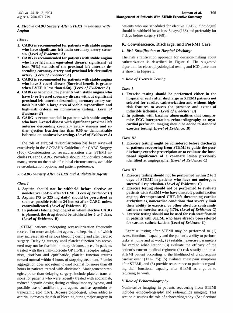
4. Elective CABG Surgery After STEMI in Patients WithAngina
Class I1. CABG is recommended for patients with stable angina
who have significant left main coronary artery steno-sis. (Level of Evidence: A)
2. CABG is recommended for patients with stable anginawho have left main equivalent disease: significant (atleast 70%) stenosis of the proximal left anterior de-scending coronary artery and proximal left circumflexartery. (Level of Evidence: A)
3. CABG is recommended for patients with stable anginawho have 3-vessel disease (Survival benefit is greaterwhen LVEF is less than 0.50). (Level of Evidence: A)
4. CABG is beneficial for patients with stable angina whohave 1- or 2-vessel coronary disease without significantproximal left anterior descending coronary artery ste-nosis but with a large area of viable myocardium andhigh-risk criteria on noninvasive testing. (Level ofEvidence: B)
5. CABG is recommended in patients with stable anginawho have 2-vessel disease with significant proximal leftanterior descending coronary artery stenosis and ei-ther ejection fraction less than 0.50 or demonstrableischemia on noninvasive testing. (Level of Evidence: A)
The role of surgical revascularization has been reviewedextensively in the ACC/AHA Guidelines for CABG Surgery(166). Consideration for revascularization after STEMI in-cludes PCI and CABG. Providers should individualize patientmanagement on the basis of clinical circumstances, availablerevascularization options, and patient preference.
5. CABG Surgery After STEMI and Antiplatelet Agents
Class I1. Aspirin should not be withheld before elective or
nonelective CABG after STEMI. (Level of Evidence: C)2. Aspirin (75 to 325 mg daily) should be prescribed as
soon as possible (within 24 hours) after CABG unlesscontraindicated. (Level of Evidence: B)
3. In patients taking clopidogrel in whom elective CABGis planned, the drug should be withheld for 5 to 7 days.(Level of Evidence: B)
STEMI patients undergoing revascularization frequentlyreceive 1 or more antiplatelet agents and heparin, all of whichmay increase risk of serious bleeding during and after cardiacsurgery. Delaying surgery until platelet function has recov-ered may not be feasible in many circumstances. In patientstreated with the small-molecule GP IIb/IIIa receptor antago-nists, tirofiban and eptifibatide, platelet function returnstoward normal within 4 hours of stopping treatment. Plateletaggregation does not return toward normal for more than 48hours in patients treated with abciximab. Management strat-egies, other than delaying surgery, include platelet transfu-sions for patients who were recently treated with abciximab,reduced heparin dosing during cardiopulmonary bypass, andpossible use of antifibrinolytic agents such as aprotinin ortranexamic acid (167). Because clopidogrel, when added toaspirin, increases the risk of bleeding during major surgery in
patients who are scheduled for elective CABG, clopidogrelshould be withheld for at least 5 days (168) and preferably for7 days before surgery (169).
K. Convalescence, Discharge, and Post-MI Care
1. Risk Stratification at Hospital Discharge
The risk stratification approach for decision-making aboutcatheterization is described in Figure 6. The suggestedalgorithm for electrophysiological testing and ICD placementis shown in Figure 5.
a. Role of Exercise Testing
Class I1. Exercise testing should be performed either in the
hospital or early after discharge in STEMI patients notselected for cardiac catheterization and without high-risk features to assess the presence and extent ofinducible ischemia. (Level of Evidence: B)
2. In patients with baseline abnormalities that compro-mise ECG interpretation, echocardiography or myo-cardial perfusion imaging should be added to standardexercise testing. (Level of Evidence: B)
Class IIb1. Exercise testing might be considered before discharge
of patients recovering from STEMI to guide the post-discharge exercise prescription or to evaluate the func-tional significance of a coronary lesion previouslyidentified at angiography. (Level of Evidence: C)
Class III1. Exercise testing should not be performed within 2 to 3
days of STEMI in patients who have not undergonesuccessful reperfusion. (Level of Evidence: C)
2. Exercise testing should not be performed to evaluatepatients with STEMI who have unstable postinfarctionangina, decompensated CHF, life-threatening cardiacarrhythmias, noncardiac conditions that severely limittheir ability to exercise, or other absolute contraindi-cations to exercise testing (170). (Level of Evidence: C)
3. Exercise testing should not be used for risk stratificationin patients with STEMI who have already been selectedfor cardiac catheterization. (Level of Evidence: C)
Exercise testing after STEMI may be performed to (1)assess functional capacity and the patient’s ability to performtasks at home and at work; (2) establish exercise parametersfor cardiac rehabilitation; (3) evaluate the efficacy of thepatient’s current medical regimen; (4) risk-stratify the post-STEMI patient according to the likelihood of a subsequentcardiac event (171–175); (5) evaluate chest pain symptomsafter STEMI; and (6) provide reassurance to patients regard-ing their functional capacity after STEMI as a guide toreturning to work.
b. Role of Echocardiography
Noninvasive imaging in patients recovering from STEMIincludes echocardiography and radionuclide imaging. Thissection discusses the role of echocardiography. (See Sections
705JACC Vol. 44, No. 3, 2004 Antman et al.August 4, 2004:671–719 Management of Patients With STEMI: Executive Summary

7.11.1.3, 7.11.1.4, and 7.11.1.5 of the full-text guidelines foradditional discussion on imaging considerations.)
Class I1. Echocardiography should be used in patients with
STEMI not undergoing LV angiography to assessbaseline LV function, especially if the patient is hemo-dynamically unstable. (Level of Evidence: C)
2. Echocardiography should be used to evaluate patients withinferior STEMI, clinical instability, and clinical suspicion ofRV infarction. (See ACC/AHA Guidelines for Clinical Ap-plication of Echocardiography (153).) (Level of Evidence: C)
3. Echocardiography should be used in patients withSTEMI to evaluate suspected complications, includingacute MR, cardiogenic shock, infarct expansion, VSR,intracardiac thrombus, and pericardial effusion. (Levelof Evidence: C)
4. Stress echocardiography (or myocardial perfusion imag-ing) should be used in patients with STEMI for in-hospital or early postdischarge assessment for inducibleischemia when baseline abnormalities are expected tocompromise ECG interpretation. (Level of Evidence: C)
Class IIa
1. Echocardiography is reasonable in patients withSTEMI to re-evaluate ventricular function during re-covery when results are used to guide therapy. (Levelof Evidence: C)
2. Dobutamine echocardiography (or myocardial perfu-sion imaging) is reasonable in hemodynamically andelectrically stable patients 4 or more days after STEMIto assess myocardial viability when required to definethe potential efficacy of revascularization. (Level ofEvidence: C)
3. In STEMI patients who have not undergone contrastventriculography, echocardiography is reasonable toassess ventricular function after revascularization.(Level of Evidence: C)
Class III1. Echocardiography should not be used for early routine
reevaluation in patients with STEMI in the absence ofany change in clinical status or revascularization pro-cedure. Reassessment of LV function 30 to 90 dayslater may be reasonable. (Level of Evidence: C)
Figure 6. Evidence-based approach to need for catheterization (cath) and revascularization after STEMI. This algorithm shows treat-ment paths for patients who initially undergo a primary invasive strategy, receive fibrinolytic therapy, or do not undergo reperfusiontherapy for STEMI. Patients who have not undergone a primary invasive strategy and have no high-risk features should undergo func-tional evaluation with one of the noninvasive tests shown. When clinically significant ischemia is detected, patients should undergocatheterization and revascularization as indicated; if no clinically significant ischemia is detected, medical therapy is prescribed afterSTEMI. *Please see Table 23 of the ACC/AHA Guidelines for the Management of Patients With Chronic Stable Angina for further defini-tion. †Please see Table 3, Section 6.3.1.6.2., and Section 7.3. in the full-text STEMI guidelines for further discussion. STEMI indicatesST-elevation myocardial infarction; EF, ejection fraction; ECG, electrocardiography.
706 Antman et al. JACC Vol. 44, No. 3, 2004Management of Patients With STEMI: Executive Summary August 4, 2004:671–719

The use of echocardiography in STEMI is discussed indetail in the ACC/AHA/ASE 2003 Guideline Update for theClinical Application of Echocardiography (153).
c. Exercise Myocardial Perfusion ImagingNoninvasive imaging in patients recovering from STEMIincludes echocardiography and radionuclide imaging. Thissection discusses the role of exercise myocardial perfusionimaging. (See Sections 7.11.1.2, 7.11.1.4, and 7.11.1.5 of thefull-text guidelines for additional discussion on imagingconsiderations.)
Class I
1. Dipyridamole or adenosine stress perfusion nuclearscintigraphy or dobutamine echocardiography beforeor early after discharge should be used in patients withSTEMI who are not undergoing cardiac catheteriza-tion to look for inducible ischemia in patients judged tobe unable to exercise. (Level of Evidence: B)
Class IIa
1. Myocardial perfusion imaging or dobutamine echocar-diography is reasonable in hemodynamically and elec-trically stable patients 4 to 10 days after STEMI toassess myocardial viability when required to define thepotential efficacy of revascularization. (Level of Evi-dence: C)
Recommended strategies for exercise test evaluations afterSTEMI are presented in Figure 6. These strategies and thedata on which they are based are reviewed in more detail inthe ACC/AHA 2002 Guideline Update for Exercise Testing(170).
d. LV FunctionNoninvasive imaging in patients recovering from STEMIincludes echocardiography and radionuclide imaging. Thissection discusses the importance of measurement of LVfunction. Either of the above imaging techniques can provideclinically useful information.
Class I
1. LVEF should be measured in all STEMI patients.(Level of Evidence: B)
Assessment of LV function after STEMI has been shownto be one of the most accurate predictors of future cardiacevents in both the prereperfusion (176) and the reperfusioneras (177,178). Multiple techniques for assessing LVfunction of patients after STEMI have important prognos-tic value. Because of the dynamic nature of LV functionrecovery after STEMI, clinicians should consider thetiming of the imaging study relative to the index eventwhen assessing LV function. (See Table 6 of the ACC/AHA/ASE 2003 Guideline Update on the Clinical Appli-cation of Echocardiography for further discussion of theimpact of timing on assessment of LV function andinducible ischemia.) (153).
e. Invasive Evaluation
Class I1. Coronary arteriography should be performed in pa-
tients with spontaneous episodes of myocardial ische-mia or episodes of myocardial ischemia provoked byminimal exertion during recovery from STEMI. (Level ofEvidence: A)
2. Coronary arteriography should be performed forintermediate- or high-risk findings on noninvasivetesting after STEMI (see Table 23 of the ACC/AHA2002 Guideline Update for the Management of Pa-tients With Chronic Stable Angina) (179). (Level ofEvidence: B)
3. Coronary arteriography should be performed if thepatient is sufficiently stable before definitive therapy ofa mechanical complication of STEMI, such as acuteMR, VSR, pseudoaneurysm, or LV aneurysm. (Level ofEvidence: B)
4. Coronary arteriography should be performed in pa-tients with persistent hemodynamic instability. (Levelof Evidence: B)
5. Coronary arteriography should be performed in sur-vivors of STEMI who had clinical heart failure duringthe acute episode but subsequently demonstrated well-preserved LV function. (Level of Evidence: C)
Class IIa1. It is reasonable to perform coronary arteriography
when STEMI is suspected to have occurred by amechanism other than thrombotic occlusion of anatherosclerotic plaque. This would include coronaryembolism, certain metabolic or hematological diseases,or coronary artery spasm. (Level of Evidence: C)
2. Coronary arteriography is reasonable in STEMI pa-tients with any of the following: diabetes mellitus,LVEF less than 0.40, CHF, prior revascularization, orlife-threatening ventricular arrhythmias. (Level of Ev-idence: C)
Class IIb1. Catheterization and revascularization may be consid-
ered as part of a strategy of routine coronary arteriog-raphy for risk assessment after fibrinolytic therapy(See Section 6.3.1.6.4.7 of the full-text guidelines).(Level of Evidence: B)
Class III1. Coronary arteriography should not be performed in
survivors of STEMI who are thought not to be candi-dates for coronary revascularization. (Level of Evi-dence: A)
The Writing Committee encourages contemporary researchinto the benefit of routine catheterization versus watchfulwaiting after fibrinolytic therapy in the contemporary era(180). (See Section 6.3.1.6.4.7 of the full-text guidelines)
f. Assessment of Ventricular Arrhythmias
Class IIb1. Noninvasive assessment of the risk of ventricular ar-
rhythmias may be considered (including signal-
707JACC Vol. 44, No. 3, 2004 Antman et al.August 4, 2004:671–719 Management of Patients With STEMI: Executive Summary

averaged ECG, 24-hour ambulatory monitoring, heartrate variability, micro T-wave alternans, and T-wavevariability) in patients recovering from STEMI. (Levelof Evidence: B)
The clinical applicability of these tests to the post-STEMIpatient is in a state of evolution. Until these issues areresolved, use these tests are used only to support routinemanagement and risk assessment.
L. Secondary Prevention
Class I1. Patients who survive the acute phase of STEMI should
have plans initiated for secondary prevention thera-pies. (Level of Evidence: A)
Secondary prevention therapies, unless contraindicated, arean essential part of the management of all patients withSTEMI (Table 4) (181); regardless of sex (182,183). Inas-much as atherosclerotic vascular disease is frequently foundin multiple vascular beds, the physician should search forsymptoms or signs of peripheral vascular disease or cerebro-vascular disease in patients presenting with STEMI.
1. Patient Education Before Discharge
Class I1. Before hospital discharge, all STEMI patients should
be educated about and actively involved in planningfor adherence to the lifestyle changes and drug thera-pies that are important for the secondary prevention ofcardiovascular disease. (Level of Evidence: B)
2. Post-STEMI patients and their family membersshould receive discharge instructions about recog-nizing acute cardiac symptoms and appropriate ac-tions to take in response (ie, calling 9-1-1 if symp-toms are unimproved or worsening 5 minutes afteronset, or if symptoms are unimproved or worsening5 minutes after 1 sublingual nitroglycerin dose) toensure early evaluation and treatment should symp-toms recur. (Level of Evidence: C)
3. Family members of STEMI patients should be advisedto learn about AEDs and CPR and be referred to aCPR training program. Ideally, such training programswould have a social support component targeting familymembers of high-risk patients. (Level of Evidence: C)
2. Lipid Management
Class I1. Dietary therapy that is low in saturated fat and cho-
lesterol (less than 7% of total calories as saturated fatand less than 200 mg/d cholesterol) should be startedon discharge after recovery from STEMI. Increasedconsumption of the following should be encouraged:omega–3 fatty acids, fruits, vegetables, soluble (vis-cous) fiber, and whole grains. Calorie intake should bebalanced with energy output to achieve and maintain ahealthy weight. (Level of Evidence: A)
2. A lipid profile should be obtained from past records,but if not available, it should be performed in all
patients with STEMI, preferably after they have fastedand within 24 hours of admission. (Level of Evidence: C)
3. The target LDL-C level after STEMI should be sub-stantially less than 100 mg/dL. (Level of Evidence: A)
a. Patients with LDL-C 100 mg/dl or above should beprescribed drug therapy on hospital discharge, withpreference given to statins. (Level of Evidence: A)
b. Patients with LDL-C less than 100 mg/dL or unknownLDL-C levels should be prescribed statin therapy onhospital discharge. (Level of Evidence: B)
4. Patients with non–high-density lipoprotein cholesterol(non HDL-C) levels less than 130 mg/dL who have anHDL-C level less than 40 mg/dL should receive specialemphasis on nonpharmacological therapy (eg, exercise,weight loss, and smoking cessation) to increase HDL-C.(Level of Evidence: B)
Class IIa1. It is reasonable to prescribe drug therapy at discharge to
patients with non–HDL-C greater than or equal to 130mg/dL, with a goal of reducing non–HDL-C to substan-tially less than 130 mg/dL. (Level of Evidence: B)
2. It is reasonable to prescribe drug therapy such asniacin or fibrate therapy to raise HDL-C levels inpatients with LDL-C less than 100 mg/dL and non–HDL-C less than 130 mg/dL but HDL-C less than 40mg/dL despite dietary and other nonpharmacologicaltherapy. (Level of Evidence: B) Dietary-supplementniacin must not be used as a substitute for prescriptionniacin, and over-the-counter niacin should be usedonly if approved and monitored by a physician.
3. It is reasonable to add drug therapy with either niacinor a fibrate to diet regardless of LDL-C and HDL-Clevels when triglyceride levels are greater than 500mg/dL. In this setting, non–HDL-C (goal substantiallyless than 130 mg/dL) should be the cholesterol targetrather than LDL-C. (Level of Evidence: B) Dietary-supplement niacin must not be used as a substitute forprescription niacin, and over-the-counter niacinshould be used only if approved and monitored by aphysician.
Early secondary prevention trials conducted before theuse of statin therapy, which used then-available drugs anddiet to lower cholesterol, demonstrated significant reduc-tions of 25% in nonfatal MIs and 14% in fatal MIs (14).Subsequently, a growing body of evidence, mainly fromlarge randomized clinical trials of statin therapy, hasfirmly established the desirability of lowering atherogenicserum lipids in patients who have recovered from aSTEMI. See Table 4 for additional discussion ofrecommendations.
3. Weight Management
Class I1. Measurement of waist circumference and calculation
of body mass index are recommended. Desirable bodymass index range is 18.5 to 24.9 kg/m2. A waistcircumference greater than 40 inches in men and 35inches in women would result in evaluation for meta-
708 Antman et al. JACC Vol. 44, No. 3, 2004Management of Patients With STEMI: Executive Summary August 4, 2004:671–719

TABLE 4. Secondary Prevention for STEMI Patients
Goals Intervention Recommendations
Smoking: Goal complete cessation Assess tobacco use. Strongly encourage patient and family to stop smoking and to avoid secondhand smoke.Provide counseling, pharmacological therapy (including nicotine replacement and bupropion), and formal smokingcessation programs as appropriate.
Blood pressure control: Goal Less than140/90 mm Hg or Less than 130/80 mm Hg ifchronic kidney disease or diabetes
If blood pressure is 120/80 mm Hg or greater:• Initiate lifestyle modification (weight control, physical activity, alcohol moderation, moderate sodium
restriction, and emphasis on fruits, vegetables, and low-fat dairy products) in all patients.If blood pressure is 140/90 mm Hg or greater or 130/80 mm Hg or greater for individuals withchronic kidney disease or diabetes:
• Add blood pressure medications, emphasizing the use of beta-blockers and inhibition of therenin-angiotensin-aldosterone system.
Lipid management: (TG less than 200 mg/dL)Primary goal LDL-C substantially less than 100mg/dL
Start dietary therapy in all patients (less than 7% of total calories as saturated fat and less than 200mg/d cholesterol). Promote physical activity and weight management. Encourage increasedconsumption of omega-3 fatty acids.Assess fasting lipid profile in all patients, preferably within 24 hours of STEMI.Add drug therapy according to the following guide:
LDL-C substantially less than 100 mg/dL (baseline or on-treatment):• Statins should be used to lower LDL-C.
LDL-C greater than or equal to 100 mg/dL (baseline or on-treatment):• Intensify LDL-C–lowering therapy with drug treatment, giving preference to statins.
Lipid management: (TG 200 mg/dL or greater)Primary goal Non–HDL-C* substantially less than130 mg/dL
If TG is greater than or equal to 150 mg/dL or HDL-C is less than 40 mg/dL:• Emphasize weight management and physical activity. Advise smoking cessation.
If TG is 200 to 499 mg/dL:• After LDL-C–lowering therapy,† consider adding fibrate or niacin.‡
If TG is greater than or equal to 500 mg/dL:• Consider fibrate or niacin‡ before LDL-C–lowering therapy.†• Consider omega-3 fatty acids as adjunct for high TG.
Physical activity: Minimum goal 30 minutes 3to 4 days per week; Optimal daily
Assess risk, preferably with exercise test, to guide prescription.Encourage minimum of 30 to 60 minutes of activity, preferably daily, or at least 3 or 4 times weekly(walking, jogging, cycling, or other aerobic activity) supplemented by an increase in daily lifestyleactivities (eg, walking breaks at work, gardening, household work). Cardiac rehabilitation/secondaryprevention programs, when available, are recommended for patients with STEMI, particularly thosewith multiple modifiable risk factors and/or those moderate- to high-risk patients in whomsupervised exercise training is warranted.
Weight management: Goal BMI 18.5–24.9 kg/m2
Waist circumference: Women: Less than 35 inchesMen: Less than 40 inches
Calculate BMI and measure waist circumference as part of evaluation. Monitor response of BMI andwaist circumference to therapy.Start weight management and physical activity as appropriate. Desirable BMI range is 18.5 to 24.9 kg/m2.If waist circumference is greater than or equal to 35 inches in women or greater than or equal to 40inches in men, initiate lifestyle changes and treatment strategies for metabolic syndrome.
Diabetes management: Goal HbA1c less than 7% Appropriate hypoglycemic therapy to achieve near-normal fasting plasma glucose, as indicated by HbA1c.Treatment of other risks (eg, physical activity, weight management, blood pressure, and cholesterolmanagement).
Antiplatelet agents/anticoagulants: Start and continue indefinitely aspirin 75 to 162 mg/d if not contraindicated. Consider clopidogrel 75mg/d or warfarin if aspirin is contraindicated. Manage warfarin to INR of 2.5 to 3.5 in post-STEMIpatients when clinically indicated or for those not able to take aspirin or clopidogrel (Figure 7).
Renin-Angiotensin-Aldosterone SystemBlockers:
ACE inhibitors in all patients indefinitely; start, early in stable high-risk patients (anterior MI, previousMI, Killip class greater than or equal to II �S3 gallop, rales, radiographic CHF�, LVEF less than 0.40).ARBs in patients who are intolerant of ACE inhibitors and who have either clinical or radiologicalsigns of heart failure or LVEF less than 0.40.Aldosterone blockade in patients without significant renal dysfunction§ or hyperkalemia who arealready receiving therapeutic doses of an ACE inhibitor, have an LVEF less than or equal to 0.40, andhave either diabetes or heart failure.
Beta-Blockers: Start in all patients. Continue indefinitely. Observe usual contraindications.
BMI indicates body mass index; in, inches; LDL-C, low-density lipoprotein cholesterol; HDL-C, high-density lipoprotein cholesterol; BMI, body mass index; INR,international normalization ratio; ACE, angiotensin converting enzyme; CHF, congestive heart failure; LVEF, left ventricular ejection fraction; ARB, angiotensin receptorblocker and TG, triglycerides.
*Non–HDL cholesterol equals total cholesterol minus HDL cholesterol.†Treat to a goal of non–HDL-C substantially less than 130 mg/dL.‡Dietary-supplement niacin must not be used as a substitute for prescription niacin, and over-the-counter niacin should be used only if approved and monitored by a physician.§Creatinine should be less than or equal to 2.5 mg/dL in men or less than or equal to 2.0 mg/dL in women.�Potassium should be less than or equal to 5.0 mEq/L.Modified with permission from Smith et al. Circulation. 2004;109:672–93 (181).
709JACC Vol. 44, No. 3, 2004 Antman et al.August 4, 2004:671–719 Management of Patients With STEMI: Executive Summary

bolic syndrome and implementation of weight-reduction strategies. (Level of Evidence: B)
2. Patients should be advised about appropriate strate-gies for weight management and physical activity(usually accomplished in conjunction with cardiacrehabilitation). (Level of Evidence: B)
3. A plan should be established to monitor the response ofbody mass index and waist circumference to therapy(usually accomplished in conjunction with cardiacrehabilitation). (Level of Evidence: B)
4. Smoking Cessation
Class I1. Patients recovering from STEMI who have a history
of cigarette smoking should be strongly encouragedto stop smoking and to avoid secondhand smoke.Counseling should be provided to the patient andfamily, along with pharmacological therapy (includ-ing nicotine replacement and bupropion) and formalsmoking-cessation programs as appropriate. (Levelof Evidence: B)
2. All STEMI patients should be assessed for a history ofcigarette smoking. (Level of Evidence: A)
5. Antiplatelet Therapy
Class I1. A daily dose of aspirin 75 to 162 mg orally should be
given indefinitely to patients recovering from STEMI.(Level of Evidence: A)
2. If true aspirin allergy is present, preferably clopidogrel(75 mg orally per day) or, alternatively, ticlopidine(250 mg orally twice daily) should be substituted.(Level of Evidence: C)
3. If true aspirin allergy is present, warfarin therapy witha target INR of 2.5 to 3.5 is a useful alternative toclopidogrel in patients less than 75 years of age who areat low risk for bleeding and who can be monitoredadequately for dose adjustment to maintain a targetINR range. (Level of Evidence: C)
Class III1. Ibuprofen should not be used because it blocks the
antiplatelet effects of aspirin. (Level of Evidence: C)
On the basis of 12 randomized trials in 18 788 patientswith prior infarction, the Antiplatelet Trialists’ Collaborationreported a 25% reduction in the risk of recurrent infarction,stroke, or vascular death in patients receiving prolongedantiplatelet therapy (36 fewer events for every 1000 patientstreated) (31). No antiplatelet therapy has proved superior toaspirin in this population, and daily doses of aspirin between80 and 325 mg appear to be effective (184). The CAPRIE(Clopidogrel versus Aspirin in Patients at Risk of IschemicEvents) trial, which compared aspirin with clopidogrel in19 185 patients at high risk for vascular events, demonstrateda modest but significant (8.6%, P equals 0.043) reduction inserious vascular events with clopidogrel compared withaspirin (185). These data suggest clopidogrel as the bestalternative to aspirin in patients with true aspirin allergy.
The use of warfarin therapy for secondary prevention ofvascular events in patients after STEMI is discussed inSection 7.12.11 of the full-text guidelines. Large randomizedtrials have demonstrated that oral anticoagulants, when givenin adequate doses, reduce the rates of adverse outcomes, atthe cost of a small increase in hemorrhagic events (186–188).In the Warfarin, Aspirin, Reinfarction Study (WARIS II),warfarin without aspirin in a dose intended to achieve an INRof 2.8 to 4.2 resulted in a significant reduction in a compositeend point (death, nonfatal reinfarction, or thromboembolicstroke) compared with therapy with aspirin alone (16.7%versus 20.0%) (186). Warfarin therapy resulted in a small butsignificant increase in major, nonfatal bleeding comparedwith therapy with aspirin alone (0.62% versus 0.17% peryear). Chronic therapy with warfarin after STEMI presents analternative to clopidogrel in patients with aspirin allergy.
6. Inhibition of Renin-Angiotensin-Aldosterone-System
Class I1. An ACE inhibitor should be prescribed at discharge
for all patients without contraindications after STEMI.(Level of Evidence: A)
2. Long-term aldosterone blockade should be prescribedfor post-STEMI patients without significant renal dys-function (creatinine should be less than or equal to 2.5mg/dL in men and less than or equal to 2.0 mg/dL inwomen) or hyperkalemia (potassium should be lessthan or equal to 5.0 mEq/L) who are already receivingtherapeutic doses of an ACE inhibitor, have an LVEFless than or equal to 0.40, and have either symptomaticheart failure or diabetes. (Level of Evidence: A)
3. An ARB should be prescribed at discharge in thoseSTEMI patients who are intolerant of an ACE inhibi-tor and have either clinical or radiological signs ofheart failure and LVEF less than 0.40. Valsartan andcandesartan have established efficacy for this recom-mendation. (Level of Evidence: B)
Class IIa1. In STEMI patients who tolerate ACE inhibitors, an
ARB can be useful as an alternative to ACE inhibitorsin the long-term management of STEMI patients,provided there are either clinical or radiological signsof heart failure or LVEF less than 0.40. Valsartan andcandesartan have established efficacy for this recom-mendation. (Level of Evidence: B)
Class IIb
1. The combination of an ACE inhibitor and an ARB maybe considered in the long-term management of STEMIpatients with persistent symptomatic heart failure andLVEF less than 0.40. (Level of Evidence: B)
The use of ACE inhibitors early in the acute phase ofSTEMI and in the hospital management phase has beendescribed earlier.
Compelling evidence now supports the broad long-termuse of ACE inhibitors after STEMI (189,190). The results of
710 Antman et al. JACC Vol. 44, No. 3, 2004Management of Patients With STEMI: Executive Summary August 4, 2004:671–719

the VALIANT study (Valsartan in Acute Myocardial Infarc-tion Trial) evaluating valsartan are discussed in Section 7.4.3of the full-text guidelines. The series of CHARM studies(Candesartan in Heart Failure Assessment in Reduction ofMortality), although focusing on the evaluation of candesar-tan in patients with chronic heart failure, provides informa-tion that can be extrapolated to the long-term management ofthe STEMI patient, because 50% to 60% of the patientsstudied had ischemic heart disease as the cause of heartfailure (191–193).
Given the extensive randomized trial and routine clinicalexperience with ACE inhibitors, they remain the logical firstagent for inhibition of the renin-angiotensin-aldosterone sys-tem in the long-term management of patients with STEMI(150,194). The ARBs valsartan and candesartan should beadministered over the long term to STEMI patients withsymptomatic heart failure who are intolerant of ACE inhibi-tors. As described in Section 7.4.3 of the full-text guidelines,the choice between an ACE inhibitor and an ARB over thelong term in patients who are tolerant of ACE inhibitors willvary with individual physician and patient preference, as wellas cost and anticipated side-effect profile (150,194).
The results of the most relevant clinical trials that testedcombinations of ACE inhibitors and ARBs have been subtlydifferent, but clinically relevant. Whereas the CHARM-Added (192) trial demonstrated a reduction in the combinedend point of heart failure hospitalization and death over ACEinhibition alone, the VALIANT study (149) reported that thecombination of captopril and valsartan was equivalent toeither alone, but with a greater number of adverse effects.Thus, when combination ACE inhibition and angiotensinreceptor blockade is considered necessary, the preferred ARBis candesartan. Although there is evidence that the combina-tion of an ACE inhibitor and an aldosterone inhibitor iseffective at reducing mortality and is well tolerated in patientswith a serum creatinine level of 2.5 mg/dL or less and a serumpotassium concentration of 5.0 mEq/L or less (see Section7.4.3 of the full-text guidelines), much less experience existswith the combination of an ARB and aldosterone inhibitor(24% of 2028 patients in the CHARM-Alternative trial) (191)and the triple combination of an ACE inhibitor, ARB, and analdosterone antagonist (17% of 2548 patients in the CHARM-Added trial) (192).
The combination of an ACE inhibitor and an ARB(valsartan 20 mg/d orally initially; titrated up to 160 mg orallytwice per day, or candesartan 4 to 8 mg/d orally initially; titratedup to 32 mg/d orally) or an ACE inhibitor and an aldosteroneinhibitor may be considered for the long-term management ofSTEMI patients with symptomatic heart failure and LVEF lessthan 0.40, provided the serum creatinine level is less than orequal to 2.5 mg/dL in men and less than or equal to 2.0 mg/dLin women and the serum potassium concentration is less than orequal to 5.0 mEq/L (See Sections 7.4.3 and 7.6.4 of the full-textguidelines.)
7. Beta-Blockers
Class I1. All patients after STEMI except those at low risk
(normal or near-normal ventricular function, success-
ful reperfusion, and absence of significant ventriculararrhythmias) and those with contraindications shouldreceive beta-blocker therapy. Treatment should beginwithin a few days of the event, if not initiated acutely,and continue indefinitely. (Level of Evidence: A)
2. Patients with moderate or severe LV failure shouldreceive beta-blocker therapy with a gradual titrationscheme. (Level of Evidence: B)
Class IIa1. It is reasonable to prescribe beta-blockers to low-risk
patients after STEMI who have no contraindications tothat class of medications. (Level of Evidence: A)
The use of beta-blockers in the early phase of STEMI andin hospital management is reviewed in Sections 6.3.1.6 and7.4.1 of the full-text guidelines. The benefits of beta-blockertherapy in patients without contraindications have been dem-onstrated with or without reperfusion, initiated early or laterin the clinical course, and for all age groups. The benefits ofbeta-blocker therapy for secondary prevention are well estab-lished (142,196). In patients with moderate or severe LVfailure, beta-blocker therapy should be administered with agradual titration scheme (197). Long-term beta-blocker ther-apy should be administered to survivors of STEMI who havesubsequently undergone revascularization, because there isevidence of a mortality benefit from their use despite revas-cularization with either CABG surgery or PCI (198).
8. Blood Pressure Control
Class I1. Blood pressure should be treated with drug therapy to
a target level of less than 140/90 mm Hg and to lessthan 130/80 mm Hg for patients with diabetes orchronic kidney disease. (Level of Evidence: B)
2. Lifestyle modification (weight control, dietary changes,physical activity, and sodium restriction) should beinitiated in all patients with blood pressure greaterthan or equal to 120/80 mm Hg. (Level of Evidence: B)
Class IIb1. A target blood pressure goal of 120/80 mm Hg for
post-STEMI patients may be reasonable. (Level ofEvidence: C)
Class III1. Short-acting dihydropyridine calcium channel block-
ing agents should not be used for the treatment ofhypertension. (Level of Evidence: B)
The Seventh Report of the Joint National Committee onPrevention, Detection, Evaluation, and Treatment of HighBlood Pressure (JNC-7) (199) recommends that patients betreated after MI with ACE inhibitors, beta-blockers, and, ifnecessary, aldosterone antagonists to a target blood pressureof less than 140/90 mm Hg, or less than 130/80 mm Hg forthose with chronic kidney disease or diabetes (199). Mostpatients will require 2 or more drugs to reach this goal, andwhen the blood pressure is greater than 20/10 mm Hg abovegoal, 2 drugs should usually be used from the outset.
711JACC Vol. 44, No. 3, 2004 Antman et al.August 4, 2004:671–719 Management of Patients With STEMI: Executive Summary

JNC-7 emphasizes the importance of lifestyle modifica-tions for all patients with blood pressure of 120/80 mm Hg orgreater (199). These modifications include weight reductionif overweight or obese, consumption of a diet rich in fruitsand vegetables and low in total fat and saturated fat, andreduction of sodium to no more than 2.4 g/d (199).
9. Diabetes Management
Class I1. Hypoglycemic therapy should be initiated to achieve
HbA1c less than 7%. (Level of Evidence: B)
Class III1. Thiazolidinediones should not be used in patients
recovering from STEMI who have New York HeartAssociation class III or IV heart failure. (Level ofEvidence: B)
10. Hormone Therapy
Class III1. Hormone therapy with estrogen plus progestin should
not be given de novo to postmenopausal women afterSTEMI for secondary prevention of coronary events.(Level of Evidence: A)
2. Postmenopausal women who are already taking estro-gen plus progestin at the time of a STEMI should notcontinue hormone therapy. However, women who arebeyond 1 to 2 years after initiation of hormone therapywho wish to continue hormone therapy for anothercompelling indication should weigh the risks and ben-efits, recognizing a greater risk of cardiovascularevents. However, hormone therapy should not be con-tinued while patients are on bedrest in the hospital.(Level of Evidence: B)
On the basis of the Heart and Estrogen/progestin Replace-ment Study (HERS) (200), the Heart and Estrogen/progestinReplacement Study Follow-up (HERS-2) (201), and theWomen’s Health Initiative (202), postmenopausal womenshould not receive combination estrogen and progestin ther-apy for primary or secondary prevention of CHD. It isrecommended that the use of hormone therapy be discontin-ued in women who have STEMI (200–202).
11. Warfarin Therapy
Class I1. Warfarin should be given to aspirin-allergic post-
STEMI patients with indications for anticoagulation asfollows:
a. Without stent implanted (INR 2.5 to 3.5). (Level ofEvidence: B)
b. With stent implanted and clopidogrel 75 mg/dadministered concurrently (INR 2.0 to 3.0). (Levelof Evidence: C)
2. Warfarin (INR 2.5 to 3.5) is a useful alternative toclopidogrel in aspirin-allergic patients after STEMIwho do not have a stent implanted. (Level of Evidence:B)
3. Warfarin (INR 2.0 to 3.0) should be prescribed forpost-STEMI patients with either persistent or parox-ysmal atrial fibrillation. (Level of Evidence: A)
4. In post-STEMI patients with LV thrombus noted on animaging study, warfarin should be prescribed for at least 3months (Level of Evidence: B) and indefinitely in patientswithout an increased risk of bleeding (Level of Evidence: C).
5. Warfarin alone (INR 2.5 to 3.5) or warfarin (INR 2.0 to 3.0)in combination with aspirin (75 to 162 mg) should beprescribed in post-STEMI patients who have no stent im-planted and who have indications for anticoagulation. (Levelof Evidence: B)
Class IIa1. In post-STEMI patients less than 75 years of age
without specific indications for anticoagulation whocan have their level of anticoagulation monitored reli-ably, warfarin alone (INR 2.5 to 3.5) or warfarin (INR2.0 to 3.0) in combination with aspirin (75 to 162 mg)can be useful for secondary prevention. (Level ofEvidence: B)
2. It is reasonable to prescribe warfarin to post-STEMIpatients with LV dysfunction and extensive regionalwall-motion abnormalities. (Level of Evidence: A)
Class IIb1. Warfarin may be considered in patients with severe
LV dysfunction, with or without CHF. (Level of Evi-dence: C)
The indications for long-term anticoagulation after STEMIremain controversial and are evolving. Although the use ofwarfarin has been demonstrated to be cost-effective com-pared with standard therapy without aspirin, the superiorsafety, efficacy and cost-effectiveness of aspirin has made itthe antithrombotic agent of choice for secondary prevention(203) (Figure 7).
12. Physical Activity
Class I1. On the basis of assessment of risk, ideally with an
exercise test to guide the prescription, all patientsrecovering from STEMI should be encouraged toexercise for a minimum of 30 minutes, preferably dailybut at least 3 or 4 times per week (walking, jogging,cycling, or other aerobic activity), supplemented by anincrease in daily lifestyle activities (eg, walking breaksat work, gardening, and household work). (Level ofEvidence: B)
2. Cardiac rehabilitation/secondary prevention pro-grams, when available, are recommended for patientswith STEMI, particularly those with multiple modifi-able risk factors and/or those moderate- to high-riskpatients in whom supervised exercise training is war-ranted. (Level of Evidence: C)
13. Antioxidants
Class III1. Antioxidant vitamins such as vitamin E and/or vitamin
C supplements should not be prescribed to patients
712 Antman et al. JACC Vol. 44, No. 3, 2004Management of Patients With STEMI: Executive Summary August 4, 2004:671–719

recovering from STEMI to prevent cardiovasculardisease. (Level of Evidence: A)
There is no convincing evidence to support lipid- orwater-soluble antioxidant supplementation in patients afterSTEMI or patients with or without established coronarydisease.
VIII. Long-Term ManagementA. Psychosocial Impact of STEMI
Class I1. The psychosocial status of the patient should be evalu-
ated, including inquiries regarding symptoms of de-pression, anxiety, or sleep disorders and the socialsupport environment. (Level of Evidence: C)
Class IIa1. Treatment with cognitive-behavioral therapy and se-
lective serotonin reuptake inhibitors can be useful forSTEMI patients with depression that occurs in the yearafter hospital discharge. (Level of Evidence: A)
Treatment of depression with combined cognitive-behavioral therapy and selective serotonin reuptake inhibitorsimproves outcome in terms of depression symptoms and
social function (204–206). It appears prudent to assessSTEMI patients for depression during hospitalization andduring the first month after STEMI and to intervene andreassess yearly in the first 5 years, as appropriate. There isevidence that the STEMI experience, with its sudden andunexpected onset, dramatic changes in lifestyle, and theadditive effort of comorbid life events, is a relatively trau-matic event and may produce impaired coping during subse-quent ischemic events (207).
B. Cardiac Rehabilitation
Class IIa1. Cardiac rehabilitation/secondary prevention pro-
grams, when available, are recommended for patientswith STEMI, particularly those with multiple modifi-able risk factors and/or those moderate- to high-riskpatients in whom supervised exercise training is war-ranted. (Level of Evidence: C)
C. Follow-Up Visit With Medical Provider
Class I1. A follow-up visit should delineate the presence or
absence of cardiovascular symptoms and functionalclass. (Level of Evidence: C)
Figure 7. Long-term antithrombotic therapy at hospital discharge after STEMI. ASA indicates aspirin; LOE, level of evidence LV, leftventricular; and INR, international normalized ratio. *Clopidogrel is preferred over warfarin because of increased risk of bleeding andlow patient compliance in warfarin trials. †For 12 months. ‡Discontinue clopidogrel 1 month after implantation of a bare metal stent orseveral months after implantation of a drug-eluting stent (3 months after sirolimus and 6 months after paclitaxel) because of the poten-tial increased risk of bleeding with warfarin and 2 antiplatelet agents. Continue aspirin and warfarin long term if warfarin is indicated forother reasons such as atrial fibrillation, LV thrombus, cerebral emboli, or extensive regional wall-motion abnormality. §An INR of 2.0 to3.0 is acceptable with tight control, but the lower end of this range is preferable. The combination of antiplatelet therapy and warfarinmay be considered in patients aged less than 75 years with low bleeding risk who can be monitored reliably.
713JACC Vol. 44, No. 3, 2004 Antman et al.August 4, 2004:671–719 Management of Patients With STEMI: Executive Summary

2. The patient’s list of current medications should bereevaluated in a follow-up visit, and appropriate titra-tion of ACE inhibitors, beta-blockers, and statinsshould be undertaken. (Level of Evidence: C)
3. The predischarge risk assessment and planned workupshould be reviewed and continued (Figure 6). This shouldinclude a check of LV function and possibly Holter moni-toring for those patients whose early post-STEMI ejectionfraction was 0.31 to 0.40 or lower, in consideration ofpossible ICD use (Figure 5). (Level of Evidence: C)
4. The healthcare provider should review and empha-size the principles of secondary prevention with thepatient and family members (Table 4) (181). (Levelof Evidence: C)
5. The psychosocial status of the patient should be eval-uated in follow-up, including inquiries regardingsymptoms of depression, anxiety, or sleep disordersand the social support environment. (Level of Evi-dence: C)
6. In a follow-up visit, the healthcare provider shoulddiscuss in detail issues of physical activity, return towork, resumption of sexual activity, and travel,including driving and flying. The metabolic equiva-lent values for various activities are provided as aresource in Table 34 of the full-text guideline. (Levelof Evidence: C)
7. Patients and their families should be asked if they areinterested in CPR training after the patient is dis-charged from the hospital. (Level of Evidence: C)
8. Providers should actively review the following issueswith patients and their families:a. The patient’s heart attack risk. (Level of Evidence: C)b. How to recognize symptoms of STEMI. (Level of
Evidence: C)c. The advisability of calling 9-1-1 if symptoms are
unimproved or worsening after 5 minutes, despitefeelings of uncertainty about the symptoms and fearof potential embarrassment. (Level of Evidence: C)
d. A plan for appropriate recognition and response to apotential acute cardiac event, including the phonenumber to access EMS, generally 9-1-1 (15). (Levelof Evidence: C)
9. Cardiac rehabilitation/secondary prevention pro-grams, when available, are recommended for patientswith STEMI, particularly those with multiple modifi-able risk factors and/or those moderate- to high-riskpatients in whom supervised exercise training is war-ranted. (Level of Evidence: C)
References1. Mehta RH, Montoye CK, Gallogly M, et al., for the GAP Steering
Committee of the American College of Cardiology. Improving quality ofcare for acute myocardial infarction: The Guidelines Applied in Practice(GAP) Initiative. JAMA 2002;287:1269–76.
2. Jencks SF, Huff ED, Cuerdon T. Change in the quality of care deliveredto medicare beneficiaries, 1998–1999 to 2000–2001. JAMA 2003;289:305–12.
3. Schneider EC, Zaslavsky AM, Epstein AM. Racial disparities in thequality of care for enrollees in medicare managed care. JAMA 2002;287:1288–94.
4. Rogers WJ, Canto JG, Lambrew CT, et al. Temporal trends in thetreatment of over 1.5 million patients with myocardial infarction in theUS from 1990 through 1999: the National Registry of MyocardialInfarction 1, 2 and 3. J Am Coll Cardiol 2000;36:2056–63.
5. American Heart Association. Heart Disease and Stroke Statistics—2004Update. Dallas, TX: American Heart Association, 2003.
5a. Wiviott SD, Morrow DA, Giugliano RP, et al. Performance of thethrombolysis in myocardial infarction risk index for early acute coro-nary syndrome in the National Registry of Myocardial Infarction: asimple risk index predicts mortality in both ST and non-ST elevationmyocardial infarction. J Am Coll Cardiol 2003;41:365A–6A.
6. Expert Panel on Detection, Evaluation, and Treatment of High BloodCholesterol in Adults. Executive Summary of the Third Report of theNational Cholesterol Education Program (NCEP) Expert Panel onDetection, Evaluation, and Treatment of High Blood Cholesterol inAdults (Adult Treatment Panel III). JAMA 2001;285:2486–97.
7. Levey AS, Coresh J, Balk E, et al. National Kidney Foundation practiceguidelines for chronic kidney disease: evaluation, classification, andstratification. Ann Intern Med 2003;139:137–47.
8. National, Heart, Lung, and Blood Institute. Act in Time to Heart Attack.Available at: http://:www.nhlbi.nih.gov/actintime, 2003. Accessed May15, 2003.
9. American College of Cardiology. Guidelines Applied in Practice.Available at: http://www.acc.org/gap/gap.htm. Accessed May 15, 2003.
10. American Heart Association. Get With the Guidelines. Available athttp://www.americanheart.org/presenter.jhtml?identifier�1165.Accessed May 15, 2003.
11. Topol EJ, Kereiakes DJ. Regionalization of care for acute ischemic heartdisease: a call for specialized centers. Circulation 2003;107:1463–6.
12. Califf RM, Faxon DP. Need for centers to care for patients with acutecoronary syndromes. Circulation 2003;107:1467–70.
13. Willerson JT. Editor’s commentary: centers of excellence. Circulation2003;107:1471–2.
14. National Cholesterol Education Program. Third Report of the Expert Panelon Detection, Evaluation, and Treatment of High Blood Cholesterol inAdults (Adult Treatment Panel III). NIH publication No. 02–5125.Bethesda, Md: National Heart, Lung, and Blood Institute, 2002. Guidelines,Related Tools, and Patient Information. Available at: http://www.nhlbi.ni-h.gov/guidelines/cholesterol/index.htm. Accessed April 12, 2003.
15. Dracup K, Alonzo AA, Atkins JM, et al. The physician’s role inminimizing prehospital delay in patients at high risk for acute myo-cardial infarction: recommendations from the National Heart AttackAlert Program. Working Group on Educational Strategies to PreventPrehospital Delay in Patients at High Risk for Acute MyocardialInfarction. Ann Intern Med 1997;126:645–51.
16. Hedges JR, Feldman HA, Bittner V, et al., for the REACT Study Group.Impact of community intervention to reduce patient delay time on use ofreperfusion therapy for acute myocardial infarction: Rapid Early Actionfor Coronary Treatment (REACT) trial. Acad Emerg Med 2000;7:862–72.
17. Canto JG, Zalenski RJ, Ornato JP, et al., for the National Registry ofMyocardial Infarction 2 Investigators. Use of emergency medicalservices in acute myocardial infarction and subsequent quality of care:observations from the National Registry of Myocardial Infarction 2.Circulation 2002;106:3018–23.
18. Goldberg R, Goff D, Cooper L, et al. Age and sex differences inpresentation of symptoms among patients with acute coronary disease:the Trial: Rapid Early Action for Coronary Treatment (REACT). CoronArtery Dis 2000;11:399–407.
19. Hutchings CB, Mann NC, Daya M, et al., for the Rapid Early Action forCoronary Treatment Study. Patients with chest pain calling 9-1-1 orself-transporting to reach definitive care: which mode is quicker? AmHeart J 2004;147:35–41.
20. Faxon D, Lenfant C. Timing is everything: motivating patients to call9–1–1 at onset of acute myocardial infarction. Circulation 2001;104:1210–1.
21. U.S. Department of Health and Human Services, Public Health Service,National Institutes of Health, National Heart, Lung, and Blood Institute.NIH publication No. 01–3313. September 2001. Available at: http://www.nhlbi.nih.gov/health/prof/heart/mi/provider.pdf. AccessedDecember 16, 2002.
22. Eisenberg MJ, Topol EJ. Prehospital administration of aspirin in patientswith unstable angina and acute myocardial infarction. Arch Intern Med1996;156:1506–10.
23. Fibrinolytic Therapy Trialists’ (FTT) Collaborative Group. Indications forfibrinolytic therapy in suspected acute myocardial infarction: collaborativeoverview of early mortality and major morbidity results from all ran-domised trials of more than 1000 patients. Lancet 1994;343:311–22.
24. Gruppo Italiano per lo Studio della Streptochinasi nell’Infarto Mio-cardico (GISSI). Effectiveness of intravenous thrombolytic treatment inacute myocardial infarction. Lancet 1986;1:397–402.
714 Antman et al. JACC Vol. 44, No. 3, 2004Management of Patients With STEMI: Executive Summary August 4, 2004:671–719

25. Armstrong PW, Collen D, Antman E. Fibrinolysis for acute myocardialinfarction: the future is here and now. Circulation 2003;107:2533–7.
26. The American Heart Association in collaboration with the InternationalLiaison Committee on Resuscitation. Guidelines 2000 for Cardiopulmo-nary Resuscitation and Emergency Cardiovascular Care: Part 7: the eraof reperfusion: section 1: acute coronary syndromes (acute myocardialinfarction). Circulation 2000;102 Suppl I:I172–203.
27. Pedley DK, Bissett K, Connolly EM, et al. Prospective observationalcohort study of time saved by prehospital thrombolysis for ST elevationmyocardial infarction delivered by paramedics. BMJ 2003;327:22–6.
28. Menown IB, Mackenzie G, Adgey AA. Optimizing the initial 12-leadelectrocardiographic diagnosis of acute myocardial infarction. EurHeart J 2000;21:275–83.
29. Case definitions for acute coronary heart disease in epidemiology andclinical research studies: a statement from the AHA Council on Epide-miology and Prevention; AHA Statistics Committee; World Heart Fed-eration Council on Epidemiology and Prevention; the European Societyof Cardiology Working Group on Epidemiology and Prevention;Centers for Disease Control and Prevention; and the National Heart,Lung, and Blood Institute. Circulation 2003;108:2543–9.
30. Come PC, Pitt B. Nitroglycerin-induced severe hypotension and brady-cardia in patients with acute myocardial infarction. Circulation 1976;54:624–8.
31. Antithrombotic Trialists’ Collaboration. Collaborative meta-analysis of ran-domised trials of antiplatelet therapy for prevention of death, myocardialinfarction, and stroke in high risk patients. BMJ 2002;324:71–86.
32. Sagar KA, Smyth MR. A comparative bioavailability study of differentaspirin formulations using on-line multidimensional chromatography.J Pharm Biomed Anal 1999;21:383–92.
33. De Luca G, Suryapranata H, Zijlstra F, et al., for the ZWOLLE Myo-cardial Infarction Study Group. Symptom-onset-to-balloon time andmortality in patients with acute myocardial infarction treated by primaryangioplasty. J Am Coll Cardiol 2003;42:991–7.
34. Boersma E, Mercado N, Poldermans D, et al. Acute myocardialinfarction. Lancet 2003;361:847–58.
35. De Luca G, Suryapranata H, Ottervanger JP, et al. Time delay totreatment and mortality in primary angioplasty for acute myocardialinfarction: every minute of delay counts. Circulation 2004;109:1223–5.
36. Boersma E, Maas AC, Deckers JW, et al. Early thrombolytic treatmentin acute myocardial infarction: reappraisal of the golden hour. Lancet1996;348:771–5.
37. Zeymer U, Tebbe U, Essen R, et al., for the ALKK-Study Group.Influence of time to treatment on early infarct-related artery patencyafter different thrombolytic regimens. Am Heart J 1999;137:34–8.
38. Weaver WD, Cerqueira M, Hallstrom AP, et al. Prehospital-initiated vshospital-initiated thrombolytic therapy: the Myocardial Infarction Triageand Intervention Trial. JAMA 1993;270:1211–6.
39. Brodie BR, Stuckey TD, Wall TC, et al. Importance of time to reper-fusion for 30-day and late survival and recovery of left ventricularfunction after primary angioplasty for acute myocardial infarction. J AmColl Cardiol 1998;32:1312–9.
40. Brodie BR, Stuckey TD, Wall TC, et al. Importance of time to reper-fusion on outcomes with primary coronary angioplasty for acute myo-cardial infarction (results from the Stent Primary Angioplasty in Myo-cardial Infarction Trial). Am J Cardiol 2001;88:1085–90.
41. Williams DO. Treatment delayed is treatment denied. Circulation 2004;109:1806-8.
42. The Task Force on the Management of Acute Myocardial Infarction ofthe European Society of Cardiology. Management of acute myocardialinfarction in patients presenting with ST-segment elevation. Eur Heart J2003;24:28–66.
43. Morrow DA, Antman EM, Charlesworth A, et al. TIMI risk score forST-elevation myocardial infarction: a convenient, bedside, clinical scorefor risk assessment at presentation: an intravenous nPA for treatment ofinfarcting myocardium early II trial substudy. Circulation 2000;102:2031–7.
44. Lee KL, Woodlief LH, Topol EJ, et al., for the GUSTO-I Investigators.Predictors of 30-day mortality in the era of reperfusion for acute myo-cardial infarction: results from an international trial of 41,021 patients.Circulation 1995;91:1659–68.
45. Morrow DA, Antman EM, Giugliano RP, et al. A simple risk index forrapid initial triage of patients with ST-elevation myocardial infarction:an InTIME II substudy. Lancet 2001;358:1571–5.
46. Krumholz HM, Chen J, Wang Y, et al. Comparing AMI mortalityamong hospitals in patients 65 years of age and older: evaluatingmethods of risk adjustment. Circulation 1999;99:2986–92.
47. Granger CB, Goldberg RJ, Dabbous O, et al., for the Global Registry ofAcute Coronary Events Investigators. Predictors of hospital mortality inthe Global Registry of Acute Coronary Events. Arch Intern Med 2003;163:2345–53.
48. Magid DJ, Calonge BN, Rumsfeld JS, et al., for the National Registry ofMyocardial Infarction 2 and 3 Investigators. Relation between hospitalprimary angioplasty volume and mortality for patients with acute MItreated with primary angioplasty vs thrombolytic therapy. JAMA 2000;284:3131–8.
49. Nallamothu BK, Bates ER. Percutaneous coronary intervention versusfibrinolytic therapy in acute myocardial infarction: is timing (almost)everything? Am J Cardiol 2003;92:824–6.
50. Bonnefoy E, Lapostolle F, Leizorovicz A, et al., for the Comparison ofAngioplasty and Prehospital Thrombolysis in Acute MyocardialInfarction study group. Primary angioplasty versus prehospital fibrino-lysis in acute myocardial infarction: a randomised study. Lancet 2002;360:825–9.
51. Steg PG, Bonnefoy E, Chabaud S, et al. Impact of time to treatment onmortality after prehospital fibrinolysis or primary angioplasty: data from theCAPTIM randomized clinical trial. Circulation 2003;108:2851–6.
52. Widimsky P, Budesinsky T, Vorac D, et al. Long distance transport forprimary angioplasty vs immediate thrombolysis in acute myocardialinfarction: final results of the randomized national multicentre trial:PRAGUE-2. Eur Heart J 2003;24:94–104.
53. Deleted in press.54. Antman EM. General hospital management. In: Julian DG, Braunwald
E, eds. Management of Acute Myocardial Infarction. London, England:WB Saunders, 1994;42–4.
54a. Lincoff AM, Califf RM, Van de Werf F, et al., for the Global Use ofStrategies To Open Coronary Arteries (GUSTO) Investigators. Mor-tality at 1 year with combination platelet glycoprotein IIb/IIIa inhibitionand reduced-dose fibrinolytic therapy vs conventional fibrinolytictherapy for acute myocardial infarction: GUSTO V randomized trial.JAMA 2002;288:2130-5.
55. Zijlstra F, Beukema WP, van’t Hof AW, et al. Randomized comparison ofprimary coronary angioplasty with thrombolytic therapy in low risk patientswith acute myocardial infarction. J Am Coll Cardiol 1997;29:908–12.
56. Ribeiro EE, Silva LA, Carneiro R, et al. Randomized trial of directcoronary angioplasty versus intravenous streptokinase in acute myo-cardial infarction. J Am Coll Cardiol 1993;22:376–80.
57. Grinfield L, Berrocal D, Bellardi J, et al. Fibrinolytics versus primaryangioplasty in acute myocardial infarction (FAP): a randomized trial ina community hospital in Argentina (abstr). J Am Coll Cardiol 1996;27:A222.
58. Zijlstra F, de Boer MJ, Hoorntje JC, et al. A comparison of immediatecoronary angioplasty with intravenous streptokinase in acute myocardialinfarction. N Engl J Med 1993;328:680–4.
59. Widimsky P, Groch L, Zelízko M, et al. Multicentre randomized trialcomparing transport to primary angioplasty vs immediate thrombolysisvs combined strategy for patients with acute myocardial infarctionpresenting to a community hospital without a catheterization laboratory:the PRAGUE study. Eur Heart J 2000;21:823–31.
60. de Boer MJ, Ottervanger JP, van’t Hof AW, et al., for the ZwolleMyocardial Infarction Study Group. Reperfusion therapy in elderlypatients with acute myocardial infarction: a randomized comparison ofprimary angioplasty and thrombolytic therapy. J Am Coll Cardiol 2002;39:1723–8.
61. Akhras F, Ousa AA, Swann G, et al. Primary coronary angioplasty orintravenous thrombolysis for patients with acute myocardial infarction?Acute and late follow-up results in a new cardiac unit (abstr). J Am CollCardiol 1997;29:A235–6.
62. Deleted in press.63. Grines CL, Browne KF, Marco J, et al., for the Primary Angioplasty in
Myocardial Infarction Study Group. A comparison of immediate angio-plasty with thrombolytic therapy for acute myocardial infarction. N EnglJ Med 1993;328:673–9.
64. Gibbons RJ, Holmes DR, Reeder GS, et al, for the Mayo Coronary CareUnit and Catheterization Laboratory Groups. Immediate angioplastycompared with the administration of a thrombolytic agent followed byconservative treatment for myocardial infarction. N Engl J Med 1993;328:685–91.
715JACC Vol. 44, No. 3, 2004 Antman et al.August 4, 2004:671–719 Management of Patients With STEMI: Executive Summary

65. Ribichini F, Steffenino G, Dellavalle A, et al. Comparison ofthrombolytic therapy and primary coronary angioplasty with liberalstenting for inferior myocardial infarction with precordial ST-segmentdepression: immediate and long-term results of a randomized study.J Am Coll Cardiol 1998;32:1687–94.
66. García E, Elízaga J, Pérez-Castellano N, et al. Primary angioplastyversus systemic thrombolysis in anterior myocardial infarction. J AmColl Cardiol 1999;33:605–11.
67. The Global Use of Strategies to Open Occluded Coronary Arteries inAcute Coronary Syndromes (GUSTO IIb) Angioplasty Substudy Inves-tigators. A clinical trial comparing primary coronary angioplasty withtissue plasminogen activator for acute myocardial infarction. N EnglJ Med 1997;336:1621–8.
68. Le May MR, Labinaz M, Davies RF, et al. Stenting versus thrombolysisin acute myocardial infarction trial (STAT). J Am Coll Cardiol 2001;37:985–91.
69. Schömig A, Kastrati A, Dirschinger J, et al., for the Stent versusThrombolysis for Occluded Coronary Arteries in Patients with AcuteMyocardial Infarction Study Investigators. Coronary stenting plus plate-let glycoprotein IIb/IIIa blockade compared with tissue plasminogenactivator in acute myocardial infarction. N Engl J Med 2000;343:385–91.
70. Vermeer F, Oude Ophuis AJ, vd Berg EJ, et al. Prospective randomisedcomparison between thrombolysis, rescue PTCA, and primary PTCA inpatients with extensive myocardial infarction admitted to a hospitalwithout PTCA facilities: a safety and feasibility study. Heart 1999;82:426–31.
71. Kastrati A, Mehilli J, Dirschinger J, et al., for the Stent versus Throm-bolysis for Occluded Coronary Arteries in Patients with Acute Myo-cardial Infarction (STOPAMI-2) Study. Myocardial salvage after coro-nary stenting plus abciximab versus fibrinolysis plus abciximab inpatients with acute myocardial infarction: a randomised trial. Lancet2002;359:920–5.
72. Andersen HR, Nielsen TT, Rasmussen K, et al., for the DANAMI-2Investigators. Thrombolytic therapy vs primary percutaneous coronaryintervention for myocardial infarction in patients presenting to hospitalswithout on-site cardiac surgery: a randomized controlled trial. JAMA2002;287:1943–51.
73. Kastrati A, Mehilli J, Dirschinger J, et al., for the Stent versus Throm-bolysis for Occluded Coronary Arteries in Patients With Acute Myo-cardial Infarction (STOPAMI-2) Study. A randomized trial of transferfor primary angioplasty versus on-site thrombolysis in patients withhigh-risk myocardial infarction: the Air Primary Angioplasty in Myo-cardial Infarction study. J Am Coll Cardiol 2002;39:1713–9.
74. Andersen HR, Nielsen TT, Rasmussen K, et al., for the DANAMI-2Investigators. A comparison of coronary angioplasty with fibrinolytictherapy in acute myocardial infarction. N Engl J Med 2003;349:733–42.
75. Hochman JS, Sleeper LA, Webb JG, et al., for the Should We Emer-gently Revascularize Occluded Coronaries for Cardiogenic Shock(SHOCK) Investigators. Early revascularization in acute myocardialinfarction complicated by cardiogenic shock. N Engl J Med 1999;341:625–34.
76. Keeley EC, Boura JA, Grines CL. Primary angioplasty versus intrave-nous thrombolytic therapy for acute myocardial infarction: a quantitativereview of 23 randomised trials. Lancet 2003;361:13–20.
76a. Melandri G. The obsession with primary angioplasty. Circulation 2003;108:e1620.
77. Rogers WJ, Dean LS, Moore PB, et al., for the Alabama Registry ofMyocardial Ischemia Investigators. Comparison of primary angioplastyversus thrombolytic therapy for acute myocardial infarction. Am JCardiol 1994;74:111–8.
78. Every NR, Parsons LS, Hlatky M, et al., for the Myocardial InfarctionTriage and Intervention Investigators. A comparison of thrombolytictherapy with primary coronary angioplasty for acute myocardialinfarction. N Engl J Med 1996;335:1253–60.
79. Tiefenbrunn AJ, Chandra NC, French WJ, et al. Clinical experience withprimary percutaneous transluminal coronary angioplasty compared withalteplase (recombinant tissue-type plasminogen activator) in patientswith acute myocardial infarction: a report from the Second NationalRegistry of Myocardial Infarction (NRMI-2). J Am Coll Cardiol 1998;31:1240–5.
80. Danchin N, Vaur L, Genès N, et al. Treatment of acute myocardialinfarction by primary coronary angioplasty or intravenous thrombolysisin the “real world”: one-year results from a nationwide French survey.Circulation 1999;99:2639–44.
81. Berger PB, Ellis SG, Holmes DR, et al. Relationship between delay inperforming direct coronary angioplasty and early clinical outcome inpatients with acute myocardial infarction: results from the Global Use ofStrategies To Open Occluded Arteries in Acute Coronary Syndromes(GUSTO-IIb) trial. Circulation 1999;100:14–20.
82. Cannon CP, Gibson CM, Lambrew CT, et al. Relationship of symptom-onset-to-balloon time and door-to-balloon time with mortality in patientsundergoing angioplasty for acute myocardial infarction. JAMA 2000;283:2941–7.
83. Brodie BR, Stuckey TD, Hansen C, et al. Benefit of coronary reper-fusion before intervention on outcomes after primary angioplasty foracute myocardial infarction. Am J Cardiol 2000;85:13–8.
84. Stone GW, Cox D, Garcia E, et al. Normal flow (TIMI-3) beforemechanical reperfusion therapy is an independent determinant ofsurvival in acute myocardial infarction: analysis from the primary angio-plasty in myocardial infarction trials. Circulation 2001;104:636–41.
85. Clements IP, Christian TF, Higano ST, et al. Residual flow to the infarctzone as a determinant of infarct size after direct angioplasty. Circulation1993;88:1527–33.
86. Juliard JM, Feldman LJ, Golmard JL, et al. Relation of mortality ofprimary angioplasty during acute myocardial infarction to door-to-Thrombolysis In Myocardial Infarction (TIMI) time. Am J Cardiol2003;91:1401–5.
87. Cragg DR, Friedman HZ, Bonema JD, et al. Outcome of patients withacute myocardial infarction who are ineligible for thrombolytic therapy.Ann Intern Med 1991;115:173–7.
88. Brodie BR, Weintraub RA, Stuckey TD, et al. Outcomes of directcoronary angioplasty for acute myocardial infarction in candidates andnon-candidates for thrombolytic therapy. Am J Cardiol 1991;67:7–12.
89. Himbert D, Juliard JM, Steg PG, et al. Primary coronary angioplasty foracute myocardial infarction with contraindication to thrombolysis. Am JCardiol 1993;71:377–81.
90. Zahn R, Schuster S, Schiele R, et al., for the Maximal IndividualTherapy in Acute Myocardial Infarction (MITRA) Study Group. Com-parison of primary angioplasty with conservative therapy in patientswith acute myocardial infarction and contraindications for thrombolytictherapy. Cathet Cardiovasc Interv 1999;46:127–33.
91. Grzybowski M, Clements EA, Parsons L, et al. Mortality benefit ofimmediate revascularization of acute ST-segment elevation myocardialinfarction in patients with contraindications to thrombolytic therapy: apropensity analysis. JAMA 2003;290:1891–8.
92. Lotfi M, Mackie K, Dzavik V, et al. Impact of delays to cardiac surgeryafter failed angioplasty and stenting. J Am Coll Cardiol 2004;43:337–42.
93. Dehmer GJ, Gantt DS. Coronary intervention at hospitals withouton-site cardiac surgery: are we pushing the envelope too far? J Am CollCardiol 2004;43:343–5.
94. Antoniucci D, Santoro GM, Bolognese L, et al. A clinical trial com-paring primary stenting of the infarct-related artery with optimal primaryangioplasty for acute myocardial infarction: results from the FlorenceRandomized Elective Stenting in Acute Coronary Occlusions(FRESCO) trial. J Am Coll Cardiol 1998;31:1234–9.
95. Rodríguez A, Bernardi V, Fernández M, et al. In-hospital and late resultsof coronary stents versus conventional balloon angioplasty in acutemyocardial infarction: Gianturco-Roubin in Acute MyocardialInfarction (GRAMI trial). Am J Cardiol 1998;81:1286–91.
96. Suryapranata H, Ottervanger JP, Nibbering E, et al. Long term outcomeand cost-effectiveness of stenting versus balloon angioplasty for acutemyocardial infarction. Heart 2001;85:667–71.
97. Saito S, Hosokawa G, Tanaka S, et al., for the PASTA Trial Investi-gators. Primary stent implantation is superior to balloon angioplasty inacute myocardial infarction: final results of the primary angioplastyversus stent implantation in acute myocardial infarction (PASTA) trial.Cathet Cardiovasc Interv 1999;48:262–8.
98. Grines CL, Cox DA, Stone GW, et al., for the Stent Primary Angio-plasty in Myocardial Infarction Study Group. Coronary angioplastywith or without stent implantation for acute myocardial infarction.N Engl J Med 1999;341:1949–56.
99. Kawashima A, Ueda K, Nishida Y, et al. Quantitative angiographicanalysis of restenosis of primary stenting using Wiktor stent for acutemyocardial infarction: results from a multicenter randomized PRISAMstudy (abstr). Circulation 1999;100 Suppl I:I856.
100. Maillard L, Hamon M, Khalife K, et al., for the STENTIM-2 Investi-gators. A comparison of systematic stenting and conventional balloonangioplasty during primary percutaneous transluminal coronary angio-
716 Antman et al. JACC Vol. 44, No. 3, 2004Management of Patients With STEMI: Executive Summary August 4, 2004:671–719

plasty for acute myocardial infarction. J Am Coll Cardiol 2000;35:1729–36.
101. Scheller B, Hennen B, Severin-Kneib S, et al. Long-term follow-up ofa randomized study of primary stenting versus angioplasty in acutemyocardial infarction. Am J Med 2001;110:1–6.
102. Zhu MM, Feit A, Chadow H, et al. Primary stent implantation comparedwith primary balloon angioplasty for acute myocardial infarction: a meta-analysis of randomized clinical trials. Am J Cardiol 2001;88:297–301.
103. Stone GW, Grines CL, Cox DA, et al., for the Controlled Abciximaband Device Investigation to Lower Late Angioplasty Complications(CADILLAC) Investigators. Comparison of angioplasty with stenting,with or without abciximab, in acute myocardial infarction. N EnglJ Med 2002;346:957–66.
104. Lemos PA, Saia F, Hofma SH, et al. Short- and long-term clinicalbenefit of sirolimus-eluting stents compared to conventional bare stentsfor patients with acute myocardial infarction. J Am Coll Cardiol 2004;43:704–8.
104a. Ellis SG, Armstrong P, Betriu A, et al. Facilitated percutaneous coro-nary intervention versus primary percutaneous coronary intervention:design and rationale of the Facilitated Intervention with EnhancedReperfusion Speed to Stop Events (FINESSE) trial. Am Heart J 2004;147:E16.
105. The TIMI Research Group. Immediate vs delayed catheterization andangioplasty following thrombolytic therapy for acute myocardialinfarction: TIMI II A results. JAMA 1988;260:2849–58.
106. Topol EJ, Califf RM, George BS, et al. A randomized trial of immediateversus delayed elective angioplasty after intravenous tissue plasminogenactivator in acute myocardial infarction. N Engl J Med 1987;317:581–8.
107. Sadanandan S, Buller C, Menon V, et al. The late open arteryhypothesis: a decade later. Am Heart J 2001;142:411–21.
108. Hochman JS, Sleeper LA, White HD, et al., for the SHOCK Investi-gators: Should We Emergently Revascularize Occluded Coronaries forCardiogenic Shock: one-year survival following early revascularizationfor cardiogenic shock. JAMA 2001;285:190–2.
109. Dzavik V, Sleeper LA, Cocke TP, et al., for the SHOCK Investigators.Early revascularization is associated with improved survival in elderlypatients with acute myocardial infarction complicated by cardiogenic shock:a report from the SHOCK Trial Registry. Eur Heart J 2003;24:828–37.
110. Dauerman HL, Goldberg RJ, Malinski M, et al. Outcomes and earlyrevascularization for patients greater than or equal to 65 years of agewith cardiogenic shock. Am J Cardiol 2001;87:844–8.
111. Dauerman HL, Ryan TJ, Piper WD, et al. Outcomes of percutaneouscoronary intervention among elderly patients in cardiogenic shock: amulticenter, decade-long experience. J Invasive Cardiol 2003;15:380–4.
112. Simoons ML, Arnold AE, Betriu A, et al. Thrombolysis with tissueplasminogen activator in acute myocardial infarction: no additionalbenefit from immediate percutaneous coronary angioplasty. Lancet1988;1:197–203.
113. Ross AM, Coyne KS, Reiner JS, et al., for the PACT (Plasminogen-activator Angioplasty Compatibility Trial) Investigators: A randomizedtrial comparing primary angioplasty with a strategy of short-actingthrombolysis and immediate planned rescue angioplasty in acute myo-cardial infarction: the PACT trial. J Am Coll Cardiol 1999;34:1954–62.
114. Montalescot G, Barragan P, Wittenberg O, et al., for the ADMIRAL(Abciximab before Direct Angioplasty and Stenting in MyocardialInfarction Regarding Acute and Long-Term Follow-up). Investigators.Platelet glycoprotein IIb/IIIa inhibition with coronary stenting for acutemyocardial infarction. N Engl J Med 2001;344:1895–903.
115. Zeymer U, Uebis R, Vogt A, et al., for the ALKK-Study Group. Ran-domized comparison of percutaneous transluminal coronary angioplastyand medical therapy in stable survivors of acute myocardial infarction withsingle-vessel disease: a study of the Arbeitsgemeinschaft Leitende Kardi-ologische Krankenhausärzte. Circulation 2003;108:1324–8.
116. Scheller B, Hennen B, Hammer B, et al., for the SIAM III StudyGroup. Beneficial effects of immediate stenting after thrombolysis inacute myocardial infarction. J Am Coll Cardiol 2003;42:634–41.
117. Braunwald E, Antman E, Beasley J, et al. ACC/AHA 2002 guidelineupdate for the management of patients with unstable angina and non–ST-segment elevation myocardial infarction: summary article: a report of theAmerican College of Cardiology/American Heart Association Task Forceon Practice Guidelines (Committee on the Management of Patients WithUnstable Angina). J Am Coll Cardiol 2002;40:1366.
118. Stenestrand U, Wallentin L. Early revascularisation and 1-year survivalin 14-day survivors of acute myocardial infarction: a prospectivecohort study. Lancet 2002;359:1805–11.
119. Alter DA, Tu JV, Austin PC, et al. Waiting times, revascularizationmodality, and outcomes after acute myocardial infarction at hospitalswith and without on-site revascularization facilities in Canada. J AmColl Cardiol 2003;42:410–9.
120. Gupta M, Chang WC, Van de Werf F, et al., for the ASSENT IIInvestigators. International differences in in-hospital revascularizationand outcomes following acute myocardial infarction: a multilevel anal-ysis of patients in ASSENT-2. Eur Heart J 2003;24:1640–50.
121. Gibson CM, Karha J, Murphy SA, et al., for the TIMI Study Group.Early and long-term clinical outcomes associated with reinfarctionfollowing fibrinolytic administration in the thrombolysis in myocardialinfarction trials. J Am Coll Cardiol 2003;42:7–16.
122. Lablanche JM, Gueret P, Blanchard D, et al. Beneficial effects of veryearly PTCA in patients with intravenous thrombolysis for acute myo-cardial infarction: data from a nation-wide registry in France (abstr).Circulation 2002;106 Suppl II:II630.
123. Fernandez-Aviles F, Alonso JJ, Castro-Beiras A, et al. Prospectiverandomized trial comparing a routine invasive strategy within 24 hoursto thrombolysis versus an ischemia-guided conservative approach toacute myocardial infarction with ST-segment elevation: the Gracia-1trial. Lancet 2004. In press.
124. ISIS-2 (Second International Study of Infarct Survival) CollaborativeGroup. Randomised trial of intravenous streptokinase, oral aspirin,both, or neither among 17,187 cases of suspected acute myocardialinfarction: ISIS-2. Lancet 1988;2:349–60.
125. Granger CB, Hirsch J, Califf RM, et al. Activated partial thromboplastintime and outcome after thrombolytic therapy for acute myocardialinfarction: results from the GUSTO-I trial. Circulation 1996;93:870–8.
126. White HD, Yusuf S. Issues regarding the use of heparin followingstreptokinase therapy. J Thromb Thrombolysis 1995;2:5–10.
127. Thrombin-specific anticoagulation with bivalirudin versus heparin inpatients receiving fibrinolytic therapy for acute myocardial infarction:the HERO-2 randomised trial. Lancet 2001;358:1855–63.
128. Bertrand ME, Rupprecht HJ, Urban P, et al. Double-blind study of thesafety of clopidogrel with and without a loading dose in combinationwith aspirin compared with ticlopidine in combination with aspirinafter coronary stenting: the Clopidogrel Aspirin Stent InternationalCooperative Study (CLASSICS). Circulation 2000;102:624–9.
129. Mehta SR, Yusuf S, Peters RJ, et al., for the Clopidogrel in Unstableangina to prevent Recurrent Events trial (CURE) Investigators. Effectsof pretreatment with clopidogrel and aspirin followed by long-termtherapy in patients undergoing percutaneous coronary intervention: thePCI-CURE study. Lancet 2001;358:527–33.
130. Steinhubl SR, Berger PB, Mann JT III, et al., for the CREDO (Clopi-dogrel for the Reduction of Events During Observation) Investigators.Early and sustained dual oral antiplatelet therapy following percutane-ous coronary intervention: a randomized controlled trial. JAMA 2002;288:2411–20.
131. CYPHER™ sirolimus-eluting coronary stent on RAPTORTM over-the-wire delivery system and CYPHER™ sirolimus-eluting coronarystent on RAPTORRAIL� rapid exchange delivery system [instructionsfor use]. Miami, FL: Cordis, a Johnson & Johnson Company, April2003.
132. TAXUS: paclitaxel-eluting coronary stent system Monorail and overthe wire coronary stent delivery system [directions for use]. Natick,MA: Boston Scientific, March 2004.
133. Patrono C, Bachmann F, Baigent C, et al. Expert consensus documenton the use of antiplatelet agents: The Task Force on the Use ofAntiplatelet Agents in Patients With Atherosclerotic CardiovascularDisease of the European Society of Cardiology. Eur Heart J 2004;25:166–81.
134. Levine GN, Kern MJ, Berger PB, et al., for the American Heart Asso-ciation Diagnostic and Interventional Catheterization Committee andCouncil on Clinical Cardiology. Management of patients undergoing per-cutaneous coronary revascularization. Ann Intern Med 2003;139:123–36.
135. Lee DP, Herity NA, Hiatt BL, et al., for the TIrofiban Given in theEmergency Room before Primary Angioplasty. Adjunctive plateletglycoprotein IIb/IIIa receptor inhibition with tirofiban before primaryangioplasty improves angiographic outcomes: results of the TIrofibanGiven in the Emergency Room before Primary Angioplasty(TIGER-PA) pilot trial. Circulation 2003;107:1497–501.
136. Finney SJ, Zekveld C, Elia A, et al. Glucose control and mortality incritically ill patients. JAMA 2003;290:2041–7.
136a. Clement S, Braithwaite SS, Magee MF, et al. Management of diabetesand hyperglycemia in hospitals. Diabetes Care 2004;27:553–97.
717JACC Vol. 44, No. 3, 2004 Antman et al.August 4, 2004:671–719 Management of Patients With STEMI: Executive Summary
137. Third Report of the National Cholesterol Education Program (NCEP)Expert Panel on Detection, Evaluation, and Treatment of High BloodCholesterol in Adults (Adult Treatment Panel III) final report. Circu-lation 2002;106:3143–421.
138. Malan SS. Psychosocial adjustment following MI: current views andnursing implications. J Cardiovasc Nurs 1992;6:57–70.
139. Havik OE, Maeland JG. Patterns of emotional reactions after a myo-cardial infarction. J Psychosom Res 1990;34:271–85.
140. Moser DK, Dracup K. Is anxiety early after myocardial infarctionassociated with subsequent ischemic and arrhythmic events? Psy-chosom Med 1996;58:395–401.
141. Frasure-Smith N, Lespérance F, Talajic M. The impact of negativeemotions on prognosis following myocardial infarction: is it more thandepression? Health Psychol 1995;14:388–98.
142. Chae Cu, Hennekens CH. Beta blockers. In: Hennekens CH, ed.Clinical Trials in Cardiovascular Disease: A Companion to Braun-wald’s Heart Disease. Philadelphia, PA: WB Saunders, 1999;79–94.
143. Latini R, Maggioni AP, Flather M, et al. ACE inhibitor use in patientswith myocardial infarction: summary of evidence from clinical trials.Circulation 1995;92:3132–7.
144. ACE Inhibitor Myocardial Infarction Collaborative Group. Indicationsfor ACE inhibitors in the early treatment of acute myocardialinfarction: systematic overview of individual data from 100,000patients in randomized trials. Circulation 1998;97:2202–12.
145. Flather MD, Yusuf S, Køber L, et al., for the ACE-Inhibitor Myo-cardial Infarction Collaborative Group. Long-term ACE-inhibitortherapy in patients with heart failure or left-ventricular dysfunction:a systematic overview of data from individual patients. Lancet2000;355:1575– 81.
146. Pitt B, Zannad F, Remme WJ, et al., for the Randomized AldactoneEvaluation Study Investigators. The effect of spironolactone on mor-bidity and mortality in patients with severe heart failure. N Engl J Med1999;341:709–17.
147. Pitt B, Remme W, Zannad F, et al., for the Eplerenone Post-AcuteMyocardial Infarction Heart Failure Efficacy and Survival Study Inves-tigators. Eplerenone, a selective aldosterone blocker, in patients withleft ventricular dysfunction after myocardial infarction. N Engl J Med.2003;348:1309–21.
148. Dickstein K, Kjekshus J, for the OPTIMAAL Steering Committee ofthe Optimal Trial in Myocardial Infarction with Angiotensin IIAntagonist Losartan (OPTIMAAL) Study Group. Effects of losartanand captopril on mortality and morbidity in high-risk patients afteracute myocardial infarction: the OPTIMAAL randomised trial:Optimal Trial in Myocardial Infarction with Angiotensin II AntagonistLosartan. Lancet 2002;360:752–60.
149. Pfeffer MA, McMurray JJ, Velazquez EJ, et al., for the Valsartan inAcute Myocardial Infarction Trial Investigators. Valsartan, captopril,or both in myocardial infarction complicated by heart failure, leftventricular dysfunction, or both. N Engl J Med 2003;349:1893–906.
150. Mann DL, Deswal A. Angiotensin-receptor blockade in acute myo-cardial infarction: a matter of dose. N Engl J Med 2003;349:1963–5.
151. Gibbons RJ, Miller TD, Christian TF. Infarct size measured by singlephoton emission computed tomographic imaging with (99m) Tc-sestamibi: a measure of the efficacy of therapy in acute myocardialinfarction. Circulation 2000;101:101–8.
152. Klocke FJ, Baird MG, Bateman TM, et al. ACC/AHA/ASNCguidelines for the clinical use of cardiac radionuclide imaging: a reportof the American College of Cardiology/American Heart Association TaskForce on Practice Guidelines (ACC/AHA/ASNC Committee to Revise the1995 Guidelines for the Clinical Use of Radionuclide Imaging). Availableat: http://www.acc.org/clinical/guidelines/radio/rni_fulltext.pdf, 2003.Accessed January 15, 2004.
153. Cheitlin MD, Armstrong WF, Aurigemma GP, et al. ACC/AHA/ASE2003 guideline update for the clinical application of echocardiography:a report of the American College of Cardiology/American Heart Asso-ciation Task Force on Practice Guidelines (ACC/AHA/ASE Com-mittee to Update the 1997 Guidelines for the Clinical Application ofEchocardiography). Available at: www.acc.org/clinical/guidelines/echo/index.pdf, 2003. Accessed January 15, 2004.
154. Mahrholdt H, Wagner A, Holly TA, et al. Reproducibility of chronicinfarct size measurement by contrast-enhanced magnetic resonanceimaging. Circulation 2002;106:2322–7.
155. Menon V, Slater JN, White HD, et al. Acute myocardial infarctioncomplicated by systemic hypoperfusion without hypotension: report ofthe SHOCK trial registry. Am J Med 2000;108:374–80.
156. Hochman JS, Boland J, Sleeper LA, et al., for the SHOCK RegistryInvestigators. Current spectrum of cardiogenic shock and effect ofearly revascularization on mortality: results of an InternationalRegistry. Circulation 1995;91:873–81.
157. Hunt SA, Baker DW, Chin MH, et al. ACC/AHA guidelines for the eval-uation and management of chronic heart failure in the adult: a report of theAmerican College of Cardiology/American Heart Association Task Forceon Practice Guidelines (Committee to Revise the 1995 Guidelines for theEvaluation and Management of Heart Failure). Available at: http://www.acc.org/clinical/guidelines/failure/hf_index.htm, 2001. AccessedJune 5, 2004.
158. Kinch JW, Ryan TJ. Right ventricular infarction. N Engl J Med 1994;330:1211–7.
159. Núñez L, de la Llana R, López Sendón J, et al. Diagnosis and treatmentof subacute free wall ventricular rupture after infarction. Ann ThoracSurg 1983;35:525–9.
160. Guidelines for cardiopulmonary resuscitation and emergency cardiaccare: Emergency Cardiac Care Committee and Subcommittees,American Heart Association: Part III: adult advanced cardiac lifesupport. JAMA 1992;268:2199–241.
161. Campbell RWF. Arrhythmias. In: Julian DG. Braunwald E, eds. Man-agement of Acute Myocardial Infarction. London, England: WBSaunders, 1994;223–40.
162. Kontoyannis DA, Anastasiou-Nana MI, Kontoyannis SA, et al. Intrave-nous amiodarone decreases the duration of atrial fibrillation associatedwith acute myocardial infarction. Cardiovasc Drugs Ther 2001;15:155–60.
163. Ryan TJ, Antman EM, Brooks NH, et al. ACC/AHA guidelines for themanagement of patients with acute myocardial infarction: 1999guideline update: a report of the American College of Cardiology/American Heart Association Task Force on Practice Guidelines (Com-mittee on Management of Acute Myocardial Infarction). J Am CollCardiol 1999;34:890–911.
164. Gregoratos G, Abrams J, Epstein AE, et al. ACC/AHA/NASPE 2002guideline update for implantation of cardiac pacemakers and antiar-rhythmia devices: summary article: a report of the American College ofCardiology/American Heart Association Task Force on PracticeGuidelines (ACC/AHA/NASPE Committee to Update the 1998Pacemaker Guidelines). J Am Coll Cardiol 2002;40:1703–19.
165. Lamas GA, Ellenbogen KA. Evidence base for pacemaker modeselection: from physiology to randomized trials. Circulation 2004;109:443–51.
166. Eagle KA, Guyton RA, Davidoff R, et al. ACC/AHA guidelines forcoronary artery bypass graft surgery: a report of the AmericanCollege of Cardiology/American Heart Association Task Force onPractice Guidelines (Committee to Revise the 1991 Guidelines forCoronary Artery Bypass Graft Surgery). American College of Car-diology/American Heart Association. J Am Coll Cardiol 1999;34:1262–347.
167. Dyke C, Bhatia D. Inhibitors of the platelet receptor glycoproteinIIb-IIIa and complications during percutaneous coronary revascular-ization: management strategies for the cardiac surgeon. J CardiovascSurg (Torino). 1999;40:505–16.
168. Yusuf S, Zhao F, Mehta SR, et al., for the Clopidogrel in UnstableAngina to Prevent Recurrent Events Trial Investigators. Effects ofclopidogrel in addition to aspirin in patients with acute coronary syn-dromes without ST-segment elevation. N Engl J Med 2001;345:494–502.
169. Physician’s Desk Reference. Clopidogrel package insert. Montvale,NJ: Medical Economics Co; 2756.
170. Gibbons RJ, Balady GJ, Bricker JT, et al. ACC/AHA 2002 guidelineupdate for exercise testing: a report of the American College of Car-diology/American Heart Association Task Force on PracticeGuidelines (Committee on Exercise Testing). Available at: http://www.acc.org/clinical/guidelines/exercise/exercise_clean.pdf, 2002.Accessed February 12, 2004.
171. Théroux P, Waters DD, Halphen C, et al. Prognostic value of exercisetesting soon after myocardial infarction. N Engl J Med 1979;301:341–5.
172. DeBusk RF, Kraemer HC, Nash E, et al. Stepwise risk stratificationsoon after acute myocardial infarction. Am J Cardiol 1983;52:1161–6.
173. Krone RJ, Gillespie JA, Weld FM, et al. Low-level exercise testingafter myocardial infarction: usefulness in enhancing clinical risk strat-ification. Circulation 1985;71:80–9.
718 Antman et al. JACC Vol. 44, No. 3, 2004Management of Patients With STEMI: Executive Summary August 4, 2004:671–719

174. Valentine PA, Frew JL, Mashford ML, et al. Lidocaine in the pre-vention of sudden death in the pre-hospital phase of acute infarction: adouble-blind study. N Engl J Med 1974;291:1327–31.
175. Ross J, Gilpin EA, Madsen EB, et al. A decision scheme for coronaryangiography after acute myocardial infarction. Circulation 1989;79:292–303.
176. Risk stratification and survival after myocardial infarction. N EnglJ Med 1983;309:331–6.
177. Zaret BL, Wackers TH, Terrin M, et al. Does left ventricular ejectionfraction following thrombolytic therapy have the same prognosticimpact described in the prethrombolytic era? Results of the TIMI IITrial (abstr). J Am Coll Cardiol 1991;17:214A.
178. Roig E, Magriñá J, Garcia A, et al. Prognostic value of exerciseradionuclide angiography in low-risk acute myocardial infarction sur-vivors. Eur Heart J 1993;14:213–8.
179. Gibbons RJ, Abrams J, Chatterjee K, et al. ACC/AHA 2002 guidelineupdate for the management of patients with chronic stable angina: areport of the American College of Cardiology/American Heart Asso-ciation Task Force on Practice Guidelines (Committee to Update the1999 Guidelines for the Management of Patients with Chronic StableAngina). Available at: www.acc.org/clinical/guidelines/stable/stable.pdf 2002. Accessed March 18, 2004.
180. O’Neill WW. “Watchful waiting” after thrombolysis: it’s time for are-evaluation. J Am Coll Cardiol 2003;42:17–9.
181. Smith SC, Blair SN, Bonow RO, et al. AHA/ACC scientificstatement: AHA/ACC guidelines for preventing heart attack anddeath in patients with atherosclerotic cardiovascular disease: 2001update: a statement for healthcare professionals from the AmericanHeart Association and the American College of Cardiology. Circu-lation 2001;104:1577–9.
182. Mosca L, Appel LJ, Benjamin EJ, et al. Evidence-based guidelines forcardiovascular disease prevention in women. Circulation 2004;109:672–93.
183. Dalal H, Evans PH, Campbell JL. Recent developments in secondaryprevention and cardiac rehabilitation after acute myocardial infarction.BMJ 2004;328:693–7.
184. Gutstein DE, Fuster V. Pathophysiologic bases for adjunctive therapiesin the treatment and secondary prevention of acute myocardialinfarction. Clin Cardiol 1998;21:161–8.
185. CAPRIE Steering Committee. A randomised, blinded, trial of clopi-dogrel versus aspirin in patients at risk of ischaemic events (CAPRIE).Lancet 1996;348:1329–39.
186. Hurlen M, Smith P, Arnesen H. Effects of warfarin, aspirin and the twocombined, on mortality and thromboembolic morbidity after myo-cardial infarction: the WARIS-II (Warfarin-Aspirin ReinfarctionStudy) design. Scand Cardiovasc J 2000;34:168–71.
187. van Es RF, Jonker JJ, Verheugt FW, et al., for the Antithrombotics inthe Secondary Prevention of Events in Coronary Thrombosis-2(ASPECT-2) Research Group. Aspirin and coumadin after acute cor-onary syndromes (the ASPECT-2 study): a randomised controlled trial.Lancet 2002;360:109–13.
188. Brouwer MA, van den Bergh PJ, Aengevaeren WR, et al. Aspirin pluscoumarin versus aspirin alone in the prevention of reocclusion afterfibrinolysis for acute myocardial infarction: results of the Antithrom-botics in the Prevention of Reocclusion In Coronary Thrombolysis(APRICOT)-2 Trial. Circulation 2002;106:659–65.
189. Yusuf S, Sleight P, Pogue J, et al., for the Heart Outcomes PreventionEvaluation Study Investigators. Effects of an angiotensin-converting-enzyme inhibitor, ramipril, on cardiovascular events in high-riskpatients. N Engl J Med 2000;342:145–53.
190. Fox KM, for the EURopean trial On reduction of cardiac events withPerindopril in stable coronary Artery disease Investigators. Efficacy ofperindopril in reduction of cardiovascular events among patients withstable coronary artery disease: randomised, double-blind, placebo-controlled, multicentre trial (the EUROPA study). Lancet 2003;362:782–8.
191. Granger CB, McMurray JJ, Yusuf S, et al., for the CHARM Investi-gators and Committees. Effects of candesartan in patients with chronicheart failure and reduced left-ventricular systolic function intolerant toangiotensin-converting-enzyme inhibitors: the CHARM-Alternativetrial. Lancet 2003;362:772–6.
192. McMurray JJ, Ostergren J, Swedberg K, et al., for the CHARM Inves-tigators and Committees. Effects of candesartan in patients withchronic heart failure and reduced left-ventricular systolic functiontaking angiotensin-converting-enzyme inhibitors: the CHARM-Addedtrial. Lancet 2003;362:767–71.
193. Yusuf S, Pfeffer MA, Swedberg K, et al., for the CHARM Investigatorsand Committees. Effects of candesartan in patients with chronic heartfailure and preserved left-ventricular ejection fraction: the CHARM-Preserved trial. Lancet 2003;362:777–81.
194. Executive Council of the Heart Failure Society of America. Impli-cations of recent clinical trials for heart failure performance measures.J Card Fail 2004;10:4–5.
195. Deleted in press.196. Antman E, Braunwald E. Acute myocardial infarction. In: Braunwald
E, Zipes DP, Libby P, eds. Heart Disease: A Textbook of Cardiovas-cular Medicine. 6th ed. Philadelphia, PA: WB Saunders, 2001:1114–251. Chapter 5.
197. Gheorghiade M, Colucci WS, Swedberg K. Beta-blockers in chronicheart failure. Circulation 2003;107:1570–5.
198. Chen J, Radford MJ, Wang Y, et al. Are beta-blockers effective inelderly patients who undergo coronary revascularization after acutemyocardial infarction? Arch Intern Med 2000;160:947–52.
199. Seventh report of the Joint National Committee on the Prevention,Detection, Evaluation, and Treatment of High Blood Pressure (JNC7): resetting the hypertension sails. Hypertension 2003;41:1178 –9.
200. Hulley S, Grady D, Bush T, et al., for the Heart and Estrogen/progestinReplacement Study (HERS) Research Group. Randomized trial ofestrogen plus progestin for secondary prevention of coronary heartdisease in postmenopausal women. JAMA 1998;280:605–13.
201. Grady D, Herrington D, Bittner V, et al., for the HERS ResearchGroup. Cardiovascular disease outcomes during 6.8 years of hormonetherapy: Heart and Estrogen/progestin Replacement Study follow-up(HERS II). JAMA 2002;288:49–57.
202. Rossouw JE, Anderson GL, Prentice RL, et al., for the Writing Groupfor the Women’s Health Initiative Investigators. Risks and benefits ofestrogen plus progestin in healthy postmenopausal women: principalresults from the Women’s Health Initiative randomized controlled trial.JAMA 2002;288:321–33.
203. Cairns JA, Markham BA. Economics and efficacy in choosing oral anti-coagulants or aspirin after myocardial infarction. JAMA 1995;273:965–7.
203a. Kris-Etherton PM, Lichtenstein AH, Howard BV, Steinberg D,Witzman JL. Antioxidant vitamin supplements and cardiovasculardisease. Circulation. In press.
204. Glassman AH, O’Connor CM, Califf RM, et al., for the SertralineAntidepressant Heart Attack Randomized Trial (SADHEART) Group.Sertraline treatment of major depression in patients with acute MI orunstable angina. JAMA 2002;288:701–9.
205. Swenson JR, O’Connor CM, Barton D, et al., for the Sertraline Anti-depressant Heart Attack Randomized Trial (SADHART) Group.Influence of depression and effect of treatment with sertraline onquality of life after hospitalization for acute coronary syndrome. Am JCardiol 2003;92:1271–6.
206. Berkman LF, Blumenthal J, Burg M, et al., for the Enhancing Recoveryin Coronary Heart Disease Patients Investigators (ENRICHD). Effectsof treating depression and low perceived social support on clinicalevents after myocardial infarction: the Enhancing Recovery in Coro-nary Heart Disease Patients (ENRICHD) Randomized Trial. JAMA2003;289:3106–16.
207. Alonzo AA. Acute myocardial infarction and posttraumatic stress dis-order: the consequences of cumulative adversity. J Cardiovasc Nurs1999;13:33–45.3
719JACC Vol. 44, No. 3, 2004 Antman et al.August 4, 2004:671–719 Management of Patients With STEMI: Executive Summary