Academy of Medicine, Singaporeams.edu.sg/view-pdf.aspx?file=media\2155_fi_738.pdf&ofile...5 and many...
41
1 College of General Dental Practitioners Singapore College of Dental Surgeons, Singapore In conjunction with College of General Dental Practitioners, Singapore Best Practice Manual for common TOSP Procedures in Dentistry 1. Introduction 2. How to use this manual 3. Guidelines a. Endodontics: Endodontic Surgery b. Endodontics: Reimplantation/Transplantation c. Implant & Related surgeries d. Trauma: Dental e. Trauma: Soft tissue f. Pathology – Cryotherapy/Laser application g. Pathology h. Orthodontics: Surgically-assisted i. Periodontal Surgery j. Teeth Excision k. Miscellaneous: Abscess l. Miscellaneous: Frenal attachment m. Miscellaneous: Foreign body n. Miscellaneous: Tongue tie o. Miscellaneous: Neuralgia College of Dental Surgeons, Singapore Academy of Medicine, Singapore
Transcript of Academy of Medicine, Singaporeams.edu.sg/view-pdf.aspx?file=media\2155_fi_738.pdf&ofile...5 and many...

1
College of General Dental Practitioners Singapore
College of Dental Surgeons, Singapore In conjunction with College of General Dental Practitioners, Singapore Best Practice Manual for common TOSP Procedures in Dentistry
1. Introduction 2. How to use this manual 3. Guidelines
a. Endodontics: Endodontic Surgery b. Endodontics: Reimplantation/Transplantation c. Implant & Related surgeries d. Trauma: Dental e. Trauma: Soft tissue f. Pathology – Cryotherapy/Laser application g. Pathology h. Orthodontics: Surgically-assisted i. Periodontal Surgery j. Teeth Excision k. Miscellaneous: Abscess l. Miscellaneous: Frenal attachment m. Miscellaneous: Foreign body n. Miscellaneous: Tongue tie o. Miscellaneous: Neuralgia
College of Dental Surgeons, Singapore Academy of Medicine, Singapore

2
1. INTRODUCTION Medisave, introduced in April 1984, is a national medical savings scheme which helps individuals put aside part of their income into their Medisave Accounts to meet their future personal or immediate family's hospitalization, day surgery and certain outpatient expenses. The Table of Surgical Procedures (TOSP) is a patient financing tool to identify procedures which qualify for use of Medisave funds as well as the respective limits of withdrawal. Since 2005, the use of Medisave was extended to private dental clinics for the financing of procedures in the TOSP done in the clinic premises. As the TOSP was last revised in 1992, many new procedures in common practice today were not in the TOSP. Similarly, some of the procedures had become obsolete. Interpretation of some codes was difficult. Much of the ambiguity of has been addressed in this latest revision of the TOSP. This Best Practice Manual is a College of Dental Surgeons, Singapore project, done in collaboration with the College of General Dental Practitioners, Singapore, based on an initiative from the Ministry of Health. The purpose of this manual is to provide dentists with a set of professional guidelines to some of the commonly used TOSP codes in dental clinics. The Ministry of Health has a set of audit guidelines which will make reference to this Best Practice Manual. Procedures submitted for Medisave claims should be consistent with the recommendations in this manual. Documentation of each surgery with detailed clinical notes and where applicable/recommended, radiographs and clinical photographs preoperatively and postoperatively are recommended as a good clinical practice. Such radiographs and photographs should have an embedded patient identifier and date. Where commercial implants and graft materials are used, the batch identifier, expiry date and other relevant information must be entered into the patient's record. Thorough documentation will assist greatly in patient management as well as in clinical audits.

3
2. HOW TO USE THE MANUAL A list of procedures done by dentists including oral and maxillofacial surgeons was extracted from the main TOSP (TOSP tab) in the attached MS Excel file. While we attempted to be as complete as possible, the list is not exhaustive and non-inclusion of any codes into this Dental TOSP does not preclude a dental practitioner from using them (Complete Dental Codes tab). Within the Dental TOSP, the committee has recommended a list of procedures that are best done in an Operating Theater instead of a dental clinic (OT Codes). These are the more complex surgeries that may require advanced facilities and staff expertise not normally found in a dental surgery. This Best Practice Manual does not cover these procedures. However, this recommendation does not imply that procedures that are not included in this list should not be done in an operating theatre. Of the remaining procedures, dentists may choose to offer patients the choice of performing the surgery within their clinics or in an operating theatre in accordance to their clinical judgment and patient preference. The committee has selected 57 codes that may be commonly used or misinterpreted and written guidelines pertaining to their use in claiming for Medisave funding. These guidelines will be used in conjunction with the audit guidelines issued by Ministry of Health.

4
3a. ENDONDONTICS: ENDODONTIC SURGERY
General Principles Through the introduction of magnification with the surgical operating microscope, use of microsurgical instruments, ultrasonic tips and biocompatible root-end fill materials, refined principles of soft tissue management and enhanced principles of wound closure, modern surgical endodontics has emerged as a highly predictable procedure compared to traditional endodontic surgery (Tsesis et al 2009, Setzer et al 2010). Retention of the natural tooth structure is still the goal of quality dental care
Code Table Description Guideline
SF708T
2C Tooth (Single-Rooted), Various Lesions, Peri-radicular Surgery
Surgery performed on the root of an anterior tooth, including:
apicoectomy (root resection, retrograde preparation and retrograde filling)
repair of root perforation or resorptive defect
retrieval of extruded root filling material or broken instruments
Pre-operative and post-operative radiographs are necessary for standard of care.
SF818T
3A Tooth (Multiple-Rooted or more than 1 tooth), Various Lesions, Peri-Radicular Surgery
Surgery performed on the root(s) of a posterior tooth or on the roots of 2 or more teeth, including:
apicoectomy (root resection, retrograde preparation and retrograde filling)
repair of root perforation or resorptive defect
retrieval of extruded root filling material or broken instruments
root amputation of a diseased or cracked root on a multi-rooted tooth
hemisection on a multi-rooted tooth to remove the diseased or cracked root and associated part of crown.
Pre-operative and post-operative radiographs are necessary for standard of care.

5
and many previously root-treated teeth that appear to be well-done, yet exhibit adverse signs or symptoms, are viable candidates for endodontic surgery. Surgical intervention is not a substitute for failure to manage properly the root canal system non-surgically, to assess thoroughly the periodontal status, and to ignore the shortcomings of the coronal restoration. Indications and contraindications Peri-radicular surgery may be considered in the following situations:
Root canal re-treatment has been carried but tooth is still exhibiting signs and symptoms of periapical disease.
Root canal re-treatment is not feasible due to: presence of a large post which may render the tooth non-restorable if
removed calcified or obliterated canals presence of separated instruments not retrievable by non-surgical
endodontic treatment perforations or resorptive areas not accessible to repair by non-
surgical endodontic treatment
Contraindications to surgical endodontic procedures include: Patient with significant medical conditions, e.g. uncontrolled diabetes,
leukemia or cancer etc Teeth that are unrestorable, e.g. extensive caries, inadequate ferrule for coronal coverage that cannot be improved with crown lengthening
Teeth with poor periodontal support, e.g. very short root, extensive gingival recession or with moderate to severe marginal periodontitis
Poor surgical access, e.g. due to limited mouth opening
Dental operating microscope (DOM) Use of a DOM is necessary during endodontic surgery as it enables easier identification of root apices and decreased angles of root resections, which help to conserve cortical bone and root length. In addition, it can help to identify causes of endodontic failure such as isthmus, lateral canals, unfilled canals, perforations, cracks and fractures, which can affect the success of endodontic surgery if left untreated. Use of a DOM or endoscope for endodontic surgery has been shown to achieve higher success rates compared to the use of loupes or without visual aids (Setzer et al 2012). Flap management A semilunar flap is no longer recommended for endodontic surgery because of poor surgical access and scar formation (Kramper et al 1984). Flap designs that are commonly used now include the intra-sulcular full thickness flap and muco-gingival flap.

6
Root resection There is no consensus on the how much root resection is necessary but at least 2-3 mm has been recommended (Gilheany et al 1994, Kim & Kratchman 2006). A 0 degree or almost 0 degree bevel is preferred in order to decrease the number of exposed cut dentinal tubules and minimize leakage (Gilheany et al 1994). Root end preparation Use of ultrasonic surgical retro-tips is recommended as they are able to produce slimmer, more parallel and deeper preparations aligned with the long axis of the tooth (Wuchenich et al 1994). Use of a bur for retro-preparation is not recommended due to poor access to the root end and the risk of producing a preparation of insufficient depth with a lingual perforation. Root end filling material Biologically acceptable materials such as Intermediate Restorative Material and Mineral Trioxide Aggregate have been shown to be highly successful and are ideal options for the choice of root end filling materials (Chong et al 2003, Lindeboom et al 2005, Tsesis et al 2009). A post–operative periapical radiograph should be taken to check adequacy of the root end filling. Amalgam should not be used as long term success rates are poor (Frank et al 1992, Dorn & Gartner 1990), possibly due to the poor seal and subsequent corrosion and leakage over time. Although gutta percha has been used successfully for obturating the canal, it is not recommended as a retrograde filling material and as far as possible, the existing gutta percha filling should not be left as the only canal seal. Root resection and burnishing of the existing gutta percha filling has been shown to have significantly lower success rates compared to placement of a root end filling (Christiansen et al 2009). Examples of wrong usage Exploratory surgery to look for root fractures or cracks on the roots should not be deemed as peri-radicular surgery, as the term “peri-radicular surgery” implies an interventional procedure to have been carried out on the root of the tooth. The level of difficulty with an exploratory surgery compared to an apicoectomy is vastly different and not justifiable for the charges for a table 2C or 3A procedure. If an attempt has been to prepare and fill the crack, with a material such as MTA or composite resin, then it may constitute as peri-radicular surgery. However, the success of such procedures is not well documented and cannot constitute as the treatment of choice. Occasionally, single rooted anterior teeth, such as the mandibular incisor, may have more than 1 canal. However, this should not be considered as a multi-rooted tooth and the appropriate code should be SF708T. References Christiansen R, Kirkevang LL, Hørsted-Bindslev P, Wenzel A. Randomized clinical trial of root-end resection followed by root-end filling with mineral trioxide aggregate or
7
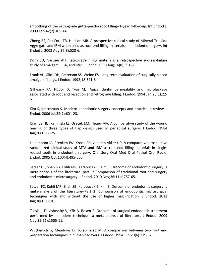
smoothing of the orthograde gutta-percha root filling--1-year follow-up. Int Endod J. 2009 Feb;42(2):105-14. Chong BS, Pitt Ford TR, Hudson MB. A prospective clinical study of Mineral Trioxide Aggregate and IRM when used as root-end filling materials in endodontic surgery. Int Endod J. 2003 Aug;36(8):520-6. Dorn SO, Gartner AH. Retrograde filling materials: a retrospective success-failure study of amalgam, EBA, and IRM. J Endod. 1990 Aug;16(8):391-3. Frank AL, Glick DH, Patterson SS, Weine FS. Long-term evaluation of surgically placed amalgam fillings. J Endod. 1992;18:391-8. Gilheany PA, Figdor D, Tyas MJ. Apical dentin permeability and microleakage associated with root end resection and retrograde filling. J Endod. 1994 Jan;20(1):22-6. Kim S, Kratchman S. Modern endodontic surgery concepts and practice: a review. J Endod. 2006 Jul;32(7):601-23. Kramper BJ, Kaminski EJ, Osetek EM, Heuer MA. A comparative study of the wound healing of three types of flap design used in periapical surgery. J Endod. 1984 Jan;10(1):17-25. Lindeboom JA, Frenken JW, Kroon FH, van den Akker HP. A comparative prospective randomized clinical study of MTA and IRM as root-end filling materials in single-rooted teeth in endodontic surgery. Oral Surg Oral Med Oral Pathol Oral Radiol Endod. 2005 Oct;100(4):495-500. Setzer FC, Shah SB, Kohli MR, Karabucak B, Kim S. Outcome of endodontic surgery: a meta-analysis of the literature--part 1: Comparison of traditional root-end surgery and endodontic microsurgery. J Endod. 2010 Nov;36(11):1757-65. Setzer FC, Kohli MR, Shah SB, Karabucak B, Kim S. Outcome of endodontic surgery: a meta-analysis of the literature--Part 2: Comparison of endodontic microsurgical techniques with and without the use of higher magnification. J Endod. 2012 Jan;38(1):1-10. Tsesis I, Faivishevsky V, Kfir A, Rosen E. Outcome of surgical endodontic treatment performed by a modern technique: a meta-analysis of literature. J Endod. 2009 Nov;35(11):1505-11. Wuchenich G, Meadows D, Torabinejad M. A comparison between two root end preparation techniques in human cadavers. J Endod. 1994 Jun;20(6):279-82.

8
3b. ENDODONTICS: REIMPLANTATION/TRANSPLANTATION Code Table Description Guideline
SF814T 3A Tooth, Dislocation,
Reimplantation and
Transplantation
This code refers to the following procedures: 1. Intentional replantation of an anterior or
posterior tooth, which includes intentional removal, inspection and treatment of the root and replacement into its own socket.
2. Transplantation of an anterior or posterior tooth, which includes intentional removal, inspection and treatment of the root and placement into another tooth socket.
Pre-operative and post-operative radiographs are necessary for standard of care.
General Principles Intentional Replantation Treatment should include an extraction technique to minimize damage to the periodontal ligament, limiting the extra-oral time of the tooth to less than 30 minutes and keeping the root moist during the extra-oral period (Kratchman 1997). Use of a tissue culture medium during the extra-oral period to maintain cellular viability of the root can be considered (Wolcott & Rossman 2003). Management of the root end is as outlined in the guidelines on peri-radicular surgery (see section on SF 708T and SF818T). Transplantation There is no established protocol for transplantation of teeth; however, the following guidelines should probably be adhered to: An extraction technique to minimize damage to the periodontal ligament Keeping the root moist during the extra-oral period to maintain cellular
viability Close approximation of the transplanted tooth to the transplanted socket
In immature and developing teeth, root canal treatment is not necessary because the apex is open and revascularization of the pulp is expected. Endodontic treatment is indicated for teeth with closed apices, usually within a month after transplantation (Kahnberg 1987, Mejare et al 2004). The prognosis for both closed and open apices is considered favorable (Kahnberg 1987, Lundberg & Isaksson 1996). Indications and contraindications Intentional Replantation In addition to the considerations for peri-radicular surgery (see section on SF708T and SF818T), intentional replantation may be considered in cases where anatomical

9
restrictions contra-indicate conventional apical surgery, such as in the maxillary and mandibular second molars, where access to root structures may be limited. Transplantation Transplantation of teeth, usually third molars, has been performed to replace a missing first or second molar. Other indications include orthodontic patients who undergo autotransplantation of maxillary premolars to replace congenitally missing mandibular premolars and mandibular premolars to replace maxillary incisors lost to trauma (Park et al 2010). Transplantation offers a viable and economic alternative to implants in selected cases for orthodontic and restorative reasons. A multi-disciplinary dental team is necessary as careful case selection and treatment planning is essential. Contraindications to the above procedures include: Patient with significant medical conditions, e.g. uncontrolled diabetes,
leukemia or cancer etc. Teeth that are non-restorable or with poor periodontal support Teeth with severely dilacerated and curved roots, which are prone to fracture
after extraction. Poor surgical access, e.g. due to limited mouth opening
Examples of wrong usage This code should not be used for the re-implantation of an avulsed tooth. In such cases, the code SF809M should be used instead. References Kahnberg KE. Autotransplantation of teeth (I). Indications for transplantation with a follow-up of 51 cases. Int J Oral Maxillofac Surg. 1987 Oct;16(5):577-85. Kratchman S. Intentional Replantation. Dent Clin North Am. 1997 Jul;41(3):603-17. Lundberg T, Isaksson S. A clinical follow-up study of 278 autotransplanted teeth. Br J Oral Maxillofac Surg. 1996 Apr;34(2):181-5. Mejàre B, Wannfors K, Jansson L. A prospective study on transplantation of third molars with complete root formation. Oral Surg Oral Med Oral Pathol Oral Radiol Endod. 2004 Feb;97(2):231-8. Park JH, Tai K, Hayashi D. Tooth autotransplantation as a treatment option: a review. J Clin Pediatr Dent. 2010 Winter;35(2):129-35. Wolcott J, Rossman LE. Intentional replantation of endodontically treated teeth: an update. Compend Contin Educ Dent. 2003 Jan;24(1):68-72, 74.
10
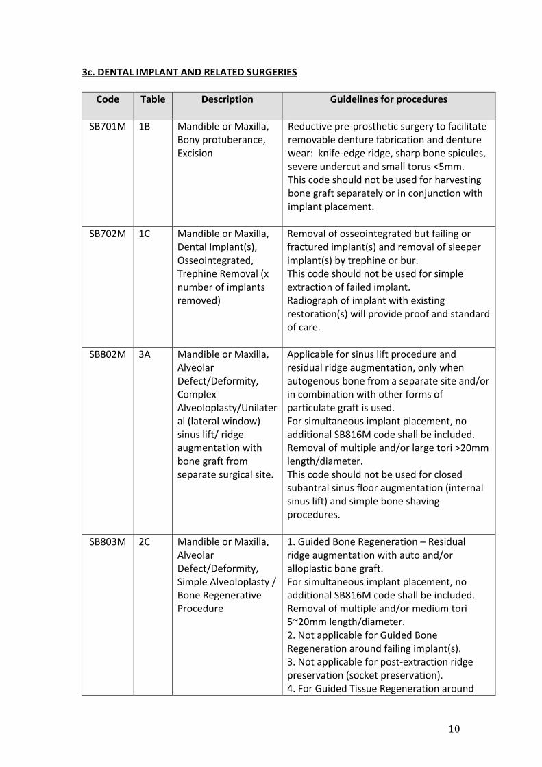
3c. DENTAL IMPLANT AND RELATED SURGERIES
Code Table Description Guidelines for procedures
SB701M 1B Mandible or Maxilla, Bony protuberance, Excision
Reductive pre-prosthetic surgery to facilitate removable denture fabrication and denture wear: knife-edge ridge, sharp bone spicules, severe undercut and small torus <5mm. This code should not be used for harvesting bone graft separately or in conjunction with implant placement.
SB702M 1C Mandible or Maxilla, Dental Implant(s), Osseointegrated, Trephine Removal (x number of implants removed)
Removal of osseointegrated but failing or fractured implant(s) and removal of sleeper implant(s) by trephine or bur. This code should not be used for simple extraction of failed implant. Radiograph of implant with existing restoration(s) will provide proof and standard of care.
SB802M 3A Mandible or Maxilla, Alveolar Defect/Deformity, Complex Alveoloplasty/Unilateral (lateral window) sinus lift/ ridge augmentation with bone graft from separate surgical site.
Applicable for sinus lift procedure and residual ridge augmentation, only when autogenous bone from a separate site and/or in combination with other forms of particulate graft is used. For simultaneous implant placement, no additional SB816M code shall be included. Removal of multiple and/or large tori >20mm length/diameter. This code should not be used for closed subantral sinus floor augmentation (internal sinus lift) and simple bone shaving procedures.
SB803M 2C Mandible or Maxilla, Alveolar Defect/Deformity, Simple Alveoloplasty / Bone Regenerative Procedure
1. Guided Bone Regeneration – Residual ridge augmentation with auto and/or alloplastic bone graft. For simultaneous implant placement, no additional SB816M code shall be included. Removal of multiple and/or medium tori 5~20mm length/diameter. 2. Not applicable for Guided Bone Regeneration around failing implant(s). 3. Not applicable for post-extraction ridge preservation (socket preservation). 4. For Guided Tissue Regeneration around

11
Code Table Description Guidelines for procedures
natural teeth, please refer to SF709M and SF710M.
SB813M 2C Mandible, Various Lesions, Alveolectomy (per quadrant)
Alveolectomy applies to reduction of multiple post-extraction sockets. Mylohyoid ridge reduction. Should not be used for surgical periodontal procedures. For simultaneous implant placement, no additional SB816M code shall be included.
SB814M 4A Mandible or Maxilla, Various Lesions, Alveoloplasty with Epithelial Graft, OR Bilateral Sinus-lift procedure OR Ridge augmentation (multiple quadrants) with bone graft harvested from separate site
Augmentation procedures with autogenous soft and hard tissue for multiple sockets or residual ridges. For simultaneous implant placement, no additional SB816M code shall be included. This code should not be used for closed subantral sinus floor augmentation (internal sinus lift) and simple bone shaving procedures.
SB816M 2C Mandible or Maxilla, Various Lesions, Insertion of Endosseous Dental Implant (single)(For multiple placement of implants, number of claims = number of implants placed )
Applicable to the actual number of implant(s) placed at the same surgical session not more than the number of missing teeth (not number of roots) to be replaced. Only implants of 3mm diameter or wider can be included. For simultaneous implant placement with bone grafts, the higher or same table applies and no separate claim for either should be included.
General Principles This section of guidelines is intended to provide simple parameters for the purpose of assignment of Medisave tables for surgical dental implant procedures only and dentists should refer to textbooks and articles published by other specialist bodies and authoritative organizations for best clinical practice recommendations. The dentist has a legal and ethical responsibility to use only implants which have favourable long term outcome assessment based on evidence, and those registered and licensed by the Singapore Health Science Authority under the Medical Devices Act.

12
Patient care should always come first with safe and effective provision of dental implants through the operators completing a structured course on dental implantology and updating themselves via continuing education. General guideline for use of dental implant and related codes i. Implant placement with or without simultaneous adjunctive site preparation Each implant placed with essential primary stability is claimable under SB816M, regardless of single or two-staged surgery, from immediate socket to healed ridge in the maxilla and mandible for the sole purpose of restoration of function by replacement of the missing tooth unit. There should be no accompanied claims on related respective surgical extraction, internal sinus lift/closed subantral elevation or minor grafting procedures, which form part of the surgical site preparation in order to achieve lasting osseointegration. ii. Staged adjunctive site preparation and implant placement Highly atrophic site and pneumatic sinus that requires significant volume of augmentation with autogenous or a combination of autogenous and allografts need adequate healing period of 3 months or longer before any attempt to implant, hence justification to separate claims under staged surgeries. Therefore no simultaneous implant placement is advisable when SB802M or SB814M code is used. In individual segment where there is a combination of grafted site to be left alone for a healing period and implant placements with primary stability, the former can be claimed as a single separate procedure and the latter, as per number inserted per tooth site. Example of combination of codes For example, case of missing #17, 16, 15, 14 where a common procedure of sinus augmentation SB802M can be claimed for 17, 16 (without simultaneous implant) plus 2 x SB816M can be claimed for 15 and 14. After a healing period of say, 6 months, 2 x SB816M for 17, 16 can be claimed separately. References: Costello BJ, Betts NJ, Barber HD, Fonseca RJ. Preprosthetic surgery for the edentulous patients. Dent Clin North Am. 1996 Jan;40(1):19-38. Parameter on placement and management of the dental implant. American Academy of Periodontology. J Periodontol. 2000 May;71(5 Suppl):870-2. Eposito M, Grusovin MG, Coulthard P, Thomsen P and Worthington HV. A 5-year Follow-up Comparative Analysis of the Efficacy of various Osseointegrated Dental Implant Systems: A Systematic Review of Randomized Controlled Clinical Trials . Int J Oral Maxillofac Implants 2005; 20:557-568 Wood M R and Vermilyea S G. Dental Literature on Evidence-Based Treatment Planning for Dental Implants: Report of the Committee on Research in Fixed

13
Prosthodontics of the Academy of Fixed Prosthodontics. J Prosthet Dent 2004; 92:447-62 State of the Science on Implant Dentistry Consensus Conference Proceedings. Int J Oral Maxillofac Implants, 2007: Special Suppl; Vol 22 2010 Guidelines of the Academy of Osseointegration for the Provision of Dental Implants and Associated Patient Care. Int J Oral Maxillofac Implants 2010; 25:620-27. Johansson LA, Isaksson S, Lindh C, Becktor JP, Sennerby L. Maxillary sinus floor augmentation and simultaneous implant placement using locally harvested autogenous bone chips and bone debris: a prospective clinical study. J Oral Maxillofac Surg. 2010 Apr; 68(4):837-44 Dental Implants in Edentulism. AMS-MOH Clinical Practice Guidelines 1/2012, Aug 2012. Ministry of Health, Singapore.

14
3d. TRAUMA: DENTAL
Code Table Description Guidelines for each code
SF809M 2C Mouth, Dislocated Teeth/Dento-Alveolar Fracture, Reduction and Immobilisation
Prior to treatment, it is good clinical practice to document the extent of the injury via photos and relevant radiographs. Some methods of immobilization that may be carried out include orthodontic bracket arch wire, acid-etch resin arch wire, arch bar and interdental wiring.
SF815T MSP Tooth, Fracture/dislocation, Removal Of Arch Bar/Dental Cap Splint
Photographic documentation of the outcome is recommended.
General principles Dislocated teeth and dentoalveolar fractures are associated with fractures of the socket wall and fractures of the alveolar process respectively. There is commonly lateral, avulsive or intrusive displacement of the teeth. Treatment of dislocated teeth and dentoalveolar fractures involve reduction and repositioning of the fractured segment and providing good stabilization to allow for osseous and periodontal healing. Indications and contraindications The indication is traumatic displacement of the teeth and alveolus requiring reduction and immobilization. Practitioners should note that prolonged splinting of teeth may lead to ankylosis. Examples of wrong usage (where applicable/available) The code SF809M should not be used for periodontal splinting of loose teeth with no history of traumatic injury. The code SF815T should not be used for removal of splints for teeth with no history of traumatic injury. This code should not be used for the intentional replantation of teeth. For that indication, please refer to SF814T (Tooth, dislocation, reimplantation and transplantation). References Peterson’s Principles of Oral Maxillofacial Surgery, Chapter 21: Management of Alveolar & Dental Fractures by Richard Leathers and Reginald Gowans.

15
3e. TRAUMA: SOFT TISSUE
Code Table Description Guidelines
SA801M 3A Mucous Membrane, Deep Laceration/Multiple Lacerations, Repair
Deep and multiple lacerations, which involve the muscular layers and require layered suturing.
SA802M 1B Mucous Membrane, Superficial Laceration equal/less than 7cm, Repair
Single layered suturing of a single shallow laceration of less than 7cm
SA803M 2B Mucous Membrane, Superficial Laceration(s) more than 7 cm, Repair
Single layered suturing of shallow lacerations of more than 7cm
General principles Repair of mucous membrane lacerations involves aiming to restore the tissues in their pre-traumatic state, minimizing bacterial wound flora, and removing any foreign bodies that may have been lodged in the tissues as a result of trauma and/or devitalized tissues that may cause post-operative infection. Closure of these lacerations will require suturing the mucosa to appose the segments, with deep lacerations usually involving a layered technique to minimize dead space and avoid significant tension at the edges. For all traumatic injuries which involve mucosal lacerations, it is good clinical practice to have photographic documentation. Indications and contraindications Intraoral mucous membrane lacerations brought about by trauma, in some cases, involving the deep muscular layers. Examples of wrong usage (where applicable/available) The code is to provide for wound apposition in the case of a traumatic injury. It should not be used for closure of a surgical incision, for example, when a flap raised for access to a bone graft donor site. This code should not be used together with Mouth, Foreign Body (Superficial), Removal (SF811M 1B), when removing a foreign body lodged within the same laceration. References Fonseca, R. et al. Oral and Maxillofacial Surgery Second Edition, Vol 2. Chapter 17: Soft Tissue Injuries.

16
3f. PATHOLOGY ‐ CRYOTHERAPY/LASER APPLICATION
General principles Cryosurgery involves deliberate destruction of the diseased tissue in a localized area using a closed delivery system of probes and nitrous oxide. The Laser, on the other hand, offers site-specific delivery of laser energy for excision of various lesions. Cryosurgery and laser application are less invasive options for management of oral lesions. The advantage of the use of Laser is the potential for reduced scarring and healing time. Indications and contraindications An indication for cryosurgery and Laser application is in the management of vascular lesions as they have a cauterizing effect on the tissue. It is also indicated in situations where the patient has special requirements, such as in the elderly or medically compromised, where a less invasive alternative is needed. Through its cauterizing effect, there is a tendency for the application of the Laser or cryotherapy to distort the histologic appearance of the tissues and caution should be taken in lesions where there is a high suspicion of malignancy to ensure there is enough undamaged specimen for histology.
Code Table New TOSP Description Guidelines for each code
SF823M 3B Mouth, Various Lesions of Oral Mucosa (large/multiple), Cryosurgical/Laser Application with Biopsy
The cryoprobe or the Laser is used as an adjunct to removal of the specimen to be sent for histology. There should be a pathology report with a histologic diagnosis.
SF824M 2B Mouth, Various / Pathologic Lesions of Oral Mucosa, Cryosurgical/Laser Application with Biopsy
SM702M 2B Mouth, Various/Pathologic Lesions of Oral Mucosa, Laser Application
The use of the Laser is meant to remove a small lesion where the diagnosis of the specimen is not in doubt and no histology is required, for example, shrinkage of haemangiomatous / lymphangiomatous lesions. As there is no histology, good clinical practice would entail photodocumentation of the lesion before and after application of the cryoprobe or Laser.

17
Examples of wrong usage The example of wrong usage is in the application of diode lasers in periodontal pockets for elimination of bacteria. It is also not to be used in the similar application for the treatment of peri-implantitis. The code is not meant for application of soft lesions/laser therapy for speeding up of healing after oral surgical procedures or as physiotherapy for the treatment of facial pain or temporomandibular joint disorders. This code applies to the oral mucosa and cannot be used for Laser tooth preparation. If the Laser is used as an alternative to a scalpel, for example, in a gingivectomy procedure, the code that should be used is the descriptive term for the procedure, ie. SF707M and the above codes are not to be used. References Farah, CS and Savage NW. Cryotherapy for treatment of oral lesions. Australian Dent Journal 2006. Vol 51:(1) 2-5.

18
3g. PATHOLOGY
Code Table Description Guidelines for each code
SF707T 1B Tongue, Various Lesions (Benign Condition) Excision Biopsy <3cm
This is meant for lesions of the tongue only. Good clinical practice entails the specimen should be sent for histology. A histology report is required.
SA804M 2B Mucous Membrane, Tumor/Cyst/Ulcer/Scar, Excision
When used for scar excision, histology is not mandatory for the code SA804M but it should be documented with photographs. When used for Tumor/Cyst/Ulcer excision, a histology report is required
SA800S 1B Skin and Mucous Membrane, Various Lesions, Excision Biopsy
Lesions excised under the code SA800S should be accompanied with a histology report. As the biopsy may yield ‘surprises’ it is good clinical practice to document this with photographs.
General principles Small benign lesions that grow on the mucosa and skin that need removal due to discomfort or progressive enlargement. Basic principles governing the surgical management of these disparate lesions involve obtaining a good margin, good wound closure and obtaining a definitive histologic diagnosis where necessary. Indications and contraindications Indication for the removal is a discrete lesion amenable to excision. The clinician has to be mindful of the fact that the lesion may be part of a much larger pathology and a good representative section must be removed for histology. Examples of wrong usage SA804M should not be used in exposure of dental implants. The intent of the code is for the excision of pathology. This code should also not be used in the case where there is harvesting of tissue from one area of the mouth to be transplanted to another, for example, connective tissue grafting and free gingival grafting. The provision is already made in the code SF700M. Disambiguation There is a slight difference in codes SA804M/2B and SA800S/1B. SA804M/2B is not a “biopsy” procedure and therefore, it is not mandatory to send all specimens under this code for histology. The clinical situation is in the excision of a scar where there is no suspicion of malevolent pathology. Requiring all specimens

19
to be sent for a histopathological exam increases costs to the patient which may sometimes be unnecessary. However, when applied to excision of tumour/cyst/ulcer, the excised specimen should be sent for histology. SA800S/1B has the descriptor of 'biopsy' attached to it and therefore must be accompanied by histology. The other criterion is that 'skin' should be part of the biopsy. Documentation of the lesion with a photograph is recommended for reference in the future.

20
3h. ORTHODONTICS: SURGICALLY‐ASSISTED Codes Table Description Guidelines for each code
SF701T 1C Tooth, Unerupted/Partially Erupted/Impacted, Surgical Exposure
Code SF701T is used for cases for a single tooth, where impaction is caused by soft tissue alone. With complete exposure of the crown and removal of overlying fibrous tissue that hinders eruption, usually the tooth moves into space quite rapidly
SF702T 2C Tooth, Unerupted/Partially Erupted/Impacted, Surgical Exposure and Bonding for traction and assisted eruption
For code SF702T, an impacted single tooth requires an appliance to move it when there is retarded eruption. In cases like this, the embedded tooth would benefit from traction appliances for movement.
SF703T 2B Teeth (2 or more) Unerupted/partially erupted/impacted, Surgical exposure
Code SF703T is used for multiple teeth, which require exposure but do not require an orthodontic appliance for assisted eruption.
SF704T 3C Teeth (2 to 3), Unerupted/partially erupted/impacted, Surgical Exposure and Bonding for traction and assisted eruption
Codes SF704T and SF705T apply to two-to-three and four or more embedded teeth respectively. The numbers should correspond to the number of teeth having traction. SF705T 4A Teeth (4 or more)
Unerupted/ partially erupted/ impacted, Surgical Exposure and Bonding for Traction and assisted eruption
General principles Surgical exposure of an embedded tooth involves removing bone and gingival tissue that may prevent proper eruption of the tooth into alignment, exposing the coronal surface of the tooth and maintaining a patent channel to facilitate unimpeded migration of the impacted tooth into its normal eruptive path. While occasionally, coronal exposure is enough to facilitate their eruption into position, traction forces may sometimes be necessary to supplement the tooth’s eruptive forces to move the tooth in place with the aid of an orthodontic traction appliance. This is commonly in the form of pins or bonded orthodontic brackets with ligature wires or chains.

21
Indications and contraindications Surgical exposure is indicated for assisting impacted teeth, to move them coronally and buccally with or without the aid of orthodontic traction appliances. Surgical exposure is contraindicated for ankylosed teeth, teeth of abnormal morphology and if there is inadequate space to accommodate the tooth in place. It is good clinical practice for the clinician to provide a reasonable amount of time for an unerupted tooth to appear in the mouth prior to attempting surgical exposure. Surgical excision, as opposed to exposure and traction, is indicated for cases where good eruptive potential is not possible. Examples of wrong usage The code does not apply for orthodontic extrusion of a retained tooth root with short clinical crown for prosthodontic restoration. It also does not cover the extrusion of an unrestorable tooth root for the purpose of enhancing bone formation for subsequent dental implant placement. References Basch, N and Baylard, J. Orthodontic extrusion: periodontal considerations and applications. J Can Dent Assoc 2004, 70(11): pp 775-780. Burch, J., Ngan, P and Hackmann, A. Diagnosis and treatment planning for unerupted premolars. J of Pediatric Dent March 1994, pp 89- 95.

22
3i. PERIODONTAL SURGERY
Code Table Description Guideline
SF703M 1A Mouth, Periodontium (within 1 quadrant), Periodontitis, open flap debridement
Access flap/Open flap debridement with or without osseous surgery performed at sites within 1 quadrant, -probing depths must be greater than 4 mm - after non-surgical therapy - pre-surgical Xrays & periodontal charting of quadrant involved must be present - can be applied to natural teeth only
SF706M 1C Mouth, Periodontium (within 2 quadrants), Periodontitis, Open flap debridement
Access flap/Open flap debridement performed with or without osseous surgery at sites within 2 quadrants - probing depths must be greater than 4 mm - after non-surgical therapy - pre-surgical Xrays & periodontal charting of quadrants involved must be present - can be applied to natural teeth only
SF704M 1B Mouth, Periodontium (within 1 quadrant), Soft tissue defect/deformity, gingivoplasty
Crown lengthening within 1 quadrant
‐ Flap must be raised‐ With or without
osseous recontouring
Removal of gingival overgrowth within 1 quadrant
‐ Incision must be made (blade/laser)
Mucogingival surgeries (1 quadrant) performed using non-autogenous

23
Code Table Description Guideline
materials, or in cases with coronally advanced flap alone
SF707M 2A Mouth, Periodontium (within 2 quadrants), Soft tissue defect/deformity, gingivoplasty
Crown lengthening within 2 quadrants
‐ Flap must be raised‐ With or without
osseous recontouring
Removal of gingival overgrowth within 2 quadrants
‐ Incision must be made (blade/laser)
Mucogingival surgeries (2 quadrants) performed using non-autogenous materials, or in cases with coronally advanced flap alone
SF705M 2B Mouth, Periodontium (within 1 quadrant), Soft tissue defect/deformity, gingivoplasty with autogenous soft/hard tissue graft
Mucogingival surgery for root coverage/gingival augmentation within 1 quadrant using autogenous tissue (free gingival graft, connective tissue graft)
‐ An additional donor site should be demonstrated.
SF708M
3A Mouth, Periodontium (within 2 quadrants), Soft tissue defect/deformity, gingivoplasty with autogenous soft/hard tissue graft
Mucogingival surgery for root coverage/gingival augmentation within 2 quadrants using autogenous tissue (free gingival graft, connective tissue graft)
‐ An extra donor site is needed.
SF709M 2C Mouth, Periodontium, Bone defect/deformity, augmentation (autogenous) /regeneration
Guided bone regeneration/guided tissue regeneration around natural teeth
‐ Autogenous bone from a donor site (additional surgical

24
Code Table Description Guideline
site, i.e., not from the same quadrant)
‐ Membrane must be used
SF710M 2A Mouth, Periodontium, bone defect/deformity, augmentation (non-autogenous) /regeneration
Guided bone regeneration/guided tissue regeneration around natural teeth - Non-autogenous bone used - Membrane must be used

25
Mouth, Periodontium, Periodontitis, open flap debridement (SF703M 1A, SF706M 1C) General principles Open flap debridement is part of surgical periodontal therapy. It involves surgical access to unresolved periodontally involved sites with probing depths greater than 4 mm, with the aim of root planing the root surfaces, possible osseous recontouring and pocket reduction. Indications and contraindications This surgery may be indicated at sites with probing depths of greater than 4 mm, with bone loss present. This surgery should not precede non-surgical periodontal therapy. The surgery is contraindicated at sites of less than 4 mm probing depths, and in presence of poor oral hygiene. Examples of wrong usage (where applicable/available) These codes cannot be used more than once for the same quadrant within the same year. These codes cannot be used for non-surgical root planning, scaling and polishing. These codes cannot be used for dental implants. References Lindhe J, Socransky SS, Nyman S, Haffajee A, Westfelt E (1982). "Critical probing depths" in periodontal therapy. J Clin Periodontol. 9:323-336. Nyman S, Rosling B, Lindhe J (1975). Effect of professional tooth cleaning on healing after periodontal surgery. J Clin Periodontol. 2 :80-86. Nyman S, Lindhe J, Rosling B (1977). Periodontal surgery in plaque-infected dentitions. J Clin Periodontol. 4:240-249. Rosling B, Nyman S, Lindhe J (1976). The effect of systematic plaque control on bone regeneration in infrabony pockets. J Clin Periodontol. 3:38-53.

26
Mouth, Periodontium, Soft tissue defect/deformity, gingivoplasty (SF704M 1B, SF707M 2A) General principles This will include surgeries related to gingival excess. This can be applied to crown lengthening procedures (involving soft tissue alone or both soft & hard tissue) or in cases where there is drug-induced gingival hyperplasia/overgrowth. Mucogingival surgeries performed using non-autogenous materials, and in cases using coronally advanced flap alone are also included under this code. Indications and contraindications Crown lengthening is indicated in teeth with inadequate tooth structure for crown or bridge abutments, or where there are subgingival margins, or where there is excessive gingival tissue show post-orthodontic treatment. Gingivoplasty/ gingivectomy can be indicated in cases with gingival hyperplasia related to drugs. The indications for use in mucogingival surgery is similar to SF 705M, SF 708 M, i.e. in cases requiring recession coverage, for increasing zone of keratinized tissue, or to thicken tissue contour. Examples of wrong usage These codes cannot be used when no flap is raised. These codes cannot be used around implants. References Brägger U, Lauchenauer D, Lang NP (1992). Surgical lengthening of the clinical crown. J Clin Periodontol. 19:58-63. Lang NP, Kiel RA, Anderhalden K (1983). Clinical and microbiological effects of subgingival restorations with overhanging or clinically perfect margins. J Clin Periodontol. 10:563-578. W.C. Tan, L.P. Lim, N.P. Lang (2007). Diagnosis, treatment and long-term maintenance of a patient with drug-associated gingival enlargement. PERIO 4: 277-286. Michael K. McGuire & E. Todd Scheyer (2010). Xenogeneic Collagen Matrix With Coronally Advanced Flap Compared to Connective Tissue With Coronally Advanced Flap for the Treatment of Dehiscence-Type Recession Defects. J Periodontol ;81:1108-1117. Myron Nevins, Marc L. Nevins Soo-Woo Kim, Peter Schupbach, David M. Kim (2011). The Use of Mucograft Collagen Matrix to Augment the Zone of Keratinized Tissue Around Teeth: A Pilot Study. Int J Periodontics Restorative Dent;31:367–373.

27
Sanz M, Lorenzo R, Aranda JJ, Martin C, Orsini M (2009). Clinical evaluation of a new collagen matrix (Mucografts prototype) to enhance the width of keratinized tissue in patients with fixed prosthetic restorations: a randomized prospective clinical trial. J Clin Periodontol; 36: 868–876.

28
Mouth, Periodontium, Soft tissue defect/deformity, gingivoplasty with autogenous soft/hard tissue graft (SF705M 2B, SF708M 3A) General principles Mucogingival surgery using autogenous tissue like free gingival graft, or connective tissue graft from another surgical site (donor site). It can be used around natural teeth only. Indications and contraindications The procedure can be performed in cases requiring recession coverage, or for increasing zone of keratinized tissue, or to thicken soft tissue contour, or for pontic site soft tissue ridge augmentation for fixed partial denture work. Examples of wrong usage (where applicable/available) These codes cannot be used around implants. References Cortellini P, Tonetti M, Baldi C, Francetti L, Rasperini G, Rotundo R, Nieri M, Franceschi D, Labriola A, Prato GP (2009). Does placement of a connective tissue graft improve the outcomes of coronally advanced flap for coverage of single gingival recessions in upper anterior teeth? A multi-centre, randomized, double-blind, clinical trial. J Clin Periodontol. 36:68-79. Cairo F, Pagliaro U, Nieri M (2008). Treatment of gingival recession with coronally advanced flap procedures: a systematic review. J Clin Periodontol. 35(8 Suppl):136-162.

29
Mouth, Periodontium, Bone defect/deformity, augmentation (autogenous) /regeneration (SF709M 2C) General principles This code is used in cases of open flap debridement with regeneration in periodontally involved teeth. It involves surgical access in sites with probing depths greater than 4 mm, with the aim of root planing the root surfaces, and guided tissue regeneration. Indications and contraindications This surgery may be indicated at sites with probing depths of greater than 4 mm, with intrabony defects or furcation involvements of molars (Class I or II), preferably in the mandibular molars. This surgery should not preceed non-surgical periodontal therapy. The surgery is contraindicated at sites of less than 4 mm probing depths, or furcation involvement Class III or in the presence of poor oral hygiene. Materials from autogenous source should be used, like autogenous bone from another surgical site or autogenous blood products like plasma rich protein. A membrane must be used with the bone graft. Examples of wrong usage This code cannot be used with dental implants. References Machtei EE, Schallhorn RG (1995). Successful regeneration of mandibular Class II furcation defects: an evidence-based treatment approach. Int J Periodontics Restorative Dent.15:146-167. Jepsen S, Eberhard J, Herrera D, Needleman I (2002). A systematic review of guided tissue regeneration for periodontal furcation defects. What is the effect of guided tissue regeneration compared with surgical debridement in the treatment of furcation defects? J Clin Periodontol. 29 Suppl 3:103-116; discussion 160-162. Kinaia BM, Steiger J, Neely AL, Shah M, Bhola M (2011). Treatment of Class II molar furcation involvement: meta-analyses of reentry results. J Periodontol. 82:413-428. Karring T, Cortellini P (1999). Regenerative therapy: furcation defects. Periodontol 2000. 19:115-137. Cortellini P, Tonetti MS (2004). Long-term tooth survival following regenerative treatment of intrabony defects. J Periodontol. 75(5):672-678. Tonetti MS, Cortellini P, Lang NP, Suvan JE, Adriaens P, Dubravec D, Fonzar A, Fourmousis I, Rasperini G, Rossi R, Silvestri M, Topoll H, Wallkamm B, Zybutz M (2004). Clinical outcomes following treatment of human intrabony defects with GTR/bone replacement material or access flap alone. A multicenter randomized controlled clinical trial. J Clin Periodontol. 31:770-776.

30
Cortellini P, Tonetti MS (2005). Clinical performance of a regenerative strategy for intrabony defects: scientific evidence and clinical experience. J Periodontol. 76:341-350. Cortellini P, Labriola A, Tonetti MS (2007). Regenerative periodontal therapy in intrabony defects: state of the art. Minerva Stomatol. 56:519-539.

31
Mouth, Periodontium, bone defect/deformity, augmentation (non‐autogenous) /regeneration (SF710M 2A) General principles Similar to SF709M, except the use of non-autogenous materials. Indications and contraindications This surgery may be indicated at sites with probing depths of greater than 4 mm, with intrabony defects or furcation involvement of molars (Class I or II), preferably in the mandibular molars. This surgery should not preceed non-surgical periodontal therapy. The surgery should be contraindicated at sites of less than 4 mm probing depths, or furcation involvement Class III in molars or in the presence of poor oral hygiene. Non-autogenous materials are used with a membrane. Examples of wrong usage This code cannot be used with dental implants. References As in (SF709M).

32
3j. TEETH EXCISION
Code Table Description Guidelines
SF800T 3C Teeth (2 to 3), Impacted, Excision with removal of bone and tooth division
Excision of 2-3 teeth where a flap was raised, bone removal with division of at least 1 tooth. Preoperative radiograph/s required for all teeth.
SF801T 3B Teeth (2 to 3), Impacted, Excision with removal of bone (without tooth division)
Excision of 2-3 teeth where surgical flap/s was/were raised, bone removal of at least 1 teeth was performed, without division of teeth. Preoperative radiograph/s required for all teeth.
SF802T 4B Teeth (4 or more), Impacted, Excision with removal of bone and tooth division
Excision of 4 or more teeth where surgical flap/s was/were raised, bone removal and division of at least 1 tooth is performed. Preoperative radiograph/s required for all teeth.
SF803T 4A Teeth (4 or more), Impacted, Excision with removal of bone (without tooth division)
Excision of 4 or more teeth where surgical flap/s was/were raised, bone removal is performed for at least 1 tooth, without division of any teeth. Preoperative radiograph/s required for all teeth.
SF810T 3A Tooth (deep, i.e. completely buried in bone), Excision with removal of bone and tooth division
Excision of 1 tooth where surgical flap was raised, bone removal is performed with division of tooth. Preoperative radiograph must demonstrate bone completely covering the impacted teeth. This code is used for deeply impacted tooth that has not erupted/retained and completely subcrestal.

33
SF811T MSP Tooth (Superficial), Unerupted/Partially Erupted/Impacted, Excision with Incision Of Overlying Soft Tissue And Removal Of Tooth
Excision of 1 tooth where a surgical flap was raised without bone removal and without division of tooth or root. Preoperative radiograph required. This code is used in surgical removal of subgingival retained roots or teeth that fractured during extraction leaving roots behind requiring surgical flap but without removal of bone and without division of roots.
SF812T 1B Tooth (superficial), Unerupted/Partially Erupted/Impacted, Excision with removal of bone (without tooth division)
Excision for 1 impacted tooth where a surgical flap was raised with bone removal and without division of tooth or root. Preoperative radiograph/s must demonstrate bone partially covering the impacted tooth or root. This code is used in surgical removal of subcrestal retained roots or teeth that fracture during extraction leaving roots requiring surgical flap and with burring of bone but without division of roots.
SF813T 2C Tooth (superficial), Unerupted/Partially Erupted/Impacted, Excision with removal of bone and tooth division
Excision for 1 impacted tooth where a surgical flap was raised with bone removal and with division of tooth or root. Preoperative X-ray required.
SF816T 2C Tooth, Simple Unerupted/Partially erupted/Impacted/Fractured, Removal of Multiple Roots
Excision for single multirooted tooth, or 2 or more tooth/roots where a surgical flap was raised with / without bone removal and with / without division of at least 1 tooth or root. Preoperative radiograph required.

34
SF817T 4A Tooth, Unerupted/Partially Erupted/Impacted, Excision with Release of Neurovascular Bundle
Excision of tooth/root(s) entrapping the inferior alveolar neurovascular bundle a surgical flap was raised with bone removal and with division of tooth or roots so as to necessitate piecemeal removal of the impacted tooth/roots to avoid unnecessary damage to the IAN bundle. Preoperative radiograph / imaging should demonstrate the IAN bundle to be in close proximity with the impacted tooth/roots.
General Principles: Teeth excision is a dental surgical procedure involving raising a flap, with or without removal of bone, with or without sectioning of the tooth and/or root(s) where an impacted tooth/retained roots or supernumerary tooth is removed. 1) An impacted tooth is one that fails to erupt into the dental arch within the expected time. The tooth becomes impacted because adjacent teeth, dense overlying bone, or excessive soft tissue prevents eruption. Because impacted teeth do not erupt, they are retained for the patient's lifetime unless surgically removed. The term unerupted includes both impacted teeth and teeth that are in the process of erupting. The most common impacted teeth are the maxillary and mandibular third molars, followed by the maxillary canines and mandibular premolars. 2) Retained roots occurs when a tooth suffers from advanced dental caries resulting in a complete crown fracture during function leaving a decaying root stump subgingival or subcrestal. Often removal of these roots require raising a surgical flap to gain access to the root stump. In certain cases where the root of a tooth is either bulbous, curved or ankylosed or the surrounding bone is sclerotic or there is hypercementosis, the extraction of the tooth may result in a fractured root stump as well requiring raising a surgical flap and/or bone removal to gain a point of purchase on the root. Indications of Teeth Excision: Excision of tooth/teeth is done for various reasons including presence of caries, periodontal disease, pericoronitis, root resorption, orthodontic treatment, cystic changes and etc. Examples of wrong usage
SF816T 2C vs SF812T 1B: A common error is to code SF816T 2C for excision of a tooth by raising a flap, bone burring and elevation of the tooth/root; SF816T is applicable for removal of multiple roots or a single root with sectioning (commensurate with the similar code SF813T 2C).

35
SF810T 3A (deep): Another common error is to assume an impacted tooth is deep (to the practitioner), without realizing that this code should be used where a tooth is incarcerated completely within bone; the applicable code is SF813T 2C.
These codes are not to be used for extractions that do not require any raising of flaps. References Contemporary Oral and Maxillofacial Surgery, 4th Edition Part II Principles of Exodontia. Larry J Peterson
36
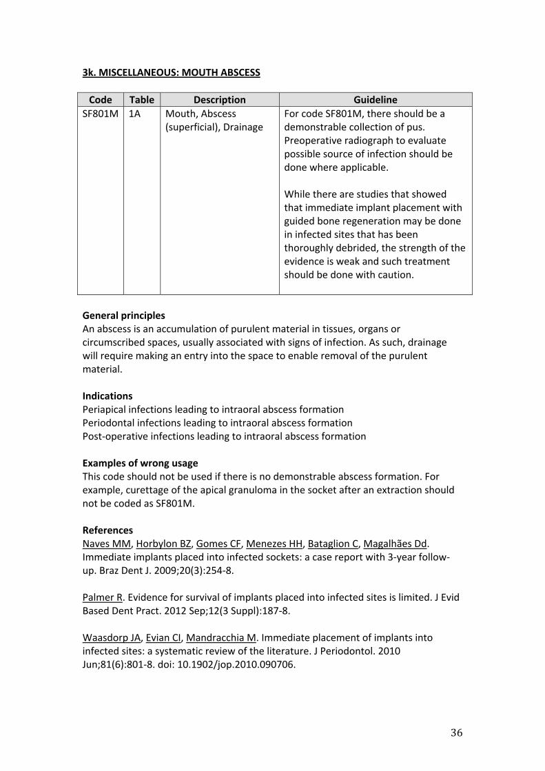
3k. MISCELLANEOUS: MOUTH ABSCESS Code Table Description Guideline
SF801M 1A Mouth, Abscess (superficial), Drainage
For code SF801M, there should be a demonstrable collection of pus. Preoperative radiograph to evaluate possible source of infection should be done where applicable. While there are studies that showed that immediate implant placement with guided bone regeneration may be done in infected sites that has been thoroughly debrided, the strength of the evidence is weak and such treatment should be done with caution.
General principles An abscess is an accumulation of purulent material in tissues, organs or circumscribed spaces, usually associated with signs of infection. As such, drainage will require making an entry into the space to enable removal of the purulent material. Indications Periapical infections leading to intraoral abscess formation Periodontal infections leading to intraoral abscess formation Post-operative infections leading to intraoral abscess formation Examples of wrong usage This code should not be used if there is no demonstrable abscess formation. For example, curettage of the apical granuloma in the socket after an extraction should not be coded as SF801M. References Naves MM, Horbylon BZ, Gomes CF, Menezes HH, Bataglion C, Magalhães Dd. Immediate implants placed into infected sockets: a case report with 3-year follow-up. Braz Dent J. 2009;20(3):254-8. Palmer R. Evidence for survival of implants placed into infected sites is limited. J Evid Based Dent Pract. 2012 Sep;12(3 Suppl):187-8. Waasdorp JA, Evian CI, Mandracchia M. Immediate placement of implants into infected sites: a systematic review of the literature. J Periodontol. 2010 Jun;81(6):801-8. doi: 10.1902/jop.2010.090706.

37
3l. MISCELLANEOUS: FRENAL ATTACHMENT Code Table Description Guideline
SF701M 1B Mouth, Frenal Attachment, Release
Release of frenal attachments should include excision of the attachments and reattachment at or above the mucogingival junction. Surgery may be done with scalpel or laser.
General Principles Frenal attachments are muco-muscular attachments of the lips to the alveolus. This is more commonly found in the maxillary midline as well as laterally in the region of the premolars. It may also be found in the mandibular labial sulcus. Lingual frenum is coded separately in the TOSP under “Tongue-tie” and is not covered here. Indications and Contraindications Frenal attachments may create spaces between teeth, typically causing a median diastema. Such frenal attachments are indicated for release to facilitate closure of the diastema. Frenal atachments may also affect prosthodontics treatment and release is indicated to facilitate such treatment. Examples of wrong usage Regardless of whether laser or scalpel is used to release the frenal attachment, this code SF701M (1B) is to be used. Codes SF823M (3B), SF824M (2B) and SM702M (2B) are not to be used even if laser is used. References Pié-Sánchez J, España-Tost AJ, Arnabat-Domínguez J, Gay-Escoda C. Comparative study of upper lip frenectomy with the CO2 laser versus the Er, Cr:YSGG laser. Med Oral Patol Oral Cir Bucal. 2012 Mar 1;17(2):e228-32. Mittal M, Murray AM, Sandler PJ. Maxillary labial fraenectomy: indications and technique. Dent Update. 2011 Apr;38(3):159-62.

38
3m. MISCELLANEOUS: FOREIGN BODY Code Table Description Guideline
SF811M 1B Mouth, Foreign Body (Superficial), Removal
Pre-operative documentation of the presentation of the foreign body with either radiographs and/or photographs will facilitate access to and complete removal. As with all codes in the TOSP, this codes requires that an incision be made to gain access to the foreign body for removal and subsequently, closure of the wound. Post operative radiograph to assess completeness of removal of the foreign body where applicable is good clinical practice.
General Principles A foreign body may be lodged in the mouth during mastication, home oral healthcare procedures, accidental injuries, intentionally by the patient (eg “charm pins”), or iatrogenically. Indications and Contraindications A foreign body is indicated for removal if it causes signs and symptoms such as pain and swelling. Removal is also indicated in the absence of signs and symptoms if the patient electively opts for removal. Examples of wrong usage This code should not be use if the foreign body is easily accessed and no incision is required. A foreign body that can be removed without an incision is similar to a simple forceps extraction of an erupted tooth. This code should not be used in combination with codes for repair of mucous membrane lacerations (SA801M, SA802M and SA803M) when the foreign body is lodged within the same laceration. References: Nil

39
3n. MISCELLANEOUS: TONGUE TIE Code Table Description Guideline
SF804T 1A Tongue, Tongue Tie, Release
For cases whereby the tongue tie is caused by a thin mucosal strand, release of tongue tie (SF804T), or frenuloplasty, may be done by means of a pair of blunt-end scissors or scalpel or laser.
SF805T 1B Tongue, Tongue Tie, Revision
For cases whereby the tongue tie is caused by a wide band of mucosa or complete fusion of the tongue to the floor of mouth, revision of tongue tie (SF805T), or frenectomy, by excision of the lingual frenulum and closure with a Z-plasty is needed to ensure adequate mobilization of the tongue. Surgery may be done using scalpel or laser.
General principles Tongue-tie is due to an abnormally short lingual frenulum which may restrict mobility of the tongue. This is a congenital anomaly and it may affect the feeding of infants. Adults also present with this condition. The degree of attachment varies from a thin mucosa strand to complete fusion to the floor of mouth. Surgical correction of tongue tie may be undertaken for the infant as well as adult. Adult patients seeking this procedure for improvement of speech should be counseled preoperatively on the benefits of speech therapy vis-à-vis surgery. Indications Surgical correction of tongue tie may be indicated in neonates when it interferes with feeding. Surgery is also indicated for older patients when it interferes with eating, speech or prosthodontics treatment. Examples of wrong use of code Use of SF805M instead of SF804M for mild cases of tongue tie caused by a thin mucosal strand. Regardless of whether laser or scalpel is used to release the tongue tie, this code SF804T and SF805T are to be used. Codes SF823M (3B), SF824M (2B) and SM702M (2B) are not to be used even if laser is used. References: Division of ankyloglossia (tongue tie) for breastfeeding - guidance NICE International procedure guidance 149. 14 December 2005 Glynn RW, Colreavy M, Rowley H, Gendy S. Division of tongue tie: review of practice through a tertiary paediatric otorhinolaryngology service. Int J Pediatr Otorhinolaryngol. 2012 Oct;76(10):1434-6. doi: 10.1016/j.ijporl.2012.06.017. Epub 2012 Jul 17. Suter VG, Bornstein MM. Ankyloglossia: facts and myths in diagnosis and treatment. J Periodontol. 2009 Aug;80(8):1204-19. doi: 10.1902/jop.2009.090086.

40
3o. MISCELLANEOUS: NEURALGIA Code Table Description Guideline
SK733N 1A Nerve, peripheral nerve block, anesthetic
Application of anesthetics (SK733N, SK734N, SK735N) as a treatment for neuralgia may be used for diagnostic or short term therapeutic purposes. Neurolytic, cryotherapy and radiofrequency thermocoagulation are use for long term/ permanent therapeutic purpose in the management of neuralgia. No single modality of treatment is effective for all patients. Detail history taking and documentation are crucial. Adjunctive therapy such as pharmacotherapy, psychotherapy and physiotherapy should be considered
SK734N 1A Nerve, peripheral nerve block, anesthetic up to 2
SK735N 1C Nerve, peripheral nerve block, anesthetic more than 2
SK736N 1B Nerve, peripheral nerve block, neurolytic
SK739N 1C Nerve, peripheral nerve block, neurolytic, cryo, radiofrequency up to 2
SK740N 2B Nerve, peripheral nerve block, neurolytic, cryo, radiofrequency, more than 2
General Principles The wording of these codes is not complete as the diagnosis is missing from the code. The intended diagnosis of these codes is “neuralgia”. As such, all the above codes should be read in the context of treatment of neuralgia. Indications and contraindications The indication for the above codes is neuralgia. Examples of wrong use of codes. These codes cannot be used when an inferior alveolar nerve block is done for the purpose of dental treatment e.g. extraction, endodontics, diagnostic blocks References: Zakrzewska JM, Akram H. Neurosurgical interventions for the treatment of classical trigeminal neuralgia. Cochrane Database Syst Rev. 2011 Sep 7;(9):CD007312. doi: 10.1002/14651858.CD007312.pub2. Poon CY. Cryotherapy in the management of trigeminal neuralgia: a review of the literature and report of three cases. Singapore Dent J. 2000 Dec;23(1 Suppl):49-55. Peters G, Nurmikko TJ. Peripheral and gasserian ganglion-level procedures for the treatment of trigeminal neuralgia. Clin J Pain. 2002 Jan-Feb;18(1):28-34.

41
Workgroup members: Chairman: Dr Chan Siew Luen Immediate Past President, College of Dental Surgeons, Singapore Members: Dr Lui Jeen Nee Chair, Chapter of Endodontists, College of Dental Surgeons, Singapore Dr Tan Wah Chin Chair, Chapter of Periodontists, College of Dental Surgeons, Singapore Dr Winston Tan Chair, Chapter of Oral & Maxillofacial Surgeons, College of Dental Surgeons, Singapore Dr Lim Eng Yong President, College of General Dental Practitioners, Singapore Dr Andrew Tay Dental Advisor, Medisave Unit, Ministry of Health Dr Asher Lim Dental Advisor, Medisave Unit, Ministry of Health Dr Dominic Leung Appointed Member