Absorption of ipratropium and L-carnitine into theorca.cf.ac.uk/79639/3/Absorption of ipratropium...
20
This is an Open Access document downloaded from ORCA, Cardiff University's institutional repository: http://orca.cf.ac.uk/79639/ This is the author’s version of a work that was submitted to / accepted for publication. Citation for final published version: Aljayyoussi, Gaith, Price, Daniel, Kreitmeyr, Katharine, Keogh, John, Smith, Mathew W., Gumbleton, Mark and Morris, Christopher J. 2015. Absorption of ipratropium and L-carnitine into the pulmonary circulation of the ex-vivo rat lung is driven by passive processes rather than active uptake by OCT/OCTN transporters. International Journal of Pharmaceutics 496 (2) , pp. 834-841. 10.1016/j.ijpharm.2015.10.036 file Publishers page: http://dx.doi.org/10.1016/j.ijpharm.2015.10.036 <http://dx.doi.org/10.1016/j.ijpharm.2015.10.036> Please note: Changes made as a result of publishing processes such as copy-editing, formatting and page numbers may not be reflected in this version. For the definitive version of this publication, please refer to the published source. You are advised to consult the publisher’s version if you wish to cite this paper. This version is being made available in accordance with publisher policies. See http://orca.cf.ac.uk/policies.html for usage policies. Copyright and moral rights for publications made available in ORCA are retained by the copyright holders.
Transcript of Absorption of ipratropium and L-carnitine into theorca.cf.ac.uk/79639/3/Absorption of ipratropium...

This is an Open Access document downloaded from ORCA, Cardiff University's institutional
repository: http://orca.cf.ac.uk/79639/
This is the author’s version of a work that was submitted to / accepted for publication.
Citation for final published version:
Aljayyoussi, Gaith, Price, Daniel, Kreitmeyr, Katharine, Keogh, John, Smith, Mathew W.,
Gumbleton, Mark and Morris, Christopher J. 2015. Absorption of ipratropium and L-carnitine into
the pulmonary circulation of the ex-vivo rat lung is driven by passive processes rather than active
uptake by OCT/OCTN transporters. International Journal of Pharmaceutics 496 (2) , pp. 834-841.
10.1016/j.ijpharm.2015.10.036 file
Publishers page: http://dx.doi.org/10.1016/j.ijpharm.2015.10.036
<http://dx.doi.org/10.1016/j.ijpharm.2015.10.036>
Please note:
Changes made as a result of publishing processes such as copy-editing, formatting and page
numbers may not be reflected in this version. For the definitive version of this publication, please
refer to the published source. You are advised to consult the publisher’s version if you wish to cite
this paper.
This version is being made available in accordance with publisher policies. See
http://orca.cf.ac.uk/policies.html for usage policies. Copyright and moral rights for publications
made available in ORCA are retained by the copyright holders.
1
Absorption of ipratropium and L-carnitine into the
pulmonary circulation of the ex-vivo rat lung is driven by
passive processes rather than active uptake by
OCT/OCTN transporters
Ghaith Aljayyoussi1, Daniel F. Price
1, Katharina Kreitmeyr
1, John P Keogh
2, Mathew W.
Smith1, Mark Gumbleton
1 Christopher J. Morris
3,4
1 School of Pharmacy & Pharmaceutical Sciences, Cardiff University, CF10 3NB
2 JPK Consulting, Hitchin, Hertfordshire, SG5 1XG
3 School of Pharmacy, University of East Anglia, Norwich Research Park, NR4 7TJ
4 Corresponding author: Dr Chris Morris, School of Pharmacy, University of East Anglia, Norwich Research Park, Norwich. NR4 7TJ Tel: +44 (0)1603 593136 Fax: +44 (0) 1603-592003. E-mail: [email protected]
KEYWORDS Organic cation transporters, OCT1, OCT2, OCT3, OCTN; SLC22; isolated
perfused rat lung (IPRL); transporters; lung; pulmonary absorption
2
ABSTRACT
The organic cation transporters OCT and OCTN have been reported to play a significant role
in the cellular uptake of substrates within in vitro lung cells. However, no studies to date have
investigated the effect of these transporters upon transepithelial absorption of substrates into
the pulmonary circulation. We investigated the contribution of OCT and OCTN transporters
to total pulmonary absorption of L-carnitine and the anti-muscarinic drug, ipratropium, across
an intact isolated perfused rat lung (IPRL). The results obtained from the IPRL were
contrasted with active transport in vitro using three human pulmonary cell lines and primary
rat alveolar epithelial cells. Ex-vivo studies showed that OCT/OCTN transporters do not play
a role in the overall pulmonary absorption of L-carnitine or ipratropium, as evidenced by the
effect of chemical inhibition of these transporters upon pulmonary absorption. In contrast, in-
vitro studies showed that OCT/OCTN transporters play a significant role in cellular
accumulation of substrates with preferential uptake of ipratropium by OCTs, and of L-
carnitine uptake by OCTNs. The results show that in-vitro uptake studies cannot be predictive
of airway to blood absorption in-vivo. Nevertheless, localised submucosal pulmonary
concentrations of inhaled drugs and their pulmonary pharmacodynamic profiles may be
influenced by OCT/OCTN transport activity.
3
1. Introduction
A number of drug candidates for inhaled therapy are cationic and are therefore potential
substrates for the SLC22 superfamily of ATP-independent polyspecific cation transporters at
the plasma membrane [1]. These transporters include the bidirectional organic cation transporters OCT 1 (SLC22A1), OCT2 (SLC22A2), OCT3 (SLC22A3) and the sodium-dependent carnitine/cation transporter proteins OCTN1 (SLC22A4) and OCTN2 (SLC22A5). The majority of inhaled drugs must cross the rate-limiting pulmonary epithelial barrier to access their pharmacological targets [2], e.g. smooth muscle cells. As such the interaction of
cationic drugs with transporter pathways expressed in pulmonary epithelium may be
important for drug access to underlying pharmacological targets. Indeed this has been the
premise of research by a number of groups (reviewed in [3-5]) exploring the expression of
OCTs/OCTNs within lung epithelium.
The localisation of OCT/OCTN transporters within intact lung has been reported using
protein immunohistochemistry in both humans [6-9] and rodents [6, 8, 10, 11]. Evidence for
expression of OCT and OCTN family members in intact human and rat lung tissue also exists
at the mRNA level [6, 8, 11-13]. Nakanishi et al. [14] showed OCT/OCTN driven
accumulation of ipratropium in lung epithelial tissue, namely tracheal epithelium, following
drug deposition in the tracheal lumen of the mouse. A number of in vitro cell culture studies
have reported substrate uptake via OCTs and OCTNs in lung epithelial cells including the
active uptake of the model OCT/OCTN cationic substrate (4-(4-dimethylaminostyryl)-N-
methylpyridinium; ASP+) in normal human bronchial cells [7] and in a range of human
bronchial epithelial cell lines [15-17]. The inhaled anti-muscarinic drug ipratropium has been
implicated as a substrate of both OCTN and OCT transporters depending upon the in vitro
model adopted [18, 19]. One report exists of the facilitative role of organic cation
transporters upon the absorptive and secretory transport of ASP+ across a cell monolayer [15]
with the results showing modest effects upon the overall absorptive transport, despite a
significant extent of cellular uptake.
Here we hypothesized that in a fully intact lung the OCT/OCTN transporters play little or no
role in the transepithelial transport of substrates into the pulmonary vasculature. To test the
hypothesis we examined in an intact perfused rat lung (IPRL) the role of OCT/OCTN
transport in pulmonary transepithelial permeability of the zwitterionic substrate, L-carnitine,
and the inhaled therapeutic cationic drug, ipratropium; archetype substrates for the pathways
under study. We found the overall solute transport into the IPRL vasculature was
predominantly driven by non-competitive passive processes that eclipse the net effect of any
OCT/OCTN-mediated transport.
2. Materials and Methods
2.1 Materials
[3H]-ipratropium bromide (70 Ci/mmol) was provided by GlaxoSmithKline (Ware, UK) and
[3H]-L-carnitine hydrochloride (70 Ci/mmol) was from American Radiochemicals Inc. (St.
Louis, MO). Unlabelled ipratropium, L-carnitine, tetraethylammonium bromide (TEA) and 1-
methyl-4-phenylpyridinium iodide (MPP+) were purchased from Sigma-Aldrich (Poole, UK).
Cell culture media and supplements were from Invitrogen (Paisley, UK) with cell culture
plastics from Corning Costar (Hemel Hempstead, UK). All other reagents and solvents were
from Fisher Scientific (Loughborough, UK) or Sigma-Aldrich. PCR primers were designed in
house and supplied by Invitrogen (Paisley, UK).
4
2.2 Methods
2.2.1 IPRL
All animal experiments adhered to the UK Animal (Scientific Procedures) Act 1986. Rats
used for all the experiments in this report weighed 250-350g. Animals were normally housed
with a 12 hour day/night cycle and fed ad libitum until the time of surgery.
To examine the transport of ipratropium and L-carnitine across an intact pulmonary barrier an
IPRL preparation was employed as previously described [20, 21]. This model includes an
intra-tracheal airway dosing technique that utilises a pressurized metered dose inhaler
(pMDI) reproducibly delivering a high extent (>95%) of deposited solute liquid aerosol dose
into the lung periphery [22]. Using the pMDI methodology the IPRL was dosed with either
vehicle control (100 µL saline) or a competitive inhibitor (in 100 µL saline), i.e. either
unlabeled solute (125 nmol unlabeled L-carnitine or ipratropium) or the selective OCT
inhibitor (MPP+). Twenty minutes later the lungs were similarly dosed with the radiolabeled
substrate (3 µCi of [3H]-L-carnitine or [
3H]-ipratropium in 100µL saline). At discrete
timepoints after lung dosing. 200µL samples were collected from the circulating perfusate
and transferred to scintillation vials for radiochemical analysis.
2.2.2 Mathematical Modelling
Pulmonary pharmacokinetic absorption parameters were calculated by fitting Equation 1 to
the individual airway to perfusate absorption data using nonlinear regression analysis
(Micromath Scientist 3.0, Missouri, USA).
% � = ∙ ∙ − −�.� Equation 1
Where, f represents the available fraction to be transported, k is the absorption rate constant
(min-1
) and t is time in minutes. Modelling of the absorption data was not improved by the
use of more complex models and Equation 1 was deemed the minimum satisfactory model to
provide precise parameter estimates for k and f.
2.2.3 In-vitro uptake studies
The in vitro active accumulation of substrates of either OCT and/or OCTN transporters, was
studied in a range of continuous lung epithelial cell lines and primary cultures of rat lung
epithelium. The human pulmonary adenocarcinoma cell line, A549 [23], and the human
bronchial epithelial cell line, BEAS-2B, were obtained from ATCC (American Type Culture
Collection; Manassas, VA). 16HBE14o- cells, generated by transformation of normal
bronchial epithelial cells, were from Dr D. C. Gruenert (University of California San
Francisco, San Francisco). Culture of these cells was performed as previously described [24],
and isolation and primary culture of rat alveolar cells to a type I pneumocyte-like phenotype
was undertaken as detailed in previous work [25].
Solute uptake studies described hereafter were conducted at incubation temperatures of both
37 ºC and 4 ºC. Radiolabelled solute was added to each well (24-well format) containing
confluent cell monolayers. The dosings were 1 µCi (15 pmol) [3H]-ipratropium or 1 µCi (15
pmol) [3H]-L-carnitine giving a final radiolabel probe concentration in each well of 50 nM
5
(300 L volume per well). Time- and temperature-dependent solute uptake was quantified at
discrete timepoints over a 60 min incubation.
Radiolabelled solute uptake studies were also undertaken in the presence of unlabelled
OCT/OCTN competitive inhibitors applied to the cells for a 30 min pre-incubation period
prior to addition of the radiolabel probes. The unlabelled inhibitors were used to achieve
concentrations of 500µM ipratropium, 100µM L-carnitine, 500µM MPP+, 5mM TEA; a no-
treatment control comprised radiolabeled solute alone in serum-free DMEM. Following the
pre-incubation period, either [3H]-ipratropium or [
3H]-L-carnitine was added to each well and
the uptake of radiolabel allowed to proceed over a 60 min incubation period. The solute
uptake experiments were terminated by two washes of the cell monolayers with ice-cold PBS
followed by the addition of ice-cold trypsin-EDTA for 5 min, after which the cells were
harvested and suspended in ice-cold DMEM and centrifuged (4 °C, 200 g) for 10 min after
which the supernatant was discarded and the cell pellet collected. This cell washing
procedure was performed three times in total. The resulting cell pellets were transferred to
scintillation vials and mixed with 3mL of BioSafe 3 scintillation fluid and the cell-associated
radioactivity quantified by liquid scintillation counting. The sodium-dependent nature of
solute uptake for both [3H]-ipratropium and [
3H]-L-carnitine was similarly conducted but
using an incubation buffer where Na+ was isotonically replaced with N-methyl-glucamine as
previously described [7].
2.2.4 Real-time Quantitative Polymerase Chain Reaction (qPCR)
Human OCT (SLC22A1, SLC22A3, SLC22A3), human OCTN (SLC22A4, SLC22A5), rat OCT (Slc22a1, Slc22a2, Slc22a3) and rat OCTN (Slc22a4, Slc22a5) mRNA sequences were
aligned using BLAST2 and the NCBI nucleotide gene search used to identify single exon
regions; Oligocalc was used to validate all used transcription variants. Table 1 shows the
primer sequences used in qPCR experiments. Total RNA was isolated (RNeasy, Qiagen,
UK) from pulmonary epithelial cells (A549, BEAS-2B, 16HBE14o- or primary rat alveolar
epithelial cells) grown under the same conditions as used in the solute uptake experiments.
Total RNA was also isolated from whole rat lung and liver tissue, with the liver serving as a
positive control that is known to express OCT/OCTN, albeit at different relative amounts.
cDNA synthesis was performed via reverse transcription using M-MLV reverse transcriptase
(Invitrogen, UK) using generic random oligonucleotide (pdN6) primers and quantified
spectrophotometrically at 260 and 280 nm by GeneQuant. The cDNA was amplified as
described elsewhere [26]. qPCR was performed by SYBR® Green chemistry using a DNA
Engine Opticon 2 Real-Time Cycler (BioRad, UK), previously described elsewhere [27]. The
increase in fluorescence emission associated with DNA amplification was calculated
throughout the run of 40 cycles and the cycle number at which fluorescence emission first
reaches exponential phase was recorded.
3. RESULTS
3.1 Pulmonary transepithelial transport of [3H]-ipratropium and [
3H]-L-carnitine
We explored if OCT/OCTN-mediated transport was a significant feature in the transepithelial
absorption (airway to pulmonary vascular space) of [3H]-ipratropium and [
3H]-L-carnitine
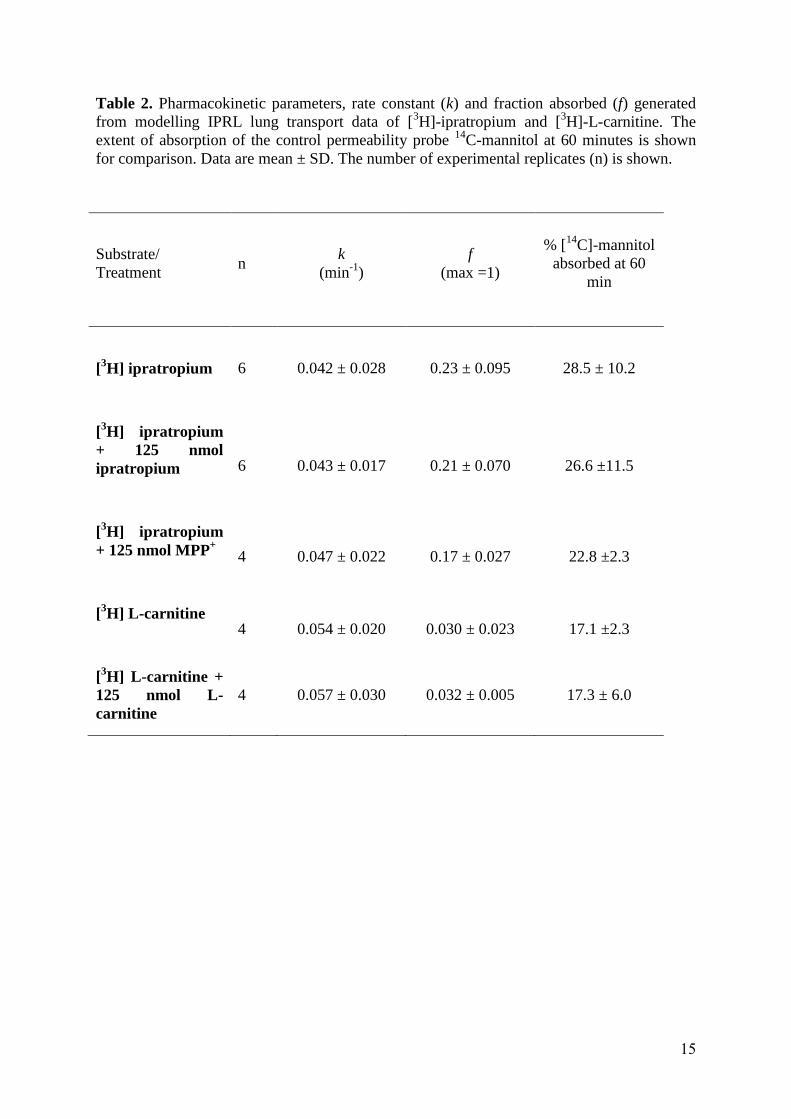
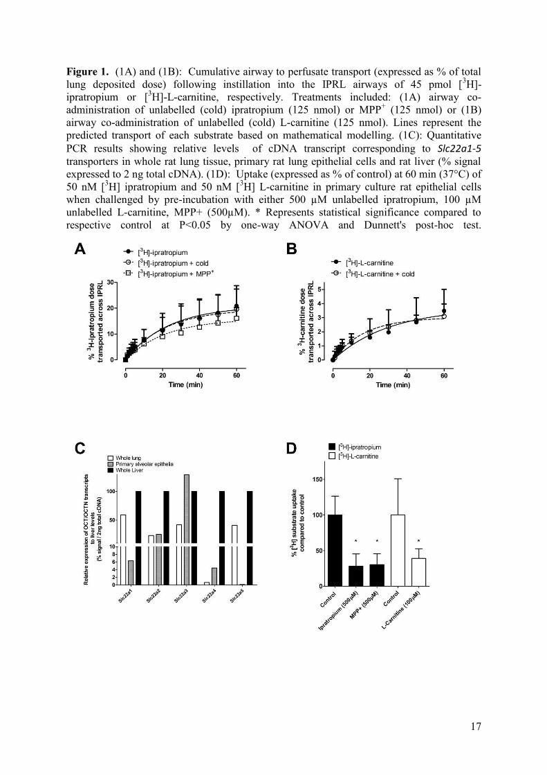
within an intact rat lung. Over the 60 min IPRL experiment approximately 20% of the lung
deposited dose of [3H]-ipratropium was absorbed to the recirculating vascular perfusate
(Figure 1A, Table 2) with approximately 3% of [3H]-L-carnitine absorbed (Figure 1B, Table
2). Pre-administration into the airways (20 min prior to that of radiolabelled substrate) of 125
6
nmol of the respective unlabelled solute (either unlabelled ipratropium or L-carnitine) failed
to significantly alter the extent or rate of absorption of the corresponding radiolabelled
species. This despite the unlabeled solute dosed at a 3000-fold excess to that of the
radiolabelled substrate. Similarly, pre-administration of 125 nmol of MPP+ failed to alter
[3H]-ipratroprium absorption profile and kinetics. None of the above treatments resulted in
perturbation of pulmonary barrier integrity or permeability as evidenced by the pulmonary
absorption of the co-administered hydrophilic paracellular probe [14
C] mannitol remaining
unaffected (p>0.05) by any treatment (Table 2).
3.2 OCT/OCTN-mediated accumulation of [3H]-ipratropium and [
3H]-L-carnitine by
primary rat pulmonary epithelial cells
Real-time qPCR confirmed the presence of Slc22a1-Slc22a5 transcripts in both rat whole
lung and in primary cultures of rat alveolar epithelial cells (Figure 1C), with the primary
cultures relatively enriched, with respect to lung tissue, in Slc22a3 and Slc22a4. As
expected, we found the uptake of [3H]-ipratropium and [
3H]-L-carnitine by the primary
alveolar epithelial cells to display temperature dependency (data not shown). Furthermore,
the accumulation of both solutes was significantly decreased (P < 0.05) by co-incubation with
their respective unlabelled solute, or in the case of [3H]-ipratropium by the OCT inhibitor
MPP+
(Figure 1D); the effect of MPP+ upon L-carnitine uptake was not examined.
3.3 OCT/OCTN-mediated accumulation of [3H]-ipratropium and [
3H]-L-carnitine by
human pulmonary cell lines
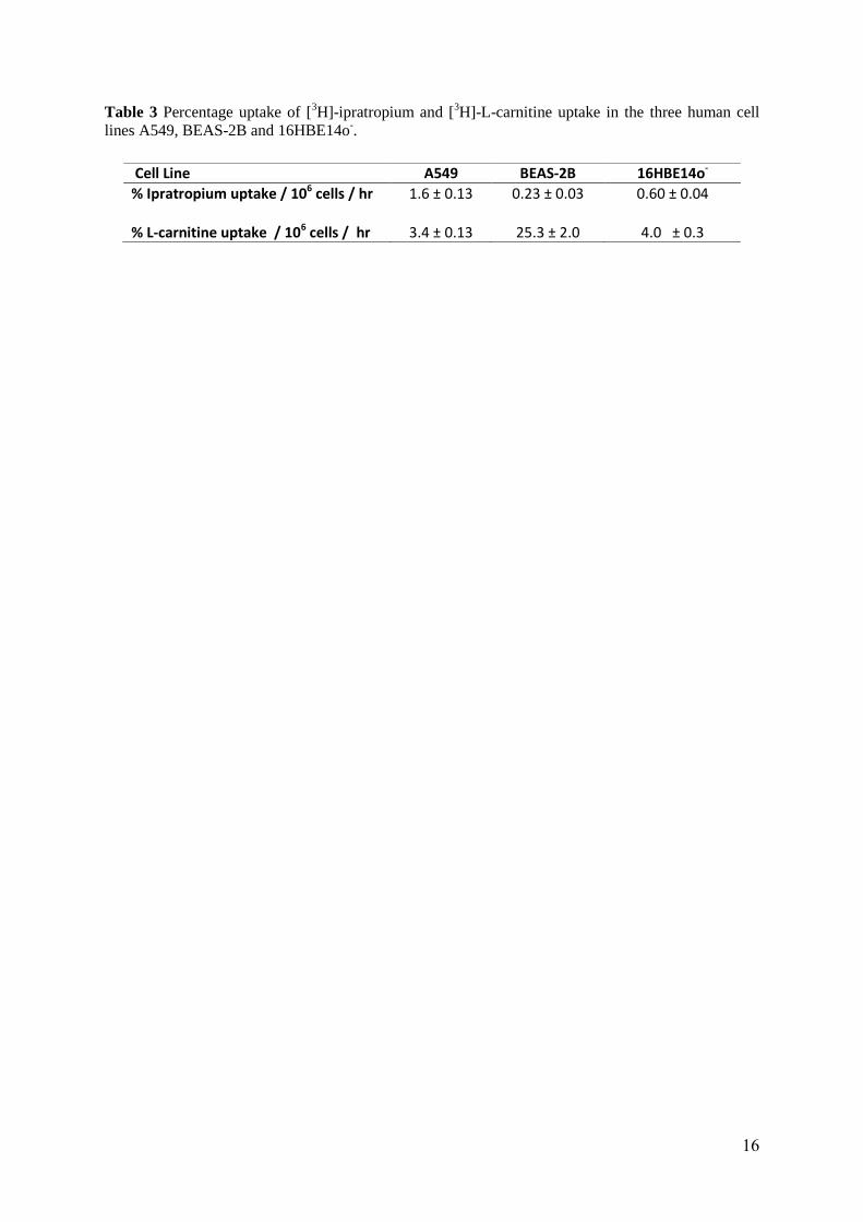
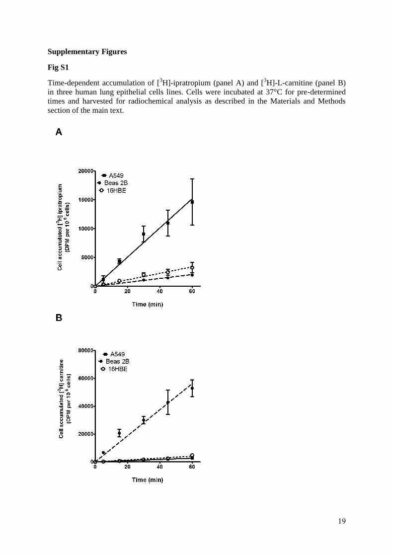
We investigated the kinetics and transporter selectivity in the accumulation of [3H]-
ipratropium and [3H]-L-carnitine using human lung epithelial cell lines (A549, BEAS-2B,
16HBE14o-) constitutively expressing OCT and OCTN transporter proteins. The
accumulation of both [3H]-ipratropium and [
3H]-L-carnitine (applied at 50 nM) was linear
over a 60 min period (Supplementary Figure 1) with parallel experiments undertaken at 4 °C
showing negligible cell-associated radioactivity (< 250 DPM/106 cells at all timepoints; data
not shown). The cell lines demonstrated differing capacities to actively accumulate [3H]-
ipratropium and [3H]-L-carnitine (Table 3) with all three cell types showing higher (P <
0.05) levels of accumulation (per 106
cells) for L-carnitine compared to ipratropium. Of
particular note was the considerable accumulation (P < 0.05) of [3H]-L-carnitine by BEAS-
2B cells, to the extent that 25% of the total applied L-carnitine radiolabel was cell-associated
at 60 min
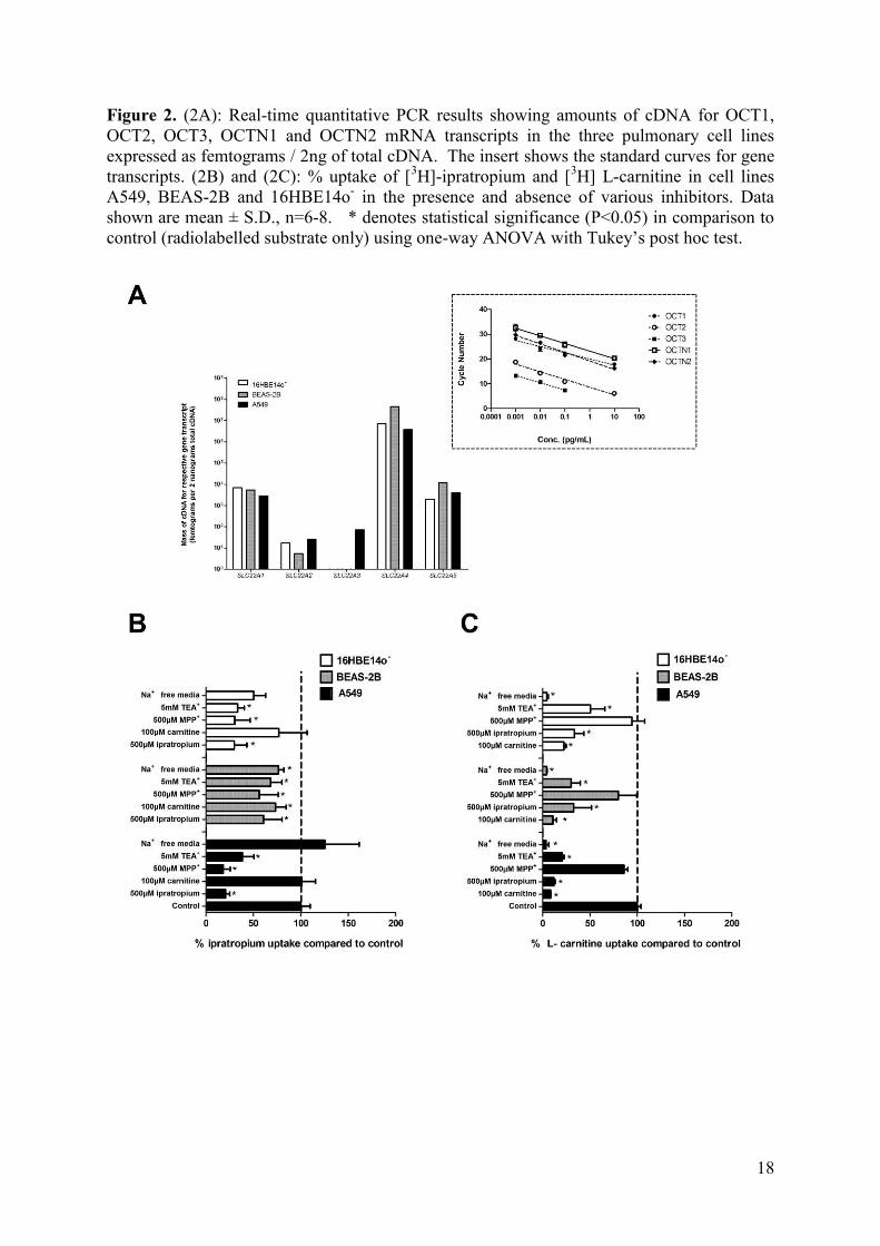
Using qPCR we evaluated solute accumulation by the various lung epithelial cell lines in the
context of the absolute levels of SLC22A1-SLC22A5 mRNA transcripts in the cells. Figure
2A shows the results of the qPCR where the mass of cDNA (femtograms) for each respective
mRNA transcript is expressed relative to a 2 ng mass of total cDNA. Consistent with our
observation of greater cellular accumulation of [3H]-L-carnitine in all three cell lines, the
combined cDNA for SLC22A4 and SLC22A5 was more abundant in each of the three cell
lines than the respective combined cDNA for SLC22A1, SLC22A2 and SLC22A3. The
BEAS-2B cell line showed the highest total SLC22A4 and SLC22A5 levels of all which was
mirrored by a greater uptake displayed by BEAS-2B for L-carnitine.
To further explore solute uptake and the interplay between constitutively expressed OCT and
OCTN transporter proteins we examined the active accumulation of [3H]-ipratropium and
[3H]-L-carnitine under challenge by various competitive inhibitors or co-factors (Figures 2B
7
and 2C respectively). Not surprisingly, in all lung epithelial cell lines the accumulation of
both radiolabelled ipratropium and L-carnitine was significantly (P < 0.05) decreased
following co-incubation with their respective unlabelled solute. Challenge with unlabelled
ipratropium resulted in a significant (P < 0.05) reduction of [3H]-L-carnitine accumulation in
all three cell lines (Figure 2C). However, the effect of unlabelled L-carnitine upon [3H]-
ipratropium accumulation (Figure 2B) resulted in a far more restricted response, with a
reduction (P < 0.05) in [3H]-ipratropium accumulation observed only in the BEAS-2B cells,
and then only at a marginal level. Co-incubation of the highly selective OCT competitive
inhibitor, MPP+, resulted in a significant (P < 0.05) inhibition of [
3H]-ipratropium
accumulation in all three cell lines with inhibitory effects most pronounced in A549 cells and
with the least affected being the BEAS-2B cells (Figure 2B). Notably MPP+ had no
significant (P > 0.05) effect upon the accumulation of the OCTN substrate [3H]-L-carnitine
(Figure 2C). Challenge with TEA, a mixed OCT/OCTN inhibitor, resulted in decreases in
the accumulation of both [3H]-ipratropium (Figure 2B) and [
3H]-L-carnitine (Figure 2C) in all
three cell lines. OCTN1 and OCTN2 display Na+-dependent transport, and in all three lung
epithelial cell lines the active accumulation of the OCTN substrate [3H]-L-carnitine was
reduced by >90% (P < 0.05) in the absence of Na+ (Figure 2C). In contrast, the accumulation
of [3H]-ipratropium was significantly less dependent on Na
+ (Figure 2B). Specifically, in the
absence of Na+ no alteration in ipratropium accumulation was observed in A549 cells. In
BEAS-2B and 16HBE14o- cell lines, Na
+ depletion had a greater effect on ipratropium
uptake (25-40% inhibition; P < 0.05), that L-carnitine accumulation. An MTT assay
substantiated that none of the competitive inhibitors had any detrimental effect upon in vitro
cell viability over the time course of experimentation (data not shown).
4. Discussion
The objective of this work was to examine the hypothesis that OCT and OCTN transporters
play little or no role in the absorption of respective archetype substrates into the pulmonary
circulation of a fully intact lung model. This set of experiments compliments a number of in-
vitro lung epithelial cell-based studies which infer that these transporters may fulfill a role in
substrate access to sub-mucosal smooth muscle targets.
Here we used an IPRL system to study transepithelial absorption from the airways, a fully
intact whole lung model retaining the relevant in-vivo biological architecture and the various
sequential and parallel, e.g. paracellular, active/facilitative and passive transcellular routes
that may contribute to various extents a substrate’s overall transepithelial penetration. We
have previously demonstrated that the IPRL can serve as a reliable and robust model that is
capable of discriminating active transport pathways for substrate movement across lung
epithelia. For example, we have shown in the IPRL that P-glycoprotein (P-gp) drug efflux
reduces the pulmonary absorption of certain P-gp substrates [28], an efflux mechanism which
can be inhibited in the IPRL by co-administration of the chemical inhibitors such as elacridar
(GF-120918). The IPRL displays similar airway perfusion characteristics to the fully intact
in-vivo rat lung [29-31]. Indeed, in the IPRL the entire lung parenchyma from the secondary
bronchi (second airway bifurcation) to the deep alveolar regions is perfused by the pulmonary
circulation [30], as it is in vivo.
The IPRL experiments (Figs 1A,B) demonstrate a non-saturable transport pathway for
ipratropium and carnitine, as well as the absence of any demonstrable inhibition of
ipratropium transport by MPP+. This demonstrates that the OCT and OCTN transporters
played no significant quantitative role in the overall transport of the substrates ipratropium
and L-carnitine across the pulmonary barrier into the pulmonary circulation. This particular
8
question has not previously been addressed. Although published studies have investigated the
in-vitro cell uptake of OCT/OCTN substrates by pulmonary epithelial cells per se.
In IRPL experiments radiolabelled [3H]-ipratropium and [
3H]-L-carnitine were administered
to the airways to achieve intraluminal concentrations at least 10-fold lower than their
respective Km values (>1 µM) for OCT/OCTN [32]. These intra-luminal concentrations were
determined to be close to those used in our parallel in-vitro uptake studies. Our calculations
were based on a rat lung epithelial lining fluid (ELF) volume of 250 - 350 µL [21]. We
found pre-administration of a 3000-fold excess of the respective unlabelled substrate did not
alter the pulmonary absorption of either [3H]-ipratropium or [
3H]-L-carnitine. Additionally,
intra-luminal dosing of high concentration of the OCT inhibitor MPP+ concentrations
(mimicking those used in in-vitro studies) did not influence [3H]-ipratropium absorption.
These findings contrast with those of the in-vitro uptake studies which unequivocally showed
saturable uptake processes for these substrates into lung epithelial cells. Nakanishi and co-workers reported that the tracheal accumulation of ipratropium in mice over a 90 second period was inhibited by both 0.2 and 1 mM carnitine and 0.1mM MPP+ [14]. Backstrom et al [33] exploited a lung slice model to investigate the tissue binding of a number of inhaled drug molecules. For ipratropium they reported a bound drug partition coefficient of the cell i.e. the ratio of intracellular to extracellular unbound drug concentration of 7.1 and for its derivative, tiotropium, a value of 1.1. The authors concluded that OCT/OCTN transporter activity was responsible for this active accumulation of ipratropium but not for tiotropium, in spite of evidence from an analogous kidney slice model that tiotropium is an OCT substrate [19]. The tissue slice models used by Backstrom and others allow for a rapid and reliable investigation of bound:unbound drug levels in tissue homogenates. However, the model does not offer opportunity to investigate specific transport processes active at the luminal epithelial barrier such as airway to blood absorption. The epithelial and multiple sub-mucosal cell types distributed across the tissue slice are simultaneously exposed to equal concentrations of drug. This does not mimic the lung microenvironment in vivo following drug inhalation and therefore does not permit the differentiation of distinct tissue binding sites in the tissue slice.
Of note, the extent of pulmonary absorption of ipratropium (bioavailability of 20% of lung
dose absorbed) was significantly greater than the zwitterion, L-carnitine (bioavailability of
3% of lung deposited dose absorbed) indicating a discrimination in the way the lung handles
these charged molecules. Differences in tissue/protein binding at the epithelial surface within
the lung, as dictated by physicochemical parameters such as logP, could serve to limit the
fraction available for absorption into the pulmonary circulation. This whole lung absorption
data nonetheless matches the low bioavailability of inhaled ipratropium in man after slow
inhalation [34] and implies that the majority of the deposited dose remains either in the
airway lumen or is localised to the lung submucosal tissue wherein lies the target smooth
muscle cells.
For the purposes of rigorous investigation we also undertook parallel in-vitro pulmonary
epithelial cell uptake studies. Consistent with others we found that OCT and OCTN
transporter proteins play a significant role in the in-vitro epithelial cell uptake of ipratropium
and L-carnitine. We found that human lung epithelial cell lines displayed differing and
opposing capacities for active uptake of ipratropium and L-carnitine. For example, BEAS-2B
cells showing the lowest capacity for ipratropium accumulation but the highest for L-
carnitine, while A549 cells showed the reverse profile suggesting these solutes utilise to some
extent distinct pathways for their active cellular accumulation. These results were in
agreement with qPCR studies which showed BEAS-2B cells to have the highest potential for
9
OCTN functionality with the combined SLC22A4 and SLC22A5 mRNA transcript levels
approximately 8500-fold greater than the combined levels of the SLC22A1, SLC22A2 and SLC22A3 mRNA transcripts. Although our studies did not seek to directly correlate mRNA
expression levels with transporter functionality in these cells lines it was noticeable that high
levels of SLC22A4 and SLC22A5 mRNA in BEAS-2B matched the extensive accumulation of
L-carnitine. We do not exclude however the contribution of another uptake transporter in L-
carnitine uptake. Similar observations have been made by Nakamura et al. [18], who reported
high expression of OCTN mRNA and a lack of OCT(1-3) mRNA in BEAS-2B cells using
semi-quantitative PCR. In contrast, Ingoglia et al [35] recently indicated that OCT3 and
OCT1 could play a role in the uptake of MPP+ in BEAS-2B cells; a cell line which, in our
hands, demonstrated negligible OCT3 expression by qPCR.
In vitro studies using overexpression kidney cell systems have shown ipratropium to
variously be a substrate for either OCT [19] or OCTN transporters [18]. Consistent with
these reports we show in the human respiratory cell lines, under conditions of constitutive
expression and allowing for interplay between the transporter subfamilies, that ipratropium
serves as a transport substrate for both OCT and OCTN but with preference for the former.
This is exemplified by Na+-free incubations, probing OCTN transporter function in
particular, that revealed dramatic reductions in the uptake of the OCTN substrate L-carnitine
in all the tested cell lines. Under the same conditions an appreciably smaller effect upon the
uptake of ipratropium was observed, with uptake in the A549 cell line (displaying the highest
relative OCT transcript levels) remaining unaffected by the absence of Na+. Consistent with
the above the OCT selective inhibitor, MPP+, had no effect on L-carnitine uptake but resulted
in significant MPP+-mediated inhibition of ipratropium accumulation; the less profound
MPP+-mediated inhibition of ipratropium accumulation in BEAS-2B cells reflecting the
considerably higher OCTN expression level in these cells that provides for a compensatory
uptake. This latter point was reinforced by experiments involving co-incubation with excess
unlabelled L-carnitine which reduced [3H]-ipratropium accumulation only in the ‘OCTN-
dominant’ BEAS-2B cells; a result that is in agreement with Nakamura et al. [18].
Primary cultures of rat lung alveolar epithelial cells display the phenotypic and biochemical
hallmarks of the in-vivo type-I pneumocyte, which presents the major constituent (95%
surface area) of the lung epithelial barrier. In these cultures we found expression of
OCT/OCTN family members and demonstrated in vitro functionality of these cells in the
saturable accumulation of both [3H]-ipratropium and [
3H]-L-carnitine; accumulation which
could be inhibited by co-exposure to their respective unlabelled species, and in the case of
ipratropium, by the OCT inhibitor, MPP+. Other studies in primary rat alveolar epithelial cells
have reported mRNA for OCT1 and OCT3 [36] by semi-quantitative PCR, while Miakotina
et al. [37] observed expression of OCT1 protein in primary rat and mouse alveolar epithelial
cells.
It is not currently understood why OCT substrates, such as ipratropium, demonstrate low
extents of absorption into the pulmonary vasculature in spite of extensive in vitro evidence of
transporter-facilitated transport into the mucosal epithelia. Unwalla et al [38] reported that the
transepithelial transport of salbutamol is increased through relaxation of epithelia tight
junctional complexes that results from the 2-receptor mediated rises in intracellular cAMP
levels. This mechanism is not expected for ipratropium which acts via the M3 muscarinic
receptor. More likely is a complex and dynamic interplay between membrane transporters in
the epithelial and submucosal lung tissue that serve in concert to limit the access of select
solutes to the pulmonary vascular bed.
10
5 Conclusions
In conclusion we find no evidence for a role of OCT/OCTN in the absorption of L-carnitine
or ipratropium across an intact lung epithelial barrier into the pulmonary circulation. This is
despite evidence in cell lines, including primary cells from the same group of rats, that the
transporters play a significant role in the uptake of the substrates in to the epithelial cells. It
follows that the rate of delivery of such molecules from airspace to lung sub-mucosal tissue
in the intact organ will predominately be driven by passive processes. However, it is not
possible to conclude that OCT and/or OCTN transporters lack influence upon localised
submucosal pulmonary concentrations, and consequently upon the pharmacodynamic (PD)
profiles for airway-administered cationic drugs. Indeed, while OCT and/or OCTN
transporters may lack significant impact upon the aggregate systemic levels of inhaled
cationic drug, these transporters may still affect localised drug concentrations (e.g. recycling)
in the pulmonary PD compartment(s) which is an issue that necessitates further PK/PD
investigations in the future.
Conflict of interest
The authors declare no conflict of interest.
REFERENCES
1. Koepsell, H. and H. Endou, The SLC22 drug transporter family. Pflugers Arch, 2004.
447(5): p. 666-76.
2. Grainger, C.I., et al., Culture of Calu-3 cells at the air interface provides a
representative model of the airway epithelial barrier. Pharm Res, 2006. 23(7): p.
1482-90.
3. Bosquillon, C., Drug transporters in the lung--do they play a role in the
biopharmaceutics of inhaled drugs? J Pharm Sci, 2010. 99(5): p. 2240-55.
4. Gumbleton, M., et al., Spatial expression and functionality of drug transporters in the
intact lung: objectives for further research. Adv Drug Deliv Rev, 2011. 63(1-2): p.
110-8.
5. Salomon, J.J. and C. Ehrhardt, Organic cation transporters in the blood-air barrier:
expression and implications for pulmonary drug delivery. Ther Deliv, 2012. 3(6): p.
735-47.
6. Bleasby, K., et al., Expression profiles of 50 xenobiotic transporter genes in humans
and pre-clinical species: a resource for investigations into drug disposition.
Xenobiotica, 2006. 36(10-11): p. 963-88.
7. Horvath, G., et al., Epithelial organic cation transporters ensure pH-dependent drug
absorption in the airway. Am J Respir Cell Mol Biol, 2007. 36(1): p. 53-60.
8. Lips, K.S., et al., Down-regulation of the non-neuronal acetylcholine synthesis and
release machinery in acute allergic airway inflammation of rat and mouse. Life Sci,
2007. 80(24-25): p. 2263-9.
9. Lips, K.S., et al., Polyspecific cation transporters mediate luminal release of
acetylcholine from bronchial epithelium. Am J Respir Cell Mol Biol, 2005. 33(1): p.
79-88.
10. Kummer, W., et al., Role of acetylcholine and polyspecific cation transporters in
serotonin-induced bronchoconstriction in the mouse. Respir Res, 2006. 7: p. 65.
11. Tamai, I., et al., Molecular and functional characterization of organic
cation/carnitine transporter family in mice. J Biol Chem, 2000. 275(51): p. 40064-72.
11
12. Ishiguro, N., et al., Transport of the dopamine D2 agonist pramipexole by rat organic
cation transporters OCT1 and OCT2 in kidney. Drug Metab Dispos, 2005. 33(4): p.
495-9.
13. Tamai, I., et al., Molecular and functional identification of sodium ion-dependent,
high affinity human carnitine transporter OCTN2. J Biol Chem, 1998. 273(32): p.
20378-82.
14. Nakanishi, T., et al., In vivo evidence of organic cation transporter-mediated tracheal
accumulation of the anticholinergic agent ipratropium in mice. J Pharm Sci, 2013.
102(9): p. 3373-81.
15. Mukherjee, M., D.I. Pritchard, and C. Bosquillon, Evaluation of air-interfaced Calu-3
cell layers for investigation of inhaled drug interactions with organic cation
transporters in vitro. Int J Pharm, 2012. 426(1-2): p. 7-14.
16. Salomon, J.J., et al., Transport of the fluorescent organic cation 4-(4-
(dimethylamino)styryl)-N-methylpyridinium iodide (ASP+) in human respiratory
epithelial cells. Eur J Pharm Biopharm, 2012. 81(2): p. 351-9.
17. Salomon, J.J., et al., Organic cation transporter function in different in vitro models
of human lung epithelium. Eur J Pharm Sci, 2015.
18. Nakamura, T., et al., Transport of ipratropium, an anti-chronic obstructive pulmonary
disease drug, is mediated by organic cation/carnitine transporters in human
bronchial epithelial cells: implications for carrier-mediated pulmonary absorption.
Mol Pharm, 2010. 7(1): p. 187-95.
19. Nakanishi, T., et al., Organic cation transporter-mediated renal secretion of
ipratropium and tiotropium in rats and humans. Drug Metab Dispos, 2011. 39(1): p.
117-22.
20. Morris, C.J., et al., Enhanced pulmonary absorption of a macromolecule through
coupling to a sequence-specific phage display-derived peptide. J Control Release,
2011. 151(1): p. 83-94.
21. Sakagami, M., et al., Expression and transport functionality of FcRn within rat
alveolar epithelium: a study in primary cell culture and in the isolated perfused lung.
Pharm Res, 2006. 23(2): p. 270-9.
22. Niven, R.W. and P.R. Byron, Solute absorption from the airways of the isolated rat
lung. I. The use of absorption data to quantify drug dissolution or release in the
respiratory tract. Pharm Res, 1988. 5(9): p. 574-9.
23. Giard, D.J., et al., In vitro cultivation of human tumors: establishment of cell lines
derived from a series of solid tumors. J Natl Cancer Inst, 1973. 51(5): p. 1417-23.
24. Endter, S., et al., RT-PCR analysis of ABC, SLC and SLCO drug transporters in
human lung epithelial cell models. J Pharm Pharmacol, 2009. 61(5): p. 583-91.
25. Campbell, L., et al., Constitutive expression of p-glycoprotein in normal lung alveolar
epithelium and functionality in primary alveolar epithelial cultures. J Pharmacol Exp
Ther, 2003. 304(1): p. 441-52.
26. Smith, M., Y. Omidi, and M. Gumbleton, Primary porcine brain microvascular
endothelial cells: biochemical and functional characterisation as a model for drug
transport and targeting. J Drug Target, 2007. 15(4): p. 253-68.
27. Bernardo, V., L.F. Ribeiro Pinto, and R.M. Albano, Gene expression analysis by real-
time PCR: experimental demonstration of PCR detection limits. Anal Biochem, 2013.
432(2): p. 131-3.
28. Al-Jayyoussi, G., et al., Selectivity in the impact of P-glycoprotein upon pulmonary
absorption of airway-dosed substrates: a study in ex vivo lung models using chemical
inhibition and genetic knockout. J Pharm Sci, 2013. 102(9): p. 3382-94.
12
29. Bhattacharya, J., Lung neovascularization: a tale of two circulations. Am J Physiol
Lung Cell Mol Physiol, 2008. 294(3): p. L417-8.
30. Hyytinen, T.A., K.J. Kairemo, and S.P. Mattila, The role of pulmonary and systemic
circulation in the tracheal blood supply in rats. Scand Cardiovasc J, 1999. 33(5): p.
274-7.
31. Magno, M.G. and A.P. Fishman, Origin, distribution, and blood flow of bronchial
circulation in anesthetized sheep. J Appl Physiol, 1982. 53(1): p. 272-9.
32. Koepsell, H., K. Lips, and C. Volk, Polyspecific organic cation transporters:
structure, function, physiological roles, and biopharmaceutical implications. Pharm
Res, 2007. 24(7): p. 1227-51.
33. Backstrom, E., et al., Development of a Novel Lung Slice Methodology for Profiling of
Inhaled Compounds. J Pharm Sci, 2015.
34. Ensing, K., et al., Pharmacokinetics of ipratropium bromide after single dose
inhalation and oral and intravenous administration. Eur J Clin Pharmacol, 1989.
36(2): p. 189-94.
35. Ingoglia, F., et al., Functional characterization of the organic cation transporters
(OCTs) in human airway pulmonary epithelial cells. Biochim Biophys Acta, 2015.
1848(7): p. 1563-72.
36. Ishiguro, N., et al., Decreased biosynthesis of lung surfactant constituent
phosphatidylcholine due to inhibition of choline transporter by gefitinib in lung
alveolar cells. Pharm Res, 2008. 25(2): p. 417-27.
37. Miakotina, O.L., et al., Adenovirus stimulates choline efflux by increasing expression
of organic cation transporter-2. Am J Physiol Lung Cell Mol Physiol, 2005. 288(1):
p. L93-102.
38. Unwalla, H.J., et al., Albuterol modulates its own transepithelial flux via changes in
paracellular permeability. Am J Respir Cell Mol Biol, 2012. 46(4): p. 551-8.
13
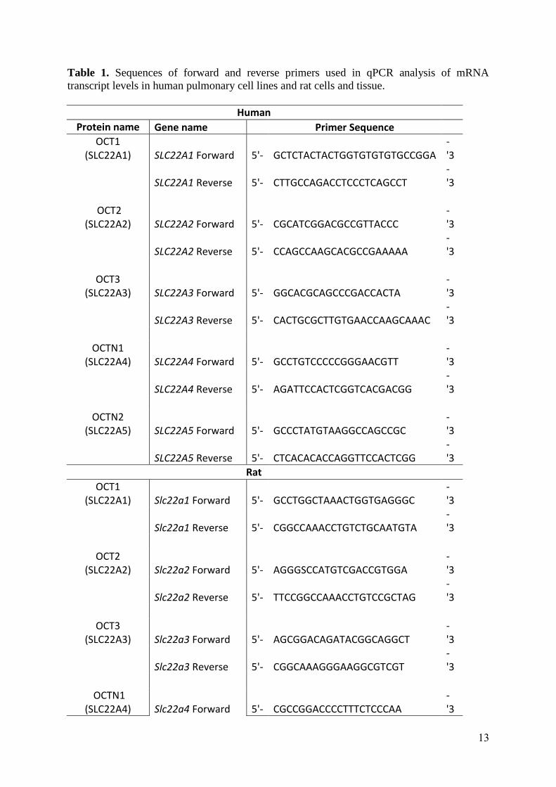
Table 1. Sequences of forward and reverse primers used in qPCR analysis of mRNA transcript levels in human pulmonary cell lines and rat cells and tissue.
Human
Protein name Gene name Primer Sequence
OCT1
(SLC22A1) SLC22A1 Forward 5'- GCTCTACTACTGGTGTGTGTGCCGGA
-
'3
SLC22A1 Reverse 5'- CTTGCCAGACCTCCCTCAGCCT
-
'3
OCT2
(SLC22A2) SLC22A2 Forward 5'- CGCATCGGACGCCGTTACCC
-
'3
SLC22A2 Reverse 5'- CCAGCCAAGCACGCCGAAAAA
-
'3
OCT3
(SLC22A3) SLC22A3 Forward 5'- GGCACGCAGCCCGACCACTA
-
'3
SLC22A3 Reverse 5'- CACTGCGCTTGTGAACCAAGCAAAC
-
'3
OCTN1
(SLC22A4) SLC22A4 Forward 5'- GCCTGTCCCCCGGGAACGTT
-
'3
SLC22A4 Reverse 5'- AGATTCCACTCGGTCACGACGG
-
'3
OCTN2
(SLC22A5) SLC22A5 Forward 5'- GCCCTATGTAAGGCCAGCCGC
-
'3
SLC22A5 Reverse 5'- CTCACACACCAGGTTCCACTCGG
-
'3
Rat
OCT1
(SLC22A1) Slc22a1 Forward 5'- GCCTGGCTAAACTGGTGAGGGC
-
'3
Slc22a1 Reverse 5'- CGGCCAAACCTGTCTGCAATGTA
-
'3
OCT2
(SLC22A2) Slc22a2 Forward 5'- AGGGSCCATGTCGACCGTGGA
-
'3
Slc22a2 Reverse 5'- TTCCGGCCAAACCTGTCCGCTAG
-
'3
OCT3
(SLC22A3) Slc22a3 Forward 5'- AGCGGACAGATACGGCAGGCT
-
'3
Slc22a3 Reverse 5'- CGGCAAAGGGAAGGCGTCGT
-
'3
OCTN1
(SLC22A4) Slc22a4 Forward 5'- CGCCGGACCCCTTTCTCCCAA
-
'3

14
Slc22a4 Reverse 5'- CAACGATGCTCCGGGGTCCC
-
'3
OCTN2
(SLC22A5) Slc22a5 Forward 5'- GGACGGCATGCGGGACTACG
-
‘3
Slc22a5 Reverse 5'- GGATGAACCAGAGAGCCCCA
-
‘3
15
Table 2. Pharmacokinetic parameters, rate constant (k) and fraction absorbed (f) generated from modelling IPRL lung transport data of [3H]-ipratropium and [3H]-L-carnitine. The extent of absorption of the control permeability probe 14C-mannitol at 60 minutes is shown for comparison. Data are mean ± SD. The number of experimental replicates (n) is shown.
Substrate/ Treatment
n k
(min-1) f
(max =1)
% [14C]-mannitol absorbed at 60
min
[3H] ipratropium
6 0.042 ± 0.028 0.23 ± 0.095 28.5 ± 10.2
[3H] ipratropium + 125 nmol ipratropium
6 0.043 ± 0.017 0.21 ± 0.070 26.6 ±11.5
[3H] ipratropium + 125 nmol MPP+
4 0.047 ± 0.022 0.17 ± 0.027 22.8 ±2.3
[3H] L-carnitine
4 0.054 ± 0.020 0.030 ± 0.023 17.1 ±2.3
[3H] L-carnitine + 125 nmol L-carnitine
4 0.057 ± 0.030 0.032 ± 0.005 17.3 ± 6.0
16
Table 3 Percentage uptake of [3H]-ipratropium and [3H]-L-carnitine uptake in the three human cell lines A549, BEAS-2B and 16HBE14o-.
Cell Line A549 BEAS-2B 16HBE14o-
% Ipratropium uptake / 106 cells / hr
1.6 ± 0.13 0.23 ± 0.03 0.60 ± 0.04
% L-carnitine uptake / 106 cells / hr 3.4 ± 0.13 25.3 ± 2.0 4.0 ± 0.3
17
Figure 1. (1A) and (1B): Cumulative airway to perfusate transport (expressed as % of total
lung deposited dose) following instillation into the IPRL airways of 45 pmol [3H]-
ipratropium or [3H]-L-carnitine, respectively. Treatments included: (1A) airway co-
administration of unlabelled (cold) ipratropium (125 nmol) or MPP+ (125 nmol) or (1B)
airway co-administration of unlabelled (cold) L-carnitine (125 nmol). Lines represent the
predicted transport of each substrate based on mathematical modelling. (1C): Quantitative
PCR results showing relative levels of cDNA transcript corresponding to Slc22a1-5
transporters in whole rat lung tissue, primary rat lung epithelial cells and rat liver (% signal
expressed to 2 ng total cDNA). (1D): Uptake (expressed as % of control) at 60 min (37°C) of
50 nM [3H] ipratropium and 50 nM [
3H] L-carnitine in primary culture rat epithelial cells
when challenged by pre-incubation with either 500 µM unlabelled ipratropium, 100 µM
unlabelled L-carnitine, MPP+ (500µM). * Represents statistical significance compared to
respective control at P<0.05 by one-way ANOVA and Dunnett's post-hoc test.
18
Figure 2. (2A): Real-time quantitative PCR results showing amounts of cDNA for OCT1,
OCT2, OCT3, OCTN1 and OCTN2 mRNA transcripts in the three pulmonary cell lines
expressed as femtograms / 2ng of total cDNA. The insert shows the standard curves for gene
transcripts. (2B) and (2C): % uptake of [3H]-ipratropium and [
3H] L-carnitine in cell lines
A549, BEAS-2B and 16HBE14o- in the presence and absence of various inhibitors. Data
shown are mean ± S.D., n=6-8. * denotes statistical significance (P<0.05) in comparison to
control (radiolabelled substrate only) using one-way ANOVA with Tukey’s post hoc test.
19
Supplementary Figures
Fig S1
Time-dependent accumulation of [3H]-ipratropium (panel A) and [3H]-L-carnitine (panel B) in three human lung epithelial cells lines. Cells were incubated at 37°C for pre-determined times and harvested for radiochemical analysis as described in the Materials and Methods section of the main text.