Abscess in adenomyosis mimicking a malignancy in a 54...
7
Abscess in adenomyosis mimicking a malignancy in a 54-year-old woman Rezzan Erguvan 1 , Mehmet M. Meydanli 2 , Alpay Alkan 3 , Mehmet N. Edali 1 , Hasan Gokce 1 and Ay¸ e Kafkasli 2 1 Department of Pathology 2 Department of Obstetrics and Gynecology and 3 Department of Radiology, School of Medicine, Inonu University, Malatya, Turkey Background: Although there are a few reports describing abscess formation in endometriotic foci no report of abscess formation arising de novo within adenomyosis appears in the literature. Preoperative diagnosis of adenomyosis is frequently difficult because of non-specific signs and symptoms. Synchronous pelvic patho- logies such as leiomyoma, endometrial polyp, endometrial hyperplasia, as well as endometrial cancer may cause differential diagnostic problems. Case: A 54-year-old postmenopausal woman complaining of inguinal pain, nightsweats and hot flashes is presented. Radiologic examinations of the pelvis revealed a 95 ´ 85 mm leiomyoma-like lesion including a 53 ´ 43 mm cystic space and 9 ´ 6 mm papillary formation within the uterus raising clinical suspicion of malignancy. A total abdominal hysterectomy and bilateral salpingo-oophorectomy were performed accompanied by a frozen section diagnosis. The frozen section revealed an abscess formation arising in a focus of adenomyosis. The postoperative period of the patient was uneventful. Conclusion: The present case, to our knowledge, is the first report representing abscess formation in adenomyosis. Abscess arising within adenomyosis can strongly raise the suspicion of endometrial cancer, particu- larly if the patient is postmenopausal. If endometrial cancer cannot be ruled out with definitive histopathological diagnosis in the preoperative period, a frozen section becomes mandatory during surgical intervention. Key words: FROZEN SECTION; TRANSVAGINAL ULTRASONOGRAPHY; UTERUS; SURGERY INTRODUCTION Adenomyosis is a common gynecological condi- tion that is characterized by the presence of endometrial glands and stroma extending beneath the endomyometrial interface which nests deep within the myometrium, whereas endometriosis refers to the occurrence of endometrial tissue out- side the uterus 1–5 or on the uterine serosa 4 . Despite its high frequency, etiology of adenomyosis is still unknown. The current leading theory suggests adenomyosis develops as a result of down-growth and invagination of the basalis endometrium into the myometrium 6 . Preoperative diagnosis of adenomyosis is fre- quently difficult because of non-specific signs and symptoms. Synchronous pelvic pathologies such as leiomyoma, endometrial polyp, endometrial hyperplasia as well as endometrial cancer may Infect Dis Obstet Gynecol 2003;11:59–64 Rezzan Erguvan, MD, Cosnuk Mah, Mehmet Buyruk Cad, Resat Turgut Sitesi B-Blok, No:75 D:26, TR-44330, Malatya, Turkey. Email: [email protected] ã 2003 The Parthenon Publishing Group 59
Transcript of Abscess in adenomyosis mimicking a malignancy in a 54...

Abscess in adenomyosis mimicking a malignancy in a54-year-old woman
Rezzan Erguvan1, Mehmet M. Meydanli2, Alpay Alkan3, Mehmet N. Edali1,Hasan Gokce1 and Ay¸e Kafkasli2
1Department of Pathology2Department of Obstetrics and Gynecology and
3Department of Radiology, School of Medicine, Inonu University, Malatya, Turkey
Background: Although there are a few reports describing abscess formation in endometriotic foci no report ofabscess formation arising de novo within adenomyosis appears in the literature. Preoperative diagnosisof adenomyosis is frequently difficult because of non-specific signs and symptoms. Synchronous pelvic patho-logies such as leiomyoma, endometrial polyp, endometrial hyperplasia, as well as endometrial cancer may causedifferential diagnostic problems.Case: A 54-year-old postmenopausal woman complaining of inguinal pain, nightsweats and hot flashes ispresented. Radiologic examinations of the pelvis revealed a 95 ´ 85 mm leiomyoma-like lesion including a53 ´ 43 mm cystic space and 9 ´ 6 mm papillary formation within the uterus raising clinical suspicion of malignancy.A total abdominal hysterectomy and bilateral salpingo-oophorectomy were performed accompanied by afrozen section diagnosis. The frozen section revealed an abscess formation arising in a focus of adenomyosis.The postoperative period of the patient was uneventful.Conclusion: The present case, to our knowledge, is the first report representing abscess formation inadenomyosis. Abscess arising within adenomyosis can strongly raise the suspicion of endometrial cancer, particu-larly if the patient is postmenopausal. If endometrial cancer cannot be ruled out with definitivehistopathological diagnosis in the preoperative period, a frozen section becomes mandatory during surgicalintervention.
Key words: FROZEN SECTION; TRANSVAGINAL ULTRASONOGRAPHY; UTERUS; SURGERY
INTRODUCTION
Adenomyosis is a common gynecological condi-tion that is characterized by the presence ofendometrial glands and stroma extending beneaththe endomyometrial interface which nests deepwithin the myometrium, whereas endometriosisrefers to the occurrence of endometrial tissue out-side the uterus1–5 or on the uterine serosa4. Despiteits high frequency, etiology of adenomyosis is still
unknown. The current leading theory suggestsadenomyosis develops as a result of down-growthand invagination of the basalis endometrium intothe myometrium6.
Preoperative diagnosis of adenomyosis is fre-quently difficult because of non-specific signs andsymptoms. Synchronous pelvic pathologies suchas leiomyoma, endometrial polyp, endometrialhyperplasia as well as endometrial cancer may
Infect Dis Obstet Gynecol 2003;11:59–64
Rezzan Erguvan, MD, Cosnuk Mah, Mehmet Buyruk Cad, Resat Turgut Sitesi B-Blok, No:75 D:26, TR-44330, Malatya,Turkey. Email: [email protected]
ã 2003 The Parthenon Publishing Group 59

cause differential diagnostic problems. This diffi-culty arises especially if the patient is postmeno-pausal with established risk factors for endometrialcancer. The respective diagnosis is rendered moredifficult in the case of inadequate biopsy specimenfor the definitive diagnosis while the radiologicfindings favor a primary uterine cancer.
Although there are a few reports describingabscess formation in endometriotic foci3,7,8, noreport of abscess formation arising de novo withinadenomyosis appears in the literature. In our reporta 54-year-old woman with a suspected malig-nancy was discovered as having adenomyosis andabscess formation arising de novo within. This isthe first case in the literature showing adeno-myosis including abscess formation.
CASE REPORT
A 54-year-old, multiparous (gravida 4, para 3,induced abortus 1), obese, postmenopausalwoman complaining of inguinal pain, nightsweatsand hot flashes was admitted to Inonu UniversityGynecology and Obstetrics Clinic in November2001. Chronic obstructive lung disease, diabetesmellitus and hypertension were present in the his-tory whereas no history of hormone replacementtherapy was noted. No history of pelvic inflamma-tory disease, intrauterine device, endometriosis,tubal pathology or diverticular disease was noted.
The duration of menopause was defined to be14 months with the patient’s menopause age being> 52 years. Except for the presence of an irregu-larly enlarged uterus, normal findings weredetected when a gynecological examination wasperformed.
Transvaginal ultrasonography revealed a95 ´ 85 mm leiomyoma-like lesion including a53 ´ 43 mm cystic space (Figure 1a) and 9 ´ 6 mmpapillary formation within the uterus (Figure 1b).Because of distortion of the endometrial cavity bythe lesion described, endometrial thickness couldnot be assessed. An endometrial biopsy was per-formed and the pathologic examination revealedinadequate tissue for the definitive diagnosis. Thepatient was hospitalized in order to rule out auterine malignancy.
Laboratory results including coagulation paneland biochemical tests did not reveal any data of
interest except for the presence of hyperglycemia.The leukocyte count was 7600/mm3 with neutro-phils 68.8%, lymphocytes 26.7%, monocytes 3%,and eosinophils 1.5%. The patient was not febrileduring her workup. Mammography and otherlaboratory examinations showed no abnormalsigns. Tumor markers including CA 125 werereported to be within normal ranges.
Magnetic resonance imaging of the pelvis wasperformed subsequently and the uterus wasdetected as greater than normal, having lobulatedcontours and heterogen density. In addition tointramural lesions with the greatest diameter of5 cm, cystic and necrotic degenerations consistentwith leiomyoma that obstruct the endocervicalcanal and a diffuse fluid accumulation in theendometrial cavity were noted. Ovaries werenormal bilaterally.
A 5 ´ 5 mm polypoid lesion on the anterior andposterior wall of the uterine fundus was detected
Abscess in adenomyosis mimicking a malignancy Erguvan et al.
60 INFECTIOUS DISEASES IN OBSTETRICS AND GYNECOLOGY
Figure 1 Transvaginal ultrasonography revealed a95 ´ 85 mm leiomyoma-like lesion consisting of a54 ´ 43 mm cystic space (a), in which a 9 ´ 6 mmpapillary structure (b) was noted
upon hysterosonography. Additionally a 1 ´ 1 cmanechoic structure protruding into the endo-metrial cavity in fundus was detected.
A fractional dilatation and curettage wasperformed under general anesthesia but the patho-logic examination revealed inadequate tissue. Ahysteroscopy was not performed because of theobstruction of the endocervical canal by one ofthe lesions described. Since it was not possibleto exclude uterine malignancy with definitivehistopathological diagnosis, the patient underwentexploratory laparotomy for clinically assumeduterine malignancy. A total abdominal hyster-ectomy and bilateral salpingo-oophorectomywere performed accompanied by a frozen sectiondiagnosis. The patient had no fever during thesurgery. Intraoperative findings did not suggestany evidence of pelvic tuberculosis, nor wassystemic tuberculosis detected. The frozen sectionrevealed an abscess formation arising in a focusof adenomyosis. The postoperative period ofthe patient was uneventful.
METHODS
Diagnostic evaluation
The material was fixed in 10% buffered formalinand processed routinely. Hematoxylin-eosinstained slides were examined.
Macroscopically 238 g weight and23 ´ 10 ´ 5.5 cm specimen was evaluated. Thick-ness of endometrium was 1 mm and it was 2 cm formyometrium. Serial sections revealed a 2 cm cysticlesion localized in myometrium adjacent toendometrium in the vicinity of left cornu uteri(Figure 2). A purulent material was drained duringthe section. The wall of the cyst had a greyishwhite–yellow color and an irregular appearance.No papillary formation on the wall was identi-fied. The right ovary had a 1 cm cystic formation.In addition a 7 mm right paratubal cyst wasnoted. The left ovary and fallopian tube weregrossly unremarkable.
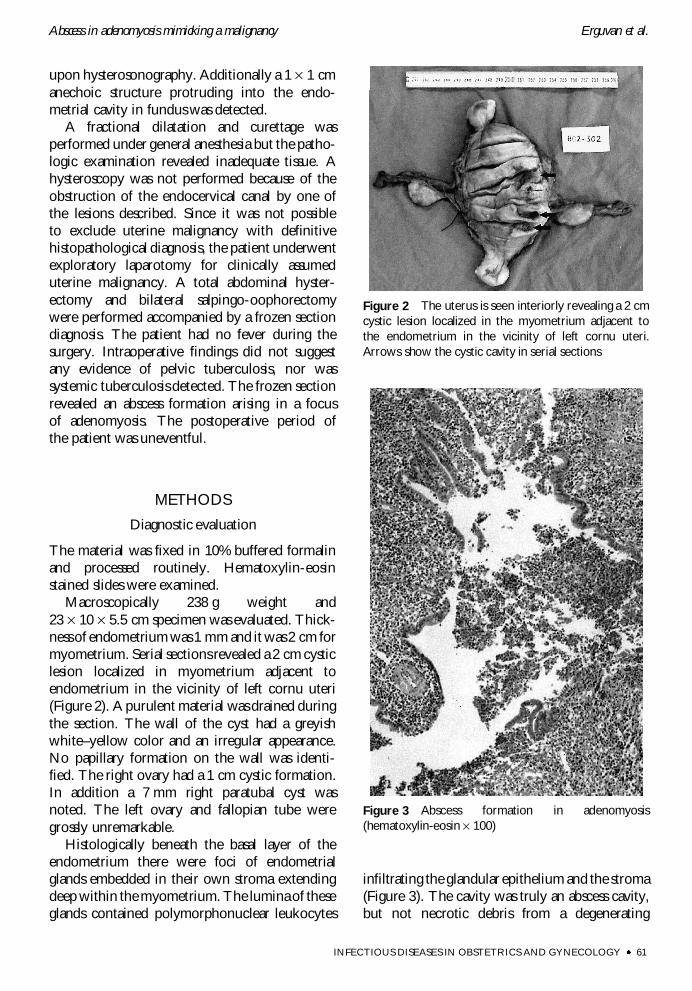
Histologically beneath the basal layer of theendometrium there were foci of endometrialglands embedded in their own stroma extendingdeep within the myometrium. The lumina of theseglands contained polymorphonuclear leukocytes
infiltrating the glandular epithelium and the stroma(Figure 3). The cavity was truly an abscess cavity,but not necrotic debris from a degenerating
Abscess in adenomyosis mimicking a malignancy Erguvan et al.
INFECTIOUS DISEASES IN OBSTETRICS AND GYNECOLOGY 61
Figure 2 The uterus is seen interiorly revealing a 2 cmcystic lesion localized in the myometrium adjacent tothe endometrium in the vicinity of left cornu uteri.Arrows show the cystic cavity in serial sections
Figure 3 Abscess formation in adenomyosis(hematoxylin-eosin ´ 100)

leiomyoma. A tissue section obtained understerile conditions was cultured for both aerobicand anaerobic microorganisms, but results wereinconclusive. The final diagnosis was abscessformation in an adenomyosis focus confirmingthe results of the frozen section in this case.
DISCUSSION
Adenomyosis is a condition in which endometrialglands and stroma are located inordinately deepin the myometrium. Endometriosis is defined asendometrial tissue occurring ectopically outsidethe uterus1–5,9 or on the uterine serosa4.
It is generally accepted that about 15% ofmultiparous women develop varying degrees ofadenomyosis in their late 30s and early 40s1,3,5,10.About 15% of women with adenomyosis haveassociated endometriosis10. Previous studieshave shown that the mean age in endometriosisis between 35–39 years and women are fre-quently multiparous5,11. Our patient is differentin that she has presented with adenomyosis post-menopausally at 54 years of age.
Although adenomyosis and endometriosisare regarded as closely related, their microscopicappearance and pathogenesis are somewhat differ-ent. It is known that they often occur indepen-dently of each other. While adenomyosis is madeup of the non-functional (basal) layer of theendometrium, endometriosis is composed ofthe functional layers so that proliferative, secretoryand menstrual changes can be seen in the latter2.
Since in some cases of adenomyosis, the affectedendometrium is functional, the most importantconsequence of adenomyosis is shedding of theendometrium during the menstrual cycle1,12. As aresult of hemorrhage within the adenomyoticfoci, menorrhagia, colicky dismenorrhea, dis-pareunia and pelvic pain occur especiallyduring the premenstrual period1,4,5,9. Egger andWeigmann11 state that the pain during menstrua-tion is due to uterine dyskinesia. However, about30–40% of patients with adenomyosis areasymptomatic; hence, this disease can be a surprisepathologic finding in a patient without menorr-hagia, dysmenorrhea, or uterine enlargement10.The pain in the present case was not of a pelvic
origin, but an inguinal pain. Since there were noother findings to cause inguinal pain, this pain mayhave been caused by the abscess itself. Foci ofadenomyosis may be affected by other diseasesaffecting the orthotopic endometrium (e.g.,hyperplasia or malignant changes). Therefore, it isimportant not to misinterpret such a condition asa deeply invasive malignancy2,4,5. These foci canalso become decidualized during pregnancy5.Adenomyosis in pregnancy can lead to ruptureof membranes2.
When the uterus is diffusely involved byadenomyosis, it is enlarged in a globoid fashion4,5,9.The most commonly affected site is the posteriorwall5,9. If it is focal (‘adenomyoma’), it mimics aleiomyoma showing a roughly spherical, intra-mural, space-occupying lesion. Its difference fromleiomyoma is not being shelled out easily from thesurrounding uninvolved myometrium4,5.
There are rare cases reported in the literaturedescribing the presence of abscess in endometriosisin different localizations, most of them being inthe ovaries3,7,8 but none in adenomyosis, to ourknowledge.
Martino and colleagues7 reported a 16-year-oldgirl who underwent surgery because of congenitalanomalies of the bladder, rectum and vagina. Inthe follow-up a right lower quadrant mass wasdiscovered and subsequently they detected threecystic midline masses at the level of umbilicus.Following the examination procedures includingfine-needle aspiration (FNA) biopsies of theselesions, they detected a secondary infection ofendometriosis arising from the fallopian tube andthe ovary. Laboratory results revealed that theetiologic agent was clostridium. They interpretedthat it was an iatrogenic infection followingFNA biopsy7.
Lipscomb and associates8 described an abscessmeasuring as large as 20 cm in the ovary and theyemphasized that it was the first case in the literaturearising de novo within an endometriosis in theirreport. They also stated that it was the second casein the literature leading to ureteral obstruction.They hypothesized that the abscess in their casemight have developed following hematogenousspread of bacteria from a urinary tract infectionbecause the same organism was recovered fromboth the patient’s urine and the abscess cavity8.
Abscess in adenomyosis mimicking a malignancy Erguvan et al.
62 INFECTIOUS DISEASES IN OBSTETRICS AND GYNECOLOGY
Egger and Weigmann11 found that the inci-dence of infection in endometriotic cysts, forma-tion of an isolated ovarian abscess, was rangingbetween 8–18% in their study of 263 patients.They stated that earlier surgical treatment ofendometriotic cysts might lead to decrease inthe risk of secondary infection11.
There are some other reports describing per-foration of the colon due to endometriosis3,13.Floberg and co-workers13 reported a 34-year-oldwoman who had an uneventful delivery anddeveloped perforation of the colon immediatelyin the postpartum stage. They detected a 5 cmabscess causing enlargement of the left ovary whichwas adhering to the sigmoid colon in the lapa-rotomy performed. They stated that the abscesshad ruptured into the peritoneal cavity and therewas also a fistula from the abscess to the colon.They discovered that the microorganisms that ledto infection were Gram-negative bacteria. Sincethey did not find any other focus of infectionlike tuba uterina which might transmit to theovary, they assumed that the ovary becameinfected from the colon via the fistula. Theyreported that the patient recovered after receivingbroad-spectrum antibiotics intravenously13.
In our patient, because there were no micro-organisms isolated from the foci of abscess in bothaerobic and anaerobic cultures, and there was noother source to lead a secondary suppurationincluding particularly the uterus itself, the causeof infection remained unknown. But it is clearin this case that this was a primary abscessformation in the adenomyosis defined for thefirst time.
Treatment modality for adenomyosis isprimarily surgical5. Because abscess formationwas localized within the adenomyotic foci, it wasregarded that antibiotic therapy was unnecessary,and total abdominal hysterectomy and bilateralsalpingo-oophorectomy was performed. Thusthe patient ameliorated after surgery and was dis-charged without any complications.
In conclusion, abscess arising within adeno-myosis can strongly raise the suspicion ofendometrial cancer, particularly if the patient ispostmenopausal. If endometrial cancer cannotbe ruled out with definitive histopathologicaldiagnosis in the preoperative period, a frozensection becomes mandatory during surgicalintervention.
REFERENCES1. Crum CP. The female genital tract. In Cotran RS,
Kumar V, Collins T, eds. Robbins Pathologic Basis ofDisease, 6th edn. Philadelphia, Pennsylvania: W.B.Saunders Co., 1999:1035–91
2. Rosai J. Female reproductive system. In Rosai J,ed. Ackerman’s Surgical Pathology, 8th edn. Missouri:Mosby-Year Book Inc., 1996:1319–564
3. Ledley GS, Shenk IM, Heiy HA. Sigmoid colonperforation due to endometriosis not associatedwith pergnancy. Am J Gastroenterol 1988;83:1424–6
4. Hendrickson MR, Longacre TA, Kempson RL.The uterine corpus. In Sternberg SS, ed. DiagnosticSurgical Pathology, 3rd edn. Philadelphia: LippincottWilliams & Wilkins, 1999:2203–5
5. Hendrickson MR, Kempson RL. Non-neoplasticconditions of the myometrium and uterineserosa. In Fox H, ed. Obstetrical and Gynaecological
Pathology, 4th edn. Churchill-Livingstone,1995:511–18
6. Ferenczy A. Pathophysiology of adenomyosis.Hum Reprod 1998;4:312–22
7. Martino CR, Haaga JR, Bryan PJ. Secondaryinfection of an endometrioma following fine-needle aspiration. Radiology 1984;151:53–4
8. Lipscomb GH, Ling FW, Photopulos GJ. Ovarianabscess arising within an endometrioma. ObstetGynecol 1991;78:951–4
9. Kraus FT. Female genitalia. In Kissane JM, ed.Anderson’s Pathology, 8th edn. Mosby, 1985:1451–545
10. Moore JG. Endometriosis and adenomyosis. InHacker NF, Moore JG, eds. Essentials of Obstetricsand Gynecology, 3rd edn. Philadelphia: WBSaunders Co., 1998:432–40
Abscess in adenomyosis mimicking a malignancy Erguvan et al.
INFECTIOUS DISEASES IN OBSTETRICS AND GYNECOLOGY 63

11. Egger H, Weigmann P. Clinical and surgicalaspects of ovarian endometriotic cysts. ArchGynecol 1982;233:37–45
12. Cotran RS, Kumar V, Robbins SL. Female genitaltract. In Cotran RS, Kumar V, Robbins SL, eds.
Robbins–Pathologic Basis of Disease, 4th edn.Philadelphia: W.B. Saunders Co., 1989:1127–80
13. Floberg J, Backdahl M, Silfersward C, ThomassenPA. Postpartum perforation of the colon due toendometriosis. Acta Obstet Gynecol Scand 1984;63:183–4
RECEIVED 07/08/02; ACCEPTED 02/01/03
Abscess in adenomyosis mimicking a malignancy Erguvan et al.
64 INFECTIOUS DISEASES IN OBSTETRICS AND GYNECOLOGY

Submit your manuscripts athttp://www.hindawi.com
Stem CellsInternational
Hindawi Publishing Corporationhttp://www.hindawi.com Volume 2014
Hindawi Publishing Corporationhttp://www.hindawi.com Volume 2014
MEDIATORSINFLAMMATION
of
Hindawi Publishing Corporationhttp://www.hindawi.com Volume 2014
Behavioural Neurology
EndocrinologyInternational Journal of
Hindawi Publishing Corporationhttp://www.hindawi.com Volume 2014
Hindawi Publishing Corporationhttp://www.hindawi.com Volume 2014
Disease Markers
Hindawi Publishing Corporationhttp://www.hindawi.com Volume 2014
BioMed Research International
OncologyJournal of
Hindawi Publishing Corporationhttp://www.hindawi.com Volume 2014
Hindawi Publishing Corporationhttp://www.hindawi.com Volume 2014
Oxidative Medicine and Cellular Longevity
Hindawi Publishing Corporationhttp://www.hindawi.com Volume 2014
PPAR Research
The Scientific World JournalHindawi Publishing Corporation http://www.hindawi.com Volume 2014
Immunology ResearchHindawi Publishing Corporationhttp://www.hindawi.com Volume 2014
Journal of
ObesityJournal of
Hindawi Publishing Corporationhttp://www.hindawi.com Volume 2014
Hindawi Publishing Corporationhttp://www.hindawi.com Volume 2014
Computational and Mathematical Methods in Medicine
OphthalmologyJournal of
Hindawi Publishing Corporationhttp://www.hindawi.com Volume 2014
Diabetes ResearchJournal of
Hindawi Publishing Corporationhttp://www.hindawi.com Volume 2014
Hindawi Publishing Corporationhttp://www.hindawi.com Volume 2014
Research and TreatmentAIDS
Hindawi Publishing Corporationhttp://www.hindawi.com Volume 2014
Gastroenterology Research and Practice
Hindawi Publishing Corporationhttp://www.hindawi.com Volume 2014
Parkinson’s Disease
Evidence-Based Complementary and Alternative Medicine
Volume 2014Hindawi Publishing Corporationhttp://www.hindawi.com