Arbuscular Mycorrhiza Inoculum to Support Sustainable Cropping Systems
About TERI TERI (The Energy and Resources
Institute) is a dynamic and flexible organization with a global vision and a local
focus. TERI’s focus is on research, in the fields of energy, environment, and sustainable development,
and on documentation and information dissemination. The genesis of these activities lie in TERI’s firm belief
that the efficient utilization of energy, sustainable use of natural resources, large-scale adoption of renewable energy
technologies, and reduction of all forms of waste would move the process of development towards the goal of sustainability.
TERI’s Mycorrhiza Network TERI’s Mycorrhiza Network is primarily responsible for
establishing the MIC (Mycorrhiza Information Centre), the CMCC (Centre for Mycorrhiza Culture Collection), and publishing
Mycorrhiza News. The Network helps scientists carry out research in mycorrhiza and promotes communication among mycorrhiza scientists.
Mycorrhiza News The Mycorrhiza News provides a forum for dissemination of scientific information on mycorrhiza research and activities; publishes state-of-the-art papers from eminent scientists; notes on important breakthroughs; brief accounts of new approaches and techniques; publishes papers complied from its RIZA database; provides information on forthcoming events on mycorrhiza and related subjects; lists important research references published during the quarter; and highlights the activities of the CMCC.
Volume 24 • Issue 3 • October 2012
2 Mycorrhiza News 24(3) • October 2012
Introduction Scutellospora savannicola (Herrer and Ferrera) Walker and Sanders is known only from Savannah on macarrera soil, i.e., clayey, sandy but not very gravelly soil with high concentrations of Iron (Ferrer and Herrera, 1980). This species of arbuscular mycorrhizal (AM) fungi was earlier placed under genus Gigaspora. Later, Walker and Sanders (1986) placed it under Scutellospora based on differentiable wall layers and knobby soil borne vesicles or auxillary cells. The spores of S. savannicola are reported to be present in soils probably during October to January (Ferrer and Herrera, 1980).Western Ghats of Goa have been randomly
surveyed for the occurrence of AM fungi by Khade (2002). Rhizospheric agro-soils of Collem from Western Ghats of Goa are acidic to near neutral, high in organic carbon and K while, total N and P levels are limiting. Micronutrients, viz., Cu, Zn, Fe, and Mn are present in high levels (Khade, 2003). Therefore, in the present study Carica papaya L., from agro-based ecosystem of Collem was surveyed for occurrence of S. savannicola post monsoons.
Materials and methodsRoot samples and the rhizosphere soil samples of papaya plants were collected during the post monsoons (December 2001) from Collem located
in Western Ghats of Goa (Plate 1 A, B). Further, freshly collected roots along with root hairs were washed in water, cleared with 10% KOH, acidified with 1N HCl, and stained in 0.05% Trypan Blue in lactoglycerol (Phillps and Hayman, 1970). Spores of AM fungi were extracted from the rhizosphere using wet sieving and decanting method (Gerdemann and Nicolson, 1963). Diagnostic slides with spores were prepared using polyvinyl alcohol lactoglycerol (PVLG) as mountant. Identification of AM fungi was carried out using Manual of identification of AM fungi (Schenck and Perez, 1990).
ResultsScutellospora savannicola (Herrer and Ferrera) Walker and Sanders recorded in the present study is taxonomically described below.Azygospores formed singly in soil, hyaline
to white, turning brown at maturity (Plate 2A), oblong - ellipsoid to broadly ellipsoidal (Plate 2A), and sometimes irregular 300- 360 x 200–300 µm in diam (Plate 2A). Azygospore is borne on a bulbous suspensor (Plate 2A) which is globose to pyriform or claviform, 33–51µm in diam at the widest part with thin wall, up to 1.4 µm thick (Plate 2B). Bulbous suspensor attached to a septate hypha (Plate 2A, Plate 2B). Generally lateral hypha extends from the bulbous suspensor and is 3–33 µm long in young
Contents
ReseaRch finding papeRsRecord of Scutellospora savannicola (Herrer and Ferrera) Walker and Sanders from GoaSharda W Khade*Department of Botany, Goa University, Taleigao Plateau, GOA 403206.
ReSeaRCH FindinG papeRS
Record of Scutellospora savannicola (Herrer and Ferrera) Walker and Sanders from Goa 2
Management of charcoal root-rot caused by Macrophomina phaseolina in groundnut by using arbuscular mycorrhizal fungi in field 5
Glomalin and its association with the rhizosphere soils of some trees in Warangal district, a.p, india 10
impact of long-term wastewater irrigation on the abundance of arbuscular mycorrhizal spores in the peri-urban soil of Varanasi 16
CMCC article
Mitochondrial genome sequences are more preferred reliable molecular markers in arbuscular Mycorrhizal Fungi over nuclear genome sequences 20
Recent references 25
Forthcoming events 28
* Current address for correspondence: Dr Sharda W. Khade, IInd floor, Darshan Apartments, Vidhyanagar colony, Near Goa Urban Co-operative bank, Miramar, Carenzalem-Post, Panaji 403002, Goa, India. Email: [email protected]

Mycorrhiza News 24(3) • October 2012 3
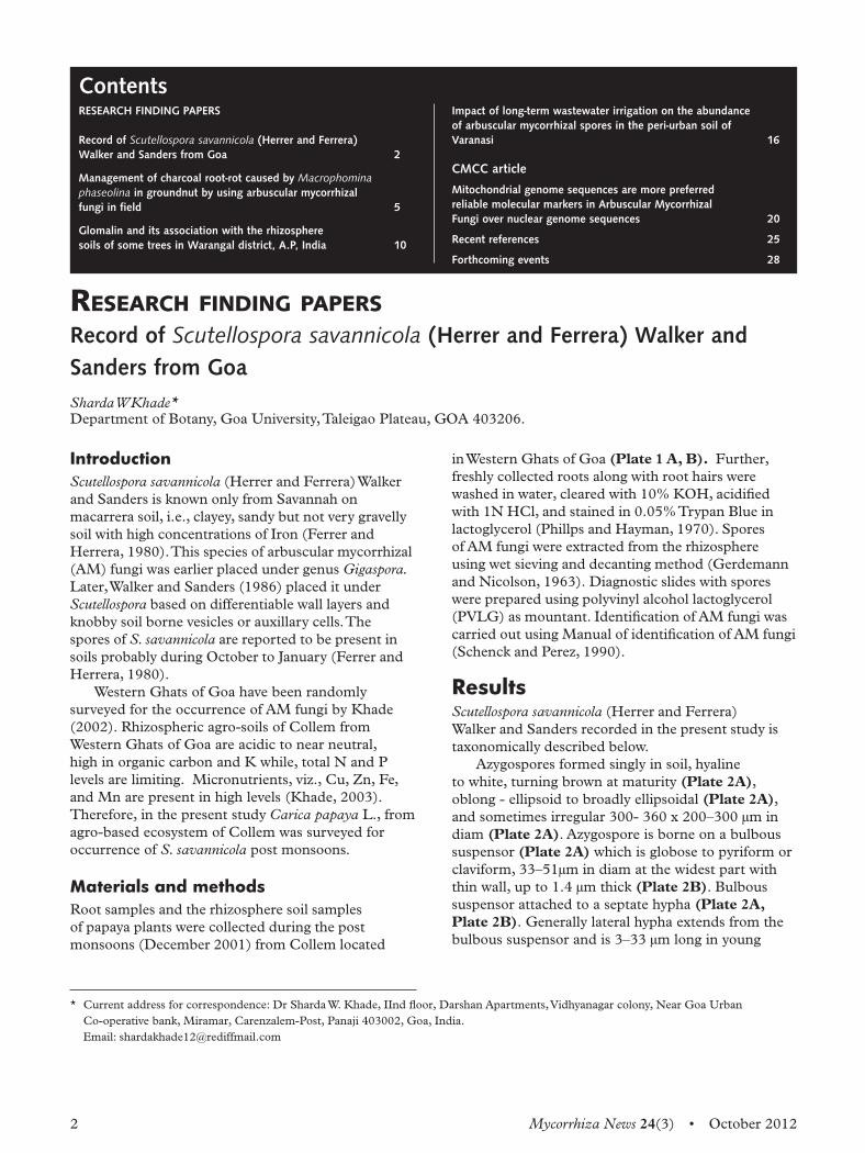
spores and much longer in mature spores (Plate 2B). Spore wall layered and 5–12 µm thick (Plate 3A). Wall composed of an exospore of two wall layers (Plate 3B), a mesospore of two membranes (Plate 3B) and a membranous endospore (Plate 3B). The external layer of the exospore is up to 3 µm thick and the internal layer upto 2.3 µm thick. The external membrane of the mesospore is up to 1.5 µm and the
internal layer upto 1.3 µm thick. The endospore is up to 1 µm thick enclosing reticulate content of the spore. Auxillary cells knobby with blunt ends, borne in groups on a short hypha.
ConclusionsIn the present study, the soil was dark brown to reddish brown, well drained gravelly with silt clay loam texture and medium water holding capacity. The present study recorded AM fungal colonization and encountered spores of S. savannicola in rhizosphere of
A
A
B
B
Plate 1 A) Western Ghats of Collem.B) Habit of Carica papaya L.
Plate 2 (A) A mature spore of Scutellospora savannicola borne on bulbous suspensor (x 200).(B) A portion of spore of Scutellospora savannicola showing bulbous suspensor (Bs) and a lateral hypha (H) extending from bulbous suspensor (x 400).
4 Mycorrhiza News 24(3) • October 2012
papaya plants from Western Ghats of Goa (Collem) during December. This supports the findings of Khade (2002), Khade et al. (2002), and Khade (2003)
who reported AM fungal association in papaya plants from Goa and presently the contention that spores of S. savannicola are also present in tropical P-deficient and iron rich agro-soils.
ReferencesFerrer R L and Herrera R A. 1980. El genero Gigaspora Gerdemann et Trappe (Endogonaceae) en Cuba.Revista Jardin Botanico Nacional Habana 1: 43–66
Gerdemann J W and Nicolson T H. 1963. Spores of mycorrhizal Endogone species extracted from soil wet sieving and decanting. Transactions of British Mycological Society 46: 235–244
Khade S W. 2002. Terminal project report on plant fungus biodiversity, inventory, conservation and utilization efforts for Western Ghats Goa region. Phase 1: 12–38
Khade S W, Bukhari M J, Jaiswal V, Gaonkar U C, Rodrigues B F. 2002. Arbuscular mycorrhizal status of medicinal plants: A field survey of AM fungal association in shrubs and trees. Journal of Economic and Taxonomic Botany 26(3): 571–578
Khade S W. 2003. Studies on arbuscular mycorrhizal status of Carica papaya L, in agrobased ecosystem of Goa. Ph.D thesis, Goa University, Goa, India, pp 1–235
Phillips J M and Hayman D S. 1970. Improved procedure for clearing roots and staining of mycorrhizal fungi for rapid assessment of infection. Transactions of British Mycological Society 55: 158–161
Schenck N C and Perez Y. 1990. Manual for identification of VA Mycorrhizal fungi. (Eds.) Schenck N C and Perez Y. INVAM, University of Florida, Gainesville. USA
Walker C and Sanders F E. 1986. Taxonomic concepts in the Endogonaceae: III. The separation of Scutellospora gen. nov. from Gigaspora Gerd. & Trappe. Mycotaxon 27: 169–182
Plate 3 (A) A portion of spore of Scutellospora savannicola showing wall layers (x 400).(B) A magnified portion of spore wall of Scutellospora savannicola with 5 layers (Two outer layers of the Exospore [Ex]; Two middle layers of the Mesospore [Me]; and One inner most layer of the endospore [En]). Arrows indicate the spore walls (x 600).
A
B
Mycorrhiza News 24(3) • October 2012 5
IntroductionChemical pesticides have always been seen as an effective methodology in control of plant pathogens in the past, but greater environment-consciousness is pushing the demand for minimum use of harmful chemical inputs. Moreover, a pesticide does not provide complete or lasting defense against soil-borne plant pathogens besides posing great threat to our lives (Johansson et al., 2004). This has led to advanced research into bio-control methods, which are beneficial for farmers who are not very economically well-off and for sustainable agricultural practices, which will benefit our environment and biodiversity as a whole. The biological control of plant pathogens by using arbuscular mycorrhizal fungi in providing resistance against various plant pathogens have been well-documented (Singh et al., 2000; Declerck et al., 2002; Ludwig-Muller, 2004; Whipps, 2004). Groundnut (A. hypogaea L.) is important as a chief oilseed crop in the Indian subcontinent. Several soil-borne plant pathogens attack groundnut plants during their growth. Among various pathogens, fungal pathogen M. phaseolina Tassi (Goid.) causes severe charcoal root-rot disease, which may assume huge economic proportions. Because of being soil-borne in nature, it remains a difficult pathogen to eradicate completely and chemical inputs are already considered both hazardous to the environment as well as costly. M. phaseolina is distributed worldwide and prevalent mainly in areas with high temperature and low rainfall (Raut and Bhombe, 1984). The main objective of this study was to evaluate the effectiveness of mycorrhizal inoculation against M. phaseolina under field conditions.
Material and methods
Plant material
The groundnut seeds were obtained from Naik Seeds, Pune, Maharashtra, India. The groundnut variety used for this study was of a local cultivar named Phule Pragati (JL-24). The seeds were surface sterilized with
Management of charcoal root-rot caused by Macrophomina phaseolina in groundnut by using arbuscular mycorrhizal fungi in field Khirood Doley* and Paramjit Kaur JiteDepartment of Botany, Mycology, University of Pune, Maharashtra, India
*Corresponding author: Khirood Doley, Department of Botany, Mycology, University of Pune, Pune – 411007, Maharashtra, IndiaE-mail: [email protected]
0.02% HgCl2 for five minutes and then washed with
distilled water.
Pathogen inoculum
The pure isolate of M. phaseolina was provided by Agharkar Research Institute and it was mass multiplied on sorghum grains (250 g), initially soaked overnight in water. About 100 gm of soaked sorghum grains were taken in saline bottles of 500 ml capacity plugged with cotton. The bottles were then sterilized for 20 minutes at 121ºC. The sterilized sorghum seeds present in saline bottles were inoculated with 5 mm mycelial disc using a cork borer (5 mm) from the active periphery of a seven-day-old pure culture of M. phaseolina in each bottle and the bottles were incubated for three weeks at temperature (28ºC ± 2ºC) for proper mycelial growth. This sorghum grain culture served as the inoculum. It was applied at the rate of 5 g after 15 days of the groundnut plant’s growth.
Isolation, identification, and inoculum preparation of AM
According to the method described by Gerdemann and Nicolson (1963), isolation of AM fungal spores was carried out by wet sieving and decanting methods. Identification of isolated fungal spores was carried out by complying keys recommended by Trappe (1982) and Schenck and Prez (1987). According to Kornerup and Wanscher (1983), determination of colour, shape, and dimensions of arbuscular mycorrhizal (AM) fungal spores was done. Mass multiplication of AM fungi [G. fasciculatum (Thaxter Sensu Gerd.) (Gerdemann and Trappe)] was done in pots with different host plants, such as Sorghum vulgare and Panicum maximum (Jacq.) and were grown on 30 cm earthen pots containing 10–15 kg of sterilized soil and sand in 1:1 proportion. Inoculations were made from this mass multiplied mycorrhizal inoculum after three months to each groundnut plant with 20 g of AM fungi inoculum mixture containing spores, colonized root pieces, and extrametrical mycelium in rhizospheric soil (obtained from the pot culture).
6 Mycorrhiza News 24(3) • October 2012
The mycorrhizal inoculum was placed below each groundnut seeds (about 3–5 cm) under the soil surface before sowing.
Field preparation for experiment
Preparation of field was carried out by ploughing it to a depth of 20 to 25 cm with shape of in-furrow system, ridge height of about 7–10 cm, inter-row distance of 15 cm, and plant-to-plant spacing of 30 cm on rows (16 foot length). The seeds of groundnut were planted by hand. Mycorrhizal (G. fasciculatum) inoculations were made with 20 g of inoculum mixture containing spores and colonized root pieces, which were obtained from the pot culture and were placed at about 3–5 cm below the seeds under the soil before sowing. Fungal inoculum of M. phaseolina mass multiplied on sorghum grains at the rate of 5 g was applied after 15 days of growth of the groundnut plant. There were four treatments replicated three times, which included: Controls (C); M. phaseolina (C+Mp); G. fasciculatum (Gf); G. fasciculatum; and M. phaseolina (Gf+Mp).
Data collection
The plants were randomly chosen as triplicates after 90 days of AM treatment and 75 days of M. phaseolina inoculations for determination of various growth responses, viz., leaf number, plant height, pod number, fresh and dry weight, and total biomass content of plants. The total chlorophyll content of the shoot was determined according to Arnon (1949); arbuscule percentage (%) and root colonization (%) was determined according to Phillips and Hayman (1970).
Mycorrhiza dependency
Mycorrhizal dependency was carried out according to Plenchette et al. (1983) by measuring the dry weights of the plants after drying the plant material to 70°C for 48 hours.
Percent Mycorrhizal Dependency = [(dry mass mycorrhizal plant − dry non-mass mycorrhizal plant) / dry mass mycorrhizal plant] x 100%
Production loss
Production losses were calculated using the equation given by Teng (1985): PL = (AY/1.0-PL) – AYWhere:PL = Production lossAY = Actual yield
Statistical analysis
The data was examined by one-way analysis of variance (ANOVA) followed by Duncan’s Multiple Range Test (DMRT). Three replications were carried out for each treatment. DMRT was applied as the post hoc test at p= 0.05. Calculations were made by using a Statistical Package for Social Sciences (SPSS) for windows version 9.0 and Microsoft Office Excel 2007 to analyse the data.
Results Growth responses groundnut plants
The data obtained from field experiment shows that G. fasciculatum inoculation on groundnut plant showed significant increase in terms of shoot length, fresh and dry weight, total biomass, total chlorophyll content, and numbers of pods as compared to their counterparts not subjected to mycorrhizal controls. In the mycorrhizal inoculated groundnut plants after 90 days of sowing, the leaf numbers were significantly higher (440.00 in Gf) than their non-mycorrhizal control counterparts (270.00 in Control). The lowest number of leaves (210.67 in C+Mp) was exhibited by diseased non-mycorrhizal groundnut plants after 90 days of sowing. But, when observed in mycorrhizal-infected groundnut plants, it showed higher number of leaves (266.67 in Gf+Mp) as per Table 1.
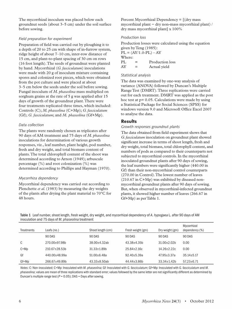
Table 1 Leaf number, shoot length, fresh weight, dry weight, and mycorrhizal dependency of A. hypogaea L. after 90 days of AM inoculation and 75 days of M. phaseolina treatment
Treatments Leafs (no.) Shoot length (cm) Fresh weight (gm) Dry weight (gm)Mycorrhizal dependency (%)
90 DAS 90 DAS 90 DAS 90 DAS 90 DAS
C 270.00±97.98b 38.00±4.32ab 43.38±4.35b 31.00±2.02b 0.00
C+Mp 210.67±28.53b 31.33±1.89b 25.84±2.16c 14.26±2.22c 0.00
Gf 440.00±48.99a 51.00±6.48a 92.40±5.39a 47.95±3.37a 35.14±5.17
Gf+Mp 266.67±49.89b 43.33±8.50ab 44.44±3.86b 33.34±1.42b 57.23±6.71
Notes: C: Non-inoculated; C+Mp: Inoculated with M. phaseolina; Gf: Inoculated with G. fasciculatum; Gf+Mp: Inoculated with G. fasciculatum and M. phaseolina; values are mean of three replications with standard error; values followed by the same letter are not significantly different as determined by Duncan’s multiple range test (P = 0.05); DAS = Days after sowing.
Mycorrhiza News 24(3) • October 2012 7
There was significant increase in plant height in mycorrhizal-treated groundnut plant (highest by 51.00 in Gf) than non-mycorrhizal groundnut plants (38.00 in Control). The plant height was lowest in non-mycorrhizal groundnut plants infected with M. phaseolina (31.33 in C+Mp) as compared to pathogenic mycorrhizal groundnut (43.33 in Gf+Mp). Similarly, the number of pods was significantly higher in mycorrhizal groundnut plants (34.00 in Gf) as compared to pathogen-infected mycorrhizal groundnut plants (27.00 in Gf+Mp). The pod number was observed to be lowest in pathogen-infected groundnut plants (20.33 in C+Mp). The groundnut plant’s fresh weight was significantly higher in mycorrhizal groundnut plant (92.40 in Gf) as compared to the non-mycorrhizal control ones (43.38 in Control). The fresh weight appears to be significantly higher in diseased mycorrhizal groundnut plants (44.44 in Gf+Mp) than pathogen-infected non-mycorrhizal groundnut plants (25.84 in C+Mp), which was observed to be the lowest. The dry weight was also significantly higher in mycorrhiza-treated groundnut plants (47.95 in Gf) than non-mycorrhizal control ones (31.00 in Control). The dry weight was higher in plants inoculated with mycorrhiza in pathogenic ones (33.34 in Gf+Mp) than non-mycorrhizal pathogenic groundnut plants, which recorded lowest dry weight (14.26 in C+Mp) (Table 1).
Total biomass
The total biomass obtained from the present results was 140.35 for only mycorrhizal groundnut treatment, which was highest followed by pathogenic mycorrhizal groundnut (77.78). Lowest total biomass was recorded as 40.09 in non-mycorrhizal groundnut plants. Control (C) treatment without any infection or inoculation showed total biomass of 74.38 (Table 2).
Production Loss
The production loss was observed to be 65.50 g/14.864 m2 for present experiment inoculated with mycorrhizal fungus.
Roots colonization
It was observed that the groundnut plant roots were able to colonize due to vesicles and the maximum colonization was observed in only G. fasciculatum treatment (89.00% for Gf) as compared to pathogenic mycorrhizal groundnut plants (45.00% in Gf+Mp) after 90 days of AM inoculation and 75 days after M. phaseolina inoculation. The colonization was observed to be negligible in non-mycorrhizal or pathogenic non-mycorrhizal groundnut plants. The percentage of arbuscule was more in only mycorrhizal- treated groundnut plants as compared to mycorrhizal-infected groundnut control ones (Table 2).
Mycorrhizal dependency
The mycorrhizal dependency was recorded to be lower (35.14%) for mycorrhizal treatment without pathogen (M. phaseolina) and in presence of pathogen M. phaseolina the mycorrhizal dependency was observed to be higher by 57.23% (Table 1). The data of present experiment revealed that the occurrence of charcoal root-rot decreased due to inoculation of G. fasciculatum on seeds of groundnut cultivar (JL-24). AM association significantly reduced disease incidence and disease severity caused by M. phaseolina as compared to control non-infected groundnut plants. The disease severity was 32.81%, 39.47%, and 32.60% for non-mycorrhizal pathogen-infected groundnut plants (C+Mp) as compared to mycorrhizal groundnut plants with pathogen (M. phaseolina) infection (30.30%, 31.11%, and 29.16%
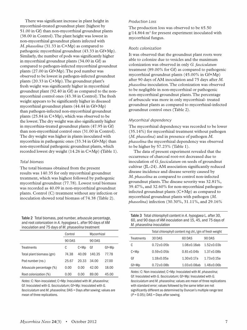
Table 2 Total biomass, pod number, arbuscule percentage, and root colonization in A. hypogaea L. after 90 days of AM inoculation and 75 days of M. phaseolina treatment
Control Mycorrhizal
90 DAS 90 DAS
Treatments C C+Mp Gf Gf+Mp
Total plant biomass (gm) 74.38 40.09 140.35 77.78
Pod number (no.) 25.67 20.33 34.00 27.00
Arbuscule percentage (%) 0.00 0.00 42.00 18.00
Root colonization (%) 0.00 0.00 89.00 45.00
Notes: C: Non-inoculated; C+Mp: Inoculated with M. phaseolina; Gf: Inoculated with G. fasciculatum; Gf+Mp: Inoculated with G. fasciculatum and M. phaseolina; DAS = Days after sowing; values are mean of three replications.
Table 3 Total chlorophyll content in A. hypogaea L. after 30, 60, and 90 days of AM inoculation and 15, 45, and 75 days of M. phaseolina inoculation
Total chlorophyll content mg chl./gm of fresh weight
Treatments 30 DAS 60 DAS 90 DAS
C 0.72±0.05b 1.08±0.18ab 1.52±0.03b
C+Mp 0.59±0.05b 0.81±0.04b 1.37±0.08b
Gf 1.18±0.05a 1.30±0.17a 1.73±0.15a
Gf+Mp 0.72±0.08b 1.03±0.08ab 1.48±0.06b
Notes: C: Non-inoculated; C+Mp: Inoculated with M. phaseolina; Gf: Inoculated with G. fasciculatum; Gf+Mp: Inoculated with G. fasciculatum and M. phaseolina; values are mean of three replications with standard error; values followed by the same letter are not significantly different as determined by Duncan’s multiple range test (P = 0.05); DAS = Days after sowing.
8 Mycorrhiza News 24(3) • October 2012
for Gf+Mp) after 30, 60, and 90 days after AM inoculation and 15, 45, and 75 days after M. phaseolina infection (Table 5). The incidences of root-rot disease caused by
M. phaseolina were reduced due to inoculation by G. fasciculatum as shown in Table 4. The disease incidence after 30, 60, and 90 days of AM inoculation and 15, 45, and 75 days after M. phaseolina infection was 53.33%, 63.33%, and 76.67% in pathogenic non-mycorrhizal (C+Mp) and 36.67%, 50.00%, and 53.33 % in mycorrhizal-infected (Gf+Mp) groundnut plants.
Discussion The present field experiment showed that groundnut plants were able to attain colonization by AM fungus G. fasciculatum. The ability of Glomus species to colonize the roots of groundnut was shown by Simpson and Daft (1990). The present study showed that due to colonization by G. fasciculatum, overall increase in the growth of groundnut plant was observed. The significant growth of groundnut plants may be attributed to mycorrhizal colonization as it is known to improve growth and provide other benefits to host plants during abiotic or biotic stresses (Singh et
al., 2000; Ludwig-Muller, 2004). It has been suggested that during colonization there is formation of vesicles and arbuscules in plants cells (Walker, 1992). The phenomenon of competition at the infection level has also been suggested. Even though both pathogen and VAM fungi colonize the same root system, but the development occurs in different cortical cells indicating some sort of competition for space (Azcon-Aguilar and Barea, 1996). So, improved growth of groundnut plants may indicate the potential of mycorrhizal plants in disease resistance.The content of total chlorophyll was affected by
infection when treated with mycorrhiza or pathogen M. phaseolina. The total chlorophyll content increased significantly in mycorrhizal groundnut when compared with non-mycorrhizal groundnut, but in the presence of pathogens chlorophyll content was found to decrease considerably, suggesting the role of the pathogens in such decrease. Similarly, degradation of chlorophyll was shown to be prevented by the increase in cytokinin content. Increase in the chlorophyll content has been showed by Allen and Allen (1981).As nearly 80% of plants on land are mycorrhizal
(Smith and Read, 2008) and in the present study inoculation with G. fasciculatum showed that colonization was higher in mycorrhizal roots of groundnut when pathogen M. phaseolina were not present. But, eventually the root colonization was found to decrease due to the presence of pathogen M. phaseolina, which suggests competition between the pathogen and AM fungus G. fasciculatum. The substrate for microbial activities is considered to be organic carbon in the rhizosphere or in the rhizoplane released from the roots of plants (Azaizeh et al., 1995). That is why carbon availability in mycorrhizal roots could suggest pathogen (M. phaseolina) resistance or decrease in incidence and severity in groundnut plants, which can be correlated with increased mycorrhizal colonization. Even Linderman (1994) and Smith (1987) suggested that both mycorrhizal fungi and root pathogens compete for the carbon compounds reaching the root.The “dependence” of a plant can be defined as its
ability to grow in the presence of colonization (Janos, 2007). Therefore, the incidence of disease and severity was recorded to be lower in pathogenic mycorrhizal groundnut as compared to healthy mycorrhizal groundnut plants. Similar results were obtained by Jaizme Vega et al. (1998) who showed increased dependency due to the presence of pathogen and lower dependency due to the absence of pathogen. The marked increase in “mycorrhizal dependency” clearly suggests the efficiency of mycorrhizal colonization by improvement in growth of groundnut plants that occurred during biotic stress exerted by the presence of pathogen M. phaseolina.
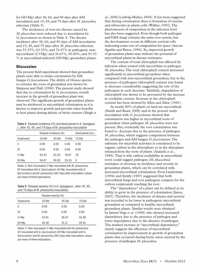
Table 4 Disease incidence (%) and dead plants in A. hypogaea L. after 15, 45, and 75 days of M. phaseolina inoculation
Disease incidence (%) Dead plants (no.)
Treatments 15 DAI 45 DAI 75 DAI 75 DAI
C 0.00 0.00 0.00 0.00
Gf 0.00 0.00 0.00 0.00
C+Mp 53.33 63.33 76.67 10
Gf+Mp 36.67 50.00 53.33 5
Notes: C: Non-inoculated; C+Mp: Inoculated with M. phaseolina; Gf: Inoculated with G. fasciculatum; Gf+Mp: Inoculated with G. fasciculatum and M. phaseolina; DAI: Days after inoculation; values are mean of three replications.
Table 5 Disease severity (%) in A. hypogaea L. after 15, 45, and 75 days of M. phaseolina inoculation
Disease severity (%)
Treatments 15 DAI 45 DAI 75 DAI
C 0.00 0.00 0.00
Gf 0.00 0.00 0.00
C+Mp 32.81 39.47 32.60
Gf+Mp 30.30 31.11 29.16
Notes: C: Non-inoculated; C+Mp: Inoculated with M. phaseolina; Gf: Inoculated with G. fasciculatum; Gf+Mp: Inoculated with G. fasciculatum and M. phaseolina; DAI: Days after inoculation; values are mean of three replications.
Mycorrhiza News 24(3) • October 2012 9
In conclusion, we may state that mycorrhizal symbiosis is universal and is associated with almost all plant species. The application of AM fungi in a field of groundnut plants infected with pathogen M. phaseolina could ensure the good health, growth, and suppression of disease in the plant.
References Arnon D J. 1949. Copper enzymes in isolated chloroplasts. J. Plant and Cell Physiology 4: 29–30.
Allen O N and Allen E K. 1981. The leguminosae, a source book of characteristics, uses, and nodulation. Madison, WI, USA: The University of Wisconsin Press.
Azaizeh H A, Marschner H, Romheld V, and Wittenmayer L. 1995. Effects of a vesicular–arbuscular mycorrhizal fungus and other soil microorganisms on growth, mineral nutrient acquisition, and root exudation of soil grown maize plants. Mycorrhiza 5: 321–327.
Azcon-Aguilar C and Barea J M. 1996. Arbuscular mycorrhizas and biological control of soil-borne plant pathogens: An overview of the mechanisms involved. Mycorrhiza 6: 457–464.
Gerdemann J W and Nicolson T H. 1963. Spores of mycorrhizal Endogene species extracted from soil by wet sieving and decanting. Transactions of the British Mycological Society 46: 235–244.
Gerdemann J W. 1975. Vesicular arbuscular mycorrhiza. In The development of and function of roots, edited by J G Torrey and D T Clarkson, pp. 575–592. London: Academic Press.
Giovannetti M, Mosse. 1980. An evaluation of techniques for measuring vesicular-arbuscular infection in roots. New Phytologist 84: 489–500.
Kornerup A and Wanscher J H. 1983. Methuen handbook of colour. 3rd Edition, pp. 252. London: E. Methuen and Co. Ltd.
Linderman R G. 1994. Role of VAM fungi in biocontrol. In Mycorrhizae and Plant Health, edited by F L Pfleger and R G Linderman, pp. 1–26. St. Paul, MN: American Phytopathology Society.
Ludwig-Muller J. 2004. From auxin homeostasis to understanding plant pathogen and plant symbiont interaction: Editor’s research interests. JPGR 23: 1–8.
Phillips J M and Hayman D S. 1970. Improved procedure for cleaning roots and staining parasitic and vesicular arbuscular mycorrhizal fungi for rapid assessment of infection. Transactions of the British Mycological Society 55: 158–160.
Declerck S, Risede J M, Rufyikiri G, and Delvaux B. 2002. Effects of arbuscular mycorrhizal fungi on severity of root rot of bananas caused by Cylindrocladium spathiphylli. Plant Pathology 51: 109–115.
Janos D P. 2007. Plant responsiveness to mycorrhizas differs from dependence upon mycorrhizas. Mycorrhiza 17: 75–91.
Jaizme-Vega M C, Sosa Hernandez B, and Hernandez J M. 1998. Interaction of arbuscular mycorrhizal fungi and the soil pathogen Fusarium oxyporum f. sp. cubense on the first stages of micropropagated Grande Naine banana. Acta Horticulturae 490: 285–95.
Johansson J F, Paul L R, and Finlay R D. 2004. Microbial interactions in the mycorrhizosphere and their significance for sustainable agriculture. FEMS Microbiology Ecology 48: 1–13.
Plenchette C, Fortin J A, and Furlan V. 1983. Growth responses of several plant species to mycorrhizae in a soil of moderate P fertility. I. Mycorrhizal dependency under field conditions. Plant Soil 70: 199–209.
Raut, J G and Bhombe B B. 1984. Longevity of M. phaseolina in sunflower seeds. Indian Phytopathology 37(2): 333–334.
Schenck N C and Perez Y. 1987. A manual for identification of vesicular-arbuscular mycorrhizal fungi, Gainesville, Florida: Synergistic Publications.
Shokes F M and Culbreath A K. 1997. Early and late leaf spots. In Compendium of Peanut Diseases, 2nd edition, edited by N K Burelle, D M Porter, R R Kabana, D H Smith, and P Subrahmanyam, pp. 17–20. St Paul, MN: American Phytopathology Society.
Simpson D and Daft M J. 1990. Effect of Glomus clarum and water stresss on growth and nitrogen- fixation in two genotypes of groundnut. Agriculture, Ecosystems & Environment. 35: 47–54.
Singh R, Adholeya A, and Mukerji K G. 2000. Mycorrhiza in control of soil-borne pathogens. In Mycorrhizal biology, pp. 173–196. London.
Smith G S. 1987. Interactions of nematodes and mycorrhizal fungi. In Vistas on Nematology, edited by J A Veech and D W Dickson, pp. 292–300. Hyattsville, MD: Soc. Nemat.
Smith S E and Read D J. 1997. Mycorrhizal Symbiosis, 2nd Edition, pp. 1–605. London: Academic Press
Smith S E and Read D J. 2008. Mycorrhizal Symbiosis, 3rd Edition. San Diego: Academic Press.
Teng P S. 1985. Construction of predictive models. 11. Forecasting crop losses. Adv. Plant Pathol. 3: 179–206.
Trappe J M. 1982. Phytopathol. 72: 1102–1108.
Walker C. 1992. Systamatics and taxonomy of the arbuscular endomycorrhizal fungi (Glomales): A possible way forward. Agronomie 12: 887–892.
Whipps J M. 2004. Prospects and limitations for mycorrhizas in biocontrol of root pathogens. Canadian Journal of Botany 82: 1198–1227.
10 Mycorrhiza News 24(3) • October 2012
Glomalin and its association with the rhizosphere soils of some trees in Warangal district, a.p, indiaV Praveen Kumar*1 and S Ram ReddyDepartment of Microbiology, Kakatiya University, Warangal - 506009 A.P
Pavan Kumar PindiDepartment of Microbiology, Palamuru University, Mahabubnagar - 509001 A.P
Introduction Plant and soil health are dependent upon the interactions of biological, physical, and chemical components of the soil. The symbiotic relationship between roots and Arbuscular Mycorrhiza (AM) fungi may also benefit the formation of soil structure (Hallett et al. 2009). Rillig et al. (2010) revealed that AM fungal mycelium alone could be sufficient to form and/or maintain water-stable soil macro-aggregates. AM fungi are considered to be the most common and ubiquitous underground endophytic fungi — serving as a crucial link within the plant and soil continuum (Wilson et al. 2009) and are treated as a principal functional component in the belowground ecosystem (Smith and Read 2008; Siddiky 2011). The rhizosphere, or root zone, is the location of the greatest flow of energy and minerals among these components (Wright and Millner, 1994). In this highly productive region, a vital symbiotic relationship exists between roots and soil-borne AM fungi (Smith and Read 1997). Mycorrhiza is the most efficient mechanism for Phosphorus (P) acquisition, especially under stress conditions. Although these fungi are not host-specific, host and fungal genotypes and soil abiotic and biotic variables have been shown to influence the nature of the symbiosis (Brundrett, 1991; Gianinazzi et al., 1995; Varma, 1995). Even though AM fungi may be important in natural and managed systems (Bever et al., 2009; Wilson et al., 2009; Klironomos et al., 2011), little is known about the factors that determine their community structure and symbiotic functioning as drivers of plant productivity; as a result, their adaptive evolution is less known (Rosendahl, 2008; Antunes et al., 2011). In addition to improving plant health, mycorrhizal fungi also contribute to soil health. Fungal hyphae improve soil structure by helping to form water-stable soil aggregates (Miller and Jastrow, 1990; Tisdall et al., 1997; Rillig and Steinberg, 2002). Mycorrhizal fungi also improve rhizosphere health by stimulating root exudation which promotes the growth of other beneficial soil microbes (Borowicz, 2001; Paul and Clark, 1996).
Glomalin is a glycoproteinaceous molecule produced by the hyphae of AM fungi (Wright and Upadhyaya, 1999; Rillig et al., 2001). Glomalin is dark reddish-brown in colour; however, after extraction it loses the brown colour associated with organic matter. The brown colour of glomalin is attributed to the incorporation of iron as a structural component that may play a role in accumulation and/or function (Wright and Upadhyaya, 1998). The identification of glomalin has led to a re-evaluation of fungal contributions to Soil Organic Matter (SOM) and aggregate stability. Glomalin was identified at the USDA in the early 1990s in course of the work to produce monoclonal antibodies reactive with AM fungi. One of these antibodies reacted with a substance on the hyphae of a number of AM species (Wright et al., 1996). This substance was named as glomalin after Glomales, the order to which all AM fungi belong. The glomalin fraction is operationally defined by its extraction procedure, but it is further characterized by total and immunoreactive protein assays (Wright et al., 1996). The evidence that glomalin is produced by AM fungi, not plant roots, was obtained in the investigation of the reaction of the monoclonal antibodies against glomalin. In a blind experiment, immunofluorescence correctly identified glomalin only on roots that were later described as having AM colonization (Morton 1990). Glomalin is reported to be present on the
extramatrical hyphae of all AMF (except Sclerocystis) tested to date (Wright et al., 1996). As hyphae degrade, this hydrophobic, highly stable glycoprotein sloughs off to coat organic matter and other soil particles. Wright et al. (1996) hypothesized that glomalin forms a conglomeration with root fragments and organic matter, thus, protecting it from degradation by microorganisms. Wright and Upadhyaya (1998) found a strong correlation between glomalin concentration and soil aggregation.Several ‘pools’ of glomalin have been identified
based on solubility characteristics: (i) easily extractable glomalin (EEG); (ii) total glomalin (TG);
* Corresponding Author, Email: [email protected]
Mycorrhiza News 24(3) • October 2012 11
and (iii) a ‘scum’ at the air-water interface that occurs during the harvesting of hyphae from pot-cultured AM fungi. Hydrophobic and/or cationic interactions may be the mechanisms by which glomalin becomes deposited on soil or organic particles, and mesh or glass beads (Wright and Upadhyaya, 1996). Glomalin may move in and out of these operationally defined pools (i.e., EEG becomes scum and scum becomes TG). Steinberg and Rillig (2003) found that following soil incubation to measure decomposition, EEG increased while TG decreased. They speculated that partial degradation decreases sorption of glomalin to soil particles, which may increase solubility and the amount in the EEG pool. The formation of humic substances or,
more likely, glomalin, would provide the organic environment that plants need for productive growth. Glomalin would have evolved this function, because supposedly it was first formed when there was no other organic matter in the soil. In the present investigations, an attempt was made to assess the occurrence and amount of EEG and TG with an objective to find the distribution of AM fungi and to find the relationship between spore number and the amount of glomalin associated with the rhizosphere soils of some trees in Warangal district, Andhra Pradesh, India.
Material and methodsIn the present investigations, 22 rhizosphere soils of different tree species (Table 1) were collected from different agro-edaphic regions of Warangal district (Mylaram (My), Chelpur (CH), and Bhupalapally (BPL)), brought to a laboratory, and stored at 4°C until experimentation. The investigations were divided into two parts. In the first part, resting spores of AM fungi were extracted from rhizosphere soil and enumerated. In the second part, a quantitative estimation of easily extractable and total glomalin (Wright and Upadhyaya, 1996) was done.
Extraction and enumeration of AM resting sporesThe extraction and enumeration of AM resting spores was done by the method suggested by Gerdemann and Nicolson (1963). Ten grams of soil collected from rhizosphere was poured into 100 ml of water, subjected to shaking on a horizontal shaker for 30 minutes, and allowed to settle. The supernatant liquid was passed through a coarse sieve (500 to 800 mm) to remove large pieces of organic matter. The lower collected liquid was passed through sieves of decreasing pore size (250, 106 and 45 µm). This process was continued till all the colloidal materials
passed through the sieves. All the debris collected on the sieves was spread on a petridish and observed under a stereo-binocular microscope for the presence of resting spores; thereafter, the total number of sporespresent was enumerated.
Extraction of EEGOne gram of soil taken in an autoclavable centrifuge tube with 8 ml of 20 mM sodium citrate (pH 7.0) was autoclaved for 30 minutes at 121°C. Later it was centrifuged for 15 min at 8000 xg. The supernatant that contained the EEG was collected, quantitatively measured, and then stored at 4°C until further use.
Extraction of TGOne gram of soil was placed in an autoclavable centrifuge tube with 8 ml of 50 mM sodium citrate (pH 8.0) and autoclaved for 60 min at 121°C and then centrifuged for 15 min at 8000 x g. The supernatant containing the protein (TG) was collected and stored at 4°C. This process was repeated until all the TG was extracted. The supernatant contains the protein (TG). The extracts obtained in each round were pooled together and the final total volume was measured with a graduated cylinder.
Quantitative estimation of glomalinThe quantitative estimation of glomalin was done using the method suggested by Lowry et al., (1951). Five millilitres of alkaline copper sulphate reagent was added to 1 ml of extract and incubated at room temperature for 10 minutes. To this 1 ml of 1 N NaOH and later 0.5 ml of FC reagent were added and the optical density of the resultant blue colour was read at 660 nm. The concentration of glomalin was calculated from the standard graph plotted for bovine serum albumin (BSA).The results obtained were subjected to statistical
analysis using Smith’s Statistical Package (SSP version 2) at alpha levels of 0.05. Analysis of variance (ANOVA) was calculated for different parameters.
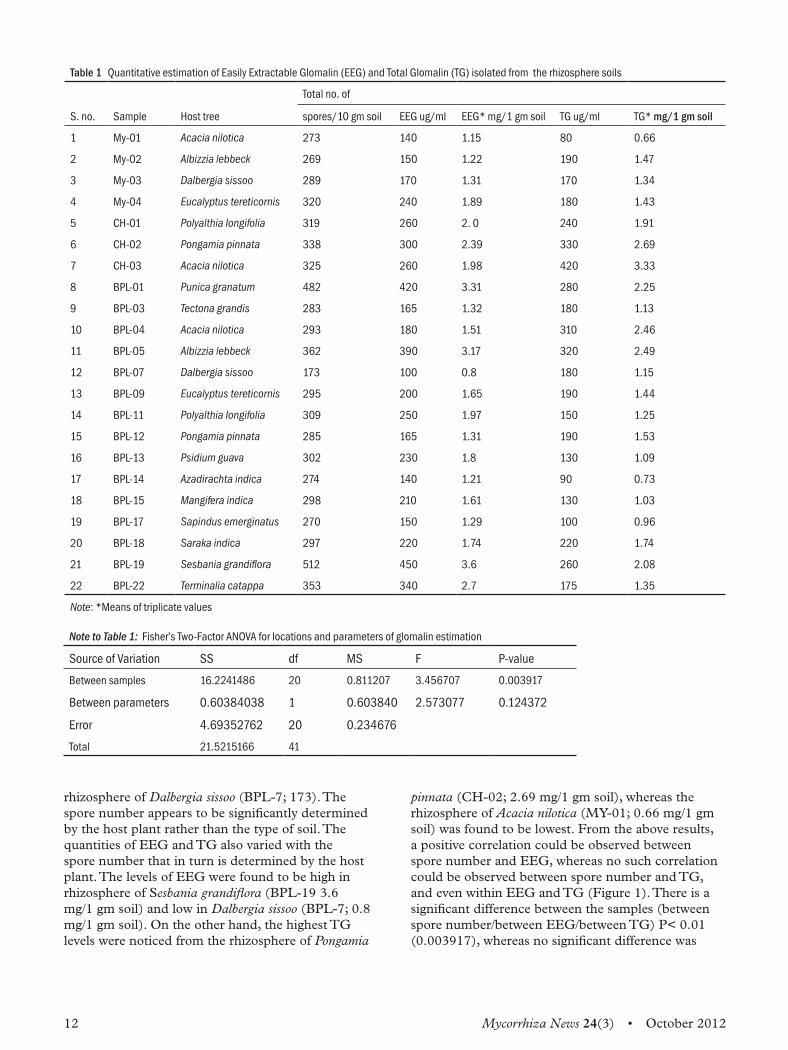
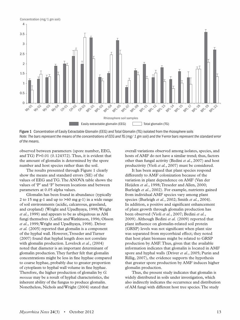
Results and discussionThe results obtained with regard to total number of spores and the quantity of EEG and TG are presented in Table 1.A critical perusal of Table 1 reveals that the AM
fungal spore population varied with the host tree species and the type of soil. The highest number of spores was recorded from rhizosphere of Sesbania grandiflora (BPL-19; 512) and the lowest from
12 Mycorrhiza News 24(3) • October 2012
rhizosphere of Dalbergia sissoo (BPL-7; 173). The spore number appears to be significantly determined by the host plant rather than the type of soil. The quantities of EEG and TG also varied with the spore number that in turn is determined by the host plant. The levels of EEG were found to be high in rhizosphere of Sesbania grandiflora (BPL-19 3.6 mg/1 gm soil) and low in Dalbergia sissoo (BPL-7; 0.8 mg/1 gm soil). On the other hand, the highest TG levels were noticed from the rhizosphere of Pongamia
pinnata (CH-02; 2.69 mg/1 gm soil), whereas the rhizosphere of Acacia nilotica (MY-01; 0.66 mg/1 gm soil) was found to be lowest. From the above results, a positive correlation could be observed between spore number and EEG, whereas no such correlation could be observed between spore number and TG, and even within EEG and TG (Figure 1). There is a significant difference between the samples (between spore number/between EEG/between TG) P< 0.01 (0.003917), whereas no significant difference was
Table 1 Quantitative estimation of Easily Extractable Glomalin (EEG) and Total Glomalin (TG) isolated from the rhizosphere soils
Total no. of
S. no. Sample Host tree spores/10 gm soil EEG ug/ml EEG* mg/1 gm soil TG ug/ml TG* mg/1 gm soil
1 My-01 Acacia nilotica 273 140 1.15 80 0.66
2 My-02 Albizzia lebbeck 269 150 1.22 190 1.47
3 My-03 Dalbergia sissoo 289 170 1.31 170 1.34
4 My-04 Eucalyptus tereticornis 320 240 1.89 180 1.43
5 CH-01 Polyalthia longifolia 319 260 2. 0 240 1.91
6 CH-02 Pongamia pinnata 338 300 2.39 330 2.69
7 CH-03 Acacia nilotica 325 260 1.98 420 3.33
8 BPL-01 Punica granatum 482 420 3.31 280 2.25
9 BPL-03 Tectona grandis 283 165 1.32 180 1.13
10 BPL-04 Acacia nilotica 293 180 1.51 310 2.46
11 BPL-05 Albizzia lebbeck 362 390 3.17 320 2.49
12 BPL-07 Dalbergia sissoo 173 100 0.8 180 1.15
13 BPL-09 Eucalyptus tereticornis 295 200 1.65 190 1.44
14 BPL-11 Polyalthia longifolia 309 250 1.97 150 1.25
15 BPL-12 Pongamia pinnata 285 165 1.31 190 1.53
16 BPL-13 Psidium guava 302 230 1.8 130 1.09
17 BPL-14 Azadirachta indica 274 140 1.21 90 0.73
18 BPL-15 Mangifera indica 298 210 1.61 130 1.03
19 BPL-17 Sapindus emerginatus 270 150 1.29 100 0.96
20 BPL-18 Saraka indica 297 220 1.74 220 1.74
21 BPL-19 Sesbania grandiflora 512 450 3.6 260 2.08
22 BPL-22 Terminalia catappa 353 340 2.7 175 1.35
Note: *Means of triplicate values
Note to Table 1: Fisher’s Two-Factor ANOVA for locations and parameters of glomalin estimation
Source of Variation SS df MS F P-value
Between samples 16.2241486 20 0.811207 3.456707 0.003917
Between parameters 0.60384038 1 0.603840 2.573077 0.124372
Error 4.69352762 20 0.234676
Total 21.5215166 41
Mycorrhiza News 24(3) • October 2012 13
observed between parameters (spore number, EEG, and TG) P>0.01 (0.124372). Thus, it is evident that the amount of glomalin is determined by the spore number and host species rather than the soil.The results presented through Figure 1 clearly
show the means and standard errors (SE) of the values of EEG and TG. The ANOVA table shows the values of ‘P’ and ‘F’ between locations and between parameters at 0.05 alpha values.Glomalin has been found in abundance (typically
2 to 15 mg g-1 and up to >60 mg g-1) in a wide range of soil environments (acidic, calcareous, grassland, and cropland) (Wright and Upadhyaya, 1998; Wright et al., 1999) and appears to be as ubiquitous as AM fungi themselves (Carlile and Watkinson, 1996; Olsson et al., 1999; Wright and Upadhyaya, 1998). Driver et al. (2005) reported that glomalin is a component of the hyphal wall. However, Treseder and Turner (2007) found that hyphal length does not correlate with glomalin production. Lovelock et al., (2004) noted that diameter is an important determinant of glomalin production. They further felt that glomalin concentrations might be less in fine hyphae compared to coarse hyphae, probably due to greater proportion of cytoplasm to hyphal wall volume in fine hyphae. Therefore, the higher production of glomalin by G. mosseae may be a result of hyphal characteristics, the inherent ability of the fungus to produce glomalin. Nonetheless, Nichols and Wright (2004) stated that
overall variations observed among isolates, species, and hosts of AMF do not have a similar trend; thus, factors other than fungal activity (Bedini et al., 2007) and host productivity (Violi et al., 2007) must be considered.It has been argued that plant species respond
differently to AMF colonization because of the variation in plant dependence on AMF (Van der Heijden et al., 1998; Treseder and Allen, 2000; Burleigh et al., 2002). For example, nutrients gained from individual AMF species vary among plant species (Burleigh et al., 2002; Smith et al., 2004). In addition, a positive and significant enhancement of plant growth through glomalin production has been observed (Violi et al., 2007; Bedini et al., 2009). Although Bedini et al. (2009) reported that plant influence on glomalin-related soil protein (GRSP) levels was not significant when plant size was separated from mycorrhizal effect; they noted that host plant biomass might be related to GRSP production by AMF. Thus, given that the available information indicates that glomalin is located in AMF spore and hyphal walls (Driver et al., 2005; Purin and Rillig, 2007), the evidence supports the hypothesis that greater spore production by AMF induces higher glomalin production.Thus, the present study indicates that glomalin is
widely distributed in soils under investigation, which also indirectly indicates the occurrence and distribution of AM fungi with different host tree species. The study
0
0.5
1
1.5
2
2.5
3
3.5
4
Easily extractable glomalin (EEG) Total glomalin (TG)
My-
01
My-
02
My-
03
My-
04
CH-01
CH-02
CH-03
BPL-01
BPL-03
BPL-04
BPL-05
BPL-07
BPL-09
BPL-11
BPL-12
BPL-13
BPL-14
BPL-15
BPL-17
BPL-18
BPL-19
BPL-22
Rhizosphere soil samples
Concentration (mg/1 gm soil)
Figure 1 Concentration of Easily Extractable Glomalin (EEG) and Total Glomalin (TG) isolated from the rhizosphere soils Note: The bars represent the means of the concentrations of EEG and TG (mg/ 1 gm soil) and the Y-error bars represent the standard error of the means.
14 Mycorrhiza News 24(3) • October 2012
further reveals the fact that glomalin plays a significant role in the formation of soil aggregates, and the stability and productivity of the plant.
AcknowledgementsThanks are due to the Head of the Department of Microbiology, Kakatiya University, for his encouragement and usage of the facilities available at the university. Financial assistance received from the UGC, New Delhi, is gratefully acknowledged.
ReferencesAntunes P M, Lehmann A, Hart M M, Baumecker M and Rillig M C. 2011. Long-term effects of soil nutrient deficiency on arbuscular mycorrhizal communities, Functional Ecology doi: 10.1111/j.1365-2435.2011.01953.x
Bedini S, Avio L, Argese E and Giovannetti M. 2007. Effects of long-term land use on arbuscular mycorrhizal fungi and glomalin-related soil protein, Agriculture, Ecosystems and Environment 120: 463–466
Bedini S, Pellegrino E, Avio L, Pellegrini S, Bazzoffi P, Argese E and Giovannetti M. 2009. Changes in soil aggregation and glomalin-related soil protein content as affected by the arbuscular mycorrhizal fungal species Glomus mosseae and Glomus intraradices, Soil Biology and Biochemistry 41: 1491–1496
Bever J D, Schultz P A, Pringle A, and Morton J B. 2009. Arbuscular mycorrhizal fungi: More diverse than meets the eye, and the ecological tale of why, BioScience, 51: 923–932
Borowicz V A. 2001. Do arbuscular mycorrhizal fungi alter plant-pathogen relations? Ecology 82: 3057–3068.
Brundrett M C. 1991. Mycorrhizas in natural ecosystems. In Adv. Ecol. Res, Vol. 21 pp. 171–313, edited by A Macfayden, M Begon, and A H Fitter. London, UK: Academic Press.
Burleigh S H, Cavagnaro T and Jakobsen I. 2002. Functional diversity of arbuscular mycorrhizas extends to the expression of plant genes involved in P nutrition, Journal of Experimental Botany 53: 1593–1601
Carlile M J and Watkinson S C. 1996. Parasites and mutualistic symbionts. In The Fungi, Chapter 7, pp. 307–371, edited by M J Carlile and S C Watkinson. New York: Academic Press.
Driver J D, Holben W E, and Rillig M C. 2005. Characterization of glomalin as a hyphal wall component of arbuscular mycorrhizal fungi, Soil Biology and Biochemistry 37:101–106.
Gerdemann J W and Nicolson T H. 1963. Spores of mycorrhizal endogone species extracted from soil by wet sieving and decanting, Transactions of the British Mycological Society 46: 235–244.
Gianinazzi S, Trouvelot A, Lovato P, Van T D, Franken P and Gianinazzi-Pearson V. 1995. Arbuscular mycorrhizal fungi in plant production of temperate agroecosystems, Critical Reviews in Biotechnology 15: 305–311.
Hallett P D, Debbie S F, Glyn B A, Rillig M C, Charlie M S, and Young I M. 2009. Disentangling the impact of AM fungi versus roots on soil structure and water transport, Plant Soil 314:183–196
Klironomos J, Zobel M, Tibbett M, Stock W D, Rillig M C, Parrent J L, Moora M, Koch A M, Facelli J M, Facelli E, Dickie I A and Bever J D. 2011. Forces that structure plant communities: Quantifying the importance of the mycorrhizal symbiosis, New Phytologist 189: 366–370
Lovelock C E, Wright S F and Nichols K A. 2004. Using glomalin as an indicator for arbuscular mycorrhizal hyphal growth: An example from a tropical rain forest soil, Soil Biology and Biochemistry 36: 1009–1012
Lowry O H, Rosebrough N J, Farr A L and Randall R J. 1951. Protein measurement with Folin phenol reagent, The Journal of Biological Chemistry 193: 265–275
Miller R M and Jastrow J D. 1990. Hierarchy of root and mycorrhizal fungal interactions with soil aggregation, Soil Biology and Biochemistry 22: 579–584
Morton J B. 1990. Species and clones of arbuscular mycorrhizal fungi (Glomales, Zygomycetes): Their role in macro- and micro-evolutionary processes, Mycotaxon 37: 493–515
Nichols K A and Wright S F. 2004. Contributions of fungi to soil organic matter in agroecosystems. In Soil Organic Matter in Sustainable Agriculture, pp. 179–198, edited by F Magdoff and R R Weil. Florida: CRC Press.
Olsson P A, Thingstrup I, Jakobsen I and Baath E. 1999. Estimation of the biomass of arbuscular mycorrhizal fungi in linseed field, Soil Biology and Biochemistry 31: 1879–1887
Paul E A and Clark F E. 1996. Soil microbiology and biochemistry, 2nd Edition. New York: Academic Press.
Purin S and Rillig M C. 2007. The arbuscular mycorrhizal fungal protein glomalin: Limitations, progress, and a new hypothesis for its function, Pedobiologia 51: 123–130
Rillig M C, Wright S F, Nichols K A, Schmidt W F, and Torn M S. 2001. Large contribution of arbuscular mycorrhizal fungi to soil carbon pools in tropical forest soils, Plant and Soil 233: 167–177
Rillig M C and Steinberg P D. 2002. Glomalin production by an arbuscular mycorrhizal fungus: A mechanism of habitat modification, Soil Biology and Biochemistry 34:1371–1374
Mycorrhiza News 24(3) • October 2012 15
Rillig M C, Mardatin N F, Leifheit E F, and Antunes P M. 2010.Mycelium of arbuscular mycorrhizal fungi increases soil water repellency and is sufficient to maintain water-stable soil aggregates, Soil Biology and Biochemistry 42: 1189–1191
Rosendahl S. 2008. Communities, populations and individuals of arbuscular mycorrhizal fungi, New Phytologist 178: 253–266
Siddiky M R K. 2011. Soil biota interactions and soil aggregation, Ph.D Thesis, p. 8. Berlin, Germany: Freie Universität.
Smith S E and Read D J. 1977. Mycorrhizal symbiosis, 1st Edition. San Diego, CA: Academic Press.
Smith S E and Read D J. 2008. Mycorrhizal Symbiosis, Third Edition, Academic Press.
Smith S E, Smith F A, and Jakobsen I. 2004. Functional diversity in arbuscular mycorrhizal (AM) symbioses: The contribution of the mycorrhizal P uptake pathway is not correlated with mycorrhizal responses in growth or total P uptake, New Phytololgist 162: 511–524
Steinberg P D and Rillig M C. 2003. Differential decomposition of arbuscular mycorrhizal fungal hyphae and glomalin, Soil Biology and Biochemistry 35: 191–194
Tisdall J M, Smith S E, and Rengasamy P. 1997. Aggregation of soil by fungal hyphae,Australian Journal of Soil Research 35: 55–60
Treseder K K and Allen M F. 2000. Mycorrhizal fungi have a potential role in soil carbon storage under elevated CO2 and nitrogen deposition, New Phytologist 147:189–200
Treseder K K and Turner K M. 2007. Glomalin in ecosystems, Soil Science Society of America Journal 711: 257–1266
Van Der Heijden M G A, Klironomos J N, Ursic M, Moutoglis P, Streitwolf-Engel R, Boller T, Wiemken A, and Sanders I R. 1998. Mycorrhizal fungal diversity determines plant biodiversity, ecosystem variability and productivity, Nature 39:69–72
Varma A. 1995. Arbucular mycorrhizal fungi: The state of the art, Critical Reviews in Biotechnology 15: 179–199
Violi H A, Treseder K K, Menge J A, Wright S F and Lovatt C J. 2007. Density dependence and interspecific interactions between arbuscular mycorrhizal fungi mediated plant growth, glomalin production, and sporulation, Canadian Journal of Botany 85: 63–75
Wilson G B, Rice C W, Rillig M C, Springer A, Hartnett D C. 2009. Soil aggregation and carbon sequestration are tightly correlated with the abundance of arbuscular mycorrhizal fungi: Results from long term field experiments, Ecology Letter 12: 452–461.
Wright S F and Millner P D. 1994. Dynamic processes of vesicluar-arbuscular mycorrhizae: A mycorrhizosystem within the agroecosystem. In Advances in Soil Science. Soil Biology: Effects on Soil Quality, pp. 29–59, edited by J L Hatfeld and B A Stewart. Boca Raton, FL: Lewis Publishers.
Wright S F, Franke-Snyder M, Morton J B and Upadhyaya A. 1996. Time-course study and partial characterization of a protein on hyphae of arbuscular mycorrhizal fungi during active colonization of roots, Plant and Soil 181: 193–203.
Wright S F and Upadhyaya A. 1996. Extraction of an abundant and unusual protein from soil and comparison with hyphal protein from arbuscular mycorrhizal fungi, Soil Science 161: 575–586
Wright S F and Upadhyaya A. 1998. A survey of soils for aggregate stability and glomalin, a glycoprotein produced by hyphae of arbuscular mycorrhizal fungi, Plant and Soil 198: 97–107
Wright S F and Upadhyaya A. 1999. Quantification of arbuscular mycorrhizal activity by the glomalin concentration on hyphae, Mycorrhiza 8: 283–285
Wright S F, Starr J L, and Paltineanu I C. 1999. Changes in aggregate stability and concentration of glomalin during tillage management transition, Soil Science Society of America Journal 63: 1825–1829
16 Mycorrhiza News 24(3) • October 2012
impact of long-term wastewater irrigation on the abundance of arbuscular mycorrhizal spores in the peri-urban soil of Varanasi
Sumita Pal *and H B Singh*Department of Mycology and Plant Pathology, Institute of Agricultural Science, Banaras Hindu University (BHU), Varanasi- 221005, India
Amitava Rakshit*Department of Soil Science and Agricultural Chemistry, Institute of Agricultural Science, Banaras Hindu University (BHU), Varanasi - 221005, India
IntroductionThe growing competition for scarce water resources, coupled with laws limiting groundwater pumping, has led to utilization of low quality water in irrigated agriculture in the Indo-Gangetic plains(Gupta and Seth, 2007). However, applying wastewater to arable lands also involves certain environmental and agricultural risks (Sharma et al., 2008). Wastewater differs from fresh water in terms of higher contents of electrolytes, dissolved organic matter, suspended solids, and biochemical and chemical oxygen demand. These varied constituents in the applied water can affect soil physico-chemical and biological properties. Increased amount of heavy metals could prove to be toxic for soil microorganisms. Although the concentration of heavy metals in waste water are low, long-term use of such waste water on agricultural lands often results in the build-up of elevated levels of these metals in soils. Among soil microorganisms like Arbuscular mycorrhizal fungi (AMF),an important biotic component of agricultural soils,are known to play a key role in the mobilization and immobilization of metal cations (Rakshit and Ghosh, 2009; Pal, 2011), thereby changing their availability for plants by effectively enlarging the rhizosphere. However, only a few studies have been carried out involving interactions between AMF and metals as a source of soil disturbance. To our knowledge, no studies have been reported on the long-term effects of increasing concentrations of waste water on the diversity of mycorrhizal propagules or on the influence of the host plant on AM fungal diversity in soils polluted by heavy metals. Our aim in this study was to determine the manner in which AM fungal diversity is affected by the addition of waste water for a long period of time.
Materials and methodsStudy area
The experiment was conducted at an urban fringe of the subtropical area of Varanasi city, situated in the
* Email:[email protected], [email protected],[email protected]
Eastern Gangetic plain (25º18´ N latitude and 83º 01´ E longitude and 76 m above the sea level) of northern India, with an average annual rainfall of 1100 mm and mean annual temperature ranges between 20–42°C and 9–28°C, respectively. This field site has been contaminated by surface application of sewage sludge and surface irrigation with waste water generated from domestic sewage, effluents discharged from small-scale fabric, plastic, battery industries, dyeing, metal plating, bicycle tyres, and heavy agricultural equipment located in the urban areas of Varanasi since the 1990s.
Soil sample collection
Soil samples were collected in triplicate from rhizosphere of 15 crop species at a depth of 0–30 cm and combined to form approximately 500 g of soil. Roots were separated and 100 g of air-dried soil was employed for extraction of AM fungal spores. Rest were air dried, crushed, and passed through a 2mm mesh-sieve and stored at ambient temperature before analysis of soil properties and concentrations of heavy metals by the standard soil analysis technique. The available Cd and Ni were extracted by DTPA solution and analysed in atomic absorption spectrophotometer. The pH of the soil was determined by using a combined electrode (Jackson,1967) and EC by conductivity metre (1:2.5 soil water suspension); organic matter by chromic acid wet digestion method (Walkley and Black,1939); plant available nitrogen by alkaline permanganate method (Subbiah and Asija,1956); phosphorus by NaHCO
3
(Olsenetal.,1954); and potassium by flame photometry (Jackson,1958). Metal concentrations and the selected soil properties are showed in Table 1. According to the Indian National Standards Institution (Awasthi, 2000), this soil is seriously contaminated with Cd as described in previous studies (Sharma et al., 2008).
Arbuscular mycorrhiza spore extraction and spore count
AMF spores were isolated from 100 g of soil by wet sieving using two sieves with aperture sizes of 425
Mycorrhiza News 24(3) • October 2012 17
and 63 µm and decanting method (Gerdemann and Nicolson, 1963), followed by sucrose centrifugation using a 1.17 M sucrose solution at a speed of 2000 rpm for 5 minutes. After centrifugation, the supernatant was poured through 50 mm pore-size mesh and quickly rinsed with tap water. Spores were counted with a Don caster dish under the dissecting microscope and grouped according to morphological characteristics.
Enzymatic activity in the rhizosphere
In the laboratory, subsamples were taken from bulk samples and were further homogenized. Large roots or shells were removed and wet soil samples were added to polypropylene centrifuge tubes for analysis of enzymatic activities. For alkaline and acid phosphatase enzymatic activity, the base substrate used was p-nitrophenol bound with phosphate (Tabatabai and Brenmer, 1969). The artificial substrate (1 mL, 0.05 M), toluene to inhibit microbial growth during incubation, a pH buffer (pH 11 for alkaline phosphatase and pH 4.5 for acid phosphatase) were incubated in closed polypropylene centrifuge tubes at 37ºC for 1 hour. At the end of incubation, enzyme activity was stopped by addition of 4mL of 0.5M NaOH; the mixture was filtered and the extract
was analysed using a UV-VIS spectrophotometer at 420 nm. Absorbance of filtrates was compared with p-nitrophenol standards. To account for non-enzymatic substrate hydrolysis, values for controls were subtracted from sample replicates. For DHA activity 2, 3, 5-triphenyltetrazolium chloride (TTC) was added as substrate to the fresh moist soil samples and incubated at 37°C for 24 hours. After incubation, the triphenyltetrazoliumformazan (TTF) formed was extracted, centrifuged, and estimated spectrophotometrically at 485 nm. Dehydrogenase activity is expressed as microgram of TTF released per gram of dry soil per 24 hours (Casida et al., 1964). The urease activity was determined by the method proposed by Kandeler and Gerber (1988) and Wang et al. (2007).
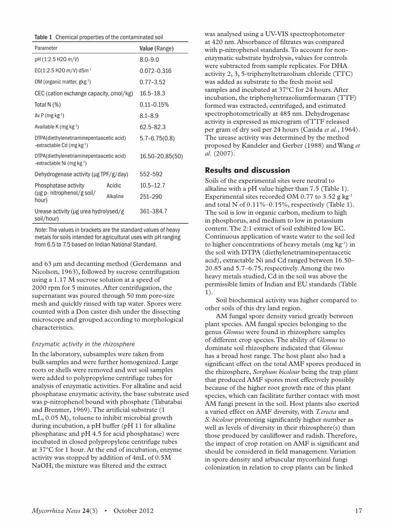
Results and discussionSoils of the experimental sites were neutral to alkaline with a pH value higher than 7.5 (Table 1). Experimental sites recorded OM 0.77 to 3.52 g kg-1
and total N of 0.11%–0.15%, respectively (Table 1). The soil is low in organic carbon, medium to high in phosphorus, and medium to low in potassium content. The 2:1 extract of soil exhibited low EC. Continuous application of waste water to the soil led to higher concentrations of heavy metals (mg kg-1) in the soil with DTPA (diethylenetriaminepentaacetic acid), extractable Ni and Cd ranged between 16.50–20.85 and 5.7–6.75, respectively. Among the two heavy metals studied, Cd in the soil was above the permissible limits of Indian and EU standards (Table 1). Soil biochemical activity was higher compared to
other soils of this dry land region.AM fungal spore density varied greatly between
plant species. AM fungal species belonging to the genus Glomus were found in rhizosphere samples of different crop species. The ability of Glomus to dominate soil rhizosphere indicated that Glomus has a broad host range. The host plant also had a significant effect on the total AMF spores produced in the rhizosphere, Sorghum bicolour being the trap plant that produced AMF spores most effectively possibly because of the higher root growth rate of this plant species, which can facilitate further contact with most AM fungi present in the soil. Host plants also exerted a varied effect on AMF diversity, with T.erecta and S. bicolour promoting significantly higher number as well as levels of diversity in their rhizosphere(s) than those produced by cauliflower and radish. Therefore, the impact of crop rotation on AMF is significant and should be considered in field management. Variation in spore density and arbuscular mycorrhizal fungi colonization in relation to crop plants can be linked
Table 1 Chemical properties of the contaminated soil
Parameter Value (Range)
pH (1:2.5 H2O m/V) 8.0–9.0
EC(1:2.5 H2O m/V) dSm-1 0.072–0.316
OM (organic matter, gkg-1) 0.77–3.52
CEC (cation exchange capacity, cmol/kg) 16.5–18.3
Total N (%) 0.11–0.15%
Av P (mg kg-1) 8.1–8.9
Available K (mg kg-1) 62.5–82.3
DTPA(diethylenetriaminepentaacetic acid) -extractable Cd (mg kg-1)
5.7–6.75(0.8)
DTPA(diethylenetriaminepentaacetic acid) -extractable Ni (mg kg-1)
16.50–20.85(50)
Dehydrogenase activity (µg TPF/g/day) 552–592
Phosphatase activity(µg p- nitrophenol/g soil/ hour)
Acidic 10.5–12.7
Alkaline 251–290
Urease activity (µg urea hydrolysed/g soil/hour)
361–384.7
Note: The values in brackets are the standard values of heavy metals for soils intended for agricultural uses with pH ranging from 6.5 to 7.5 based on Indian National Standard.
18 Mycorrhiza News 24(3) • October 2012
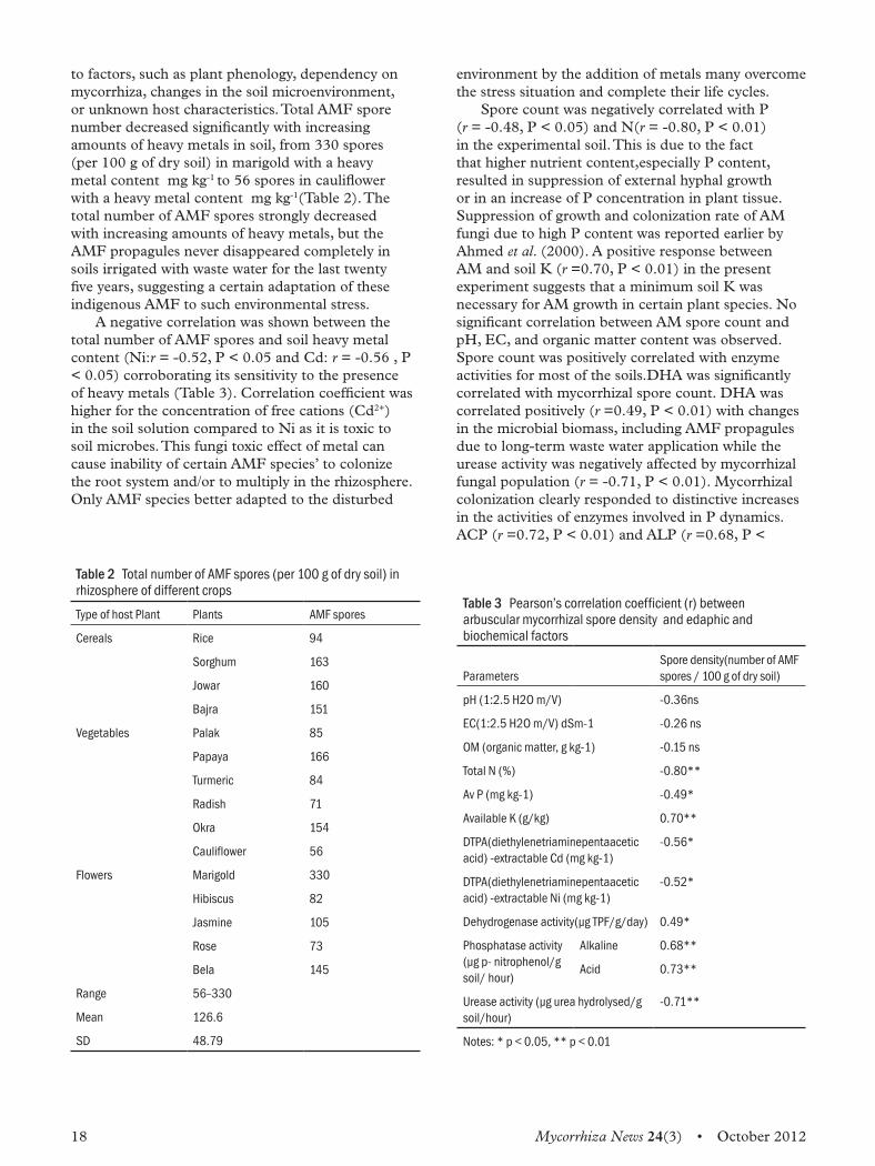
to factors, such as plant phenology, dependency on mycorrhiza, changes in the soil microenvironment, or unknown host characteristics. Total AMF spore number decreased significantly with increasing amounts of heavy metals in soil, from 330 spores (per 100 g of dry soil) in marigold with a heavy metal content mg kg-1 to 56 spores in cauliflower with a heavy metal content mg kg-1(Table 2). The total number of AMF spores strongly decreased with increasing amounts of heavy metals, but the AMF propagules never disappeared completely in soils irrigated with waste water for the last twenty five years, suggesting a certain adaptation of these indigenous AMF to such environmental stress.A negative correlation was shown between the
total number of AMF spores and soil heavy metal content (Ni:r = -0.52, P < 0.05 and Cd: r = -0.56 , P < 0.05) corroborating its sensitivity to the presence of heavy metals (Table 3). Correlation coefficient was higher for the concentration of free cations (Cd2+) in the soil solution compared to Ni as it is toxic to soil microbes. This fungi toxic effect of metal can cause inability of certain AMF species’ to colonize the root system and/or to multiply in the rhizosphere. Only AMF species better adapted to the disturbed
Table 2 Total number of AMF spores (per 100 g of dry soil) in rhizosphere of different crops
Type of host Plant Plants AMF spores
Cereals Rice 94
Sorghum 163
Jowar 160
Bajra 151
Vegetables Palak 85
Papaya 166
Turmeric 84
Radish 71
Okra 154
Cauliflower 56
Flowers Marigold 330
Hibiscus 82
Jasmine 105
Rose 73
Bela 145
Range 56–330
Mean 126.6
SD 48.79
environment by the addition of metals many overcome the stress situation and complete their life cycles.Spore count was negatively correlated with P
(r = -0.48, P < 0.05) and N(r = -0.80, P < 0.01) in the experimental soil. This is due to the fact that higher nutrient content,especially P content, resulted in suppression of external hyphal growth or in an increase of P concentration in plant tissue. Suppression of growth and colonization rate of AM fungi due to high P content was reported earlier by Ahmed et al. (2000). A positive response between AM and soil K (r =0.70, P < 0.01) in the present experiment suggests that a minimum soil K was necessary for AM growth in certain plant species. No significant correlation between AM spore count and pH, EC, and organic matter content was observed. Spore count was positively correlated with enzyme activities for most of the soils.DHA was significantly correlated with mycorrhizal spore count. DHA was correlated positively (r =0.49, P < 0.01) with changes in the microbial biomass, including AMF propagules due to long-term waste water application while the urease activity was negatively affected by mycorrhizal fungal population (r = -0.71, P < 0.01). Mycorrhizal colonization clearly responded to distinctive increases in the activities of enzymes involved in P dynamics. ACP (r =0.72, P < 0.01) and ALP (r =0.68, P <
Table 3 Pearson’s correlation coefficient (r) between arbuscular mycorrhizal spore density and edaphic and biochemical factors
ParametersSpore density(number of AMF spores / 100 g of dry soil)
pH (1:2.5 H2O m/V) -0.36ns
EC(1:2.5 H2O m/V) dSm-1 -0.26 ns
OM (organic matter, g kg-1) -0.15 ns
Total N (%) -0.80**
Av P (mg kg-1) -0.49*
Available K (g/kg) 0.70**
DTPA(diethylenetriaminepentaacetic acid) -extractable Cd (mg kg-1)
-0.56*
DTPA(diethylenetriaminepentaacetic acid) -extractable Ni (mg kg-1)
-0.52*
Dehydrogenase activity(µg TPF/g/day) 0.49*
Phosphatase activity(µg p- nitrophenol/g soil/ hour)
Alkaline 0.68**
Acid 0.73**
Urease activity (µg urea hydrolysed/g soil/hour)
-0.71**
Notes: * p < 0.05, ** p < 0.01
Mycorrhiza News 24(3) • October 2012 19
0.01) was positively correlated with the development of mycorrhizal spores. As heavy metals cannot be chemically degraded, bio-remediation of metal-polluted soils is limited mainly to immobilization through phytostabilization by promoting plant growth to reduce or eliminate the bioavailability of metals. In this context, role of indigenous AMF constitutes an important functional component of the soil-plant ecosystem that is critical for sustainable productivity in wastewater treated soils.
ReferencesAwasthi S K. 2000.Prevention of Food Adulteration Act No. 37 of 1954: Central and state rules as amended for 1999. Third Ed. Delhi: Ashoka Law House.
Casida L E Jr, Klein D A, and Santoro T. 1964.Soil dehydrogenase activity. Soil Science 98: 371–376.
Gerdemann J W and Nicolson T H. 1963.Spores of mycorrhizal Endogone species extracted from soil by wet sieving and decanting. Transactions of the British Mycological Society 46: 235–246.
Gupta R K and Seth A. 2007. A review of resource conserving technologies for sustainable management of the rice–wheat cropping system of the Indo-Gangetic Plains (GCP). Crop Protection 26:436–447.
Jackson M L. 1967. Soil chemical analysis, p. 498. New Delhi: Prentice Hall of India.
Kandeler E and Gerber H. 1988. Short-term assay of soil urease activity using colorimetric determination of ammonium. Biology and Fertility of Soils 6: 68–72.
Olsen S R, Cole C V, Watanabe F S, and Dean L A. 1954. Estimation of available phosphorus in soils by extraction with sodium bicarbonate. USDA, Circ. 939. Washington, DC: US Government Printing Office.
Pal S. 2011. Arbuscular Mycorrhiza: Useful tool for heavy metal bioremediation. International Journal of Agriculture, Environment and Biotechnology 4(4):397–401.
Rakshit A and Ghosh S. 2009.Bioremediation: Concepts and country experiences, p.175. Hyderabad:ICFAI University Press.
Sharma R K, Agrawal M, and Marshall F M. 2008.Heavy metals (Cu, Cd, Zn and Pb) contamination of vegetables in Urban India: A case Study in Varanasi. Environment Pollution 154: 254–263.
Subbiah B V and Asija G L. 1956. A rapid procedure for estimation of available nitrogen in soils. Current Science 25(8): 259–260.
Tabatabai M A and Bremner J M. 1969. Use of pnitrophenyl phosphate for assay of soil phosphatase activity. Soil Biology and Biochemistry 1: 301–307.
Wang M Y, Xia R X, Wu Q S, Liu J H, and Hu L M. 2007. Influence of arbuscular mycorrhizal fungi on microbes and enzymes of soils from different cultivated densities of red clover. Annals of Microbiology 57:1–7.
Walkley A and Black I A. 1939. An examination of Degtjareff method for determining soil organic matter and a proposed modification of the chromic acid titration method. Soil Science 37:29–37.
20 Mycorrhiza News 24(3) • October 2012
Mitochondrial genome sequences are more preferred reliable molecular markers in arbuscular Mycorrhizal Fungi over nuclear genome sequencesBurla Sashidhar and Alok Adholeya
Introduction Arbuscular Mycorrhiza Fungi (AMF) are vital components of the microbial soil community, forming the most commonly occurring underground network, probably the oldest and largest symbiotic association between the roots of more than 80% of all terrestrial plant species and members of the phylum Glomeromycota. AMF help plants in several ways such as by improving plant fitness by increasing soil health, seedling establishment, plant fecundity, and tolerance to some root pathogens. It also aids in improving soil nutrients uptake, especially of poor mobility nutrients like phosphorus which play an important role in carbon and nitrogen recycling, sequestration of toxic heavy metals, water relation and formation, etc., and the fungi itself is benefited from the plants for carbon nutrition. It is universally accepted that the more AMF species are present in one ecosystem, the more diverse the ecosystem will be. But, some AMF individuals perform better than others in improving the plants fitness and directly affect crop yields as a whole. Despite many positive effects on plant nutrition, knowledge of AMF genetics is limited because of its obligate biotrophic nature; single spore contains many nuclei with genetic differences. Until now, attempts to generate strain-specific genetic markers have proved unreliable. The current review is based on a newly published research article by Formey et al. (2012) on the method to generate reliable strain-specific genetic markers in different AMFs by designing specific primers for Polymerase Chain Reaction (PCR) that are able to differentiate five laboratory strains of Rhizophagus irregularis, all of which have been previously problematic to differentiate. Formey et al. (2012) found that intra- and interstrain variability in different isolates of AMF based on mitochondrial genome sequences that are invaded by mobile selfish DNA elements.
Nuclear genome polymorphism is high in arbuscular mycorrhizal fungiAMF play an important role in altering the plant
community structure and consequently increase plant diversity and productivity. In recent years, most of the research work on AMF was focused mainly on taxonomy, phylogeny, ecology, genetics, and functional symbiosis with application of different traditional and molecular methods but associated with a number of difficulties in studying AMF. Recently, Ehinger et al. (2012) found the relative abundance of four alleles of a single locus Bg112 after three successive generations. They have found that ‘families’ of single spore isolates derived from a single spore culture in the previous generation were more similar to one another than to families from other single spore isolates. The genetic diversity contained in one initial spore repeatedly gave rise to genetically different variants of the fungus with novel phenotypes. None of the alleles became extinct, and no new alleles were observed in this study. Traditionally, AMF identification was based
on the morphology of the spores (Gerdemann and Trappe, 1974; Morton, 1988; Walker, 1992). Other biochemical methods of identification and characterization of AMF were based on fatty acids profiling (Graham et al., 1995), spore protein profiling through SDS-PAGE (Avio and Giovannetti, 1998), and isozymes analysis of malate dehydrogenase and esterase loci (Dodd et al., 1996). These methods are not widely used but can complement the other methods of characterization and do not solely identify the AMF diversity. Intraspore rDNA variations were well reported in the genome of a single nucleus as well as in other organisms and/or among nuclei residing in the same cell (Buckler et al., 1997). The genetic studies on multiple nuclei in the glomeromycotan mycelia raised many contradictory statements; some reported that nuclei residing in single spore are heterokaryotic (Kuhn et al., 2001; Hijri and Sanders, 2005) while others data indicated that they are homokaryotic (Pawlowska and Taylor, 2004). A heterokaryotic genetic system implies absence of a fixed nuclear genotype for a fungal isolate, with populations of nuclei changing within a species. To address this issue, monoaxenic cultures of G.
centRe foR MycoRRhizal cultuRe collection
Mycorrhiza News 24(3) • October 2012 21
intraradices was grown in vitro using transformed carrot roots and amplified fragment length polymorphism (AFLP) analysis on G. intraradices showed a high degree of genetic and phenotypic diversity among individual isolates (Koch et al., 2004). rRNA genes of the genomic DNA are available
in high copy number with highly conserved as well as variable sections and hence they were explored as molecular marker for identification and characterization of AMF. These sequences allow in distinguishing taxa at many different levels (Saiki et al., 1988). The first studies on the subgroup of order Glomales on molecular phylogeny using 18S rDNA subunit sequence as a basis for detection of AMF was conducted by designing the specific primer VANS1 (Simon et al., 1992) and an identification system using SSCP later (Simon et al., 1993) was proposed. From then, AMF are molecularly characterized using different sets of primers at different regions of the nuclear encoded rDNA genes containing internal transcribed spacers ITS1, ITS2, and the 5.8S gene because of its high polymorphic nature. The regions of small-subunit (SSU) rRNA gene (Schussler et al., 2001), the regions of large-subunit (LSU) rRNA gene (Alves et al., 2006; Clapp et al., 2001; Raab et al., 2005), and a combination of all rRNA gene sequences that constitute complete 1500 bp fragment of rDNA locus was used as identification tool in AMF (Stockinger et al., 2010). Sometimes this has been proved with respect to single spore rDNA gene sequence analysis of the intra-species variations (Hijri et al., 1999; Jansa et al., 2002; Nilsson et al., 2008; Sokolski et al., 2010) are more than the inter-species variations (Borstler et al., 2008; Rosendahl, 2008), and because of this, rDNA single genetic locus does not give a clear cut-off between intra-species and inter-species variation. Hence, other highly conserved house-keeping protein-encoding genes were used as targets for the marker development for AMFs, i.e., actin gene sequence analysis (Helgason et al., 2003), β-tubulin gene sequence analyses (Corradi et al., 2004a; Corradi et al., 2004b; Msiska and Morton 2009), and P-type H+-ATPase (Corradi et al., 2004a; Ferrol et al., 2000) have been investigated, but success rate was very low. While genes for elongation factor (ef1-alpha), V-H+-ATPase, and F-ATPase subunit sequences were analyses (Sokolski et al., 2010) and found efficacious only for discriminating between unrelated Glomus species.A number of other molecular methods have been
investigated for studying the genetic diversity analysis and species identification. Which includes terminal restriction fragment length polymorphism (T-RFLP) with fluorescently labelled primers combined with restriction digestion to analyse the sequence variation
either from single or mixed-species DNA samples (Dickie and Fitzjohn, 2007) and increasingly used as high an throughput fingerprinting technique in AMF ecology. But, the peak-profile of T-RFLP and database T-RFLP also raised a number of errors. Croll et al. (2008) used 10 simple sequence repeat (SSR), introns of a nuclear gene as molecular markers while Mathimaran et al. (2008) used a set of 18 SSR markers to analyse genetic variation among eight different isolates of G. intraradices. Only two isolates from these sets appeared to be identical clones. Genetic diversity among fungal isolates was high, but isolates from far distance locations like from two different continents were not substantially different (Borstler et al., 2008). These results complemented those of Koch et al. (2004) and indicated that much of the global genetic diversity of G. intraradices could be represented just within one field site. Multilocus genotypes also have been identified from individual field collected spores of G. mosseae using markers from single copy nuclear genes GmFOX2, GmTOR2, and GmGIN1 (Stukenbrock and Rosendahl, 2005). An inorganic phosphate transporter gene sequence (852-bp section) has been used to discriminate between 10 Glomus fungal species represented by 25 strains. This gene was particularly valuable in differentiating between different similar morphotypes with nucleotide and amino acid sequence differences greater than 3%. This gene was also proposed as a reliable barcode for the Glomeromycetes (Sokolski et al., 2011). Neither the SSR or RFLP markers listed earlier, nor the ‘Single Nucleotide Polymorphisms’ of Stukenbrock and Rosendahl (2005) and inorganic phosphate transporter gene have been successfully applied to the mycorrhizal roots from the field so far.
Nuclear genome verses mitochondrial genomeThere has been a long debate over genome size and nuclear organization in AMF. Different studies conducted have showed different results varying in genome size and ploidy level. A large amount of sequencing data was generated (Martin et al., 2008), but the sequence assembly and annotation seems not efficient and still not possible because of its high genetic polymorphism among nuclei of a single spore. A few reports estimated the nuclear DNA content of AMF isolates but none of these studies have shown similar results on genome sizes. Hijri and Sanders (2004) showed size of the nuclear genome was high and the genome size and ploidy level of DAOM197198 isolate has already been determined in G. intraradices DAOM197198 which was also chosen for the first complete genome sequencing project on AMF (Martin et al., 2008) but it was a
22 Mycorrhiza News 24(3) • October 2012
long hard road to get the large genome sequence data assemble and annotate. The average genome size of AMF seems to be highly variable where Hijri and Sanders (2004), reported 15.7 Mb in G. intraradices DAOM197198 and 37 Mb in Glomus etunicatum (Hijri and Sanders, 2005), while it was reported 250 Mb in Glomus versiforme (Bianciotto and Bonfante, 1992) and 740 Mb in Gigaspora margarita (Hosny et al., 1997). Recently, Sedzielewska et al., (2011) showed that the genome size of G. intraradices AMykor isolate was 150 Mb which was in comparison with the nuclear DNA content of G. intraradices DAOM197198 and an Arabidopsis thaliana as reference genome sizes, measured using FC and Feulgen DNA image densitometry. These values are 10 times bigger than previously reported (Hijri and Sanders, 2004) but complement the results of FC data, indicate the true picture on the genome size of G. intraradices (Sedzielewska et al., 2011).In contrast, mitochondrial genome size is
comparatively small (usually < 100 kb) which ranges widely from 70 783 bp for DAOM197198 to 87 754 bp for MUCL_43204 (Formey et al., 2012). It has been shown that mitochondrial DNA in AMF was homogeneous within the isolates of the same species, which made it possible to sequence and assemble complete mtDNA sequence of an isolate G. intraradices by Lee and Young (2009). The sequencing showed that the genes are homogenous as there was no evidence of substantial sequence variations and hence mtDNA can act as good marker for identification which was true in terms of a long history as molecular markers in other organisms. In ectomycorrhizal fungal molecular identification and species characterization was carried out successfully using mtLSU rRNA analysis on roots colonized with EMF and the large sets of data generated were available for comparative study (Bruns et al., 1998). The first sequences from the mitochondrial genome of the AMF have been documented the presence of unique sequences without substantial variations in an isolates of G. intraradices and G. proliferum mtLSU rRNA region (Raab et al., 2005). Borstler et al. (2008) showed by using mtLSU rRNA gene, combined with RFLP approach that the genetic diversity within G. intraradice is substantially higher than previously expected. These results suggested that mtLSU rDNA gene sequences provide useful information on intra-specific variation that occurs in closely related Glomus species (Thiery et al., 2010) and these variations are mostly because of the presence/absence nature of introns. Based on the advancement in the mitochondrial genome sequencing projects more AMFs mtDNA genomes were sequenced and assembled. Till date, there are at least eight different AMF mtDNA genome that have been sequenced, two
of them were strains of G. irregularis – FACE#494 (Lee and Young, 2009) and DAOM197198 (HQ189519.1). Two other species of AM fungi – Gigaspora margarita (Pelin et al., 2012) and Gigaspora rosea (Nadimi et al., 2012) were sequence and have been published. Four different strains of G. irregularis AMF mtDNA genomes, MUCL_43204, MUCL_46239 and MUCL_46240/MUCL_46241 were sequenced and were submitted to Genbank with the accession numbers JQ514224, JQ514223 and JQ514225, respectively (Formey et al., 2012).
Mobile genetic elements invade randomly in Rhizophagus irregularies mitochondrial genome and showed no variation within strainThis wealth of mtDNA genome sequence information was then studied further for comparative analysis of mtDNA genomes to intra- and interstrain variability. All the mt DNA genomes are circular with an average GC content of 37% (Formey et al., 2012). The strains exhibit a different number of predicted open reading frames (ORFs), compared to FACE#494 having the lowest ORF content (19), while MUCL_43204 having the highest (34). One important feature was that all strains have the same classic set of proteins that was already annotated in the FACE#494 strain (Formey et al., 2012). These variations in the number of predicted ORFs could partly explain the variation in genome size. Intraspecific polymorphic regions would be helpful to design markers for fundamental genetic studies in vitro or in the field, or for quality control of AM fungal inoculum. These variations have prompted to explore these mitochondrial genes as a potential alternative to nuclear markers because of their intra-strain sequence homogeneity and high copy number per spore (i.e., each AMF spore typically contains hundreds of mitochondria). The mt genomes are unique and have been
shown homogeneous within isolates of the same species of each strain. Further, some mitochondrial intergenic regions vary substantially among isolates of G. irregularis and closely related species, with the presence of numerous insertions/deletions and eroded mobile elements. The presence of this polymorphism is mainly because of three types of variability generating element (VGE) viz., homing endonucleases (HEOs), DNA polymerase domain-containing open reading frames (DPDCO) and small inverted repeats (SIR) (Formey et al., 2012). The discovery these VGEs highlights the great intraspecific plasticity of the G. irregularis mt genome. These mobile genetic elements have eroded the mitochondrial genomes of G. irregularis strains, which range from just over 70 to 88 kb. Such interstrain variation had already been
Mycorrhiza News 24(3) • October 2012 23
reported in other fungi (e.g., Schizosaccharomyces pombe; Schafer, 2003), but it is important and yet unknown from where these mobile genetic elements were originated and how stable these variations exist within the isolate.
ReferencesAlves da Silva G, E Lumini, L Costa Maia, P Bonfante, and V Bianciotto. 2006. Phylogenetic analysis of Glomeromycota by partial LSU rDNA sequences. Mycorrhiza 16: 183–189.
Avio L and M Giovannetti (1998). The protein pattern of spores of arbuscular mycorrhizal fungi: Comparison of species, isolates and physiological stages. Mycological Research 102 (08): 985–990.
Bianciotto V and P Bonfante (1992). Quantification of nuclear DNA content of two arbuscular mycorrhizal fungi. Mycol Res. 96: 1071–1076.
Borstler B, PA Raab, O Thiery, JB Morton, D Redecker (2008). Genetic diversity of the arbuscular mycorrhizal fungus Glomus intraradices as determined by mitochondrial large subunit rRNA gene sequences is considerably higher than previously expected. New Phytologist 180 (2): 452–465.
Bruns TD, TM Szaro, M Gardes, KW Cullings, JJ Pan, DL Taylor, TR Horton, A Kretzer, M Garbelottoand, and Y Li (1998). A sequence database for the identification of ectomycorrhizal Basidiomycetes by phylogenetic analysis. Molecular Ecology 7 (3): 257–272.
Buckler ESI, A Ippolito, and TP Holtsford (1997). The evolution of ribosomal DNA: Divergent paralogues and phylogenetic implications. Genetics 145: 821–832.
Clapp JP, A Rodriguez, and JC Dodd (2001). Inter- and intra-isolate rRNA large subunit variation in Glomus coronatum spores. New Phytol. 149: 539–554.
Corradi N, G Kuhn, and IR Sanders (2004a). Monophyly of β-tubulin and H+-ATPase gene variants in Glomus intraradices: Consequences for molecular evolutionary studies of AM fungal genes. Fungal Genet. Biol. 41: 262–273.
Corradi N, M Hijri, L Fumagalli, and IR Sanders (2004b). Arbuscular mycorrhizal fungi (Glomeromycota) harbour ancient fungal tubulin genes that resemble those of the chytrids (Chytridiomycota). Fungal Gen. Biol. 41: 1037–1045.
Croll D, L Wille, H A Gamper, N Mathirmaran, P J Lammers, N Corradi, I R Sanders (2008). Genetic diversity and host plant preferences revealed by simple sequence repeat and mitochondrial markers in a population of the arbuscular mycorrhizal fungus Glomus intraradices. New Phytologist 178 (3): 672–687.
Dickie IA and RG Fitzjohn (2007). Using terminal restriction fragment length polymorphism (T-RFLP) to identify mycorrhizal fungi: A methods review. Mycorrhiza 17: 259–270.
Dodd JC, S Rosendahl, M Giovannetti, A Broome, L Lanfranco and C Walker (1996). Inter- and intraspecific variation within the morphologically-similar arbuscular mycorrhizal fungi Glomus mosseae and Glomus coronatum. New Phytol. 133: 113–122.
Ehinger MO, D Croll, AM Koch, and IR Sanders (2012). Significant genetic and phenotypic changes arising from clonal growth of a single spore of an arbuscular mycorrhizal fungus over multiple generations. New Phytologist 196: 853–861.
Ferrol N, JM Barea, C Azcon-Aguilar (2000). The plasma membrane H+-ATPase gene family in the arbuscular mycorrhizal fungus Glomus mosseae. Curr Genet 37: 112–118.
Formey D, M Moles, A Haouy, B Savelli, O Bouchez, G Becard, and C Roux (2012). Comparative analysis of mitochondrial genomes of Rhizophagus irregularis – syn. Glomus irregulare – reveals a polymorphism induced by variability generating elements. New Phytologist 196: 1217–1227.
Gerdemann J W and J M Trappe (1974). The Endogonales in the Pacific Northwest. Mycol. Mem. 5: 29–30.
Graham JH, NC Hodge, JB Morton (1995). Fatty acid methyl ester profiles for characterization of glomalean fungi and their endomycorrhizae. Appl Environ Microbiol. 61: 58–64.
Helgason T, I J Watson, and J P M Young (2003).Phylogeny of the Glomerales and Diversisporales (Fungi: Glomeromycota) from actin and elongation factor 1-alpha sequences. FEMS Microbiol. Lett. 229: 127–132.
Hijri M and IR Sanders (2004). The arbuscular mycorrhizal fungus Glomus intraradices is haploid and has a small genome size in the lower limit of eukaryotes. Fungal Genet. Biol. 41: 253–261.
Hijri M and IR Sanders (2005). Low gene copy number shows that arbuscular mycorrhizal fungi inherit genetically different nuclei. Nature 433: 160–163.
Hijri M, M Hosny, D van Tuinen, and H Dulieu (1999). Intraspecific ITS polymorphism in Scutellospora castanea (Glomales, Zygomycota) is structured within multinucleate spores. Fungal Genet. Biol. 26: 141–151.
Hosny M, JP Pais de Barros, V Gianinazzi-Pearson, H Dulieu (1997). Base composition of DNA from glomalean fungi: High amounts of methylated cytosine. Fungal Genet. Biol. 22: 103–111.
Jansa J, A Mozafar, S Banke, BA McDonald, and E Frossard (2002). Intra- and intersporal diversity of ITS rDNA sequences in Glomus intraradices assessed by cloning and sequencing, and by SSCP analysis. Mycol. Res. 106: 670–681.
Koch AM, G Kuhn, P Fontanillas, L Fumagalli, J Goudet, IR Sanders (2004). High genetic variability and low local diversity in a population of arbuscular mycorrhizal fungi. Proc. Natl. Acad. Sci., USA 101: 2369–2374.
24 Mycorrhiza News 24(3) • October 2012
Kuhn G, M Hijri, and IR Sanders (2001). Evidence for the evolution of multiple genomes in arbuscular mycorrhizal fungi. Nature 414: 745–748.
Lee J and JPW Young (2009). The mitochondrial genome sequence of the arbuscular mycorrhizal fungus Glomus intraradices isolate 494 and implications for the phylogenetic placement of Glomus. New Phytologist 183: 200–211.
Martin F, V Gianinazzi-Pearson, M Hijri, P Lammers, N Requena, IR Sanders, Y Shachar-Hill, H Shapiro, GA Tuskan, JP Young. 2008. The long hard road to a completed Glomus intraradices genome. New Phytol. 180: 747–750.
Mathimaran N, L Falquet, K Ineichen, C Picard, D Redecker, T Boller, A Wiemken (2008). Microsatellites for disentangling underground networks: Strain-specific identification of Glomus intraradices, an arbuscular mycorrhizal fungus. Fungal Gen. Biol. 45: 812–817.
Morton JB (1988). Taxonomy of VA mycorrhizal fungi: Classification, nomenclature, and identification. Mycotaxon. 32: 267–324.
Msiska Z and JB Morton (2009). Phylogenetic analysis of the Glomeromycota by partial β-tubulin gene sequences. Mycorrhiza 19: 247–254.
Nadimi M, D Beaudet, L Forget, M Hijri, BF Lang (2012). Group I intronmediated trans-splicing in mitochondria of Gigaspora rosea, and a robust phylogenetic affiliation of arbuscular mycorrhizal fungi with Mortierellales. Molecular Biology and Evolution 29 (9): 2199–2210.
Nilsson RH, E Kristiansson, M Ryberg, N Hallenberg, and K-H Larsson (2008). Intraspecific ITS variability in the kingdom Fungi as expressed in the international sequence databases and its implications for molecular species identification. Evol. Bioinform. 4: 193–201.
Pawlowska TE and JW Taylor (2004). Organization of genetic variation in individuals of arbuscular mycorrhizal fungi. Nature 427: 733–737.
Pelin A, J-F Pombert, A Salvioli, L Bonen, P Bonfante, N Corradi (2012). The mitochondrial genome of the arbuscular mycorrhizal fungus Gigaspora margarita reveals two unsuspected trans-splicing events of group I introns. New Phytologist 194: 836–845.
Raab PA, A Brennwald, and D Redecker (2005). Mitochondrial large ribosomal subunit sequences are homogeneous within isolates of Glomus (arbuscular mycorrhizal fungi, Glomeromycota). Mycol. Res. 109: 1315–1322.
Rosendahl S (2008). Communities, populations and individuals of arbuscular mycorrhizal fungi. New Phytol. 178: 253–266.
Saiki RK, DH Gelfand, S Stoffels, J Scharf, R Higuchi, GT Horn, KB Mullis, and HA Erlich (1988). Primer-directed enzymatic amplification of DNA with a thermostable DNA polymerase. Science 239: 487–491.
Schafer B (2003). Genetic conservation versus variability in mitochondria: The architecture of the mitochondrial genome in the petite-negative yeast Schizosaccharomyces pombe. Curr. Genet. 43: 311–326.
Schussler A, H Gehrig, D Schwarzott, and C Walker (2001). Analysis of partial Glomales SSU rRNA gene sequences: Implications for primer design and phylogeny. Mycol. Res. 105: 5–15.
Sedzielewska KA, J Fuchs, EM Temsch, K Baronian, R Watzke, and G Kunze (2011). Estimation of the Glomus intraradices nuclear DNA content. New Phytologist 192: 794–797.
Simon L, M Lalonde, and TD Bruns (1992). Specific amplification of 18S fungal ribosomal genes from vesicular arbuscular endomycorrhizal fungi colonizing root. Appl. Environ. Microbiol. 58: 291–295.
Simon L, RC Levesque, and M Lalonde (1993). Identification of endomycorrhizal fungi colonizing roots by fluorescent single-stranded conformation polymorphism-polymerase chain reaction. Appl. Environ Micobiol. 59: 4211–4215.
Sokolski S, Y Dalpe, S Seguin, D Khasa, CA Levesque and Y Piche (2010). Conspecificity of DAOM197198, the model arbuscular mycorrhizal fungus, with Glomus irregulare: molecular evidence with three protein-encoding genes. Botany 88: 829–838.
Sokolski S, Y Dalpe, and Y Piche (2011). Phosphate Transporter Genes as Reliable Gene Markers for the Identification and Discrimination of Arbuscular Mycorrhizal Fungi in the Genus Glomus. Appl. Env. Microbiol 77 (5): 1888–1891.
Stockinger H, M Krüger, and A Schüßler (2010). DNA barcoding of arbuscular mycorrhizal fungi. New Phytol. 187: 265–268.
Stukenbrock EH and S Rosendahl (2005). Development and amplifications of multiple co-dominant genetic markers for single spores of arbuscular mycorrhizal fungi by nested multiplex PCR. Fungal Gen. Biol. 42: 73–80.
Thiery O, B Börstler, K Ineichen, D Redecker (2010). Evolutionary dynamics of introns and homing endonuclease ORFs in a region of the large subunit of the mitochondrial rRNA in Glomus species (arbuscular mycorrhizal fungi, Glomeromycota). Molecular Phylogenetics and Evolution 55: 599–610.
Walker C (1992) Systematics and taxonomy of the arbuscular mycorrhizal fungi (Glomales): A possible way forward. Agronomie 12: 887–897.
Mycorrhiza News 24(3) • October 2012 25
Recent RefeRencesThe latest additions to the network’s database on mycorrhiza are published here for the members’ information. The list consists of papers from the following journals.
P Agriculture, Ecosystems & Environment P Applied Clay ScienceP Applied Soil Ecology P Basic and Applied Ecology P Ecological Modelling P Environmental and Experimental BotanyP Fungal Ecology P Journal of Integrative Agriculture P Journal of Microbiological Methods
P Journal of Plant Physiology P Pedobiologia P Pedosphere P Phytochemistry P Plant ScienceP Procedia Engineering P Scientia Horticulturae P Soil and Tillage ResearchP Soil Biology and Biochemistry
Arocena J M, Velde B, Robertson S J. 2012
Weathering of biotite in the presence of arbuscular mycorrhizae in selected agricultural crops. Applied Clay Science 6: 12–17
University of Northern British Columbia, Prince George, BC, Canada V2N4Z9
Asensio Dolores, Rapparini Francesca, Peñuelas Josep. 2012
AM fungi root colonization increases the production of essential isoprenoids vs. nonessential isoprenoids especially under drought stress conditions or after jasmonic acid application. Phytochemistry 77: 149–161 Global Ecology Unit CREAF-CEAB-CSIC, Center for Ecological Research and Forestry Applications, Edifici C,Universitat Autònoma de Barcelona, 08193 Barcelona, Spain
Brito Isabel, Goss Michael J, Carvalho Mário de, Chatagnier Odile, Tuinen Diederik van. 2012
Impact of tillage system on arbuscular mycorrhiza fungal communities in the soil under Mediterranean conditions. Soil and Tillage Research 121: 63–67
Universidade de Évora – ICAAM, Apartado 94, 7002 – 554 Évora, Portugal
Chmura, Damian, Gucwa-Przepióra Ewa. 2012
Interactions between arbuscular mycorrhiza and the growth of the invasive alien annual Impatiens parviflora DC: A study of forest type and soil properties in nature reserves (S Poland). Applied Soil Ecology 62: 71–80
[Institute of Engineering and Environmental Protection, Faculty of Materials and Environmental Sciences, University of Bielsko-Biała, 2 Willowa Str, PL 43-309 Bielsko-Biała, Poland]
Estrada Beatriz, Aroca Ricardo, Barea José Miguel, Ruiz-Lozano Juan Manuel. 2012
Native arbuscular mycorrhizal fungi isolated from a saline habitat improved maize antioxidant systems and plant tolerance to salinity Plant Science, In Press, Accepted Manuscript, Available online 3 December 2012
Departamento de Microbiología del Suelo y Sistemas Simbióticos. Estación Experimental del Zaidín (CSIC). Profesor Albareda n° 1, 18008 Granada, Spain
Fujimura Kei E, Egger Keith N. 2012
Host plant and environment influence community assembly of High Arctic root-associated fungal communities. Fungal Ecology 5(4): 409–418
Ecosystem Science & Management Program, University of Northern British Columbia, Prince George, BC, V2N 4Z9, Canada
George Carmen, Wagner Marcel, Kücke Martin, Rillig Matthias C. 2012
Divergent consequences of hydrochar in the plant–soil system: Arbuscular mycorrhiza, nodulation, plant growth and soil aggregation effects. Applied Soil Ecology 59: 68–72
Freie Universität Berlin, Institut für Biologie, Plant Ecology, Altensteinstr. 6, D-14195 Berlin, Germany
Name of the author(s) and year of publication
Title of the article, name of the journal, volume number, issue number, page numbers (address of the first author or of the corresponding author is marked with an asterisk)
26 Mycorrhiza News 24(3) • October 2012
Name of the author(s) and year of publication
Title of the article, name of the journal, volume number, issue number, page numbers (address of the first author or of the corresponding author is marked with an asterisk)
Gorzelak Monika A, Hambleton Sarah, Massicotte Hugues B. 2012
Community structure of ericoid mycorrhizas and root-associated fungi of Vaccinium membranaceum across an elevation gradient in the Canadian Rocky Mountains. Fungal Ecology 5(1): 36–45
[Biology Department, University of British Columbia – Okanagan, 3333 University Way, Kelowna, BC, Canada V1V 1V7]
Gorzelak Monika A, Holland Taylor C, Xing Xiaoke, Hart Miranda M. 2012
Molecular approaches for AM fungal community ecology: A primer Journal of Microbiological Methods 90(2): 108–114
Biology, University of British Columbia Okanagan, Kelowna, BC, Canada V1V 1V7
Hani Antoun. 2012 Beneficial Microorganisms for the Sustainable Use of Phosphates in Agriculture. Procedia Engineering 46: 62–67
Département des Sols et de Génie agroalimentaire & Centre de Recherche en Horticulture, Université Laval Québec Canada G1 V 0A6
Koorem Kadri, Saks Ülle, Sõber Virve, Uibopuu Annika, Öpik Maarja, Zobel Martin, Moora Mari. 2012
Effects of arbuscular mycorrhiza on community composition and seedling recruitment in temperate forest understory. Basic and Applied Ecology 13(8): 663–672
Department of Botany, Institute of Ecology and Earth Sciences, University of Tartu, Lai 40, 51005 Tartu, Estonia
Lehman R Michael, Taheri Wendy I, Osborne Shannon L, Buyer Jeffrey S, Douds Jr David D. 2012
Fall cover cropping can increase arbuscular mycorrhizae in soils supporting intensive agricultural production. Applied Soil Ecology 61: 300–304
U.S. Department of Agriculture, Agricultural Research Service, North Central Agricultural Research Laboratory, 2923 Medary Ave, Brookings, SD 57006, United States
Megan H Ryan, John A. Kirkegaard. 2012
The agronomic relevance of arbuscular mycorrhizas in the fertility of Australian extensive cropping systems Original Research Article. Agriculture, Ecosystems & Environment 163(1): 37–53
School of Plant Biology, M081, and Institute of Agriculture, The University of Western Australia, 35 Stirling Highway, Crawley, WA 6009, Australia
Oztekin Golgen Bahar, Tuzel Yuksel, Tuzel I. Hakki. 2012
Does mycorrhiza improve salinity tolerance in grafted plants? Scientia Horticulturae, In Press, Corrected Proof, Available online 22 March 2012 Ege University, Faculty of Agriculture, Department of Horticulture, 35100 Bornova-Izmir, Turkey
Piao He-Chun, Liu Cong-Qiang, Wang Shi-Jie. 2012
Isotopic evaluation of the role of arbuscular mycorrhizae in the nitrogen preference in Chinese fir seedlings. Pedobiologia 55(3): 167–174 The State Key Laboratory of Environmental Geochemistry, Institute of Geochemistry, Chinese Academy of Sciences, Guiyang 55002, China
Pena Rodica, Simon Judy, Rennenberg Heinz, Polle Andrea. 2012
Ectomycorrhiza affect architecture and nitrogen partitioning of beech (Fagus sylvatica L.) seedlings under shade and drought Environmental and Experimental Botany, In Press, Accepted Manuscript, Available online 28 November 2012
Büsgen-Institut, Abteilung: Forstbotanik und Baumphysiologie, Georg-August Universität Göttingen, Büsgenweg 2, 3077 Göttingen, Germany
Qiang-sheng WU, Ying-ning ZOU, Chun-yan LIU, Ting LU. 2012
Interacted Effect of Arbuscular Mycorrhizal Fungi and Polyamines on Root System Architecture of Citrus Seedlings. Journal of Integrative Agriculture 11(10): 1675–1681 College of Horticulture and Gardening, Yangtze University, Jingzhou 434025, P R China
Mycorrhiza News 24(3) • October 2012 27
Name of the author(s) and year of publication
Title of the article, name of the journal, volume number, issue number, page numbers (address of the first author or of the corresponding author is marked with an asterisk)
Ricardo Aroca, Ricardo Ruiz-Lozano, Ángel María Zamarreño, Antonio Paz José, José María García-Mina, José Pozo María, Antonio López-Ráez Juan. 2012
Arbuscular mycorrhizal symbiosis influences strigolactone production under salinity and alleviates salt stress in lettuce plants. Journal of Plant Physiology 170(1): 47–55
Department of Soil Microbiology and Symbiotic Systems, Estación Experimental del Zaidín-Consejo Superior de Investigaciones Científicas (EEZ-CSIC), Profesor Albareda 1, 18008 Granada, Spain
Veresoglou S D. 2012 Arbuscular Mycorrhiza Prevents Suppression of Actual Nitrification Rates in the (Myco-)Rhizosphere of Plantago lanceolata. Pedosphere 22(2): 225–229
Faculty of Agriculture, Laboratory of Ecology and Environmental Protection, Aristotle University of Thessaloniki, 541 24 Thessaloniki (Greece)
Veresoglou Stavros D, Chen Baodong, Rillig Matthias C. 2012
Arbuscular mycorrhiza and soil nitrogen cycling. Soil Biology and Biochemistry 46: 53–62 [Freie Universität Berlin – Institut für Biologie, Dahlem Center of Plant Sciences, Plant Ecology, Altensteinstr. 6, D-14195 Berlin, Germany]
Veresoglou Stavros D, Halley John M. 2012
A model that explains diversity patterns of arbuscular mycorrhizas Ecological Modelling 231: 146–152
Freie Universität Berlin – Institut für Biologie, Dahlem Center of Plant Sciences, Plant Ecology, Altensteinstr. 6, D-14195 Berlin, Germany
Wakelin Steve, Mander Carolyn, Gerard Emily, Jansa Jan, Erb Angela, Young Sandra, Condron Leo, O’Callaghan Maureen. 2012
Response of soil microbial communities to contrasted histories of phosphorus fertilisation in pastures. Applied Soil Ecology 61: 40–48
AgResearch Ltd., Lincoln Science Centre, Cnr Springs Road and Gerald Street, Private Bag 4749, Christchurch 8140, New Zealand
Yi Liu, Vivienne Gianinazzi-Pearson, Christine Arnould, Daniel Wipf, Bin Zhao, Tuinen Diederik van. 2012
Fungal genes related to calcium homeostasis and signalling are up-regulated in symbiotic arbuscular mycorrhiza interactions. Fungal Biology, In Press, Accepted Manuscript, Available online 30 November 2012
College of Life Science and Technology, State Key Laboratory of Agricultural Microbiology, Huazhong Agricultural University, Wuhan 430070, China
Yu Z X, Zhang Q, Yang H S, Tang J J, Weiner J, Chen X. 2012
The effects of salt stress and arbuscular mycorrhiza on plant neighbour effects and self-thinning. Basic and Applied Ecology 13(8): 673–680
College of Life Sciences, Zhejiang University, No. 866 Yuhangtang Road, Hangzhou 310058, China
Zhang Guoyi, Raza Wasim, Wang Xiaohui, Ran Wei, Shen Qirong. 2012
Systemic modification of cotton root exudates induced by arbuscular mycorrhizal fungi and Bacillus vallismortis HJ-5 and their effects on Verticillium wilt disease. Applied Soil Ecology 61: 85–91
Jiangsu Key Lab for Organic Solid Waste Utilization, College of Resources and Environmental Sciences, Nanjing Agricultural University, Nanjing 210095, PR China
Zubek Szymon, Stefanowicz Anna M, Błaszkowski Janusz, Niklińska Maria, Seidler-Łożykowska Katarzyna. 2012
Arbuscular mycorrhizal fungi and soil microbial communities under contrasting fertilization of three medicinal plants . Applied Soil Ecology 59: 106–115
Laboratory of Mycology, Institute of Botany, Jagiellonian University, Lubicz 46, 31-512 Kraków, Poland
foRthcoMing events confeRences, congResses, seMinaRs, syMposiuMs, and woRkshops
Editor Alok Adholeya • Associate Editor T P Sankar • Assistant Editor Roshni Sengupta
Printed and published by Dr R K Pachauri on behalf of The Energy and Resources Institute, Darbari Seth Block, IHC Complex, Lodhi Road, New Delhi – 110 003, and printed at Multiplexus (India), C-440, DSIDC, Narela Industrial Park, Narela, Delhi – 110 040.
ISSN 0970-695X Regd No. 49170/89 `38/-
New Delhi, India6–12 January 2013
International Conference on Mycorrhiza – ICOM-7 Dr Alok Adholeya, Director, Biotechnology & Bioresources, Centre for Mycorrhizal Research, The Energy and Resources Institute (TERI), Darbari Seth Block India Habitat Centre, Lodhi Road, New Delhi–110 003, IndiaAn invitation to participate in field trip during ICOM7Date: January 9, 2013Time: 8:00 am – 6:30 pmDestination: TERI Gram and mycorrhizal field trial demonstration in farmer’s fields at HaryanaContact: [email protected]
Tel . +11 (0) 24682100, Cell. 9811628889 • Fax +44 (0) 24682145E–mail [email protected] • Website www.teriin.org
Arusha, Tanzania.28 January –1 February 2013
International Plant Virus Epidemiology Symposium Website http://www.iita.org/ipve
Dubai, UAE18–21 February 2013
2nd Biotechnology World CongressP.O. Box 121223, SAIF Zone, Sharjah, U.A.E. Tel : +971-6-5575783Fax: +971-6-5575784E-mail: [email protected] or [email protected] Website: www.biotechworldcongress.com
Singapore.18–19 March 2013
3rd Annual International Conference on Advances in Biotechnology BIOTECH 2013Global Science & Technology Forum (GSTF), 10 Anson Road, International Plaza, Singapore 079903 Tel :+65 6327 0166 Fax +65 6327 0162E-mail:[email protected] , [email protected] site:http://www.advbiotech.org
Sitges (near Barcelona), Spain13–15 May 2013
BITE2013 — 3rd International Conference on Bio-Sensing Technology
Tel . +86 10 8520 8673E–mail [email protected], [email protected] site: http://www.biosensingconference.com/
Brno, Czech Republic20–25 May 2013
Biosecurity in naturals forests, stands and plantations, genomics and biotechnology for biosecurity in forestry Dpt. of Forest Protection and Wildlife Management, Faculty of Forestry and Wood Technology, Mendel University, Zemedelska 3, 613 00 Brno, Czech Republic
Tel . +420 739 341 961 • E–mail: [email protected]
Asheville, Nc, United States26 May–1 June 2013
Tree Biotechnology 2013 Conference - Forest Biotechnology: Meeting the Needs of a Changing World E–mail [email protected] & [email protected]: http://forestbiotech.org/news/2012/may-26-june-1-2013-tree-biotechnology-2013-conference-asheville-nc
Greece1–4 September 2013
7th EPSO Conference E–mail [email protected] Web site: http://www.epsoweb.org/7th-epso-conference-1-4-september-2013-greece