VMC Flowchart - Do you need a Verified Mark Certificate (VMC)?

About Science Prof Online PowerPoint Resources
• Science Prof Online (SPO) is a free science education website that provides fully-developed Virtual Science Classrooms, science-related PowerPoints, articles and images. The site is designed to be a helpful resource for students, educators, and anyone interested in learning about science.
• The SPO Virtual Classrooms offer many educational resources, including practice test questions, review questions, lecture PowerPoints, video tutorials, sample assignments and course syllabi. New materials are continually being developed, so check back frequently, or follow us on Facebook (Science Prof Online) or Twitter (ScienceProfSPO) for updates.
• Many SPO PowerPoints are available in a variety of formats, such as fully editable PowerPoint files, as well as uneditable versions in smaller file sizes, such as PowerPoint Shows and Portable Document Format (.pdf), for ease of printing.
• Images used on this resource, and on the SPO website are, wherever possible, credited and linked to their source. Any words underlined and appearing in blue are links that can be clicked on for more information. PowerPoints must be viewed in slide show mode to use the hyperlinks directly. • Several helpful links to fun and interactive learning tools are included throughout the PPT and on the Smart Links slide, near the end of each presentation. You must be in slide show mode to utilize hyperlinks and animations. •This digital resource is licensed under Creative Commons Attribution-ShareAlike 3.0: http://creativecommons.org/licenses/by-sa/3.0/
Alicia Cepaitis, MS Chief Creative Nerd Science Prof Online Online Education Resources, LLC [email protected]
From the Virtual Microbiology Classroom on ScienceProfOnline.com Image: Compound microscope objectives, T. Port
Tami Port, MS Creator of Science Prof Online Chief Executive Nerd Science Prof Online Online Education Resources, LLC [email protected]
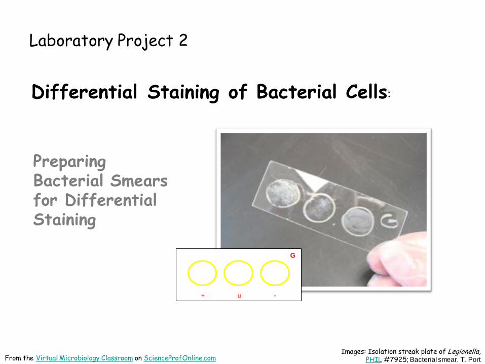
Laboratory Project 2
Differential Staining of Bacterial Cells:
Images: Isolation streak plate of Legionella, PHIL #7925; Bacterial smear, T. Port From the Virtual Microbiology Classroom on ScienceProfOnline.com
Preparing Bacterial Smears for Differential Staining
+ u -
G
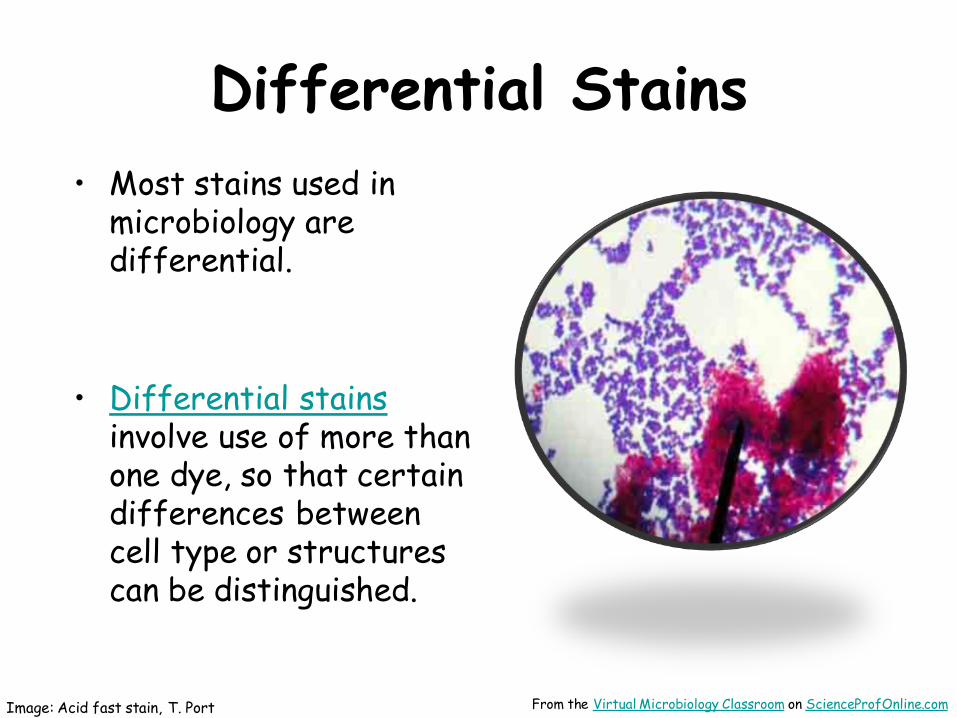
Differential Stains
• Most stains used in microbiology are differential.
• Differential stains involve use of more than one dye, so that certain differences between cell type or structures can be distinguished.
Image: Acid fast stain, T. Port From the Virtual Microbiology Classroom on ScienceProfOnline.com

Inoculation Loop & Aseptic Technique
• You will be using an unknown bacteria that you will be identifying in a future lab.
• To transfer the bacteria to your slide and make bacterial the smears, you will use an inoculation loop
(aka smear loop, inoculation wand or microstreaker).
• Simple tool used to retrieve an inoculum from a culture of microorganisms.
• Always sterilize in microincinerator until loop becomes red hot before and after each use.
• By doing this, the same tool can be reused in different experiments without fear of cross-contamination.
• Be sure that your inoculation loop has cooled before using it to retrieve inoculum or streak a plate!
• If you hear medium sizzle when you touch it with loop, the loop is too hot!
Images:; Isolation streak plate of Legionella, PHIL #7925 Inoculation loop, Jeffrey M. Vinocur; Microincinerator, T. Port From the Virtual Microbiology Classroom on ScienceProfOnline.com

When obtaining a bacterial sample from a tube or plate of media do so gently! The bacteria is growing as a thin film on top of the media! Don’t scrape so hard that you
have pieces of agar in your sample!
From the Virtual Microbiology Classroom on ScienceProfOnline.com Image: E. coli growing on TSY agar in
slant tube and in Petri dish, T. Port
If obtaining bacterial sample from slant tubes: - never pick up test tube by the cap. - do NOT set cap down on lab bench - flame neck of the test tube before & after obtaining sample.

Gram Stain • Distinguishes between two large groups of microorganisms:
- purple staining, Gram-positive cells
- pink staining, Gram-negative cells
• What is the difference in cell structure of Gram+ vs Gram- baacteria?
+ u -
G To prepare Gram bacterial smear for staining:
• Draw three circles on slide using wax pen. • Also include a “G” to identify that slide will be Gram stained. • Flip slide over. • Use DI water dropper to place very small drop of water inside each
circle. • Using a sterilized inoculation loop, take a small sample of your
unknown. Be gentle! The bacteria is on the surface of the medium. • Swirl into the water in the center circle of your slide. • Q: Why are there two additional circles on our slide? • Use same method to add controls to circle on left and right. • Heat fix the slide on top of your microincinerator. Allow it to stay
in the platform for 5 minutes after water has completely evaporated.
From the Virtual Microbiology Classroom on ScienceProfOnline.com
Watch video of How to Prepare a
Bacterial Smear for Gram Staining

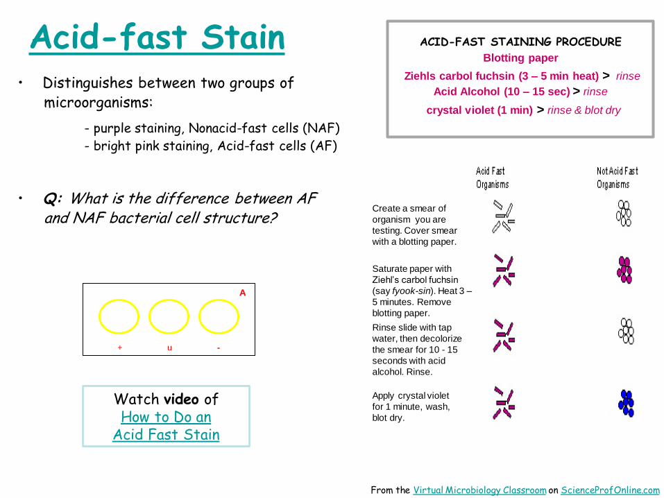
Acid-fast Stain • Distinguishes cells that have mycolic acid in cell wall, from those that do not:
- purple staining, Nonacid-fast cells (NAF)
- bright pink staining, Acid-fast cells (AF)
• What is the difference in cell structure between acid-fast and nonacid-fast bacterial cells?
+ u -
A
To prepare Acid-fast bacterial smear for staining:
• Draw three circles on slide using wax pen. • Also include an “A” to identify that slide will be Acid-fast stained. • Flip slide over. • Use DI water dropper to place very small drop of water inside each
circle. • Using a sterilized inoculation loop, take a small sample of your
unknown. Be gentle! The bacteria is on the surface of the medium. • Swirl into the water in the center circle of your slide. • Q: What (+) and (-) control can we use for this stain? • Use same method to add controls to circle on left and right. • Heat fix the slide on top of your microincinerator. Allow it to stay
in the platform for 5 minutes after water has completely evaporated.
From the Virtual Microbiology Classroom on ScienceProfOnline.com
Watch video of How to Prepare a
Bacterial Smear for Acid Fast Staining

Endospore Stain • Distinguishes between two things:
- endospores, which stain green
- vegetative cells, which stain pink
• Q: What is an endospore?
• Q: What two genera of endospore-producing bacteria have we studied in class?
+ u -
E To prepare Endospore bacterial smear for staining:
• Draw three circles on slide using wax pen. • Also include an “E”to identify that slide will be Endospore stained. • Flip slide over. • Use DI water dropper to place very small drop of water inside each
circle. • Using a sterilized inoculation loop, take a small sample of your
unknown. Be gentle! The bacteria is on the surface of the medium. • Swirl into the water in the center circle of your slide. • Q: What (+) and (-) control can we use for this stain? • Use same method to add controls to circle on left and right. • Heat fix the slide on top of your microincinerator. Allow it to stay
in the platform for 5 minutes after water has completely evaporated.
From the Virtual Microbiology Classroom on ScienceProfOnline.com
Watch video of How to Prepare a
Bacterial Smear for Endospore Staining

Laboratory Project 2
Differential Staining of Bacterial Cells
Preforming the Gram,
Acid-fast & Endospore stains
b u e
E
Images: Acid fast stain & mordant step of Gram stain, both by T. Port From the Virtual Microbiology Classroom on ScienceProfOnline.com

Gram Stain • Distinguishes between two large groups of microorganisms:
- purple staining, Gram-positive cells
- pink staining, Gram-negative cells
• Q: What is the difference between Gram+ and Gram- bacterial cell wall structure?
+ u -
G
GRAM STAINING PROCEDURE
Crystal violet (1 min) > rinse Iodine (1 min) > rinse
Acetone Alcohol (10–15 sec) > rinse Safrinin (1 min) > rinse & blot dry
From the Virtual Microbiology Classroom on ScienceProfOnline.com
Watch video of How to Do a Gram Stain
Acid-fast Stain • Distinguishes between two groups of microorganisms:
- purple staining, Nonacid-fast cells (NAF)
- bright pink staining, Acid-fast cells (AF)
• Q: What is the difference between AF and NAF bacterial cell structure?
+ u -
A
Create a smear of
organism you are
testing. Cover smear
with a blotting paper.
Saturate paper with
Ziehl’s carbol fuchsin
(say fyook-sin). Heat 3 –
5 minutes. Remove
blotting paper.
Rinse slide with tap
water, then decolorize
the smear for 10 - 15
seconds with acid
alcohol. Rinse.
Apply crystal violet
for 1 minute, wash,
blot dry.
ACID-FAST STAINING PROCEDURE
Blotting paper
Ziehls carbol fuchsin (3 – 5 min heat) > rinse
Acid Alcohol (10 – 15 sec) > rinse
crystal violet (1 min) > rinse & blot dry
From the Virtual Microbiology Classroom on ScienceProfOnline.com
Watch video of How to Do an
Acid Fast Stain

Endospore Stain • Distinguishes between two things:
- endospores, which stain green
- vegetative cells, which stain pink
• Some bacteria produce endospores; dormant, highly-resistant structures that can survive environmental extremes (desiccation, heat, harmful chemicals).
• Most notable genera: Bacillus and Clostridium
• Endospores cannot be stained by normal staining
procedures because their walls are practically
impermeable.
• Endospore stain uses heat to drive the primary stain,
(malachite green) into the endospore.
• Q: What color or colors will I see in my endospore + control?
What color or colors will I see in my endospore – control?
+ u -
E
ENDOSPORE STAINING PROCEDURE
Malachite Green (5 min heat) > rinse
Safrinin (1 min) > rinse & blot dry
From the Virtual Microbiology Classroom on ScienceProfOnline.com
Watch video of How to Do an
Endospore Stain

Gram Stain Examples
Staphylococcus epidermidis Escherichia coli
Mixed Sample of S. epidermidis & E. coli
Images: All Gram stain images by T. Port From the Virtual Microbiology Classroom on ScienceProfOnline.com
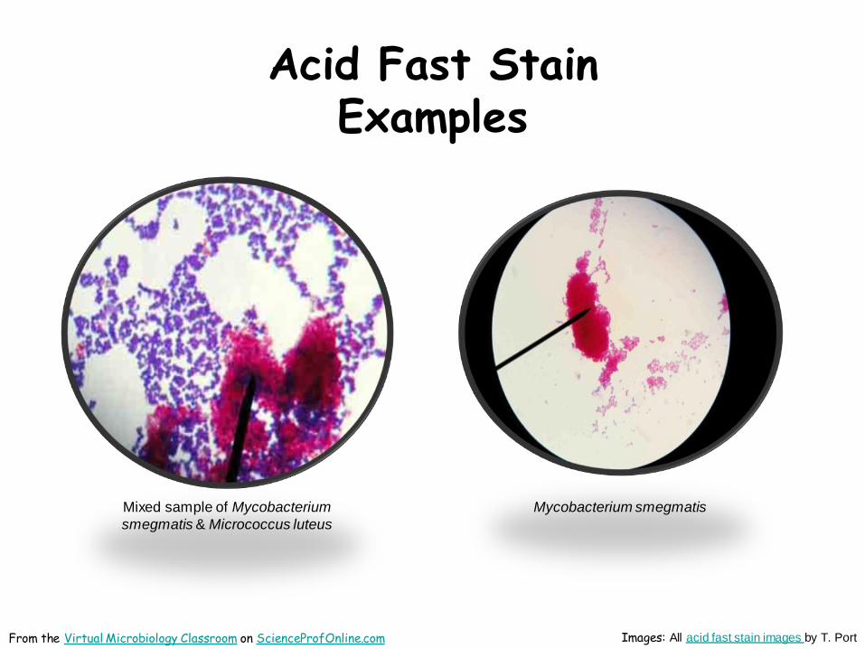
Acid Fast Stain Examples
Mixed sample of Mycobacterium
smegmatis & Micrococcus luteus
Mycobacterium smegmatis
Images: All acid fast stain images by T. Port From the Virtual Microbiology Classroom on ScienceProfOnline.com

Endospore Stain Examples
Images: All endospore stain images by T. Port
Bacillus
cereus
From the Virtual Microbiology Classroom on ScienceProfOnline.com

Confused?
Here are links to fun resources that further
explain streak plate technique and differential staining:
• Bacterial Identification Laboratory Main Page on the Virtual Microbiology Classroom
of Science Prof Online.
• How to Prepare a Bacterial Smear for Gram Staining, video from Science Prof Online (SPO).
• Gram Stain Interactive Tutorial. This is an extremely useful tutorial that shows, step-by-step, what happens in Gram-positive and Gram-negative cells during Gram staining.
• How to Prepare a Bacterial Smear for Acid Fast Staining, video from SPO.
• Acid-fast Stain Animated Tutorial. The staining procedure depicted in this tutorial differs a bit from how we do it in lab, but this tutorial is still very useful. Shows the steps of the staining procedure and the resulting color of Acid-fast and Nonacid-fast cells.
• How to Prepare a Bacterial Smear for Endospore Staining, video from SPO.
• Endospore Stain PowerPoint. Although this is just a PPT, it does have useful information and images for students learning about the endospore stain.
(You must be in PPT slideshow view to click on links.) From the Virtual Microbiology Classroom on ScienceProfOnline.com

Are microbes intimidating you?
Do yourself a favor. Use the…
Virtual Microbiology Classroom (VMC) !
The VMC is full of resources to help you succeed,
including: • practice test questions
• review questions
• study guides and learning objectives
You can access the VMC by going to the Science Prof Online website www.ScienceProfOnline.com
Images: C. diff., Giant Microbes; Prokaryotic cell, Mariana Ruiz