
ABiological Sensor Iron Available to Bacteria in Their Habitats … · BIOLOGICAL SENSOR FOR IRON...
8
APPLIED AND ENVIRONMENTAL MICROBIOLOGY, June 1994, p. 1934-1941 Vol. 60, No. 6 0099-2240/94/$04.00+0 Copyright © 1994, American Society for Microbiology A Biological Sensor for Iron Available to Bacteria in Their Habitats on Plant Surfaces J. E. LOPER'* AND S. E. LINDOW2 Horticultural Crops Research Laboratoty, Agricultural Research Service, U.S. Department of Agriculture, and Department of Botany and Plant Pathology, Oregon State University, Corvallis, Oregon, Iand Department of Plant Pathology, University of California, Berkeley, Califomia2 Received 16 November 1993/Accepted 15 March 1994 A sensor responsive to iron was constructed by fusing a promoterless ice nucleation activity gene (inaZ) to an iron-regulated promoter of a genomic region involved in pyoverdine (fluorescent siderophore) (pvd) production in Pseudomonas syringae. Cells of Pseudomonas fluorescens and P. syringae that contained the pvd-inaZ fusion expressed iron-responsive ice nucleation activity in the bean rhizosphere and phyllosphere, respectively, and in culture. Addition of Fe(III) to leaves or soil reduced the apparent transcription of the pvd-inaZ reporter gene, as shown by a reduction in the number of ice nuclei produced, indicating that Fe(III) was primarily responsible for mediating transcription of the pvd-inaZ gene even in natural environments. A Pseudomonas sp. strain having an intact iceC gene, which conferred Fe-insensitive expression of ice nucleation activity, was included in all studies to account for small strain- or environment-dependent differences in the ability of bacterial cells to produce ice nuclei. Thus, a comparison of the ice nucleation activity conferred by pvd-inaZ with the activity conferred by iceC revealed the bioavailability of iron in culture or natural habitats. The relative ice nucleation activities expressed by strains containing iceC orpvd-inaZ indicated that, while not abundant, Fe(III) is not present at extremely low concentrations at all microsites colonized by bacteria on plant surfaces. Biological sensors that are constructed by fusing inaZ to chemically responsive promoters provide a novel way to characterize chemical constituents of microbial habitats. A variety of environmental factors, including the availability of nutrients, influence the growth, activity, and gene expression of microorganisms in culture and presumably in natural habi- tats. One of these factors, iron availability, profoundly affects gene expression in prokaryotes and is particularly important in the activity and metabolism of bacteria (54). Although iron is abundant, the extreme insolubility of ferric hydroxide limits the equilibrium concentration of free iron available in aerobic aqueous environments at pH 7 to approximately 10-18 M (44). Iron exists in the form of insoluble oxides in soils at neutral or alkaline pH values (28, 29). Although iron is essential to virtually all forms of life, excessive concentrations are toxic (53). Thus, most organisms have systems for specific chelation and regulated transport of Fe(III) into the cell. Most micro- organisms use siderophores and corresponding membrane receptors for iron acquisition (35). Siderophores, which are low-molecular-weight compounds produced under iron-limit- ing conditions, chelate the ferric ion with a high specific activity and serve as vehicles for the transport of Fe(III) into microbial cells (36). Transport of iron into the cells is mediated by a membrane receptor that specifically recognizes a ferric-sid- erophore complex (37). Fluorescent pseudomonads are common inhabitants of the plant phyllosphere and rhizosphere and are important as phytopathogens, as incitants of frost injury, and as agents for biological control of plant disease. The fluorescent pseudo- monads are characterized by the production of yellow-green * Corresponding author. Mailing address: Horticultural Crops Re- search Laboratory, Agricultural Research Service, U.S. Department of Agriculture, 3420 N.W. Orchard Ave., Corvallis, OR 97330. Phone: (503) 750-8771. Fax: (503) 750-8764. Electronic mail address: Loper [email protected]. pigments, called pyoverdines or pseudobactins, that fluoresce under UV light and function as siderophores (1). Pyoverdines inhibit fungal growth by sequestering iron as ferric-pyoverdine complexes (30, 42, 48). Although pyoverdine-mediated com- petition for Fe(III) has been proposed as a mechanism for biological control of certain plant diseases caused by soilborne fungi (30, 42, 48), there is no evidence indicating how com- monly the growth of fungi or bacteria is limited by the lack of available iron in soil or on plant surfaces. Bacteria do not form uniform populations spatially on leaf and root surfaces, but instead are often found in localized assemblages where the resources provided by the plant are more favorable for growth or survival (9, 21). Iron availability at the sites colonized by bacteria, in contrast to the iron contents of bulk environmental samples, cannot be quantified by conventional methods. A goal of this study was to develop a method by which the levels of iron available in microbial habitats could be assessed. In this study, a reporter gene conferring a phenotype that is readily detected and quantified in environmental samples was placed under the regulatory control of an iron-regulated promoter; the resulting gene fusion provided a tool to estimate the availability of iron in habitats occupied by bacteria on plant surfaces. Of the available reporter genes, we selected inaZ (23), which confers ice nucleation activity, for use in this study. The inaZ reporter is composed of an ice nucleation gene from Pseudomonas syringae (12) that is devoid of its native promoter and encodes an outer membrane protein (InaZ) that catalyzes ice formation at -2 to - 10°C, thereby limiting the supercool- ing of water (11, 27, 55). Ice nucleation activity is related quantitatively to the InaZ protein content of a cell (23, 50) and can be quantified conveniently by a droplet-freezing assay of unprocessed aqueous suspensions of environmental samples (24). Because neither plant tissue nor soil contains significant numbers of ice nuclei active at -5°C or above (2, 26), even low 1934 on February 8, 2019 by guest http://aem.asm.org/ Downloaded from
Transcript of ABiological Sensor Iron Available to Bacteria in Their Habitats … · BIOLOGICAL SENSOR FOR IRON...

APPLIED AND ENVIRONMENTAL MICROBIOLOGY, June 1994, p. 1934-1941 Vol. 60, No. 60099-2240/94/$04.00+0Copyright © 1994, American Society for Microbiology
A Biological Sensor for Iron Available to Bacteria in TheirHabitats on Plant Surfaces
J. E. LOPER'* AND S. E. LINDOW2
Horticultural Crops Research Laboratoty, Agricultural Research Service, U.S. Department ofAgriculture, andDepartment of Botany and Plant Pathology, Oregon State University, Corvallis, Oregon, Iand
Department of Plant Pathology, University of California, Berkeley, Califomia2
Received 16 November 1993/Accepted 15 March 1994
A sensor responsive to iron was constructed by fusing a promoterless ice nucleation activity gene (inaZ) toan iron-regulated promoter of a genomic region involved in pyoverdine (fluorescent siderophore) (pvd)production in Pseudomonas syringae. Cells of Pseudomonas fluorescens and P. syringae that contained thepvd-inaZ fusion expressed iron-responsive ice nucleation activity in the bean rhizosphere and phyllosphere,respectively, and in culture. Addition of Fe(III) to leaves or soil reduced the apparent transcription of thepvd-inaZ reporter gene, as shown by a reduction in the number of ice nuclei produced, indicating that Fe(III)was primarily responsible for mediating transcription of the pvd-inaZ gene even in natural environments. APseudomonas sp. strain having an intact iceC gene, which conferred Fe-insensitive expression of ice nucleationactivity, was included in all studies to account for small strain- or environment-dependent differences in theability of bacterial cells to produce ice nuclei. Thus, a comparison of the ice nucleation activity conferred bypvd-inaZ with the activity conferred by iceC revealed the bioavailability of iron in culture or natural habitats.The relative ice nucleation activities expressed by strains containing iceC orpvd-inaZ indicated that, while notabundant, Fe(III) is not present at extremely low concentrations at all microsites colonized by bacteria on plantsurfaces. Biological sensors that are constructed by fusing inaZ to chemically responsive promoters provide anovel way to characterize chemical constituents of microbial habitats.
A variety of environmental factors, including the availabilityof nutrients, influence the growth, activity, and gene expressionof microorganisms in culture and presumably in natural habi-tats. One of these factors, iron availability, profoundly affectsgene expression in prokaryotes and is particularly important inthe activity and metabolism of bacteria (54). Although iron isabundant, the extreme insolubility of ferric hydroxide limits theequilibrium concentration of free iron available in aerobicaqueous environments at pH 7 to approximately 10-18 M (44).Iron exists in the form of insoluble oxides in soils at neutral oralkaline pH values (28, 29). Although iron is essential tovirtually all forms of life, excessive concentrations are toxic(53). Thus, most organisms have systems for specific chelationand regulated transport of Fe(III) into the cell. Most micro-organisms use siderophores and corresponding membranereceptors for iron acquisition (35). Siderophores, which arelow-molecular-weight compounds produced under iron-limit-ing conditions, chelate the ferric ion with a high specific activityand serve as vehicles for the transport of Fe(III) into microbialcells (36). Transport of iron into the cells is mediated by amembrane receptor that specifically recognizes a ferric-sid-erophore complex (37).
Fluorescent pseudomonads are common inhabitants of theplant phyllosphere and rhizosphere and are important asphytopathogens, as incitants of frost injury, and as agents forbiological control of plant disease. The fluorescent pseudo-monads are characterized by the production of yellow-green
* Corresponding author. Mailing address: Horticultural Crops Re-search Laboratory, Agricultural Research Service, U.S. Department ofAgriculture, 3420 N.W. Orchard Ave., Corvallis, OR 97330. Phone:(503) 750-8771. Fax: (503) 750-8764. Electronic mail address: [email protected].
pigments, called pyoverdines or pseudobactins, that fluoresceunder UV light and function as siderophores (1). Pyoverdinesinhibit fungal growth by sequestering iron as ferric-pyoverdinecomplexes (30, 42, 48). Although pyoverdine-mediated com-petition for Fe(III) has been proposed as a mechanism forbiological control of certain plant diseases caused by soilbornefungi (30, 42, 48), there is no evidence indicating how com-monly the growth of fungi or bacteria is limited by the lack ofavailable iron in soil or on plant surfaces. Bacteria do not formuniform populations spatially on leaf and root surfaces, butinstead are often found in localized assemblages where theresources provided by the plant are more favorable for growthor survival (9, 21). Iron availability at the sites colonized bybacteria, in contrast to the iron contents of bulk environmentalsamples, cannot be quantified by conventional methods. A goalof this study was to develop a method by which the levels ofiron available in microbial habitats could be assessed.
In this study, a reporter gene conferring a phenotype that isreadily detected and quantified in environmental samples wasplaced under the regulatory control of an iron-regulatedpromoter; the resulting gene fusion provided a tool to estimatethe availability of iron in habitats occupied by bacteria on plantsurfaces. Of the available reporter genes, we selected inaZ(23), which confers ice nucleation activity, for use in this study.The inaZ reporter is composed of an ice nucleation gene fromPseudomonas syringae (12) that is devoid of its native promoterand encodes an outer membrane protein (InaZ) that catalyzesice formation at -2 to - 10°C, thereby limiting the supercool-ing of water (11, 27, 55). Ice nucleation activity is relatedquantitatively to the InaZ protein content of a cell (23, 50) andcan be quantified conveniently by a droplet-freezing assay ofunprocessed aqueous suspensions of environmental samples(24). Because neither plant tissue nor soil contains significantnumbers of ice nuclei active at -5°C or above (2, 26), even low
1934
on February 8, 2019 by guest
http://aem.asm
.org/D
ownloaded from
BIOLOGICAL SENSOR FOR IRON 1935
levels of inaZgene expression by an introduced ice nucleation-active bacterium can be detected in environmental samples.Furthermore, the ice nucleation phenotype is expressed inmany gram-negative bacteria (23) and can be quantified fromcells grown under a wide range of environmental conditions(24, 39). Because ice nucleation activity does not depend onrapid cell growth (2, 26), it is a useful reporter of geneexpression in quiescent cells, such as those that may be foundin natural habitats.
MATERUILS AND METHODS
Bacterial strains, plasmids, and growth conditions. P. syrin-gae 31 is an ice nucleation-active (Ina' or Ice'), epiphyticbacterium that was originally isolated from a corn leaf surface(2). Strain 31R1, a spontaneous mutant of strain 31, wasselected for its resistance to rifampin. Strain 31R1-P6, an Ina-mutant of strain 31R1 derived by deletion of a region withinthe inaZ gene, was obtained from J. Lindemann (DNA PlantTechnologies, Oakland, Calif.). Pseudomonas fluorescens Pf-5(14), which was isolated from the cotton rhizosphere, wasobtained from C. R. Howell (Agricultural Research Service,U.S. Department of Agriculture, College Station, Tex.). Plas-mid pSFL12 (32) contains genes involved in pyoverdine pro-duction in P. syringae 31 cloned in broad-host-range cosmidpLAFR1 (10), which confers tetracycline resistance. PlasmidpVSP61 (obtained from William Tucker, DNA Plant Technol-ogies) confers kanamycin resistance and contains replicons ofpVS1 (15) and pACY184 (6). In preliminary experiments,pVSP61 was maintained stably in P. syringae 31R1-P6 and P.fluorescens Pf-5 inhabiting phyllospheres and rhizospheres,respectively, for at least 7 days after inoculation. The inaZ geneof P. syringae S203 (12) was provided by G. J. Warren (DNAPlant Technologies). The iceC gene of P. syringae Cit7R1 (41)is virtually identical to inaZ in its amino acid sequence (25).Pseudomonas spp. were grown routinely on nutrient agar(Difco Laboratories, Detroit, Mich.) supplemented with 1%(wt/vol) glycerol; Escherichia coli was grown on Luria-Bertanimedium (46). When it was necessary, the media were supple-mented with tetracycline (25 ,ug/ml), kanamycin (50 p,g/ml),rifampin (100 ,ug/ml), spectinomycin (50 ,ug/ml), or ampicillin(100 pLg/ml).DNA biochemistry. Plasmid DNA was isolated by the alka-
line lysis procedure, and ligations, restriction digestions, aga-rose gel electrophoresis, and Southern hybridizations wereperformed by standard methods (46). Mobilizable plasmidswere introduced into Pseudomonas spp. by triparental matings,using pRK2013 as a helper plasmid (8).
Identification of an iron-sensitive promoter. PlasmidpSFL12 was mutagenized with Tn3-HoHol by using previouslydescribed methods (51). Following matings with E. coli donorsharboring pSFL12::Tn3-HoHol, colonies of P. syningae 31R1that harbored fusion plasmids were selected on medium 925(20) containing myo-inositol (10 g/liter) as a sole carbonsource, tetracycline, ampicillin, and 5-bromo-4-chloro-3-in-doyl-p-D-galactopyranoside (X-Gal) (40 ,ug/ml). After 3 daysof incubation at 27°C, the colonies were screened for a bluecolor, which indicated that there was a Tn3-HoHol insertiondownstream of a promoter.Assessment of transcriptional activity and pyoverdine pro-
duction in culture. Pseudomonas spp. were grown at 25°C in aminimal salts medium (SM) (31) supplemented with differentconcentrations of FeCl3. The effects of other micronutrientswere evaluated in SM supplemented with ZnSO4 (10-5 to 10-7M), CuCl2 (2 x 10⁻⁶ to 10-7 M), or MnCl2 (10- to 10-7 M);
the maximum concentrations of the elements used were thegreatest concentrations that did not reduce cell growth signif-icantly. After 48 h of growth, ,3-galactosidase activity wasassessed by the method of Miller (34). The ice nucleationactivity of cells was quantified by the droplet-freezing assay(24). The concentrations of pyoverdine in culture supernatantswere estimated by determining the A405 of the ferric-pyover-dine complex. FeCl3 was added to a culture supernatant to afinal concentration of 10-3 M, the preparation was mixed for30 min, diluted 1:1 in 1 M potassium phosphate buffer (pH7.5), and centrifuged to remove the precipitate, and the A405was measured. Values for ,-galactosidase activity, ice nucle-ation activity, and pyoverdine concentration were normalizedfor the number of bacterial cells, as determined by the opticaldensity at 560 nm of the cultures. The values reported beloware the means obtained from three replicate cultures.Assessment of transcriptional activity of Pseudomonas spp.
on plant surfaces. The ice nucleation activities of Pseudomonasspp. inhabiting rhizospheres and phyllospheres of beans(Phaseolus vulgaris L. cv. Bush Blue Lake) were determined.Except where noted below, the bacterial inoculum was grownin SM supplemented with 10-3 M FeCl3. Cells were harvestedfrom cultures by centrifugation and resuspended in steriledeionized water to an optical density at 560 nm of 0.1. Thesuspensions were diluted to final densities of approximately 105and 106 CFU/ml for inoculation of leaves and roots, respec-tively. For rhizosphere studies, bean seeds were surface steril-ized in a 1% hypochlorite solution for 10 min, rinsed thor-oughly in deionized water, and placed on moist paper towelsfor 3 days. The roots of the resulting bean seedlings weredipped in aqueous suspensions of P. fluorescens Pf-5 andplanted in pots containing Warden sandy-silt loam soil (-0.3bar, pH 7.2, 14 mg of Fe per kg). In some experiments, asolution of the ferric-sodium salt of EDTA (FeEDTA) wasmixed into the soil at a concentration of 300 ,ug/kg prior toplanting. Soil amendment with FeEDTA, which has a stabilityconstant of 25 (28), is thought to increase the biologicalavailability of iron in the rhizosphere (19, 47). For phyllo-sphere studies, bean seeds were planted in acid-washed sandand watered with Hoagland's solution. Leaf surfaces wereinoculated by dipping an entire plant into a bacterial suspen-sion and covering it loosely with a plastic bag. After inocula-tion, the plants were grown for 5 days in environmental growthchambers maintained at 25°C with a 12-h photoperiod. Indi-vidual leaflets from trifoliate leaves or individual root systemswere placed in a washing buffer (40) and sonicated for 5 min toremove bacterial cells from tissue surfaces or rhizosphere soil.The washing buffer containing suspended bacterial cells wasdiluted, and bacterial population size was determined byspreading aliquots of dilutions on King's medium B (17)containing rifampin, using previously described methods (33,40). The ice nucleation activities of bacteria inhabiting therhizosphere or phyllosphere were determined by the droplet-freezing assay (24). In an experiment to evaluate the effect ofthe rhizosphere environment and harvesting procedures on icenucleation activity, the roots of bean seedlings were inoculatedwith a suspension of ice nucleation-active P. fluorescens cells,and the seedlings were planted in soil and immediately har-vested. The ice nucleation activity of the inoculated cells andpopulation size were determined as described above. For eachtreatment we used five plants, and each experiment wasrepeated.
VOL. 60, 1994
on February 8, 2019 by guest
http://aem.asm
.org/D
ownloaded from

1936 LOPER AND LINDOW
300
250
'- 2000
s,150e0(a 100cu
50
0-7 -6.5 -6 -5.5 -5 -4.5 -4 -3.5
2A
1.5 o0
'a)
0L-
i .
a)
0.5 0a.
-3
Log ([FeCI3])
FIG. 1. ,B-Galactosidase activity and pyoverdine production by P.syringae 31R1(pJEL1395) grown in SM containing different concentra-tions of iron. Symbols: M, 3-galactosidase activity, in Miller units (34);V, pyoverdine production, as determined by A405 of the ferric-pyoverdine complex. The 3-galactosidase activity and pyoverdineproduction values were normalized for the number of bacterial cells, asdetermined by measuring the turbidity of cultures at 560 nm. Valuesare the means obtained from three replicate cultures.
RESULTS
B
C
R pVSP61
KmR
R RinaZ
inaZ4-
R pVSP61 R R H R
KmR inaZ pvd4--
R pVSP61 R R
KmR iceC4-0
pvd-inaZ
iceC
I kb
FIG. 2. Ice nucleation reporter gene construction. (A) inaZ (12)devoid of its native promoter and cloned as a 3.4-kb EcoRI fragmentin the opposite orientation to the plac promoter of the pUC8polylinker. (B) pvd-inaZ, consisting of the promoterless inaZ genecloned downstream from an 8.0-kb EcoRI fragment involved inpyoverdine production (pvd) in P. syringae. (C) iceC (41) driven by itsnative, iron-constitutive promoter and contained within a 9.5-kbEcoRI fragment. The three constructions were cloned in plasmidvector pVSP61, which is maintained stably in Pseudomonas spp.Restriction endonuclease cut site abbreviations: R, EcoRI; H, HindIll.
syringae 31. The Tn3-HoHol insertion of pJEL1395 was lo-cated 1.0 kb from the left side of the 8-kb EcoRI fragment andwas oriented such that the lacZ gene was transcribed fromright to left, as the fragment is shown in Fig. 2. We thereforesuspected that the iron-regulated promoter from which lacZ
Identification of an iron-regulated promoter. A genomicregion whose transcription was regulated strongly by iron wasidentified by Tn3-HoHol mutagenesis of plasmid pSFL12(32), which contains cloned genes involved in pyoverdinebiosynthesis (pvd) in P. syringae 31. Colonies of P. syringae31R1 containing pSFL12 with random insertions of Tn3-HoHol were evaluated for 3-galactosidase activity. We iden-tified 40 colonies that were blue on medium 925 containingX-Gal but were white on medium 925 containing X-Gal and10-4 M FeCl3. Plasmid pJEL1395, which was isolated fromone of the 40 colonies, did not confer ,-galactosidase activityon E. coli DH5o or HB1O1. The level of pyoverdine productionand the level of 3-galactosidase activity of P. syringae harboringpJEL1395 decreased by 300-fold as the concentration of addedFeCl3 increased from 10' to 10-6 M (Fig. 1). In SM amendedwith 10-6 M FeCl3, the level of 3-galactosidase activity was atthe lower limit of detection. Therefore, further decreases inthe level of 3-galactosidase activity with increases in the FeCl3concentration to values greater than 10-6 M would not havebeen detectable. The Fe(III)-mediated change in transcriptionof the region involved inpvd biosynthesis was apparent in cellsat all stages of growth in culture (data not shown). Other traceelements (Mn2", Zn2+, Cu2+) had only small effects on the3-galactosidase activity conferred by pJEL1395 or on pyover-dine production in P. syringae. Thus, of the elements evaluated,only iron substantially affected the transcriptional activity ofthe pvd promoter; this effect paralleled the specific effect ofFe(III) on pyoverdine production.The single Tn3-HoHol insertion of pJEL1395 was located
on an 8-kb EcoRI fragment of pSFL12. Plasmid pSFL12restores fluorescence to nine mutants of P. syringae 31R1 thatare deficient in pyoverdine production (32). A subclone ofpSFL12, which contained the 8-kb EcoRI fragment cloned intoplasmid pVSP61, restored fluorescence to three of the ninepyoverdine-deficient mutants of P. syringae 31R1. Thus, the8-kb region was involved in pyoverdine production in P.
= - 1 _%
A1
o -2a)~~~~~~V
O -3a)
040--5
-7 -6.5 -6 -5.5 -5 -4.5 -4 -3.5 -3
Log (FeCl3)
FIG. 3. (A) Iron-regulated ice nucleation activity expressed by P.syningae 31R1-P6 containing iceC (0) or pvd-inaZ (v). The icenucleation activities of cells that were cultured at 25°C with shaking inSM (31) amended with various concentrations of FeCl3 were quanti-fied by performing the droplet-freezing assay at temperature of -5°C(24). Ice nucleation activity values were normalized for the number ofbacterial cells, as determined by measuring the turbidity of cultures at560 nm. (B) Difference in ice nucleation activities expressed by cells ofP. syringae 31R1-P6 containing pvd-inaZ and cells containing iceCgrown as described above.
6------ ----
APPL. ENVIRON. MICROBIOL.
on February 8, 2019 by guest
http://aem.asm
.org/D
ownloaded from

BIOLOGICAL SENSOR FOR IRON 1937
I -1
=. -24
m -3a)
.S -41-
cmo -5-J
(v).cj 2 1
cu 1
QL -1-0-i
° -2
CE3-4
-7 -6.5 -6 -5.5 -5 -4.5 -4 -3.5 -3
Log (FeC13)FIG. 4. Iron-regulated ice nucleation activity expressed by P. fluo-
rescens Pf-5 containing iceC (@) or pvd-inaZ (V). The ice nucleationactivities of cells that were cultured at 25°C with shaking in SM (31)amended with various concentrations of FeCl3 were quantified byperforming the droplet-freezing assay at temperature of -5°C (24). Icenucleation activity values were normalized for the number of bacterialcells, as determined by measuring the turbidity of cultures at 560 nm.
(B) Difference in ice nucleation activities expressed by cells of P.fluorescens Pf-5 containingpvd-inaZ and cells containing iceC grown as
described above.
was transcribed was present on the 8-kb EcoRI fragment ofpSFL12.
Construction of an iron sensor. A pvd-inaZ fusion wasconstructed by fusing the 8-kb EcoRI fragment of pSFL12 to a
promoterless inaZ gene (Fig. 2). Constructions were intro-duced by conjugation into P. fluorescens Pf-5 and P. syringae31R1-P6, neither of which produced detectable ice nuclei inthe absence of an introduced ice nucleation gene. The con-struction containing the promoterless inaZ gene (Fig. 2) didnot confer ice nucleation activity on either strain. The icenucleation activities of P. syringae and P. fluorescens strainsconferred by pvd-inaZ were regulated dramatically by iron(Fig. 3 and 4). Transcription of the iron-regulatedpvd gene(s)was detected as high levels of ice nucleation activity in cellsgrown in media containing low levels of Fe(III) (i.e., <10-6 Madded FeCl3) (Fig. 3 and 4). At such low FeCl3 concentrations,every cell produced at least one ice nucleus active at -5°C, themaximum level of ice nucleation activity that can be quantifiedby the droplet-freezing assay. Cells containing pvd-inaZ ex-
pressed 10,000- to 100,000-fold-greater ice nucleation activityat low concentrations of FeCl3 (.10-6 M) than at highconcentrations of FeCl3 (-10-4 M). In contrast, the icenucleation activity of cells containing iceC, the ice nucleationgene of P. syringae transcribed from its native promoter, was
not affected substantially by the FeCl3 concentration in theculture medium, indicating that InaZ protein production andfunction were not affected greatly by iron availability. Thus, a
comparison of the ice nucleation activities of cells containingpvd-inaZ and cells containing iceC provided a way to assess
0
4
2
0
-2
4
a)
-,6
0 5 10 15 20 25
Time (h)
FIG. 5. Ice nucleation activities and rhizosphere populations of P.fluorescens Pf-5 containing pvd-inaZ. Root systems of beans were
dipped in suspensions of bacterial cells grown in SM amended withi0-' M FeCl3 (V), 10-5 M FeCl3 (-), or 10' M FeCl3 (0), and thenthe beans were planted in Warden sandy-silt loam soil. The rhizos-phere population size (A) and the in situ ice nucleation activity (B) ofP. fluorescens Pf-5 containingpvd-inaZ were estimated periodically for24 h following inoculation of root systems.
iron availability to Pseudomonas spp. It is important to notethat at low FeCl3 concentrations (.10-6 M added FeCl3), theice nucleation activity expressed by cells containing pvd-inaZwas greater than the activity expressed by cells containing iceC,whereas at high FeCI3 concentrations (-10-4 M), the icenucleation activity expressed by cells containing pvd-inaZ was
less than the activity expressed by cells containing iceC (Fig. 3Band 4B). The low levels of ice nucleation activity expressed bystrains containing pvd-inaZ in iron-replete media (210-4 Madded FeCl3) were attributed to low levels of constitutivetranscriptional activity of the pvd promoter.
Transcriptional activity in the rhizosphere. The ice nucle-ation activity of P. fluorescens was not influenced by therhizosphere environment or by the methods used to obtainbacterial samples from rhizosphere soil; the cultured cells usedas the inoculum expressed levels of ice nucleation activityequivalent to the levels expressed by similar cells retrieved
TABLE 1. Ice nucleation activity expressed by P. fluorescens Pf-5cells containing pvd-inaZ or iceC constructions in the
bean rhizosphere
Soil Population size Ice nucleation activityConstruction treatment' (log [CFU/g])b (log [ice nuclei/cell])b
iceC None 5.4 a -1.1 aiceC FeEDTA 5.4 a -0.9 apvd-inaZ None 5.5 a -2.9 bpvd-inaZ FeEDTA 5.5 a <D
a For the FeEDTA treatment a solution of FeEDTA was mixed into the soilto a concentration of 300 jig/kg soil prior to planting.
b Population size and ice nucleation activity were assessed 5 days after plantswere inoculated with bacteria containing the constructions. Each value is themean of five replicates. Values followed by the same letter are not statisticallydifferent as determined by the Waller-Duncan test at P = 0.05. <D, below thelimit of detection, which was -5.5 log (ice nuclei per cell) in this experiment.
_ A
I I
B
I Ie
VOL. 60, 1994
on February 8, 2019 by guest
http://aem.asm
.org/D
ownloaded from
1938 LOPER AND LINDOW
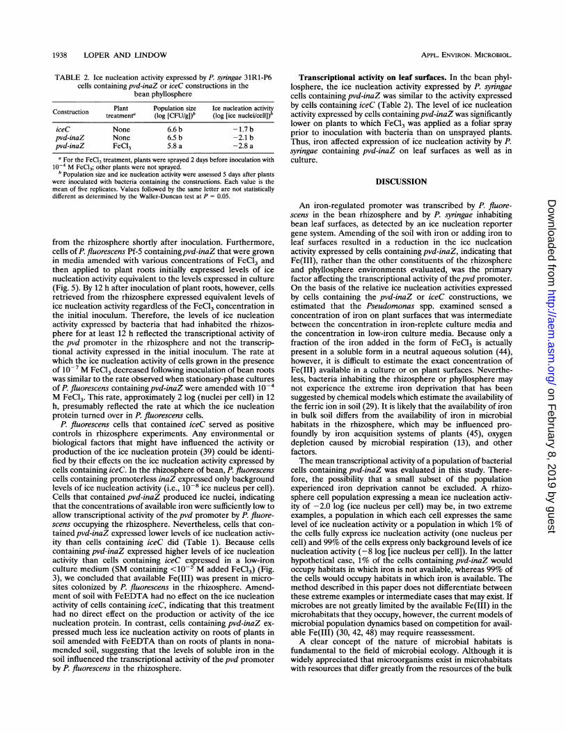
TABLE 2. Ice nucleation activity expressed by P. syringae 31R1-P6cells containing pvd-inaZ or iceC constructions in the
bean phyllospherePlant Population size Ice nucleation activityConstruction treatment' (log [CFU/g])b (log [ice nuclei/cell])b
iceC None 6.6 b -1.7 bpvd-inaZ None 6.5 b -2.1 bpvd-inaZ FeCl3 5.8 a -2.8 a
a For the FeCl3 treatment, plants were sprayed 2 days before inoculation with10'- M FeCI3; other plants were not sprayed.
b Population size and ice nucleation activity were assessed 5 days after plantswere inoculated with bacteria containing the constructions. Each value is themean of five replicates. Values followed by the same letter are not statisticallydifferent as determined by the Waller-Duncan test at P = 0.05.
from the rhizosphere shortly after inoculation. Furthermore,cells of P. fluorescens Pf-5 containingpvd-inaZ that were grownin media amended with various concentrations of FeCl3 andthen applied to plant roots initially expressed levels of icenucleation activity equivalent to the levels expressed in culture(Fig. 5). By 12 h after inoculation of plant roots, however, cellsretrieved from the rhizosphere expressed equivalent levels ofice nucleation activity regardless of the FeCl3 concentration inthe initial inoculum. Therefore, the levels of ice nucleationactivity expressed by bacteria that had inhabited the rhizos-phere for at least 12 h reflected the transcriptional activity ofthe pvd promoter in the rhizosphere and not the transcrip-tional activity expressed in the initial inoculum. The rate atwhich the ice nucleation activity of cells grown in the presenceof 10'- M FeCl3 decreased following inoculation of bean rootswas similar to the rate observed when stationary-phase culturesof P. fluorescens containing pvd-inaZ were amended with 10i-M FeCl3. This rate, approximately 2 log (nuclei per cell) in 12h, presumably reflected the rate at which the ice nucleationprotein turned over in P. fluorescens cells.
P. fluorescens cells that contained iceC served as positivecontrols in rhizosphere experiments. Any environmental orbiological factors that might have influenced the activity orproduction of the ice nucleation protein (39) could be identi-fied by their effects on the ice nucleation activity expressed bycells containing iceC. In the rhizosphere of bean, P. fluorescenscells containing promoterless inaZ expressed only backgroundlevels of ice nucleation activity (i.e., 108 ice nucleus per cell).Cells that contained pvd-inaZ produced ice nuclei, indicatingthat the concentrations of available iron were sufficiently low toallow transcriptional activity of the pvd promoter by P. fluore-scens occupying the rhizosphere. Nevertheless, cells that con-tained pvd-inaZ expressed lower levels of ice nucleation activ-ity than cells containing iceC did (Table 1). Because cellscontaining pvd-inaZ expressed higher levels of ice nucleationactivity than cells containing iceC expressed in a low-ironculture medium (SM containing <10- M added FeCl3) (Fig.3), we concluded that available Fe(III) was present in micro-sites colonized by P. fluorescens in the rhizosphere. Amend-ment of soil with FeEDTA had no effect on the ice nucleationactivity of cells containing iceC, indicating that this treatmenthad no direct effect on the production or activity of the icenucleation protein. In contrast, cells containing pvd-inaZ ex-pressed much less ice nucleation activity on roots of plants insoil amended with FeEDTA than on roots of plants in nona-mended soil, suggesting that the levels of soluble iron in thesoil influenced the transcriptional activity of the pvd promoterby P. fluorescens in the rhizosphere.
Transcriptional activity on leaf surfaces. In the bean phyl-losphere, the ice nucleation activity expressed by P. syringaecells containing pvd-inaZ was similar to the activity expressedby cells containing iceC (Table 2). The level of ice nucleationactivity expressed by cells containingpvd-inaZ was significantlylower on plants to which FeCl3 was applied as a foliar sprayprior to inoculation with bacteria than on unsprayed plants.Thus, iron affected expression of ice nucleation activity by P.syringae containing pvd-inaZ on leaf surfaces as well as inculture.
DISCUSSION
An iron-regulated promoter was transcribed by P. fluore-scens in the bean rhizosphere and by P. syringae inhabitingbean leaf surfaces, as detected by an ice nucleation reportergene system. Amending of the soil with iron or adding iron toleaf surfaces resulted in a reduction in the ice nucleationactivity expressed by cells containing pvd-inaZ, indicating thatFe(III), rather than the other constituents of the rhizosphereand phyllosphere environments evaluated, was the primaryfactor affecting the transcriptional activity of thepvd promoter.On the basis of the relative ice nucleation activities expressedby cells containing the pvd-inaZ or iceC constructions, weestimated that the Pseudomonas spp. examined sensed aconcentration of iron on plant surfaces that was intermediatebetween the concentration in iron-replete culture media andthe concentration in low-iron culture media. Because only afraction of the iron added in the form of FeCl3 is actuallypresent in a soluble form in a neutral aqueous solution (44),however, it is difficult to estimate the exact concentration ofFe(III) available in a culture or on plant surfaces. Neverthe-less, bacteria inhabiting the rhizosphere or phyllosphere maynot experience the extreme iron deprivation that has beensuggested by chemical models which estimate the availability ofthe ferric ion in soil (29). It is likely that the availability of ironin bulk soil differs from the availability of iron in microbialhabitats in the rhizosphere, which may be influenced pro-foundly by iron acquisition systems of plants (45), oxygendepletion caused by microbial respiration (13), and otherfactors.The mean transcriptional activity of a population of bacterial
cells containing pvd-inaZ was evaluated in this study. There-fore, the possibility that a small subset of the populationexperienced iron deprivation cannot be excluded. A rhizo-sphere cell population expressing a mean ice nucleation activ-ity of -2.0 log (ice nucleus per cell) may be, in two extremeexamples, a population in which each cell expresses the samelevel of ice nucleation activity or a population in which 1% ofthe cells fully express ice nucleation activity (one nucleus percell) and 99% of the cells express only background levels of icenucleation activity (-8 log [ice nucleus per cell]). In the latterhypothetical case, 1% of the cells containing pvd-inaZ wouldoccupy habitats in which iron is not available, whereas 99% ofthe cells would occupy habitats in which iron is available. Themethod described in this paper does not differentiate betweenthese extreme examples or intermediate cases that may exist. Ifmicrobes are not greatly limited by the available Fe(III) in themicrohabitats that they occupy, however, the current models ofmicrobial population dynamics based on competition for avail-able Fe(III) (30, 42, 48) may require reassessment.A clear concept of the nature of microbial habitats is
fundamental to the field of microbial ecology. Although it iswidely appreciated that microorganisms exist in microhabitatswith resources that differ greatly from the resources of the bulk
APPL. ENVIRON. MICROBIOL.
on February 8, 2019 by guest
http://aem.asm
.org/D
ownloaded from

BIOLOGICAL SENSOR FOR IRON 1939
samples accessible to scientific study, characterization of thesemicrohabitats has eluded scientific investigation. Concentra-tions of elements or chemical compounds that influence bio-logical systems are currently assessed primarily by chemicalprocedures, usually by analyzing samples that are large relativeto the size of bacteria. Although such analytical methods aresensitive indicators of chemical concentrations in bulk environ-mental samples, they do not assess whether a chemical ispresent in a form that is available biologically to the organismsthat occupy natural habitats. Numerous physicochemical fac-tors influence the form of a chemical compound or element insolution or soil (28); because the chemical form has a profoundeffect on the biological activity of a chemical, methods thatevaluate only the concentration of a chemical do not providean accurate assessment of the biological relevance of thechemical to an ecosystem. Thus, it is sometimes observed thatchemical concentrations that are determined do not have theexpected effects on biological communities or indicator organ-isms (52). There is a need for new methods to determinebiologically meaningful levels of chemicals present in disturbedand natural ecosystems. Sensors based on a bioluminescence(lux) reporter system have been developed to assess biologi-cally available forms of Hg(II) (49) and naphthalene (18) inenvironmental samples such as surface water amended withsubstrates required for bacterial growth or soil slurries. Be-cause the ice nucleation activity expressed by bacterial cellsinhabiting natural substrates can be quantified directly, theinaZ gene was selected as a reporter system for the sensor
assessing the iron available to bacteria occupying naturalhabitats. In the future, inaZ sensors responsive to a variety ofchemical signals may allow characterization of many chemicalcomponents of microbial habitats on plant surfaces, in soil, or
in aqueous environments.The ice nucleation reporter system provided a sensitive,
convenient, and inexpensive tool for the study of bacterial geneexpression in natural habitats. Because of its sensitivity, the icenucleation reporter gene system may be uniquely suited tostudies evaluating the chemical environment of microbialhabitats. For example, P-galactosidase activity was a usefulreporter of the pvd promoter in culture only when the level oftranscriptional activity was relatively high (i.e., in P. syringaecells grown in media containing -10-6 M added FeCl3) (Fig.1). In fact, progressive decreases in the level of transcription ofthis gene with increases in the FeCl3 concentration to valuesgreater than 106 M were not observed with lacZ fusionsbecause of undetectably low levels of P-galactosidase activity(Fig. 1). In contrast, the ice nucleation reporter system pro-vided a more sensitive way to assess transcriptional activity; icenucleation activity was detected in P. syringae cells containingpvd-inaZ grown in media containing up to 10-3 M added FeCl3(Fig. 3). Because the transcriptional activities of the pvdpromoter expressed by Pseudomonas spp. occupying habitats inthe phyllosphere or rhizosphere were in the range detectablewith the inaZ reporter system, such activity may not have beendetected with a less sensitive reporter gene, such as lacZ. Aphosphate sensor, constructed by fusing a phosphate-respon-sive promoter to a promoterless lacZ gene, provided a way toassess phosphate availability to Pseudomonas putida occupyingthe rhizosphere of plants grown in sand or sterile soil, but thissensor was not sensitive enough to assess phosphate availabilityto the same bacteria occupying the rhizosphere of plants grownin a nonsterile soil (7). The ice nucleation reporter gene systemis particularly useful in habitats where the transcriptionalactivity of target genes is low or only a small number of cellsare present. Both situations are likely to occur in natural
environments such as the rhizospheres of plants grown innative soils.Many phenotypes expressed by microorganisms are readily
detected and quantified in culture, whereas the same pheno-types are difficult to detect in natural environments. Pyover-dine production by Pseudomonas spp. is an example of such a
phenotype. Typically, siderophore concentrations in soil are
estimated by performing a bioassay with indicator organismsthat grow only if a specific ferric siderophore or a general classof ferric siderophores (i.e., ferric siderophores with catechol or
hydroxamate groups) provides an exogenous source of iron (3,38, 43). Bioassays have not been useful for specifically assess-
ing pyoverdines in soil, however, because no indicator strainthat specifically utilizes a single ferric pyoverdine as an ironsource has been identified. Pseudomonas spp. commonly utilizea ferric pyoverdine(s) produced by other strains of Pseudomo-nas spp. (5, 22) and ferric siderophores produced by membersof diverse genera of bacteria and fungi (16). Pseudobactin, a
pyoverdine produced by P. fluorescens B1O, was detectedrecently in a rhizosphere at a concentration of 3.5 x 10- ' Mby an immunoassay in which specific monoclonal antibodieswere used (4). Because the transcriptional activity of the pvdpromoter is proportional to pyoverdine production in Pseudo-monas spp. (Fig. 1), ice nucleation activity conferred by thepvd-inaZ fusion may provide a reliable way to assess pyover-dine production by Pseudomonas spp. Because of the relativeconvenience of ice nucleation activity assays, in situ expressionof pyoverdine biosynthesis genes can be readily assessed as an
estimate of pyoverdine production by Pseudomonas spp. occu-
pying microhabitats. Future studies using the complementaryapproaches of assessing the transcriptional activity of the pvdpromoter and estimating pseudobactin concentration with an
immunoassay should provide considerable insight into factorsthat influence pyoverdine production by Pseudomonas spp.occupying natural habitats.
ACKNOWLEDGMENTS
We are grateful to M. D. Henkels for assisting with the growthchamber experiments and for preparing the figures; S. Carnegie forassisting with the construction and testing of the gene fusions; C. R.Howell, J. Lindemann, W. Tucker, and G. J. Warren for providingstrains; and E. Clark and M. Wilson for reviewing the manuscript.
This research was supported in part by grant 91-37030-6802 from theU.S. Department of Agriculture Competitive Grants Program.
REFERENCES
1. Abdallah, M. A. 1991. Pyoverdins and pseudobactins, p. 139-153.In G. Winkelmann (ed.), Handbook of microbial iron chelates.CRC Press, Boca Raton, Fla.
2. Arny, D. C., S. E. Lindow, and C. D. Upper. 1976. Frost sensitivityof Zea mays increased by application of Pseudomonas syningae.Nature (London) 262:282-284.
3. Bossier, P., M. Hofte, and W. Verstraete. 1988. Ecological signif-icance of siderophores in soil. Adv. Microb. Ecol. 10:385-414.
4. Buyer, J. S., M. G. Kratzke, and L. J. Sikora. 1993. A method fordetection of pseudobactin, the siderophore produced by a plant-growth-promoting Pseudomonas strain, in the barley rhizosphere.Appl. Environ. Microbiol. 59:677-681.
5. Buyer, J. S., and J. Leong. 1986. Iron transport-mediated antago-nism between plant growth-promoting and plant-deleteriousPseudomonas strains. J. Biol. Chem. 261:791-794.
6. Chang, A. C. Y., and S. N. Cohen. 1978. Construction andcharacterization of amplifiable multicopy DNA cloning vehiclesderived from the P1SA cryptic miniplasmid. J. Bacteriol. 134:1141-1156.
7. De Weger, L. A., L. C. Dekkers, A. J. van der Bij, and B. J. J.
VOL. 60, 1994
on February 8, 2019 by guest
http://aem.asm
.org/D
ownloaded from

1940 LOPER AND LINDOW
Lugtenberg. 1994. Use of phosphate reporter bacteria to studyphosphate limitation in the rhizosphere and in bulk soil. Mol.Plant Microbe Interact. 7:32-38.
8. Figurski, D. H., and D. R. Helinski. 1979. Replication of an
origin-containing derivative of plasmid RK2 dependent on a
plasmid function provided in trans. Proc. NatI. Acad. Sci. USA76:1648-1652.
9. Foster, R. C., A. D. Rovira, and T. W. Cock. 1983. Ultrastructureof the root-soil interface. The American Phytopathological Soci-ety, St. Paul, Minn.
10. Friedman, A. M., S. R. Long, S. E. Brown, W. J. Buikema, andF. M. Ausubel. 1982. Construction of a broad host range cosmidcloning vector and its use in the genetic analysis of Rhizobiummutants. Gene 18:289-296.
11. Govindarajan, A. G., and S. E. Lindow. 1988. Size of bacterialice-nucleation sites measured in situ by radiation inactivationanalysis. Proc. NatI. Acad. Sci. USA 85:1334-1338.
12. Green, R. L., and G. J. Warren. 1985. Physical and functionalrepetition in a bacterial ice nucleation gene. Nature (London)317:645-648.
13. H0jberg, O., and J. S0rensen. 1993. Microgradients of microbialoxygen consumption in a barley rhizosphere model system. Appl.Environ. Microbiol. 59:431-437.
14. Howell, C. R., and R. D. Stipanovic. 1979. Control of Rhizoctoniasolani on cotton seedlings with Pseudomonas fluorescens and withan antibiotic produced by the bacterium. Phytopathology 69:480-482.
15. Itoh, Y., and D. Haas. 1985. Cloning vectors derived from thePseudomonas plasmid pVS1. Gene 36:27-36.
16. Jurkevitch, E., Y. Hadar, and Y. Chen. 1992. Differential sid-erophore utilization and iron uptake by soil and rhizospherebacteria. Appl. Environ. Microbiol. 58:119-124.
17. King, E. O., M. K. Ward, and D. E. Raney. 1954. Two simplemedia for the demonstration of pyocyanin and fluorescin. J. Lab.Clin. Med. 44:301-307.
18. King, J. M. H., P. M. DiGrazia, B. Applegate, R. Burlage, J.Sanseverino, P. Dunbar, F. Larimer, and G. S. Sayler. 1990.Rapid, sensitive bioluminescent reporter technology for naphtha-lene exposure and biodegradation. Science 249:778-781.
19. Kloepper, J. W., J. Leong, M. Teintze, and M. N. Schroth. 1980.Enhanced plant growth by siderophores produced by plantgrowth-promoting rhizobacteria. Nature (London) 286:885-886.
20. Langley, R. A., and C. I. Kado. 1972. Studies on Agrobacteriumtumefaciens. Conditions for mutagenesis by N-methyl-N'-nitro-N-nitrosoguanidine and relationships of A. tumefaciens mutants to
crown-gall tumor induction. Mutat. Res. 14:277-286.21. Leben, C., M. N. Schroth, and D. C. Hildebrand. 1970. Coloniza-
tion and movement of Pseudomonas syringae on healthy beanseedlings. Phytopathology 60:677-680.
22. Leong, J., W. Bitter, M. Koster, V. Venturi, and P. J. Weisbeek.1991. Molecular analysis of iron transport in plant growth-promot-ing Pseudomonas putida WCS358. Biol. Metals 4:36-40.
23. Lindgren, P. B., R. Frederick, A. G. Govindarajan, N. J. Panopou-los, B. J. Staskawicz, and S. E. Lindow. 1989. An ice nucleationreporter gene system: identification of inducible pathogenicitygenes in Pseudomonas syringae pv. phaseolicola. EMBO J. 8:1291-1301.
24. Lindow, S. E. 1990. Bacterial ice nucleation activity, p. 185-198. InS. Klement, K. Rudolf, and D. C. Sands (ed.), Methods inphytobacteriology. Akademiai Kiad6, Budapest.
25. Lindow, S. E. Unpublished data.26. Lindow, S. E., D. C. Arny, and C. D. Upper. 1982. Bacterial ice
nucleation: a factor in frost injury to plants. Plant Physiol.70:1084-1089.
27. Lindow, S. E., E. Lahue, A. G. Govindarajan, N. J. Panopoulos,and D. Gies. 1989. Localization of ice nucleation activity and theiceC gene product in Pseudomonas syringae and Escherichia coli.Mol. Plant Microbe Interact. 2:262-272.
28. Lindsay, W. L. 1979. Chemical equilibria in soils. John Wiley andSons, New York.
29. Lindsay, W. L., and A. P. Schwab. 1982. The chemistry of iron insoils and its availability to plants. J. Plant Nutr. 5:821-840.
30. Loper, J. E., and J. S. Buyer. 1991. Siderophores in microbialinteractions on plant surfaces. Mol. Plant Microbe Interact. 4:5-13.
31. Loper, J. E., and S. E. Lindow. 1987. Lack of evidence for in situfluorescent pigment production by Pseudomonas syringae pv. sy-ringae on bean leaf surfaces. Phytopathology 77:1449-1454.
32. Loper, J. E., C. S. Orser, N. J. Panopoulos, and M. N. Schroth.1984. Genetic analysis of fluorescent pigment production inPseudomonas syringae pv. syringae. J. Gen. Microbiol. 130:1507-1515.
33. Loper, J. E., T. V. Suslow, and M. N. Schroth. 1984. Lognormaldistribution of bacterial populations in the rhizosphere. Phytopa-thology 74:1454-1460.
34. Miller, J. H. 1972. Experiments in molecular genetics. Cold SpringHarbor Laboratory, Cold Spring Harbor, N.Y.
35. Neilands, J. B. 1981. Iron absorption and transport in microorgan-isms. Annu. Rev. Nutr. 1:27-46.
36. Neilands, J. B. 1981. Microbial iron compounds. Annu. Rev.Biochem. 50:715-731.
37. Neilands, J. B. 1982. Microbial envelope proteins related to iron.Annu. Rev. Microbiol. 36:285-309.
38. Nelson, M., C. R. Cooper, D. E. Crowley, C. P. P. Reid, and P. J.Szaniszlo. 1988. An Escherichia coli bioassay of individual sid-erophores in soil. J. Plant Nutr. 11:915-924.
39. Nemecek-Marshall, M., R. LaDuca, and R. Fall. 1993. High-levelexpression of ice nuclei in a Pseudomonas syringae strain is inducedby nutrient limitation and low temperature. J. Bacteriol. 175:4062-4070.
40. O'Brien, R. D., and S. E. Lindow. 1988. Effect of plant species andenvironmental conditions on ice nucleation activity of Pseudomo-nas syringae on leaves. Appl. Environ. Microbiol. 54:2281-2286.
41. Orser, C., B. J. Staskawicz, N. J. Panopoulos, D. Dahlbeck, andS. E. Lindow. 1985. Cloning and expression of bacterial icenucleation genes in Escherichia coli. J. Bacteriol. 164:359-366.
42. O'Sullivan, D. J., and F. O'Gara. 1992. Traits of fluorescentPseudomonas spp. involved in suppression of plant root pathogens.Microbiol. Rev. 56:662-676.
43. Powell, P. E., P. J. Szaniszlo, and C. P. P. Reid. 1983. Confirmationof occurrence of hydroxamate siderophores in soil by a novelEschenichia coli bioassay. Appl. Environ. Microbiol. 46:1080-1083.
44. Raymond, K. N., and C. J. Carrano. 1979. Coordination chemistryand microbial iron transport. Acc. Chem. Res. 12:183-190.
45. Romheld, V. 1987. Existence of two different strategies for theacquisition of iron in higher plants, p. 353-374. In G. Winkelmann,D. van der Helm, and J. B. Neilands (ed.), Iron transport inmicrobes, plants and animals. VCH Publisher, Weinheim, Ger-many.
46. Sambrook, J., E. F. Fritsch, and T. Maniatis. 1989. Molecularcloning: a laboratory manual, 2nd ed. Cold Spring Harbor Labo-ratory, Cold Spring Harbor, N.Y.
47. Scher, F. M., and R. Baker. 1982. Effects of Pseudomonas putidaand a synthetic iron chelator on induction of soil suppressivenessto Fusarium wilt pathogens. Phytopathology 72:1567-1573.
48. Schippers, B., A. W. Bakker, and P. A. H. M. Bakker. 1987.Interactions of deleterious and beneficial rhizosphere microorgan-isms and the effect of cropping practices. Annu. Rev. Phytopathol.25:339-358.
49. Selifonova, O., R. Burlage, and T. Barkay. 1993. Bioluminescentsensors for detection of bioavailable Hg(II) in the environment.Appl. Environ. Microbiol. 59:3083-3090.
50. Southworth, M. W., P. K. Wolber, and G. J. Warren. 1988.Nonlinear relationship between concentration and activity of abacterial ice nucleation protein. J. Biol. Chem. 263:15211-15216.
51. Stachel, S. E., G. An, C. Flores, and E. W. Nester. 1985. A Tn3lacZ transposon for the random generation of ,B-galactosidasegene fusions: application to the analysis of gene expression inAgrobacterium. EMBO J. 4:891-898.
52. Walton, B. T., T. A. Anderson, M. S. Hendricks, and S. S. Talmage.
APPL. ENVIRON. MICROBIOL.
on February 8, 2019 by guest
http://aem.asm
.org/D
ownloaded from

BIOLOGICAL SENSOR FOR IRON 1941
1989. Physicochemical properties as predictors of organic chemicaleffects on soil microbial respiration. Environ. Toxicol. Chem.8:53-63.
53. Weinberg, E. D. 1989. Cellular regulation of iron assimilation. Q.Rev. Biol. 64:261-290.
54. Weinberg, E. D. 1990. Roles of trace metals in transcriptional
control of microbial secondary metabolism. Biol. Metals 2:191-196.
55. Wolber, P. K., C. A. Deininger, M. W. Southworth, J. Vandeker-ckhove, M. van Montagu, and G. J. Warren. 1986. Identificationand purification of a bacterial ice-nucleation protein. Proc. NatI.Acad. Sci. USA 83:7256-7260.
VOL. 60, 1994
on February 8, 2019 by guest
http://aem.asm
.org/D
ownloaded from


![IRON SHARPENS IRON “iron [does sharpen] iron…one man [does sharpen] another…” (Proverbs 27:17).](https://static.fdocuments.net/doc/165x107/56649c925503460f9494dd37/iron-sharpens-iron-iron-does-sharpen-ironone-man-does-sharpen-another.jpg)