
ABERRANT PROMOTER HYPERMETHYLATION OF … Barbosa... · En conclusión, los resultados muestran que...
206
INSTITUTO DE INVESTIGAÇÃO E FORMAÇÃO AVANÇADA ÉVORA, SETEMBRO 2014 ORIENTADORES: Professor Doutor Javier Sáenz de Santamaría Professora Doutora Áurea Gómez Durán Professor Doutor Fernando Capela e Silva Tese apresentada à Universidade de Évora para obtenção do Grau de Doutor em Biologia Marta Sofia Carranca Barbosa METHYLATION STATUS ANALYSIS AND CLINICOPATHOLOGICAL SIGNIFICANCE ABERRANT PROMOTER HYPERMETHYLATION OF AHR GENE IN HUMAN GLIOMAS
Transcript of ABERRANT PROMOTER HYPERMETHYLATION OF … Barbosa... · En conclusión, los resultados muestran que...

INSTITUTO DE INVESTIGAÇÃO E FORMAÇÃO AVANÇADA
ÉVORA, SETEMBRO 2014
ORIENTADORES: Professor Doutor Javier Sáenz de Santamaría
Professora Doutora Áurea Gómez Durán
Professor Doutor Fernando Capela e Silva
Nome do Professor
Tese apresentada à Universidade de Évora
para obtenção do Grau de Doutor em Biologia
Marta Sofia Carranca Barbosa
METHYLATION STATUS ANALYSIS AND CLINICOPATHOLOGICAL SIGNIFICANCE
ABERRANT PROMOTER HYPERMETHYLATION OF
AHR GENE IN HUMAN GLIOMAS

ii

iii
As long as our brain is a mystery, the universe, the reflection
of the structure of the brain, will also be a mystery.
Santiago Ramón y Cajal (1852-1934)
Spanish pathologist, histologist, neuroscientist
and 1906 Nobel Laureate for Physiology or Medicine

iv

v
To my beloved nephew and godson, Tiago

vi

vii
ACKNOWLEDGMENTS
I am grateful for this opportunity to acknowledge all those who have made this thesis
possible.
Dr. Luís Gonçalves, thanks for this wonderful opportunity but most of all, thank you for
believing and bet on me. The years that I worked beside you were an honor and a pleasure.
To my supervisors, Dr. Javier Santamaría and Drª. Áurea Durán (Department of Pathology,
Hospital Infanta Cristina, Badajoz) a big thank, for receiving me in your department, for
providing the conditions necessary for the completion of this project and for your support
during the writing of this thesis.
To my supervisor, Dr. Fernando Capela e Silva, from the Department of Biology (University
of Évora), for all the help in academic procedures and for reviewing this thesis, a huge
acknowledgment.
To my colleagues, Eva Afonso and Ruth Sardinha, a special thanks. Your collaboration and
understanding were essential in the development of this project. To Ruth, who traveled this
long road ahead of me, thanks for the valuable advices.
To Frederico Patrício and Carlos Alves, from Merck Serono, my sincere thanks for the
valuable financial support.
A big thank to Dr. Russell Alpizar from the Department of Mathematics (University of Évora)
for his valuable help in the statistical analysis, his professionalism and sympathy.
To Gaspar , for being the most loyal, faithful and companion friend, and undoubtedly the
best listener. Your presence, your silliness was all that I needed in the most desperate
moments.
My sister, Bruna, thank you for your love, generosity, understanding and your big heart.
Believe in yourself and take care of you.
To my parents, Genoveva and José, for all your love, dedication, support and guidance. For
always being unconditionally with me. Without you none of this would be possible and I
would not be who I am. Thanks and sorry for everything.
Last, but not least, my nephew Tiago, to whom I dedicate this thesis with all my love and
affection. You are my greatest source of strength and happiness that allows me to overcome
the "fears in the belly" of life.
Marta Barbosa

viii
Resumo
ix
Título: Hipermetilação aberrante do promotor do gene AHR em gliomas
humanos: análise do estado de metilação e significado clinicopatológico
RESUMO
O silenciamento epigenético de genes supressores de tumores desempenha um papel
importante na tumorigénese humana. Embora alguns estudos sugiram um possível papel do
gene AHR no desenvolvimento dos gliomas, o mecanismo envolvido é ainda desconhecido.
Recentemente verificou-se que o gene AHR se encontra epigeneticamente subregulado em
doenças hematológicas malignas devido à hipermetilação do seu promotor.
De modo a desvendar o papel do AHR no glioma humano, o estado de metilação do
promotor do AHR foi analisado, por MSP, numa série de 188 gliomas, e foi efectuada
análise estatística para investigar a existência de associação entre o estado de metilação do
AHR e os parâmetros clínico-patológicos dos pacientes.
O promotor do AHR encontra-se frequentemente hipermetilado, em 46,8% dos casos de
glioma, mas não no tecido normal cerebral. Nos astrocitomas difusos, oligodendrogliomas e
glioblastomas o promotor do AHR encontra-se hipermetilado em 36%, 35,1% e 55,5% dos
casos, respectivamente. Não se detectou hipermetilação nos casos de astrocitomas
pilocíticos. Foram encontradas associações significativas entre a hipermetilação do AHR e a
idade dos pacientes e o grau histológico do tumor.
Em geral, os resultados mostram que a hipermetilação do promotor do gene AHR é um
evento frequente, que é específico das células tumorais e que ocorre na fase inicial do
desenvolvimento dos gliomas. Apoiam a hipótese de que o AHR poderá agir como um gene
supressor de tumor e que a hipermetilação do DNA poderá desempenhar um papel
importante na inactivação funcional do AHR nestes tumores. Assim, o estado de metilação
do promotor do AHR poderá representar um biomarcador útil para a detecção precoce e
diagnóstico em pacientes com glioma e, portanto, esta temática deverá ser alvo de mais
investigações.
PALAVRAS CHAVE:
Receptor Aril Hidrocarboneto, Gliomas, Hipermetilação do DNA, Epigenética, Biomarcador

x

Abstract
xi
Title: Aberrant promoter hypermethylation of AHR gene in human gliomas:
methylation status and clinicopathological significance
ABSTRACT
Epigenetic silencing of tumor suppressor genes plays important role in human tumorigenesis.
Although some studies suggest a possible role of the AHR in the development of gliomas,
the mechanism is still unknown. Recently, AHR gene was found to be epigenetically
downregulated through promoter hypermethylation in hematological malignancies.
In order to understand the potential role of AHR in human glioma, the AHR promoter
methylation status was investigated, by MSP, in a series of 188 gliomas, and statistical
analyses were conducted to investigate the association between AHR methylation status and
the patient’s clinicopathological parameters.
AHR promoter was frequently hypermethylated, in 46.8% of glioma cases, but not in normal
brain tissue. Diffuse astrocytomas, oligodendrogliomas and glioblastomas presented AHR
promoter hypermethylated in 36%, 35.1% and 55.5%, respectively. No hypermethylation was
found in pilocytic astrocytomas. Significant associations were found between AHR promoter
hypermethylation and patient age and histological tumor grade.
Overall, results show that aberrant hypermethylation of AHR promoter is a frequent event, is
tumor specific and occurs in an early phase in the development of human gliomas. Support
the hypothesis that AHR may act as a tumor suppressor and DNA hypermethylation might
play an important role in the functional inactivation of AHR in these tumors. Thus, the
methylation status of AHR promoter might represent a valuable biomarker for early detection
and diagnosis in glioma patients and deserves further investigations.
KEYWORDS:
Aryl Hydrocarbon Receptor, Gliomas, DNA hypermethylation, Epigenetic, Biomarker

xii

Resumen
xiii
Título: Hipermetilación aberrante del promotor del gen AHR en los gliomas
humanos: estado de metilación y el significado clínicopatologico
RESUMEN
El silenciamiento de los genes supresores de tumores juega un papel importante en el
desarrollo tumoral. Aunque algunos pocos estudios sugieren un posible papel de AHR en el
desarrollo de los gliomas, el mecanismo sigue siendo desconocido. Recientemente, se ha
encontrado que en neoplasias hematológicas, el gen AHR se encuentra regulado
negativamente a través de la hipermetilación de su promotor.
Con el fin de determinar el papel potencial de AHR en glioma humano, se ha investigado el
estado de metilación de su promotor mediante MSP en una serie de 188 gliomas y se han
analizado estadísticamente los resultados para establecer la asociación entre la
hipermetilación del promotor de AHR y las características clínico-patológicas de los
pacientes.
El promotor de AHR se encuentra frecuentemente hipermetilado en gliomas, 46.8% de los
casos, pero no en tejido cerebral normal. Astrocitomas difusos, oligodendrogliomas y
glioblastomas presentan hipermetilación del promotor de AHR en un 36%, 35.1% y 55,5%
de los casos, respectivamente. En astrocitomas pilocíticos no se observó hipermetilación del
promotor de AHR en ninguno de los casos estudiados. Al mismo tiempo, se evidenció
asociación estadísticamente significativa entre la hipermetilación del promotor de AHR y la
edad del paciente y el grado histológico del tumor.
En conclusión, los resultados muestran que la hipermetilación aberrante del promotor del
gen AHR es un evento frecuente, específico de tumor y que se produce en una etapa
temprana en el desarrollo de los gliomas humanos. Los datos obtenidos permiten plantear la
hipótesis de que AHR podría actuar como supresor tumoral, y la hipermetilación de su
promotor desempeñaría un papel importan en la inactivación funcional de AHR en estos
tumores. Por otro lado, el estado de metilación del promotor de AHR podría constituirse
como un biomarcador útil en la detección temprana y el diagnóstico en pacientes con
glioma, sin embargo, a tal efecto más estudios precisan llevarse a cabo, abriendo así una
interesante línea de investigaciones futuras.
PALABRAS CLAVE:
Aril Hydrocarbon Receptor, gliomas, hipermetilación del ADN, epigenética, biomarcadores

xiv

Abbreviations
xv
ABBREVIATIONS
AHR Aryl Hydrocarbon Receptor
AHRR Aryl Hydrocarbon Receptor Repressor
ALL Acute lymphoblastic leukemia
ARNT Aryl hydrocarbon receptor nuclear translocator
BBB Blood brain barrier
BCNU Carmustine
BRAF V-raf murine sarcoma viral oncogene homolog B1
CBTRUS Central Brain Tumor Registry of the United States
CIMP CpG island methylator phenotype
CNS Central Nervous System
CSF Cerebrospinal Fluid
DNA Deoxyribonucleic acid
DNMT DNA methyltransferase
EGFR Epidermal growth factor receptor
FDA Food and Drug Administration
FFPE Formalin-fixed paraffin-embedded
GFAP Glial fibrillary acidic protein
H&E Hematoxylin-Eosin
IDH Isocitrate dehydrogenase
LOH Loss of Heterozigoty
LTBP-1 Latent TGF-beta binding proteins
MCL Mantle cell lymphoma
MGMT O(6)-methylguanine-DNA methyltransferase
MMR Mismatch Repair Gene
mRNA Messenger Ribonucleic acid
MSP Methylation Specific Polimerase Chain Reaction
NCCN National Comprehensive Cancer Network
NF1 Neurofibromatosis type 1
OS Overall Survival
PCR Polimerase Chain Reaction
PFS Progression - free Survival
Abbreviations
xvi
PTEN Phosphatase and tensin homology
RT Radiotherapy
TCDD 2,3,7,8-tetrachlorodibenzo-p-dioxin
TGF-β Transforming Growth Factor Beta
TMZ Temozolomide
TP53 Tumor Suppressor Protein p53
TRAMP Transgenic Adenocarcinoma of the Mouse Prostate
TTF Tumor Treating Fields
VEGF Vascular Endothelial Growth Factor
WHO World Health Organization

Table of contents
xvii
TABLE OF CONTENTS
ACKNOWLEDMENTS vii
RESUMO ix
ABSTRACT xi
RESUMEN xiii
ABBREVIATIONS xv
TABLE OF CONTENTS xvii
LIST OF TABLES xxiii
LIST OF FIGURES xxiv
INTRODUCTION 1
AIMS OF THE STUDY 2
CHAPTER 1 - Review of literature 3
1.1. Anatomy and Histology of the Central Nervous System 3
1.2. Tumors of the Central Nervous System 4
1.3. Grading of Tumors of the Central Nervous System 4
1.4. Astrocytic tumors 5
1.4.1. Pilocytic astrocytoma (WHO grade I) 5
1.4.1.1. Epidemiological and clinical features 5
1.4.1.2. Macroscopy and Histopathology 6
1.4.2. Diffuse astrocytoma (WHO grade II) 7
1.4.2.1. Epidemiological and clinical features 7
1.4.2.2. Macroscopy and Histopathology 7
1.4.3. Anaplastic astrocytoma (WHO grade III) 8
1.4.3.1. Epidemiological and clinical features 8
1.4.3.2. Macroscopy and Histopathology 9
1.4.4. Glioblastoma (WHO grade IV) 9
1.4.4.1. Epidemiological and clinical features 9

Table of contents
xviii
1.4.4.2. Macroscopy and Histopathology 10
1.5. Oligodendroglial tumors 11
1.5.1. Oligodendroglioma (WHO grade II) 11
1.5.1.1. Epidemiological and clinical features 11
1.5.1.2. Macroscopy and Histopathology 12
1.5.2. Anaplastic Oligodendroglioma (WHO grade III) 13
1.5.2.1. Epidemiological and clinical features 13
1.5.2.2. Macroscopy and Histopathology 14
1.6. Prognostic factors of gliomas 14
1.7. Immunophenotype of gliomas 16
1.8. Genetic alterations in gliomas 17
1.8.1. EGFR gene amplification/overexpression 18
1.8.2. PTEN gene mutation 18
1.8.3. TP53 gene mutation 19
1.8.4. IDH gene mutation 20
1.8.5. BRAF fusion gene 22
1.8.6. LOH of chromossomes 1p and 19q 23
1.9. Epigenetics 25
1.9.1. DNA methylation 25
1.9.2. DNA methylation in normal cells 26
1.9.3. DNA methylation and cancer 26
1.9.3.1. DNA Hypomethylation 27
1.9.3.2. DNA Hypermethylation 28
1.9.4. DNA methylation in pre-malignant lesions 32
1.9.5. Aging, DNA methylation and cancer 32
1.9.6. Use of demethylating agents to induce gene reexpression 34
1.9.7. DNA methylation as a biomarker 35
1.9.7.1. Diagnostic and tumor classification marker 36
Table of contents
xix
1.9.7.2. Detection of aberrant DNA methylation in biological fluids as early detection tool for cancer
37
1.9.7.3. Detection of aberrant DNA methylation in serum and cerebrospinal fluid in glioma patients
38
1.9.7.4. Prognostic marker 40
1.9.7.5. Predictive marker 41
1.9.8. DNA methylation as therapeutic target in cancer 41
1.9.8.1. DNMT Inhibitors 42
1.10. Aberrant DNA methylation in Gliomas 44
1.10.1. DNA methylation in pilocytic astrocytomas 44
1.10.2. DNA Methylation in oligodendrogliomas 45
1.10.3. DNA methylation in diffuse astrocytomas, anaplastic astrocytomas and glioblastomas
46
1.10.4. MGMT gene promoter methylation in gliomas 48
1.10.5. DNA hypomethylation in gliomas 53
1.10.6. Glioma - CpG island methylator phenotype (G-CIMP) 54
1.11. Current clinical management of gliomas 55
1.11.1. Surgical resection 55
1.11.2. Radiation therapy 57
1.11.3. Systemic therapy 58
1.11.3.1. Blood-brain barrier 58
1.11.3.2. Adjuvant chemotherapy 59
1.11.3.2.1. Temozolomide 59
1.11.3.2.2. Combined PCV chemotherapy for anaplastic gliomas 61
1.11.3.3. Recurrence chemotherapy 61
1.11.4. Antigiogenic targeted therapy 62
1.11.4.1. Anti-VEGF therapy - Bevacizumab 62
1.11.5. Tumor Treating Fields Therapy for recurrent Glioblastoma 64
1.11.6. Local intracavitary chemotherapy - Casmustine wafers 65
1.12. DNA methylation analysis methods and technical considerations 66

Table of contents
xx
1.12.1. Selection of a method for DNA methylation analysis of specific CpG islands 66
1.12.2. Formalin-fixed paraffin embedded tissues 66
1.12.3. Sodium Bisulfite Treatment 68
1.12.4. Bisulfite-treated DNA 69
1.12.5. Methylation-specific Polymerase Chain Reaction (MSP) 70
1.12.6. Methylation-Specific PCR (MSP) Primers 70
1.12.7. Bisulfite genomic sequencing 71
1.12.8. Combined bisulfite restriction analysis (COBRA) 72
1.12.9. Pyrosequencing 72
1.12.10. Methylight 74
1.13. The Aryl Hydrocarbon Receptor 76
1.13.1. The Aryl Hydrocarbon Receptor structure 76
1.13.2. The AHR signalling pathway 77
1.13.3. Ligands of AHR 78
1.13.4. Role of the AHR in the acute toxicity and teratogenicity of 2,3,7,8-tetrachlorodibenzo-p-dioxin (TCDD)
79
1.13.5. AHR regulates distinct dioxin-dependent and dioxin-independent gene batteries
80
1.13.6. AHR genetic polymorphisms 81
1.13.7. Developmental role of AHR 82
1.13.8. Tumor suppressor gene role of AHR in the absence of xenobiotics 83
1.13.9. Role of AHR in gliomas carcinogenesis 84
1.13.9.1. AHR expression in normal brain and gliomas 84
1.13.9.2. Epigenetic regulation of AHR expression in human cancer 85
1.14. The Transforming growth factor beta 86
1.14.1. TGF-β role in human carcinogenesis 87
1.14.2. TGF-β and gliomagenesis 87
1.14.3. TGF-β as a therapeutic target in gliomas 89
1.14.4. Crosstalk between AHR and TGF-β signalling pathways 90
Table of contents
xxi
1.14.5. AHR and TGF-β signalling pathways in human gliomas 90
CHAPTER 2 - Material and methods 93
2.1. Tissue samples selection 93
2.2. Diagnostic review 93
2.3. Clinicopathological data 93
2.4. Ethical issues 94
2.5. DNA extraction and purification 94
2.6. DNA Quantification and quality control 95
2.7. Bisulfite treatment and purification of genomic DNA 95
2.8. Methylation-specific polymerase chain reaction (MSP) 97
2.9. Electrophoresis 98
2.10. Data interpretation 98
2.11. Statistical analysis 99
CHAPTER 3 – Results 101
3.1. Clinicopathological classification 101
3.2. AHR promoter methylation status in human normal brain tissue 102
3.3. AHR promoter methylation status in human glioma 102
3.4. Association of AHR promoter methylation status with tumor histological grade in human gliomas
102
3.5. Association of AHR promoter methylation status with age, gender and tumor location in human gliomas
103
3.6. AHR promoter methylation status in pilocytic astrocytomas (WHO Grade I) 104
3.7. AHR promoter methylation status in diffuse astrocytomas (WHO Grade II) 104
3.8. AHR promoter methylation status in oligodendrogliomas (WHO Grade II and III) 105
3.9. AHR promoter methylation status in glioblastomas (WHO Grade IV) 106
3.10. Association of AHR promoter methylation status with age, gender and tumor location in glioblastoma (WHO grade IV)
106
3.11. AHR gene promoter methylation status impact in overall survival in glioblastoma
107
3.12. Age impact in overall survival in glioblastoma 108
Table of contents
xxii
3.13. AHR gene promoter methylation status impact in overall survival in glioblastoma, depending on the patient’s age
109
3.14. Association between AHR and MGMT genes promoter methylation status in glioblastoma
110
3.15. Methylation status of AHR and MGMT genes impact in overall survival in glioblastoma
111
CHAPTER 4 - Discussion 115
4.1. Use of MSP to evaluate the AHR promoter methylation status in formalin-fixed paraffin-embedded archive tissues
117
4.2. AHR promoter hypermethylation is a frequent event in glioma 118
4.3. AHR promoter hypermethylation is an early event in glioma 119
4.4. AHR promoter hypermethylation is a tumor specific event in human gliomas 120
4.5. AHR methylation status is associated with tumor histological grade 121
4.6. AHR methylation status and it association with the patient’s clinicopathological parameters
123
4.7. Patient age and overall survival in glioblastoma patients 124
4.8. AHR promoter methylation status is independent of the MGMT promoter methylation status in glioblastoma patients
124
4.9. AHR promoter methylation status and overall survival in glioblastoma patients 125
4.10. AHR promoter hypermethylation and gene expression in human gliomas 125
4.11. AHR is involved in gliomagenesis possibly via TGF-β pathway 128
CONCLUSION 131
REFERENCES 133
APPENDICES 165
Appendix 1 _ List of genes found to be promoter hypermethylated in human gliomas
167
Appendix 2 _ Gliomas systemic therapy according to NCCN recommendations 169
Appendix 3 _ Methods for the analysis of DNA methylation 171
Annex 1 _ WHO 2007 Grading of Tumors of the Central Nervous System 173

List of tables
xxiii
LIST OF TABLES
CHAPTER 2
Table 1 _ Bisulfite conversion thermal cycler conditions 96
Table 2 _ Sets of primers used for Methylation-specific polymerase chain
reaction (MSP)
97
CHAPTER 3
Table 3 _ Distribution of glioma classification according to WHO classification
system
101
Table 4 _ Frequency and percentage of AHR promoter methylation by
patient’s age group at diagnosis
104
Table 5 _ Associations between clinicopathological data of patients and AHR
methylation status in diffuse astrocytoma and oligodendroglioma
105
Table 6 _ Association between clinicopathological data of patients and AHR
methylation status in glioblastoma
107
Table 7 _ Association between AHR and MGMT methylation status in
glioblastoma.
111
Table 8 _ Median overall survival (in months) of glioblastoma patients
depending on AHR and MGMT genes promoter methylation status
112

List of figures
xxiv
LIST OF FIGURES
CHAPTER 1
Figure 1 _ Histological features of pilocytic astrocytoma (WHO grade I) 6
Figure 2 _ Histological features of diffuse astrocytoma (WHO grade II) 8
Figure 3 _ Histological features of anaplastic astrocytoma (WHO grade III) 9
Figure 4 _ Histological features of glioblastoma (WHO grade IV) 11
Figure 5 _ Histological features of oligodendroglioma (WHO grade II) 13
Figure 6 _ Histological features of anaplastic oligodendroglioma (WHO grade
III)
14
Figure 7 _ Schematic representation of the molecular pathogenesis of
astroctyomas
22
Figure 8 _ Gene inactivation by methylation of a promoter CpG island 29
Figure 9 _ Genetic and epigenetic mechanisms that inactivate cancer genes 30
Figure 10 _ Distribution of hypermethylated genes in human cancer 31
Figure 11 _ DNA methylation as biomarker in cancer diagnosis 35
Figure 12 _ Therapeutic effects of DNA methylation inhibition and gene
reactivation in cancer
43
Figure 13 _ The DNA repair process mediated by O6-methylguanine
methyltransferase (MGMT)
49
Figure 14 _ Mechanism of enhanced chemosensitivity resulting from
epigenetic of the MGMT gene
50
Figure 15 _ MGMT expressing in glioblastoma 51
Figure 16 _ Bevacizumab action mechanism 63
Figure 17 _ Tumor Treating Fields (TTF) 64
Figure 18 _ Conversion of cytosine to 5-methylcytosine by DNA
methyltransferase (DNMT)
69
Figure 19 _ DNA Methylation analyses by Pyrosequencing. 73
Figure 20 _ Schematic of the theoretical basis of MethyLight technology 75
Figure 21 _ Schematic representation of functional domains of AHR 78
Figure 22 _ Schematic representation of the AHR signalling pathway 78
Figure 23 _ Examples of the diversity of AHR ligands 79
Figure 24 _ Possible multiple roles of TGF-β in tumor pathogenesis 89

List of figures
xxv
CHAPTER 2
Figure 25 _ Sodium bisulfite DNA modification is a three step process 95
Figure 26 _ Example of bands pattern for unmethylated and methylated DNA
sequences in polyacrylamide gel
99
Figure 27 _ Graphic showing percentage of AHR promoter unmethylated
(dark gray) and methylated (light gray), in normal brain tissue (NB), Pilocytic
astrocytoma (PA), Diffuse astocytoma (DA), Oligodendrogliomas (OGD) and
Glioblastoma (GBM)
103
Figure 28 _ Kaplan-Meier overall survival (months) analysis in glioblastoma
patients according to methylation status of AHR and MGMT gene
108
Figure 29 _ Kaplan-Meier overall survival (months) analysis in glioblastoma
patients according to age group (years)
109
Figure 30 _ Kaplan-Meier overall survival (months) analysis in glioblastoma
patients according to age group (years) in AHR unmethylated gene and AHR
methylated gene
110
Figure 31 _ Methylation analysis of the AHR promoter region by methylation
specific PCR
113

xxvi
Introduction
1
INTRODUCTION
Gliomas are the most common tumors of the human Central Nervous System, accounting for
about of 65% of primary brain tumors. Gliomas are classified by the World Health
Organization (WHO) into four malignancy grades, from I to IV. WHO grade I and II are
regarded as low grade gliomas whereas grade III to IV as high grade gliomas.
Glioblastoma is the most common and most aggressive of all human gliomas. At present
there are limited therapeutic options for advanced or recurrent glioblastoma and despite
multimodal aggressive treatment comprising surgery, radio and chemotherapy, the median
survival of glioblastoma patients, after diagnosis, is still approximately 12 to 14 months.
Although histologic evaluation remains the gold standard for glioma diagnosis, diagnostic
difficulty may arise from tumor heterogeneity, overlapping morphologic features and tumor
sampling. An understanding of the genetic and epigenetic background processes involved in
the gliomagenesis is therefore critical for the diagnosis, prognosis and development of
rational, targeted therapies.
Epigenetic can be defined as mitotically heritable changes in gene expression that are not
due to changes in the primary deoxyribonucleic acid (DNA) sequence. Individually or in
combination with genetic mechanisms, epigenetic alterations, such as aberrant promoter
hypermethylation, affect the expression of tumor suppressor genes and DNA repair genes,
leading to their silencing. A distinguishing feature of epigenetic change, as compared with
genetic change, is it reversibility, which makes aberrant DNA methylation an attractive target
and offers a good opportunity for the development of epigenetic therapy, diagnosis and
prevention in cancer management.
Gene silencing by hypermethylation in gliomas affect genes involved in key cellular functions
like cell cycle, tumor suppression, DNA repair, tumor invasion and apoptosis. However, and
despite the several studies that have been made in recent years, the pattern of epigenetic
gene silencing remains diffusely characterized in gliomas.
The best known is O6-methylguanine-DNA methyltransferase gene promoter methylation,
determining tumors response to DNA alkylating agents and being an independent prognostic
factor for patient survival.
Introduction
2
The aryl hydrocarbon receptor (AHR) is a transcription factor which has been attributed a
role in human carcinogenesis, cell cycle progression and transforming growth factor-β
signalling.
The role of AHR in gliomagenesis, although suggested, has not been elucidated yet, and the
number of studies on the potential role of AHR signalling in gliomas is still very limited.
Recently, AHR has been found to be silenced by promoter hypermethylation in a significant
number of acute lymphoblastic leukaemia and mantle cell lymphoma cases. Based on that, it
was sought to determine whether AHR could also be deregulated in gliomas due to
hypermethylation of its promoter.
AIMS OF THE STUDY:
Glioma carcinogenesis has been demonstrated to be a multifactorial process that possible
involves genetic and epigenetic factors. For innovating early diagnosis, biomarkers and new
therapeutic strategies, it is necessary to explore the molecular mechanisms of glioma
development and progression, among them, epigenetic alterations.
The main aim, in the present study, was to evaluate the potential role of AHR gene in human
gliomas development and progression. For this reason, it was performed an analysis of the
AHR promoter methylation status, using the technique of methylation specific polimerase
chain reaction (MSP), in a series of 188 human gliomas of different grades (WHO grade I,II,
III and IV) and 20 samples of human normal brain tissue.
The specific aims of the study were, 1) to evaluate the AHR gene promoter methylation
status in normal brain tissue, 2) to evaluate the AHR gene promoter methylation status in
human gliomas (grades I,II,III and IV), 3) to determine the frequency of AHR gene promoter
methylation in human gliomas, 4) to analyze the association between the AHR methylation
status and patient´s clinicopathological features (age, gender, tumor location, overall survival
and MGMT promoter methylation status) and, finnaly, 5) to reflect and hypothesize about the
possible role of the AHR in the origin and progression of gliomas and to identify possible
mechanisms involved.

Review of literature
3
CHAPTER 1 | Review of literature
1.1. Anatomy and Histology of the Central Nervous System
The Nervous System is the most complete system of the body whose main components,
brain, spinal cord, peripheral nerves and nodes, are closely intertwined. This system
performs sensory, motor, cognitive, memory and autonomic functions (Rubin and Strayer,
2011).
The brain and spinal cord form the Central Nervous System (CNS). These structures consist
of nerve cells, neurons, and a group of specialized cells that are designated for supporting
glial cells, including astrocytes, oligodendrocytes, the ependymal cells (which form
macroglia) and microglial cells (Stevens and Lowe, 2002).
Glial cells are derived from neuroectoderm (macroglia) or bone marrow (microglia). Glial cells
interact structurally and metabolically with neurons and are critical in a variety of normal
functions and mechanisms in response to injury such as inflammation, repair, fluid balance
and energy metabolism (Kumar et al., 2010). Astrocytes are glial cells of larger size, which
feature a star shaped and oval or slightly irregular nucleus with open chromatin pattern. Its
star shaped is due to the existence of multiple cytoplasmic terminations departing from body
cells, which are rich in glial fibrillary acidic protein (GFAP) (Stevens and Lowe, 2002).
There are two types of astrocytes: fibrous astrocytes are present in greater amounts in the
cerebral white matter and their terminations are very rich in GFAP while protoplasmic
astrocytes are more numerous in the gray matter and their terminations are not as rich in
GFAP (Kumar et al., 2010).
A special feature is the ability of astrocytes to interact with the blood vessels of the brain,
forming plaque and inducing changes in the vascular endothelium level, causing a blood
brain barrier (BBB) that allows to control the exchanges between blood, cerebrospinal fluid
and brain. Astrocytes are also the main cells responding to injury (Stevens and Lowe, 2002).
Oligodendrocytes are the cells responsible for the production of CNS myelin, and each cell
has several cellular extensions that myalinized multiple neurons. Have small round nuclei
with dense chromatin and cytoplasm which forms a thin halo around the core resulting from a
histological artifact of preparation (Kumar et al., 2010; Stevens and Lowe, 2002).

Review of literature
4
1.2. Tumors of the Central Nervous System
In 2012 were diagnosed worldwide, about 14 million of new malignant tumors, of which about
2% are malignant tumors of the CNS. In Europe, CNS tumors have an incidence rate of
5.5/100.000 and a mortality rate of 3.8/100.000, are more prevalent in males and in the age
group from 50 to 75 years (Ferlay et al., 2013).
In Portugal, CNS tumors have an estimate incidence rate of 5.3/100.000. The latest data
published nationally relate to the year 2007, during which 752 new cases of CNS tumors
were diagnosed: 13%, 44% and 19% of these new cases were diagnosed as astrocytoma,
glioblastoma and oligodendroglioma, respectively (RORENO, 2013).
CNS tumors, despite being part of tumors with lower incidence rate, are among the group of
the five tumors with the higher ratio incidence/mortality which means that they are extremely
lethal tumors and present a frustrating challenge for oncologists and pathologists worldwide.
1.3. Grading of tumors of the Central Nervous System
Primary CNS tumors comprise a heterogeneous group of benign and malignant tumors, the
most common of which are tumors of glial cells, collectively referred to as gliomas. The gold
standard for CNS tumors diagnosis is still based on the histologic examination of sampled
tissue. The histological classification of CNS tumors is important because tumors category
and subtyping predicts biological behavior and prognosis more precisely and affect
therapeutic decisions.
The first systematic classification of gliomas according to defined histological criteria and
their putative histogenetic origin dates back to the publication of Bailey and Cusching in 1926
(Bailey and Cushing, 1926).
Today, the World Health Organization (WHO) classification of CNS tumors, revised in 2007,
is the standard accepted and used worldwide classification system (Louis et al., 2007)
(Annex 1). According to this classification, gliomas are classified based on the presumed cell
of origin in astrocytomas (derived from astrocytes or their precursors), oligodendrogliomas
(derived from oligodendrogliomas or their precursors), oligoastrocytomas (mixed lineage) or
ependymal tumors (derived from ependymal or their precursors), and subdivided inside a
malignancy scale from grade I to IV.

Review of literature
5
WHO grade I comprise biologically benign lesions with low proliferative potential and high
chances of cure after surgical resection alone. The WHO grade II are generally infiltrative
tumors which although present low proliferative activity, often recur, and tend to progress to
higher grades of malignancy, namely grade III and later grade IV tumors. Denominated
anaplastic tumors, WHO grade III, are rapidly growing tumors showing histological evidence
of malignancy including nuclear atypia and brisk mitotic activity. The highest malignancy
grade, WHO grade IV, includes tumors presenting malignancy features like mitotic activity,
angiogenesis and necrosis. These tumors are resistant to radio and chemotherapy and are
associated with rapid disease evolution and a fatal outcome (Louis et al., 2007).
The WHO grading is one of a group of criteria helping in predict response to therapy and
outcome, together with clinical findings, radiological features, surgical resection and genetic
alterations (Louis et al., 2007).
1.4. Astrocytic tumors
Astrocytic tumors, or astrocytomas, are the most common intracranial neoplasm, accounting
for approximately 75% of all gliomas (Ostrom et al., 2013). Despite sharing a same cell of
origin, tumors included in this category present different characteristics with respect to
location, age and gender, morphological features, growth, invasiveness and progression, and
clinical course.
Four major clinicopathological entities are recognized: pilocytic astrocytoma (WHO grade I),
diffuse astrocytoma (WHO grade II), anaplastic astrocytoma (WHO grade III) and
glioblastoma (WHO grade IV).
1.4.1. Pilocytic astrocytoma (WHO grade I)
1.4.1.1. Epidemiological and clinical features
Pilocytic astrocytoma represent 5 to 6% of all gliomas and has an incidence of 0.37 per 100
000 persons per year. Is the most frequent brain tumor in children, usually develops within
the first two decades of life, and has similar frequencies in both males and females (Louis et
al., 2007; Ostrom et al., 2013).
Signs and symptoms may include focal neurological deficits but also non-localized signs
such as macrocephaly, headache, endocrinopathy and increased intracranial pressure. Arise
most frequently in the optic nerve, optic chiasm and hypothalamus, thalamus and basal
ganglia, cerebral hemispheres, cerebellum and brain stem, and less frequently in the spinal
cord (Louis et al., 2007).
Review of literature
6
Pilocytic astrocytoma occurs sporadically and in association with neurofibromatosis type 1
(NF1). Approximately 15% of patients with NF1 develop pilocytic astrocytoma, particularly of
the optic nerve, and about one third of patients with a pilocytic astrocytoma in optic nerve
have NF1 (Lewis et al., 1984).
1.4.1.2. Macroscopy and Histopathology
These tumors appear as soft and grey nodules, and variably sized cysts are common
(Weidner et al., 2009). Cyst formation is an important diagnostic feature in WHO grade I
tumors (Palma et al., 1983).
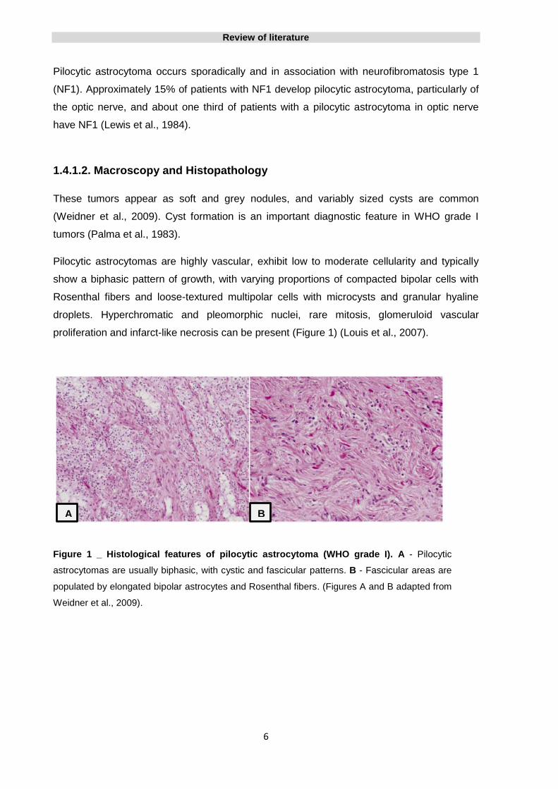
Pilocytic astrocytomas are highly vascular, exhibit low to moderate cellularity and typically
show a biphasic pattern of growth, with varying proportions of compacted bipolar cells with
Rosenthal fibers and loose-textured multipolar cells with microcysts and granular hyaline
droplets. Hyperchromatic and pleomorphic nuclei, rare mitosis, glomeruloid vascular
proliferation and infarct-like necrosis can be present (Figure 1) (Louis et al., 2007).
Figure 1 _ Histological features of pilocytic astrocytoma (WHO grade I). A - Pilocytic
astrocytomas are usually biphasic, with cystic and fascicular patterns. B - Fascicular areas are
populated by elongated bipolar astrocytes and Rosenthal fibers. (Figures A and B adapted from
Weidner et al., 2009).
A B
Review of literature
7
1.4.2. Diffuse astrocytoma (WHO grade II)
1.4.2.1. Epidemiological and clinical features
Diffuse astrocytoma, or low grade astrocytoma, as some prefer to designate it, represent
about 10% of all gliomas. According to Central Brain Tumor Registry of the United States
(CBTRUS) data, diffuse astrocytoma has an annual incidence rate of 1.3/1 million population.
The peak incidence is in young adults between the ages of 30 and 40 years, with a male
gender preference and a male: female ratio of 1.18:1 (Louis et al., 2007; Ostrom et al.,
2013).
Subtle signs such as speech difficulties, changes in sensation, vision and motor change may
be present in an early phase, but the commonest symptom are seizures (Louis et al., 2007).
1.4.2.2. Macroscopy and Histopathology
About a third of cases of diffuse astrocytomas develop preferentially in the frontal and
temporal lobes, but it may be located in any region of the CNS (Louis et al, 2007).
Macroscopically, are often difficult to distinguish from adjacent invaded structures, with
enlargement and distortion of these anatomical structures. Local mass lesions are grey or
yellow-white, with indistinct boundaries, smaller or larger cysts, granular areas and zones of
firmness or softening may be seen (Louis et al., 2007).
Diffuse astrocytoma is composed of well differentiated fibrillary, gemistocytic or, rarely,
protoplasmic astrocytes on the background of a loosely structured, often microcystic tumor
matrix. When compared with the normal brain tissue, the cellularity is moderately increased.
Occasional nuclear atypia is present, and mitotic activity is generally absent, however, a
single mitosis is not sufficient for the diagnosis of anaplastic astrocytoma. Other histologic
features suggestive of anaplasia, such as, necrosis or microvascular proliferation are not
present in these tumors (Louis et al., 2007).
Normal astrocytes have an oval-to-elongate or round nucleus and a cytoplasm which show
no Hematoxylin-Eosin (H&E) staining. Reactive astrocytes present an enlarged, eccentric
nucleus and a stainable eosinophilic, defined cytoplasm (Figure 2) (Louis et al., 2007).

Review of literature
8
Figure 2 _ Histological features of diffuse astrocytoma (WHO grade II). A – Increased cellularity
and some nuclear atypia. B – Microcystic change is a diagnostic feature os astrocytoma but is also
seen in oligodendroglioma. C – Minimally hypercellular astrocytoma with an irregular distribution of
elongated nuclei and nuclear atypia. (Figures adapted from Weidner et al., 2009).
1.4.3. Anaplastic astrocytoma (WHO grade III)
1.4.3.1. Epidemiological and clinical features
Anaplastic astrocytoma is a high-grade glioma, that may arise from diffuse astrocytoma
(WHO grade II) or de novo (without evidence of a less malignant precursor lesion).
These tumors account for approximately 6% of all gliomas, affect males more frequently than
females (ratio 1.1:1) and the peak of incidence in adults aged between 30 to 50 years. In
most cases, anaplastic astrocytoma will progress to glioblastoma, showing a mean time to
progression of about 2 years (Louis et al., 2007; Ostrom et al., 2013).
Review of literature
9
1.4.3.2. Macroscopy and Histopathology
Like in diffuse astrocytoma, anaplastic astrocytoma present as a mass with tendency to
infiltrate the surrounding brain tissue leading to marker enlargement of neighbour structures.
However cysts are uncommon and areas of granularity, opacity and soft consistency are
frequently present (Louis et al., 2007).
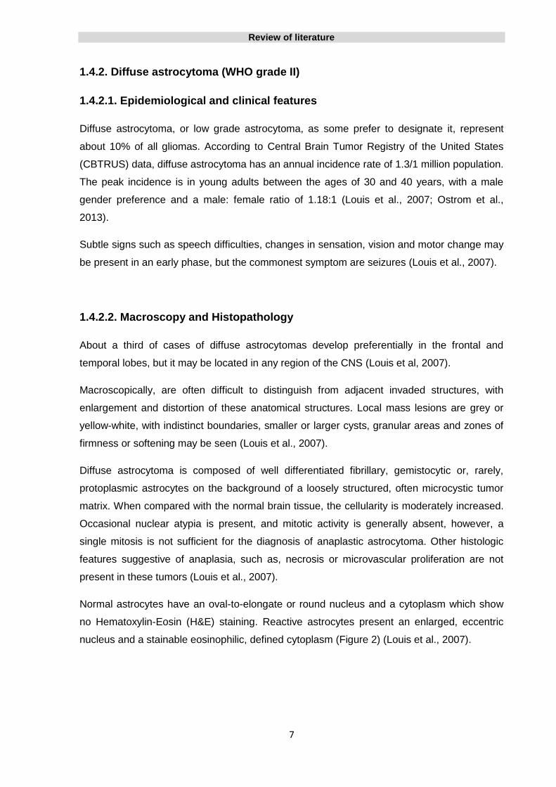
Anaplastic astrocytoma exhibit hypercellularity, cytologic anaplasia and mitotic activity (more
than one figure). Show a variable degree of cytoplasmatic pleomorphism or nuclear
elongation and pleomorphism, or both, when compared with diffuse astrocytoma.
Microvascular proliferation and necrosis are absent (Figure 3) (Louis et al., 2007).
Figure 3 _ Histological features of anaplastic astrocytoma (WHO grade III). A and B –
Characteristic hypercellularity, elongation and hyperchromasia of nuclei, atypia and brisk mitotic
activity. (Figures A and B adapted from Weidner et al., 2009).
1.4.4. Glioblastoma (WHO grade IV)
1.4.4.1. Epidemiological and clinical features
Glioblastoma, also termed glioblastoma multiforme, is the most frequent brain tumor and
represent about 15% of all intracraninal neoplasm and 60 to 75% of astrocytic tumors
(Ohgaki and Kleihues, 2005).
A B
Review of literature
10
The peak of incidence of these tumors is between 45 and 75 years of age and the clinical
history is short, frequently less than 3 months, characterized by signs and symptoms such
as, intracranial pressure, headache, nausea, epileptic seizures and also neurological
symptoms like personality changes (Louis et al., 2007).
The majority of glioblastoma cases (>90%) are primary glioblastoma that develop rapidly de
novo, without clinical or histological evidence of a less malignant precursor lesion. Secondary
glioblastoma, represent about 5% of all glioblastoma, and develop slowly through
progression from diffuse astrocytoma (WHO grade II) or anaplastic astrocytoma (WHO grade
III) (Louis et al., 2007).
The mean age of primary glioblastoma is about 60 years of age and it develop more
frequently in male (male/female ratio of 1.33), whereas secondary glioblastoma develop in
younger patients (45 years) and is more frequent in women (male/female ratio of 0.65). The
median survival of primary and secondary glioblastoma is 4.7 and 7.8 months, respectively
(Ohgaki et al., 2004; Ohgaki and Kleihues, 2005).
1.4.4.2. Macroscopy and Histopathology
Typical histopathological features include nuclear atypia, cellular pleomorphism, mitotic
activity, vascular thrombosis, microvascular proliferation and necrosis (Louis et al., 2007).
Microvascular proliferation and necrosis are histopathological hallmarks of glioblastoma
diagnosis. Microvascular proliferation consists of multi-layered, mitotically active endothelial
cells together with smooth muscle cells (Nagashima et al., 1987). Necrosis may comprise
more than 80% of the tumor and consists of multiple, small, irregulary-shaped band-like or
serpiginous foci, surrounded by radially oriented, densely packed, small fusiform gliomas
cells in a “pseudopalisading” pattern (Figure 4) (Louis et al., 2007).
Primary and secondary glioblastoma are histologically indistinguishable, but a growing
number of evidence has been suggesting that these two entities evolve through different
molecular mechanisms.

Review of literature
11
Figure 4 _ Histological features of glioblastoma (WHO grade IV). A – Cytologic pleomorphism and severe nuclear atypia. B – Microvascular proliferation. C – Pseudopalisading necrosis. (Figures adapted from Weidner et al., 2009).
1.5. Oligodendroglial tumors
Oligodendroglial tumors account for approximately 8.4% of all gliomas (Ostrom et al., 2013).
Based on their degree of malignancy and other histopathological features, two histological
subtypes are distinguished: oligodendroglioma (WHO grade II) and anaplastic
oligodendroglioma (WHO grade III).
1.5.1. Oligodendroglioma (WHO grade II)
1.5.1.1. Epidemiological and clinical features
Oligodendroglioma represents about 2.5% of all primary brain tumors and 5-6% of all
gliomas (Ohgaki and Kleihues, 2005). The incidence rate is about 0.27 per 100,000 persons
and is more frequent in males than in females by a ratio of 1.2:1.The average age on onset
of oligodendroglioma is between 35 and 55 years (Ostrom et al., 2013).
A B
C
Review of literature
12
Seizures are the most frequently symptom encountered in oligodendroglioma, followed by
headache, increased intracranial pressure, focal neurological deficits and cognitive or mental
changes (Lebrun et al., 2004).
1.5.1.2. Macroscopy and Histopathology
Oligodendrogliomas exhibit predominantly cerebral hemispheric locations favouring the
frontotemporal region, in about 50 to 65 % of cases, followed by temporal, parietal and
occipital lobes, respectively.
Macroscopically, oligodendroglioma appear as soft, gelatinous grayish-pink masses with well
delineated borders. In some areas the tumor may present a grilly texture due to the presence
of calcifications. Regions of cystic degeneration and intratumoral haemorrhages can be also
present (Louis et al., 2007).
The microscopic appearance of most oligodendroglioma is highly distinctive; the tumor it is
diffusely infiltrating with moderate cellularity and is composed of monomorphic cells with
uniform, round nuclei, perinuclear halo and increased chromatin density. The perinuclear
halos, denominated honeycomb pattern, are artifacts, resulting from the processing of tissue,
seen in the paraffin embedded tissue but not in smear preparation nor in frozen sections
(Louis et al., 2007).
Microcalcifications, cystic degeneration, dense network of branching capillaries (chicken wire
pattern), satellitosis and occasionally mini-gemistocytes are also present. Nuclear atypia can
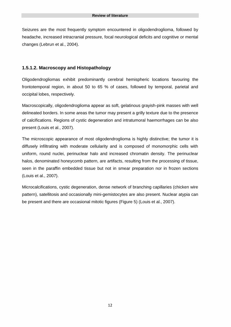
be present and there are occasional mitotic figures (Figure 5) (Louis et al., 2007).

Review of literature
13
Figure 5 _ Histological features of Oligodendroglioma (WHO grade II). A – Cells
perinuclear halos forming the honeycomb pattern. B – Cells with clear cytoplasm and well
defined plasma membrane. C – Dense network of branching capillaries. D –
Microcalcifications. (Figures A, B and C adapted from Louis et al., 2007. Figure D adapted from
Weidner et al., 2009).
1.5.2. Anaplastic oligodendroglioma (WHO grade III)
1.5.2.1. Epidemiological and clinical features
Anaplastic oligodendroglioma accounts for approximately 1.2% of all primary brain tumors
and represents about 20% of all oligodendroglial tumors (Ohgaki and Kleihues, 2005).
Anaplastic oligodendroglioma have an annual incidence of approximately 0.11 per 100,000
population and its incidence peak is between 45 and 50 years. They are slightly more
common in men, with a male / female ratio of 1.3 / 1 (Ostrom et al., 2013).
Anaplastic oligodendroglioma may develop de novo or be the result of a progression from a
grade II oligodendroglioma. The median time to progression between oligodendroglioma and
the development of anaplastic oligodendroglioma is about 6 to 7 years. Regarding anaplastic
oligodendroglioma developed de novo, the most important symptom is seizures (Lebrun et
al., 2004).
A B
C D
Review of literature
14
A
D C
B
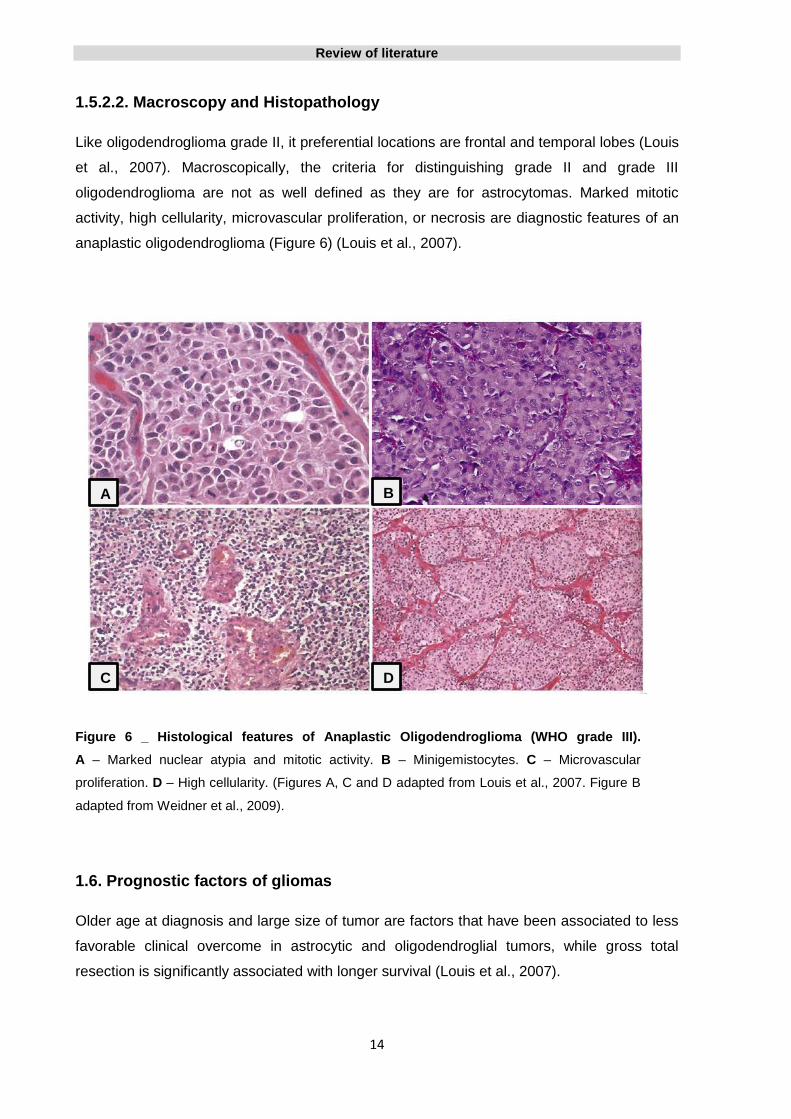
1.5.2.2. Macroscopy and Histopathology
Like oligodendroglioma grade II, it preferential locations are frontal and temporal lobes (Louis
et al., 2007). Macroscopically, the criteria for distinguishing grade II and grade III
oligodendroglioma are not as well defined as they are for astrocytomas. Marked mitotic
activity, high cellularity, microvascular proliferation, or necrosis are diagnostic features of an
anaplastic oligodendroglioma (Figure 6) (Louis et al., 2007).
Figure 6 _ Histological features of Anaplastic Oligodendroglioma (WHO grade III).
A – Marked nuclear atypia and mitotic activity. B – Minigemistocytes. C – Microvascular
proliferation. D – High cellularity. (Figures A, C and D adapted from Louis et al., 2007. Figure B
adapted from Weidner et al., 2009).
1.6. Prognostic factors of gliomas
Older age at diagnosis and large size of tumor are factors that have been associated to less
favorable clinical overcome in astrocytic and oligodendroglial tumors, while gross total
resection is significantly associated with longer survival (Louis et al., 2007).

Review of literature
15
Some glioblastoma may develop from a diffuse or anaplastic astrocytoma (secondary
glioblastoma) or it may manifest de novo (primary glioblastoma). Secondary glioblastoma
prognosis is better than in primary glioblastoma. However, this fact seems to be related with
the fact that, on average, patients with a primary glioblastoma are older than those with
secondary ones (Louis et al., 2007).
Studies have shown that patients with diffuse astrocytomas have worse prognosis when
compared to oligodendrogliomas, and the tumor malignancy grade is a proved independent
prognostic factor in gliomas .The survival of patients and choice of treatment depends mostly
on the malignancy grade of gliomas (Louis et al., 2007).
Presence of mitotic activity is in general a bad prognostic sign and is valid to all glial tumors
(Louis et al., 2007). According to WHO classification, the presence of a single mitosis is not a
criterion for anaplastic astrocytoma diagnosis. The literature provides contradictory data
concerning prognostic significance of a single mitosis. However, several authors suggested
that a single mitosis does not determine a poor prognosis. They found that survival of
patients with astrocytomas with single mitosis is similar to that of patients with WHO grade II
astrocytomas and is significantly better than that with astrocytomas with a larger number of
mitoses (Coons and Pearl, 1998; Giannini et al., 1999; Perry et al., 1999).
In general, higher proliferation rates significantly correlate with worse prognosis (Louis et al.,
2007). The most used technique to evaluate cell proliferation is to perform
immunohistochemistry technique using KI-67/MIB-1 antibodies. This antibody is not
expressed only in cells in the resting phase G0. The growth fraction, as determined by the
antibodies KI-67/MIB-1, is usually less than 4% (with a median of 2.5%) in diffuse
astrocytoma, range between 5 to 10% in anaplastic astrocytoma and 15 to 20% in
glioblastoma, and below 5% in oligodendrogliomas.
Microvascular proliferation, a histopathological hallmark of glioblastoma, plays a large role in
the tumor growth and spread, and is an indicator of a high malignancy grade glioma. Leon et
al have shown that density of microvessels is a prognostic factor of astroglial tumors (Leon et
al., 1996).
Necrosis is also a distinguishing feature of glioblastoma and is incompatible with a diagnosis
of anaplastic astrocytoma, though it may be present in anaplastic oligodendrogliomas.
Clinical studies indicate that as the degree of necrosis advances, the patient's prognosis
worsens, and so necrosis is pointed as one of strongest predictors of bad prognosis in
gliomas patients (Raza et al., 2002).

Review of literature
16
Karnofsky‘s ten-tiered scale is used to evaluate the patient’s ability to manage every-day life
and work. Karnofsky Performance Status (KPS) at diagnosis and other measures of mental
and physical functionality also predict survival for glioblastoma and anaplastic astrocytoma
patients; a higher preoperative Karnofsky score is a strong predictor of a more favorable
clinical outcome (Louis et al., 2007).
Besides the clinical prognostic factor presented, several molecular prognostic factor have
also been identified in gliomas, and will be presented later in the appropriate sections.
1.7. Immunophenotype of gliomas
Immunohistochemical markers are important and rapidly evolving tools in the classification
and neuropathological diagnosis of malignant gliomas. Currently, the most clinically useful of
these markers, for classification of gliomas, are GFAP, OLIG2, Synaptophysin, S-100,
keratins and vimentin.
Glial fibrillary acidic protein (GFAP) is an intermediate filament expressed by normal glial
cells and glial tumor cells whose sensitivity as a marker of glial differentiation is around
100%. GFAP can be detected in nearly all malignant gliomas but is negative in carcinomas,
lymphomas, melanomas and sarcomas. The fibrillary matrix, composed by neoplastic cell
processes and reactive astrocytes, may forms a diffuse GFAP positive background. Despite
GFAP is expressed more intensely and more frequently in astrocytomas it is also expressed
in oligodendrogliomas (Brat et al., 2008; Louis et al., 2007). In retrospective studies of GFAP
expression by immunohistochemistry, staining was noted in 100% of astrocytic tumors, in
58% of oligodendrogliomas and 79% of anaplastic oligodendrogliomas (Cosgrove et al.,
1989; Dehghani et al., 1998).
Cytokeratin expression usually indicates epithelial differentiation supporting a diagnosis of
metastatic carcinoma rather than malignant gliomas. However, the evaluation of the
cytokeratins in gliomas should be cautious because malignant gliomas often show
immunoreactivity to cytokeratins, especially CK AE1/3 (Cosgrove et al., 1993; Kriho et al.,
1997). CK AE1/3 staining was found in 66% of diffuse astrocytomas, 83% of anaplastic
astrocytomas and 83% of glioblastomas (Cosgrove et al., 1993). In a different study, CK
AE1/3 expression was present in 96% of glioblastoma, while only 4% showed expression of
CK8.18, CK7 and CK20 (Oh and Prayson, 1999). This immunoreactivity is attributed to a
cross-reactivity phenomenon of the antibodies with GFAP. For these reason, epithelial
differentiation in metastatic carcinoma should be preferably investigated using cytokeratins
Review of literature
17
antibodies which are negative in astrocytomas, such as CK8.18 or even other epithelial
marker like epithelial membrane antigen (EMA).
S-100 protein is expressed in almost 100% of malignant gliomas and for this reason is not a
good marker for differential diagnosis from other S-100 positive tumors, such as, melanoma
(Nakapoulou et al., 1990).
Vimentin, an intermediate filament protein, shows a pattern of immunoreactivity similar to that
of GFAP and vimentin-positive cells may lack GFAP expression. Vimentin is expressed in
astrocytic tumors, and a tendency to be expressed more consistently is found in high grade
astrocytomas. In oligodendroglioma, vimentin is infrequently expressed in low grade tumors
and more often found in anaplastic oligodendroglioma (Louis et al., 2007; Dehghani et al.,
1998).
There is no distinct single antibody available to discriminate reliably between oligodendroglial
and astrocytic tumors. OLIG2 immunoreactiviy is slightly stronger in oligodendroglioma but is
also seen in other glial tumors (Ligon et al., 2004). MAP2 is constantly expressed in
oligodendroglioma, but also found in 92% of diffuse astrocytoma and glioblastoma (Blümcke
et al., 2004). WT1 in oligodendroglioma is usually restricted to single WT1 positive tumor
cells or completely absent, while in high grade astrocytic tumor is strongly expressed in 83-
92% (Schittenhelm et al., 2009). Nogo-A is found in 71% oligodendroglioma and 24%
glioblastoma but is absent in diffuse astrocytoma (Kuhlmann et al., 2008).
1.8. Genetic alterations in gliomas
In last decades, several genetic alterations were identified in gliomas that may be used to
facilitate glioma classification, especially in cases that present inconclusive or borderline
histological features, and possibly impact future treatment strategies.
Many of these genetic alterations have been investigated in regard to their diagnostic,
prognostic and or predictive implications, but only few were qualified as clinically relevant
biomarkers. A prognostic factor is any measurement available at the time of surgery that
correlates with disease-free or overall survival in the absence of systematic adjuvant and
thus conveys information on the natural course of the disease. In contrast, a predictive factor
should provide information associated with the response to a given therapy
(Riemenschneider et al., 2010).

Review of literature
18
1.8.1. EGFR gene amplification/overexpression
First reported by Libermann and co-workers in 1985, epidermal growth factor
receptor (EGFR) gene amplification and overexpression is a hallmark of glioblastoma,
specifically primary glioblastoma, affecting approximately 40% of these tumors. This
alteration is rare in secondary glioblastomas (<10%) (Ohgaki et al., 2004; Ware et al., 2003;
Yip et al., 2008; Watanabe et al., 1996).
Many glioblastoma showing EGFR gene amplification has EGFR mutations. EGFRvIII is the
best described and most common mutant type and is present in more than 50% of cases.
This truncated mutant variant is characterized by genomic deletion of exons 2-7 what results
in an active oncogenic form (Sugawa et al., 1990; Aldape et al., 2004; Gan et al., 2009).
The presence of EGFR amplification and EGFRvIII can be used to support high grade glioma
diagnosis when histologic criteria are doubtful. Their prognostic value remains unclear,
existing several contradictory studies (Nikiforova and Hamilton, 2011; Heimberger et al.,
2005).
EGFR signaling pathway is an attractive target for the development of new therapies in
glioma (Mischel et al., 2003; Kuan et al., 2000; Belda-Iniesta et al., 2008). Although the
predictive value of these changes is not yet clear, there are some studies showing that
EGFR amplification and EGFRvIII expression may provide a response to tyrosine kinase
inhibitor therapies, especially when there is PTEN expression (Mellinghoff et al., 2005; Haas-
Kogan et al., 2005).
A recent study demonstrates that EGFR amplification and rearrangement are early events in
tumorigenesis and EGFRvIII expression is restricted by epigenetic mechanisms, suggesting
that drugs that modulate the epigenome might be used successfully in glioblastoma tumors
(Del Vecchio et al., 2012).
1.8.2. PTEN gene mutation
The tumor suppressor gene phosphatase and tensin homology (PTEN), located at 10q23.3,
present mutations in about 15 to 40% of primary glioblastoma (Dahia, 2000; Knobbe et al.,
2002; Koul et al, 2008), being rare in secondary glioblastoma and other gliomas (Tohma et
al., 1998; Ohgaki et al., 2009).

Review of literature
19
PTEN deletions may occur but in a very low percentage below 2% (Knobbe and
Reifenberger, 2003).
Loss of chromosome 10 is associated with mutation or deletions of the PTEN gene in
glioblastoma and other human cancers (Colman, 2008).
Loss of heterozigoty (LOH) at 10q, frequently found at 10q23-24, 10q25-pter, 10p14-p15 or
with loss of the entire long arm of chromosome 10 (Nikiforova and Hamilton, 2011), is the
most frequent genetic alteration in glioblastomas (60-80% of cases). Is common in
glioblastomas and anaplastic astrocytomas (35-60%) but is less frequent in anaplastic
oligodendrogliomas (10%) (Ohgaki et al., 2009).
Studies suggest PTEN mutations and 10q LOH as poor prognostic markers in WHO grade III
and IV gliomas. The loss of 10q is associated with tumor progression (Koul, 2008; Hill et al.,
2003).
An inverse relationship between tumor grade and PTEN expression in gliomas revealed that
PTEN mutations are relatively late events in these tumors development, occurring during the
evolution from low grade to high grade gliomas (Ermoian et al., 2002).
1.8.3. TP53 gene mutation
TP53 is the gene that encodes the tumor suppressor protein p53 and is located at 17q13.1.
Somatic mutations of the TP53 gene have been reported to occur frequently in most of
human tumors, including gliomas (Greenblatt et al., 1994).
Mutation of TP53 gene is the most common molecular alteration in WHO grade II gliomas
being found in about 60% of cases (Parsons et al., 2008). Anaplastic astrocytomas and
secondary glioblastomas show a percentage of mutations similar to diffuse gliomas while the
rate of primary glioblastomas mutated is significantly lower (about 28%) (Watanabe et al.,
1997; Ohgaki et al., 2004). In oligodendrogliomas, TP53 mutations are found in 13% of
cases (Okamoto et al., 2004).
TP53 mutations are an early event in diffuse astrocytomas tumorigenesis. This alteration is
frequently found in the first biopsy and their frequency does not increase in recurrences
(Lang et al., 1994; Riemenschneider et al., 2009; van de Kelft, 1997).

Review of literature
20
Until now, studies are contradictory and do not confirm TP53 as an independent predictive or
prognostic of patient outcome in gliomas, but it has a role in diagnosis as it can help to
distinguish tumor grade (Ohgaki et al., 2004; Ohgaki et al., 2005; Schmidt et al., 2002).
1.8.4. IDH gene mutation
Mutations in the gene encoding the human cytosolic NADPH-dependent isocitrate
dehydrogenase (IDH), located on chromosome 2q33, were fist described by Parsons et al in
about of 12% of glioblastomas, mostly in secondary glioblastomas (Parsons et al., 2008;
Riemenschneider et al., 2010).
Subsequent studies identified IDH1 mutations at much higher frequencies in grade II and
grade III gliomas, in about 60 to 90% of cases (Balss et al., 2008; Watanabe et al., 2009;
Yan et al., 2009; Ichimura et al., 2009; Nobusawa et al., 2009; Sanson et al., 2009;
Hartmann et al., 2010).
All the described IDH1 mutations were found in codon 132 (exon 4) for arginine (R132)
(Parsons et al., 2009; Ichimura et al., 2009) and appear to be rare in other common human
tumors (Bleeker et al., 2009). The R132H mutations constitutes >90% of the IDH1 mutations
seen in gliomas (Yan et al., 2009).
Approximately 70% of the secondary glioblastomas carry IDH1 mutations while this is
observed only in 5 to 10% of primary glioblastomas, what reinforces the hypothesis that
glioblastomas, primary and secondary, have different molecular origins (Ohgaki et al., 2007;
Balss et al., 2008; Ichimura et al., 2009; Yan et al., 2009; Watanabe et al., 2009).
The vast majority of low grade diffuse astrocytomas contained both an IDH1 mutation and a
TP53 mutation and a similar majority of oligodendroglioma showed both IDH1 mutation and
1p/19q loss. These results suggest that IDH1 mutations are very early genetic events in
gliomagenesis, before TP53 mutations or loss of 1p/19q occur, and that diffuse astrocytomas
and oligodendrogliomas may originate from common glial precursor cells carrying IDH1
mutations (Watanabe et al., 2009; Sanson et al., 2009).
Other studies suggested that IDH1 mutations might occur after formation of a low grade
glioma and drive the progression of the tumor to a glioblastoma (Parsons et al., 2009; Yan et
al., 2009).

Review of literature
21
Mutations in IDH2 gene, affecting the arginine in position 172, occurs infrequently in
astrocytic gliomas but are present in about 3-5% of oligodendrogliomas (Yan et al., 2009;
Hartmann et al., 2010). IDH1 and IDH2 mutations were found to be mutually exclusive (Yan
et al., 2009).
In pilocytic astrocytomas, IDH1 and IDH2 mutations are absent, what possibly means they
arise through a different way and may be derived from a different type of glial progenitor (Yan
et al., 2009; Watanabe et al., 2009; Ichimura et al., 2009).
Sanson and colleagues (2009) found that 90% of the 1p19q codeleted gliomas have IDH1
mutations and that IDH1 mutations are rare in gliomas with EGFR amplification, what is
supported by the fact that 1p19q deletions and EGFR amplification are alterations mutually
exclusive (Sanson et al., 2009; Idbaih et al., 2008).
IDH mutations have diagnostic value, are associated with better outcome and young age,
and have been shown to be a powerful independent prognostic factor for longer survival in
gliomas WHO grade II, III and IV (Sanson et al., 2009; Parsons et al., 2008; Yan et al., 2009;
Combs et al., 2011; van den Bent et al., 2010; Nikiforova and Hamilton, 2011). A follow-up
study showed improved outcome for IDH1 and IDH2 mutated tumors, with median overall
survival 31 months versus 15 months for glioblastoma lacking mutations and 65 months
versus 20 months for anaplastic astrocytoma (Yan et al., 2009).
IDH1 and IDH2 mutations analysis can be helpful for discrimination between primary and
secondary glioblastoma and in case of a differential diagnosis of low grade glioma versus
pilocytic astrocytoma or even low grade glioma versus reactive gliosis, what can sometimes
be difficult solely on the basis of histopathological criteria particularly with small biopsies
(Horbinski et al., 2009; Korshunov et al., 2009; Watanabe et al., 2009; Yan et al., 2009;
Jansen et al., 2010).
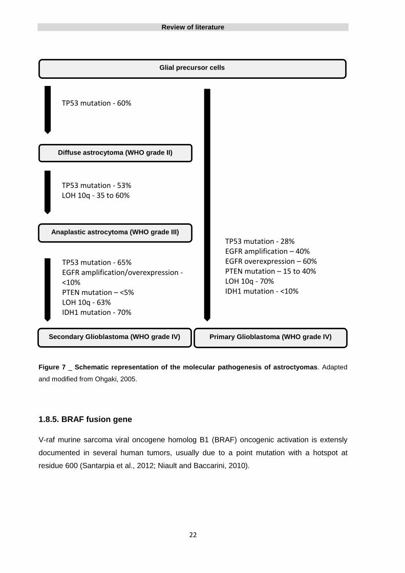
For a better perception of the changes that occur in different astrocytic tumors, inclusive the
differences between both primary and secondary glioblastomas, see the representation of
Figure 7.
Review of literature
22
Figure 7 _ Schematic representation of the molecular pathogenesis of astroctyomas. Adapted
and modified from Ohgaki, 2005.
1.8.5. BRAF fusion gene
V-raf murine sarcoma viral oncogene homolog B1 (BRAF) oncogenic activation is extensly
documented in several human tumors, usually due to a point mutation with a hotspot at
residue 600 (Santarpia et al., 2012; Niault and Baccarini, 2010).
Diffuse astrocytoma (WHO grade II)
Primary Glioblastoma (WHO grade IV) Secondary Glioblastoma (WHO grade IV)
Anaplastic astrocytoma (WHO grade III)
Glial precursor cells
TP53 mutation - 60%
TP53 mutation - 53% LOH 10q - 35 to 60%
TP53 mutation - 28% EGFR amplification – 40% EGFR overexpression – 60% PTEN mutation – 15 to 40% LOH 10q - 70% IDH1 mutation - <10%
TP53 mutation - 65% EGFR amplification/overexpression - <10% PTEN mutation – <5% LOH 10q - 63% IDH1 mutation - 70%

Review of literature
23
Recently, BRAF gene duplication and fusion, or less frequently point mutation, have been
identified in pilocytic astrocytomas and result in mitogen-activated protein kinase (MAPK)
signalling pathway activation (Pfister et al., 2008; Jones et al., 2008; Bar et al., 2008; Jones
et al., 2012).
A tandem duplication at 7q34 produce a novel oncogenic BRAF fusion gene, KIAA1549:
BRAF, which is present in about of 66% of pilocytic astrocytomas but rare in diffuse
astrocytomas (Jones et al., 2008; Sievert et al., 2009).
KIAA1549: BRAF fusion transcript seems to be a characteristic event in pediatric pilocytic
astrocytoma, presenting a frequency of 79% in tumors diagnosed in the first decade of life,
while in adult patients older than 40 years, with pilocytic astrocytoma, it frequency is about
7% (Hasselblatt et al., 2011).
BRAF point mutations (V600E and ins 3bp at 598) and SRGAP3:RAF1 fusion can rarely
occur in pilocytic astrocytomas and are mechanisms that also originate MAPK pathway
activation (Jones et al., 2008; Jones et al., 2009; Eisenhardt, 2011).
Because KIAA1549:BRAF fusion is so rare in diffuse astrocytomas it might allow the
differential diagnosis between pilocytic astrocytomas and low grade diffuse astrocytomas
especially when used in conjunction with the mutational analysis of the IDH1 gene, since this
gene mutations are generally absent in pilocytic astrocytomas (Korshunov et al., 2009;
Nikiforova and Hamilton, 2011).
The studies suggest that the BRAF fusion gene do not have any prognostic value but might
become an important diagnostic tool and a promissing therapeutic target (Jones et al., 2008;
Jones et al., 2009; Horbinski et al., 2010; Jeuken and Wesseling, 2010).
1.8.6. LOH of chromossomes 1p and 19q
First described in oligodendroglial tumors by Reifenberger and coworkers in 1994, the
combined deletion of chromosomes 1p and 19q is the consequence of an unbalanced
translocation between chromosomes 1 and 19 [t(1;19)(q10;p10)] where in most cases the
entire 1p/19q arms are involved (Felsberg et al., 2004; Jenkins et al., 2006; Ransom et al.,
1992; Bello et al., 1994; Ducray et al., 2009).

Review of literature
24
1p/19q codeletion is the most common genetic alteration and is present in up to 90% of
oligodendrogliomas, 50-70% of anaplastic oligodendroglioma and less than 10% of astrocytic
gliomas, including glioblastoma (Cairncross and Jenkins, 2008; Aldape et al., 2007). Despite
this change is uncommon in glioblastoma, when present seem to predict shorter survival,
possibly due true genomic instability (Smith et al., 2000). In anaplastic oligodendrogliomas is
a good prognostic factor, meaning of longer survival, and a strong predictor of sensitivity to
radio and chemotherapy but in grade II oligodendrogliomas the data are not clear
(Cairncross et al., 1998; Cairncross et al., 2006; Brandes et al., 2006; Kouwenhoven et al.,
2009; Bauman et al., 2000; van den Bent et al., 2006).
The higher frequency of 1p/19q codeletion in lower grade tumors and the fact this alteration
is retained at the time of progression may suggest this is an early event in tumor formation
(Hofer and Lassman, 2010; Lavon et al., 2007).
There is a strong association between 1p/19q codeletion and oligodendrogliomas that have
the classic features, namely, uniformly round nuclei and small nucleoli with even celular
distribution, prominente perinuclear “halos” and chicken-wire vascular pattern (Burger et al.,
2002; Kouwenhoven et al., 2009; Giannini et al., 2008). However morphology alone cannot
predict 1p/19q status (Scheie et al., 2008).
Loss of 1p/19q is virtually always associated with IDH1 mutations (Yan et al., 2009) and is
mutually exclusive from TP53 mutations, 10q deletion, EGFR and other genes amplifications
(Nutt et al., 2005; Bourne et al., 2010; Reifenberger and Louis, 2003).
Is a good prognostic factor, meaning of longer survival, and a strong predictor of sensitivity to
radio and chemotherapy, particularly to the combination of procarbazine, lomustine and
vincristine (PCV), in anaplastic oligodendrogliomas but in grade II oligodendrogliomas the
data are not clear (Cairncross et al., 1998; Cairncross et al., 2006; Brandes et al., 2006;
Kouwenhoven et al., 2009; Bauman et al., 2000; van den Bent et al., 2006).
Because is most common in oligodendrogliomas, 1p/19q deletion status is often used to
support a diagnosis of oligodendroglioma when the histology is ambiguous (Aldape et al.,
2007).

Review of literature
25
1.9. Epigenetics
Epigenetic is described as a heritable change in gene expression without an alteration in the
DNA sequence (Holliday, 1987).
In addition to genetic alterations, epigenetic abnormalities are associated with all cancer
types and it is now apparent that not only genetic, but also epigenetic changes might be
responsible for cancer initiation and progression.
Epigenetic modifications are reversible but nonetheless are very stable and can be generally
divided into three main mechanisms: DNA methylation, histone modification and chromatin
remodelling. Disruption of any of these three mechanisms leads to inappropriate gene
expression, resulting in cancer development and other diseases (Jones and Baylin, 2002). Of
these mechanisms, the most frequent and the best studied in mammalian genome is DNA
methylation.
1.9.1. DNA methylation
DNA methylation is the covalent addition or subtraction of a methyl group to a cytosine
nucleotide in a sequence of DNA. In humans, DNA methylation is catalysed by enzymes,
named DNA methyltransferases (DNMTs). DNMTs family encompasses three main
enzymes, DNMT1, DNMT3A and DNMT3B, which are divided according to their
maintenance and/or de novo functions. Maintenance DNMT1 binds methyl groups to the
hemimethylated DNA during replication whereas de novo DNMT3A and DNMT3B add methyl
groups to CpG dinucleotides of unmethylated DNA (Okano et al., 1999; Esteller, 2008).
In addition to the DNMTs, also play an important role in DNA methylation, demethylases
(namely 5-methylcytosine glycosylase and MBD2b), methylation centers triggering DNA
methylation and methylation protection centers (Costello and Plass, 2001).
In mammalian cells, methylation is mostly restricted to cytosine (C) residues that precede
guanine (G) residues, denominated CpG dinucleotides. In general, CpG dinucleotides are
underrepresented in the mammalian genome but can be found at relatively high frequency in
short genomic sequences know as CpG islands (Bird, 1986).
The overall frequency of CpG dinucleotides in the genome is substantially lower than what
would be mathematically predicted, probably because DNA methylation has progressively

Review of literature
26
depleted the genome of CpG dinucleotides over the course of time. This depletion is related
to the propensity of methylated C to deaminate, forming T, and if this mutation is not
repaired, the C to T change remains and represents the most common type of genetic
polymorphism in human populations.
CpG islands are DNA segments of 0.5 to 2.5 kb in size, which are rich in cytosine-guanine
dinucleotides (have a frequency of CpG dinucleotides approximately five times greater that
the genome as a whole) and comprise 1-2% of the entire genome (Bird, 1986).
CpG islands are often located in the promoter or 5’ exonal sequences of genes. About 50 to
60% of human genes harbour CpG islands in their promoter sequences and these CpG
islands are normally protected from methylation, while CpG dinucleotides outside CpG
islands are commonly methylated (Larsen et al., 1992; Antequera and Bird, 1993; Costello
and Plass, 2001). It has been proposed that these patterns of methylation are responsible for
transcriptionally active and inactive zones in human genome (Costello and Plass, 2001).
1.9.2. DNA methylation in normal cells
The deletion of any of three methyltransferases genes from mice is lethal, suggesting that
methylation has additional and indispensable functions in mammal (Okano et al., 1999).
In normal cells, CpG islands methylation in the promoter region is a normal event that occurs
to regulate gene expression, as mechanism for control of specific gene expression in normal
tissues in adults. The most prominent examples are the inactivation of the x chromosome in
females (methylation of CpG island is present on the inactivated x chromosome), the control
of certain tissue-specific genes which are silenced in almost tissues except malignant tumors
and imprinted genes (only the paternally or maternally inherited allele of the gene is
expressed, the inactivation of the allele which is not expressed is associated with promoter
methylation) (Ideraabdullah et al., 2008).
1.9.3. DNA methylation and cancer
As compared with normal cells, cancer cells show major disruptions in their DNA
methylations patterns. Cancer cells exhibit two apparently opposing changes is their pattern
of DNA methylation, both playing important roles in tumorigenic process: overall decrease in
DNA methylation (DNA Hypomethylation) and increased methylation of CpG islands (DNA
Hypermethylation). Whether genome hypomethylation and CpG island hypermethylation are

Review of literature
27
linked by a common underlying mechanism or result from distinct abnormalities in the cancer
is currently unknown.
DNA hypomethylation and hypermethylation occur at specific but distinct sites within cancer
genome what suggests different aetiologies and both are present in pre-malignant lesions
indicating that they are not simply a consequence of the malignant state. Hypermethylation
at promoter CpG islands is the most studied and has a much clear role in carcinogenesis.
Therefore, DNA methylation can act as a double-edged sword, promoting the neoplastic
process by local hypermethylation resulting in silencing of tumor suppressor genes and in
parallel by globlal hypomethylation triggering reactivation of cellular proto-oncogenes.
1.9.3.1. DNA Hypomethylation
The low level of DNA methylation in tumors as compared to their normal tissue counterparts
is denominated as DNA hypomethylation. In human tumors, DNA hypomethylation, was
identified even earlier than hypermethylation (Feinberg and Vogelstein, 1983) and in human
genome is often found in repeated DNA sequences such as satellite DNA, LINE, SINE, viral
retrotransposon sequence and single copy genes (Elrich, 2002).
Although analysis of DNA methylation at a genome-wide level has revealed that virtually all
human tumors exhibit overall decreased levels of DNA methylation, it role in cancer
development remains to be elucidated (Costello and Plass, 2001). Meanwhile, it has been
proposed that global hypomethylation may contribute to oncogenesis by generation of
chromosome instability, latent retrotransposon activation and oncogene loss of imprinting
and consequent activation (Esteller, 2008).
DNA hypomethylation is a common event in solid tumors such as prostate tumors, cervical
cancer and in some hematologic malignancies such as leukaemia (Kim et al., 1994; Bedford
and van Helden, 1987). Many genes are activated and expressed through hypomethylation
in human tumors including HRAS, cyclin D2 and Maspin in gastric cancer, carbonic
anhydrase IX in renal cell and S100 calcium-binding protein A4 in colon cancer and
melanoma-associated MAGE gene in melanoma and glioblastoma. Human papillomavirus
HPV16 is also activated by hypomethylation in cervical cancer (Wilson et al., 2007).
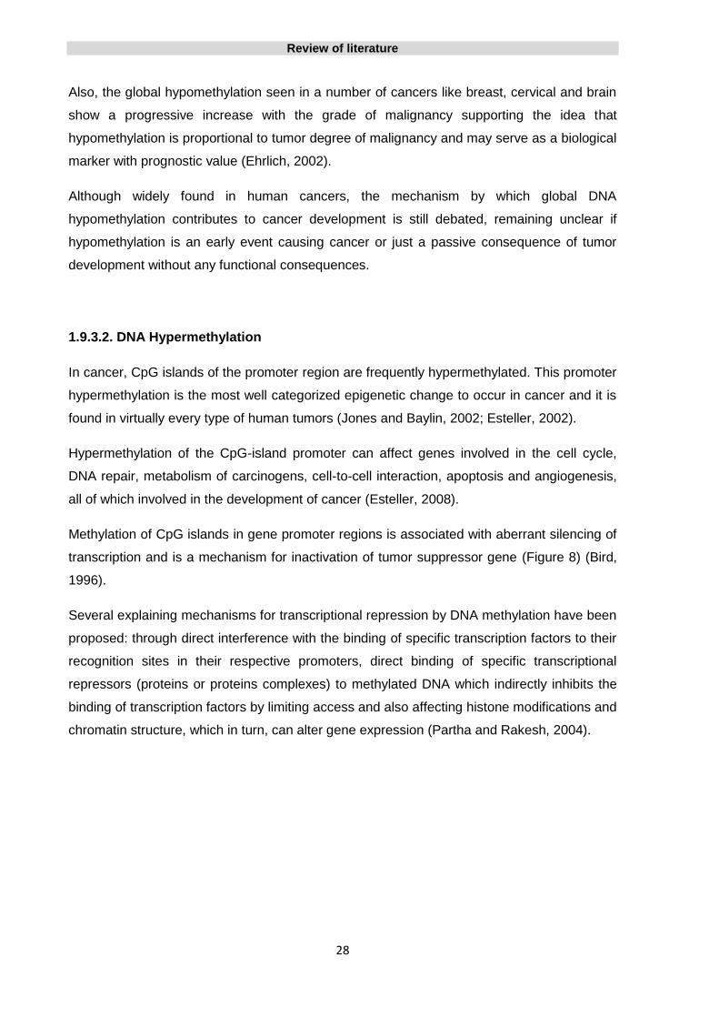
Review of literature
28
Also, the global hypomethylation seen in a number of cancers like breast, cervical and brain
show a progressive increase with the grade of malignancy supporting the idea that
hypomethylation is proportional to tumor degree of malignancy and may serve as a biological
marker with prognostic value (Ehrlich, 2002).
Although widely found in human cancers, the mechanism by which global DNA
hypomethylation contributes to cancer development is still debated, remaining unclear if
hypomethylation is an early event causing cancer or just a passive consequence of tumor
development without any functional consequences.
1.9.3.2. DNA Hypermethylation
In cancer, CpG islands of the promoter region are frequently hypermethylated. This promoter
hypermethylation is the most well categorized epigenetic change to occur in cancer and it is
found in virtually every type of human tumors (Jones and Baylin, 2002; Esteller, 2002).
Hypermethylation of the CpG-island promoter can affect genes involved in the cell cycle,
DNA repair, metabolism of carcinogens, cell-to-cell interaction, apoptosis and angiogenesis,
all of which involved in the development of cancer (Esteller, 2008).
Methylation of CpG islands in gene promoter regions is associated with aberrant silencing of
transcription and is a mechanism for inactivation of tumor suppressor gene (Figure 8) (Bird,
1996).
Several explaining mechanisms for transcriptional repression by DNA methylation have been
proposed: through direct interference with the binding of specific transcription factors to their
recognition sites in their respective promoters, direct binding of specific transcriptional
repressors (proteins or proteins complexes) to methylated DNA which indirectly inhibits the
binding of transcription factors by limiting access and also affecting histone modifications and
chromatin structure, which in turn, can alter gene expression (Partha and Rakesh, 2004).
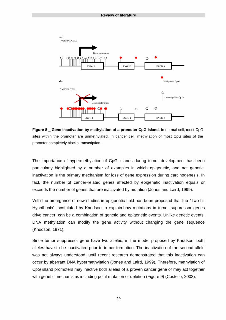
Review of literature
29
Figure 8 _ Gene inactivation by methylation of a promoter CpG island. In normal cell, most CpG
sites within the promoter are unmethylated. In cancer cell, methylation of most CpG sites of the
promoter completely blocks transcription.
The importance of hypermethylation of CpG islands during tumor development has been
particularly highlighted by a number of examples in which epigenetic, and not genetic,
inactivation is the primary mechanism for loss of gene expression during carcinogenesis. In
fact, the number of cancer-related genes affected by epigenetic inactivation equals or
exceeds the number of genes that are inactivated by mutation (Jones and Laird, 1999).
With the emergence of new studies in epigenetic field has been proposed that the “Two-hit
Hypothesis”, postulated by Knudson to explain how mutations in tumor suppressor genes
drive cancer, can be a combination of genetic and epigenetic events. Unlike genetic events,
DNA methylation can modify the gene activity without changing the gene sequence
(Knudson, 1971).
Since tumor suppressor gene have two alleles, in the model proposed by Knudson, both
alleles have to be inactivated prior to tumor formation. The inactivation of the second allele
was not always understood, until recent research demonstrated that this inactivation can
occur by aberrant DNA hypermethylation (Jones and Laird, 1999). Therefore, methylation of
CpG island promoters may inactive both alleles of a proven cancer gene or may act together
with genetic mechanisms including point mutation or deletion (Figure 9) (Costello, 2003).
Review of literature
30
Figure 9 _ Genetic and epigenetic mechanisms that inactivate cancer genes. The
mechanisms can act alone or in various combinations to cause biallelic inactivation of a cancer
gene. (Adapted from Costello and Plass, 2001).
This assumption is now being used to identify novel tumor suppressor genes in regions
of chromosomal loss where searches for mutated genes have failed to identify
candidate cancer genes. Certain chromosomal regions seem to be more prone to
hypermethylation events than others, being the short arm of chromosome 3 (3p) the
chromosome region where more promoter CpG islands hypermethylation have been
identified (Figure 10) (Esteller, 2007).
Many of the genes modified by promoter hypermethylation have classic tumor
suppressor function. Aberrant CpG island hypermethylation in tumor suppressor genes
or tumor-related genes is a frequent finding in human cancers of various tissue types,
and each type has its own distinct methylation pattern. Generally genes frequently
methylated in a specific cancer type differed from those methylated in other cancer
types. These characteristic allows that different types of cancer can be reliably
distinguished by their unique tumor-type and gene-specific DNA hypermethylation
pattern. These specific DNA methylation pattern is usually designed by DNA
“hypermethylome” (Costello et al., 2000a).

Review of literature
31
The first discovery of methylation in a CpG island of a tumor suppressor gene in a
human cancer was that of retinoblastoma gene (Rb) in 1989 (Greger et al., 1989).
Then, the subsequent characterization of tumor suppressor genes, such as, VHL, p16,
MLH1 and BRAC1, in which transcriptional silencing was associated with the
hypermethylation gave rive to a new interest in epigenetic matter (reviewed in Esteller,
2002).
Figure 10 _ Distribution of hypermethylated genes in human cancer. (Adapted from
Esteller, 2007).
The reason why some genes are hypermethylated and others remained unmethylated in
specific tissue types is unclear. One explanation is that certain genes become
hypermethylated preferentially because their hypermethylation confers a selective growth
and survival advantage to a given cancer cell (Park and Claus, 2007). For example, BRCA1
gene undergoes promoter hypermethylation only in breast and ovarian tumors because only
in these tumor types does the lack of this transcript have important cellular consequences
(Esteller et al., 2000b).
When analysing tumor types regarding to DNA hypermethylation, tumors from the
gastrointestinal tract emerge as the most methylated while ovarian tumors and sarcomas are
described as the less hypermethylated. These can be explained by the fact that
hypermethylated genes in the tumor types described as less hypermethylated have not yet
been found or that tumors more exposed to external carcinogen agents, like those from
gastrointestinal tract, are for this reason more hypermethylated types (Esteller, 2007).

Review of literature
32
1.9.4. DNA methylation in pre-malignant lesions
Promoter hypermethylation is often an early event in the natural history of human cancer and
might be a key initiating event (Esteller and Corn, 2001). Alterations of DNA methylation
patterns often precede malignancy as extensive CpG island hypermethylation can be
detected in benign lesions that precede neoplastic process.
Early occurrence of DNA hypermethylation has been established in preneoplastic pancreatic
intraepithelial lesions that precede pancreatic carcinogenesis (Hanoun et al., 2010), in
chronic cholecystitis that may be a precursor lesion for gallbladder cancer (García et al.,
2009), in endometrial hyperplasia preceding endometrial cancer (Esteller et al., 1999a;
Banno et al., 2006), in basal cell hyperplasia squamous metaplasia and carcinoma in situ of
the lung (Belinsky et al., 1998), in colorectal adenomas (Judson et al., 2006) and in
premalignant stages of gastric carcinoma (Kang et al., 2001).
1.9.5. Aging, DNA methylation and cancer
The use of methylated genes is based on the assumption that methylation at specific sites in
the promoter regions of certain genes is confined to malignancy or premalignant lesions.
Other factors, however, may affect gene methylation such as aging, benign diseases and
exposure to environmental influences. To be useful as biomarkers, cancer associated or
predisposing alterations have therefore to be distinguishable from age-associated DNA
methylation changes.
Aging has long been considered one of the most important risk factors for the development
of cancers, and has generally been attributed to the cumulative exposure to carcinogens
over time, as well as the time required to have the multiple hits needed for the onset of
cancer. At the same time, several studies suggested that age-related CpG island methylation
is a universal phenomenon and one of the most frequent events contributing to
hypermethylation in cancers (Ahuja and Issa, 2000).
Studies for the relationship between aging and DNA methylation have demonstrated that
aging is associated with genomic DNA hypomethylation and gene specific promoter DNA
hypermethylation. Total methylcytosine contents are prone to decrease by aging, leading to
genomic hypomethylation in most vertebrate tissues, whereas promoter regions tend to
undergo hypermethylation in many genes.

Review of literature
33
The first link between aging and changes in DNA methylation was initially reported for the
estrogen receptor (ER) gene in normal human colon. ER methylation was undetectable in
young patients, but partial methylation of ER can be observed in normal tissues of older
patients, and increases with age. ER hypermethylation is then found in all colonic adenomas
and cancers (Issa et al., 1994). Posteriorly, other study showed that both N33 and MyoD
genes also have a pattern of methylation in the normal colorectal mucosa that increases as a
function of age (Ahuja et al., 1998)
Also in colon, when analyzing the methylation status of various genes, Toyota and coworkers
found that 70% of genes showed hypermethylation in normal colon tissue with increased
hypermethylation in adenomas and colon cancer, while in other genes hypermethylation was
found only in cancer (Toyota et al., 1999b). In a similar study in breast cancer, in about half
of genes, hypermethylation was present in normal tissue and cancer, while the remaining
half is de-novo hypermethylated (Huang et al., 1999).
Supporting these findings, a study performed in monozygotic twins reveals that the patterns
of epigenetic modifications in young twins are indistinguishable but diverge as they become
older, so differences in epigenetic patterns in genetically identical individuals could be
explained by the influence of both external and internal factors, such as aging (Fraga et al.,
2005).
The results of the studies also suggest that age-related methylation is gene and tissue-
specific and only certain promoter are susceptible to this process. Also, and since age-
related methylation is widespread while de-novo methylation in cancer is highly selective for
certain genes, it is likely that each is governed by different mechanisms (Ahuja and Issa,
2000).
With regard to this topic, some limitations have been identified in the identification of
methylation associated with aging, particularly the percentage of methylated cells present in
the analyzed samples. An important point in studying the phenomenon of age-related
methylation is that the normal tissue adjacent to cancer may not always be representative of
the cell populations that are predisposed to neoplasia (Ahuja and Issa, 2000). Fujii and co-
workers (1998) analyzed 39 primary breast cancer tissues and found that HIC-1 gene
(Hypermethylated in cancer 1 gene which is a candidate tumor suppressor gene, located at
17p13.3) was hypermethylated in 67% of cases. Interestingly, they also verified that normal
breast tissue, largely composed of supportive stromal cells, was unmethylated for HIC-1,
while the purified normal breast epithelium was highly methylated.
Review of literature
34
In human glioblastoma samples, a striking association between age at diagnosis and the
incidence of ER and N33 genes methylation status was found (Li et al., 1998). ER and N33
methylation was significantly more frequent in tumors of older patients, however, in normal
brain, it was not observed methylation of these two genes. As referred before, in the colon,
methylation of ER gene can be observed in normal tissues, and increases with age (Issa et
al., 1994). According to Li and co-workers (1998), these apparently discordant results can be
explained by age-related methylation in a small population of glial cells that is not detectable
by Southern blot analysis (i.e. representing less than 3% of nucleated cells in the brain since
3% was the approximate limit for methylation detection in their studies) that becomes
predisposed to glioblastoma tumorigenesis in the process.
1.9.6. Use of demethylating agents to induce gene reexpression
An usual way to demonstrate that CpG island hypermethylation of a given gene is crucial for
the cancer cell is to study the effects of its reexpression. A hypermethylated gene is silent
however its structure is intact and so the use of demethylating agent may restore its normal
functionality (Esteller, 2002).
Hypermethylation of the CpG island of MLH1 promoter (gene involved in DNA mismatch
repair) is found in the majority of sporadic primary colorectal cancers (about of 84%) with
microsatellite instability (MSI), and is often associated with loss of MLH1 protein expression.
Such methylation also occurred, but was less common, in microsatellite stable tumors, as
well as in MSI tumors with known mutations of a mismatch repair gene (MMR).
Reversal of the MLH1 promoter methylation with 5-aza-2′-deoxycytidine, a demethylating
drug, in colorectal cancer cell lines not only resulted in reexpression of MLH1 protein, but
also in restoration of the MMR capacity in MMR-deficient cell lines. These results suggest
that DNA methylation associated with transcriptional silencing of MLH1 is the underlying
cause of MMR defects in most sporadic colorectal cancers having a MSI+ phenotype
(Herman et al., 1998).
Death-associated protein kinase (DAPK) is a serine/threonine kinase whose expression is
required for gamma interferon-induced apoptosis. In leukaemia cell lines, apoptosis induced
by gamma-interferon is inhibited by the epigenetic silencing of its mediator DAPK. Treatment
of these cell lines with 5-aza-2′-deoxycytidine induce DAPK reexpression by hypomethylating
its promoter and cells became sensitive to γ interferon treatment (Katzenellenbogen et al.,
1999).
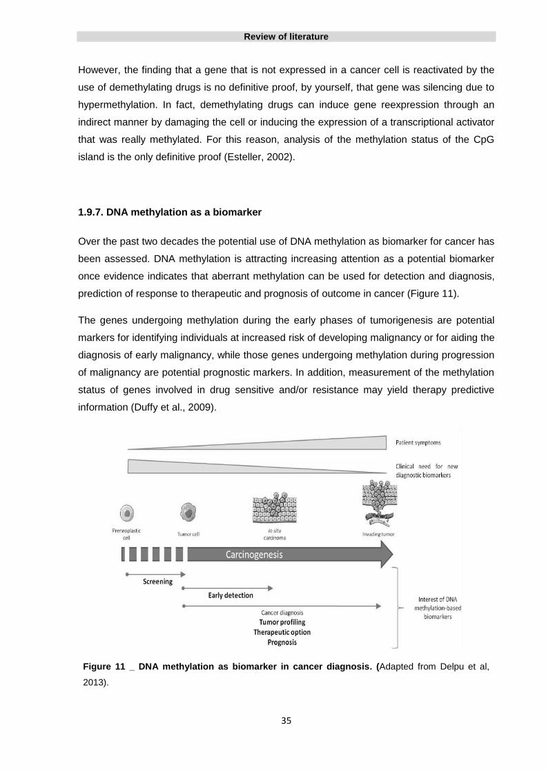
Review of literature
35
However, the finding that a gene that is not expressed in a cancer cell is reactivated by the
use of demethylating drugs is no definitive proof, by yourself, that gene was silencing due to
hypermethylation. In fact, demethylating drugs can induce gene reexpression through an
indirect manner by damaging the cell or inducing the expression of a transcriptional activator
that was really methylated. For this reason, analysis of the methylation status of the CpG
island is the only definitive proof (Esteller, 2002).
1.9.7. DNA methylation as a biomarker
Over the past two decades the potential use of DNA methylation as biomarker for cancer has
been assessed. DNA methylation is attracting increasing attention as a potential biomarker
once evidence indicates that aberrant methylation can be used for detection and diagnosis,
prediction of response to therapeutic and prognosis of outcome in cancer (Figure 11).
The genes undergoing methylation during the early phases of tumorigenesis are potential
markers for identifying individuals at increased risk of developing malignancy or for aiding the
diagnosis of early malignancy, while those genes undergoing methylation during progression
of malignancy are potential prognostic markers. In addition, measurement of the methylation
status of genes involved in drug sensitive and/or resistance may yield therapy predictive
information (Duffy et al., 2009).
Figure 11 _ DNA methylation as biomarker in cancer diagnosis. (Adapted from Delpu et al,
2013).
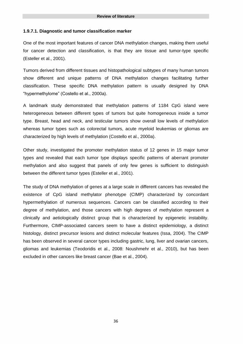
Review of literature
36
1.9.7.1. Diagnostic and tumor classification marker
One of the most important features of cancer DNA methylation changes, making them useful
for cancer detection and classification, is that they are tissue and tumor-type specific
(Esteller et al., 2001).
Tumors derived from different tissues and histopathological subtypes of many human tumors
show different and unique patterns of DNA methylation changes facilitating further
classification. These specific DNA methylation pattern is usually designed by DNA
“hypermethylome” (Costello et al., 2000a).
A landmark study demonstrated that methylation patterns of 1184 CpG island were
heterogeneous between different types of tumors but quite homogeneous inside a tumor
type. Breast, head and neck, and testicular tumors show overall low levels of methylation
whereas tumor types such as colorectal tumors, acute myeloid leukemias or gliomas are
characterized by high levels of methylation (Costello et al., 2000a).
Other study, investigated the promoter methylation status of 12 genes in 15 major tumor
types and revealed that each tumor type displays specific patterns of aberrant promoter
methylation and also suggest that panels of only few genes is sufficient to distinguish
between the different tumor types (Esteller et al., 2001).
The study of DNA methylation of genes at a large scale in different cancers has revealed the
existence of CpG island methylator phenotype (CIMP) characterized by concordant
hypermethylation of numerous sequences. Cancers can be classified according to their
degree of methylation, and those cancers with high degrees of methylation represent a
clinically and aetiologically distinct group that is characterized by epigenetic instability.
Furthermore, CIMP-associated cancers seem to have a distinct epidemiology, a distinct
histology, distinct precursor lesions and distinct molecular features (Issa, 2004). The CIMP
has been observed in several cancer types including gastric, lung, liver and ovarian cancers,
gliomas and leukemias (Teodoridis et al., 2008: Noushmehr et al., 2010), but has been
excluded in other cancers like breast cancer (Bae et al., 2004).
Review of literature
37
1.9.7.2. Detection of aberrant DNA methylation in biological fluids as early detection
tool for cancer
For many types of human tumors, symptoms are often not presented until the primary tumors
have invaded surrounding tissue and/or metastasized, therefore the late presentation of
neoplastic process prevents the timely detection of cancer, resulting in high mortality. The
discovery of epigenetic biomarkers thus may prove to be extremely useful for early detection
and prevention of cancer.
The benefits of early detection of cancer have led researchers to evaluate methylation
markers as potential screening tools. Although using primary tissue to study methylation
profiles and generate new potential biomarkers is of interest, acquiring this biological material
is only achievable by means of invasive techniques. Many studies described the application
of assays, for methylation markers known to be present in tumors, to other diagnostic
samples, namely body fluids, that may contain DNA, such as serum, plasma, blood, urine,
saliva, sputum, bronchoalveolar lavage, ejaculates and ductal fluid. These samples can be
obtained using minimally or even non-invasive diagnostic techniques.
Promoter hypermethylation of the glutathione S-transferase P1 gene (GSTP1) is the most
frequent DNA alteration in prostatic carcinoma but is not found in normal prostatic tissue or
other normal tissues. Matched specimens of primary tumor, peripheral blood lymphocytes
(normal control), and simple voided urine were collected from 28 patients with prostate
cancer. From the 22 prostate tumors that were positive for GSTP1 methylation, in 27% of
cases, the corresponding urine-sediment DNA was positive for GSTP1 methylation,
indicating the presence of neoplastic DNA in the urine. There was no case where a urine-
sediment DNA gave a positive methylation result in the absence of methylation in the
corresponding tumor (Cairns et al., 2001).
In breast cancer patients, from a panel of six genes, GSTP1, RAR_2, p16INK4a, p14ARF,
RASSF1A, and DAP-kinase, aberrant promoter hypermethylation of one or more genes was
found in all 22 tumors (100% diagnostic coverage) and identical gene hypermethylation
pattern was detected in 18 of 22 (82%) matched nipple aspirate fluid. In contrast,
hypermethylation was absent in benign and normal breast tissue and nipple aspirate DNA
from healthy women (Krassenstein et al., 2004).
Wong et al reported that p16 gene was methylated in 16 of 22 patients with hepatocellular
carcinoma and of the 16 patients with methylated p16 in their tumors, 13 also had
methylation in their plasma/serum samples. No methylated p16 sequences were detected in
the peripheral plasma/ serum of the six hepatocellular carcinomas cases without these

Review of literature
38
changes in the tumor, in 38 patients with chronic hepatitis/cirrhosis, or in 10 healthy control
subjects (Wong et al., 1999).
Aberrant methylation of eight gene promoters (CDH1, APC, MGMT, RASSF1A, GSTP1, p16,
RAR-_2, and ARF) was examined in the tumor and the matched bronchoalveolar lavage
(BAL) from 31 patients with primary lung cancer, as well in 10 normal control samples.
Promoter hypermethylation of at least one of the genes studied was detected in all 31 lung
primary tumors. Methylation was detected in 68% (21/31) BAL samples, and in every case,
aberrant methylation in BAL was accompanied by concordant methylation in the matched
tumor samples. Aberrant methylation was not detected in the BAL of lung cancer patients
without methylation in the corresponding tumor (Topaloglu et al., 2004).
Many of these biological samples can be obtained with minimal invasiveness and thus are
suitable for large population screening in the detection of a wide variety of cancers.
1.9.7.3. Detection of aberrant DNA methylation in serum and cerebrospinal fluid in
glioma patients
Examination of serum for circulating tumor DNA with abnormal methylation patterns offers a
possible method for early detection of several cancers. Aberrant CpG island
hypermethylation rarely occur in non-neoplastic and normally differentiated cells. Therefore,
the DNA released from tumor cells can be detected with a notable degree of sensitivity, even
in the presence of excess of DNA from normal cells and this represents a remarkable
potential clinical application.
There are several sources of extracellular DNA namely necrotic cell death that releases high-
molecular-weight DNA into the bloodstream and programmed cell death (apoptosis) (Lavon
et al., 2010).
Although brain tumors may shed free DNA into the extracellular space at the same rate as
systemic tumors, several anatomic and physiologic differences make it uncertain how much
of this DNA may reach systemic circulation while it is still detectable. Primary brain tumors
are confined to the cranial vault, where their extracellular space drains largely into the
cerebrospinal fluid (CSF), which, following circulation, will eventually clear into the
bloodstream (Lavon et al, 2010).

Review of literature
39
Several studies seems to confirm that the blood–brain barrier (BBB) does not interfere
significantly with the leakage of tumor-specific DNA into the global circulation, which makes
the detection of methylation biomarkers in the serum of CNS cancer patients possible.
In a series of 20 astrocytic and 20 oligodendroglial tumors and the
corresponding serum samples, and serum samples from 10 healthy individuals, p16 gene
methylation was reported in 12 of the 20 tissues with astrocytoma, but in only 1 of the tissues
with oligodendroglioma. Similar methylations were detected in the serum of 9 of the 12
patients with aberrant methylation in the tumor tissues. No methylated p16 sequences were
detected in the peripheral serum of the patients having tumors without
these methylation changes or in the 10 healthy controls (Wakabayashi et al., 2009).
Paired tumor-serum samples from 70 patients with high-grade astrocytomas and
oligodendrogliomas of various grades were analyzed to ascertain the methylation status of
MGMT and PTEN genes promoters by MSP. Statistically significant tumor-serum
concordance was found for MGMT methylation in both astrocytic tumors and oligodendroglial
tumors allowing the conclusion that serum DNA in glial tumors is informative for aberrant
gene promoter methylation analysis during the course of the disease (Lavon et al., 2010).
MSP was also used to determine the methylation status of the promoters for p16/(INK4a),
MGMT, p73, and RARβ genes within glioma tissue and corresponding plasma. It was found
that patients with high grade gliomas have large amounts of DNA in the plasma. Of these
primary brain tumors, 90 percent contained methylated gene promoters, and in over 60
percent of these patients the same methylated promoters present in the tumor were also
found in the plasma (Weaver ey al., 2006).
In a similar study, the methylation profiles of MGMT, RASSF1A, p15INK4B, and p14ARF
genes were evaluated in serum free-circulating DNA and the corresponding tumor tissue in a
group of 33 primary or metastatic CNS cancer patients. All the tested genes were found to be
methylated to a different extent in both serum and tumor samples and the gene methylation
profiles observed in serum, in most cases, matched the methylation profiles detected in
paired tumor samples (Majchrzak-Celińska et al., 2013).
Likewise, when analyzed gene methylation in primary tumor, blood and cerebrospinal fluid, in
patients diagnosed with malignant glioma, a relation was also found. Promoter
hypermethylation of four tumor-related genes, MGMT, p16INK4a, TIMP-3 and THBS1, in
serum and CSF from glioma patients, is a specific event in that hypermethylation was not
detected in any of the 20 control serum and the same methylation profiles were found in the
serum, CSF, and the corresponding tumor, while hypermethylation was not detected in the
serum or CSF of glioma patients without methylation in the corresponding tumor. Highly

Review of literature
40
elevated MGMT, p16INK4a, and THBS1 methylation level in gliomas serum was an
independent factor predicting inferior overall survival in the cohort in study (Liu et al., 2010).
It has also been suggested that MSP assay in serum DNA could be a good predictive tool for
selecting glioblastoma patients to be treated with BCNU or alternatively with the combination
of temozolamide plus cisplatin. The methylation status of MGMT, p16, DAPK, and RASSF1A
genes was assessed in tumor and matching serum of a series of 28 glioblastoma patients
treated with either BCNU or temozolamide plus cisplatin. Methylated MGMT, p16, DAPK,
and RASSF1A were found in serum DNA of glioblastoma patients, with a good and highly
significant correlation between serum and primary tumor tissue. Moreover, serum MGMT
methylation predicted response and time to progression in BCNU-treated glioblastoma
patients (Balãna et al., 2003).
The results from the studies cited above, illustrate that both circulating tumor-derived DNA
and CSF harbor the methylation markers found in the originating tumor tissue and are
eligible for clinical biomarker assessment.
It allows the conclusion that DNA methylation patterns measured in peripheral blood and
CSF in glioma patients have great potential to be useful and informative biomarkers of
cancer risk and prognosis. However, large systematic and unbiased prospective studies will
be needed in order to develop clinically feasible assays.
1.9.7.4. Prognostic marker
Hypermethylation events occur already at the early stages of cancer development and may
provide prognostic information, as they might reflect both the growth advantage and
malignant potential of cancer cells. Over the years there were several genes whose it’s
promoter hypermethylation has been linked to patient prognosis.
Hypermethylation of the E-cadherin gene was significantly associated with shorter disease
free survival in diffuse gastric cancer and oral tongue carcinomas, and with early recurrence
in gastric stomal tumor (Graziano et al., 2004; Chang et al., 2002; House et al., 2003).
Aberrant methylation of laminin-5 gene was a negative prognostic marker in prostate and
bladder tumors tissues (Sathyanarayana et al., 2003; Sathyanarayana et al., 2004).

Review of literature
41
Promoter hypermethylation of RAS association domain family Protein1A (RASSF1A) gene
has been shown to be predictive of poor prognosis in many tumors including prostate (Ge et
al., 2014), breast (Martins et al., 2011), endometrial (Jo et al., 2006), lung (Wang et al., 2004)
and bladder (Gao et al., 2012) cancers, hepatoblastoma (Sugawara et al., 2007) and clear-
cell renal cell carcinoma (Kawai et al., 2010).
1.9.7.5. Predictive marker
DNA methylation might help to predict the clinical phenotypes in individual patients and may
be a proper biomarker to optimize individualized cancer treatment. However, the knowledge
of epigenetics in drug response is still in its infancy.
In breast cancer patients, methylation of ESR1 and ARH1 genes can predict survival in
tamoxifen-treated and non-tamixofen treated patients, respectively (Widschwendter et al.,
2004), and the estrogen receptor β promoter was associated with positive response to
tamoxifen treatment in recurrent breast cancer (Chang et al., 2005). In melanoma, inherent
or chemotherapy acquired drug resistance was found to be mediated through the inactivation
of APAF-1 gene due to promoter hypermethylation (Soengas et al., 2001). In ovarian cancer,
BRAC1 promoter hypermethylation was associated with sensitivity to platinum-based
chemotherapy (Chaudhry et al., 2009). In glioblastoma patients, and as will be discussed in
more detail further ahead, MGMT promoter methylation status can predict response to
alkylating based chemotherapy, and represents the most compelling example of DNA
methylation status utility as predictive biomarker until to date (Esteller et al., 2000a) .
1.9.8. DNA methylation as therapeutic target in cancer
A distinguish feature of epigenetic changes in comparison with genetic changes is that they
are reversible. Inhibition of DNA methylation and consequent re-activation of genes related to
tumor development is an attractive avenue for the development of novel therapeutics (Figure
12). This strategy is particularly appealing because, in normal cells these genes are not
normally regulated by DNA methylation and therefore the toxicity of inhibitors of DNA
methylation to non-cancer cells could potentially be well below that seen with conventional
cytotoxic anti-cancer agents.

Review of literature
42
1.9.8.1. DNMT Inhibitors
Since DNA methylation is established and maintained by enzymatic reactions, aberrant DNA
methylation patterns are potentially reversible by DNMT inhibitors. DNA methylation
inhibitors are divided in nucleosidic and non-nucleosidic DNMTs inhibitors. Although many of
them mediate inhibitory effects of DNA methylation in preclinical studies, only few of them
are clinically used.
The nucleosidic DNA methylation inhibitors are incorporated in replicating DNA and/or RNA,
trap DNMTs and target theses enzymes for degradation. DNA synthesis in the absent of
DNMTs results in hypomethylation in the cells and results in gene expression reactivation.
There are three main nucleosidic DNA methylation inhibitors: Azacytidine or 5-azacytidine
(Vidaza®, Celgene, Summit, NJ, USA), Decitabine or 5-aza-2’-deoxycytidine (Dacogen, MHI
Pharma, Bloomington, MN, USA) and Zebularine or 1-(b-D-ribofuranosyl)-1,2
dihydropyrimidin-2-one (Tocris Bioscience) (Delpu et al., 2013).
Azacytidine and Decitabine are approved by the U.S. Food and Drug Administration (FDA)
and currently used for the treatment of acute myeloid leukemia (AML) and myelodysplastic
syndrome (Issa and Kantarjian, 2009; Delpu et al., 2013). Promising results, in recent studies
in xenografed mouse models, demonstrate that low doses of Azacytidine and Decitabine
have antitumor effects on solid tumors such as those from breast, colon and lungs (Tsai et
al., 2012).
Zebularine is mostly studied for its therapeutic activity on AML, however it has not been
pursued clinically because of toxicity in preclinical models (Issa and Kantarjian, 2009; Delpu
et al, 2013).
Various non-nucleoside analogues were also report to inhibit DNA methylation including
Hydralazine, Procainamide derivates, Flavanoids and others such as RG108 (phthalimido-L-
tryptophan), MG98 (DNMT1 antisense oligonucleotide) and SHI-1027 (lipophilic quinolone).
The action mechanisms from these drugs that leads to DNA demethylation is still unclear and
despite some promising results, trials are being conducted to evaluated their real anti-tumor
effects (Issa and Kantarjian, 2009; Delpu et al., 2013).
Review of literature
43
Figure 12 _ Therapeutic effects of DNA methylation inhibition and gene reactivation in
cancer. DNA methylation is maintained postreplication by the action of DNA methyltransferases.
Nucleoside DNA methylation inhibitors lead to degradation of the main DNMTs and continued
replication results in passive demethylation that eventually results in reactivated gene expression.
Activated gene expression, in turn, has effects on multiple pathways, each of which could contribute
to a clinical response. Adapted from Issa and Kantarjian, 2009.

Review of literature
44
1.10. Aberrant DNA methylation in gliomas
Although the role of genetic mechanisms in oncogene activation and tumor suppressor
inactivation in gliomas is well documented, the pattern of epigenetic alterations still remains
poor characterized. Most studies of the epigenetic changes in gliomas have focused on DNA
methylation and a large number of genes involved in various cellular processes including cell
cycle regulation, tumor suppression, DNA repair, apoptosis, cell proliferation, invasion and
angiogenesis have been identified promoter hypermethylated in gliomas. Some of these
genes are listed in Appendix 1.
1.10.1. DNA methylation in pilocytic astrocytoma
Analysing a gene panel comprising several genes, ARF, CDKN2B, RB1, APC, CDH1, ESR1,
GSTP1, TGFBR2, THBS1, TIMP3, PTGS2, CTNNB1, CALCA, MYOD1 and HIC1, pilocytic
astrocytomas showed no evidence of CpG island hypermethylation, but were significantly
hypomethylated for all the genes relative to control tissues whereas diffuse astrocytomas and
oligodendrogliomas showed distinct methylation profiles. The authors suggest the use of
MYOD1 hypomethylation as a discriminator of pilocytic astrocytoma from other astrocytoma
(Uhlmann et al., 2003).
The methylation status of NF1 gene, which is strongly associated with the development of
pilocytic astrocytoma, was investigated in 30 pilocytic astrocytoma. However, no methylation
at the NF1 promoter region in pilocytic astrocytomas was found (Ebinger et al., 2005).
In the other hand, the frequency of aberrant CpG island methylation of ten tumor-associated
genes (MGMT, GSTP1, DAPK, p14ARF, THBS1, TIMP-3, p73, p16INK4A, RB1 and TP53) was
determined in 13 pilocytic astrocytomas concluding that CpG island methylation is a common
event in pilocytic astrocytomas. Comparison of the pilocytic astrocytoma methylation profile
to previous findings on the same genes in other astrocytic tumors showed methylation rates
for GSTP1, DAPK, p14ARF and TP53 similar to those found in diffuse astrocytomas. The
data for p16INK4A and THBS1 presented high methylation rates in all astrocytoma
subgroups including pilocytic astrocytoma. Pilocytic astrocytoma displayed the lowest
methylation rate (8%) for MGMT against 50-71% methylation in other astrocytoma subgroups
(Gonzalez-Gomez et al., 2003b).
In other study, in a series of 16 pilocytic astrocytomas, the methylation frequency of the RB1,
p14ARF and p16INK4a genes was 19%, 6%, and 44%, respectively (Bello et al., 2004).

Review of literature
45
In a series of 62 pilocytic astrocytomas, the global DNA methylation profiles were analyzed
using a DNA methylation microarray for 450,000 CpG sites in the human genome. The
differential expression and methylation of key genes in brain development has identified two
subgroups of pilocytic astrocytomas according to tumor location, infratentorial versus
supratentorial. These results support previous genetic evidences which were also related to
differential gene expression based on tumor location (Lambert et al., 2013).
The tumor suppressor gene AKAP12 (scaffold protein A-kinase anchor protein 12)
expression and promoter methylation status was evaluated in 194 human gliomas, including
pilocytic and diffusely infiltrating astrocytomas, and 23 normal brain samples. The results
show that AKAP12 is expressed in normal brain, strongly upregulated in pilocytic
astrocytomas and weakly expressed in diffuse astrocytomas of WHO grade II to
IV. Accordingly, methylation analyses revealed specific hypermethylation of AKAP12
promoter in diffuse astrocytomas but not in pilocytic astrocytomas (Goeppert et al., 2013).
The small number of studies and the contradictory findings regarding the methylation status
presented in pilocytic astrocytomas require further studies to determine the true methylation
pattern of these tumors.
1.10.2. DNA Methylation in oligodendroglioma
Oligodendrogliomas also exhibit complex profiles of epigenetic alterations that are common
and likely to be related to the development and prognosis of these tumors.
In order to determine the frequency and timing of hypermethylation during carcinogenesis of
nonastrocytic tumors, promoter methylation status of 10 tumor-associated genes in a series
of 41 oligodendrogliomas was analysed. A high frequency of aberrant promoter methylation
of the MGMT, GSTP1, TP14(ARF), THBS1, TIMP3, and TP73 genes was observed in
oligodendrogliomas grade II and III. Promoter methylation seems to occur early in the
carcinogenesis process once it is already present in the low-grade forms (Alonso et al.,
2003).
In patients with oligodendroglial tumors, EMP3 hypermethylation is found in approximately
63% of cases and is strongly associated with both IDH1/IDH2 mutations, 1p/19q codeletion
and also with longer overall survival, which emphasizes its relevance as a prognostic marker
in these tumors (Mellai et al., 2013).
Review of literature
46
In a series of 43 oligodendroglial tumors, the hypermethylation of CpG islands at the gene
promoter region of 11 tumor-related genes (MGMT, RB1, ER, p73, p16INK4a, GST-pi, VHL,
DAPK, hMLH1 p15INK4b, and p14ARF), involved in the carcinogenesis of oligodendroglial
tumors, was evaluated. The result showed that hypermethylation of CpG islands is a
common epigenetic event that is associated with the development of oligodendroglial tumors.
MGMT promoter hypermethylation was detected at 60% of oligodendrogliomas and was
significantly associated with loss of chromosome 19q and with combined loss of
chromosomes 1p and 19q. More importantly, several candidate tumor suppressor genes
such as p16INK4a, p15INK4b, and p73 that were previously reported as unmutated in
oligodendroglial tumors were found to be hypermethylated in their CpG islands (Dong et al,
2001).
1.10.3. DNA methylation in diffuse astrocytoma, anaplastic astrocytoma and
glioblastoma
Astrocytic gliomas often show epithelial membrane protein 3 gene (EMP3) hypermethylation
and aberrant expression. DNA methylation of EMP3 may be an early alteration in
astrocytoma, being found in 80% of anaplastic, diffuse astrocytoma and secondary
glioblastoma. By the other hand, only 17% of primary glioblastoma present EMP3
methylation and frequently overexpressed EMP3. Therefore, the silencing of EMP3 may be a
marker that distinguishes primary from secondary glioblastoma (Kunitz et al., 2007).
Several other studies have noted differences between primary and secondary glioblastomas
with respect to epigenetic changes. Overall, secondary glioblastoma has a higher frequency
of promoter methylation than primary glioblastoma (Ohgaki and Kleihues, 2007).
RB1 gene, considered as a tumor suppressor gene since function as inhibitor of cell cycle
progression, is one of the most frequently epigenetically altered gene in glioblastoma.
Promoter methylation of RB1 was found to be approximately three times more common in
secondary glioblastoma than in primary glioblastoma (43% versus 14%, respectively). A
correlation between loss of RB1 expression and RB1 promoter methylation was found and
results indicated that RB1 promoter methylation is a late event in astrocytoma progression
(Nakamura et al., 2001b).
PTEN promoter methylation is frequent in diffuse astrocytomas and secondary glioblastoma
whereas in primary glioblastoma is a rare occurrence (Wiencke et al., 2006). Correlation

Review of literature
47
between PTEN promoter methylation and expression is not consistent because loss of PTEN
function may also occur by others mechanisms including PTEN mutations and LOH at the
PTEN locus on chromosome 10q23 (Baeza et al., 2003).
In an analysis of a gene panel comprising several genes, seven (CDKN2B, PTGS2, CALCA,
MYOD1, THBS1, TIMP3 and CDH1) have showed tumor specific methylation changes in
gliomas. For each tumor subtype altered methylation profiles were found in comparison with
normal brain DNA as well as significantly different methylation profiles between the different
tumor subtypes: pilocytic astrocytomas were always hypomethylated for all the genes in
study whereas diffuse astrocytomas and oligodendrogliomas showed distinct methylation
profiles. The authors demonstrated that pilocytic astrocytomas, astrocytomas and
oligodendrogliomas can all be discriminated from each other. This study also represents the
first report of hypermethylation of CDH1, PTGS2 and CALCA in gliomas (Uhlmann et al.,
2003).
The progression of glioma over time and recurrence are also associated with distinct
methylation patterns. The p14ARF hypermethylation, for example, is associated with malignant
progression and shorter survival in astrocytoma (Watanabe et al., 2007) and p15 promoter
hypermethylation is an important prognostic factor for survival in glioblastoma patients
(Wemmert et al., 2009). SOCS3 promoter hypermethylation was identified in 35% of the
glioblastoma analysed and was significantly associated with poorer patient outcome (Martini
et al., 2008).
Martinez et al, have shown that promoter hypermethylation of CASP8 gene was associated
with relapsed glioblastoma and DNA methylation pattern is different in relapsed glioblastoma
as compared to the corresponding primary tumor in two thirds of cases (Martinez et al.,
2007).
One of the hallmarks of gliomas is their marked ability to invade normal brain tissue what
makes cure by surgery alone essentially impossible. Genes involved in invasion and
metastasis can also be affected by promoter hypermethylation in gliomas. PCDH--A11 gene
show hypermethylation in 87% of glioblastoma and 88% of diffuse astrocytomas and is
epigenetically silenced in gliomas. It was concluded that the inactivation of this cell-to-cell
contact molecule might be involved in the invasive properties of glioma cells (Waha et al.,
2005). Other example, the CST6 gene, putative metastasis suppressor gene, was found to
be methylated in 74% of high grade gliomas (Kim et al., 2006).
Review of literature
48
Promoter hypermethylation can modulate sensitivity to drugs and radiotherapy in gliomas.
Tumor suppressor genes previously implicated in apoptosis and sensitivity to chemotherapy,
such as EMP3, RUNX3 and TES, have been found to be hypermethylated in gliomas in
several studies (Mueller et al., 2007; Kunitz et al., 2007).
One particular important example of epigenetic silencing of a drug resistance gene is
promoter hypermethylation of MGMT gene.
1.10.4. MGMT gene promoter methylation in gliomas
O-6-Methylguanine-DNA Methyltransferase (MGMT) is a gene located on chromosome
10q26 which encodes a DNA-repair protein that removes mutagenic and cytotoxic alkyl
adducts from the O6 position of guanine, an important site of DNA alkylation. It consists of 5
exons and 4 introns and spans greater than 300kb. MGMT promoter region is composed by
1.2 kb, includes the first exon and part of the fisrt intron and is rich in repetitive GC
sequences including a CpG island (Pegg et al., 2007).
Alkylation of DNA at the O6 position of guanine is an important step in the formation of
mutations in cancer, primarily due to the tendency of the O6-methylguanine to pair with
thymine during replication, resulting in a conversion of guanine-cytosine to adenine-thymine
pairs in DNA. Furthermore, the O6-alkylguanine-DNA adduct may cross-link with the
opposite cytosine residues, blocking DNA replication. MGMT protect cells against these
lesions, transferring the alkyl group from the O6-guanine in DNA to an active cysteine within
its own sequence in a reaction that inactivates one MGMT molecule for each lesion repaired
(Pegg, 1990) (Figure 13).
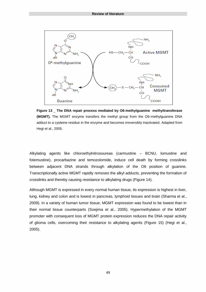
Review of literature
49
Figure 13 _ The DNA repair process mediated by O6-methylguanine methyltransferase
(MGMT). The MGMT enzyme transfers the methyl group from the O6-methylguanine DNA
adduct to a cysteine residue in the enzyme and becomes irreversibly inactivated. Adapted from
Hegi et al., 2005.
Alkylating agents like chloroethylnitrosoureas (carmustine – BCNU, lomustine and
fotemustine), procarbazine and temozolomide, induce cell death by forming crosslinks
between adjacent DNA strands through alkylation of the O6 position of guanine.
Transcriptionally active MGMT rapidly removes the alkyl adducts, preventing the formation of
crosslinks and thereby causing resistance to alkylating drugs (Figure 14).
Although MGMT is expressed in every normal human tissue, its expression is highest in liver,
lung, kidney and colon and is lowest in pancreas, lymphoid tissues and brain (Sharma et al.,
2009). In a variety of human tumor tissue, MGMT expression was found to be lowest than in
their normal tissue counterparts (Soejima et al., 2005). Hypermethylation of the MGMT
promoter with consequent loss of MGMT protein expression reduces the DNA repair activity
of glioma cells, overcoming their resistance to alkylating agents (Figure 15) (Hegi et al.,
2005).

Review of literature
50
Figure 14 _ Mechanism of enhanced chemosensitivity resulting from epigenetic of the
MGMT gene. Gliomas with a methylated MGMT promoter have transcriptional silencing of
MGMT protein. DNA adducts produced by carmustine (or other alkylating drug) in these
tumors are not efficiently removed, leading to tumor-cell death and drug toxicity. In contrast,
gliomas with an unmethylated MGMT express MGMT protein, which removes guanine
adducts from the DNA produced by the administration of carmustine (or other alkylating
drug), resulting in resistance to alkylating drugs. Adapted from Esteller et al., 2000a.
In a study, among more than 500 primary human tumors examined, MGMT hypermethylation
was present in a subset of specific types of cancer while all normal tissues and expressing
cancer cell lines were unmethylated. In gliomas and colorectal carcinomas, aberrant
methylation was detected in 40% of the tumors, whereas in non-small cell lung carcinomas,
lymphomas, and head and neck carcinomas, this alteration was found in about 25% of the
tumors. In leukemias, pancreatic carcinomas, melanoma, renal and bladder carcinomas
MGMT methylation was found rarely and in non-glial brain tumors, breast, ovarian and
endometrial cancers was not found at all (Esteller et al., 1999b). The presence of aberrant
MGMT promoter hypermethylation was associated with loss of MGMT protein, suggesting
that epigenetic inactivation of MGMT by promoter hypermethylation is a common event in
primary human neoplasia and plays an important role in primary human neoplasia (Esteller et
al., 1999b).

Review of literature
51
Because MGMT gene is not commonly mutated or deleted (Esteller et al., 2000a), MGMT
expression silencing in human tumors has been mostly associated with MGMT promoter
hypermethylation (Soejima et al., 2005).
In human gliomas, MGMT promoter hypermethylation has been found in approximately 40%
of primary glioblastoma and 75% of secondary glioblastoma. In diffuse astrocytomas, MGMT
promoter methylation is present in about of 30 - 45% of cases, while In oligodendrogliomas
and anaplastic oligodendrogliomas is reported in approximately 30 and 60% of cases,
respectively (Cankovic et al., 2007; van den Bent et al., 2011; Nakamura et al., 2001c).
In 1998, was establish, for the first time, a correlation between overall survival (OS) and
MGMT expression levels in glioma patients treated with radiotherapy and bis-
chloroethylnitrosourea (BCNU or carmustine). Of 64 patients with malignant glioma, the
overall median survival for patients with high versus low MGMT levels was 8 and 29 months,
respectively. MGMT was a predictive marker of survival in patients with malignant glioma and
was independent of other previously described prognostic variables such as age,
performance status or histology (Jaeckle et al., 1998).
In 2000, Esteller et al found that methylation of the MGMT promoter in high
grade gliomas was a useful predictor of the responsiveness of the tumors to alkylating
agents, namely carmustine. The MGMT promoter was methylated in gliomas from 19 of 47
patients and was associated with responsiveness to carmustine and prolonged overall and
Figure 15 _ MGMT expressing in glioblastoma. a) Lack of nuclear MGMT expression in
tumor cells from a glioblastoma with MGMT promoter methylation (note staining of proliferating
microvascular cells as an internal control). b) Strong MGMT positivity in tumor and vascular
cells in a MGMT unmethylated glioblastoma. Adapted from Weller et al., 2010.
Review of literature
52
disease-free survival. It was an independent and stronger prognostic and predictive factor
than age, stage, tumor grade, or performance status (Esteller et al., 2000a).
A phase III trial showed that the addition of concomitant and adjuvant temozolomide to
radiotherapy, for newly diagnosed glioblastoma patients, has result in a clinically meaningful
and statistically significant OS benefit, when compared with patients who received
radiotherapy alone (Stupp et al., 2005).
Hegi and collegues tested the relationship between MGMT silencing by promoter
hypermethylation and the survival of glioblastoma patients in a randomized trial comparing
radiotherapy alone with radiotherapy combined with concomitant and adjuvant treatment with
temozolomide. Patients who had a methylated MGMT promoter benefited from radiotherapy
combined with temozolomide treatment, showing a significantly longer OS (21.7 months),
whereas those who did not have a methylated MGMT promoter presented an OS of only
12.7 months. It was also found that MGMT promoter hypermethylation was a favorable
prognostic factor in glioblastoma patients, independently of treatment received (Hegi et al.,
2005).
More recently, these results were corroborated by several studies (Dunn et al., 2009;
Brandes et al., 2008). Importantly, in a five year study, the methylation status of the MGMT
promoter was the strongest predictor for outcome and a benefit of combined therapy
(radiotherapy plus temozolamide chemotherapy) was recorded in all clinical prognostic
subgroups. Overall survival was 27.2%, 16.0%, 12.1%, and 9.8% in patients receiving
radiotherapy plus temozolomide, versus 10.9%, 4.4%, 3.0%, and 1.9% with radiotherapy
alone, at 2, 3, 4 and 5 years, respectively (Stupp et al., 2009).
In a prospective study, the prognostic value of genetic alterations characteristic of
glioblastoma, namely TP53 mutation, p53 immunoreactivity, epidermal growth factor
receptor, cyclin-dependent kinase CDK 4 or murine double minute 2 amplification, CDKN2A
homozygous deletion, allelic losses on chromosome arms 1p, 9p, 10q, and 19q, MGMT
promoter methylation, and IDH1 mutations, was evaluated in 301 patients diagnosed with
glioblastoma. MGMT promoter methylation and IDH1 mutations were associated with longer
OS and progression-free survival (PFS) in patients receiving temozolomide and allowed for
stratification into prognostically distinct subgroups whereas no other molecular factor was
associated with outcome (Weller et al., 2009).

Review of literature
53
Although several studies suggest the existence of a relationship between the methylation
status of the MGMT promoter, response to treatment with alkylating agents and outcome,
other studies still do not support these findings (Schaich et al., 2009; Blanc et al., 2004; Wick
et al., 2007; Costa et al., 2010).
As example, in a Portuguese multicentre study, no statistically significant associations were
found between MGMT promoter methylation status and glioblastoma patients outcome
treated with radiotherapy combined with concomitant and adjuvant temozolamide. The
median OS was 13 and 16 months for patients with unmethylated and methylated MGMT
promoter, respectively (Costa et al., 2010).
Despite that today there is still some controversy surrounding these matter, the promoter
methylation status of MGMT gene is commonly assessed both in clinical trials and routine
diagnostics, due to the potential prognostic and predictive roles in GBM patients.
1.10.5. DNA hypomethylation in gliomas
Melanoma Antigen Family A1 (MAGEA1), which belongs to a broad category of genes
commonly called cancer-testis antigens, expression in glioblastoma is correlated with the
level of hypomethylation at key CpG sites within its promoter. Searching for a possible
relationship between MAGEA1 expression and the extent of genomic hypomethylation in
primary glioblastomas and cultured glioma cell lines, MAGEA1 expression was found to be
silenced in normal brain, but found to be reactivated in tumors that exhibited the most
dramatic genome-wide hypomethylation (Cadieux et al., 2006).
The SRY (sex determining region Y)-box 2 (SOX2) gene was found to be highly
overexpressed in human glioblastoma. Molecular alterations found in SOX2, namely SOX2
amplification, did not justify the high frequency of SOX2 overexpression. When compared to
normal human astrocytes, SOX2 promoter is hypomethylated, and this epigenetic alteration
seems to be the leading mechanism responsible for SOX2 overexpression in human
glioblastoma and to contribute to the invasive phenotype of glioblastoma (Alonso et al.,
2011).
OCT4, a transcription factor that plays a key role in regulating the self-renewal ability of
embryonic stem cells, is highly expressed in primary gliomas. The methylation levels of
OCT4 gene promoter and exon are significantly reduced in comparison with the normal
control group and are negatively correlated with OCT4 gene expression suggesting that

Review of literature
54
OCT4 is epigenetically regulated by DNA hypomethylation in primary gliomas (Shi et al.,
2013).
When evaluating the promoter methylation status of coagulation factor X (F10) gene in
tumors of 96 glioma patients, F10 hypomethylation was detected in 82.3%
of glioma specimens but not in any of normal brain control tissues. A statistically significant
correlation was found between F10 hypomethylation, protein expression, and OS, verifying
that patients in F10 hypomethylated group and with F10 overexpression presented an OS
shorter (Liu et al., 2012).
In a similar study, POTE ankyrin domain family, member H (POTEH) was hypomethylated in
81.3% gliomas but not in normal brain tissues, and a correlation between
POTEH hypomethylation, protein expression and overall survival was also found (Liu et al,
2011). These findings indicate the F10 and POTEH genes promoter hypomethylation as
putative biomarkers for glioma prognosis (Liu et al., 2012; Liu et al., 2011).
Epigenetic silencing of Kazal-Type Serine Peptidase Inhibitor Domain 1 (KAZALD1) gene
was associated with malignant transformation, progression and prognosis in glioma. In a
large cohort including 119 glioma samples, KAZALD1 was hypomethylated in high-
grade glioma and it expression scores of high-grade glioma samples were higher compared
to the scores of low-grade gliomas. Also,in high-grade gliomas, OS was shorter for patients
with KAZALD1 hypomethylation or overexpression compared to those without and decreased
KAZALD1 expression in glioma inhibited cell proliferation and invasion (Wang et al., 2013).
1.10.6. Glioma - CpG island methylator phenotype (G-CIMP)
A CpG island methylator phenotype (CIMP) was first characterized in human colorectal
cancer as cancer-specific CpG island hypermethylation of a subset of genes in a subset of
tumors (Toyota et al., 1999a).
In the context of The Cancer Genome Atlas (TCGA), promoter DNA methylation alterations
in 272 glioblastoma tumors were profiled. In this report was identified and characterized a
distinct molecular subgroup in human gliomas with highly concordant DNA methylation of a
large number of gene loci, indicative of a CpG island methylator phenotype (G-CIMP).
Review of literature
55
This G-CIMP was associated with secondary or recurrent (treated) tumors, younger age at
the time of diagnosis and favourable prognosis within glioblastomas as a whole and also
within the proneural subset. It was also highly associated with IDH1 mutation across all
glioma tumor grades, which may be explained by metabolic alterations resulting from these
mutations (Kreth et al., 2012; Noushmehr et al., 2010). These findings identify G-CIMP as a
distinct subset of human gliomas on molecular and clinical grounds (Noushmehr et al.,
2010).
1.11. Current clinical management of gliomas
Gliomas range from pilocytic astrocytomas, which are noninvasive and surgically curable, to
glioblastoma, which is highly invasive and virtually incurable (NCCN, 2013).
The involvement of an interdisciplinary team, including neurosurgeons, radiation therapists,
oncologists, neurologists and neuroradiologists is essential in the appropriate management
of glioma patients (NCCN, 2013).
According to the National Comprehensive Cancer Network (NCCN) guidelines, the standard
therapy for gliomas involves surgical resection to the maximal extent possible with adjuvant
radiotherapy and systemic therapy. Despite these guidelines are updated annually and
because this field evolves continually, the professionals should use all of the available
information to achieve the best clinical options for their patients (NCCN, 2013). For a
comprehensive view of the current treatment options available see Appendix 2.
1.11.1. Surgical resection
Surgical intervention is indicated in almost all glioma patients at least at some point during
the course of their disease and has three principal goals: obtaining a histological diagnosis
and grading, providing symptom relief and improving patient survival by reducing the tumor
burden. In some cases, surgery will even result in a cure (NCCN, 2013).
There are four surgical options: stereotactic biopsy, open biopsy, subtotal resection and
complete or gross total resection (NCCN, 2013).

Review of literature
56
Biopsy results can be misleading, because gliomas often have varying degrees of cellularity,
mitosis or necrosis from one region to another, thus, small samples can provide a lower
histologic grade (NCCN, 2013).
The decision with regard to the aggressiveness of surgery for primary brain tumors is
complex and dependent of several factor such as: patient’s age and performance status,
proximity to eloquent areas of the brain, feasibility of decreasing the mass effect with
aggressive surgery, resectability of the tumor and time since last surgery (in patients with
recurrent disease) (Sawaya et al., 1998).
In order to document the extent of disease after surgical intervention, a magnetic resonance
imaging (MRI) scan, with and without contrast, in the 24 to 72 hours after surgery, is
recommended (NCCN, 2013).
In pilocytic astrocytoma, surgery is often the treatment of choice and total resection is often
possible. Sometimes the location can preclude the access to the tumor and lead to
incomplete or no resection at all. Radiation therapy appears not to be needed for patients
undergoing gross total removal. In patients undergoing subtotal resection, close follow-up
with additional surgery or radiation therapy at the time of tumor progression is recommended.
Patients who have biopsy only should receive radiation therapy (Forsyth et al., 1993).
There is little consensus about the optimal treatment strategy for diffusely infiltrating low-
grade gliomas, including astrocytomas and oligodendrogliomas (WHO grade II), being it
clinical management one of the most controversial areas in neurooncology (Lang and Gilbert,
2006).
In low grade astrocytoma the role of maximal tumor resection is not well defined. There are
retrospective studies suggests a survival benefit from aggressive surgical resection (Soffietti
et al., 1989; Smith et al., 2008) while others report no difference (Shaw et al., 1989). On this
basis, and taking into account that the tumor may contain higher grade foci not reflected in a
small sample, the complete excision may decrease the risk of future differentiation to a
higher malignant lesion and a large tumor burden resection may also enhance the effect of
radiotherapy, the actual recommendation for treating a low grade astrocytoma is the most
complete excision of the tumor as possible, without compromising eloquent brain areas
(NCCN, 2013).
Due to their frequent localization in the frontal lobes and present a distinct tumor margins,
oligodendrogliomas are often amenable to excision (NCCN, 2013).
Review of literature
57
In anaplastic gliomas and glioblastomas, when compared with biopsy, resection is a strong
prognostic factor (Laws et al., 2003).
In patients with glioblastoma treated with surgery and radiation therapy, resection of the
mass followed by radiation therapy is associated with longer survival times than radiation
therapy after biopsy alone, especially when other predictive variables such as age, Karnofsky
Performance Status, location and degree of necrosis, are favorable (Wood et al., 1988;
Lacroix et al., 2001; Devaux et al., 1993; Simpson et al., 1993).
At recurrence a new resection may improve the patient’s outcome and the extent of resection
is an important predictor of overall survival (Barker et al., 1998; Bloch et al., 2012). On the
other hand, tumor involvement of eloquent/critical brain regions, poor Karnofsky performance
status and large tumor volume are factors associated with poor postoperative survival (Park
et al., 2010).
1.11.2. Radiation therapy
There is ongoing controversy about the proper radiotherapeutic management of low grade
gliomas, namely about the optimum timing of radiation therapy, the optimum radiotherapy
(RT) dose and also the addition of chemotherapy to RT (NCCN, 2013).
While some clinicians doubt the efficacy of postoperative RT, others advise routine
postoperative RT. In the EORTC 22845 trial, 314 patients with low-grade astrocytoma,
oligodendroglioma, mixed oligoastrocytoma, and incompletely resected pilocytic
astrocytoma, were assigned to early RT or no immediate therapy (control group). Despite
median PFS was 5.3 years in the early RT group against 3.4 years in the control group, the
OS was similar between the two groups (7.4 years and 7.2 years, respectively). Highlight the
fact that seizures were better controlled in the early RT group (van de Bent et al., 2005).
These results are supported by other trial where of 290 eligible patients, the irradiated group
showed a significant improvement in time to progression but also not in OS when compared
with other patients who did not receive any treatment after surgery until the tumor showed
progression (Karim et al., 2002).
On the other hand, some studies have suggested a benefit of early RT, with an increase in
survival with early versus delayed therapy (Shibamoto et al., 1993; Shaw et al., 1989).

Review of literature
58
The standard radiation dose for low grade glioma is 45 to 54 Gy, delivered in 1.8 to 2.0 Gy
fractions, and data indicate that lower doses of RT are probably as effective as higher doses
of radiation for these tumors (NCCN, 2013; Shaw et al., 2002).
In high grade gliomas, external beam RT after surgery is standard adjuvant therapy. In a
prospective, randomized study was compared the use of best conventional care, 1,3-bis(2-
chloroethyl)-1-nitrosourea (BCNU) and/or RT in the treatment of 303 patients diagnosed with
anaplastic glioma. The median survival of patients who received best conventional care,
BCNU, RT and BCNU plus RT was 14 weeks, 18.5 weeks, 35 weeks and 34.5 week,
respectively (Walker et al., 1978).
In other prospective, randomized trial, from 118 patients diagnosed with high grade glioma
(grade III and IV), those who received conventional care, but no RT or chemotherapy,
presented a median survival of 5.2 months while those patients who received RT presented
a median survival of 10.8 months (Kristiansen et al., 1981).
The standard radiation dose for high grade glioma is 60 Gy, delivered in 1.8 to 2.0 Gy
fractions. In grade III gliomas a slightly lower dose of 55-57 Gy can be applied (NCCN,
2013).
1.11.3. Systemic therapy
1.11.3.1. Blood-brain barrier
The blood-brain barrier (BBB) is a diffusion barrier essential for the normal function of the
CNS which impedes influx of most compounds from blood to brain. BBB is composed by
endothelial cells, astrocytes and pericytes. Tight junctions, present between the endothelial
cells, form a diffusion barrier which selectively excludes most of hydrophilic and larger
lipophilic molecules from blood from entering the brain (Ballabh et al., 2004).
A critical issue in the management of brain tumors is adequate delivery of chemotherapeutic
agents to the tumor. This delivery may be impeded by the BBB. Alternative drug
administration techniques and BBB disruption ways have been developed however have not
demonstrated any significant improvements in the antitumor effect.
Appendix 2 presents a summary of currently available systemic therapies for the treatment of
gliomas and that will be addressed in the following sections of this chapter.

Review of literature
59
1.11.3.2. Adjuvant chemotherapy
1.11.3.2.1. Temozolomide
Temozolomide (TMZ) (Temodar®; Temodal®; Schering-Plough Corporation, Kenilworth, NJ)
is an orally alkylating agent that has demonstrated antitumor activity as a single agent in the
treatment of malignant gliomas.
Unlike many other chemotherapeutic agents, TMZ can easily cross the BBB, so that CSF
levels can reach at least 20% to 40% of those observed in the plasma (Ostermann et al.,
2004).
Following oral absorption, TMZ is spontaneously hydrolyzed in aqueous solution, with almost
100% bioavailability, to methyl-triazeno-imidazole-carboxamide (MTIC). MTIC is rapidly
converted to the inactive 5-aminoimidazole-4-carboxamide (AIC) and to the electrophilic
alkylating methyldiazonium cation that transfers a methyl group to DNA. The DNA methyl
adducts are responsible for cytotoxicity (Vilano et al., 2009).
TMZ exerts its cytotoxic action via DNA methylation at the N-7 and O-6 positions of guanine,
and the O-3 position of adenine. Although methylation of the O-6 position of guanine
represents only a small fraction of the total DNA lesions induced by TMZ, it remains the
major effector of its cytotoxic action. Methylation of guanine at the O-6 position results in
“mismatch” incorporation of thymidine instead of cytosine (Omar and Mason, 2010).
This error is recognized by the mismatch repair (MMR) enzyme system that attempts to
excise thymidine. Since guanine persists, a series of futile replication and repair cycles
ensue, ultimately resulting in apoptotic cell death (Omar and Mason, 2010).
The DNA repair enzyme MGMT, an enzyme that removes methyl adducts at the O-6 position
of guanine added by TMZ, is expressed in gliomas and has been implicated in the resistance
of tumor cells to alkylating agents (Esteller et al., 2000a; Hegi et al., 2004).
TMZ was granted accelerated approval by FDA in 1999 for the treatment of recurrent
anaplastic astrocytoma, with subsequent approval, in 2005, for the first-line therapy of
glioblastoma (Cohen, 2005).
The 1999 approval, for the treatment of anaplastic astrocytoma patients, was based on a
22% response rate (12 of 54 patients). Also, the median PFS was 4.4 months and median
OS was 15.9 months (Yung et al., 1999).
Review of literature
60
Concurrent TMZ and RT followed by adjuvant TMZ (clinically termed Stupp regimen) is
currently the standard of care for newly diagnosed glioblastoma and is the standard first-line
chemotherapy of malignant gliomas (Stupp et al., 2005).
TMZ is the only anticancer drug that has been shown in a phase III study to improve survival
in glioblastoma when administered with concomitant radiotherapy (Stupp et al., 2005).
In Stupp et al trial, 573 patients from 85 institutions in 15 countries were randomly assigned
to receive standard focal radiotherapy alone (286 patients, control group) or standard RT
plus concomitant daily TMZ, followed by adjuvant TMZ (287 patients, Stupp regimen group)
(Stupp et al., 2005).
Concomitant chemotherapy consisted of TMZ at a dose of 75 mg per square meter per day,
given 7 days per week from the first day of RT until the last day of radiotherapy, but for no
longer than 49 days. After a 4-week break, patients were then to receive up to six cycles of
adjuvant TMZ according to the standard 5-day schedule every 28 days. The dose was 150
mg per square meter for the first cycle and was increased to 200 mg per square meter
beginning with the second cycle, so long as there were no hematologic toxic effects (Stupp et
al., 2005).
Stupp regimen results in a 2.5 month prolongation of median survival among glioblastoma
patients compared with the control group; the median survival was 14.6 months with RT plus
TMZ and 12.1 months with RT alone. Relevantly, the two-year survival rate was 26.5 percent
in the group given RT plus TMZ, as compared with 10.4 percent with RT alone (Stupp et al.,
2005).
The results of this study led to the FDA approval, in 2005, for the use of TMZ in the treatment
of newly diagnosed glioblastoma multiforme concomitantly with RT.
Similar results were seen in a smaller phase II trial. In a series of 130 patients with newly
diagnosed glioblastoma, the median OS time was significantly better in the group receiving
TMZ concomitant with RT and followed by TMZ, versus the group receiving RT alone (13.4 v
7.7 months, respectively), and the 1-year OS was 56.3% v 15.7% respectively (Athanassiou
et al., 2005).
TMZ has a well-tolerated safety profile and the most common toxic side effects associated
with their administration included anorexia, constipation, alopecia, headache, fatigue,
convulsions, nausea, vomiting, and hematologic toxicity (Cohen et al., 2005).

Review of literature
61
Despite there have been some safety concerns regarding to combined treatment with
surgery and placement of carmustine wafers, followed by standard adjuvant treatment with
RT and concomitant and subsequent chemotherapy with TMZ, the results of recent studies
show that it is a safe and feasible procedure, without any adjunctive complication (Dixit et al.,
2011; Salvati et al., 2011).
1.11.3.2.2. Combined PCV chemotherapy for anaplastic gliomas
Chromosomal deletions on the 1p and 19q are predictive for good response to radio and
chemotherapy, and meaning of longer survival in anaplastic oligodendroglial tumors.
Both the RTOG study 9402 and the EORTC study 26951 compared adjuvant PCV in
combination with RT, to RT only in anaplastic oligodendrogliomas. The addition of PCV after
RT increases both OS and PFS in anaplastic oligodendroglial tumors and 1p/19q-codeleted
tumors derive more benefit from adjuvant PCV compared with non-1p/19q-deleted tumors
(van de Bent et al., 2013; Cairncross et al., 2013).
1.11.3.3. Recurrence chemotherapy
Together with TMZ, the most commonly used chemotherapy agents for recurrent gliomas are
nitrosoureas (carmustine - BCNU and lomustine - CCNU), procarbazine, vinca-alkaloids
(vincrisitine), platinum based drugs (cisplatin, carboplatin) and cyclophosphamide. The most
used drug combination is called PCV (i.e. procarbazine, CCNU and vincristine) (Wick et al.,
2010; Triebels et al., 2004; Chamberlain and Tsao-Wei, 2004; Chamberlain et al., 2006;
Brandes et al., 2003). In the particular case of anaplastic astrocytomas and
oligodendrogliomas, irinotecan and etoposide can also be used (Chamberlain et al., 2008;
Fulton et al., 1996).
Prior to the introduction of TMZ, nitrosoureas and PCV were the standard chemotherapeutic
regimens for recurrent malignant glioma. Over the past several years TMZ has become the
treatment of choice in these tumors, however the responses tend to be modest and therefore
TMZ has been evaluated in combination with other chemotherapeutic agents in order to
improve the therapeutic benefit.

Review of literature
62
1.11.4. Antigiogenic targeted therapy
1.11.4.1. Anti-VEGF therapy - Bevacizumab
Angiogenesis is the process by which new blood vessels form from existing vasculature by
endothelial cell migration and proliferation (Chamberlain, 2010). Glioblastoma is one of the
most vascularized cancers and vascular endothelial growth factor (VEGF) is an important
regulator of angiogenesis that is highly expressed within brain tumors (Louis et al., 2007).
Bevacizumab (Avastin®, Genentech, South San Francisco, CA) is a recombinant humanized
monoclonal IgG1 antibody that selectively binds to and neutralizes the biologic activity of
human VEGF. Bevacizumab inhibits the binding of VEGF to its receptors, Flt-1 and KDR, on
the surface of endothelial cells. Neutralization of the biologic activity of VEGF can result in
the reduction of tumor vascularization and subsequent reduction in tumor growth (Figure 16)
(Cohen et al., 2009).
Bevacizumab was granted accelerated FDA approval in 2009 as a single-agent therapy for
use in recurrent glioblastoma following prior chemotherapy or RT (Cohen et al., 2009), based
in to phase two trials results (Friedman et al., 2009; Kreisl et al., 2009).
The prognosis for patients with recurrent glioblastoma multiforme is poor, with a median
survival of 3 to 6 months.
The first ever phase II study with the use of bevacizumab, in combination with irinotecan, for
patients with recurrent malignant glioma, showed a 6-month PFS of 46% and a 6-month OS
of 77%, with moderate toxicity (Vredenburgh et al., 2007).
Posteriorly, in 2009, two phase II trials evaluating the efficacy of bevacizumab, alone or in
combination with irinotecan, in patients with recurrent glioblastoma were conducted
(Friedman et al., 2009; Kreisl et al., 2009).
In the bevacizumab-alone group of patients, 6-month PFS rate of 42.6% was reported, and
median OS was 9.2 months while in the bevacizumab-plus-irinotecan group, those values
were 50.3% and 8.7 months, respectively (Friedman et al., 2009).
Kreisl and colleagues reported a 6-month PFS of 29% and a median OS of approximately 8
months, when single-agent activity of bevacizumab, in patients with recurrent glioblastoma,
was evaluated (Kreisl et al., 2009).

Review of literature
63
Overall, bevacizumab treatment is generally well tolerated in patients with recurrent
glioblastoma but adverse events as hypertension, impaired wound healing, colonic
perforation and thromboembolism can occurred (NCCN, 2013).
Trials with bevacizumab plus erlotinib (Sathornsumetee et al., 2010), etoposide (Reardon et
al., 2009), TMZ (Verhoeff et al., 2010), cetuximab (Hasselbalch et al., 2010) or carboplatin
(Reardon et al., 2012) have been also reported. These regimens were associated with
similar PFS benefit and radiographic response when compared with other bevacizumab-
containing regimens, namely bevacizumab alone or in association with irinotecan.
Preliminary randomized Phase III trial results do not recommend the routine use of
bevacizumab in combination with standard RT and TMZ in patients with newly diagnosed
glioblastoma, based on the lack of proven survival benefit and the increased risk of toxicity
associated with combination therapy (Gilbert et al., 2013; Cloughsey et al., 2013).
Figure 16_ Bevacizumab action mechanism. Bevacizumab binds VEGF, preventing
interaction with its receptors and activation of downstream signaling pathway. Adapted from
Rini, 2007.

Review of literature
64
1.11.5. Tumor Treating Fields Therapy for recurrent Glioblastoma
The NovoTTF-100A™ System (Novocure Ltd., Haifa, Israel) is a portable medical device
approved by the FDA, in 2011, to deliver Tumor Treating Fields (TTF) therapy, for the
treatment of recurrent glioblastoma (Stupp et al., 2012; Fonkem and Wong, 2012).
This noninvasive technology, works by emitting a low-intensity, intermediate-frequency (200
kHz), alternating electric field administered via insulated transducer arrays applied onto the
scalp. The electric field penetrates the brain and inhibits the growth and proliferation of
glioblastoma by interfering with tumor cell mitosis at anaphase (Figure 17) (Stupp et al.,
2012; Fonkem and Wong, 2012).
Results from a Phase III clinical trial comparing chemotherapy-free treatment with TTF
versus active chemotherapy in the treatment of patients with recurrent glioblastoma, indicate
that the efficacy of NovoTTF-100A is equivalent to standard-of-care chemotherapy. Despite
no improvement in OS was demonstrated, the efficacy and activity with TTF device is
comparable to chemotherapy regimens and the toxicity levels, adverse events and quality of
life clearly favored TTF (Stupp et al., 2012).
Figure 17 _ Tumor Treating Fields (TTF) therapy. Exemplary T1-weighted, post
contrast, MRI scans of recurrent glioblastoma patient before (Left) and after (Right)
TTF treatment. Complete response after 8 months of treatment. Adapted from Kirson et al,
2007.
Review of literature
65
1.11.6. Local intracavitary chemotherapy - Casmustine wafers
The toxicity and poor BBB penetration associated with systemic chemotherapy have led to
the development of different approaches aimed at delivering therapeutic agent directly into
the tumor allowing this way increase the exposure of tumor cells to the drug and reduce the
systemic complications.
Casmustine (BCNU) wafers (Gliadel®, MGI Pharma, Bloomington, MN) were approved in
1996 as the first brain cancer treatment to deliver chemotherapy directly to the tumor site.
BCNU wafer is a sterile wafer containing 192.3 mg of biodegradable copolymer polifeprosan
20 and 7.7 mg of BCNU. The polifeprosan was selected as a potentially appropriate means
to deliver BCNU chemotherapy controlled release after direct implantation of the wafers
because it supports gradual release and is hydroponic, thereby protecting BCNU from
exposure to water which would result in hydrolysis and deactivation (Domb et al., 1999). Up
to 8 BCNU wafers can be implanted into the surgical resection cavity, and have the ability to
release high concentration of BCNU at the tumor surrounding tissue in a controlled way over
a period of up to 3 weeks, and after 6 to 8 weeks the wafers are dissolved (Juratli et al.,
2013). Implantation of the drug-impregnated polymer at the tumor site allows prolonged local
exposure with minimal systemic exposure.
BCNU wafers were first approved in 1996 for the treatment of recurrent glioblastoma, as an
adjunct to surgery, and were subsequently approved in 2003 for first-line treatment of high
grade malignant glioma, as an adjunct to surgery and radiation (Brem et al., 1995; Westphal
et al., 2003).
In a randomized, placebo-controlled, prospective study to evaluate the effectiveness of
BCNU wafers, in 222 patients with recurrent malignant glioma requiring re-operation, the
median survival of the 110 patients who received BCNU polymers was 31 weeks compared
with 23 weeks for the 112 patients who received only placebo. Among patients with
glioblastoma, 6-month survival in those treated with BCNU wafers was 50% greater than in
those treated with placebo. The results suggest that interstitial chemotherapy delivered with
polymers directly to brain tumors at the time of surgery seems to be a safe and effective
treatment for recurrent malignant gliomas (Brem et al., 1995).
Later, in a phase III trial, 240 patients were randomized to receive either BCNU or placebo
wafers at the time of primary surgical resection. The median survival was 13.9 months for the
BCNU wafer-treated group and 11.6 months for the placebo-treated group with a 29%
reduction in the risk of death in the treatment group. This study confirms that

Review of literature
66
local chemotherapy with BCNU wafers is well tolerated and offers a survival benefit to
patients with newly diagnosed malignant glioma (Westphal et al., 2003).
Despite the above, adverse advents related to the use of carmustine wafers, such as
seizures, brain edema, intracranial infections and healing abnormalities, have been
frequently reported (Juratli et al., 2013).
1.12. DNA methylation analysis methods and technical considerations
1.12.1. Selection of a method for DNA methylation analysis of specific CpG
islands
In the past 20 years the repertoire of methylation methodology has expanded greatly and a
variety of methods have been used to screen the genome for novel markers. These methods
allow the identification of a single CpG site that differs in its methylation status between two
samples, for example, a normal sample compared to a tumor sample or even to subtypes of
the same type of tumor compared between each other.
The choice of a method for DNA methylation analysis should be mainly based on the goals of
the testing, required assay performance and the material and human resources available.
Several factors have also to be taken into account, such as, how many CpG sites will be
tested in each sample, the expected heterogeneity of normal and tumor cells within samples
and the amount and quality of starting DNA material in each sample. Some of the techniques
available for DNA methylation analysis, the most commonly used, are described in sections
below and in Appendix 3 are referenced some others techniques currently available.
1.12.2. Formalin-fixed paraffin embedded tissues
In pathology archives, tissue samples are stored as formalin-fixed paraffin
embedded (FFPE) blocks. FFPE tissue samples are the largest source of clinical material
from normal controls and pathological tissues and their use is of inestimable research and
clinical value.
The FFPE archives enclose large numbers of tissue samples that had been obtained for
diagnostic and therapeutic purposes. After a diagnosis was reached on an FFPE tissue
biopsy or resection specimen, excess material is usually available for further studies,
including molecular analysis.

Review of literature
67
The need for suitable quality of DNA recovered from FFPE is necessary for the success of
molecular analysis, namely DNA methylation analysis. Also, the use of archived material,
formalin-fixed paraffin embedded tissues may significantly impact the choice of the method
used in these analysis (Ludyga et al., 2012).
When analyzing DNA extracted from FFPE tissue, there is a risk of lower analysis quality
since DNA is fragmented, a fact that may influence both bisulfite treatment and further
polymerase chain reaction (PCR).
The storage time of the blocks can influence the quality of the DNA extracted. In general,
DNA fragmentation is associated with the storage time: the older the FFPE the shorter the
fragments obtained (Funabashi et al., 2012; Ludyga et al., 2012).
According to several authors, the most critical step for obtaining intact DNA is formalin
fixation (Bonin et al., 2003; Funabashi et al., 2012). Formalin is 37-40% formaldehyde in
water, stabilized by 10% methanol. Tissues are routinely fixed in a phosphate buffered 10%
solution of Formalin. Formaldehyde reacts with amino groups of basic amino acids such as
lysine, asparagine, arginine, histidine and glutamine, leading to the formation of higly reactive
methylol adducts. A subsequent condensation reaction of adducts occurs through Schiff
base formation. This results in the formation of methylene bridges with amine, guanidyl,
phenol, imizadol and indole groups of several other amino acids like arginine, asparagine,
glutamine, histidine, tryptophan and tyrosine, leading to inter and intra-molecular cross-
linking of proteins (Berg et al., 2010).
In spite of the many advantages formalin fixation of tissue samples for diagnostic purposes
has to offer, the use of FFPE material for molecular analysis remain problematic. While the
tissue morphology is well preserved, the degradation of nucleic acids continues, in particular
due to a time dependent decrease of pH.
DNA extraction from FFPE tissue requires special protocols because the material is often
scarce, degraded and can contain substances that inhibit the molecular procedures. Among
others commercial kits, those from Qiagen® have shown satisfactory results for extraction of
DNA from FFPE tissue, allowing obtaining DNA with high purity degree (Ludyga et al., 2012).
Formalin also decreases the efficiency of PCR. There are several reasons for the failure of
PCR using DNA isolated from FFPE tissues: the generation of DNA-protein cross-linkages
due to formaldehyde solution resulting in nucleic acid fragmentation; the presence of
remnants of substances that inhibit the amplification reaction such as formalin or inhibit the

Review of literature
68
proteinase K used in the extraction procedure such as xylene; and the risk of contamination
during the manipulation of samples (Farrugia et al., 2010).
In the literature it has been reported that the average length of PCR products generated from
FFPE samples is between 100 to 300 bp. In FFPE samples, the use of amplification products
smaller than 150 bp is recommended, as well as repetitions in the PCR and electrophoresis
steps, for full and effective analysis of the DNA fragments to be suited (Funabashi et al.,
2012).
Regarding to epigenetic studies, methylation analysis successfully performed with DNA
isolated from 30 year old FFPE tissue has been reported (Kristensen et al., 2009).
1.12.3. Sodium Bisulfite Treatment
The analysis of DNA methylation was revolutionized by the introduction of sodium bisulfite
conversion of genomic DNA. As an epigenetic event, methylation is not preserved during
amplification processes such as PCR. Bisulfite treatment of DNA transforms an epigenetic
event to a genetic change which is then able to be analyzed using PCR-based methods.
Methylation of the 5’-position of cytosine residues is a reversible covalent modification of
DNA, resulting in production of 5-methyl-cytosine. Bisulfite deaminates non-methylated
cytosine residues to uracil, leaving methylated cytosines unchanged.
The chemistry of cytosine deamination by sodium bisulfite involved three steps (Figure 18):
the addition of bisulfite to the 5-6 double bond of cytosine designed sulphonation, hydrolic
deamination of the resulting cytosine-bisulfite derivate to give a uracil-bisulfite derivate and
alkali desulfonation by removal of the sulphonate group by an alkali treatment, to give uracil
(Clark et al., 1994).
Bisulfite treatment of DNA is technically the most challenging part of methylation detection
because it leads to further DNA fragmentation. Partial conversion could also be problematic
because it leads to false positive results. To control appropriately for bisulfite effect,
methylated and unmethylated controls must be treated in parallel with patients samples, to
determine whether complete conversion has occurred (Cankovic et al., 2013).

Review of literature
69
Today, several different techniques rely on the ability of sodium bisulfite to efficiently convert
unmethylated cytosine to uracil without affecting 5-methylcytosine. The protocol described by
Frommer and colleagues (1992) has been widely used and a variety of commercial kits are
now available for this purpose, although most are still relatively time-consuming and labor
intensive.
a)
Figure 18 _ a) Conversion of
cytosine to 5-methylcytosine
by DNA methyltransferase
(DNMT). DNMT catalyses the
transfer of a methyl group
(CH3) from S-
adenosylmethionine (SAM) to
the 5-carbon position of
cytosine.
b) Sodium bisulfite
conversion of genomic DNA.
It involves three steps:
1. Sulphonation
2. Hydrolic deamination
3. Alkali desulphonation.
Adapted from Clark et al, 1994.
b)
1.12.4. Bisulfite-treated DNA
The stability of bisulfite-treated DNA is reduced due to nucleotide mispairing and incomplete
complementarity. Although there are minimal data on the effects of temperature and time of
storage on the stability of bisulfite-treated DNA, the analysis should be performed soon after
the conversion to minimize further DNA degradation. Until more data are available, ultra-low
temperature storage conditions (-70ºC or below) should be used if converted DNA must be
stored before analysis. It is also advisable to include appropriate controls to validate the
results obtained from assays with such DNA (Sepulveda et al., 2009).

Review of literature
70
1.12.5. Methylation-specific Polymerase Chain Reaction (MSP)
Methylation-specific PCR (MSP) was originally described in 1996 and is a technique which
can rapidly assess the methylation status of virtually any group of CpG sites within a CpG
island, and was the first assay independent of the use of methylation-sensitive restriction
enzymes. Among the several techniques developed over the years to analyze CpG DNA
methylation in genomic DNA, MSP is the most commonly used (Herman et al., 1996).
First, the genomic DNA is treated with sodium bisulfite (converting all unmethylated, but not
methylated, cytosines to uracil) and then a PCR is performed using primers that bind
specifically to bisulfite converted methylated or unmethylated DNA, leading to specific
amplification (Herman et al., 1996).
In its classic format, a gel based MSP assay, the presence of a band on a gel in each
reaction determines the methylation state, is a nonquantitative assay, the results are
reported as methylated or unmethylated, and cannot distinguish between low and high levels
of a methylated target sequence (Herman et al., 1996).
MSP requires only small quantities of DNA, is sensitive to 0.1% methylated alleles of a given
CpG island locus, and can be performed on DNA extracted from paraffin-embedded
samples. MSP eliminates the false positive results inherent to previous PCR-based
approaches which relied on differential restriction enzyme cleavage to distinguish methylated
from unmethylated DNA (Herman et al., 1996).
PCR analysis after bisulfite conversion is the method of choice when sensitive detection is
desired. Other advantages of this technique are easy of design and low complexity of the
reaction. The main disadvantages are that it is only qualitative and not quantitative, it cannot
be performed in a high-throughput manner and it allows screening of only a very small
stretch (<40bp) of DNA within a CpG island for methylated residues.
1.12.6. Methylation-Specific PCR (MSP) Primers
MSP is a particularly demanding application as, in order to provide reliable results, it requires
high specificity to discriminate between cytosine and thymine bases derived from methylated
and unmethylated cytosines following bisulfite conversion.

Review of literature
71
Right primer design is crucial for successful PCR amplification of bisulfite-modified DNA and
once after sodium bisulfite treatment the sense and antisense strands are no longer
complementary, the primers should be designed for either strand.
In order to discriminate between a methylated and unmethylated DNA fragment, primers
should contain as much CpG sites as possible within their sequence (at least one CpG) and
preferably at the far 3’-end. At least one of the last three bases at 3’-end of the primer has to
be a CpG “C”. Primers should also have an adequate number of non-CpG Cs in their
sequence to amplify only the bisulfite-modified DNA. Primers with more non-CpG Cs are
preferred (Barekati et al., 2010).
Primer for methylated DNA and unmethylated DNA should contain the same CpG sites in
their sequence, but they may differ in length and start position.
The two sets of primers for methylated and unmethylated DNA should ideally have a similar
annealing temperature and similar Tm values (máx Tm difference of 5ºC).
MSP generally requires longer primers because bisulfite modification decreases the GC
content of DNA templates and produces long stretches of “T” in the sequence that makes
difficult to achieve acceptable Tm values and stability of primers. Usually, primers with a
length of approximately 20-30 mer yield successful results (Barekati et al., 2010).
Amplification of a product size greater than 500bp is difficult using bisulfite-modified DNA
template because DNA degradation occurs by bisulfite modification. For this reason, primer
sets should amplify product with a size range between 100 to 500bp (Barekati et al., 2010).
1.12.7. Bisulfite genomic sequencing
Direct bisulfite sequencing for analysis of DNA methylation levels is a technique that was
developed by Frommer and colleagues, in 1992, in order to determine CpG site-specific
changes. This technique provides positive identification of 5-methylcytosine residues
and yields strand-specific sequences of individual molecules in genomic DNA. The method
consists in bisulfite modification of genomic DNA (converting cytosine to uracil while but 5-
methylcytosine remains unchanged). The DNA sequence is then amplified by PCR with two
sets of specific primers and all uracil and thymine residues have been amplified as thymine
and only 5-methylcytosine residues have been amplified as cytosine. Finally the PCR
products can be sequenced directly or as single clones: direct sequencing provides an
estimate of the average methylation status of each CpG site in all molecules while

Review of literature
72
sequencing of the cloned PCR products provides information on individual molecules
(Frommer et al., 1992; Clark et al., 1994).
1.12.8. Combined bisulfite restriction analysis (COBRA)
Combined bisulfite restriction analysis (COBRA) is a quantitative technique used to
determine DNA methylation levels at specific gene loci in small amounts of genomic DNA,
based on the fact that digestion of PCR products with certain restriction enzymes can be
used to distinguish between methylated and unmethylated DNA (Xiong and Laird, 1997).
DNA sequences of interest are treated with bisulfite, in a standard way, amplified by PCR
and subjected to restriction digestion with an enzyme that contains cytosines only within CG
sites in its recognition sequence (such as BstUI [CGCG] or TaqI [TCGA]). If the cytosines are
methylated, then the enzyme will still cut the site; if they were not methylated, then the
restriction site will be lost. As a control for complete bisulfite conversion, a second enzyme
may be used that lacks CG sites within its recognition sequence. Hsp92II (CATG) is a good
example of such an enzyme. Any cleavage by Hsp92II would indicate either incomplete
conversion or nonsymmetrical methylation (Sadri and Hornsby, 1996; Xiong and Laird,
1997).
Therefore, in the mixed population of resulting PCR fragments, the fraction that has retained
restriction site that contains a CpG(s) should be a direct reflection of the percentage DNA
methylation at that site in the original genomic DNA. The most accurate and reliable method
to quantitate the relative amounts of digested and undigested PCR products is to perform an
unlabeled PCR reaction, followed by a purification step to ensure subsequent complete
cutting, then restriction digestion, polyacrylamide gel electrophoresis, electroblotting, oligo
hybridization and phosphorimager quantitation. This method, despite is relatively labour-
intensive, is cost effective (Sadri and Hornsby, 1996; Xiong and Laird, 1997).
1.12.9. Pyrosequencing
Bisulfite pyrosequencing is a quantitative methodology, alternative to the traditional dideoxy
(Sanger) sequencing approach, based on the detection of emitted light during synthesis of
the complementary strand by an exonuclease-deficient DNA polymerase. Biotin-labelled,
single-stranded PCR products generated from bisulfite-treated DNA are used as a template
with an internal primer to perform the pyrosequencing reaction. When nucleotides are
incorporated, in a predetermined order, pyrophosphate is released and converted to ATP by

Review of literature
73
the enzyme ATP sulfurylase. The ATP molecules provide energy for the enzyme luciferase to
oxidize luciferin in a reaction that generates light (Figure 19). DNA methylation ratios are
calculated from the levels of light emitted from each nucleotide incorporated at individual
CpG positions in a strand-dependent manner. The methylation detection limit at individual
CpG sites is approximately 5% and the results are displayed as an average methylation level
for each CpG position assayed across all amplification products generated during a PCR
reaction.
This technique allows the identification of heterogeneous DNA methylation patterns but does
not provide information at a single allele resolution. Is a methodology suited to analyze short
DNA sequences, up to 100-bp in length, such as those typically extracted from formalin-fixed
paraffin-embedded specimens. Quantitative epigenotypes are obtained using this protocol in
approximately 4 h for up to 96 DNA samples when bisulfite-treated DNA is already available
as the starting material (Tost and Gut, 2007; Mikesa et al., 2011).
Figure 19 _ DNA Methylation analyses by
Pyrosequencing. The reaction cascade
generates light for every incorporated nucleotide,
the intensity of which is proportional to the number
of bases incorporated.

Review of literature
74
1.12.10. Methylight
Methylight is a high-throughput quantitative methylation assay, sodium-bisulfite-dependent,
which relies on fluorescence-based real-time PCR (TaqMan) technology (Figure 20). It
requires no further manipulations after the PCR step and can very accurately determine the
relative prevalence of a particular pattern of DNA methylation. Is also a highly sensitive
assay, capable of detecting very low frequencies of hypermethylated alleles in the presence
of a 10,000-fold excess of unmethylated alleles (Eads et al., 2000).
Four types of MethyLight reactions were described, depending on which oligonucleotides are
designed to discriminate the methylation status: (i) only the primers, (ii) only the TaqMan
probe, (iii) both primers and probe, or (iv) none (in cases where a control reaction is required
to discriminate the converted DNA). Using these reactions, several variations of MethyLight
have been proposed to address different biological questions. The most commonly used
MethyLight methodology uses two primers and a TaqMan probe designed to bind the
methylated allele specifically and requires a reference gene for normalization. It is important
to note that MethyLight, depending on the method subtype, can assess the methylation
status of all CpG sites covered by the TaqMan probes.
The highest advantages of MethyLight, as compared to other existing techniques, are: its
potential to allow the extremely rapid screening of hundreds to thousands of samples, the
decrease of chance of sample contamination and error and of the amount of labor involved in
DNA methylation analysis, because the assay is completed at the PCR step without the need
for further gel electrophoretic separation or hybridization, and the compatibility with small
biopsies and paraffin-embedded tissues once it requires only minute amounts of DNA of
modest quality. MethyLight has shown higher levels of accuracy and lower rates of false
negatives when compared with previously described techniques being for that frequently
used to validate other techniques for DNA methylation studies (Eads et al., 2000; Campan et
al., 2009).

Review of literature
75
Figure 20 _ Schematic of the theoretical basis of MethyLight technology. Genomic DNA is first
chemically modified by sodium bisulfite. This generates methylation-dependent sequence differences
at CpG dinucleotides by converting unmethylated cytosine residues (locations indicated by white
circles) to uracil, while methylated cytosine residues (locations indicated by black circles) are retained
as cytosine. Fluorescence-based PCR is then performed with primers that either overlap CpG
methylation sites or that do not overlap any CpG dinucleotides. Sequence discrimination can occur
either at the level of the PCR amplification process or at the level of the probe hybridization process,
or both. Sequence discrimination at the PCR amplification level requires the primers and probe
(application D), or just the primers (application C), to overlap potential methylation sites (CpG
dinucleotides). Only two [fully methylated (M) and fully unmethylated (U)] of the many theoretical
methylation permutations are shown. The MethyLight assay can also be designed such that
sequence discrimination does not occur at the PCR amplification level. If neither the primers nor the
probe overlap sites of CpG dinucleotides (application A), then no methylation-dependent sequence
discrimination occurs at the PCR amplification or probe hybridization level. This reaction represents
amplification of the converted genomic DNA without bias to methylation status, which can serve as a
control for the amount of input DNA. When just the probe overlaps methylation sites (application B),
then sequence discrimination can occur through probe hybridization. The design of separate probes
for each sequence variant resulting from different methylation patterns (22 = 4 probes in the case of
two CpGs, as illustrated) can potentially serve as a quantitative version of the MethyLight technology.
(From Eads et al,. 2000).

Review of literature
76
1.13. The Aryl Hydrocarbon Receptor
1.13.1. The Aryl Hydrocarbon Receptor structure
The Aryl Hydrocarbon Receptor (AHR), also called the dioxin receptor, was identified in 1976
by Poland et al. Is a ligand-activated transcription factor member of basic helix-loop-helix
Per-ARNT-Sim (bHLH/PAS) proteins superfamily which also includes the transcriptional
regulators Period (Per), AHR nuclear translocator protein (ARNT) and Single minded (Sim)
(Poland et al., 1976; Whitlock, 1993; Hankinson, 1995).
The bHLH/PAS family of transcription factors is important for regulation of diverse biological
processes including the induction of drugs metabolizing enzymes in response to xenobiotic
exposure, circadian rhythms1 regulation, hypoxia response and development of the CNS
(McIntosh et al., 2010; Gu et al., 2000).
The AHR gene is located on chromosome 7p15 and consists of 12 exons, including a
noncoding exon (exon 12) (Micka et al., 1997).
AHR has a uniform molecular design that is composed of three functional domains, including
a highly conserved N-terminal bHLH domain, a pair of degenerate PAS repeats
(denominated PAS.A and PAS.B), and a poorly conserved C-terminal transactivation domain
(Figure 21) (Kewley et al., 2004).
The PAS domain of the AHR mediates heterodimerization, DNA recognition, ligand binding
and chaperone interactions; the bHLH domain is involved in DNA binding and support of
dimerization, and comprise the nuclear localization sequence (NLS) and the nuclear export
sequence (NES); the C-terminal is highly variable and contains the transactivation domain
(TAD) responsible for activating transcription after DNA binding (Figure 21) (Stevens et al.,
2009).
The promoter of AHR gene did not contain a TATA box nor a CAAT box and is GC-rich,
multiple GC boxes are present close to the determined transcription initiation sites. It also
show several well conserved regions, containing binding sites for known transcription factors,
such as Sp1, all features of a housekeeping gene (Mimura et al., 1994; Schmidt et al, 1993;
Eguchi et al., 1994). Binding sites for putative transcription factors such as c-Myc, NF-E1,
AP1 and AP2 have also been observed in upstream region of the AHR promoter.
1 Circadian rhythms are physical, mental and behavioural changes, found in most living beings, that follow a roughly 24-hour
cycle, responding primarily to light and darkness in an organism’s environment.

Review of literature
77
AHR gene and homologues have been identified in many mammalian, in several non
mammalian vertebrate and also in invertebrate species (Hahn, 2002). Studies have
demonstrated that AHR is expressed, in a tissue, cell and developmentally specific manner,
in several human tissues including heart, pancreas and liver, showing its highest levels in
lung and placenta and with lower levels of expression found in brain, kidney, and skeletal
muscle (Dolwick et al., 1993).
1.13.2. The AHR signalling pathway
In the absence of a ligand, AHR is found in the cytoplasm in a complex with the chaperone2
heat-shock protein 90 (hsp90), the co-chaperone3 p23 and an immunophilin homologous
protein known as ARA9 (AHR-associated protein 9), AIP (AHR-interacting protein) or also
XAP2 (hepatitis B virus X-associated protein 2) (Nguyen et al., 2008).
Following ligand binding the AHR changes its conformation and translocates into the
nucleus, dissociates from the hsp90 complex and forms a heterodimer with the ARNT (also
known as HIF-1β). The AHR/ARNT complex then binds to specific DNA recognition
sequences termed xenobiotic responsive elements (XREs, also known as DREs, dioxin
responsive elements) found in the promoter regions of a variety of genes, resulting in
increased gene transcription. This AHR-ARNT heterodimer has been shown to regulate the
expression of several genes involved in diverse signalling pathways (Reyes et al., 1992).
The subset of AHR target genes includes the members of cytochrome P450 family 1
(CYP1A1, CYP1A2 and CYP1B1), NAD(P)H dehydrogenase quinone 1 (NQO1), aldehyde
dehydrogenase 3 family, member A1 (ALDH3A1), Uridine Diphospate
glucuronosyltransferase 1 family polypeptide A6 (UGT1A6) and glutathione S-
transferase Ya subunit (GST-YA), and is usually collectively referred to as “AHR gene
battery”. These genes are commonly upregulated following AHR activation, and encode
phase I and phase II xenobiotic metabolizing enzymes, which function to metabolize
activating compounds and thus provide a vital role in the detoxification of xenobiotics (Hao
and Whitelaw, 2013).
After nuclear translocation, the AHR is exported to cytoplasm and targeted for degradation
via the ubiquitin-proteosome pathway. The AHR activity in the cell is also negatively
2 Chaperones are proteins found in both prokaryotes and eukaryotes, which facilitate the correct assembly or disassembly of
newly synthesized oligomeric protein complexes, participating in transmembrane targeting and protein folding. 3 Co-chaperones are proteins that assist chaperones in protein folding and other functions.
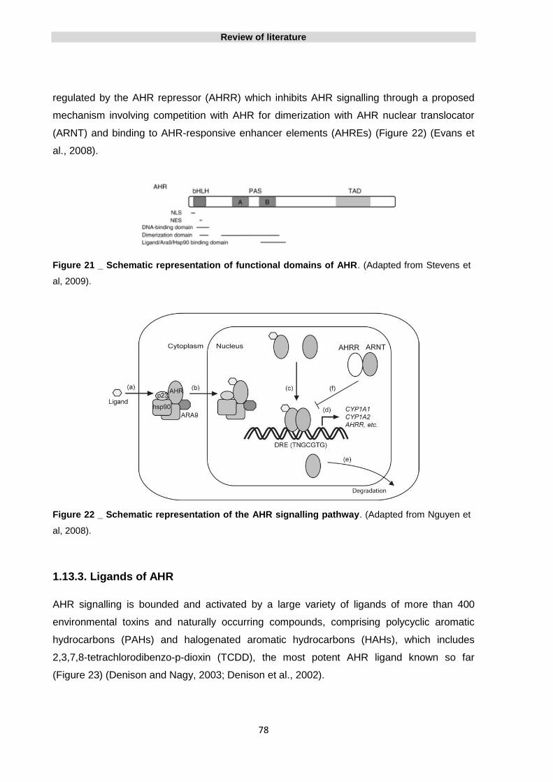
Review of literature
78
regulated by the AHR repressor (AHRR) which inhibits AHR signalling through a proposed
mechanism involving competition with AHR for dimerization with AHR nuclear translocator
(ARNT) and binding to AHR-responsive enhancer elements (AHREs) (Figure 22) (Evans et
al., 2008).
Figure 21 _ Schematic representation of functional domains of AHR. (Adapted from Stevens et
al, 2009).
Figure 22 _ Schematic representation of the AHR signalling pathway. (Adapted from Nguyen et
al, 2008).
1.13.3. Ligands of AHR
AHR signalling is bounded and activated by a large variety of ligands of more than 400
environmental toxins and naturally occurring compounds, comprising polycyclic aromatic
hydrocarbons (PAHs) and halogenated aromatic hydrocarbons (HAHs), which includes
2,3,7,8-tetrachlorodibenzo-p-dioxin (TCDD), the most potent AHR ligand known so far
(Figure 23) (Denison and Nagy, 2003; Denison et al., 2002).

Review of literature
79
Beyond the exogenous ligands, several endogenous compounds have been proposed has
also capable of AHR-mediated induction such as tryptophan derivate, indole acetic acid,
bilirubin, biliverdin and arachidonic acid metabolites, among others (Figure 23) (Heath-
Pagliuso et al., 1998; Phelan et al., 1998; Denison and Nagy, 2003).
Figure 23_ Examples of the diversity of AHR ligands. Representative synthetic ligands: 2,3,7,8-
tetraclorodibenzo-p-dioxin (TCDD), 1,3,6,8-tetrachlorodibenzofuran (TCDF), 3-methylcholanthene
(3MC), indro[3,2-b]carbozole (IZC). Natural ligands: Lipoxin A4, Tryptamine, Bilirrubin, 2-[1’H-indole-
3’carbonyl]-thiazole-4-carboxylic acid methyl ester (ITE), Indirubin, Indigo, 6-formylindolo [3,2-b]
carbazole (FICZ). Adapted from Fujii-Kuriyama and Mimura, 2005.
1.13.4. Role of the AHR in the acute toxicity and teratogenicity of 2,3,7,8-
tetrachlorodibenzo-p-dioxin (TCDD)
Exposure of laboratory rodents to TCDD produces a variety of toxicities including
hepatoxicity, reproductive toxicity, teratogenicity, neuroxicity, dermal toxicity, immune
suppression, epithelial hyperplasia and tumor promotion.
In an effort to distinguish AHR-mediated TCDD toxicities from those resulting from alternative
pathways, AHR-null mice were compared with wild-type mice regarding their susceptibility to
acute TCDD-induced toxicity. AHR-deficient mice are relatively unaffected by doses of TCDD
(2000 micrograms/kg) 10-fold higher than that found to induce severe toxic and pathologic

Review of literature
80
effects in littermates expressing a functional AHR. Analyses of liver, thymus, heart, kidney,
pancreas, spleen, lymph nodes, and uterus from AHR-deficient mice identified no significant
TCDD-induced lesions (Fernandez-Salguero et al., 1996).
The results suggest that the pathological changes induced by TCDD in the liver and thymus
are mediated entirely by the AHR. However, it is important to note that at high doses of
TCDD, AHR-deficient mice displayed limited vasculitis and scattered single cell necrosis in
their lungs and livers, respectively. The mechanisms responsible for these apparently
receptor-independent processes remain unclear but may involve novel, alternative pathways
for TCDD-induced toxicity (Fernandez-Salguero et al., 1996).
1.13.5. AHR regulates distinct dioxin-dependent and dioxin-independent gene
batteries
The AHR acts as a ligand-dependent transcription factor but the spectrum of genes regulated
by the AHR is incompletely defined, and the specific genes whose AHR-mediated
dysregulation by dioxins leads to major forms of dioxin toxicity are largely unknown. At the
same time, a number of AHR target genes, independent of exogenous ligand, that do not fit
into a xenobiotic metabolising role, have been identified.
In order to assess AHR-dependence, Tijet and colleagues used expression arrays to
compare gene expression in wild-type AHR and AHR-null mouse liver cells and so identify
the batteries of genes whose expression in vivo is affected by the AHR status only, by TCDD
only, or by the combination of AHR and the TCDD. This way, three distinct sets of genes
were characterized, those affected by AHR genotype independent of TCDD, those
responsive to TCDD in an AHR-dependent manner and those responsive to TCDD in an
AHR-independent manner (Tijet et al., 2006).
When none TCDD was administrated, constitutive expression of numerous genes were
substantially higher in AHR +/+ mice than in AHR -/- mice, in a total of 392 probesets.
Although not all of these probesets are necessarily regulated directly by the AHR gene, the
impact of AHR status on constitutive expression of numerous genes suggests multiple and
diverse roles for the AHR in normal physiology in addition to the AHR’s ability to mediate
gene expression in response to xenobiotic ligands. As an example, the mRNA for Serpina12,
a proteinase inhibitor, was 220-fold more abundant in liver from AHR +/+ mice than from
AHR -/-. CYP1A2 gene, whose expression was already known as inducible by TCDD via the
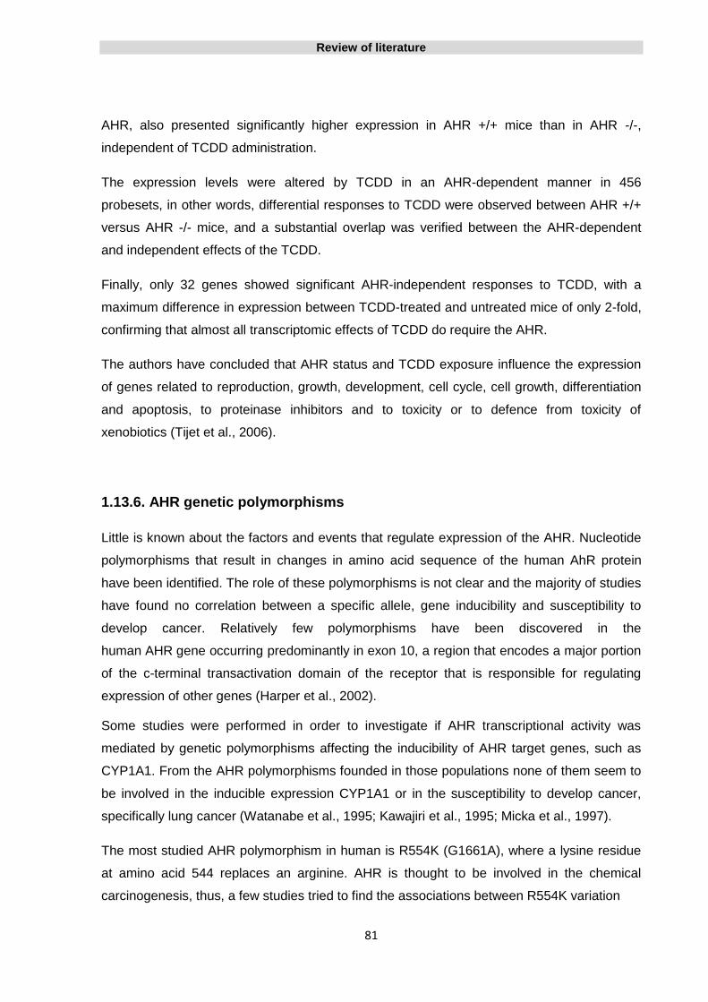
Review of literature
81
AHR, also presented significantly higher expression in AHR +/+ mice than in AHR -/-,
independent of TCDD administration.
The expression levels were altered by TCDD in an AHR-dependent manner in 456
probesets, in other words, differential responses to TCDD were observed between AHR +/+
versus AHR -/- mice, and a substantial overlap was verified between the AHR-dependent
and independent effects of the TCDD.
Finally, only 32 genes showed significant AHR-independent responses to TCDD, with a
maximum difference in expression between TCDD-treated and untreated mice of only 2-fold,
confirming that almost all transcriptomic effects of TCDD do require the AHR.
The authors have concluded that AHR status and TCDD exposure influence the expression
of genes related to reproduction, growth, development, cell cycle, cell growth, differentiation
and apoptosis, to proteinase inhibitors and to toxicity or to defence from toxicity of
xenobiotics (Tijet et al., 2006).
1.13.6. AHR genetic polymorphisms
Little is known about the factors and events that regulate expression of the AHR. Nucleotide
polymorphisms that result in changes in amino acid sequence of the human AhR protein
have been identified. The role of these polymorphisms is not clear and the majority of studies
have found no correlation between a specific allele, gene inducibility and susceptibility to
develop cancer. Relatively few polymorphisms have been discovered in the
human AHR gene occurring predominantly in exon 10, a region that encodes a major portion
of the c-terminal transactivation domain of the receptor that is responsible for regulating
expression of other genes (Harper et al., 2002).
Some studies were performed in order to investigate if AHR transcriptional activity was
mediated by genetic polymorphisms affecting the inducibility of AHR target genes, such as
CYP1A1. From the AHR polymorphisms founded in those populations none of them seem to
be involved in the inducible expression CYP1A1 or in the susceptibility to develop cancer,
specifically lung cancer (Watanabe et al., 1995; Kawajiri et al., 1995; Micka et al., 1997).
The most studied AHR polymorphism in human is R554K (G1661A), where a lysine residue
at amino acid 544 replaces an arginine. AHR is thought to be involved in the chemical
carcinogenesis, thus, a few studies tried to find the associations between R554K variation
Review of literature
82
and the risks of lung cancer and others human cancers, but failed (Kawajiri et al., 1995; Luo
et al., 2013). However a report showed significant and adverse association of R554K with
survival for soft tissue sarcoma (Berwick et al., 2004).
The available data lead to believe that mutations causing alterations in AHR gene expression
may either be quite rare or might even be incompatible with survival when leading to a
marked loss of expression as the AHR plays also a role in cell cycle regulation (Puga et al.,
2002).
1.13.7. Developmental role of AHR
While the AHR was discovered due to its role in drug metabolism, increasing evidence
suggests that the primary function of mammalian AHR is probably related to normal
development, namely in liver and vascular development, immune system function and cell
growth and differentiation (Mulero-Navarro et al., 2006).
Phylogenetic analysis suggests that the ability of mammalian and vertebrate AHR to sense
xenobiotics and regulate biotransformation enzymes was acquired at a later stage of
evolution as part of an adaptive response mechanism, and that the original function of the
AHR may have been as a developmental regulatory gene (Hahn, 2002).
Unlike mammalian AHR, the invertebrate AHR orthologs, although working as transcription
factors, do not bind to any of the classic know AHR ligands. This evidence strongly supports
the fact that xenobiotic binding is an independent function of the developmental role of the
AHR (Hahn, 2002).
Even more elucidative of the physiological and developmental importance of the AHR is the
fact that AHR knock null mice, generated in separated laboratories, display several
physiological defects, namely, growth retardation, peripheral immune system deficiency,
reduced liver size, abnormalities in vascular structure, portal tract fibrosis, female decreased
fertility, epidermal hyperplasia and hyperproliferation of hair follicles and lung and liver
tumors (Mimura et al., 1997; Schmidt et al., 1996; Fernandez-Salguero et al., 1995).
AHR-null mice do not express receptor protein and typical AHR target genes, such as
CYP1A1 and CYP1A2, are not induced upon the administration of TCDD (Fernandez-
Salguero et al., 1995). AHR is also known to cross-talk with a variety of other cell signalling
pathways.
Review of literature
83
All the arguments above point to a critical endogenous role of AHR, especially during normal
tissue development and homeostasis, lend further support for the existence of an
endogenous ligand and also suggesting that the driving force for the evolutionary
conversation of AHR not only lies on its role in xenobiotic metabolism but also in normal cell
development.
1.13.8. Tumor suppressor gene role of AHR in the absence of xenobiotics
For several decades AHR gene has been studied as a receptor for environmental
contaminants, as a mediator of chemical toxicity and regulator of xenobiotic-induced
carcinogenesis. However its role in the process of tumor initiation and development under
normal physiological conditions, meaning, in the absence of xenobiotics, is just beginning to
be understood (Barouki et al., 2007).
Epidemiological and experimental animal data have provided evidences for an association
between abnormal AHR function and cancer, suggesting that AHR may function as a tumor
suppressor gene that becomes silenced in the process of tumor formation (Fan et al., 2010).
AHR has been shown to act as a tumor suppressor in a mouse model of prostate cancer
denominated Transgenic Adenocarcinoma of the Mouse Prostate (TRAMP). The AHR-/-
TRAMP mice develop prostate tumors with greater frequency than AHR +/+ TRAMP mice.
Among other hypotheses, it was proposed that AHR may affect prostate carcinogenesis
through cross-talk with other signalling pathways, including the androgen signalling cascade.
However, the exact mechanism through which the AHR enhances the process of prostate
carcinogenesis remains to be determined (Fritz et al., 2007).
Peng and colleagues (2009) also investigated the susceptibility of AHR-/- mice to develop
liver tumors and they also found that a significant enhancement of tumor incidence in those
when compared with AHR +/+ mice, supporting the idea that loss of the AHR gene acts like a
tumor suppressor and can be a critical event in liver cancer progression (Peng et al, 2008).
These results are supported by other study (Fan et al., 2010).
Those data implied that AHR acts as a tumor suppressor gene but the underlying molecular
mechanism is still unknown.

Review of literature
84
1.13.9. Role of AHR in gliomas carcinogenesis
There have been a limited number of investigations on the potential role of AHR signalling in
gliomas. However, the role of AHR in glioma carcinogenesis in the absence of exogenous
ligand remains to be determined.
1.13.9.1. AHR expression in normal brain and gliomas
In rats, AHR and ARNT mRNAs were found widely distributed throughout the brain,
especially in hypothalamic and brainstem regions involved in the regulation of appetite and
circadian rhythms (Petersen et al., 2000). In other study, and also in murine cells, both
astrocytes and endothelial cells express abundant levels of AHR protein in brain (Filbrandt et
al., 2004).
In 2009, Gramatzki and coworkers, in order to predict the possible value of targeting AHR in
human glioma patients in vivo, have studied the AHR expression in human normal brain and
in a large series of human gliomas of WHO grade II-IV, using immunohistochemistry
(Gramatzki et al., 2009).
The AHR gene was expressed in normal brain regions including cortex and white matter of
frontal, parietal, temporal and occipital lobes, basal ganglia, thalamus, hippocampus,
midbrain and cerebellum. On the other hand, no AHR expression was found in pyramidal
hippocampal neurons, nigral neurons of the midbrain, pyramidal cortical neurons or granular
cerebellar cells (Gramatzki et al., 2009).
A strong AHR expression was seen especially in astrocytes. Expression was observed in the
cytoplasm and strongly in the nuclei of glial cells but not in endothelial cells of proliferating
vessels. Both in normal brain and in gliomas the pattern of expression was heterogeneous
(Gramatzki et al., 2009).
The mean AHR expression levels showed a decrease with WHO grade, showing the lowest
levels in grade IV glioma, being the difference in the expression levels, between grade II
gliomas when compared with grade IV gliomas, statistically significant. The authors have
also found that the nuclear staining intensity was higher in WHO grade IV gliomas when
compared with the grade II and III gliomas (Gramatzki et al., 2009).

Review of literature
85
1.13.9.2. Epigenetic regulation of AHR expression in human cancer
Surprisingly few studies have been focused on the mechanisms that control expression of
the human AHR gene. There are numerous examples in which alterations of the level of AHR
expression determines the responsiveness of downstream pathways, however, the
underlying cellular and molecular mechanisms responsible for the observed changes in AHR
expression remain for the most part elusive (Harper et al., 2006).
It was demonstrated that expression of the AHR gene may be down regulated by epigenetic
mechanisms, namely by DNA hypermethylation. Mulero – Navarro and co-workers analysed
the promoter methylation status as an epigenetic mechanism regulating AHR expression in
19 human tumor cell lines, representing 16 human tumor types (Mulero-Navarro et al., 2006).
They found that only cell lines from acute lymphoblastic leukaemia and chronic myeloid
leukaemia had significant AHR promoter hypermethylation. Twenty-one human primary
acute lymphoblastic leukaemia tumors were also analysed and 33% of the patients
presented AHR promoter hypermethylation (Mulero-Navarro et al., 2006).
In non-lympohoid human tumors, namely, from breast, colon, lung, melanoma,
osteosarcoma, testis carcinoma, neuroblastoma, rhabdomyosarcoma and choriocarcinoma
minimal AHR promoter hypermethylation was found (Mulero-Navarro et al., 2006).
The results from this study suggest that promoter hypermethylation could represent a
mechanism regulating AHR expression in human tumor cells in a cell-specific manner, points
AHR as a negative regulator of cell growth and proliferation in acute lymphoblastic leukemia
in the absence of xenobiotics and supports the putative role of tumor suppressor of AHR
gene (Mulero-Navarro et al., 2006).
AHR expression was also found to be regulated through CpG promoter hypermethylation in
mantle cell lymphoma (MCL), in 32% of samples, and is correlated with higher proliferation,
increased number of chromosomal abnormalities and shorter survival of the patients. Five
genes, including AHR, (SOX9, HOXA9, AHR, NR2F2, and ROBO1) were frequently
methylated in these tumors suggesting that a subset of MCL might show a CpG island
methylator phenotype (CIMP) that may influence the behaviour of the tumors (Enjuanes et
al., 2011).

Review of literature
86
1.14. The Transforming growth factor beta
Transforming growth factor-beta (TGF-β) is a multifunctional regulatory polypeptide that is
the prototypical member of a large family of cytokines which is comprised of over thirty
members including activins, nodals, bone morphogenetic proteins, growth and differentiation
factors. Is secreted by several cell types and controls many aspects of cellular function,
including cellular proliferation, differentiation, migration, apoptosis, adhesion, angiogenesis,
immune surveillance, and survival (Jakowlew, 2006).
Three different isoforms have been identified in mammals, namely TGF-β1, TGF-β2 and
TGF-β3, which share about 80% amino acid sequence identity (Roberts, 1998).
The active form of the TGF-β is a 25kDa dimer of two polypeptides stabilized by hydrophobic
interactions strengthened by a bisulfite bond. Before binding to its receptors, TGF-β is
activated from a large latent complex comprised of Latent TGF-β binding protein (LTBP) and
Latency Associated Peptide (LAP).
Once activated, TGF-β signals through two classes of receptors, the TGF-β type I receptor
(TβRI) and TGF-β type I receptor (TβRII). Type I and II receptors are serine/threonine kinase
receptors that form a heterodimeric complex upon TGF-β binding. TGF-β interacts with TβRII
which catalyses the phosphorylation of the TβRI and allows the subsequent incorporation of
the TβRI generating a ligand-receptor complex formed by a ligand dimer, two TβRI and two
TβRII. The only known function of the TβRII is to activate the TβRI. Once activated by the
TβRII, TβRI phosphorylates Smads through its catalytic domain.
Smad proteins are transcription factor and only known TGF-β receptor substrates capable of
signal transduction. Activated Smads regulate diverse biological effects by partnering with
transcription factors resulting in cell-state specific modulation of transcription. The activity of
the Smad pathway is terminated through a number of mechanisms including ubiquitinylation
and proteosomic destruction of the Smads (Weiss and Attisano, 2013; Seoane, 2006; Rich,
2003).
In addition, TGF-β can signal through Smad-independent pathways (Moustakas and Heldin,
2005).

Review of literature
87
1.14.1. TGF-β role in human carcinogenesis
The role of TGF-β in cancer is complex once it has a dual role acting as both a tumor
suppressor in early tumorigenesis and tumor promoter in later cancer stages (Figure 24)
(Derynck et al., 2001; Akhurst et al., 2001).
In normal epithelial cells and early well-differentiated epithelial tumor cells TGF-β acts as a
suppressor of cell proliferation and apoptosis promoter (Massagué, 1998).
Studies shown that pancreatic cancers have non-functional TGF-β pathways that suggest
that inactivation of TGF-β signalling pathways plays an important role in human pancreatic
tumorigenesis and support the tumor suppressor role of TGF-β in those tumors (Goggins et
al., 1998; Villanueva et al., 1998).
In contrast, in human colorectal cancer TGF-β is expressed in high levels and is associated
with disease progression (Tsushima et al., 1996). In prostate cancer, TGF-β1 upregulation is
associated with angiogenesis, metastasis and poor patient prognosis (Wikström et al., 1998).
Furthermore, a significantly greater number of invasive breast carcinomas expressed higher
levels of TGF-β than in situ breast carcinomas and the strongest expression was observed in
invasive carcinomas with associated lymph node metastasis, supporting a relationship
between TGF-β production and tumor progression (Walker and Dearing, 1992; Gorsch et al.,
1992).
The oncogenic role of TGF-β has prompted the development of therapeutic strategies based
on the inhibition of the TGF-β pathway but a better understanding of the mechanisms that
mediate the malignant transformation of TGF-β will improve the development of rational and
successful therapeutic strategies.
1.14.2. TGF-β and gliomagenesis
TGF-β has been shown to play a role in many pathological disorders involving the central
nervous system, including neurodegenerative diseases and brain tumors (Vivien and Ali,
2006).
Among the multiple pathways associated with gliomas, the TGF-β pathway plays a very
crucial role in regulating the behaviour of these tumors. Elevated levels of TGF-β have been

Review of literature
88
reported in the blood serum os patients with malignant gliomas and a striking correlation was
observed between elevated TGF-β levels and high tumor grade, advanced tumor stage and
poor patient outcome (Rich, 2003; Sasaki et al., 1995; Platten et al., 2001).
In human gliomas, TGF-β expression has been pointed as a factor playing an important role
in cell growth, metastasis, invasion, angiogenesis and induction of immunosuppression
(Merzak et al., 1994; Platten et al., 2001; Derynck et al., 2001).
TGF-β and its ligands and receptor are expressed in both low grade and high grade gliomas,
but were not detected in normal brain and gliosis (Samuels et al, 1989; Yamada et al, 1995;
Kjellman et al, 2000). Unlike most cancers where TGF-β1 is the isoform most commonly
overexpressed, in gliomas TGF-β2 is the most abundant isoform upregulated and is involved
in tumor progression (Hülper et al., 2011; Hau et al., 2011).
An inverse correlation between TGF-β expression in gliomas and survival, suggest that TGF-
beta plays an important role in the malignant progression of gliomas (Merzak et al., 1994;
Platten et al., 2001; Wick et al., 2001).
High TGF-β/SMAD activity is associated with aggressiveness, high proliferation and poor
prognosis in glioma patients, and is dependent on PDGF-B methylation status (Bruna et al.,
2007). TGF-β is able to promote proliferation through PDGF-B induction in glioma cells,
acting as an oncogenic factor, in those with unmethylated PDGF-B promoter. In about 50%
of human gliomas presenting low expression of PDGF-B, the PDGF-B promoter is found to
be methylated. These tumors do not have a hyperactive TGF-β/SMAD activity and are
usually less aggressive (Bruna et al., 2007).
A connection between angiogenesis process and TGF-β was demonstatred in several
studies. Data suggest that the overproduction of TGF-β by hamster ovary tumor cells can
contribute to neovascularization and may help promote tumor development in vivo. In
glioblastoma, TGF-β has a key role in regulating vascular phenotype of these tumors,
suggesting that, TGF-β may represent a new target for vascular normalization therapy (Ueki
et al., 1992; Dieterich et al., 2012).
Despite the data regarding high TGF-β expression in different human gliomas, the
mechanisms underlying this expression, signalling and role of TGF-β in promoting
tumorigenesis in glioma it is far from known.

Review of literature
89
1.14.3. TGF-β as a therapeutic target in gliomas
The observation that TGF- β acts on multiple levels to promote the malignant phenotype of
gliomas, has prompted the development of approaches to antagonize the biological effect of
TGF- β as a promising experimental strategy to manage malignant gliomas.
Recent therapeutic approaches targeting the TGF-β pathway comprise antisense
oligonucleotides, neutralizing antibodies, antagonistic antibodies or small molecule inhibitors.
Despite a number of agents are currently evaluated in early clinical studies in glioma
patients, all these approaches seem to faced problems caused by limitations in passing the
BBB, but are nevertheless promising (Kaminska et al., 2013; Joseph et al., 2013).
Figure 24_ Possible multiple roles of TGF-β in tumor pathogenesis. TGF-β can induce
apoptosis or inhibit proliferation of nontransformed cells but loses its growth-inhibitory potential as
cells progress to later stages of tumorigenesis. In the later stages of tumor development TGF-β is
actively secreted by tumor cells or stromal cells and contributes to cell growth, invasion, metastasis,
and decrease in host anti-tumor immune responses. Adapted from Kaminska et al., 2005.

Review of literature
90
1.14.4. Crosstalk between AHR and TGF-β signalling pathways
TGF-β, such as the tumor necrosis factor- α (TNF-α) and the epidermal growth factor (EGF),
were found to be downstream targets of AHR signalling (Haarmann-Stemmann et al., 2009).
Prior work has established a link between the AHR and TGF-β signalling pathways and
shown that TGF-β levels and activity increase in AHR -/- mouse cells (Elizondo et al., 2000;
Gonzalez and Fernandez-Salguero, 1998; Guo et al., 2004). Furthermore, many features of
the pathology that arise from the absence of the AHR gene are similar to the ramifications
that arise from an increase in TGF-β levels (Guo et al., 2004).
AHR and TGF-β signalling pathways are known to cross-regulate each other in a cell type-
specific manner and seem to converge to the regulation of the same cellular processes
(Wolff et al., 2001; Chang et al., 2007; Gomez-Duran et al., 2009).
In their work, Guo and colleagues, proposed to study the role of TGF-β in the pathology that
results from the loss of the AHR by comparison of global gene expression profiles of AHR
+/+ and AHR -/- vascular smooth muscle cells from mouse aorta. They found that AHR
represses TGF-β gene expression and affects the gene expression of several TGF-β related
genes. Based on that, the authors concluded that the simplest explanation for the AHR -/-
pathology is that AHR normally represses TGF-β signalling pathway in the wild-type cell
(Guo et al., 2004).
1.14.5. AHR and TGF-β signalling pathways in human gliomas
Latent TGF-β-binding protein-1 (LTBP-1) is a transcriptional target of AHR (Santiago-Josefat
et al, 2004; Corchero et al, 2004) and has been characterized as crucial in the process of
TGF-β activation in gliomas (Tritschler et al., 2009).
LTBP-1 was identified as an important modulator of TGF-β activation in glioma cells. Gene-
transfer mediated overexpression of LTBP-1 in glioma cell lines results in an increase in
TGF-β activity and also in the enhanced Smad2 phosphorylation, an intracellular marker
of TGF-beta activity. Oppositely, LTBP-1 gene silencing reduces TGF-β activity and Smad2
phosphorylation without affecting TGF-beta protein levels (Tritschler et al., 2009).
Several studies suggest that the AhR is involved in TGF-β and LTBP-1 signalling. In their
study, Santiago-Josefat and colleagues analysed differential patterns of LTBP-1 expression
between wild-type (AHR+/+) and null (AHR–/–) mouse embryo fibroblasts and found that
Ltbp-1 mRNA and protein expression were markedly increased in AHR–/–. This way they

Review of literature
91
identified mouse Ltbp-1 as a new negatively regulated AHR-target gene not involved in
xenobiotic metabolism and shown that LTBP-1 overexpression in the absence of AHR is
related to increased levels of TGF-β activation (Santiago-Josefat et al., 2004).
In mice liver, AHR was also identified as a negative regulator of liver fibrosis, possibly
through the control of LTBP-1 and TGF-β activities. Results suggest that LTBP-1 targets
TGF-β to specific areas of the liver and shown that the absence of AHR increased TGF-β
activity (Corchero et al., 2004).
Accordingly, it was found that LTBP-1 expression in human gliomas increases significantly
with WHO grade while in normal brain no expression was detected in 85% of cases, and in
the remaining cases only a weak diffuse expression staining was present (Tritschler et al.,
2009).
This way, TGF-β overexpression is pointed to be in the root of the greater tumorigenesis of
the AHR-/- genotype in human cancer (Fan et al., 2010). As referred before, the TGF-β has a
dual role during carcinogenesis, acting as a tumor suppressor in earlier phases and then as
tumor promoter in later tumorigenesis phases (Massagué, 1998.) The switch occurs as a
consequence of its being produced in high amounts in the tumor, which stimulates
tumorigenesis by allowing tumor cells to escape immune surveillance and promoting
angiogenesis.

92

Material and methods
93
CHAPTER 2 | Material and methods
2.1. Tissue samples selection
Brain tumor histological samples, fixed in buffered formalin and embedded in paraffin, were
obtained in the archives from the Pathology Department of Complejo Hospitalario
Universitario, Badajoz, Spain.
The inclusion criteria for the selection of the samples were previous histopathologic
diagnosis of astrocytic (WHO grade I, II and IV) or oligodendroglial tumor (WHO grade II and
III), diagnosis dating from the year 2003 to 2007, and sufficient tissue material available for
obtaining high-quality DNA for methylation analysis.
In accordance with the inclusion criteria described above, 188 tumor samples were
considered valid to be included in the study.
Twenty normal brain tissue samples were also obtained from the archives from the
Pathology Department of Complejo Hospitalario Universitario, Badajoz, Spain.
2.2. Diagnostic review
The histological slides, stained with hematoxylin-eosin (H&E), referring to all cases were
reviewed by two experienced pathologists and the tumors were graded and classified
according to the WHO classification (World Health Organization Classification of tumors of
the nervous system, Louis et al., 2007).
Blocks and areas most representative of tumor were selected to posterior DNA extraction and
it was confirmed that normal tissue samples do not exhibit any areas of tumor tissue.
2.3. Clinicopathological data
Clinicopathological data, including gender, age at the time of the diagnosis and tumor
location, were collected from the electronic medical records of the patients.
For cases diagnosed as glioblastoma (WHO grade IV) the promoter methylation status of the
MGMT gene and overall survival (months) were also collected.

Material and methods
94
Overall survival data of 66 glioblastoma patients were calculated from the date of the
operation to death. Standard therapy for these patients was surgical resection, carmustine
implants (Gliadel®), radiotherapy and concomitant and adjuvant temozolomide. The overall
survival and therapy data were provided by Neurosurgery Department of Complejo
Hospitalario Universitario, Badajoz, Spain.
2.4. Ethical issues
The study was conducted according to the Declaration of Helsinki principles and the medical
investigation law (ley 14/2007, de 3 de julio, de Investigación biomédica), currently into force
in Spain. All samples used in this study were collected before the year 2007, therefore, and
in accordance with the applicable law, no informed consents of the patients were collected.
In order to maintain the confidentiality of patient and all related data the samples were
anonymized using an acronym followed by a sequential number, namely, PA (Pilocytic
Astrocytoma), DA (Diffuse astrocytoma), OGD (Oligodendroglioma), GBM (Glioblastoma)
and NB (normal brain tissue).
2.5. DNA extraction and purification
Genomic DNA from buffered formalin fixed and paraffin embedded (FFPE) samples was
extracted by use of the QIAamp® DNA FFPE Tissue Kit (Qiagen, Hilden, Germany),
according to the manufacturer’s instructions.
QIAamp® DNA FFPE Tissue Kit is a commercially available spin column-based nucleic acid
extraction kit. Is a simple and quick approach to extracting nucleic acids from small
biological samples and combines the selective purification of DNA using a silica-gel-
membrane, which is based on a simple three step process of bind, wash and elute, with
previous proteinase k lysis at 56ºC.
By sample, 10 µm thick consecutive tissue sections (varying in number depending on the
size of the sample) were dewaxed in xylene and rehydrated in a decreasing alcohol series
and incubated in Buffer ATL and proteinase k at 56ºC, overnight.
After lysis was complete, the samples were incubated one hour at 90 ° C in order to reverse
the formalin crosslinking of nucleic acids.

Material and methods
95
Series of centrifugations were performed with buffers AW1 and AW2 so that the DNA binds
to the silica membrane and the contaminants were eliminated.
Lastly, 40 μl buffer ATE (elution buffer) was applied in the column membrane, incubated 5
minutes at room temperature and centrifuged to get DNA in an eppendorf tube. The
extracted DNA was stored at - 20°C.
2.6. DNA quantification and quality control
DNA quality control and concentration were estimated by spectrophotometry using
NanoPhotometer® (Implen GmbH, München, Germany). Readings were taken at
wavelengths of 260 nm and 280 nm which allow calculation of the concentration of nucleic
acid (µg/µl) in the sample and gives the amount of protein in the sample, respectively.
2.7. Bisulfite treatment and purification of genomic DNA
Bisulfite modification of DNA is the most commonly used, "gold standard" method for DNA
methylation studies since it was described for 5-methylcytosine detection (Frommer et al.,
1992; Clark et al., 1994).
The bisulfite DNA modification is a three step process (Figure 25), based on the different
sensitivity of cytosine and 5-methylcytosine to deamination by sodium bisulfite under acidic
conditions leading to unmethylated cytosine conversion to uracil whereby the unreactive 5-
methylcytosine remains a cytosine.
Figure 25 _ Sodium bisulfite DNA
modification is a three step process. Step
1: Sulfonation (addition of bisulfite to the 5-6
double bond of cytosine). Step 2: Hydrolytic
deamination (hydrolytic deamination of the
resulting cytosine-bisulfite derivate to give a
uracil-bisulfite derivate). Step 3: Alkaline
desulfonation (removal of sulfonate group by
an alkali treatment, to give uracil). Adapted
from Kilgore et al., 2007.

Material and methods
96
Genomic DNA from tumor and non-tumor samples was modified by treatment with sodium
bisulfite, using the EpiTect® Bisulfite Kit (Qiagen, Hilden, Germany), according to
manufacturer's protocol.
At room temperature, two bisulfite reactions were prepared for each sample: in two 200 µl
eppendorfs, containing 0.5 µg and 1 µg of DNA, were added the remaining components of
the bisulfite reaction (Nuclease-free water, Bisulfite Mix and DNA Protect Buffer) in a total
volume of reaction of 140µl. In order to perform the bisulfite DNA conversion the tubes were
placed into the thermal cycler with the following program conditions:
Table 1 _ Bisulfite conversion thermal cycler conditions.
Step Time (minutes) Temperature (ºC)
Denaturation 5 95
Incubation 25 60
Denaturation 5 95
Incubation 85 60
Denaturation 5 95
Incubation 175 60
Hold Indefinite 20
Once the bisulfite conversion was complete a mixture of Buffer BL and RNA carrier was
added to the bisulfite reactions and the reactions were transferred to the Epitect spin column.
The Buffer BL and RNA carrier mixture promotes the binding of the DNA to the column
membrane. Then, residual sodium bisulfite was removed using Buffer BW, desulfonation was
performed using Buffer BD and finally DNA was desalted with the addition of Buffer BW, in a
series of consecutive centrifugations.
The treated DNA was eluted in 20-40 μl buffer EB and bisulphite modified DNA was stored at
-20°C. The modified DNA was used as a template for polymerase chain reaction analysis.
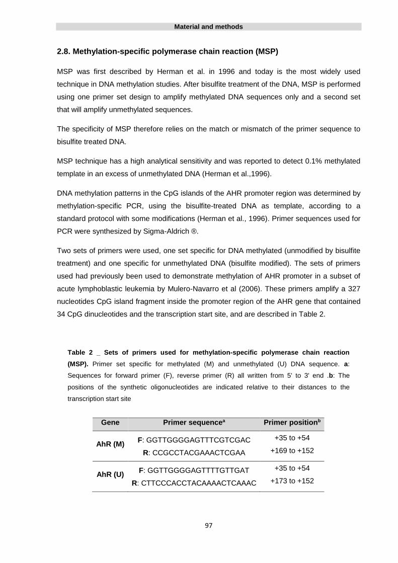
Material and methods
97
2.8. Methylation-specific polymerase chain reaction (MSP)
MSP was first described by Herman et al. in 1996 and today is the most widely used
technique in DNA methylation studies. After bisulfite treatment of the DNA, MSP is performed
using one primer set design to amplify methylated DNA sequences only and a second set
that will amplify unmethylated sequences.
The specificity of MSP therefore relies on the match or mismatch of the primer sequence to
bisulfite treated DNA.
MSP technique has a high analytical sensitivity and was reported to detect 0.1% methylated
template in an excess of unmethylated DNA (Herman et al.,1996).
DNA methylation patterns in the CpG islands of the AHR promoter region was determined by
methylation-specific PCR, using the bisulfite-treated DNA as template, according to a
standard protocol with some modifications (Herman et al., 1996). Primer sequences used for
PCR were synthesized by Sigma-Aldrich ®.
Two sets of primers were used, one set specific for DNA methylated (unmodified by bisulfite
treatment) and one specific for unmethylated DNA (bisulfite modified). The sets of primers
used had previously been used to demonstrate methylation of AHR promoter in a subset of
acute lymphoblastic leukemia by Mulero-Navarro et al (2006). These primers amplify a 327
nucleotides CpG island fragment inside the promoter region of the AHR gene that contained
34 CpG dinucleotides and the transcription start site, and are described in Table 2.
Table 2 _ Sets of primers used for methylation-specific polymerase chain reaction
(MSP). Primer set specific for methylated (M) and unmethylated (U) DNA sequence. a:
Sequences for forward primer (F), reverse primer (R) all written from 5' to 3' end .b: The
positions of the synthetic oligonucleotides are indicated relative to their distances to the
transcription start site
Gene Primer sequencea Primer positionb
AhR (M)
F: GGTTGGGGAGTTTCGTCGAC
R: CCGCCTACGAAACTCGAA
+35 to +54
+169 to +152
AhR (U)
F: GGTTGGGGAGTTTTGTTGAT
R: CTTCCCACCTACAAAACTCAAAC
+35 to +54
+173 to +152
Material and methods
98
PCR reactions were performed in a MyCyclerTM thermal cycler (Bio - Rad Laboratories Inc.,
CA, USA) under the following conditions: initial denaturing at 95°C for 5 min, followed by 42
cycles of denaturing at 95°C for 1 min, annealing at 62°C for 1 min and at 72°C for 1 min,
and final extension for 5 min at 72°C.
PCR reactions were carried out in a total 25 μl reaction mixture containing 1 × PCR buffer
(Qiagen®), 2 mM MgCl2 (Qiagen®), 0.2 mM dNTPs (Invitrogen ®), each primer at 0.14 μM, 24
µU/µl Hotstar taq ® plus DNA polymerase (Qiagen®), and 2-4 µl bisulfite-modified genomic
DNA.
In each PCR reaction, a methylated control DNA (CpGenomeTM Universal Methylated DNA,
Millipore) was used as a positive control. In addition, a negative/unmethylated control DNA
(EpiTect® Control DNA unmethylated, Qiagen) was included to ensure the specificity of the
results.
2.9. Electrophoresis
After amplification, aliquots of 10 µl of methylation-specific PCR (MSP) products were
analyzed on 8% polyacrylamide gel, stained with ethidium bromide, and visualized under
ultraviolet (UV) illumination. Results were recorded with a digital imaging system and
analyzed with the software Quantity one® 1-D Analysis Software (Bio Rad®).
2.10. Data interpretation
The technique used in this study relies on sodium bisulfite treatment of DNA, which converts
unmethylated cytosines to uracils while leaving methylated cytosines unaffected. The
modified sequences are then amplified with specific primers, and the amplified products are
identified using gel electrophoresis. This standard MSP approach offers only qualitative
analysis and cannot discern relative amounts of methylation.
The data interpretation consists in direct visualization of presence or absence of a defined
band in a certain expected length. In each polyacrylamide gel was added a molecular weight
marker consisting of a series of bands of varying known lengths that allow us to estimate the
length of the other bands existing on the gel.

Material and methods
99
For both samples and controls (methylated and unmethylated) two PCR reactions were
performed, one with primer sequence specific for methylated (M) and one for the
unmethylated sequence (U).
In methylated control, a band should only arise in the reaction performed with primers M,
while for the unmethylated control band should appear only in the reaction that was
performed with primers U. The samples were considered methylated when two bands
emerged, one at each primer (M and U) and the unmethylated ones exhibit only one band on
the primer U (Figure 26).
All the discordant or unclear results were confirmed by repeating the bisulfite treatment, MSP
and electrophoresis.
Figure 26 _ Example of bands pattern for unmethylated and methylated
DNA sequences in polyacrylamide gel. Note that samples were considered
methylated when two bands emerged (PCR reaction with primer M and U) and
unmethylated when only one band is visible (PCR reaction with primer M and
U).
2.11. Statistical Analysis
The AHR gene promoter methylation status in human gliomas was evaluated in association
with the clinicopathological parameters of patients including gender, histologic grade, tumor
location and MGMT methylation status by using Pearson Chi-Square or Fisher’s Exact Test
when sample size was small.
To evaluate a possible correlation of AHR methylation status and patient’s age, Pearson
correlation test was performed.

Material and methods
100
Overall survival was calculated from the date of the operation until death or the date of the
last follow-up. Survival analyses were carried out on 66 patients who undergone surgery,
radiotherapy and chemotherapy with concomitant and adjuvant temozolomide, plus
carmustine wafers implantation. Overall survival was illustrated using Kaplan-Meier plots.
The overall survival according to the status of AHR methylation in glioblastoma was
compared using a log-rank test. For comparing survival time distribution between groups the
log-rank test was used.
Statistical analysis was carried out with the software of IBM SPSS Statistics 21.0 (IBM SPSS
Inc., Chicago, IL). In all analysis, P-values <0.05 were considered statistically significant.

Results
101
CHAPTER 3 | Results
3.1. Clinicopathological classification
In total, samples of 188 cases were considered valid and were included in the study. Eight
cases were excluded from the study because the existing material was considered
insufficient (biopsies of very small size) or presenting poor quality (existence of large areas of
necrosis) to perform the extraction technique and subsequent MSP analysis and also
because do not have a diagnosis of glioma malignancy grade I, II, III or IV.
The mean age at diagnosis of the included patients was 51.6 years (range: 4-78; SD: 16.3).
The male to female ratio was 1:0.6 (male n=118, female n=70).
All cases were reviewed by experienced pathologists and classified according to WHO
classification system (Louis, 2007) (Table 3): 3,7% (n=7) cases were classified as pilocytic
astrocytoma (Grade I), 13.3% (n=25) as diffuse astrocytoma (Grade II), 19,7% (n=37) as
oligodendroglioma (Grade II and III) and 63,3% (n=119) were classified as glioblastoma
(Grade IV).
Table 3 _ Distribution of glioma classification according to WHO classification system (Louis
et al., 2007).
Type of glioma, grade (WHO,2007) n (%)
Pilocytic Astrocytoma, Grade I 7 (3.7)
Diffuse astrocytoma, Grade II 25 (13.3)
Oligodendroglioma, Grade II and III 37 (19.7)
Glioblastoma, Grade IV 119 (63.3)
Total 188 (100)
It was possible to collect data on tumor localization of 78 cases. The most common locations
were the frontal and temporal lobes both with 38.5% (n = 30) of cases, followed by the
parietal lobe with 17.9% (n = 14) and finally the occipital lobe at 5.1% (n = 4) cases.

Results
102
3.2. AHR promoter methylation status in human normal brain tissue
To study the potential role of AHR promoter methylation status in human gliomas, the
methylation status of AHR promoter in normal human brain tissue was first established.
Normal brain tissue derived from FFPE samples was analysed for the AHR promoter
methylation by MSP and served as control group. According to the bibliography reviewed,
the AHR promoter methylation status in human normal brain tissue was never reported
before.
Normal brain tissues samples from 20 patients were obtained, 11 female and 9 male with a
mean age of 49.95 years (range: 24-75; SD:15.86 ).
Interestingly, hypermethylation of AHR promoter was not observed in any of 20 normal brain
samples (Figure 27).
3.3. AHR promoter methylation status in human glioma
To characterize methylation patterns in human gliomas MSP assay was performed. Contrary
to what was verified in the normal brain tissue, aberrant hypermethylation in AHR promoter
was frequently observed in human glioma histological types. From a total of 188 gliomas,
46,8% (n=88) were methylated for AHR promoter and 53.2% (n=100) show no methylation
(Figure 27).
A significantly association is found when comparing AHR methylation status in normal brain
control and glioma samples (P=0.000).
These data suggest that the hypermethylation of the AHR gene promoter is a tumor-specific
event in human gliomas.
3.4. Association of AHR promoter methylation status with tumor histological
grade in human glioma
In the present study gliomas histologically classified into different malignancy grades,
according to WHO classification (Louis et al., 2007) were included: pilocytic astrocytomas
(grade I), diffuse astrocytoma (grade II), oligodendroglioma (grade II and III) and
glioblastoma (grade IV), being grade I and grade IV the least and the most malignant grade,
respectively.

Results
103
Analysing the relation between the methylation status of AHR promoter and the histological
classification of glioma a statistical association could be observed between them (P=0.005).
The methylation frequence increased along with the tumor grade being that gliomas with the
higher malignancy degree, namely, glioblastoma (grade IV) were more likely to be
methylated and, oppositely, pilocytic astrocytomas (grade I) showed no methylation (Figure
27).
Figure 27 _ Percentage of AHR promoter unmethylated (dark gray) and methylated (light gray),
in normal brain tissue (NB), Pilocytic astrocytoma (PA), Diffuse astocytoma (DA),
Oligodendrogliomas (OGD) and Glioblastoma (GBM).
3.5. Association of AHR promoter methylation status with age, gender and
tumor location in human glioma
When comparing the patient’s age at diagnosis and AHR promoter methylation status in
gliomas a significant association was found (P=0.026), the percentage of cases presenting
methylation of AHR increased with age, being more prevalent from the age group of 50
years, and it seems to be associated with older patient age at diagnosis (Table 4).
100 100
64 64,9
44,5
53,2
0 0
36 35,1
55,5
46,8
0
10
20
30
40
50
60
70
80
90
100
NB(n=20)
PA (n=7) DA(n=25)
OGD(n=37)
GBM(n=119)
Total(n=188)
Unmethylated
Methylated
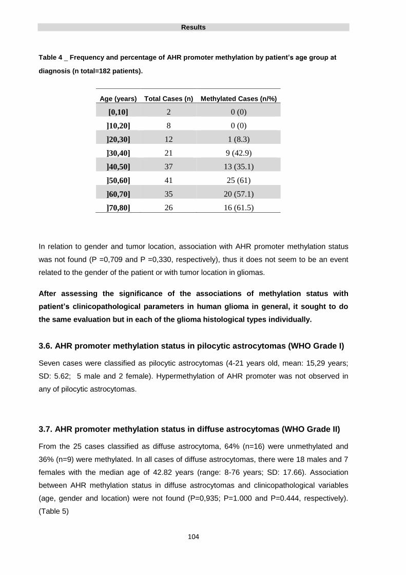
Results
104
Table 4 _ Frequency and percentage of AHR promoter methylation by patient’s age group at
diagnosis (n total=182 patients).
Age (years) Total Cases (n) Methylated Cases (n/%)
[0,10] 2 0 (0)
]10,20] 8 0 (0)
]20,30] 12 1 (8.3)
]30,40] 21 9 (42.9)
]40,50] 37 13 (35.1)
]50,60] 41 25 (61)
]60,70] 35 20 (57.1)
]70,80] 26 16 (61.5)
In relation to gender and tumor location, association with AHR promoter methylation status
was not found (P =0,709 and P =0,330, respectively), thus it does not seem to be an event
related to the gender of the patient or with tumor location in gliomas.
After assessing the significance of the associations of methylation status with
patient’s clinicopathological parameters in human glioma in general, it sought to do
the same evaluation but in each of the glioma histological types individually.
3.6. AHR promoter methylation status in pilocytic astrocytomas (WHO Grade I)
Seven cases were classified as pilocytic astrocytomas (4-21 years old, mean: 15,29 years;
SD: 5.62; 5 male and 2 female). Hypermethylation of AHR promoter was not observed in
any of pilocytic astrocytomas.
3.7. AHR promoter methylation status in diffuse astrocytomas (WHO Grade II)
From the 25 cases classified as diffuse astrocytoma, 64% (n=16) were unmethylated and
36% (n=9) were methylated. In all cases of diffuse astrocytomas, there were 18 males and 7
females with the median age of 42.82 years (range: 8-76 years; SD: 17.66). Association
between AHR methylation status in diffuse astrocytomas and clinicopathological variables
(age, gender and location) were not found (P=0,935; P=1.000 and P=0.444, respectively).
(Table 5)

Results
105
3.8. AHR promoter methylation status in oligodendrogliomas (WHO Grade II
and III)
A total of 37 OGD were included, 21 (56.8%) patients were males and 16 (43.2%) were
female. The mean age of oligodendroglioma patients was 41.05 years (range: 14-77 years;
SD: 14.05). MSP revealed methylation of the AHR promoter region in 13 cases (35.1%)
while the remaining 24 cases (64,9%) were not methylated for AHR promoter.
Representative samples are shown in figure 31.
Association between AHR methylation status in oligodendrogliomas and clinicopathological
variables, gender and tumor location, were not found (P=0.793 and P=1.00 respectively) but
a significant association with age is present in these tumors P=0.016 (Table 5).
Table 5 _ Associations between clinicopathological data of patients and AHR
methylation status in diffuse astrocytoma and oligodendroglioma (n – total number of
patients).
Variables n
AHR status Statistic significance
P<0.05 Methylated Unmethylated
Dif
fuse a
str
oc
yto
ma
(Gra
de
II)
Age (years) P=0.935
Gender P=1.000
Male 25
7 11
Female 2 5
Location P=0.444
Frontal
10
4 3
Temporal 0 2
Parietal 0 0
Occipital 1 0
Olig
od
en
dro
gli
om
a
(Gra
de
II a
nd
III)
Age (years) P=0.016
Gender P=0.793
Male 37
7 14
Female 6 10
Location P=1.000
Frontal
10
2 5
Temporal 1 2
Parietal 0 0
Occipital 0 0

Results
106
3.9. AHR promoter methylation status in glioblastomas (WHO Grade IV)
From the 119 patients diagnosed as glioblastoma, 74 (62.2%) were males and 45 (37.8%)
females and the mean age of these patients was 58.83 years (range: 27-78; SD: 10.68).
The methylation status of the AHR promoter was evaluated by MSP in 119 glioblastomas.
Promoter hypermethylation was detected in 55.5% (n=66) of cases, while 44.5% (n=53) were
not methylated (Figure 27). Representative samples are shown in figure 31.
3.10. Association of AHR promoter methylation status with age, gender and tumor
location in glioblastoma (WHO grade IV)
The patients with a glioblastoma diagnosis were divided in two groups according to their age:
one group consists of the individuals aged less than 60 years while the other group is
composed by the individuals with 60 or more years.
A Pearson Correlation test was performed in order to ascertain if there is any relationship
between age group and methylation status of the AHR in glioblastoma. A significant
association (P=0.037) was found between these two parameters what leads to believe that
the AHR gene methylation is a factor related to patient age in glioblastoma.
Association between AHR methylation status in glioblastoma and clinicopathological
variables gender and location was not found (P>0.05).
The table 6 shows the details of associations between AHR gene methylation and
clinicopathological parameters in glioblastoma patients.

Results
107
Table 6 _ Association between clinicopathological data of patients and AHR methylation
status in Glioblastoma (n – total number of patients).
Variables n AHR status Statistic significance
P<0.05 Methylated Unmethylated
Age (years) P=0.037
<60 116
30 30
≥60 34 22
Gender P=0.692
Male 119
40 34
Female 26 19
Location P=0.622
Frontal
58
8 8
Temporal 11 14
Parietal 9 5
Occipital 2 1
3.11. AHR gene promoter methylation status impact in overall survival in glioblastoma
Overall survival (OS) data of 66 glioblastoma patients, who undergone surgery, radiotherapy
and chemotherapy with concomitant and adjuvant temozolomide (Stupp protocol) plus
carmustine wafers, were collected. OS was calculated from the date of the operation until
death or the date of the last follow-up.
To evaluate the potential role of AHR methylation status in determining the prognosis of
glioblastoma patients treated in accordance with the STUPP protocol plus BCNU wafers,
survival analysis using Kaplan-Meier curves was conducted (P values were generated using
the log-rank test).
The results showed that AHR methylation is not associated with longer survival which
indicates that is not a significant predictor of OS in glioblastoma patients. The median
survival among patients with the unmethylated AHR promoter was 12 months (95% CI: 6.3-
17.7 months) as compared with 17 months (95% CI: 11,6-22.4 months) in patients with
methylated AHR promoter. Although methylated patients had a better prognosis than
unmethylated patients, the difference was not significant (Fig. 3-A; P = 0.658).

Results
108
Figure 28 _ Kaplan-Meier overall survival (months) analysis in glioblastoma patients according
to methylation status of (A) AHR gene (log-rank test, χ2 = 0.196, df = 1 ,p = 0.658) and (B) MGMT
gene (log-rank test, χ2 = 0.149, df = 1,P = 0.700).
3.12. Age impact in overall survival in glioblastoma
When the patient sample was divided into two groups by age (<60 years and ≥60 years) and
as expected, it was observed glioblastoma patient OS correlation with age (significant at the
level 0.01; P=0.005), noting that overall survival decreases with age and is lower in the age
group above 60 years. The median survival of patients aged <60 years and ≥60 years was
21 months (95% CI: 17.6-24.4 months) and 12 months (95% CI: 5.8-18.2 months) months,
respectively.
In figure 29 it can be seen the Kaplan-Meier overall survival (in months) analysis in
glioblastoma patients according to age group (in years).

Results
109
Figure 29_ Kaplan-Meier overall survival (months) analysis in glioblastoma patients according
to age group (years) (log-rank test, χ2 = 7.839, df = 1 ,P = 0.005).
3.13. AHR gene promoter methylation status impact in overall survival in glioblastoma,
depending on the patient’s age
In order to know whether the methylation status had any influence on the OS of patients of
different age groups (<60 and ≥ 60 years) survival Kaplan - Meier curve was applied for each
group (Figure 30).
In patients who did not show methylation of the AHR promoter the median OS was 18 and 7
months for the groups <60 and ≥ 60 years, respectively (Figure 30-A).
With regard to patients whose AHR promoter was found methylated, median survival of
patients younger than 60 years was 21 months and patients with 60 or more years was 12
months (Figure 30-B).
Patients, in both age groups (<60 and ≥ 60 years), in which the AHR promoter was
methylated presented median overall survival greater than patients of the same age group
where there was no AHR promoter methylation. A strong and significantly association was
found between these two groups with a P value = 0.01.

Results
110
Figure 30 _ Kaplan-Meier overall survival (months) analysis in glioblastoma patients according
to age group (years) in (A) AHR Unmethylated gene and (B) AHR Methylated gene (log-rank test, χ2
= 6,583 ,df = 1,P = 0.01, Adjusted for AHR methylation status).
3.14. Association between AHR and MGMT genes promoter methylation status in
glioblastoma
To further study the importance of promoter methylation of newly identified epigenetically
silenced gene, AHR, and revealing its relationship with already known epigenetic markers in
glioblastoma, like MGMT gene, could help to better understand glioblastoma’s biology.
Therefore, in the course of this study, data on the methylation status of the MGMT gene of
110 patients, from the same patients group which were assessed for the status of AHR
methylation, were collected.
Of the 110 cases, 64 (58%) were methylated in the AHR gene and 46 (42%) in the MGMT
gene, and 29 cases (26%) are co-methylated for both genes (Table 7).
When analysing the methylation status of the two genes, AHR and MGMT, there was no
significant association between these two parameters (P>0.05) (Table 7).
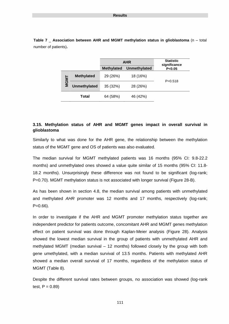
Results
111
Table 7 _ Association between AHR and MGMT methylation status in glioblastoma (n – total
number of patients).
AHR Statistic significance
P<0.05 Methylated Unmethylated M
GM
T
Methylated 29 (26%) 18 (16%)
P=0.518
Unmethylated 35 (32%) 28 (26%)
Total 64 (58%) 46 (42%)
3.15. Methylation status of AHR and MGMT genes impact in overall survival in
glioblastoma
Similarly to what was done for the AHR gene, the relationship between the methylation
status of the MGMT gene and OS of patients was also evaluated.
The median survival for MGMT methylated patients was 16 months (95% CI: 9.8-22.2
months) and unmethylated ones showed a value quite similar of 15 months (95% CI: 11.8-
18.2 months). Unsurprisingly these difference was not found to be significant (log-rank;
P=0.70). MGMT methylation status is not associated with longer survival (Figure 28-B).
As has been shown in section 4.8, the median survival among patients with unmethylated
and methylated AHR promoter was 12 months and 17 months, respectively (log-rank;
P=0.66).
In order to investigate if the AHR and MGMT promoter methylation status together are
independent predictor for patients outcome, concomitant AHR and MGMT genes methylation
effect on patient survival was done through Kaplan-Meier analysis (Figure 28). Analysis
showed the lowest median survival in the group of patients with unmethylated AHR and
methylated MGMT (median survival – 12 months) followed closely by the group with both
gene umethylated, with a median survival of 13.5 months. Patients with methylated AHR
showed a median overall survival of 17 months, regardless of the methylation status of
MGMT (Table 8).
Despite the different survival rates between groups, no association was showed (log-rank
test, P = 0.89)

Results
112
Table 8 _ Median overall survival (in months) of glioblastoma patients depending on AHR and
MGMT genes promoter methylation status.
AHR
Methylated Unmethylated M
GM
T Methylated 17 12
Unmethylated 17 13.5

Results
113
(A)
(B)
(C) (D) (E) (F)
Figure 31 _ Methylation analysis of the AHR promoter region by methylation specific PCR.
PCR product was analysed by polyacrylamide gel electrophoresis. (A) MSP control reactions
consisted of a methylated control (CTR M) (CpGenomeTM Universal Methylated DNA, Millipore) used
as a positive control and a negative/unmethylated control (CTR U) (EpiTect® Control DNA
unmethylated, Qiagen). Arrows showing molecular weight marker (501, 489, 404, 331, 242, 190, 147
and 110 bp). The presence of bands in both the unmethylated PCR product (U) and methylated PCR
product (M) lanes reflects a methylated AHR promoter. The lack of a band in the lane corresponding
to methylated- specific primers (M) reflects the absence of AHR promoter methylation.
Representative results of methylation-specific PCR analysis of AHR promoter in (B) normal brain
tissues, (C) Pilocytic astrocytomas, (D) Diffuse astrocytomas, (E) Oligodendrogliomas, (F)
Glioblastomas.
PA.1
M U
NB.8
M U
NB.14
M U
NB.20
M U
NB.1
M U
PA.2
M U
PA.5
M U
PA.7
M U
DA.7
M U
DA.12
M U
DA.4
M U
OGD.5
M U
OGD.16
M U
OGD.10
M U
OGD.7
M U
DA.15
M U
GBM.25
M U
CTR M
M U
CTR U
M U
GBM.20
M U
GBM.105
M U
GBM.37
M U
190 bp 147 bp 110 bp

Results
114

Discussion
115
CHAPTER 4 | Discussion
Understanding of the molecular alterations that occur during tumorigenesis, and identification
of novel markers for cancer diagnosis and novel targets for treatment, may be important for
the improvements in tumor diagnosis, treatment and prevention.
Oncogene activation and/or tumor suppressor gene inactivation play a critical role during
tumor development and progression. It has emerged that epigenetic events, like DNA
methylation, can lead to tumor suppressor gene inactivation as an alternative mechanism to
genetic events, such as gene mutation, deletion or rearrangement.
Hypermethylation is a regional event that occurs frequently in GC-rich sequence, called CpG
islands, and often located within the 5’ regulatory non-transcribed regions of genes. Aberrant
methylation of CpG islands of the promoter region of genes plays a role in the development
of various tumors and has become an important area of investigation in assessing the
mechanisms of tumor suppressor and regulatory gene inactivation. It was shown that tumor
suppressor genes can be transcriptionally silenced when their promoter region is
hypermethylated. Meanwhile, the methylation status of tumor suppressor genes has been
investigated and profiled for a number of human cancers.
Human gliomas are the most common malignant tumors in the adult CNS. Glioblastoma is
the most common, malignant and lethal of human gliomas and accounts for 50 to 60% of
primary brain tumors. Despite intensive research throughout the past two decades, there has
been little progress in the treatment of glioblastoma and the median survival after diagnosis
is approximately 12 to 14 months (Louis et al., 2007).
The most common molecular alterations, that characterize human gliomas and could be
used in current clinical practice, diagnosis and therapeutic decision making are Ki-67
proliferation marker, amplification and overexpression of EGFR gene, mutations involving
PTEN, TP53 and IDH genes, duplication, fusion and/or mutation in BRAF gene, 1p/19q
deletions and MGMT promoter methylation status (see Chapter 1, point 1.8. Genetic
alterations in gliomas, for revision).
AHR is a cytosolic ligand-activated transcription factor which belongs to the bHLH/PAS
family of transcriptional regulators which is located in the 7p15 region (Micka et al., 1997).
Despite it has been largely studied because of its critical role in xenobiotic-induce toxicity and

Discussion
116
carcinogenesis, physiologic functions of AHR in the absence of xenobiotics are beginning to
be understood.
Data provide substantial support for an association between abnormal AHR function and
cancer, implicating that abnormal expression of AHR plays an important role in multiple
stages of tumor promotion and progression. Few works from several labs suggested that
AHR may function as a tumor suppressor gene that becomes silenced during the process of
tumor formation, but the underlying molecular mechanism is still unknown (Barouki et al.,
2007; Fan et al., 2010).
Loss of gene expression associated with promoter methylation is a well described
phenomenon in a variety of tumors (Baylin and Herman, 2000) and several recent studies
have shown that aberrant promoter DNA methylation contributes to gene silencing and may
participate in the carcinogenesis of human cancer.
It was found that AHR expression is markedly down-regulated in patients of hematologic
tumors as acute lymphoblastic leukemia (ALL) and mantle cell lymphoma (MCL) (Mulero-
Navarro et al., 2006; Enjuanes et al., 2011).
Since gene silencing of AHR gene due to aberrant promoter hypermethylation has already
been reported in human ALL and MCL it was hypothesized that AHR may be also
downregulated in gliomas due to hypermethylation of it promoter.
To analyze the potential of AHR as methylation biomarker in human glioma, the AHR
promoter methylation status was evaluated in a large series of 188 glioma samples, including
pilocytic astrocytomas, diffuse astrocytomas, oligodendrogliomas and glioblastomas, and 20
normal brain tissue samples, using the MSP technique.
In the present study, AHR promoter was found to be frequently hypermethylated in human
gliomas and according to the present knowledge, this was the first report evaluating and
reporting the presence of promoter hypermethylation of AHR in gliomas. Also, and in order to
understand the clinical implications of AHR promoter methylation status, this variable was
investigated in association with the patients clinical parameters, age, gender, histologic
classification, tumor location and MGMT promoter methylation status. Overall survival
analysis was conducted in glioblastoma patients who were treated with the STUPP protocol
and BCNU wafers to investigate the prognostic and/or predictive value of AHR promoter
methylation status.

Discussion
117
4.1. Use of MSP to evaluate the AHR promoter methylation status in formalin-fixed
paraffin-embedded archive tissues
Despite currently there are several methods for determining the methylation status of the
promoter of a gene, the most widely used technique is MSP analysis after bisulfite treatment
(Herman et al., 1996).
MSP is easy to perform and is not an expensive method, does not require any special
equipment or consumables aside from what is present in most medical laboratories, so it can
be easily incorporated into the workflow of a molecular laboratory (Christians et al., 2012).
MSP detects CpG island methylation with high sensitivity and specificity, particularly when
high-quality DNA is analyzed. Significant risks of false-positive and false-negative results
have been reported, especially when the DNA quality and/or quantity is low as in cases of
DNA extracted from paraffin-embedded material (Hamilton et al., 2011).
The major possible limitations adjacent to the use of MSP after bisulfite treatment and
formalin-fixed paraffin-embedded archive tissues, in assessing the methylation status of the
promoter of AHR in this study, were identified and measures were taken to overcome these
possible limitations so that the results obtained were not committed and were the most
reliable as possible.
Because of tumor heterogeneity, AHR promoter methylation status may depend on the site
of surgical sample collection within a glioma specimen, therefore, careful selection of testing
samples was an essential step of the testing protocol. For that reason, all H&E slides from
each case were evaluated by experienced pathologists, the tissue block with the largest
number of tumor cells was selected and also blocks/areas with extensive areas of necrosis
were discarded.
The genomic DNA extraction was performed in samples from formalin fixed and paraffin
embedded blocks using QIAamp® DNA FFPE Tissue Kit. This commercial kit is specially
designed for purifying DNA from formalin-fixed, paraffin-embedded tissue sections. It uses
special lysis conditions to release DNA from tissue sections and to overcome inhibitory
effects caused by formalin crosslinking of nucleic acids and uses QIAamp MinElute spin
columns for purification of high-quality DNA in small volumes. All obtained DNA evaluated for
quality control and concentration were estimated by spectrophotometry using
NanoPhotometer®. This procedure ensured that all DNA samples used contained sufficient
concentration and quality to perform the subsequent techniques without compromise results.

Discussion
118
Bisulfite treatment of DNA is technically the most challenging step of methylation analysis
because it leads to DNA fragmentation and when incomplete bisulfite conversion occurred it
may leads to false-positive or false-negative results, mispriming, lower sensitivity and
specificity (Cankovic et al., 2013).
In order to avoid false negative results, which may be due to several reasons including
recovery of partially sectioned cell nuclei, incomplete DNA modification during bisulfite
treatment, or loss of DNA during the testing procedure, appropriate quality control measures
were selected to assure that all assay steps were well controlled (Cankovic et al., 2013).
Positive methylated DNA and negative unmethylated DNA controls were incorporated, in
parallel with patient specimens during the bisulfite reaction and PCR amplification, in order to
assure that optimal conditions were maintained during all testing steps.
MSP is believed not to be prone to errors by contamination, since normal cells such as
infiltrating lymphocytes have an unmethylated promoter and therefore the contaminating
material does not produce a methylated MSP signal. Furthermore, it was confirmed that none
of the cases of normal brain tissue included in this study, showed methylation of the AHR
promoter, so it is expected that the existing normal tissue in the tumor sample has no
influence on the result, not working as a contaminant and not give false-positive results, in
the evaluation of the status of methylation of AHR promoter in gliomas
4.2. AHR promoter hypermethylation is a frequent event in glioma
The methylation analysis focused a CpG island that contained the transcription start site and
34 CpG dinucleotides susceptible to be methylated within AHR promoter. Mulero-Navarro
and coworkers analyzed hypermethylation in this promoter region as a potential mechanism
controlling AHR expression, and found that it cause gene silencing of AHR gene (Mulero-
Navarro et al., 2006).
From a variety of tumors of various origins, such as breast, lung, colon, skin, among others,
the authors found that AHR promoter was hypermethylated only in acute lymphoblastic
leukemia (ALL), in about 33% of cases. In other study, AHR promoter hypermethylation in
mantle cell lymphoma (MCL) was found in 32% of cases (Mulero-Navarro et al., 2006;
Enjuanes et al., 2011).
The results from the present study showed that promoter hypermethylation is a frequent
event in human gliomas. Gliomas were found to be AHR promoter hypermethylated in about
47% of cases while the remaining 53% were not methylated. By analyzing each histologic
Discussion
119
type of glioma individually, it was found that the AHR promoter is frequently hypermethylated
in 36%, 35.1% and 55.5% of diffuse astrocytomas, oligodendrogliomas and in glioblastomas,
respectively. The percentage obtained, especially in grade II gliomas, is very similar to those
obtained by Mulero-Navarro and Enjuanes in hematologic tumors.
Many researchers have previously reported the methylation frequency of several others
genes in the various histological types of human gliomas. PTEN promoter is reported as
hypermethylated in 43% of diffuse astrocytomas (Wiencke et al., 2006) and 35% of
glioblastoma (Baeza et al., 2003); in glioblastoma GATA6, CD81, DR4 and CASP8 are
hypermethylated in 68,4%, 46.1%, 41.3% and 56%, respectively (Skiriute et al., 2012), p73 is
methylated in 18% of cases (Watanabe et al., 2002) and TMS1/ASC in 21% of cases
(Matinez et al., 2007); p14ARF is promoter hypermethylated in 40% and 38.9% of grade II
astrocytomas and high grade gliomas, respectively (He et al., 2010), for just naming a few
examples among many others currently described.
Compared with the methylation markers suggested previously, AHR has a relatively high
frequency of promoter hypermethylation in human gliomas, especially in glioblastoma
samples where 55,5% of cases were hypermethylated, and so can be regarded as one of the
relevant epigenetic markers in these tumors.
4.3. AHR promoter hypermethylation is an early event in glioma
Gene promoter hypermethylation is an early event, can even be detected in benign lesions
that precede neoplastic process, and so is pointed as a key initiating event in human cancer
(Esteller and Corn, 2001).
There is increasing evidence that altered methylation patterns of tumor-associated genes are
already present in the early stages of astrocytic tumors, such as WHO grade II astrocytomas
and oligodendrogliomas, and that aberrant methylation of genes is more prevalent than
genetic alterations and may have consequences for the development of low-
grade astrocytomas (Costello et al., 2000b).
In an attempt to identify AHR as a new candidate gene that might be involved in the early
development of gliomas, WHO grade II astrocytomas and oligodendrogliomas were included
in the present study and the AHR promoter methylation status was evaluated using MSP.

Discussion
120
Methylation of AHR was found to be present in a high percentage of WHO grade II tumors, in
about 35% of cases, and was detected in both the early and late stages of glioma, indicating
that the inactivation of the AHR gene might be essential in the early development of gliomas
and persist through the course of development. These results suggest that aberrant promoter
methylation of AHR gene may serve as a useful biomarker during the follow-up of low-grade
gliomas.
4.4. AHR promoter hypermethylation is a tumor specific event in human gliomas
CpG islands, which are found in the promoters of approximately 60% of the coding genes in
the mammalian genome, are generally unmethylated in normal cells. On the other hand, the
hypermethylation of these promoter regions is found in virtually every type of human cancer
and is associated with the inappropriate transcriptional silencing of genes (Bird, 1996;
Esteller, 2002).
Twenty samples of normal brain tissue adjacent to tumor tissues were included in this study,
and served as normal control group. The results showed that AHR promoter was not
hypermethylated in any of the normal tissue samples. In concern to gliomas samples, AHR
promoter hypermethylation was detected in 46.8% of cases. Taken together, these data
clearly indicating that promoter hypermethylation occurs specifically in cancer cells but not in
normal brain cells, which means it is a tumor-specific event, and is an indicator that AHR
promoter hypermethylation may be involved in the pathogenesis of human gliomas.
In previous studies, it was found that the promoter of several genes, such as p14ARF,
p15INK4B, p16INK4B, RB (Yin et al., 2002; He et al., 2011), LATS1 and LATS2 (Jiang et al.,
2006), WIF-1 (Yang et al., 2010), TMS1/ASC (Stone et al., 2004), SLC22A18 (Chu et al.,
2011), LRRC4 (Zhang et al., 2008) and PTEN (Wiencke et al., 2007), revealed no
methylation in normal brain tissue samples, while in the samples of gliomas tumors, the
promoter of these genes presented hypermethylated.
The distinct methylation status of a gene in benign and malignant tissues is a prerequisite to
determine a gene as an effective molecular biomarker. Because AHR is not found to be
hypermethylated in normal brain tissue, AHR promoter methylation status could discriminate
between normal and tumoral tissues with a high sensitivity and specificity, suggesting that it
may be a promising epigenetic biomarker for assistant diagnosis and cancer early detection.
Discussion
121
The AHR promoter hypermethylation is an epigenetic change that arises early in the
carcinogenic process and that is specific tumor in gliomas. This molecular change has the
potential to be used in early detection of gliomas, including in biological samples such as
blood and cerebrospinal fluid. Previous studies show that analysis of DNA methylation can
be performed in biological samples, such as blood and cerebrospinal fluid, which are
obtained from relatively less aggressive then histological biopsies, with a good and highly
significant correlation with the primary tumor tissue (Balãna et al., 2003; Liu et al., 2010;
Majchrzak-Celińska et al., 2013; Lavon et al., 2010; Wakabayashi et al., 2009).
4.5. AHR methylation status is associated with tumor histological grade
The MSP analysis showed that the promoter hypermethylation frequency of AHR gene was
significantly higher in high grade gliomas compared to low grade gliomas, and a significant
trend to the more malignant glioma (glioblastoma) likely to be more methylated is present.
Percentages of 36%, 35.1% and 55.5% were found when AHR promoter methylation status
was analyzed in diffuse astrocytoma (Grade II), oligodendrogliomas (grade II and III) and
glioblastoma (grade IV), respectively. As previously mentioned, AHR methylation was not
detected in any cases of pilocytic astrocytoma (grade I).
AHR has been shown to act as a tumor suppressor gene and be involved in tumor
progression in prostate and liver cancers (Fritz et al., 2007; Fan et al., 2010). Also, in ALL,
AHR is associated with cell growth and proliferation and, in MCL, the AHR silencing through
hypermethylation is correlated with higher proliferation (Mulero-Navarro et al., 2006:
Enjuanez et al., 2011). Based on that, it can be speculate that tumors with a hypermethylated
AHR have a high proliferation rate, and so AHR may play a key role in the development of
low-grade gliomas and in their subsequent progression to higher malignancy histological
glioma grades.
Still with regard to the differences found between the different histological types, it was found
that AHR promoter methylation is present both in diffuse astrocytomas and
oligodendrogliomas but is absent in pilocytic astrocytomas
As a number of molecular genetic alterations have been identified in gliomas over the last
two decades, it has become clear that each subtype has a distinct genotype and adult and
pediatric gliomas develop through different molecular pathogenesis.
Discussion
122
In adults, diffuse astrocytomas (WHO grade II) and oligodendrogliomas share frequent
mutations of IDH1/IDH2. While astrocytomas of grade II and III often harbor TP53 mutations
in addition to IDH1 mutations, oligodendrogliomas rarely have TP53 mutations but frequently
show concurrent total 1p/19q loss and IDH1 mutations (Watanabe et al, 2009; Sanson et al,
2009). These facts suggest that astrocytomas and oligodendrogliomas share a common cell
of origin.
On the other hand, pilocytic astrocytomas (WHO grade I) rarely show these changes but
display an otherwise unique genetic profile. More than 80% of pilocytic astrocytomas, which
typically arise in children, have alterations of the MAPK pathway predominantly involving
the KIAA1549-BRAF fusion gene or BRAF mutations. The BRAF fusion gene is so specific to
pilocytic astrocytomas that it is considered as a diagnostic marker (Pfister et al., 2008; Jones
et al., 2008; Bar et al., 2008; Horbinski et al., 2010; Jeuken and Wesseling, 2010). These
facts strongly suggest that diffuse astrocytomas, oligodendrogliomas and pilocytic
astrocytomas develop through a distinct molecular pathogenesis.
With respect to known epigenetic alterations in pilocytic astrocytomas, in particular
hypermethylation of CpG island, and although there are some studies with contradictory
results, in most studies the genes analyzed showed no CpG island hypermethylation and,
conversely, were significantly hypomethylated relative to control tissues. At the same time,
astrocytomas and oligodendrogliomas were found to be frequently hypermethylated
(Uhlmann et al., 2003; Goeppert et al., 2013).
Together, these results provide evidence that the molecular mechanisms involved in the
development of pilocytic astrocytomas and other gliomas are different, and are in accordance
with the results obtained in the present study.
Of the seven cases of pilocytic astrocytomas included in the study, none of them presented
hypermethylation of AHR promoter, while in turn, diffuse astrocytomas and
oligodendrogliomas have presented a relative high and similar rate of AHR promoter
hypermethylation of about 35%.
The absence of methylation in pilocytic astrocytomas suggests that AHR does not play a role
in the pathogenesis of these tumors. The fact that AHR gene present hypermethylation of its
promoter in a similar proportion of cases in astrocytomas and oligodendrogliomas, may
reflect the fact that the pathway leading to these two entities is quite similar or even the
same, and the AHR gene may play a role in its development. Based on this, AHR promoter
methylation status analysis could also be used to help in differential diagnosis between
pilocytic astrocytomas and diffuse astrocytomas and oligodendrogliomas. However, it should
be noted that only 7 pilocytic astrocytoma samples were included in this study, and therefore
Discussion
123
all the results obtained and conclusions drawn should be accepted with caution and require
confirmation in further studies with a larger number of samples.
4.6. AHR methylation status and it association with the patient’s clinicopathological
parameters
To investigate the factors that might influence AHR promoter methylation in human gliomas,
a possible association between AHR promoter methylation status and clinical patient
parameters, such as age, gender and tumor location was evaluated.
There was no association between gender and tumor location, and methylation of AHR,
however there was a striking association between age at diagnosis and the incidence of AHR
hypermethylation in oligodendroglioma and glioblastoma patients. Patients with methylated
AHR were significantly older than those whose tumor lacked methylation.
The process of aging is a risk factor in cancer development and has been pointed as a major
event responsible for DNA hypermethylation in human cancer (Ahuja and Issa, 2000). Age
related gene hypermethylation was found in normal tissue, and tumors of older patients, and
an increase along with age was verified (Issa et al., 1994)
In the present study, 20 controls of normal brain tissue from patients with ages ranging
between 24 and 75 years were included. No promoter hypermethylation of the AHR gene
was found in any of the control tissues, even in older patients, which seems to indicate that
methylation of the promoter of the AHR gene is not associated with aging process.
The association between AHR hypermethylation with an older age at diagnosis, in
oligodendroglioma and glioblastoma patients, is in accordance with the fact that AHR
hypermethylation was also associated with the malignancy grade of the tumor. Also, the
median age of incidence is higher in patients with grade III and IV gliomas, when compared
with lower grade gliomas. In glioblastoma and anaplastic oligodendroglioma the median age
is 45 to 75 and 45 to 50 years, respectively (Louis et al., 2007; Ostrom et al., 2013).
In accordance, the glioblastoma (grade IV) and oligodendroglioma (grade II and III) patients
were those which represented the higher malignancy grades included in this study and so,
this can explain the association found.
Discussion
124
4.7. Patient age and overall survival in glioblastoma patients
Since the young age at diagnosis has been consistently associated with longer survival in
patients with glioblastoma, age has been identified as a major therapy-independent factor in
patient survival (Krex et al., 2007; Weller et al., 2009).
After dividing the patient sample into two groups by age (<60 years versus >60 years), and
as expected, a significant correlation between overall survival and patient age was found,
with a decrease in time along with age. Patients with age under 60 years presented a median
OS of 21 months, while older patients above 60 years showed a median OS of only 12
months. Like as had been proposed previously, age appears to be an independent
prognostic indicator in glioblastoma patients.
4.8. AHR promoter methylation status is independent of the MGMT promoter
methylation status in glioblastoma patients
The availability of methylation data on several genes in the same tumors allowed us to study
potential interactions between these various events.
MGMT promoter methylation status seems to have prognostic and predictive value in
glioblastoma patients treated with alkylating chemotherapy such as TMZ (Hegi et al., 2005).
However, and despite promoter methylation status of MGMT is already commonly assessed
in clinical routine for therapeutic purposes, the validity of MGMT methylation as a prognostic
and predictive indicator is still controversial (Schaich et al., 2009; Blanc et al., 2004; Wick et
al., 2007; Costa et al., 2010).
The data regarding MGMT promoter methylation status from 110 of the 119 glioblastoma
patients included in the study were collected. Those glioblastoma patients were uniformly
treated according STUPP protocol and BCNU wafers implantation.
Of those 110 glioblastoma patients, it was found that approximately 42% were MGMT
promoter hypermethylated while about 58% of the patients were hypermethylated for AHR
promoter. The frequency of MGMT promoter methylation was in agreement with previously
described results in glioblastoma, which range from 36-45% (Esteller et al., 1999b; Cankovic
et al., 2007; Nakamura et al., 2001c; Jaeckle et al., 1998).

Discussion
125
This study found no significant association between the methylation status
of MGMT and AHR (P > 0.05) despite relative high co-methylation of these genes, in 26% of
cases.
4.9. AHR promoter methylation status and overall survival in glioblastoma patients
Of 119 glioblastoma patients who were included in this study it was only possible to collect
data on overall survival in 66 cases. All patients were given uniform treatment in accordance
with the Stupp protocol plus BCNU wafers. The association between AHR promoter
hypermethylation status and clinical outcome in those patients was determined.
Patients with methylated AHR promoter presented a longer median OS, 17 months, than
those with unmethylated AHR promoter, 12 months. However, although methylated patients
seem to have a better prognosis, the difference was not significant and the methylation
status of AHR does not appear to play a prognostic role in this patients.
Although no significant, these results are in accordance with others verified for the MGMT
gene, once MGMT promoter hypermethylation is associated with longer OS in glioblastoma
patients, but are not in agreement with those obtained previously in cases of MCL, where
was found that patients with methylation of AHR had a significantly shorter OS (p = 0.0376)
compared to patients with unmethylated AHR (Enjuanes et al., 2011) and also results relative
to other genes, such as p14ARF and SOCS3, where an association between hypermethylation
of its promoter and a shorter survival was also be found (Watanabe et al, 2007; Martini et al,
2008).
4.10. AHR promoter hypermethylation and gene expression in human gliomas
In mammals, CpG islands are found in the proximal promoter regions of almost half of the
genes and are generally unmethylated in normal cells, while in cancer cells, the
hypermethylation of these promoter regions is the most well categorized epigenetic change
to occur (Larsen, 1992; Antequera and Bird, 1993; Costello and Plass, 2001). Promoter
CpGs hypermethylation is found in virtually every type of human tumors and result in
transcriptional silencing of the gene and subsequent loss of protein expression (Jones and
Baylin, 2002; Esteller, 2002).

Discussion
126
The correlation between DNA methylation and gene inactivation is a prerequisite for the
identification and functional validation of novel tumor suppressor genes (How Kit et al.,
2012).
Inactivation of tumor suppressor genes plays an important role in malignant brain tumor
formation and progression. Genetic mechanisms such as mutation, deletion, and structural
chromosome rearrangement are known to inactivate tumor suppressor genes. Today, it is
well known that also aberrant methylation of CpG islands is one of the major modes of
inactivation of tumor suppressor genes in cancer and a growing list of genes are being
identified as abnormal methylation of promoter having CpG islands.
The abnormal function of AHR gene has been associated with tumorigenesis pointing to a
possible role of AHR as a tumor suppressor gene which is silenced during tumor
development. In fact, AHR has been implied to act as a tumor suppressor in prostate and
liver tumors, however the underlying molecular mechanism is still unknown (Fan et al., 2010;
Fritz et al., 2007; Peng et al., 2008).
AHR is expressed in normal brain tissue, however in gliomas the expression is frequently
lost. In human gliomas, the median AHR expression levels showed a decrease from WHO
grade II astrocytomas, to lower levels in Grade III anaplastic astrocytomas and the lowest
levels in grade IV glioblastoma (Gramatzki et al., 2009). The mechanisms that alter/silence
the expression levels of the AHR gene in cancer, and in the specific case of gliomas,
nevertheless are still unknown.
Mutations causing alterations in AHR gene expression are described as be quite rare or even
incompatible with survival when leading to a marked loss of expression, suggesting that
other mechanisms are at the origin of the loss of expression of AHR (Puga et al., 2002).
Based on these issues and trying to identify the mechanisms that regulate the activity of the
AHR in tumor cells, in 2006, Mulero-Navarro and co-workers found that AHR was
epigenetically silenced by promoter hypermethylation in 33% of patients with acute
lymphoblastic leukemia (ALL). Also, and in order to address whether the low and lost
expression of AHR was even due to promoter hypermethylation, treatments with AZA were
performed and the AHR mRNA expression was significantly restored. The authors have also
evaluated the AHR promoter status in human tumor cell lines from breast, colon, lung,
melanoma, osteosarcoma, rhabdomyosarcoma, testis carcinoma, choriocarcinoma,
neuroblastoma and embryonic kidney, but minimal AHR promoter hypermethylation was
observed (Mulero-Navarro et al., 2006).

Discussion
127
In a more recent study, the methylation status of the AHR gene was investigated in mantle
cell lymphoma (MCL). AHR was promoter hypermethylated in 32% of samples. Also, there
was a significant inverse correlation between methylation and mRNA levels of AHR, and
normal lymph node samples showed significant higher expression levels of AHR than
primary MCL (Enjuanes et al., 2011).
Both studies mentioned above concluded that promoter hypermethylation of the AHR gene is
frequent and is the source of gene silencing in the tumors under study.
In the present study, and hypothesized that the AHR gene could also be epigenetically
regulated by its promoter methylation in human glioma, a series of 188 gliomas (WHO
grades I, II and IV) was analyzed by MSP. The methylation specific primers used were the
same used by Mulero-Navarro in their study. Within a CpG island, a sequence with 327
nucleotides (-133 to 194), containing 34 CpG dinucleotides susceptible to be methylated,
was analyzed (Mulero-Navarro et al., 2006).
AHR gene was found to be promoter hypermethylated in 46.8% of human gliomas and was
frequently hypermethylated in 36%, 35.1% and 55.5% of diffuse astrocytomas,
oligodendrogliomas and in glioblastomas, respectively. In grade I pilocytic astrocytomas no
AHR promoter hypermethylation was found. Another result was that there is a statistical
association between the methylation status of AHR promoter and the histological grade of
gliomas, with a significant trend to the more malignant glioma likely to be more methylated.
The facts that previous studies show, that there is loss of expression of AHR in gliomas
which increases with the degree of malignancy, in conjunction with the results obtained in
this study, that there is a frequent promoter hypermethylation of AHR and that
hypermethylation also is more frequent as the degree malignancy of gliomas increases,
suggests that the AHR gene is epigenetically silenced in human gliomas due to
hypermethylation of its promoter, like as seen in ALL and MCL.
However, the major limitation of this study is that analysis of AHR protein expression was not
performed, such as the correlation between expression of AHR protein and the promoter
methylation status of its gene, thus it can only be hypothesized that the AHR is silenced due
to methylation of its promoter.
Although is very tempting to hypothesize that frequent AHR promoter methylation may
induce down-regulation of AHR protein expression, however, the frequent methylation of
AHR in glioma should be regarded as an epigenetic biomarker but not a therapeutic target
until the association between the down-regulation of AHR and its promoter methylation is

Discussion
128
confirmed.The knowledge of the exact role of AHR gene in the development and progression
of gliomas can open doors in the diagnosis and clinical management of these tumors.
4.11. AHR is involved in gliomagenesis possibly via TGF-β pathway
Although it was found that the AHR gene is frequently hypermethylated and most likely
silenced in human gliomas, the process by which it may be involved in the development and
progression of these tumors is still unknown and must be investigated in the future.
A likely hypothesis, already vaguely explored in some studies, of the way in which the AHR
gene may be involved in tumorigenesis is through its interaction with the TGF-β gene and
regulation of it signaling pathway (Elizondo et al., 2000; Gonzalez and Fernandez-Salguero,
1998; Guo et al., 2004).
Deregulation of TGF-β expression or signaling has been implicated in the pathogenesis of a
variety of diseases, including cancer. TGF-β has a dual role in oncogenesis. It is a strong
inhibitor of proliferation of normal epithelial cells and astrocytes, and is considered a tumor
suppressor factor (Bruna et al., 2007). On the other hand, in some tumor types, and
specifically in high grade gliomas, TGF-β becomes an oncogenic factor.
TGF-β is pointed as having major functions in glioma biology, namely by controlling cell
proliferation, mediating invasion, acting as angiogenic factor promoting once it stimulates
production of VEGF, and allowing gliomas cells to escape from host immunity (Kaminska et
al., 2013).
Under physiological conditions TGF-β is expressed at a very low level in the brain. However,
TGF-β is frequently and highly expressed in gliomas and elevated TGF-β activity confers
poor prognosis in glioma patients (Rich, 2003; Samuels et al., 1989; Kjellman et al., 2000).
Also, a comparative study of expression of TGF-β in gliosis and gliomas tissues found a
correlation between TGF-β expression and tumor grade (Yamada et al., 1995).
TGF-β overexpression is pointed to be in the root of the greater tumorigenesis of the AHR-/-
genotype in human cancer. Studies have demonstrated that TGF-β levels and activity
increase in AHR -/- mouse cells and that many features of the pathology that arise from the
absence of the AHR gene are similar to those that arise from an increase in TGF-β levels
(Guo et al., 2004; Elizondo et al., 2000; Gonzalez and Fernandez-Salguero, 1998; Fan et al.,
2010).
Discussion
129
Furthermore, several lines of evidence indicate that the AHR is a negative regulator of TGF-β
levels, in other words, that AHR normally represses TGF-β gene expression and affects the
gene expression of several TGF-β related genes, and that both ligand-independent and
ligand –dependent AHR activity has been shown to regulate TGF-β signaling in various
systems (Puga et al., 2005; Harper et al., 2006; Chang et al., 2007; Guo et al., 2004).
Based on the foregoing, the results obtained thus suggest that promoter hypermethylation
and subsequent silencing of the AHR gene could be the cause of TGF-β cascade activation
and it overexpression in human gliomas. Confirming this hypothesis, the inhibition of TGF-β
signalling by reactivation of AHR gene may thus be a potential strategy in gliomas clinical
management.

130

Conclusion
131
CONCLUSION
The development and progression of gliomas may likely be due to a multistep process that
involves the functional inactivation of tumor suppressor genes, as well as the activation of
oncogenes. Given the limitations of current therapies, understanding the pathways that lead
to tumor progression should remain a high priority in cancer research.
Genetic and epigenetic studies involving cohorts of gliomas patients have provided important
information for understanding the role of key genes in the development and progression of
gliomas.
In the present study, MSP analysis of the AHR promoter hypermethylation status of 188
samples from gliomas patients and 20 samples of normal brain tissue was performed.
According to the literature reviewed, this was the first time that the methylation status of AHR
promoter was evaluated in human gliomas and, moreover, this study was performed on a
series comprised of a large number of cases.
AHR gene was frequently hypermethylated in gliomas but not in normal brain tissue samples
which indicates that AHR promoter hypermethylation is a tumor specific process likely to be
related to glioma carcinogenesis and may serve as a promising biomarker for diagnosis.
The results presented allows to conclude that promoter hypermethylation of AHR gene is an
early event and increased with tumor malignancy grade, suggesting that hypermethylation
could be a mechanism involved during the development and progression of gliomas.
None of the pilocytic astrocytomas included were hypermethylated, which suggests that AHR
gene is not involved in the development of these tumors, and this finding can be explored in
order to help in differential diagnosis between pilocytic astrocytomas and other entities which
frequently have presented AHR promoter hypermethylated, such as, diffuse astrocytomas
and oligodendrogliomas.
Evidence suggest that aberrant promoter hypermethylation of AHR is most likely related with
AHR silencing in gliomas, suggesting and supporting the idea that glioma patients may
benefit by use of demethylating drug treatments. TGF-β pathway is pointed has been
associated with AHR silencing effects in the developing of human tumors

Conclusion
132
These findings suggest a potential clinical application of AHR promoter methylation status
and future confirmatory studies to support these findings and characterizing the role of AHR
gene and it relation with TGF-β pathway in gliomagenesis are needed.
It would also be interesting assess the methylation status of the AHR gene in other tumors of
the Central Nervous System, such as ependymoma, medulloblastoma or neuroblastoma, in
order to trying to understand if this epigenetic alteration is exclusive of the specific group of
gliomas or, on the contrary, is also found in other types of tumors of this system.
References
133
REFERENCES
Ahuja N, Issa JP. Aging, methylation and cancer. Histol Histopathol. 2000 Jul;15(3):835-42.
Akhurst RJ, Derynck R. TGF-beta signaling in cancer--a double-edged sword. Trends Cell Biol. 2001 Nov;11(11):S44-51.
Aldape K, Burger P and Perry A. Clinicophatologic aspects of 1p/19q loss and the diagnosis of oligodendroglioma. Arch Pathol Lab Med. 2007; 131: 242-251.
Aldape KD, Ballman K, Furth A, Buckner JC, Giannini C, Burger PC, Scheithauer BW, Jenkins RB, James CD. Immunohistochemical detection of EGFRvIII in high malignancy grade astrocytomas and evaluation of prognostic significance. J Neuropathol Exp Neurol. 2004 Jul;63(7):700-7.
Alonso ME, Bello MJ, Gonzalez-Gomez P, Arjona D, Lomas J, de Campos JM, Isla A, Sarasa JL, Rey JA. Aberrant promoter methylation of multiple genes in oligodendrogliomas and ependymomas. Cancer Genet Cytogenet. 2003 Jul 15;144(2):134-42.
Alonso MM, Diez-Valle R, Manterola L, Rubio A, Liu D, Cortes-Santiago N, Urquiza L, já uregi P, Lopez de Munain A, Sampron N, Aramburu A, Tejada-Solís S,Vicente C, Odero MD, Bandrés E, García-Foncillas J, Idoate MA, Lang FF, Fueyo J, Gomez-Manzano C. Genetic and epigenetic modifications of Sox2 contribute to the invasive phenotype of malignant gliomas. PLoS One. 2011;6(11):e26740.
Antequera F, Bird A. Number of CpG islands and genes in human and mouse. Proc Natl Acad Sci U S A. 1993 Dec 15;90(24):11995-9.
Athanassiou H, Synodinou M, Maragoudakis E, Paraskevaidis M, Verigos C, Misailidou D, Antonadou D, Saris G, Beroukas K, Karageorgis P. Randomized phase II study of temozolomide and radiotherapy compared with radiotherapy alone in newly diagnosed glioblastoma multiforme. J Clin Oncol. 2005 Apr 1;23(10):2372-7.
Bae YK, Brown A, Garrett E, Bornman D, Fackler MJ, Sukumar S, Herman JG, Gabrielson E. Hypermethylation in histologically distinct classes of breast cancer. Clin Cancer Res. 2004 Sep 15;10(18 Pt 1):5998-6005.
Baeza N, Weller M, Yonekawa Y, Kleihues P, Ohgaki H. PTEN methylation and expression in glioblastomas. Acta Neuropathol. 2003 Nov;106(5):479-85.
Bailey P, Cushing H. A classification of tumors of the glioma group on a histogenetic basis with a correlated study of prognosis. Lippincott, Philadelphia,1926.
Ballabh P, Braun A, Nedergaard M. The blood-brain barrier: an overview: structure, regulation, and clinical implications. Neurobiol Dis. 2004 Jun;16(1):1-13.
Balss J, Meyer J, Mueller W, Korshunov A, Hartmann C, von Deimling A. Analysis of the IDH1 codon 132 mutation in brain tumors. Acta Neuropathol. 2008; 116(6):597-602.
Banno K, Yanokura M, Susumu N, Kawaguchi M, Hirao N, Hirasawa A, Tsukazaki K, Aoki D. Relationship of the aberrant DNA hypermethylation of cancer-related genes with carcinogenesis of endometrial cancer. Oncol Rep. 2006 Dec;16(6):1189-96.
Bar EE, Lin A, Tihan T, Burger PC, Eberhart CG. Frequent gains at chromosome 7q34 involving BRAF in pilocytic astrocytoma. J Neuropathol Exp Neurol. 2008 Sep;67(9):878-87.
http://www.ncbi.nlm.nih.gov/pubmed?term=Diez-Valle%20R%5BAuthor%5D&cauthor=true&cauthor_uid=22069467
http://www.ncbi.nlm.nih.gov/pubmed?term=Misailidou%20D%5BAuthor%5D&cauthor=true&cauthor_uid=15800329
http://www.ncbi.nlm.nih.gov/pubmed?term=Misailidou%20D%5BAuthor%5D&cauthor=true&cauthor_uid=15800329
http://www.ncbi.nlm.nih.gov/pubmed?term=Gabrielson%20E%5BAuthor%5D&cauthor=true&cauthor_uid=15447983
http://www.ncbi.nlm.nih.gov/pubmed?term=Gabrielson%20E%5BAuthor%5D&cauthor=true&cauthor_uid=15447983

References
134
Barekati Z, Radpour R, Kohler C, Zhong XY. Specificity of methylation assays in cancer research: a guideline for designing primers and probes. Obstet Gynecol Int. 2010;2010. pii: 870865.
Barker FG 2nd, Chang SM, Gutin PH, Malec MK, McDermott MW, Prados MD, Wilson CB. Survival and functional status after resection of recurrent glioblastoma multiforme. Neurosurgery. 1998 Apr;42(4):709-20; discussion 720-3.
Barouki R, Coumoul X, Fernandez-Salguero PM. The aryl hydrocarbon receptor, more than a xenobiotic-interacting protein. FEBS Lett. 2007 Jul 31;581(19):3608-15.
Bauman GS, Ino Y, Ueki K, Zlatescu MC, Fisher BJ, Macdonald DR, Stitt L, Louis DN, Cairncross JG. Allelic loss of chromosome 1p and radiotherapy plus chemotherapy in patients with oligodendrogliomas. Int J Radiat Oncol Biol Phys. 2000 Oct 1;48(3):825-30.
Bedford MT, van Helden PD. Hypomethylation of DNA in pathological conditions of the human prostate. Cancer Res. 1987; 47(20):5274-6.
Belda-Iniesta C et al. Epidermal growth factor receptor and glioblastoma multiforme: molecular basis for a new approach. Clin Transl Oncol. 2008; 10:73-77.
Belinsky SA, Nikula KJ, Palmisano WA, Michels R, Saccomanno G, Gabrielson E, Baylin SB, Herman JG.Aberrant methylation of p16(INK4a) is an early event in lung cancer and a potential biomarker for early diagnosis. Proc Natl Acad Sci U S A. 1998 Sep 29;95(20):11891-6.
Bello MJ, Gonzalez-Gomez P, Alonso ME, Anselmo NP, Arjona D, Amiñoso C, Lopez-Marin I, De Campos JM, Isla A, Vaquero J, Casartelli C, Rey JA. Methylation analysis of cell cycle control genes RB1, p14ARF and p16INK4a in human gliomas. Cancer Therapy Jul 24; 2:187-194, 2004
Bello MJ, Vaquero J, de Campos JM, Kusak ME, Sarasa JL, Saez-Castresana J, Pestana A, Rey JA. Molecular analysis of chromosome 1 abnormalities in human gliomas reveals frequent loss of 1p in oligodendroglial tumors. Int J Cancer. 1994 Apr 15;57(2):172-5.
Berg D, Hipp S, Malinowsky K, Böllner C, Becker KF. Molecular profiling of signalling pathways in formalin-fixed and paraffin-embedded cancer tissues. Eur J Cancer. 2010 Jan;46(1):47-55.
Berwick M, Matullo G, Song YS, Guarrera S, Dominguez G, Orlow I, Walker M, Vineis P. Association between aryl hydrocarbon receptor genotype and survival in soft tissue sarcoma. J Clin Oncol. 2004 Oct 1;22(19):3997-4001.
Bird AP. CpG-rich islands and the function of DNA methylation. Nature. 1986 May 15-21;321(6067):209-13.
Bird AP. The relationship of DNA methylation to cancer. Cancer Surv. 1996;28:87-101.
Blanc JL, Wager M, Guilhot J, Kusy S, Bataille B, Chantereau T, Lapierre F, Larsen CJ, Karayan-Tapon L. Correlation of clinical features and methylation status of MGMT gene promoter in glioblastomas. J Neurooncol. 2004 Jul;68(3):275-83.
Bleeker FE, Lamba S, Leenstra S, Troost D, Hulsebos T, Vandertop WP, Frattini M, Molinari F, Knowles M, Cerrato A, Rodolfo M, Scarpa A, Felicioni L, Buttitta F,Malatesta S, Marchetti A, Bardelli A. IDH1 mutations at residue p.R132 (IDH1(R132)) occur frequently in high-grade gliomas but not in other solid tumors. Hum Mutat. 2009 Jan;30(1):7-11.
http://www.ncbi.nlm.nih.gov/pubmed?term=Macdonald%20DR%5BAuthor%5D&cauthor=true&cauthor_uid=11020580
http://www.ncbi.nlm.nih.gov/pubmed?term=Malinowsky%20K%5BAuthor%5D&cauthor=true&cauthor_uid=19914823
http://www.ncbi.nlm.nih.gov/pubmed/?term=cpg-rich+islands+and+the+function+of+dna+methylation+*+bird
http://www.ncbi.nlm.nih.gov/pubmed?term=Chantereau%20T%5BAuthor%5D&cauthor=true&cauthor_uid=15332332

References
135
Bloch O, Han SJ, Cha S, Sun MZ, Aghi MK, McDermott MW, Berger MS, Parsa AT. Impact of extent of resection for recurrent glioblastoma on overall survival: clinical article. J Neurosurg. 2012 Dec;117(6):1032-8.
Blümcke I, Müller S, Buslei R, Riederer BM, Wiestler OD. Microtubule-associated protein-2 immunoreactivity: a useful tool in the differential diagnosis of low-grade neuroepithelial tumors. Acta Neuropathol. 2004 Aug;108(2):89-96.
Bonin S, Petrera F, Niccolini B, Stanta G. PCR analysis in archival postmortem tissues. Mol Pathol. 2003 Jun;56(3):184-6.
Bourne D and Schiff D. Update on molecular findings, management and outcome in low-grade gliomas. Nature Reviews Neurology. 2010; 6: 695-701.
Brandes AA, Tosoni A, Cavallo G, Reni M, Franceschi E, Bonaldi L, Bertorelle R, Gardiman M, Ghimenton C, Iuzzolino P, Pession A, Blatt V, Ermani M; GICNO. Correlations between O6-methylguanine DNA methyltransferase promoter methylation status, 1p and 19q deletions, and response to temozolomide in anaplastic and recurrent oligodendroglioma: a prospective GICNO study. J Clin Oncol. 2006 Oct 10;24(29):4746-53.
Brandes AA, Basso U, Vastola F, Tosoni A, Pasetto LM, Jirillo A, Lonardi S, Paris MK, Koussis H, Monfardini S, Ermani M. Carboplatin and teniposide as third-line chemotherapy in patients with recurrent oligodendroglioma or oligoastrocytoma: a phase II study. Ann Oncol. 2003 Dec;14(12):1727-31.
Brandes AA, Franceschi E, Tosoni A, Blatt V, Pession A, Tallini G, Bertorelle R, Bartolini S, Calbucci F, Andreoli A, Frezza G, Leonardi M, Spagnolli F, Ermani M. MGMT promoter methylation status can predict the incidence and outcome of pseudoprogression after concomitant radiochemotherapy in newly diagnosed glioblastoma patients. J Clin Oncol. 2008 May 1;26(13):2192-7.
Brat DJ, Prayson RA, Ryken TC, Olson JJ. Diagnosis of malignant glioma: role of neuropathology. J Neurooncol. 2008 Sep;89(3):287-311.
Brem H, Piantadosi S, Burger PC, Walker M, Selker R, Vick NA, Black K, Sisti M, Brem S, Mohr G, et al.Placebo-controlled trial of safety and efficacy of intraoperative controlled delivery by biodegradable polymers of chemotherapy for recurrent gliomas. The Polymer-brain Tumor Treatment Group. Lancet. 1995 Apr 22;345(8956):1008-12.
Bruna A, Darken RS, Rojo F, Ocaña A, Peñuelas S, Arias A, Paris R, Tortosa A, Mora J, Baselga J, Seoane J. High TGFbeta-Smad activity confers poor prognosis in glioma patients and promotes cell proliferation depending on the methylation of the PDGF-B gene. Cancer Cell. 2007 Feb;11(2):147-60.
Cadieux B, Ching TT, VandenBerg SR, Costello JF. Genome-wide hypomethylation in human glioblastomas associated with specific copy number alteration, methylenetetrahydrofolate reductase allele status, and increased proliferation. Cancer Res. 2006 Sep 1;66(17):8469-76.
Cairncross G, Berkey B, Shaw E, Jenkins R, Scheithauer B, Brachman D, Buckner J, Fink K, Souhami L, Laperierre N, Mehta M, Curran W. Phase III trial of chemotherapy plus radiotherapy compared with radiotherapy alone for pure and mixed anaplastic oligodendroglioma: Intergroup Radiation Therapy Oncology Group Trial 9402. J Clin Oncol. 2006 Jun 20;24(18):2707-14.
http://www.ncbi.nlm.nih.gov/pubmed?term=McDermott%20MW%5BAuthor%5D&cauthor=true&cauthor_uid=23039151
http://www.ncbi.nlm.nih.gov/pubmed?term=Franceschi%20E%5BAuthor%5D&cauthor=true&cauthor_uid=16954518
http://www.ncbi.nlm.nih.gov/pubmed?term=Bertorelle%20R%5BAuthor%5D&cauthor=true&cauthor_uid=16954518
http://www.ncbi.nlm.nih.gov/pubmed?term=Monfardini%20S%5BAuthor%5D&cauthor=true&cauthor_uid=14630676
http://www.ncbi.nlm.nih.gov/pubmed?term=Franceschi%20E%5BAuthor%5D&cauthor=true&cauthor_uid=18445844
http://www.ncbi.nlm.nih.gov/pubmed?term=Bertorelle%20R%5BAuthor%5D&cauthor=true&cauthor_uid=18445844
http://www.ncbi.nlm.nih.gov/pubmed?term=Oca%C3%B1a%20A%5BAuthor%5D&cauthor=true&cauthor_uid=17292826
http://www.ncbi.nlm.nih.gov/pubmed?term=Cairncross%20G%5BAuthor%5D&cauthor=true&cauthor_uid=16782910
References
136
Cairncross G, Jenkins R. Gliomas with 1p/19q codeletion: a.k.a. oligodendroglioma. Cancer J. 2008 Nov-Dec;14(6):352-7.
Cairncross G, Wang M, Shaw E, Jenkins R, Brachman D, Buckner J, Fink K, Souhami L, Laperriere N, Curran W, Mehta M. Phase III trial of chemoradiotherapy for anaplastic oligodendroglioma: long-term results of RTOG 9402. J Clin Oncol. 2013 Jan 20;31(3):337-43.
Cairncross JG, Ueki K, Zlatescu MC, Lisle DK, Finkelstein DM, Hammond RR, Silver JS, Stark PC, Macdonald DR, Ino Y, Ramsay DA, Louis DN. Specific genetic predictors of chemotherapeutic response and survival in patients with anaplastic oligodendrogliomas. J Natl Cancer Inst. 1998;90(19):1473-9.
Cairns P, Esteller M, Herman JG, Schoenberg M, Jeronimo C, Sanchez-Cespedes M, Chow NH, Grasso M, Wu L, Westra WB, Sidransky D. Molecular detection of prostate cancer in urine by GSTP1 hypermethylation. Clin Cancer Res. 2001 Sep;7(9):2727-30.
Campan M, Weisenberger DJ, Trinh B, Laird PW. MethyLight. Methods Mol Biol. 2009;507:325-37.
Cankovic M, Mikkelsen T, Rosenblum ML, Zarbo RJ.A simplified laboratory validated assay for MGMT promoter hypermethylation analysis of glioma specimens from formalin-fixed paraffin-embedded tissue. Lab Invest. 2007 Apr;87(4):392-7.
Cankovic M, Nikiforova MN, Snuderl M, Adesina AM, Lindeman N, Wen PY, Lee EQ. The role of MGMT testing in clinical practice: a report of the association for molecular pathology. J Mol Diagn. 2013 Sep;15(5):539-55.
Chamberlain MC, Tsao-Wei DD, Groshen S. Salvage chemotherapy with cyclophosphamide for recurrent temozolomide-refractory anaplastic astrocytoma. Cancer. 2006 Jan 1;106(1):172-9.
Chamberlain MC, Tsao-Wei DD. Salvage chemotherapy with cyclophosphamide for recurrent, temozolomide-refractory glioblastoma multiforme. Cancer. 2004 Mar 15;100(6):1213-20.
Chamberlain MC, Wei-Tsao DD, Blumenthal DT, Glantz MJ. Salvage chemotherapy with CPT-11 for recurrent temozolomide-refractory anaplastic astrocytoma. Cancer. 2008 May 1;112(9):2038-45.
Chamberlain MC. Emerging clinical principles on the use of bevacizumab for the treatment of malignant gliomas. Cancer. 2010 Sep 1;116(17):3988-99.
Chang HG, Kim SJ, Chung KW, Noh DY, Kwon Y, Lee ES, Kang HS. Tamoxifen-resistant breast cancers show less frequent methylation of the estrogen receptor beta but not the estrogen receptor alpha gene. J Mol Med (Berl). 2005 Feb;83(2):132-9.
Chang HW, Chow V, Lam KY, Wei WI, Yuen A. Loss of E-cadherin expression resulting from promoter hypermethylation in oral tongue carcinoma and its prognostic significance. Cancer. 2002 Jan 15;94(2):386-92.
Chang X, Fan Y, Karyala S, Schwemberger S, Tomlinson CR, Sartor MA, Puga A. Ligand-independent regulation of transforming growth factor beta1 expression and cell cycle progression by the aryl hydrocarbon receptor. Mol Cell Biol. 2007 Sep;27(17):6127-39.
http://www.ncbi.nlm.nih.gov/pubmed?term=Cairncross%20G%5BAuthor%5D&cauthor=true&cauthor_uid=19060598
http://www.ncbi.nlm.nih.gov/pubmed?term=Cairncross%20G%5BAuthor%5D&cauthor=true&cauthor_uid=23071247
http://www.ncbi.nlm.nih.gov/pubmed?term=Laperriere%20N%5BAuthor%5D&cauthor=true&cauthor_uid=23071247
http://www.ncbi.nlm.nih.gov/pubmed?term=Schoenberg%20M%5BAuthor%5D&cauthor=true&cauthor_uid=11555585
http://www.ncbi.nlm.nih.gov/pubmed?term=Rosenblum%20ML%5BAuthor%5D&cauthor=true&cauthor_uid=17260000

References
137
Chaudhry P, Srinivasan R, Patel FD. Utility of gene promoter methylation in prediction of response to platinum-based chemotherapy in epithelial ovarian cancer (EOC). Cancer Invest. 2009 Oct;27(8):877-84.
Chu SH, Feng DF, Ma YB, Zhang H, Zhu ZA, Li ZQ, Jiang PC. Promoter methylation and downregulation of SLC22A18 are associated with the development and progression of human glioma. J Transl Med. 2011 Sep 21;9:156.
Clark SJ, Harrison J, Paul CL, Frommer M. High sensitivity mapping of methylated cytosines. Nucleic Acids Res. 1994 Aug 11; 22(15):2990-7.
Cloughsey T, Wick W, Mason W, Henriksson R, Saran F, Nishikawa R, Hilton M, Abrey L, Chinot O. Phase III Trial of Bevacizumab Added to Standard Radiotherapy and Temozolomide for Newly Diagnosed Glioblastoma: Final Progression-Free Survival and Preliminary Overall Survival Results from AVAglio. Neurology. 2013;80 (Meeting Abstracts 1): (PL02.002)
Cohen MH, Johnson JR, Pazdur R. Food and Drug Administration Drug approval summary: temozolomide plus radiation therapy for the treatment of newly diagnosed glioblastoma multiforme. Clin Cancer Res. 2005 Oct 1;11(19 Pt 1):6767-71.
Cohen MH, Shen YL, Keegan P, Pazdur R. FDA Drug Approval Summary: Bevacizumab (Avastin®) as Treatment of Recurrent Glioblastoma Multiforme. Oncologist. 2009 Nov;14(11):1131-8.
Colman H and Aldape K. Molecular Predictors in glioblastoma. Arch Neurol. 2008; 65(7):877-883.
Combs S et al. Prognostic significance of IDH-1 and MGMT in patients with glioblastoma: one step forward and one step back? Radiation Oncology. 2011; 6: 115-119.
Coons SW, Pearl DK. Mitosis identification in diffuse gliomas.Implication for tumor grading. Cancer 1998;82:1550-5.
Corchero J, Martín-Partido G, Dallas SL, Fernández-Salguero PM. Liver portal fibrosis in dioxin receptor-null mice that overexpress the latent transforming growth factor-beta-binding protein-1. Int J Exp Pathol. 2004 Oct;85(5):295-302.
Cosgrove M, Fitzgibbons PL, Sherrod A, Chandrasoma PT, Martin SE.Intermediate filament expression in astrocytic neoplasms. Am J Surg Pathol. 1989 Feb;13(2):141-5.
Cosgrove MM, Rich KA, Kunin SA, Sherrod AE, Martin SE. Keratin intermediate filament expression in astrocytic neoplasms: analysis by immunocytochemistry, western blot, and northern hybridization. Mod Pathol. 1993 May;6(3):342-7.
Costa BM, Caeiro C, Guimarães I, Martinho O, Jaraquemada T, Augusto I, Castro L, Osório L, Linhares P, Honavar M, Resende M, Braga F, Silva A, Pardal F,Amorim J, Nabiço R, Almeida R, Alegria C, Pires M, Pinheiro C, Carvalho E, Lopes JM, Costa P, Damasceno M, Reis RM. Prognostic value of MGMT promoter methylation in glioblastoma patients treated with temozolomide-based chemoradiation: a Portuguese multicentre study. Oncol Rep. 2010 Jun;23(6):1655-62.
Costello JF, Berger MS, Huang HS, Cavenee WK. Silencing of p16/CDKN2 expression in human gliomas by methylation and chromatin condensation. Cancer Res. 1996 May 15;56(10):2405-10.
Costello JF, Frühwald MC, Smiraglia DJ, Rush LJ, Robertson GP, Gao X, Wright FA, Feramisco JD, Peltomäki P, Lang JC, Schuller DE, Yu L, Bloomfield CD, Caligiuri
http://www.ncbi.nlm.nih.gov/pubmed?term=Srinivasan%20R%5BAuthor%5D&cauthor=true&cauthor_uid=19548140
http://www.ncbi.nlm.nih.gov/pubmed?term=Smiraglia%20DJ%5BAuthor%5D&cauthor=true&cauthor_uid=10655057
http://www.ncbi.nlm.nih.gov/pubmed?term=Robertson%20GP%5BAuthor%5D&cauthor=true&cauthor_uid=10655057
References
138
MA, Yates A, Nishikawa R, Su Huang H, Petrelli NJ, Zhang X, O'Dorisio MS, Held WA, Cavenee WK, Plass C.Aberrant CpG-island methylation has non-random and tumour-type-specific patterns. Nat Genet. 2000a Feb;24(2):132-8.
Costello JF, Plass C, Cavenee WK. Aberrant methylation of genes in low-grade astrocytomas. Brain Tumor Pathol. 2000b;17(2):49-56.
Costello JF, Plass C. Methylation matters. J Med Genet. 2001 May;38(5):285-303.
Dahia PL. PTEN, a unique tumor suppressor gene. Endocr Relat Cancer. 2000 Jun;7(2):115-29.
Dehghani F, Schachenmayr W, Laun A, Korf HW. Prognostic implication of histopathological, immunohistochemical and clinical features of oligodendrogliomas: a study of 89 cases. Acta Neuropathol. 1998 May;95(5):493-504.
Del Vecchio CA, Giacomini CP, Vogel H, Jensen KC, Florio T, Merlo A, Pollack JR, Wong AJ. EGFRvIII gene rearrangement is an early event in glioblastoma tumorigenesis and expression defines a hierarchy modulated by epigenetic mechanisms. Oncogene. 2012 Jul 16.
Delpu Y, Cordelier P, Cho WC, Torrisani J.DNA methylation and cancer diagnosis. Int J Mol Sci. 2013 Jul 18;14(7):15029-58.
Denison MS, Pandini A, Nagy SR, Baldwin EP, Bonati L. Ligand binding and activation of the Ah receptor. Chem Biol Interact. 2002 Sep 20;141(1-2):3-24.
Denison MS, Nagy SR. Activation of the aryl hydrocarbon receptor by structurally diverse exogenous and endogenous chemicals. Annu Rev Pharmacol Toxicol. 2003;43:309-34.
Derynck R, Akhurst RJ, Balmain A. TGF-beta signaling in tumor suppression and cancer progression. Nat Genet. 2001 Oct;29(2):117-29.
Devaux BC, O'Fallon JR, Kelly PJ. Resection, biopsy, and survival in malignant glial neoplasms. A retrospective study of clinical parameters, therapy, and outcome. J Neurosurg. 1993 May;78(5):767-75.
Di Vinci A, Casciano I, Marasco E, Banelli B, Ravetti GL, Borzì L, Brigati C, Forlani A, Dorcaratto A, Allemanni G, Zona G, Spaziante R, Gohlke H, Gardin G, Merlo DF, Mantovani V, Romani M. Quantitative methylation analysis of HOXA3, 7, 9, and 10 genes in glioma: association with tumor WHO grade and clinical outcome. J Cancer Res Clin Oncol. 2012 Jan;138(1):35-47.
Dieterich LC, Mellberg S, Langenkamp E, Zhang L, Zieba A, Salomäki H, Teichert M, Huang H, Edqvist PH, Kraus T, Augustin HG, Olofsson T, Larsson E,Söderberg O, Molema G, Pontén F, Georgii-Hemming P, Alafuzoff I, Dimberg A. Transcriptional profiling of human glioblastoma vessels indicates a key role of VEGF-A and TGFβ2 in vascular abnormalization. J Pathol. 2012 Nov;228(3):378-90.
Dixit S, Hingorani M, Achawal S, Scott I. The sequential use of carmustine wafers (Gliadel®) and post-operative radiotherapy with concomitanttemozolomide followed by adjuvant temozolomide: a clinical review. Br J Neurosurg. 2011 Aug;25(4):459-69.
Dolwick KM, Schmidt JV, Carver LA, Swanson HI, Bradfield CA. Cloning and expression of a human Ah receptor cDNA. Mol Pharmacol. 1993 Nov;44(5):911-7.
http://www.ncbi.nlm.nih.gov/pubmed?term=Caligiuri%20MA%5BAuthor%5D&cauthor=true&cauthor_uid=10655057
http://www.ncbi.nlm.nih.gov/pubmed?term=Su%20Huang%20H%5BAuthor%5D&cauthor=true&cauthor_uid=10655057
http://www.ncbi.nlm.nih.gov/pubmed?term=O'Dorisio%20MS%5BAuthor%5D&cauthor=true&cauthor_uid=10655057
http://www.ncbi.nlm.nih.gov/pubmed?term=Giacomini%20CP%5BAuthor%5D&cauthor=true&cauthor_uid=22797070
http://www.ncbi.nlm.nih.gov/pubmed?term=O%27Fallon%20JR%5BAuthor%5D&cauthor=true&cauthor_uid=8468607
http://www.ncbi.nlm.nih.gov/pubmed?term=Di%20Vinci%20A%5BAuthor%5D&cauthor=true&cauthor_uid=21947269
http://www.ncbi.nlm.nih.gov/pubmed?term=Borz%C3%AC%20L%5BAuthor%5D&cauthor=true&cauthor_uid=21947269
http://www.ncbi.nlm.nih.gov/pubmed?term=Dorcaratto%20A%5BAuthor%5D&cauthor=true&cauthor_uid=21947269
http://www.ncbi.nlm.nih.gov/pubmed?term=Dieterich%20LC%5BAuthor%5D&cauthor=true&cauthor_uid=22786655

References
139
Domb AJ, Israel ZH, Elmalak O, Teomim D, Bentolila A. Preparation and characterization of carmustine loaded polyanhydride wafers for treating brain tumors. Pharm Res. 1999 May;16(5):762-5.
Dong SM, Pang JC, Poon WS, Hu J, To KF, Chang AR, Ng HK. Concurrent hypermethylation of multiple genes is associated with grade of oligodendroglial tumors. J Neuropathol Exp Neurol. 2001 Aug;60(8):808-16.
Ducray F , Hallani S and Idbaih A. Diagnostic and prognostic markers in gliomas. Curr Opin Oncol. 2009; 21: 537-542.
Duffy MJ, Napieralski R, Martens JW, Span PN, Spyratos F, Sweep FC, Brunner N, Foekens JA, Schmitt M; EORTC PathoBiology Group. Methylated genes as new cancer biomarkers. Eur J Cancer. 2009 Feb;45(3):335-46.
Dunn J, Baborie A, Alam F, Joyce K, Moxham M, Sibson R, Crooks D, Husband D, Shenoy A, Brodbelt A, Wong H, Liloglou T, Haylock B, Walker C. Extent of MGMT promoter methylation correlates with outcome in glioblastomas given temozolomide and radiotherapy. Br J Cancer. 2009 Jul 7;101(1):124-31.
Eads CA, Danenberg KD, Kawakami K, Saltz LB, Blake C, Shibata D, Danenberg PV, Laird PW. MethyLight: a high-throughput assay to measure DNA methylation. Nucleic Acids Res. 2000 Apr 15;28(8):E32.
Ebinger M, Senf L, Wachowski O, Scheurlen W. No aberrant methylation of neurofibromatosis 1 gene (NF1) promoter in pilocytic astrocytoma in childhood. Pediatr Hematol Oncol. 2005 Jan-Feb;22(1):83-7.
Eguchi H, Hayashi S, Watanabe J, Gotoh O, Kawajiri K. Molecular cloning of the human AH receptor gene promoter. Biochem Biophys Res Commun. 1994 Aug 30;203(1):615-22.
Ehrlich M. DNA methylation in cancer: too much, but also too little. Oncogene. 2002; 21(35):5400-13.
Eisenhardt AE, Olbrich H, Röring M, Janzarik W, Anh TN, Cin H, Remke M, Witt H, Korshunov A, Pfister SM, Omran H, Brummer T. doi: 10.1002/ijc.25893. Epub 2011 Jul 25. Functional characterization of a BRAF insertion mutant associated with pilocytic astrocytoma. Int J Cancer. 2011 Nov 1;129(9):2297-303.
Elizondo G, Fernandez-Salguero P, Sheikh MS, Kim GY, Fornace AJ, Lee KS, Gonzalez FJ.Altered cell cycle control at the G(2)/M phases in aryl hydrocarbon receptor-null embryo fibroblast. Mol Pharmacol. 2000 May;57(5):1056-63.
Enjuanes A, Fernàndez V, Hernández L, Navarro A, Beà S, Pinyol M, López-Guillermo A, Rosenwald A, Ott G, Campo E, Jares P. Identification of methylated genes associated with aggressive clinicopathological features in mantle cell lymphoma. PLoS One. 2011;6(5):e19736.
Ermoian RP, Furniss CS, Lamborn KR, Basila D, Berger MS, Gottschalk AR, Nicholas MK, Stokoe D, Haas-Kogan DA. Dysregulation of PTEN and protein kinase B is associated with glioma histology and patient survival. Clin Cancer Res. 2002 May;8(5):1100-6.
Esteller M , Corn PG, Baylin SB, Herman JG. A gene hypermethylation profile of human cancer. Cancer Res. 2001 Apr 15;61(8):3225-9.
http://www.ncbi.nlm.nih.gov/pubmed?term=Danenberg%20KD%5BAuthor%5D&cauthor=true&cauthor_uid=10734209
References
140
Esteller M, Catasus L, Matias-Guiu X, Mutter GL, Prat J, Baylin SB, Herman JG. hMLH1 promoter hypermethylation is an early event in human endometrial tumorigenesis. Am J Pathol. 1999a Nov;155(5):1767-72.
Esteller M, Garcia-Foncillas J, Andion E, Goodman SN, Hidalgo OF, Vanaclocha V, Baylin SB, Herman JG. Inactivation of the DNA-repair gene MGMT and the clinical response of gliomas to alkylating agents. N Engl J Med. 2000a Nov 9;343(19):1350-4.
Esteller M, Hamilton SR, Burger PC, Baylin SB, Herman JG. Inactivation of the DNA repair gene O6-methylguanine-DNA methyltransferase by promoter hypermethylation is a common event in primary human neoplasia. Cancer Res. 1999b Feb 15;59(4):793-7.
Esteller M, Silva JM, Dominguez G, Bonilla F, Matias-Guiu X, Lerma E, Bussaglia E, Prat J, Harkes IC, Repasky EA, Gabrielson E, Schutte M, Baylin SB,Herman JG. Promoter hypermethylation and BRCA1 inactivation in sporadic breast and ovarian tumors. J Natl Cancer Inst. 2000b Apr 5;92(7):564-9.
Esteller M. Epigenetic gene silencing in cancer: the DNA hypermethylome. Hum Mol Genet. 2007 Apr 15;16 Spec No 1:R50-9.
Esteller M. Epigenetics in cancer. N Engl J Med. 2008; 358(11):1148-59.
Esteller M. CpG island hypermethylation and tumor suppressor genes: a booming present, a brighter future. Oncogene. 2002 Aug 12;21(35):5427-40.
Evans BR, Karchner SI, Allan LL, Pollenz RS, Tanguay RL, Jenny MJ, Sherr DH, Hahn ME. Repression of aryl hydrocarbon receptor (AHR) signaling by AHR repressor: role of DNA binding and competition for AHR nuclear translocator. Mol Pharmacol. 2008 Feb;73(2):387-98.
Fan Y, Boivin GP, Knudsen ES, Nebert DW, Xia Y, Puga A. The aryl hydrocarbon receptor functions as a tumor suppressor of liver carcinogenesis. Cancer Res. 2010 Jan 1;70(1):212-20.
Farrugia A, Keyser C, Ludes B. Efficiency evaluation of a DNA extraction and purification protocol on archival formalin-fixed and paraffin-embedded tissue. Forensic Sci Int. 2010 Jan 30;194(1-3):e25-8.
Feinberg AP, Vogelstein B. Hypomethylation distinguishes genes of some human cancers from their normal counterparts. Nature. 1983; 301(5895):89-92.
Felsberg J, Erkwoh A, Sabel MC, Kirsch L, Fimmers R, Blaschke B, Schlegel U, Schramm J, Wiestler OD, Reifenberger G. Oligodendroglial tumors: refinement of candidate regions on chromosome arm 1p and correlation of 1p/19q status with survival. Brain Pathol. 2004 Apr;14(2):121-30.
Ferlay J, Soerjomataram I, Ervik M, Dikshit R, Eser S, Mathers C, Rebelo M, Parkin DM, Forman D, Bray, F. GLOBOCAN 2012 v1.0, Cancer Incidence and Mortality Worldwide: IARC CancerBase No. 11 [Internet]. Lyon, France: International Agency for Research on Cancer; 2013. Available from: http://globocan.iarc.fr, accessed on 23/06/2014.
Fernandez-Salguero PM, Hilbert DM, Rudikoff S, Ward JM, Gonzalez FJ. Aryl-hydrocarbon receptor-deficient mice are resistant to 2,3,7,8-tetrachlorodibenzo-p-dioxin-induced toxicity. Toxicol Appl Pharmacol. 1996 Sep;140(1):173-9.
http://www.ncbi.nlm.nih.gov/pubmed?term=Vanaclocha%20V%5BAuthor%5D&cauthor=true&cauthor_uid=11070098
http://www.ncbi.nlm.nih.gov/pubmed?term=Gabrielson%20E%5BAuthor%5D&cauthor=true&cauthor_uid=10749912
http://www.ncbi.nlm.nih.gov/pubmed/?term=farrugia+*+efficiency+evaluation+of+a+dna+extraction+*+2010

References
141
Fernandez-Salguero P, Pineau T, Hilbert DM, McPhail T, Lee SS, Kimura S, Nebert DW, Rudikoff S, Ward JM, Gonzalez FJ. Immune system impairment and hepatic fibrosis in mice lacking the dioxin-binding Ah receptor. Science. 1995 May 5;268(5211):722-6.
Filbrandt CR, Wu Z, Zlokovic B, Opanashuk L, Gasiewicz TA. Presence and functional activity of the aryl hydrocarbon receptor in isolated murine cerebral vascular endothelial cells and astrocytes. Neurotoxicology. 2004 Jun;25(4):605-16.
Foltz G, Ryu GY, Yoon JG, Nelson T, Fahey J, Frakes A, Lee H, Field L, Zander K, Sibenaller Z, Ryken TC, Vibhakar R, Hood L, Madan A.Genome-wide analysis of epigenetic silencing identifies BEX1 and BEX2 as candidate tumor suppressor genes in malignant glioma. Cancer Res. 2006 Jul 1;66(13):6665-74.
Fonkem E, Wong ET. NovoTTF-100A: a new treatment modality for recurrent glioblastoma. Expert Rev Neurother. 2012 Aug;12(8):895-9.
Forsyth PA, Shaw EG, Scheithauer BW, O'Fallon JR, Layton DD Jr, Katzmann JA. Supratentorial pilocytic astrocytomas. A clinicopathologic, prognostic, and flow cytometric study of 51 patients. Cancer. 1993 Aug 15;72(4):1335-42.
Fraga MF, Ballestar E, Paz MF, Ropero S, Setien F, Ballestar ML, Heine-Suñer D, Cigudosa JC, Urioste M, Benitez J, Boix-Chornet M, Sanchez-Aguilera A, Ling C, Carlsson E, Poulsen P, Vaag A, Stephan Z, Spector TD, Wu YZ, Plass C, Esteller M. Epigenetic differences arise during the lifetime of monozygotic twins. Proc Natl Acad Sci U S A. 2005 Jul 26;102(30):10604-9.
Friedman HS, Prados MD, Wen PY, Mikkelsen T, Schiff D, Abrey LE, Yung WK, Paleologos N, Nicholas MK, Jensen R, Vredenburgh J, Huang J, Zheng M,Cloughesy T. Bevacizumab alone and in combination with irinotecan in recurrent glioblastoma. J Clin Oncol. 2009 Oct 1;27(28):4733-40.
Fritz WA, Lin TM, Cardiff RD, Peterson RE. The aryl hydrocarbon receptor inhibits prostate carcinogenesis in TRAMP mice. Carcinogenesis. 2007 Feb;28(2):497-505.
Frommer M, McDonald LE, Millar DS, Collis CM, Watt F, Grigg GW, Molloy PL, Paul CL. A genomic sequencing protocol that yields a positive display of 5-methylcytosine residues in individual DNA strands. Proc Natl Acad Sci U S A. 1992 Mar 1;89(5):1827-31.
Fujii H, Biel MA, Zhou W, Weitzman SA, Baylin SB, Gabrielson E. Methylation of the HIC-1 candidate tumor suppressor gene in human breast cancer. Oncogene. 1998 Apr 23;16(16):2159-64..
Fujii-Kuriyama Y, Mimura J. Molecular mechanisms of AhR functions in the regulation of cytochrome P450 genes. Biochem Biophys Res Commun. 2005 Dec 9;338(1):311-7.
Fulton D, Urtasun R, Forsyth P. Phase II study of prolonged oral therapy with etoposide (VP16) for patients with recurrent malignant glioma. J Neurooncol. 1996 Feb;27(2):149-55.
Furnari FB, Fenton T, Bachoo RM, Mukasa A, Stommel JM, Stegh A, Hahn WC, Ligon KL, Louis DN, Brennan C, Chin L, DePinho RA, Cavenee WK. Malignant astrocytic glioma: genetics, biology, and paths to treatment. Genes Dev. 2007 Nov 1;21(21):2683-710.
http://www.ncbi.nlm.nih.gov/pubmed?term=Filbrandt%20CR%5BAuthor%5D&cauthor=true&cauthor_uid=15183014
http://www.ncbi.nlm.nih.gov/pubmed?term=Gasiewicz%20TA%5BAuthor%5D&cauthor=true&cauthor_uid=15183014
http://www.ncbi.nlm.nih.gov/pubmed?term=Sibenaller%20Z%5BAuthor%5D&cauthor=true&cauthor_uid=16818640
http://www.ncbi.nlm.nih.gov/pubmed?term=O%27Fallon%20JR%5BAuthor%5D&cauthor=true&cauthor_uid=8339223
http://www.ncbi.nlm.nih.gov/pubmed?term=Ballestar%20ML%5BAuthor%5D&cauthor=true&cauthor_uid=16009939
http://www.ncbi.nlm.nih.gov/pubmed?term=Paleologos%20N%5BAuthor%5D&cauthor=true&cauthor_uid=19720927

References
142
Gan HK, Kaye AH, Luwor RB. The EGFRvIII variant in glioblastoma multiforme. J Clin Neurosci. 2009 Jun;16(6):748-54.
Gao T, Wang S, He B, Pan Y, Song G, Gu L, Chen L, Nie Z, Xu Y, Li R. The association of RAS association domain family Protein1A (RASSF1A) methylation states and bladder cancer risk: a systematic review and meta-analysis. PLoS One. 2012;7(11):e48300.
García P, Manterola C, Araya JC, Villaseca M, Guzmán P, Sanhueza A, Thomas M, Alvarez H, Roa JC. Promoter methylation profile in preneoplastic and neoplastic gallbladder lesions. Mol Carcinog. 2009 Jan;48(1):79-89.
Ge YZ, Xu LW, Jia RP, Xu Z, Feng YM, Wu R, Yu P, Zhao Y, Gui ZL, Tan SJ, Song Q. The association between RASSF1A promoter methylation and prostate cancer: evidence from 19 published studies. Tumour Biol. 2014 Apr;35(4):3881-90.
Giannini C, Burger PC, Berkey BA, Cairncross JG, Jenkins RB, Mehta M, Curran WJ, Aldape K. Anaplastic oligodendroglial tumors: refining the correlation among histopathology, 1p 19q deletion and clinical outcome in Intergroup Radiation Therapy Oncology Group Trial 9402. Brain Pathol. 2008 Jul;18(3):360-9.
Giannini C, Scheithauer BW, Burger PC, Christensen MR,Wollan PC, Sebo TJ, et al. Cellular proliferation in pilocyticand diffuse astrocytomas. J Neuropathol Exp Neurol 1999;58(1):46-53.
Gilbert MR, Dignam J, Won M, Blumenthal DT, Vogelbaum MA, Aldape KD, Colman H, Chakravarti A, Jeraj R, Armstrong TS, Wefel JS, Brown PD, Jaeckle KA, Schiff D, Atkins JN, Brachman D, Werner-Wasik M, Komaki R, Sulman EP, Mehta MP. RTOG 0825: Phase III double-blind placebo-controlled trial evaluating bevacizumab (Bev) in patients (Pts) with newly diagnosed glioblastoma (GBM). J Clin Oncol 31, 2013 (suppl; abstr 1)
Goeppert B, Schmidt CR, Geiselhart L, Dutruel C, Capper D, Renner M, Vogel
MN, Zachskorn C, Zinke J, Campos B, Schmezer P, Popanda O, Wick W, Weller
M, Meyermann R, Schittenhelm J, Harter PN, Simon P, Weichert W, Schirmacher
P, Plass C, Mittelbronn M. Differential expression of the tumor suppressor A-kinase
anchor protein 12 in human diffuse and pilocytic astrocytomas is regulated by
promoter methylation. J Neuropathol Exp Neurol. 2013 Oct;72(10):933-41.
Goggins M, Shekher M, Turnacioglu K, Yeo CJ, Hruban RH, Kern SE. Genetic alterations of the transforming growth factor beta receptor genes in pancreatic and biliary adenocarcinomas. Cancer Res. 1998 Dec 1;58(23):5329-32.
Gomez-Duran A, Carvajal-Gonzalez JM, Mulero-Navarro S, Santiago-Josefat B, Puga A, Fernandez-Salguero PM. Fitting a xenobiotic receptor into cell homeostasis: how the dioxin receptor interacts with TGFbeta signaling. Biochem Pharmacol. 2009 Feb 15;77(4):700-12.
Gömöri E, Pál J, Mészáros I, Dóczi T, Matolcsy A. Epigenetic inactivation of the hMLH1 gene in progression of gliomas. Diagn Mol Pathol. 2007 Jun;16(2):104-7.
Gonzalez FJ, Fernandez-Salguero P.The aryl hydrocarbon receptor: studies using the AHR-null mice. Drug Metab Dispos. 1998 Dec;26(12):1194-8.
Gonzalez-Gomez P, Bello MJ, Arjona D, Lomas J, Alonso ME, De Campos JM, Vaquero J, Isla A, Gutierrez M, Rey JA. Promoter hypermethylation of multiple genes in astrocytic gliomas. Int J Oncol. 2003a Mar;22(3):601-8.
http://www.ncbi.nlm.nih.gov/pubmed?term=Geiselhart%20L%5BAuthor%5D&cauthor=true&cauthor_uid=24042196
http://www.ncbi.nlm.nih.gov/pubmed?term=Turnacioglu%20K%5BAuthor%5D&cauthor=true&cauthor_uid=9850059

References
143
Gonzalez-Gomez P, Bello MJ, Lomas J, Arjona D, Alonso ME, Amiñoso C, De Campos JM, Vaquero J, Sarasa JL, Casartelli C, Rey JA. Epigenetic changes in pilocytic astrocytomas and medulloblastomas. Int J Mol Med. 2003b May;11(5):655-60.
Gorsch SM, Memoli VA, Stukel TA, Gold LI, Arrick BA. Immunohistochemical staining for transforming growth factor beta 1 associates with disease progression in human breast cancer. Cancer Res. 1992 Dec 15;52(24):6949-52.
Gramatzki D, Pantazis G, Schittenhelm J, Tabatabai G, Köhle C, Wick W, Schwarz M, Weller M, Tritschler I. Aryl hydrocarbon receptor inhibition downregulates the TGF-beta/Smad pathway in human glioblastoma cells. Oncogene. 2009 Jul 16;28(28):2593-605
Graziano F, Arduini F, Ruzzo A, Bearzi I, Humar B, More H, Silva R, Muretto P, Guilford P, Testa E, Mari D, Magnani M, Cascinu S. Prognostic analysis of E-cadherin gene promoter hypermethylation in patients with surgically resected, node-positive, diffuse gastric cancer. Clin Cancer Res. 2004 Apr 15;10(8):2784-9.
Greenblatt MS, Bennett WP, Hollstein M, Harris CC. Mutations in the p53 tumor suppressor gene: clues to cancer etiology and molecular pathogenesis. Cancer Res. 1994 Sep 15;54(18):4855-78.
Greger V, Passarge E, Höpping W, Messmer E, Horsthemke B. Epigenetic changes may contribute to the formation and spontaneous regression of retinoblastoma. Hum Genet. 1989 Sep;83(2):155-8.
Gu YZ, Hogenesch JB, Bradfield CA. The PAS superfamily: sensors of environmental and developmental signals. Annu Rev Pharmacol Toxicol. 2000;40:519-61.
Guo J, Sartor M, Karyala S, Medvedovic M, Kann S, Puga A, Ryan P, Tomlinson CR. Expression of genes in the TGF-beta signaling pathway is significantly deregulated in smooth muscle cells from aorta of aryl hydrocarbon receptor knockout mice. Toxicol Appl Pharmacol. 2004 Jan 1;194(1):79-89.
Haarmann-Stemmann T, Bothe H, Abel J. Growth factors, cytokines and their receptors as downstream targets of arylhydrocarbon receptor (AhR) signaling pathways. Biochem Pharmacol. 2009 Feb 15;77(4):508-20.
Haas-Kogan DA, Prados MD, Tihan T, Eberhard DA, Jelluma N, Arvold ND, Baumber R, Lamborn KR, Kapadia A, Malec M, Berger MS, Stokoe D. Epidermal growth factor receptor, protein kinase B/Akt, and glioma response to erlotinib. J Natl Cancer Inst. 2005 Jun 15;97(12):880-7.
Hahn ME. Aryl hydrocarbon receptors: diversity and evolution. Chem Biol Interact. 2002 Sep 20;141(1-2):131-60.
Hamilton MG, Roldán G, Magliocco A, McIntyre JB, Parney I, Easaw JC. Determination of the methylation status of MGMT in different regions within glioblastoma multiforme. J Neurooncol. 2011 Apr;102(2):255-60.
Hankinson O. The aryl hydrocarbon receptor complex. Annu Rev Pharmacol Toxicol. 1995;35:307-40.
Hanoun N, Delpu Y, Suriawinata AA, Bournet B, Bureau C, Selves J, Tsongalis GJ, Dufresne M, Buscail L, Cordelier P, Torrisani J. The silencing of microRNA 148a production by DNA hypermethylation is an early event in pancreatic carcinogenesis. Clin Chem. 2010 Jul;56(7):1107-18.
http://www.ncbi.nlm.nih.gov/pubmed?term=Casartelli%20C%5BAuthor%5D&cauthor=true&cauthor_uid=12684707
http://www.ncbi.nlm.nih.gov/pubmed?term=K%C3%B6hle%20C%5BAuthor%5D&cauthor=true&cauthor_uid=19465936
http://www.ncbi.nlm.nih.gov/pubmed?term=Tritschler%20I%5BAuthor%5D&cauthor=true&cauthor_uid=19465936
http://www.ncbi.nlm.nih.gov/pubmed?term=Greenblatt%20MS%5BAuthor%5D&cauthor=true&cauthor_uid=8069852
http://www.ncbi.nlm.nih.gov/pubmed?term=Hogenesch%20JB%5BAuthor%5D&cauthor=true&cauthor_uid=10836146
http://www.ncbi.nlm.nih.gov/pubmed?term=Bradfield%20CA%5BAuthor%5D&cauthor=true&cauthor_uid=10836146
http://www.ncbi.nlm.nih.gov/pubmed?term=Medvedovic%20M%5BAuthor%5D&cauthor=true&cauthor_uid=14728982
http://www.ncbi.nlm.nih.gov/pubmed?term=Tomlinson%20CR%5BAuthor%5D&cauthor=true&cauthor_uid=14728982
http://www.ncbi.nlm.nih.gov/pubmed?term=Tsongalis%20GJ%5BAuthor%5D&cauthor=true&cauthor_uid=20431052

References
144
Hao N, Whitelaw ML. The emerging roles of AhR in physiology and immunity. Biochem Pharmacol. 2013 Sep 1;86(5):561-70.
Harper PA, Wong Jm, Lam MS, Okey AB. Polymorphisms in the human AH receptor. Chem Biol Interact. 2002 Sep 20;141(1-2):161-87.
Harper PA, Riddick DS, Okey AB. Regulating the regulator: factors that control levels and activity of the aryl hydrocarbon receptor. Biochem Pharmacol. 2006 Jul 28;72(3):267-79.
Hartmann C et al. Patients with IDH1 wild type anaplastic astrocytomas exhibit worse prognosis than IDH1-mutated glioblastomas, and IDH1 mutation status accounts for the unfavorable prognostic effect of higher age: implications for classification of gliomas. Acta Neuropathol. 2010; 120:707-718.
Hasselbalch B, Lassen U, Hansen S, Holmberg M, Sørensen M, Kosteljanetz M, Broholm H, Stockhausen MT, Poulsen HS. Cetuximab, bevacizumab, and irinotecan for patients with primary glioblastoma and progression after radiation therapy and temozolomide: a phase II trial. Neuro Oncol. 2010 May;12(5):508-16.
Hasselblatt M, Riesmeier B, Lechtape B, Brentrup A, Stummer W, Albert FK, Sepehrnia A, Ebel H, Gerss J, Paulus W. BRAF-KIAA1549 fusion transcripts are less frequent in pilocytic astrocytomas diagnosed in adults. Neuropathol Appl Neurobiol. 2011 Dec;37(7):803-6.
Hau P, Jachimczak P, Schlaier J, Bogdahn U. TGF-β2 signaling in high-grade gliomas. Curr Pharm Biotechnol. 2011 Dec;12(12):2150-7.
He J, Qiao JB, Zhu H. p14ARF promoter region methylation as a marker for gliomas diagnosis. Med Oncol. 2011 Dec;28(4):1218-24.
Heath-Pagliuso S, Rogers WJ, Tullis K, Seidel SD, Cenijn PH, Brouwer A, Denison MS. Activation of the Ah receptor by tryptophan and tryptophan metabolites. Biochemistry. 1998 Aug 18;37(33):11508-15.
Hegi ME, Diserens AC, Godard S, Dietrich PY, Regli L, Ostermann S, Otten P, Van Melle G, de Tribolet N, Stupp R. Clinical trial substantiates the predictive value of O-6-methylguanine-DNA methyltransferase promoter methylation in glioblastoma patients treated with temozolomide. Clin Cancer Res. 2004 Mar 15;10(6):1871-4.
Hegi ME, Diserens AC, Gorlia T, Hamou MF, de Tribolet N, Weller M, Kros JM, Hainfellner JA, Mason W, Mariani L, Bromberg JE, Hau P, Mirimanoff RO,Cairncross JG, Janzer RC, Stupp R. MGMT gene silencing and benefit from temozolomide in glioblastoma. N Engl J Med. 2005 Mar 10;352(10):997-1003.
Heimberger AB, Suki D, Yang D, Shi W, Aldape K. The natural history of EGFR and EGFRvIII in glioblastoma patients. J Transl Med. 2005 Oct 19;3:38.
Herman JG, Graff JR, Myöhänen S, Nelkin BD, Baylin SB.Methylation-specific PCR: a novel PCR assay for methylation status of CpG islands. Proc Natl Acad Sci U S A. 1996 Sep 3;93(18):9821-6.
Herman JG, Umar A, Polyak K, Graff JR, Ahuja N, Issa JP, Markowitz S, Willson JK, Hamilton SR, Kinzler KW, Kane MF, Kolodner RD, Vogelstein B, Kunkel TA,Baylin SB. Incidence and functional consequences of hMLH1 promoter hypermethylation in colorectal carcinoma. Proc Natl Acad Sci U S A. 1998 Jun 9;95(12):6870-5.
References
145
Hill C, Hunter SB, Brat DJ. Genetic markers in glioblastoma: prognostic significance and future therapeutic implications. Adv Anat Pathol. 2003 Jul;10(4):212-7.
Hofer S, Lassman AB. Molecular markers in gliomas: impact for the clinician. Target Oncol. 2010 Sep;5(3):201-10.
Holliday R. The inheritance of epigenetic defects. Science. 1987 Oct 9;238(4824):163-70.
Horbinski C, Hamilton RL, Nikiforov Y, Pollack IF. Association of molecular alterations, including BRAF, with biology and outcome in pilocytic astrocytomas. Acta Neuropathol. 2010 May;119(5):641-9.
Horbinski C, Kofler J, Kelly LM, Murdoch GH, Nikiforova MN. Diagnostic use of IDH1/2
mutation analysis in routine clinical testing of formalin-fixed, paraffin-embedded
glioma tissues. J Neuropathol Exp Neurol. 2009 Dec;68(12):1319-25.
House MG, Guo M, Efron DT, Lillemoe KD, Cameron JL, Syphard JE, Hooker CM, Abraham SC, Montgomery EA, Herman JG, Brock MV. Tumor suppressor gene hypermethylation as a predictor of gastric stromal tumor behavior. J Gastrointest Surg. 2003 Dec;7(8):1004-14; discussion 1014.
How Kit A, Nielsen HM, Tost J. DNA methylation based biomarkers: practical considerations and applications. Biochimie. 2012 Nov;94(11):2314-37.
Huang TH, Perry MR, Laux DE. Methylation profiling of CpG islands in human breast cancer cells. Hum Mol Genet. 1999 Mar;8(3):459-70.
Hülper P, Schulz-Schaeffer W, Dullin C, Hoffmann P, Harper J, Kurtzberg L, Lonning S, Kugler W, Lakomek M, Erdlenbruch B. Tumor localization of an anti-TGF-β antibody and its effects on gliomas. Int J Oncol. 2011 Jan;38(1):51-9.
Ichimura K, Pearson D, Kocialkowski S, Bäcklund L, Chan R, Jones D and Collins V. IDH1 mutations are present in the majority of common adult gliomas but rare in primary glioblastomas. Neuro-Oncology. 2009; 341-47.
Idbaih A, Marie Y, Lucchesi C, Pierron G, Manié E, Raynal V, Mosseri V, Hoang-Xuan K, Kujas M, Brito I, Mokhtari K, Sanson M, Barillot E, Aurias A, Delattre JY,Delattre O. BAC array CGH distinguishes mutually exclusive alterations that define clinicogenetic subtypes of gliomas. Int J Cancer. 2008 Apr 15;122(8):1778-86.
Ideraabdullah FY, Vigneau S, Bartolomei MS. Genomic imprinting mechanisms in mammals. Mutat Res. 2008 Dec 1;647(1-2):77-85
Issa JP, Kantarjian HM. Targeting DNA methylation. Clin Cancer Res. 2009 Jun 15;15(12):3938-46.
Issa JP, Ottaviano YL, Celano P, Hamilton SR, Davidson NE, Baylin SB. Methylation of the oestrogen receptor CpG island links ageing and neoplasia in human colon. Nat Genet. 1994 Aug;7(4):536-40.
Issa JP. CpG island methylator phenotype in cancer. Nat Rev Cancer. 2004 Dec;4(12):988-93.
Jaeckle KA, Eyre HJ, Townsend JJ, Schulman S, Knudson HM, Belanich M, Yarosh DB, Bearman SI, Giroux DJ, Schold SC. Correlation of tumor O6 methylguanine-DNA methyltransferase levels with survival of malignant astrocytoma patients treated with bis-chloroethylnitrosourea: a Southwest Oncology Group study. J Clin Oncol. 1998 Oct;16(10):3310-5.
http://www.ncbi.nlm.nih.gov/pubmed?term=Mani%C3%A9%20E%5BAuthor%5D&cauthor=true&cauthor_uid=18076069
http://www.ncbi.nlm.nih.gov/pubmed?term=Hoang-Xuan%20K%5BAuthor%5D&cauthor=true&cauthor_uid=18076069

References
146
Jakowlew SB. Transforming growth factor-beta in cancer and metastasis. Cancer Metastasis Rev. 2006 Sep;25(3):435-57.
Jansen M, Yip M and Louis D. Molecular pathology in adult neuro-oncology: an update on diagnostic, prognostic and predictive markers. Lancet Neurol. 2010; 9(7): 717-726.
Jenkins RB, Blair H, Ballman KV, Giannini C, Arusell RM, Law M, Flynn H, Passe S, Felten S, Brown PD, Shaw EG, Buckner JC. A t(1;19)(q10;p10) mediates the combined deletions of 1p and 19q and predicts a better prognosis of patients with oligodendroglioma. Cancer Res. 2006 Oct 15;66(20):9852-61.
Jeuken JW, Wesseling P. MAPK pathway activation through BRAF gene fusion in pilocytic astrocytomas; a novel oncogenic fusion gene with diagnostic, prognostic, and therapeutic potential. J Pathol. 2010 Dec;222(4):324-8.
Jiang Z, Li X, Hu J, Zhou W, Jiang Y, Li G, Lu D. Promoter hypermethylation-mediated down-regulation of LATS1 and LATS2 in human astrocytoma. Neurosci Res. 2006 Dec;56(4):450-8.
Jo H, Kim JW, Kang GH, Park NH, Song YS, Kang SB, Lee HP. Association of promoter hypermethylation of the RASSF1A gene with prognostic parameters in endometrial cancer. Oncol Res. 2006;16(4):205-9.
Jones DT, Gronych J, Lichter P, Witt O, Pfister SM. MAPK pathway activation in pilocytic astrocytoma. Cell Mol Life Sci. 2012 Jun;69(11):1799-811.
Jones DT, Kocialkowski S, Liu L, Pearson DM, Bäcklund LM, Ichimura K, Collins VP. Tandem duplication producing a novel oncogenic BRAF fusion gene defines the majority of pilocytic astrocytomas. Cancer Res. 2008 Nov 1;68(21):8673-7.
Jones DT, Kocialkowski S, Liu L, Pearson DM, Ichimura K, Collins VP. Oncogenic RAF1 rearrangement and a novel BRAF mutation as alternatives to KIAA1549:BRAF fusion in activating the MAPK pathway in pilocytic astrocytoma. Oncogene. 2009 May 21;28(20):2119-23.
Jones PA, Baylin SB. The fundamental role of epigenetic events in cancer. Nat Rev Genet. 2002 Jun;3(6):415-28
Jones PA, Laird PW. Cancer epigenetics comes of age. Nat Genet. 1999 Feb;21(2):163-7.
Joseph JV, Balasubramaniyan V, Walenkamp A, Kruyt FA. TGF-β as a therapeutic target in high grade gliomas - promises and challenges. Biochem Pharmacol. 2013 Feb 15;85(4):478-85.
Judson H, Stewart A, Leslie A, Pratt NR, Baty DU, Steele RJ, Carey FA.Relationship between point gene mutation, chromosomal abnormality, and tumour suppressor gene methylation status in colorectal adenomas. J Pathol. 2006 Nov;210(3):344-50.
Juratli TA, Schackert G, Krex D. Current status of local therapy in malignant gliomas--a clinical review of three selected approaches. Pharmacol Ther. 2013 Sep;139(3):341-58.
Kaminska B, Kocyk M, Kijewska M. TGF beta signaling and its role in glioma pathogenesis. Adv Exp Med Biol. 2013;986:171-87.
Kaminska B, Wesolowska A, Danilkiewicz M. TGF beta signalling and its role in tumour pathogenesis. Acta Biochim Pol. 2005;52(2):329-37.
http://www.ncbi.nlm.nih.gov/pubmed/?term=promoter+hypermethylation-mediated+down-regulation+of+lats1

References
147
Kang GH, Shim YH, Jung HY, Kim WH, Ro JY, Rhyu MG. CpG island methylation in premalignant stages of gastric carcinoma. Cancer Res. 2001 Apr 1;61(7):2847-51.
Karim AB, Afra D, Cornu P, Bleehan N, Schraub S, De Witte O, Darcel F, Stenning S, Pierart M, Van Glabbeke M. Randomized trial on the efficacy of radiotherapy for cerebral low-grade glioma in the adult: European Organization for Research and Treatment of Cancer Study 22845 with the Medical Research Council study BRO4: an interim analysis. Int J Radiat Oncol Biol Phys. 2002 Feb 1;52(2):316-24.
Katzenellenbogen RA, Baylin SB, Herman JG. Hypermethylation of the DAP-kinase CpG island is a common alteration in B-cell malignancies. Blood. 1999 Jun 15;93(12):4347-53.
Kawai Y, Sakano S, Suehiro Y, Okada T, Korenaga Y, Hara T, Naito K, Matsuyama H, Hinoda Y. Methylation level of the RASSF1A promoter is an independent prognostic factor for clear-cell renal cell carcinoma. Ann Oncol. 2010 Aug;21(8):1612-7.
Kawajiri K, Watanabe J, Eguchi H, Nakachi K, Kiyohara C, Hayashi S. Polymorphisms of human Ah receptor gene are not involved in lung cancer. Pharmacogenetics. 1995 Jun;5(3):151-8.
Kawakami K, Ruszkiewicz A, Bennett G, Moore J, Grieu F, Watanabe G, Iacopetta B (2006) DNA hypermethylation in the normal colonic mucosa of patients with colorectal cancer. Br J Cancer 94: 593–598
Kewley RJ, Whitelaw ML, Chapman-Smith A. The mammalian basic helix-loop-helix/PAS family of transcriptional regulators. Int J Biochem Cell Biol. 2004 Feb;36(2):189-204.
Kilgore, JA, Hoose SA, Gustafson TL, Porter W, Kladde MP. Single-molecule and population probing of chromatin structure using DNA methyltransferases. Methods. 2007 Mar; 41(3):320-32.
Kim YI, Giuliano A, Hatch KD, Schneider A, Nour MA, Dallal GE, Selhub J, Mason JB. Global DNA hypomethylation increases progressively in cervical dysplasia and carcinoma. Cancer. 1994; 74(3):893-9.
Kirson ED, Dbalý V, Tovarys F, Vymazal J, Soustiel JF, Itzhaki A, Mordechovich D, Steinberg-Shapira S, Gurvich Z, Schneiderman R, Wasserman Y, Salzberg M,Ryffel B, Goldsher D, Dekel E, Palti Y. Alternating electric fields arrest cell proliferation in animal tumor models and human brain tumors. Proc Natl Acad Sci U S A. 2007 Jun 12;104(24):10152-7.
Kjellman C, Olofsson SP, Hansson O, Von Schantz T, Lindvall M, Nilsson I, Salford LG, Sjögren HO, Widegren B. Expression of TGF-beta isoforms, TGF-beta receptors, and SMAD molecules at different stages of human glioma. Int J Cancer. 2000 May 20;89(3):251-8.
Kleihues P, Kiessling M, Janzer RC (1987). Morphological markers in neurooncology. Curr Top Pathol 77: 307-338.
Knobbe CB, Merlo A, Reifenberger G. Pten signaling in gliomas. Neuro Oncol. 2002 Jul;4(3):196-211.
Knobbe CB, Reifenberger G. Genetic alterations and aberrant expression of genes related to the phosphatidyl-inositol-3'-kinase/protein kinase B (Akt) signal transduction pathway in glioblastomas. Brain Pathol. 2003 Oct;13(4):507-18.
http://www.ncbi.nlm.nih.gov/pubmed?term=De%20Witte%20O%5BAuthor%5D&cauthor=true&cauthor_uid=11872276
http://www.ncbi.nlm.nih.gov/pubmed?term=Gustafson%20TL%5BAuthor%5D&cauthor=true&cauthor_uid=17309843

References
148
Knudson AG Jr. Mutation and cancer: statistical study of retinoblastoma. Proc Natl Acad Sci U S A. 1971 Apr;68(4):820-3.
Korshunov A, Meyer J, Capper D, Christians A, Remke M, Witt H, Pfister S, von Deimling A, Hartmann C. Combined molecular analysis of BRAF and IDH1 distinguishes pilocytic astrocytoma from diffuse astrocytoma. Acta Neuropathol. 2009 Sep;118(3):401-5.
Koul D. PTEN signaling pathways in glioblastoma. Cancer Biol Ther. 2008 Sep;7(9):1321-5.
Kouwenhoven MC, Gorlia T, Kros JM, Ibdaih A, Brandes AA, Bromberg JE, Mokhtari K, van Duinen SG, Teepen JL, Wesseling P, Vandenbos F, Grisold W, Sipos L, Mirimanoff R, Vecht CJ, Allgeier A, Lacombe D, van den Bent MJ. Molecular analysis of anaplastic oligodendroglial tumors in a prospective randomized study: A report from EORTC study 26951. Neuro Oncol. 2009 Dec;11(6):737-46.
Krassenstein R, Sauter E, Dulaimi E, Battagli C, Ehya H, Klein-Szanto A, Cairns P. Detection of breast cancer in nipple aspirate fluid by CpG island hypermethylation. Clin Cancer Res. 2004 Jan 1;10(1 Pt 1):28-32.
Kreisl TN, Kim L, Moore K, Duic P, Royce C, Stroud I, Garren N, Mackey M, Butman JA, Camphausen K, Park J, Albert PS, Fine HA. Phase II trial of single-agent bevacizumab followed by bevacizumab plus irinotecan at tumor progression in recurrent glioblastoma. J Clin Oncol. 2009 Feb 10;27(5):740-5.
Krex D, Klink B, Hartmann C, von Deimling A, Pietsch T, Simon M, Sabel M, Steinbach JP, Heese O, Reifenberger G, Weller M, Schackert G; German Glioma Network. Long-term survival with glioblastoma multiforme. Brain. 2007 Oct;130(Pt 10):2596-606.
Kriho VK, Yang HY, Moskal JR, Skalli O. Keratin expression in astrocytomas: an immunofluorescent and biochemical reassessment. Virchows Arch. 1997 Aug;431(2):139-47.
Kristensen LS, Wojdacz TK, Thestrup BB, Wiuf C, Hager H, Hansen LL. Quality assessment of DNA derived from up to 30 years old formalin fixed paraffin embedded (FFPE) tissue for PCR-based methylation analysis using SMART-MSP and MS-HRM. BMC Cancer. 2009 Dec 21;9:453.
Kristiansen K, Hagen S, Kollevold T, Torvik A, Holme I, Nesbakken R, Hatlevoll R, Lindgren M, Brun A, Lindgren S, Notter G, Andersen AP, Elgen K. Combined modality therapy of operated astrocytomas grade III and IV. Confirmation of the value of postoperative irradiation and lack of potentiation of bleomycin on survival time: a prospective multicenter trial of the Scandinavian Glioblastoma Study Group. Cancer. 1981 Feb 15;47(4):649-52.
Kuan CT, Wikstrand CJ, Bigner DD. EGFRvIII as a promising target for antibody-based brain tumor therapy. Brain Tumor Pathol. 2000;17(2):71-8.
Kuhlmann T, Gutenberg A, Schulten HJ, Paulus W, Rohde V, Bruck W. Nogo-a expression in glial CNS tumors: a tool to differentiate between oligodendrogliomas and other gliomas? Am J Surg Pathol. 2008 Oct;32(10):1444-53.
Kumar V, Abbas AK and Fausto N. Robbins and Cotran Pathologic Basis of Disease. 8th ed. Philadelphia: Saunders. 2010.
Kunitz A, Wolter M, van den Boom J, Felsberg J, Tews B, Hahn M, Benner A, Sabel M, Lichter P, Reifenberger G, von Deimling A, Hartmann C. DNA hypermethylation and
http://www.ncbi.nlm.nih.gov/pubmed?term=Mirimanoff%20R%5BAuthor%5D&cauthor=true&cauthor_uid=19224764
http://www.ncbi.nlm.nih.gov/pubmed?term=Mirimanoff%20R%5BAuthor%5D&cauthor=true&cauthor_uid=19224764
http://www.ncbi.nlm.nih.gov/pubmed?term=Camphausen%20K%5BAuthor%5D&cauthor=true&cauthor_uid=19114704
http://www.ncbi.nlm.nih.gov/pubmed?term=Steinbach%20JP%5BAuthor%5D&cauthor=true&cauthor_uid=17785346
http://www.ncbi.nlm.nih.gov/pubmed?term=Steinbach%20JP%5BAuthor%5D&cauthor=true&cauthor_uid=17785346
http://www.ncbi.nlm.nih.gov/pubmed?term=Kristiansen%20K%5BAuthor%5D&cauthor=true&cauthor_uid=6164465
References
149
aberrant expression of the EMP3 gene at 19q13.3 in Human Gliomas. Brain Pathol. 2007 Oct;17(4):363-70.
Lacroix M, Abi-Said D, Fourney DR, Gokaslan ZL, Shi W, DeMonte F, Lang FF, McCutcheon IE, Hassenbusch SJ, Holland E, Hess K, Michael C, Miller D,Sawaya R. A multivariate analysis of 416 patients with glioblastoma multiforme: prognosis, extent of resection, and survival. J Neurosurg. 2001 Aug;95(2):190-8.
Lambert SR, Witt H, Hovestadt V, Zucknick M, Kool M, Pearson DM, Korshunov A, Ryzhova M, Ichimura K, Jabado N, Fontebasso AM, Lichter P, Pfister SM,Collins VP, Jones DT. Differential expression and methylation of brain developmental genes define location-specific subsets of pilocytic astrocytoma. Acta Neuropathol. 2013 Aug;126(2):291-301.
Lang FF, Miller DC, Koslow M, Newcomb EW. Pathways leading to glioblastoma multiforme: a molecular analysis of genetic alterations in 65 astrocytic tumors. J Neurosurg. 1994 Sep;81(3):427-36.
Lang FF, Gilbert MR, Diffusely infiltrative low-grade gliomas in adults. J Clin Oncol. 2006 Mar 10;24(8):1236-45.
Larsen F, Gundersen G, Lopez R, Prydz H. CpG islands as gene markers in the human genome. Genomics. 1992 Aug;13(4):1095-107.
Lavon I, Zrihan D, Zelikovitch B, Fellig Y, Fuchs D, Soffer D and Siegal T. Longitudinal Assessment of genetic and epigenetic markers in oligodendrogliomas. Clin Cancer Res. 2007; 13 (5): 1429-37.
Lavon I, Refael M, Zelikovitch B, Shalom E, Siegal T. Serum DNA can define tumor-specific genetic and epigenetic markers in gliomas of various grades. Neuro Oncol. 2010 Feb;12(2):173-80.
Laws ER, Parney IF, Huang W, Anderson F, Morris AM, Asher A, Lillehei KO, Bernstein M, Brem H, Sloan A, Berger MS, Chang S; Glioma Outcomes Investigators. Survival following surgery and prognostic factors for recently diagnosed malignant glioma: data from the GliomaOutcomes Project. J Neurosurg. 2003 Sep;99(3):467-73.
Lebrun C, Fontaine D, Ramaioli A, Vandenbos F, Chanalet S, Lonjon M, Michiels JF, Bourg V, Paquis P, Chatel M, Frenay M; Nice Brain Tumor Study Group. Long-term outcome of oligodendrogliomas. Neurology. 2004 May 25;62(10):1783-7.
Leon SP, Folkerth RD, Black PM. Microvessel density is a prognostic indicator for patients with astroglial brain tumors. Cancer. 1996 Jan 15;77(2):362-72.
Levenson VV.DNA methylation biomarkers of cancer: moving toward clinical application. Pharmacogenomics. 2004 Sep;5(6):699-707.
Lewis RA, Gerson LP, Axelson KA, Riccardi VM, Whitford RP. von Recklinghausen neurofibromatosis. II. Incidence of optic gliomata. Ophthalmology. 1984 Aug;91(8):929-35.
Li Q, Jedlicka A, Ahuja N, Gibbons MC, Baylin SB, Burger PC, Issa JP. Concordant methylation of the ER and N33 genes in glioblastoma multiforme. Oncogene. 1998 Jun 18;16(24):3197-202.
Libermann TA, Nusbaum HR, Razon N, Kris R, Lax I, Soreq H, Whittle N, Waterfield MD, Ullrich A, Schlessinger J. Amplification, enhanced expression and possible
http://www.ncbi.nlm.nih.gov/pubmed?term=Nice%20Brain%20Tumor%20Study%20Group%5BCorporate%20Author%5D

References
150
rearrangement of EGF receptor gene in primary human brain tumours of glial origin. Nature. 1985 Jan 10-18;313(5998):144-7.
Ligon KL, Alberta JA, Kho AT, Weiss J, Kwaan MR, Nutt CL, Louis DN, Stiles CD, Rowitch DH.The oligodendroglial lineage marker OLIG2 is universally expressed in diffuse gliomas. J Neuropathol Exp Neurol. 2004 May;63(5):499-509.
Liu BL, Cheng JX, Zhang W, Zhang X, Wang R, Lin H, Huo JL, Cheng H. Quantitative detection of multiple gene promoter hypermethylation in tumor tissue, serum, and cerebrospinal fluidpredicts prognosis of malignant gliomas. Neuro Oncol. 2010 Jun;12(6):540-8.
Liu X, Tang H, Wang Z, Huang C, Zhang Z, She X, Wu M, Li G. F10 gene hypomethylation, a putative biomarker for glioma prognosis. J Neurooncol. 2012 May;107(3):479-85.
Liu X, Tang H, Zhang Z, Li W, Wang Z, Zheng Y, Wu M, Li G. POTEH hypomethylation, a new epigenetic biomarker for glioma prognosis. Brain Res. 2011 May 19;1391:125-31.
Louis DN, Ohgaki H, Wiestler OD, Cavenee WK. WHO classification of Tumours of the Central Nervous System, 4rd edition.2007. IARC Press, Lyon, Fance.
Luczak MW, Jagodziński PP. The role of DNA methylation in cancer development. Folia Histochem Cytobiol. 2006;44(3):143-54.
Ludyga N, Grünwald B, Azimzadeh O, Englert S, Höfler H, Tapio S, Aubele M. Nucleic acids from long-term preserved FFPE tissues are suitable for downstream analyses. Virchows Arch. 2012 Feb;460(2):131-40.
Luo C, Zou P, Ji G, Gu A, Zhao P, Zhao C. The aryl hydrocarbon receptor (AhR) 1661G>A polymorphism in human cancer: a meta-analysis. Gene. 2013 Jan 15;513(1):225-30.
Majchrzak-Celińska A, Paluszczak J, Kleszcz R, Magiera M, Barciszewska AM, Nowak S, Baer-Dubowska W. Detection of MGMT, RASSF1A, p15INK4B, and p14ARF promoter methylation in circulating tumor-derived DNA of central nervous system cancer patients. J Appl Genet. 2013 Aug;54(3):335-44.
Martini M, Pallini R, Luongo G, Cenci T, Lucantoni C, Larocca LM. Prognostic relevance of SOCS3 hypermethylation in patients with glioblastoma multiforme. Int J Cancer. 2008 Dec 15;123(12):2955-60.
Martins AT, Monteiro P, Ramalho-Carvalho J, Costa VL, Dinis-Ribeiro M, Leal C, Henrique R, Jerónimo C. High RASSF1A promoter methylation levels are predictive of poor prognosis in fine-needle aspirate washings of breast cancer lesions. Breast Cancer Res Treat. 2011 Aug;129(1):1-9.
Massagué J.TGF-beta signal transduction. Annu Rev Biochem. 1998;67:753-91.
McIntosh BE, Hogenesch JB, Bradfield CA. Mammalian Per-Arnt-Sim proteins in environmental adaptation. Annu Rev Physiol. 2010;72:625-45.
Mellai M, Piazzi A, Caldera V, Annovazzi L, Monzeglio O, Senetta R, Cassoni P, Schiffer D. Promoter Hypermethylation of the EMP3 Gene in a Series of 229 Human Gliomas. Biomed Res Int. 2013;2013:756302.
Mellinghoff IK, Wang MY, Vivanco I, Haas-Kogan DA, Zhu S, Dia EQ, Lu KV, Yoshimoto K, Huang JH, Chute DJ, Riggs BL, Horvath S, Liau LM, Cavenee WK, Rao PN,
http://www.ncbi.nlm.nih.gov/pubmed?term=Paluszczak%20J%5BAuthor%5D&cauthor=true&cauthor_uid=23661397
http://www.ncbi.nlm.nih.gov/pubmed?term=Hogenesch%20JB%5BAuthor%5D&cauthor=true&cauthor_uid=20148691

References
151
Beroukhim R, Peck TC, Lee JC, Sellers WR, Stokoe D, Prados M, Cloughesy TF, Sawyers CL, Mischel PS. Molecular determinants of the response of glioblastomas to EGFR kinase inhibitors. N Engl J Med. 2005 Nov 10;353(19):2012-24.
Merzak A, McCrea S, Koocheckpour S, Pilkington GJ. Control of human glioma cell growth, migration and invasion in vitro by transforming growth factor beta 1. Br J Cancer. 1994 Aug;70(2):199-203.
Micka J, Milatovich A, Menon A, Grabowski GA, Puga A, Nebert DW. Human Ah receptor (AHR) gene: localization to 7p15 and suggestive correlation of polymorphism with CYP1A1 inducibility. Pharmacogenetics. 1997 Apr;7(2):95-101.
Mimura J, Yamashita K, Nakamura K, Morita M, Takagi TN, Nakao K, Ema M, Sogawa K, Yasuda M, Katsuki M, Fujii-Kuriyama Y. Loss of teratogenic response to 2,3,7,8-tetrachlorodibenzo-p-dioxin (TCDD) in mice lacking the Ah (dioxin) receptor. Genes Cells. 1997 Oct;2(10):645-54.
Mimura J, Ema M, Sogawa K, Ikawa S, Fujii-Kuriyama Y. A complete structure of the mouse Ah receptor gene. Pharmacogenetics. 1994 Dec;4(6):349-54.
Mischel P and Cloughesy T. Targeted molecular therapy of GBM. Brain Pathol. 2003; 13:52-61.
Moustakas A, Heldin CH. Non-Smad TGF-beta signals. J Cell Sci. 2005 Aug 15;118(Pt 16):3573-84.
Mueller W, Nutt CL, Ehrich M, Riemenschneider MJ, von Deimling A, van den Boom D, Louis DN. Downregulation of RUNX3 and TES by hypermethylation in glioblastoma. Oncogene. 2007 Jan 25;26(4):583-93.
Mulero-Navarro S, Carvajal-Gonzalez JM, Herranz M, Ballestar E, Fraga MF, Ropero S, Esteller M, Fernandez-Salguero PM. The dioxin receptor is silenced by promoter hypermethylation in human acute lymphoblastic leukemia through inhibition of Sp1 binding. Carcinogenesis. 2006 May;27(5):1099-104.
Nagashima T, Hoshino T, Cho KG. Proliferative potential of vascular components in human glioblastoma multiforme. Acta Neuropathol 1987,73: 301-305.
Nakamura M, Watanabe T, Klangby U, Asker C, Wiman K, Yonekawa Y, Kleihues P, Ohgaki H. p14ARF deletion and methylation in genetic pathways to glioblastomas. Brain Pathol. 2001a Apr;11(2):159-68.
Nakamura M, Yonekawa Y, Kleihues P, Ohgaki H. Promoter hypermethylation of the RB1 gene in glioblastomas. Lab Invest. 2001b Jan;81(1):77-82.
Nakamura M, Watanabe T, Yonekawa Y, Kleihues P, Ohgaki H. Promoter methylation of the DNA repair gene MGMT in astrocytomas is frequently associated with G:C --> A:T mutations of the TP53 tumor suppressor gene. Carcinogenesis. 2001c Oct;22(10):1715-9.
National Comprehensive Cancer Network. NCCN Clinical Practice Guidelines in Oncology – Central Nervous System Cancers. Version 2.2013: NCCN; 2013. Available from: http://www.nccn.org/professionals/physician_gls/pdf/cns.pdf.
Nguyen LP, Bradfield CA. The search for endogenous activators of the aryl hydrocarbon receptor. Chem Res Toxicol. 2008 Jan;21(1):102-16.
Niault TS, Baccarini M. Targets of Raf in tumorigenesis. Carcinogenesis. 2010 Jul;31(7):1165-74.
http://www.ncbi.nlm.nih.gov/pubmed?term=Cloughesy%20TF%5BAuthor%5D&cauthor=true&cauthor_uid=16282176
http://www.ncbi.nlm.nih.gov/pubmed?term=Pilkington%20GJ%5BAuthor%5D&cauthor=true&cauthor_uid=8054266

References
152
Nikiforova M and Hamilton R. Molecular Diagnostics of gliomas. Arch Pathol Lab Med. 2011; 135: 558-568.
Nobusawa S, Watanabe T, Kleihues P, Ohgaki H. IDH1 mutations as molecular signature and predictive factor of secondary glioblastomas. Clin Cancer Res. 2009;15(19):6002-7.
Noushmehr H, Weisenberger DJ, Diefes K, Phillips HS, Pujara K, Berman BP, Pan F, Pelloski CE, Sulman EP, Bhat KP, Verhaak RG, Hoadley KA, Hayes DN,Perou CM, Schmidt HK, Ding L, Wilson RK, Van Den Berg D, Shen H, Bengtsson H, Neuvial P, Cope LM, Buckley J, Herman JG, Baylin SB, Laird PW, Aldape K;Cancer Genome Atlas Research Network. Identification of a CpG island methylator phenotype that defines a distinct subgroup of glioma. Cancer Cell. 2010 May 18;17(5):510-22.
Nutt CL. Molecular genetics of oligodendrogliomas: a model for improved clinical management in the field of neurooncology. Neurosurg Focus. 2005 Nov 15;19(5):E2.
Oh D, Prayson RA. Evaluation of epithelial and keratin markers in glioblastoma multiforme: an immunohistochemical study. Arch Pathol Lab Med. 1999 Oct;123(10):917-20.
Ohgaki H, Dessen P, Jourde B, Horstmann S, Nishikawa T, Di Patre PL, Burkhard C, Schüler D, Probst-Hensch NM, Maiorka PC, Baeza N, Pisani P, Yonekawa Y, Yasargil MG, Lütolf UM, Kleihues P. Genetic pathways to glioblastoma: a population-based study. Cancer Res. 2004; 64(19):6892-9.
Ohgaki H, Kleihues P, Genetic Pathways to Primary and Secondary Glioblastoma. The American Journal of Pathology 2007, 170(5):1445-1453.
Ohgaki H, Kleihues P. Population-based studies on incidence, survival rates, and genetic alterations in astrocytic and oligodendroglial gliomas. J Neuropathol Exp Neurol. 2005 Jun;64(6):479-89.
Ohgaki H. Genetic pathways to glioblastomas. Neuropathology. 2005 Mar;25(1):1-7.
Ohgaki, H and Kleihues P. Genetic alterations and signaling pathways in the evolution of gliomas. Cancer Sci. 2009; 100(12): 2235-2241.
Okamoto Y, Di Patre PL, Burkhard C, Horstmann S, Jourde B, Fahey M, Schüler D, Probst-Hensch NM, Yasargil MG, Yonekawa Y, Lütolf UM, Kleihues P, Ohgaki H. Population-based study on incidence, survival rates, and genetic alterations of low-grade diffuse astrocytomas and oligodendrogliomas. Acta Neuropathol. 2004 Jul;108(1):49-56.
Okano M, Bell DW, Haber DA, Li E. DNA methyltransferases Dnmt3a and Dnmt3b are essential for de novo methylation and mammalian development. Cell. 1999 Oct 29;99(3):247-57.
Omar AI, Mason WP. Temozolomide: The evidence for its therapeutic efficacy in malignant astrocytomas. Core Evid. 2010 Jun 15;4:93-111.
Ostermann S, Csajka C, Buclin T, Leyvraz S, Lejeune F, Decosterd LA, Stupp R. Plasma and cerebrospinal fluid population pharmacokinetics of temozolomide in malignant glioma patients. Clin Cancer Res. 2004 Jun 1;10(11):3728-36.
Ostrom QT, Gittleman H, Farah P, Ondracek A, Chen Y, Wolinsky Y, Stroup NE, Kruchko C, Barnholtz-Sloan JS. CBTRUS statistical report: Primary brain and central nervous system tumors diagnosed in the United States in 2006-2010. Neuro Oncol. 2013 Nov;15 Suppl 2:ii1-56.
References
153
Palma L, Russo A, Mercuri S. Cystic cerebral astrocytomas in infancy and childhood: long-term results. Childs Brain. 1983;10(2):79-91.
Park JK, Hodges T, Arko L, Shen M, Dello Iacono D, McNabb A, Olsen Bailey N, Kreisl TN, Iwamoto FM, Sul J, Auh S, Park GE, Fine HA, Black PM. Scale to predict survival after surgery for recurrent glioblastoma multiforme. J Clin Oncol. 2010 Aug 20;28(24):3838-43.
Park YJ, Claus R, Weichenhan D, Plass C. Genome-wide epigenetic modifications in cancer. Prog Drug Res. 2011;67:25-49.
Parsons, D. et al. An Integrated Genomic Analysis of Human Glioblastoma Multiforme. Science. 2008; 321(5897): 1807.
Partha M. Das, Rakesh Singal. DNA Methylation and Cancer. Journal of Clinical Oncology. 2004 Nov 22;22.
Pegg AE, Fang Q, Loktionova NA. Human variants of O6-alkylguanine-DNA alkyltransferase. DNA Repair (Amst). 2007 Aug 1;6(8):1071-8.
Pegg AE.Mammalian O6-alkylguanine-DNA alkyltransferase: regulation and importance in response to alkylating carcinogenic and therapeutic agents. Cancer Res. 1990 Oct 1;50(19):6119-29.
Peng L, Mayhew CN, Schnekenburger M, Knudsen ES, Puga A. Repression of Ah receptor and induction of transforming growth factor-beta genes in DEN-induced mouse liver tumors. Toxicology. 2008 Apr 18;246(2-3):242-7.
Perry A, Jenkins RB, O’Fallon JR, Schaefer PL, Kimmel DW,Mahoney MR, et al. Clinicopathologic study of 85 similarlytreated patients with anaplastic astrocytic tumors. An analysisof DNA content (ploidy), cellular proliferation, and p53 expression. Cancer 1999;86(4):672-83.
Petersen SL, Curran MA, Marconi SA, Carpenter CD, Lubbers LS, McAbee MD. Distribution of mRNAs encoding the arylhydrocarbon receptor, arylhydrocarbon receptor nuclear translocator, and arylhydrocarbon receptor nuclear translocator-2 in the rat brain and brainstem. J Comp Neurol. 2000 Nov 20;427(3):428-39.
Pfister S, Janzarik WG, Remke M, Ernst A, Werft W, Becker N, Toedt G, Wittmann A, Kratz C, Olbrich H, Ahmadi R, Thieme B, Joos S, Radlwimmer BRAF gene duplication constitutes a mechanism of MAPK pathway activation in low-grade astrocytomas. J Clin Invest. 2008 May;118(5):1739-49.
Phelan D, Winter GM, Rogers WJ, Lam JC, Denison MS. Activation of the Ah receptor signal transduction pathway by bilirubin and biliverdin. Arch Biochem Biophys. 1998 Sep 1;357(1):155-63.
Platten M, Wick W, Weller M. Malignant glioma biology: role for TGF-beta in growth, motility, angiogenesis, and immune escape. Microsc Res Tech. 2001 Feb 15;52(4):401-10.
Poland A, Glover E, Kende AS. Stereospecific, high affinity binding of 2,3,7,8-tetrachlorodibenzo-p-dioxin by hepatic cytosol. Evidence that the binding species is receptor for induction of aryl hydrocarbon hydroxylase. J Biol Chem. 1976 Aug 25;251(16):4936-46.
Puga A, Tomlinson CR, Xia Y. Ah receptor signals cross-talk with multiple developmental pathways. Biochem Pharmacol. 2005 Jan 15;69(2):199-207.
http://www.ncbi.nlm.nih.gov/pubmed?term=Weichenhan%20D%5BAuthor%5D&cauthor=true&cauthor_uid=21141723
http://www.ncbi.nlm.nih.gov/pubmed?term=Carpenter%20CD%5BAuthor%5D&cauthor=true&cauthor_uid=11054704
http://www.ncbi.nlm.nih.gov/pubmed?term=Radlwimmer%20B%5BAuthor%5D&cauthor=true&cauthor_uid=18398503

References
154
Puga A, Marlowe J, Barnes S, Chang CY, Maier A, Tan Z, Kerzee JK, Chang X, Strobeck M, Knudsen ES. Role of the aryl hydrocarbon receptor in cell cycle regulation. Toxicology. 2002 Dec 27;181-182:171-7.
Ransom DT, Ritland SR, Kimmel DW, Moertel CA, Dahl RJ, Scheithauer BW, Kelly PJ, Jenkins RB. Cytogenetic and loss of heterozygosity studies in ependymomas, pilocytic astrocytomas, and oligodendrogliomas. Genes Chromosomes Cancer. 1992 Nov;5(4):348-56.
Raza SM, Lang FF, Aggarwal BB, Fuller GN, Wildrick DM, Sawaya R. Necrosis and glioblastoma: a friend or a foe? A review and a hypothesis. Neurosurgery. 2002 Jul;51(1):2-12; discussion 12-3.
Reardon DA, Desjardins A, Peters KB, Gururangan S, Sampson JH, McLendon RE, Herndon JE 2nd, Bulusu A, Threatt S, Friedman AH, Vredenburgh JJ,Friedman HS. Phase II study of carboplatin, irinotecan, and bevacizumab for bevacizumab naïve, recurrent glioblastoma. J Neurooncol. 2012 Mar;107(1):155-64.
Reardon DA, Desjardins A, Vredenburgh JJ, Gururangan S, Sampson JH, Sathornsumetee S, McLendon RE, Herndon JE 2nd, Marcello JE, Norfleet J,Friedman AH, Bigner DD, Friedman HS. Metronomic chemotherapy with daily, oral etoposide plus bevacizumab for recurrent malignant glioma: a phase II study. Br J Cancer. 2009 Dec 15;101(12):1986-94.
Reifenberger G, Louis DN. Oligodendroglioma: toward molecular definitions in diagnostic neuro-oncology. J Neuropathol Exp Neurol. 2003 Feb;62(2):111-26.
Reifenberger J, Reifenberger G, Liu L, James C, Wechsler W, Collins V. Molecular genetic analysis of oligodendroglial tumors shows preferential allelic deletions on 19q and 1p. Am J Pathol. 1994; 145(5): 1175–1190.
Reyes H, Reisz-Porszasz S, Hankinson O. Identification of the Ah receptor nuclear translocator protein (Arnt) as a component of the DNA binding form of the Ah receptor. Science. 1992 May 22;256(5060):1193-5.
Rich JN. The role of transforming growth factor-beta in primary brain tumors. Front Biosci. 2003 Jan 1;8:e245-60.
Riemenschneider M and Reifenberger G. Molecular Neuropathology of Gliomas. Int J Mol Sci. 2009; 10; 184-212.
Riemenschneider MJ, Jeuken JW, Wesseling P, Reifenberger G. Molecular diagnostics of gliomas: state of the art. Acta Neuropathol. 2010 Nov;120(5):567-84.
Rini BI. Vascular endothelial growth factor-targeted therapy in renal cell carcinoma: current status and future directions. Clin Cancer Res. 2007 Feb 15;13(4):1098-106.
Roberts AB. Molecular and cell biology of TGF-beta. Miner Electrolyte Metab. 1998;24(2-3):111-9.
RORENO. Registo Oncológico Nacional 2007. Instituto Português de Oncologia do Porto Francisco Gentil - EPE. Porto, 2013. Available from http://www.roreno.com.pt/images/stories/pdfs/ro_nacional2007.pdf, accessed on 23/06/2014.
Rubin R, and Strayer D. Rubin´s Pathology: Clinicopathologic Foundations of Medicine. 6th ed. Philadelphia: Lippincott Williams & Wilkins. 2011.
http://www.ncbi.nlm.nih.gov/pubmed?term=Desjardins%20A%5BAuthor%5D&cauthor=true&cauthor_uid=21986722
http://www.ncbi.nlm.nih.gov/pubmed?term=Gururangan%20S%5BAuthor%5D&cauthor=true&cauthor_uid=21986722
http://www.ncbi.nlm.nih.gov/pubmed?term=Desjardins%20A%5BAuthor%5D&cauthor=true&cauthor_uid=19920819

References
155
Sadri R, Hornsby PJ. Rapid analysis of DNA methylation using new restriction enzyme sites created by bisulfite modification. Nucleic Acids Res. 1996 Dec 15;24(24):5058-9.
Salvati M, D'elia A, Frati A, Brogna C, Santoro A, Delfini R. Safety and feasibility of the adjunct of local chemotherapy with biodegradable carmustine (BCNU) wafers to the standard multimodal approach to high grade gliomas at first diagnosis. J Neurosurg Sci. 2011 Mar;55(1):1-6.
Samuels V, Barrett JM, Bockman S, Pantazis CG, Allen MB Jr. Immunocytochemical study of transforming growth factor expression in benign and malignant gliomas. Am J Pathol. 1989 Apr;134(4):894-902
Sanson M, Marie Y, Paris S, Idbaih A, Laffaire J, Ducray F, El Hallani S, Boisselier B, Mokhtari K, Hoang-Xuan K, Delattre JY. Isocitrate dehydrogenase 1 codon 132 mutation is an important prognostic biomarker in gliomas. J Clin Oncol. 2009;27(25):4150-4.
Santarpia L, Lippman SM, El-Naggar AK. Targeting the MAPK-RAS-RAF signaling pathway in cancer therapy. Expert Opin Ther Targets. 2012 Jan;16(1):103-19.
Santiago-Josefat B, Mulero-Navarro S, Dallas SL, Fernandez-Salguero PM. Overexpression of latent transforming growth factor-beta binding protein 1 (LTBP-1) in dioxin receptor-null mouseembryo fibroblasts. J Cell Sci. 2004 Feb 29;117(Pt 6):849-59.
Sathornsumetee S, Desjardins A, Vredenburgh JJ, McLendon RE, Marcello J, Herndon JE, Mathe A, Hamilton M, Rich JN, Norfleet JA, Gururangan S,Friedman HS, Reardon DA. Phase II trial of bevacizumab and erlotinib in patients with recurrent malignant glioma. Neuro Oncol. 2010 Dec;12(12):1300-10.
Sathyanarayana UG, Maruyama R, Padar A, Suzuki M, Bondaruk J, Sagalowsky A, Minna JD, Frenkel EP, Grossman HB, Czerniak B, Gazdar AF. Molecular detection of noninvasive and invasive bladder tumor tissues and exfoliated cells by aberrant promoter methylation of laminin-5 encoding genes. Cancer Res. 2004 Feb 15;64(4):1425-30.
Sathyanarayana UG, Padar A, Suzuki M, Maruyama R, Shigematsu H, Hsieh JT, Frenkel EP, Gazdar AF. Aberrant promoter methylation of laminin-5-encoding genes in prostate cancers and its relationship to clinicopathological features. Clin Cancer Res. 2003 Dec 15;9(17):6395-400.
Sawaya R, Hammoud M, Schoppa D, Hess KR, Wu SZ, Shi WM, Wildrick DM. Neurosurgical outcomes in a modern series of 400 craniotomies for treatment of parenchymal tumors. Neurosurgery. 1998 May;42(5):1044-55; discussion 1055-6.
Schaich M, Kestel L, Pfirrmann M, Robel K, Illmer T, Kramer M, Dill C, Ehninger G, Schackert G, Krex D. A MDR1 (ABCB1) gene single nucleotide polymorphism predicts outcome of temozolomide treatment in glioblastoma patients. Ann Oncol. 2009 Jan;20(1):175-81.
Scheie D, Cvancarova M, Mørk S, Skullerud K, Andresen PA, Benestad I, Helseth E, Meling T, Beiske K. Can morphology predict 1p/19q loss in oligodendroglial tumours? Histopathology. 2008 Nov;53(5):578-87.
Schittenhelm J, Beschorner R, Simon P, Tabatabai G, Herrmann C, Schlaszus H, Capper D, Weller M, Meyermann R, Mittelbronn M. Diagnostic value of WT1 in neuroepithelial tumours. Neuropathol Appl Neurobiol. 2009 Feb;35(1):69-81.
http://www.ncbi.nlm.nih.gov/pubmed?term=Allen%20MB%20Jr%5BAuthor%5D&cauthor=true&cauthor_uid=2705509
http://www.ncbi.nlm.nih.gov/pubmed?term=El-Naggar%20AK%5BAuthor%5D&cauthor=true&cauthor_uid=22239440
http://www.ncbi.nlm.nih.gov/pubmed?term=Desjardins%20A%5BAuthor%5D&cauthor=true&cauthor_uid=20716591
http://www.ncbi.nlm.nih.gov/pubmed?term=Gururangan%20S%5BAuthor%5D&cauthor=true&cauthor_uid=20716591
http://www.ncbi.nlm.nih.gov/pubmed?term=Sagalowsky%20A%5BAuthor%5D&cauthor=true&cauthor_uid=14973053
http://www.ncbi.nlm.nih.gov/pubmed?term=Shigematsu%20H%5BAuthor%5D&cauthor=true&cauthor_uid=14695140
http://www.ncbi.nlm.nih.gov/pubmed?term=Cvancarova%20M%5BAuthor%5D&cauthor=true&cauthor_uid=18983467
References
156
Schmidt JV, Su GH, Reddy JK, Simon MC, Bradfield CA. Characterization of a murine Ahr null allele: involvement of the Ah receptor in hepatic growth and development. Proc Natl Acad Sci U S A. 1996 Jun 25;93(13):6731-6.
Schmidt JV, Carver LA, Bradfield CA. Molecular characterization of the murine Ahr gene. Organization, promoter analysis, and chromosomal assignment. J Biol Chem. 1993 Oct 15;268(29):22203-9.
Schmidt N, Windmann S, Reifenberger G, Riemenschneider MJDNA hypermethylation and histone modifications downregulate the candidate tumor suppressor gene RRP22 on 22q12 in human gliomas. Brain Pathol. 2012 Jan;22(1):17-25.
Schmidt, M et al. Impact of genotype and morphology on the prognosis of glioblastoma. Journal of Nueropathology and Experimental Neurology. 2002; 64(4): 321-328.
Seoane J. Escaping from the TGFbeta anti-proliferative control. Carcinogenesis. 2006 Nov;27(11):2148-56.
Sepulveda AR, Jones D, Ogino S, Samowitz W, Gulley ML, Edwards R, Levenson V, Pratt VM, Yang B, Nafa K, Yan L, Vitazka P. CpG methylation analysis--current status of clinical assays and potential applications in molecular diagnostics: a report of the Association for Molecular Pathology. J Mol Diagn. 2009 Jul;11(4):266-78.
Sharma S, Salehi F, Scheithauer BW, Rotondo F, Syro LV, Kovacs K. Role of MGMT in tumor development, progression, diagnosis, treatment and prognosis. Anticancer Res. 2009 Oct;29(10):3759-68.
Shaw E, Arusell R, Scheithauer B, O'Fallon J, O'Neill B, Dinapoli R, Nelson D, Earle J, Jones C, Cascino T, Nichols D, Ivnik R, Hellman R, Curran W, Abrams R. Prospective randomized trial of low- versus high-dose radiation therapy in adults with supratentorial low-grade glioma: initial report of a North Central Cancer Treatment Group/Radiation Therapy Oncology Group/Eastern Cooperative Oncology Group study. J Clin Oncol. 2002 May 1;20(9):2267-76.
Shaw EG, Daumas-Duport C, Scheithauer BW, Gilbertson DT, O'Fallon JR, Earle JD, Laws ER Jr, Okazaki H. Radiation therapy in the management of low-grade supratentorial astrocytomas. J Neurosurg. 1989 Jun;70(6):853-61.
Shi J, Shi W, Ni L, Xu X, Su X, Xia L, Xu F, Chen J, Zhu J. OCT4 is epigenetically regulated by DNA hypomethylation of promoter and exon in primary gliomas. Oncol Rep. 2013 Jul;30(1):201-6.
Shibamoto Y, Kitakabu Y, Takahashi M, Yamashita J, Oda Y, Kikuchi H, Abe M. Supratentorial low-grade astrocytoma. Correlation of computed tomography findings with effect of radiation therapy and prognostic variables. Cancer. 1993 Jul 1;72(1):190-5.
Sievert AJ, Jackson EM, Gai X, Hakonarson H, Judkins AR, Resnick AC, Sutton LN, Storm PB, Shaikh TH, Biegel JA. Duplication of 7q34 in pediatric low-grade astrocytomas detected by high-density single-nucleotide polymorphism-based genotype arrays results in a novel BRAF fusion gene. Brain Pathol. 2009 Jul;19(3):449-58.
Simpson JR, Horton J, Scott C, Curran WJ, Rubin P, Fischbach J, Isaacson S, Rotman M, Asbell SO, Nelson JS, et al. Influence of location and extent of surgical resection on survival of patients with glioblastoma multiforme: results of three consecutive Radiation Therapy Oncology Group (RTOG) clinical trials. Int J Radiat Oncol Biol Phys. 1993 May 20;26(2):239-44.
http://www.ncbi.nlm.nih.gov/pubmed?term=Sepulveda%20AR%5BAuthor%5D&cauthor=true&cauthor_uid=19541921
http://www.ncbi.nlm.nih.gov/pubmed?term=O%27Fallon%20J%5BAuthor%5D&cauthor=true&cauthor_uid=11980997
http://www.ncbi.nlm.nih.gov/pubmed?term=Gilbertson%20DT%5BAuthor%5D&cauthor=true&cauthor_uid=2715812
http://www.ncbi.nlm.nih.gov/pubmed?term=O%27Fallon%20JR%5BAuthor%5D&cauthor=true&cauthor_uid=2715812

References
157
Skiriute D, Vaitkiene P, Saferis V, Asmoniene V, Skauminas K, Deltuva VP, Tamasauskas A. MGMT, GATA6, CD81, DR4, and CASP8 gene promoter methylation in glioblastoma. BMC Cancer. 2012 Jun 6;12:218.
Smith JS, Perry A, Borell TJ, Lee HK, O'Fallon J, Hosek SM, Kimmel D, Yates A, Burger PC, Scheithauer BW, Jenkins RB. Alterations of chromosome arms 1p and 19q as predictors of survival in oligodendrogliomas, astrocytomas, and mixed oligoastrocytomas. J Clin Oncol. 2000 Feb;18(3):636-45.
Smith JS, Chang EF, Lamborn KR, Chang SM, Prados MD, Cha S, Tihan T, Vandenberg S, McDermott MW, Berger MS. Role of extent of resection in the long-term outcome of low-grade hemispheric gliomas. J Clin Oncol. 2008 Mar 10;26(8):1338-45.
Soejima H, Zhao W, Mukai T. Epigenetic silencing of the MGMT gene in cancer. Biochem Cell Biol. 2005 Aug;83(4):429-37.
Soengas MS, Capodieci P, Polsky D, Mora J, Esteller M, Opitz-Araya X, McCombie R, Herman JG, Gerald WL, Lazebnik YA, Cordón-Cardó C, Lowe SW. Inactivation of the apoptosis effector Apaf-1 in malignant melanoma. Nature. 2001 Jan 11;409(6817):207-11.
Soffietti R, Chiò A, Giordana MT, Vasario E, Schiffer D. Prognostic factors in well-differentiated cerebral astrocytomas in the adult. Neurosurgery. 1989 May;24(5):686-92.
Stevens A and Lowe J. Human histology. 2nd ed. Barcelona: Mosby. 2002.
Stevens EA, Mezrich JD, Bradfield CA. The aryl hydrocarbon receptor: a perspective on potential roles in the immune system. Immunology. 2009 Jul;127(3):299-311.
Stone AR, Bobo W, Brat DJ, Devi NS, Van Meir EG, Vertino PM. Aberrant methylation and down-regulation of TMS1/ASC in human glioblastoma. Am J Pathol. 2004 Oct;165(4):1151-61.
Stupp R, Hegi ME, Mason WP, van den Bent MJ, Taphoorn MJ, Janzer RC, Ludwin SK, Allgeier A, Fisher B, Belanger K, Hau P, Brandes AA, Gijtenbeek J,Marosi C, Vecht CJ, Mokhtari K, Wesseling P, Villa S, Eisenhauer E, Gorlia T, Weller M, Lacombe D, Cairncross JG, Mirimanoff RO; European Organisation for Research and Treatment of Cancer Brain Tumour and Radiation Oncology Groups; National Cancer Institute of Canada Clinical Trials Group. Effects of radiotherapy with concomitant and adjuvant temozolomide versus radiotherapy alone on survival in glioblastoma in a randomised phase III study: 5-year analysis of the EORTC-NCIC trial. Lancet Oncol. 2009 May;10(5):459-66.
Stupp R, Mason WP, van den Bent MJ, Weller M, Fisher B, Taphoorn MJ, Belanger K, Brandes AA, Marosi C, Bogdahn U, Curschmann J, Janzer RC, Ludwin SK, Gorlia T, Allgeier A, Lacombe D, Cairncross JG, Eisenhauer E, Mirimanoff RO; European Organisation for Research and Treatment of Cancer Brain Tumor and Radiotherapy Groups; National Cancer Institute of Canada Clinical Trials Group. Radiotherapy plus concomitant and adjuvant temozolomide for glioblastoma. N Engl J Med. 2005 Mar 10;352(10):987-96.
Stupp R, Wong ET, Kanner AA, Steinberg D, Engelhard H, Heidecke V, Kirson ED, Taillibert S, Liebermann F, Dbalý V, Ram Z, Villano JL, Rainov N, Weinberg U, Schiff D, Kunschner L, Raizer J, Honnorat J, Sloan A, Malkin M, Landolfi JC, Payer F, Mehdorn M, Weil RJ, Pannullo SC, Westphal M, Smrcka M, Chin L,Kostron
http://www.ncbi.nlm.nih.gov/pubmed?term=Vandenberg%20S%5BAuthor%5D&cauthor=true&cauthor_uid=18323558
http://www.ncbi.nlm.nih.gov/pubmed?term=Vandenberg%20S%5BAuthor%5D&cauthor=true&cauthor_uid=18323558
http://www.ncbi.nlm.nih.gov/pubmed?term=McDermott%20MW%5BAuthor%5D&cauthor=true&cauthor_uid=18323558
http://www.ncbi.nlm.nih.gov/pubmed?term=Bradfield%20CA%5BAuthor%5D&cauthor=true&cauthor_uid=19538249
http://www.ncbi.nlm.nih.gov/pubmed?term=Gijtenbeek%20J%5BAuthor%5D&cauthor=true&cauthor_uid=19269895
http://www.ncbi.nlm.nih.gov/pubmed?term=Eisenhauer%20E%5BAuthor%5D&cauthor=true&cauthor_uid=19269895
http://www.ncbi.nlm.nih.gov/pubmed?term=Curschmann%20J%5BAuthor%5D&cauthor=true&cauthor_uid=15758009
http://www.ncbi.nlm.nih.gov/pubmed?term=Eisenhauer%20E%5BAuthor%5D&cauthor=true&cauthor_uid=15758009
http://www.ncbi.nlm.nih.gov/pubmed?term=Taillibert%20S%5BAuthor%5D&cauthor=true&cauthor_uid=22608262
http://www.ncbi.nlm.nih.gov/pubmed?term=Taillibert%20S%5BAuthor%5D&cauthor=true&cauthor_uid=22608262
http://www.ncbi.nlm.nih.gov/pubmed?term=Liebermann%20F%5BAuthor%5D&cauthor=true&cauthor_uid=22608262
References
158
H, Hofer S, Bruce J, Cosgrove R, Paleologous N, Palti Y, Gutin PH. NovoTTF-100A versus physician's choice chemotherapy in recurrent glioblastoma: a randomised phase III trial of a novel treatment modality. Eur J Cancer. 2012 Sep;48(14):2192-202.
Sugawa N, Ekstrand AJ, James CD, Collins VP. Identical splicing of aberrant epidermal growth factor receptor transcripts from amplified rearranged genes in human glioblastomas. Proc Natl Acad Sci U S A. 1990 Nov;87(21):8602-6.
Sugawara W, Haruta M, Sasaki F, Watanabe N, Tsunematsu Y, Kikuta A, Kaneko Y. Promoter hypermethylation of the RASSF1A gene predicts the poor outcome of patients with hepatoblastoma. Pediatr Blood Cancer. 2007 Sep;49(3):240-9.
Teodoridis JM, Hardie C, Brown R. CpG island methylator phenotype (CIMP) in cancer: causes and implications. Cancer Lett. 2008 Sep 18;268(2):177-86.
Tijet N, Boutros PC, Moffat ID, Okey AB, Tuomisto J, Pohjanvirta R. Aryl hydrocarbon receptor regulates distinct dioxin-dependent and dioxin-independent gene batteries. Mol Pharmacol. 2006 Jan;69(1):140-53.
Tohma Y, Gratas C, Biernat W, Peraud A, Fukuda M, Yonekawa Y, Kleihues P, Ohgaki H. PTEN (MMAC1) mutations are frequent in primary glioblastomas (de novo) but not in secondary glioblastomas. J Neuropathol Exp Neurol. 1998 Jul;57(7):684-9.
Topaloglu O, Hoque MO, Tokumaru Y, Lee J, Ratovitski E, Sidransky D, Moon CS. Detection of promoter hypermethylation of multiple genes in the tumor and bronchoalveolar lavage of patients with lung cancer. Clin Cancer Res. 2004 Apr 1;10(7):2284-8.
Tost J, Gut IG. DNA methylation analysis by pyrosequencing. Nat Protoc. 2007;2(9):2265-75.
Toyota M, Ahuja N, Ohe-Toyota M, Herman JG, Baylin SB, Issa JP. CpG island methylator phenotype in colorectal cancer. Proc Natl Acad Sci USA. 1999a. 96: 8681–8686
Toyota M, Ho C, Ahuja N, Jair KW, Li Q, Ohe-Toyota M, Baylin SB, Issa JP. Identification of differentially methylated sequences in colorectal cancer by methylated CpG island amplification. Cancer Res. 1999b May 15;59(10):2307-12.
Triebels VH, Taphoorn MJ, Brandes AA, Menten J, Frenay M, Tosoni A, Kros JM, Stege EB, Enting RH, Allgeier A, van Heuvel I, van den Bent MJ. Salvage PCV chemotherapy for temozolomide-resistant oligodendrogliomas. Neurology. 2004 Sep 14;63(5):904-6.
Tritschler I, Gramatzki D, Capper D, Mittelbronn M, Meyermann R, Saharinen J, Wick W, Keski-Oja J, Weller M. Modulation of TGF-beta activity by latent TGF-beta-binding protein 1 in human malignant glioma cells. Int J Cancer. 2009 Aug 1;125(3):530-40.
Tsai HC, Li H, Van Neste L, Cai Y, Robert C, Rassool FV, Shin JJ, Harbom KM, Beaty R, Pappou E, Harris J, Yen RW, Ahuja N, Brock MV, Stearns V, Feller-Kopman D, Yarmus LB, Lin YC, Welm AL, Issa JP, Minn I, Matsui W, Jang YY, Sharkis SJ, Baylin SB, Zahnow CA. Transient low doses of DNA-demethylating agents exert durable antitumor effects on hematological and epithelial tumor cells. Cancer Cell. 2012 Mar 20;21(3):430-46.
Tsushima H, Kawata S, Tamura S, Ito N, Shirai Y, Kiso S, Imai Y, Shimomukai H, Nomura Y, Matsuda Y, Matsuzawa Y. High levels of transforming growth factor beta 1 in
http://www.ncbi.nlm.nih.gov/pubmed?term=Tsunematsu%20Y%5BAuthor%5D&cauthor=true&cauthor_uid=16937357
http://www.ncbi.nlm.nih.gov/pubmed?term=Ratovitski%20E%5BAuthor%5D&cauthor=true&cauthor_uid=15073103
http://www.ncbi.nlm.nih.gov/pubmed?term=Ohe-Toyota%20M%5BAuthor%5D&cauthor=true&cauthor_uid=10344734

References
159
patients with colorectal cancer: association with disease progression. Gastroenterology. 1996 Feb;110(2):375-82.
Ueki N, Nakazato M, Ohkawa T, Ikeda T, Amuro Y, Hada T, Higashino K.
Excessive production of transforming growth-factor beta 1 can play an important role
in the development of tumorigenesis by its action for angiogenesis: validity of
neutralizing antibodies to block tumor growth. Biochim Biophys Acta. 1992 Oct
27;1137(2):189-96.
van de Kelft E. Molecular pathogenesis of astrocytoma and glioblastoma multiforme. Acta Neurochir (Wien). 1997;139(7):589-99.
van den Bent MJ, Carpentier AF, Brandes AA, Sanson M, Taphoorn MJ, Bernsen HJ, Frenay M, Tijssen CC, Grisold W, Sipos L, Haaxma-Reiche H, Kros JM, van Kouwenhoven MC, Vecht CJ, Allgeier A, Lacombe D, Gorlia T. Adjuvant procarbazine, lomustine, and vincristine improves progression-free survival but not overall survival in newly diagnosed anaplastic oligodendrogliomas and oligoastrocytomas: a randomized European Organisation for Research and Treatment of Cancer phase III trial. J Clin Oncol. 2006 Jun 20;24(18):2715-22.
van den Bent MJ, Afra D, de Witte O, Ben Hassel M, Schraub S, Hoang-Xuan K, Malmström PO, Collette L, Piérart M, Mirimanoff R, Karim AB; EORTC Radiotherapy and Brain Tumor Groups and the UK Medical Research Council. Long-term efficacy of early versus delayed radiotherapy for low-grade astrocytoma and oligodendroglioma in adults: the EORTC 22845 randomised trial. Lancet. 2005 Sep 17-23;366(9490):985-90.
van den Bent MJ, Brandes AA, Taphoorn MJ, Kros JM, Kouwenhoven MC, Delattre JY, Bernsen HJ, Frenay M, Tijssen CC, Grisold W, Sipos L, Enting RH,French PJ, Dinjens WN, Vecht CJ, Allgeier A, Lacombe D, Gorlia T, Hoang-Xuan K. Adjuvant procarbazine, lomustine, and vincristine chemotherapy in newly diagnosed anaplastic oligodendroglioma: long-term follow-up of EORTC brain tumor group study 26951. J Clin Oncol. 2013 Jan 20;31(3):344-50.
van den Bent MJ, Dubbink HJ, Marie Y, Brandes AA, Taphoorn MJ, Wesseling P, Frenay
M, Tijssen CC, Lacombe D, Idbaih A, van Marion R, Kros JM, Dinjens WN, Gorlia
T, Sanson M.IDH1 and IDH2 mutations are prognostic but not predictive for
outcome in anaplastic oligodendroglial tumors: a report of the European
Organization for Research and Treatment of Cancer Brain Tumor Group. Clin
Cancer Res. 2010 Mar 1;16(5):1597-604.
van den Bent MJ, Gravendeel LA, Gorlia T, Kros JM, Lapre L, Wesseling P, Teepen JL, Idbaih A, Sanson M, Smitt PA, French PJ. A hypermethylated phenotype is a better predictor of survival than MGMT methylation in anaplastic oligodendroglial brain tumors: a report from EORTC study 26951. Clin Cancer Res. 2011 Nov 15;17(22):7148-55.
Verhoeff JJ, Lavini C, van Linde ME, Stalpers LJ, Majoie CB, Reijneveld JC, van Furth WR, Richel DJ. Bevacizumab and dose-intense temozolomide in recurrent high-grade glioma. Ann Oncol. 2010 Aug;21(8):1723-7.
Villano JL, Seery TE, Bressler LR. Temozolomide in malignant gliomas: current use and future targets. Cancer Chemother Pharmacol. 2009 Sep;64(4):647-55.
Villanueva A, García C, Paules AB, Vicente M, Megías M, Reyes G, de Villalonga P, Agell N, Lluís F, Bachs O, Capellá G. Disruption of the antiproliferative TGF-beta signaling
http://www.ncbi.nlm.nih.gov/pubmed?term=de%20Witte%20O%5BAuthor%5D&cauthor=true&cauthor_uid=16168780
http://www.ncbi.nlm.nih.gov/pubmed?term=Hoang-Xuan%20K%5BAuthor%5D&cauthor=true&cauthor_uid=16168780
http://www.ncbi.nlm.nih.gov/pubmed?term=Mirimanoff%20R%5BAuthor%5D&cauthor=true&cauthor_uid=16168780
http://www.ncbi.nlm.nih.gov/pubmed?term=Hoang-Xuan%20K%5BAuthor%5D&cauthor=true&cauthor_uid=23071237
http://www.ncbi.nlm.nih.gov/pubmed?term=Garc%C3%ADa%20C%5BAuthor%5D&cauthor=true&cauthor_uid=9788440

References
160
pathways in human pancreatic cancer cells. Oncogene. 1998 Oct 15;17(15):1969-78.
Vivien D, Ali C. Transforming growth factor-beta signalling in brain disorders. Cytokine Growth Factor Rev. 2006 Feb-Apr;17(1-2):121-8.
Vredenburgh JJ, Desjardins A, Herndon JE 2nd, Marcello J, Reardon DA, Quinn JA, Rich JN, Sathornsumetee S, Gururangan S, Sampson J, Wagner M, Bailey L, Bigner DD, Friedman AH, Friedman HS. Bevacizumab plus irinotecan in recurrent glioblastoma multiforme. J Clin Oncol. 2007 Oct 20;25(30):4722-9.
Waha A, Güntner S, Huang TH, Yan PS, Arslan B, Pietsch T, Wiestler OD, Waha A. Epigenetic silencing of the protocadherin family member PCDH-gamma-A11 in astrocytomas. Neoplasia. 2005 Mar;7(3):193-9.
Wakabayashi T, Natsume A, Hatano H, Fujii M, Shimato S, Ito M, Ohno M, Ito S, Ogura M, Yoshida J. p16 promoter methylation in the serum as a basis for the molecular diagnosis of gliomas. Neurosurgery. 2009 Mar;64(3):455-61; discussion 461-2.
Walker MD, Alexander E Jr, Hunt WE, MacCarty CS, Mahaley MS Jr, Mealey J Jr, Norrell HA, Owens G, Ransohoff J, Wilson CB, Gehan EA, Strike TA. Evaluation of BCNU and/or radiotherapy in the treatment of anaplastic gliomas. A cooperative clinical trial. J Neurosurg. 1978 Sep;49(3):333-43.
Walker RA, Dearing SJ.Transforming growth factor beta 1 in ductal carcinoma in situ and invasive carcinomas of the breast. Eur J Cancer. 1992;28(2-3):641-4.
Wang H, Feng Y, Bao Z, Jiang C, Yan W, Wang Y, Zhang C, Liu Y, Zhang Q, Zhang W, Jiang C. Epigenetic silencing of KAZALD1 confers a better prognosis and is associated with malignant transformation/progression in glioma. Oncol Rep. 2013 Nov;30(5):2089-96.
Wang J, Lee JJ, Wang L, Liu DD, Lu C, Fan YH, Hong WK, Mao L. Value of p16INK4a and RASSF1A promoter hypermethylation in prognosis of patients with resectable non-small cell lung cancer. Clin Cancer Res. 2004 Sep 15;10(18 Pt 1):6119-25.
Ware M, Berger M and Binder D. Molecular biology of glioma tumorigenesis. Histol Histopathol. 2003; 18:207-216.
Watanabe K, Tachibana O, Sata K, Yonekawa Y, Kleihues P, Ohgaki H. Overexpression of the EGF receptor and p53 mutations are mutually exclusive in the evolution of primary and secondary glioblastomas. Brain Pathol. 1996 Jul;6(3):217-23; discussion 23-4.
Watanabe K, Sato K, Biernat W, Tachibana O, von Ammon K, Ogata N, Yonekawa
Y, Kleihues P, Ohgaki H. Incidence and timing of p53 mutations during astrocytoma
progression in patients with multiple biopsies. Clinical Cancer Research. 1997
Apr;3(4):523-30.
Watanabe T, Katayama Y, Yoshino A, Yachi K, Ohta T, Ogino A, Komine C, Fukushima T. Aberrant hypermethylation of p14ARF and O6-methylguanine-DNA methyltransferase genes in astrocytoma progression. Brain Pathol. 2007 Jan;17(1):5-10.
Watanabe T, Nakamura M, Yonekawa Y, Kleihues P, Ohgaki H. Promoter hypermethylation and homozygous deletion of the p14ARF and p16INK4a genes in oligodendrogliomas. Acta Neuropathol. 2001a Mar;101(3):185-9.
http://www.ncbi.nlm.nih.gov/pubmed?term=Desjardins%20A%5BAuthor%5D&cauthor=true&cauthor_uid=17947719
http://www.ncbi.nlm.nih.gov/pubmed?term=Gururangan%20S%5BAuthor%5D&cauthor=true&cauthor_uid=17947719
References
161
Watanabe T, Nobusawa S, Kleihues P and Ohgaki H. IDH1 Mutations are early events in the development os astrocytomas and oligodendrogliomas. The American Journal of Pathology. 2009; 174(4): 1149-53.
Watanabe T, Yokoo H, Yokoo M, Yonekawa Y, Kleihues P, Ohgaki H. Concurrent inactivation of RB1 and TP53 pathways in anaplastic oligodendrogliomas. J Neuropathol Exp Neurol. 2001b Dec;60(12):1181-9.
Watanabe T, Huang H, Nakamura M, Wischhusen J, Weller M, Kleihues P, Ohgaki H.
Methylation of the p73 gene in gliomas. Acta Neuropathol. 2002 Oct;104(4):357-62.
Watanabe J, Yang JP, Eguchi H, Hayashi S, Imai K, Nakachi K, Kawajiri K. An Rsa I polymorphism in the CYP2E1 gene does not affect lung cancer risk in a Japanese population. Jpn J Cancer Res. 1995 Mar;86(3):245-8.
Weaver KD, Grossman SA, Herman JG. Methylated tumor-specific DNA as a plasma biomarker in patients with glioma. Cancer Invest. 2006 Feb;24(1):35-40.
Weidner N, Cote R, Suster and Weiss L. Modern Surgical Pathology. 2nd edition. Philadelphia: Saunders.2009. p. 1984-1998.
Weiss A, Attisano L. The TGFbeta superfamily signaling pathway. Wiley Interdiscip Rev Dev Biol. 2013 Jan-Feb;2(1):47-63.
Weller M, Felsberg J, Hartmann C, Berger H, Steinbach JP, Schramm J, Westphal M, Schackert G, Simon M, Tonn JC, Heese O, Krex D, Nikkhah G, Pietsch T,Wiestler O, Reifenberger G, von Deimling A, Loeffler M. Molecular predictors of progression-free and overall survival in patients with newly diagnosed glioblastoma: a prospective translational study of the German Glioma Network. J Clin Oncol. 2009 Dec 1;27(34):5743-50.
Weller M, Stupp R, Reifenberger G, Brandes AA, van den Bent MJ, Wick W, Hegi ME. MGMT promoter methylation in malignant gliomas: ready for personalized medicine? Nat Rev Neurol. 2010 Jan;6(1):39-51.
Wemmert S, Bettscheider M, Alt S, Ketter R, Kammers K, Feiden W, Steudel WI, Rahnenführer J, Urbschat S. p15 promoter methylation - a novel prognostic marker in glioblastoma patients. Int J Oncol. 2009 Jun;34(6):1743-8.
Westphal M, Hilt DC, Bortey E, Delavault P, Olivares R, Warnke PC, Whittle IR, Jääskeläinen J, Ram Z. A phase 3 trial of local chemotherapy with biodegradable carmustine (BCNU) wafers (Gliadel wafers) in patients with primary malignant glioma. Neuro Oncol. 2003 Apr;5(2):79-88.
Whitlock JP Jr. Mechanistic aspects of dioxin action. Chem Res Toxicol. 1993 Nov-Dec;6(6):754-63.
Wick A, Felsberg J, Steinbach JP, Herrlinger U, Platten M, Blaschke B, Meyermann R, Reifenberger G, Weller M, Wick W. Efficacy and tolerability of temozolomide in an alternating weekly regimen in patients with recurrent glioma. J Clin Oncol. 2007 Aug 1;25(22):3357-61.
Wick W, Puduvalli VK, Chamberlain MC, van den Bent MJ, Carpentier AF, Cher LM, Mason W, Weller M, Hong S, Musib L, Liepa AM, Thornton DE, Fine HA. Phase III study of enzastaurin compared with lomustine in the treatment of recurrent intracranial glioblastoma. J Clin Oncol. 2010 Mar 1;28(7):1168-74.
http://www.ncbi.nlm.nih.gov/pubmed?term=Wischhusen%20J%5BAuthor%5D&cauthor=true&cauthor_uid=12200621
http://www.ncbi.nlm.nih.gov/pubmed?term=Steinbach%20JP%5BAuthor%5D&cauthor=true&cauthor_uid=19805672
http://www.ncbi.nlm.nih.gov/pubmed?term=Steinbach%20JP%5BAuthor%5D&cauthor=true&cauthor_uid=17664483
http://www.ncbi.nlm.nih.gov/pubmed?term=Herrlinger%20U%5BAuthor%5D&cauthor=true&cauthor_uid=17664483

References
162
Wick W, Platten M, Weller M. Glioma cell invasion: regulation of metalloproteinase activity by TGF-beta.. J Neurooncol. 2001 Jun;53(2):177-85.
Widschwendter M, Siegmund KD, Müller HM, Fiegl H, Marth C, Müller-Holzner E, Jones PA, Laird PW. Association of breast cancer DNA methylation profiles with hormone receptor status and response to tamoxifen. Cancer Res. 2004 Jun 1;64(11):3807-13.
Wiencke JK, Zheng S, Jelluma N, Tihan T, Vandenberg S, Tamgüney T, Baumber R, Parsons R, Lamborn KR, Berger MS, Wrensch MR, Haas-Kogan DA,Stokoe D. Methylation of the PTEN promoter defines low-grade gliomas and secondary glioblastoma. Neuro Oncol. 2007 Jul;9(3):271-9.
Wikström P, Stattin P, Franck-Lissbrant I, Damber JE, Bergh A. Transforming growth factor beta1 is associated with angiogenesis, metastasis, and poor clinical outcome in prostate cancer. Prostate. 1998 Sep 15;37(1):19-29.
Wilson AS, Power BE, Molloy PL. DNA hypomethylation and human diseases. Biochim Biophys Acta. 2007; 1775(1):138-62.
Wolff S, Harper PA, Wong JM, Mostert V, Wang Y, Abel J. Cell-specific regulation of human aryl hydrocarbon receptor expression by transforming growth factor-beta(1). Mol Pharmacol. 2001 Apr;59(4):716-24.
Wong IH, Lo YM, Zhang J, Liew CT, Ng MH, Wong N, Lai PB, Lau WY, Hjelm NM, Johnson PJ. Detection of aberrant p16 methylation in the plasma and serum of liver cancer patients. Cancer Res. 1999 Jan 1;59(1):71-3.
Wood JR, Green SB, Shapiro WR. The prognostic importance of tumor size in malignant gliomas: a computed tomographic scan study by the Brain Tumor Cooperative Group. J Clin Oncol. 1988 Feb;6(2):338-43.
Xiong Z, Laird PW.COBRA: a sensitive and quantitative DNA methylation assay. Nucleic Acids Res. 1997 Jun 15;25(12):2532-4.
Yamada N, Kato M, Yamashita H, Nistér M, Miyazono K, Heldin CH, Funa K. Enhanced expression of transforming growth factor-beta and its type-I and type-II receptors in human glioblastoma. Int J Cancer. 1995 Aug 9;62(4):386-92.
Yan, H. et al. IDH1 and IDH2 mutations in gliomas. N Engl J Med. 2009; 360: 765-773.
Yang Z, Wang Y, Fang J, Chen F, Liu J, Wu J, Wang Y. Expression and aberrant promoter methylation of Wnt inhibitory factor-1 in human astrocytomas. J Exp Clin Cancer Res. 2010 Mar 24;29:26.
Yin D, Xie D, Hofmann WK, Miller CW, Black KL, Koeffler HP. Methylation, expression, and mutation analysis of the cell cycle control genes in human brain tumors. Oncogene. 2002 Nov 28;21(54):8372-8.
Yip S, Iafrate AJ, Louis DN. Molecular diagnostic testing in malignant gliomas: a practical update on predictive markers. J Neuropathol Exp Neurol. 2008; 67(1):1-15.
Yung WK, Prados MD, Yaya-Tur R, Rosenfeld SS, Brada M, Friedman HS, Albright R, Olson J, Chang SM, O'Neill AM, Friedman AH, Bruner J, Yue N, Dugan M,Zaknoen S, Levin VA. Multicenter phase II trial of temozolomide in patients with anaplastic astrocytoma or anaplastic oligoastrocytoma at first relapse. Temodal Brain Tumor Group. J Clin Oncol. 1999 Sep;17(9):2762-71.
http://www.ncbi.nlm.nih.gov/pubmed?term=Vandenberg%20S%5BAuthor%5D&cauthor=true&cauthor_uid=17504928
http://www.ncbi.nlm.nih.gov/pubmed?term=Nist%C3%A9r%20M%5BAuthor%5D&cauthor=true&cauthor_uid=7635563
http://www.ncbi.nlm.nih.gov/pubmed?term=Rosenfeld%20SS%5BAuthor%5D&cauthor=true&cauthor_uid=10561351

References
163
Zhang Z, Li D, Wu M, Xiang B, Wang L, Zhou M, Chen P, Li X, Shen S, Li G. Promoter hypermethylation-mediated inactivation of LRRC4 in gliomas. BMC Mol Biol. 2008 Nov 3;9:99.

164

Appendices
165
APPENDICES

166

Appendices
167
Appendix 1 _ List of genes found to be promoter hypermethylated in human gliomas.
GENE Name Function Reference
BEX 1/2 Brain expressed, X-
linked 1/2 Cell cycle progression,
apoptosis Foltz et al, 2006
CALCA Calcitonin-related polypeptide alpha
Signaling, regulation Uhlmann et al, 2003
CASP8 Caspase 8, apoptosis-
related cysteine peptidase
Apoptosis Skiriute et al, 2012; Martinez et
al, 2007
CD81 Cluster of differentiation
81 Cell surface protein
Martinez et al, 2009; Skiriute et al, 2012
CDH1 Cadherin type 1, E-cadherin (epithelial)
Cell adhesion Uhlmann et al, 2003
CDH13 Cadherin 13, H-cadherin
(heart) Cell adhesion Piperi el al, 2010
CDKN2B Cyclin-dependent kinase
inhibitor 2B Cell cycle regulation Uhlmann et al, 2003
CST6 Cystatin E/M Metastases, protease
inhibiton Kim et al,2006
DNMT1 DNA (cytosine-5-)-methyltransferase 1
DNA methylation Gomori et al, 2012
DR4 HLA-DR4 Apoptosis Skiriute et al, 2012
EGFR Epidermal growth factor
receptor Cell surface receptor Gomori et al, 2012
EMP3 Epithelial membrane
protein 3 Cell-cell interation,
proliferation Kunitz et al, 2007
FZD9 Frizzled family receptor
9 Development of the central nervous system
Martinez et al, 2009
GATA6 GATA binding protein 6 Proliferation Skiriute et al, 2012; Martinez et
al, 2009
GFAP Glial fibrillary acidic
protein
Cell communication, mitosis and functioning
of the blood brain barrier
Restrepo et al, 2011
HOXA Homeobox A Cell growth and differentiation
Martinez et al, 2009; Di Vinci et al, 2012
LATS Large tumor suppressor
kinase Cell cycle regulation Jiang et al, 2006
LRRC4 Leucine rich repeat
containing 4
Nervous system development and
differentiaition Zhang et al, 2008
MEST Mesoderm specific
transcript Development Martinez et al, 2009
MGMT O-6-methylguanine-DNA
methyltransferase DNA repair
Gomori et al, 2012; Piperi el al, 2010; Skiriute et al, 2012
MLH1 MutL homolog 1) DNA repair Gomori et al, 2007
MYOD1 Myogenic differentiation
1 Cell differentiation Uhlmann et al, 2003
P14ARF/ CDKN2A
Cyclin-Dependent Kinase Inhibitor 2A
(P14) Cell cycle regulation
He et al, 2010; Nakamura et al, 2001a; Watanabe et al, 2007
P16 INK4a
/CDKN2A
Cyclin-Dependent Kinase Inhibitor 2A
(P16) Cell cycle regulation Costello et al, 1996
Cont.

Appendices
168
cont.
GENE Name Function Reference
PCDH--A11 Protocadherin-gamma
subfamily A11 Invasion, cell adhesion Waha et al, 2005
PEG3 Paternally expressed 3 Cell proliferation and
apoptosis Otsuka et al, 2009
PRKCDBP Protein kinase C, delta
binding protein Cell cycle regulation,
apoptosis Martinez et al, 2009
PTEN Phosphatase and tensin
homolog Tumor suppressor,
proliferation Wiencke et al, 2006; Baeza et
al, 2003
PTGS2 Prostaglandin-
endoperoxide synthase 2
Synthesizing Uhlmann et al, 2003
RARβ
Retinoic acid receptor
Cell growth and differentiation
Piperi el al, 2010
RASSF1A Ras association domain
family member 1 Apoptosis and cell cycle
regulation Piperi el al, 2010
RB1 Retinoblastoma 1 Cell cycle regulation Gonzalez-Gomez et al, 2003a;
Nakamura et al, 2001b
RRP22 RAS-related on
chromosome 22 isoform b
Cell growth, apoptosis Schmidt et al, 2012
RUNX Runt-related
transcription factor 1 Transcription factor,
proliferation Mueller et al,2007
SLC22A18 Solute carrier family 22,
member 18 Drug resistance, Tumor
supressor Chu et al, 2011
SOCS3 Suppressor of cytokine
signaling 3 Tumor supressor Martini et al, 2008
Sox2 SRY (sex determining
region Y)-box 2 Migration Alonso et al, 2011
TES Testis derived transcript Tumor supressor,
protein-protein interactions
Mueller et al,2007; Martinez et al, 2009
THBS1 Thrombospondin 1 Angiogenesis Uhlmann et al, 2003
TIMP3 TIMP metallopeptidase
inhibitor 3 Metastasis, protease
inhibiton Uhlmann et al, 2003
TMS1/ASC
Target of Methylation induced
Silencing/Apoptosis Speck like protein
containing a CARD
Apoptosis Stone et al, 2004; Martinez et al,
2007
TNFRSF10A Tumor necrosis factor receptor superfamily,
member 10a Necrosis Martinez et al, 2009
TP53 Tumor protein p53 Cell cycle regulating,
apoptosis Amatya et al, 2005
TP73 Tumor protein p73 Cell cycle regulating,
apoptosis Watanabe et al, 2002
WIF-1 WNT inhibitory factor 1 Cell cycle regulation and
proliferation Yang et al, 2010

169
Appendix 2 _ Gliomas systemic therapy according to NCCN recommendations (NCCN, 2013).
Low grade astrocytoma/
oligodendroglioma
Anaplastic astrocytoma/
oligodendroglioma Glioblastoma
Adjuvant
therapy
Temozolomide
Temozolomide or PCV with deferred RT
Concurrent (with RT) temozolomide
RT and PCV for 1p19q co-deleted (for anaplastic
oligodendrogliomas)
Concurrent (with RT) temozolomide
Post RT temozolomide
Temozolomide
Recurrence/
salvage
therapy
Temozolomide
Nitrosourea
Combination PCV
(lomustine+procarbazine+vincristine)
Platinum based regimens (cisplatin or carboplatin)
Temozolomide
Nitrosourea
Combination PCV
(lomustine+procarbazine+vincristine)
Bevacizumab
Bevacizumab + chemotherapy (irinotecan,
carmustine/lomustine, temozolomide)
Irinotecan
Cyclophosphamide
Platinum based regimens (cisplatin or carboplatin)
Etoposide
Bevacizumab
Bevacizumab + chemotherapy (irinotecan,
carmustine/lomustine, temozolomide)
Temozolomide
Nitrosourea
Combination PCV
Cyclophosphamide
Platinum-based regimens (cisplatin or
carboplatin)
*Consider Tumor Treating Fields Therapy

170

171
Appendix 3 _ Methods for the analysis of DNA methylation. Adapted from How Kit et al, 2012.
Methodology DNA pre-treatment Principle of analysis
HeavyMethyl Bisulfite conversion
Blockers, designed to bind unmethylated DNA only, ensure the amplification of methylated DNA with merthylation independent
amplification primers (MIP). A probe hybridizing to CpG sites containing a fluorophore and a quencher is designed to detect the
amplification of methylated molecules.
MS-SNuPE/SNaPshot Bisulfite conversion The region of interest is amplified with MIP primers. Labelled ddNTPs are used to extend a primer that terminates at the CpG site. The
products are visualized on a gel or by capillary electrophoresis.
MS-HRM Bisulfite conversion High-resolution melting curve analysis following quantiatitve real time RT-PCR yields quantitative methylation levels. Inclusion of a few
CpG sites in the 5’ end of MIP primers allows for the amplification of unmethylated as well as methylated DNA.
SMART-MSP Bisulfite conversion
At least one CpG site at or near the 3’ ensures amplification of methylated DNA only. By including non-CpG cytosines in the primer
sequence and having stringent annealing temperatures, amplification of incomplete converted- and non- target DNA is limited. Further,
melting curve analysis allow for the exclusion of false positive results.
MS-MLPA Methylation-sensitive restriction enzymes MLPA primers containing an additionally restriction site are hybridized to the target of interest. Methylation-sensitive endo-nucleases
digest the probes hybridized to unmethylated targets. Thus, only methylated targets are amplified.
Restriction Landmark
genomic scanning (RLGS) Methylation-sensitive restriction enzymes
Digested DNA is radiolabeled and separated by 2D gel electrophoresis. Spot intensities are linked to methylation levels of a fragment,
which can be quantified and compared between samples.
Differential methylation
hybridization (DMH)
Resctrition enzyme digestion followed by
depletion of repetitive elements and
methylation-sensitive restriction enzymes
Digested DNA is radiolabeled in a linker mediated PCR reaction and hybridized to a nylon membrane or a microarray containing CpG
island tags.
Amplification of inter-
methylated sites (AIMS)
Methylation-sensitive and –insensitive
restriction enzyme digestion
Digested DNA is ligated to adaptors and followed by PCR amplifying methylated regions. Amplicons are separated on a high-resolution
polyacrylamide gel, cloned and sequenced for identification.
Epigenotyping arrays
(GoldenGate, Infinium) Bisulfite conversion
GoldeGate: ligation of allele-specific probes to another probe coupled to PCR with fluorescent primers.
Infinium: single base extension of a specific probe corresponding to a specific CpG coupled to different fluorescent dyes.

172
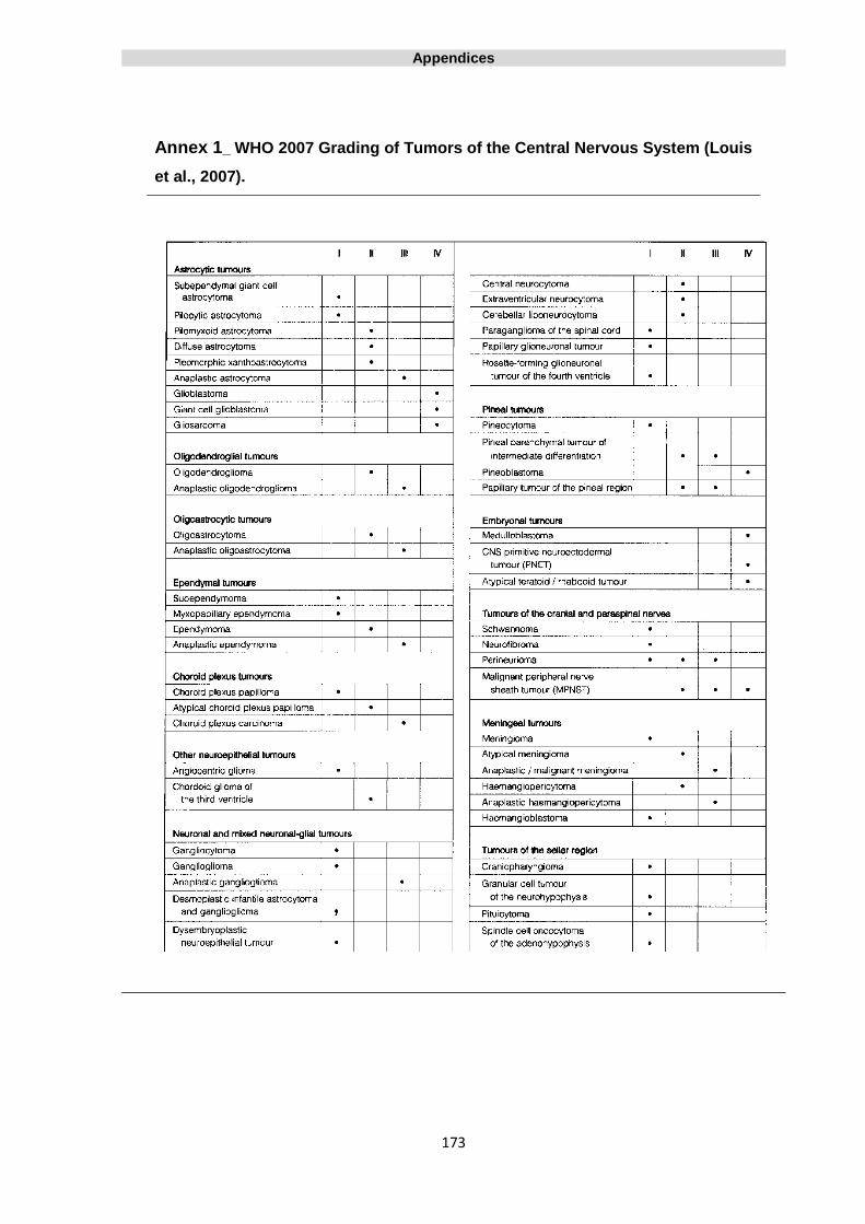
Appendices
173
Annex 1_ WHO 2007 Grading of Tumors of the Central Nervous System (Louis
et al., 2007).

174

175

176
Contactos:
Universidade de Évora
Instituto de Investigação e Formação Avançada - IIFA
Palácio do Vimioso | Largo Marquês de Marialva, Apart. 94
7002-554 Évora | Portugal
Tel: (+351) 266 706 581
Fax: (+351) 266 744 677
email: [email protected]


![Promoter hypermethylation profiling of distant breast ... · phenotype of distant breast cancer metastases [14–16]. Extensive knowledge of the hypermethylation status of tumor suppressor](https://static.fdocuments.net/doc/165x107/5d21f00788c993722e8c67ea/promoter-hypermethylation-profiling-of-distant-breast-phenotype-of-distant.jpg)