Aberrant cerebro-cerebellar functional connectivity and its … · 2019-08-19 · Furthermore, the...
57
저작자표시-비영리-변경금지 2.0 대한민국 이용자는 아래의 조건을 따르는 경우에 한하여 자유롭게 l 이 저작물을 복제, 배포, 전송, 전시, 공연 및 방송할 수 있습니다. 다음과 같은 조건을 따라야 합니다: l 귀하는, 이 저작물의 재이용이나 배포의 경우, 이 저작물에 적용된 이용허락조건 을 명확하게 나타내어야 합니다. l 저작권자로부터 별도의 허가를 받으면 이러한 조건들은 적용되지 않습니다. 저작권법에 따른 이용자의 권리는 위의 내용에 의하여 영향을 받지 않습니다. 이것은 이용허락규약 ( Legal Code) 을 이해하기 쉽게 요약한 것입니다. Disclaimer 저작자표시. 귀하는 원저작자를 표시하여야 합니다. 비영리. 귀하는 이 저작물을 영리 목적으로 이용할 수 없습니다. 변경금지. 귀하는 이 저작물을 개작, 변형 또는 가공할 수 없습니다.
Transcript of Aberrant cerebro-cerebellar functional connectivity and its … · 2019-08-19 · Furthermore, the...

저 시-비 리- 경 지 2.0 한민
는 아래 조건 르는 경 에 한하여 게
l 저 물 복제, 포, 전송, 전시, 공연 송할 수 습니다.
다 과 같 조건 라야 합니다:
l 하는, 저 물 나 포 경 , 저 물에 적 된 허락조건 명확하게 나타내어야 합니다.
l 저 터 허가를 면 러한 조건들 적 되지 않습니다.
저 에 른 리는 내 에 하여 향 지 않습니다.
것 허락규약(Legal Code) 해하 쉽게 약한 것 니다.
Disclaimer
저 시. 하는 원저 를 시하여야 합니다.
비 리. 하는 저 물 리 목적 할 수 없습니다.
경 지. 하는 저 물 개 , 형 또는 가공할 수 없습니다.
Aberrant cerebro-cerebellar functional connectivity and its phenomenological
manifestation in individuals at ultra-high risk for psychosis and
with first-episode schizophrenia
Minji Bang
Department of Medicine
The Graduate School, Yonsei University
Aberrant cerebro-cerebellar functional connectivity and its phenomenological
manifestation in individuals at ultra-high risk for psychosis and
with first-episode schizophrenia
Directed by Professor Suk Kyoon An
The Doctoral Dissertation submitted to the Department of Medicine, the Graduate School of Yonsei University
in partial fulfillment of the requirement for the degree of Doctor of Philosophy
Minji Bang
December 2016

ACKNOWLEDGEMENTS
I would like to express my deep gratitude to everyone who
encouraged me in my work.
First of all, my most sincere thanks go to Professor Suk
Kyoon An, whose kind encouragement and willing assistance
helped bring this thesis to fruition. His deep insight and
enthusiasm for the truth have always inspired me to go further.
I also wish to record my thanks to Professor Dong Goo Kim,
Jun Soo Kwon, Hae-Jeong Park, and Sang Chul Chong for their
thoughtful comments and guidance in writing this thesis. Also,
special thanks to my colleagues in the Section of Self, Affect,
and Neuroscience, Institute of Behavioral Science in Medicine,
who provided various kinds of help and support to me.
Finally, I would like to thank my family members. I could
not have done it without them.
Minji Bang
<TABLE OF CONTENTS>
ABSTRACT ······································································ 1
I. INTRODUCTION ···························································· 3
II. MATERIALS AND METHODS ·········································· 8
1. Participants ································································· 8
2. Assessment of ipseity disturbances ····································· 11
3. Assessment of conversion to overt psychosis in UHR
participants ································································ 11
4. Neuroimaging data acquisition ········································· 12
5. Image preprocessing ······················································ 13
6. Connectivity analysis ····················································· 14
7. Statistical analysis ························································ 16
III. RESULTS ································································· 17
1. Functional connectivity of the cerebellar seeds ······················· 17
2. Group differences in the cerebro-cerebellar functional
connectivity ································································ 18
3. Associations with ipseity disturbances ································· 22
IV. DISCUSSION ······························································ 25
V. CONCLUSION ····························································· 32

REFERENCES ·································································· 33
APPENDIX ······································································ 45
ABSTRACT (IN KOREAN) ·················································· 46
LIST OF FIGURES
Figure 1. Resting-state functional connectivity of the
cerebellar default mode network seeds ·············· 17
Figure 2. (A) Cerebral regions showing significant group
differences in the ANCOVA model
(B) Post-hoc comparisons within significant clusters
among the four groups ··························· 19
Figure 3. Dot plots of the cerebellar functional connectivity
with significant cerebral clusters in the entire group
of UHR participants ··································· 21
Figure 4. The mean FCQ-S scores of the four groups ········ 22
Figure 5. Cerebral regions showing significant correlations
with the FCQ-S scores in the respective groups of
UHR and FES participants ···························· 23
LIST OF TABLES
Table 1. Demographic and clinical profiles of the
participants ··············································· 10
Table 2. MNI coordinates of cerebral regions showing
significantly decreased functional connectivity with
the cerebellum in the ANCOVA model ·············· 20
Table 3. MNI coordinates of cerebral regions showing
significant correlations between the cerebro-
cerebellar connectivity and the FCQ-S scores in the
respective groups of UHR and FES participants ···· 24
1
ABSTRACT
Aberrant cerebro-cerebellar functional connectivity
and its phenomenological manifestation in individuals
at ultra-high risk for psychosis and with first-episode schizophrenia
Minji Bang
Department of Medicine
The Graduate School, Yonsei University
(Directed by Professor Suk Kyoon An)
From a phenomenological perspective, ipseity disturbances have been
suggested as the fundamental psychopathology of schizophrenia; however, the
underlying neural mechanisms remain unclear. Here, we investigated the cerebro-
cerebellar default mode network (DMN) connectivity during rest and its
association with ipseity disturbances in individuals at ultra-high risk (UHR) for
psychosis and patients with first-episode schizophrenia (FES). Thirty-three UHR
individuals, including eight converters, 18 FES patients, and 56 healthy controls
underwent functional magnetic resonance imaging during rest at baseline. All
UHR participants were assessed for conversion to overt psychosis every month
2
during a follow-up period. Seed-based functional connectivity analyses using the
cerebellar DMN seeds were performed, followed by between-group comparisons.
Correlation analyses were conducted to examine the relationship between the
cerebro-cerebellar functional connectivity and the self-reported severity of ipseity
disturbances in the UHR and FES groups, respectively. Compared to healthy
controls, converted UHR and FES participants showed decreased functional
connectivity between the cerebellum and the right anterior prefrontal cortex, left
presupplementary motor area, and precuneus, whereas UHR participants without
conversion showed comparable functional connectivity to healthy controls.
Furthermore, the degree of the cerebellar functional connectivity with several
cerebral regions was significantly associated with more-severe ipseity
disturbances in the respective groups of UHR and FES participants. Our findings
support the notion that schizophrenia is a disorder of ipseity, which appears to be
associated with aberrant cerebro-cerebellar functional connectivity. These results
also imply that the underlying neuropathological changes associated with ipseity
disturbances can be detected in UHR individuals who will later develop
schizophrenia spectrum psychosis. Such aberrations in cerebro-cerebellar
networks may help to predict future psychosis in UHR individuals, thus providing
an opportunity to apply early interventions to prevent the onset of schizophrenia
spectrum psychosis.
------------------------------------------------------------------------------------------------ Key words: schizophrenia, ultra-high risk for psychosis, cerebellum, default
mode network, ipseity, phenomenology
3
Aberrant cerebro-cerebellar functional connectivity
and its phenomenological manifestation in individuals
at ultra-high risk for psychosis and with first-episode schizophrenia
Minji Bang
Department of Medicine
The Graduate School, Yonsei University
(Directed by Professor Suk Kyoon An)
I. INTRODUCTION
The current diagnosis of schizophrenia in the Diagnostic and Statistical
Manual of Mental Disorders (DSM) is based on a description of the observable
and measurable features of psychopathology. Nevertheless, this diagnosis does
not capture the true nature of schizophrenia, since the DSM only describes the
minimum criteria, which are reliable but do not have guaranteed validity.1
Over the past few years, studies have begun to explore subjective self-
experiences to determine the fundamental psychopathology of schizophrenia
from a phenomenological perspective.2-5 Subjective experiences of the self, also
known as the minimal self or ipseity, constitute the most basic levels of our
4
experiences, which normally remain “in the tacit background” of conscious
awareness.6-8 However, once ipseity is disturbed, individuals no longer
experience themselves or their surroundings in a usual way. These anomalous
changes are conceptualized as hyperreflexivity (heightened awareness toward
some aspects of oneself that normally remain pre-reflective) and diminished self-
presence (decreased feeling of being a subject of everyday experiences), which
lead to disruptions in the cognitive-perceptual experiential field.6,7 Such
anomalous self-experiences have been found to be selectively aggregated in
schizophrenia spectrum disorders,9-12 and already present in the prodrome of
schizophrenia.13-15 Moreover, higher levels of anomalous self-experiences
reportedly predict diagnostic conversion to overt psychosis in genetic16 and
clinical high-risk individuals.15 These findings suggest that ipseity disturbances
are a phenomenological manifestation of the core pathogenesis of schizophrenia.
Nonetheless, the neural mechanisms responsible for the anomalous self-
experiences currently remain unclear.
Recent neuroimaging studies have demonstrated that the cortical midline
structures are functionally relevant to self-referential processing.17 These areas
substantially overlap with the default mode network (DMN), which is active
when attention is allocated to intrinsic feelings and thoughts.18 The DMN is
known to mediate a broad range of self-related mental operations: integration of
self-relevant information from the inner and outer world,19 self-referential mental
simulations and judgments,20 autobiographical memory retrieval,21 self-other
representations,22 and perspective taking of others.21,23 Given this relevance of the
DMN to self-referential mental activities, the DMN has been postulated to be a
5
neural correlate of ipseity.24 However, mental operations mediated by the DMN
are not purely pre-reflective but instead, are accompanied by higher cognitive
aspects of the self. Because the sense of ipseity is immediately and automatically
given to oneself without conscious awareness,25 it is necessary to consider a more
implicit mechanism of self-referential processing to investigate the neural
underpinning of ipseity itself.
The sense of ipseity is closely related to the sense of agency and the sense
of ownership,6,25 which are generated by comparing the predicted outcome of our
intention to the actual feedback from movement.26,27 Based on neuroimaging
findings,28,29 the cerebellum is considered the center of this comparator system,
operating as an internal model that represents the implicit dynamics of
movements and thoughts.30,31 As the internal model becomes more sophisticated
throughout our lives, motor and cognitive schemas become more stable, thus
enabling us to experience a pre-reflective feeling of control over our own body
and mind.32 Given that ipseity is a solid referential point in the field of first-person
experiences, the cerebellum has been hypothesized to play an important role in
the sense of ipseity.33 Therefore, investigating cerebro-cerebellar connectivity
within the DMN may better disclose the pre-reflective aspect of the self, which
appears to be mediated not only via the cerebral DMN but also through
coordination with the cerebellum.
Aberrant functional connectivity in the cerebral DMN has been repeatedly
found in patients with schizophrenia; however, the findings vary across studies.
Although the most common finding is hyperconnectivity of the cerebral
DMN,18,34 multimodal neuroimaging studies have identified both structural and
6
functional disruptions of the cerebral DMN in schizophrenia.35,36 Regarding the
cerebro-cerebellar DMN, decreased connectivity was found in patients with
chronic schizophrenia,37 while a mixed pattern of connectivity was also found in
patients with first-episode schizophrenia.38,39 Based on the functional relevance
of the cerebral DMN to self-referential processing, the above findings have been
interpreted as reflecting a state of preoccupation with one’s own inner world.18
However, given the functional organization between the cerebral DMN and the
cerebellum as an internal model,40 aberrant cerebro-cerebellar connectivity may
be related to the disturbances of pre-reflective self-experiences in schizophrenia,
although the link between them has not yet been empirically examined. Since it
is known that ipseity disturbances can be detected more precisely prior to
psychotic elaborations,5,41 examining the disturbances in individuals at ultra-high
risk (UHR) for psychosis may help to reveal the true gestalt of schizophrenia as
a disorder of ipseity. Moreover, UHR individuals have an advantage of being
relatively less contaminated by secondary morbidity due to advances illness, as
well as FES patients do. Although some recent studies have reported that the
functional connectivity of the cerebro-cerebellar DMN was increased in genetic
high-risk38,39 and UHR42 individuals compared to that in healthy individuals,
differences between converters and non-converters were not investigated.
Therefore, further investigations are needed to specify what neurobiological
mechanisms are related to the conversion risk in UHR individuals and their
relevance to ipseity disturbances from a phenomenological perspective.
The aim of the present study was to investigate the cerebro-cerebellar DMN
functional connectivity and its association to ipseity disturbances across healthy
7
controls, UHR individuals with and without conversion to overt psychosis, and
FES patients. A resting-state functional magnetic resonance imaging (fMRI)
paradigm was chosen to evaluate implicit and pre-reflective experiences of the
self in the absence of specific thoughts or external stimuli. We first hypothesized
that UHR individuals and FES patients would show aberrant functional
connectivity within the cerebro-cerebellar DMN when compared to healthy
controls. More specifically, converted UHR individuals were expected to show a
similar pattern of aberrant functional connectivity with schizophrenia, whereas
non-converters were not. Finally, we predicted that aberrant cerebro-cerebellar
DMN connectivity would be associated with the severity of ipseity disturbances
in UHR individuals and FES patients.
8
II. MATERIALS AND METHODS
1. Participants
Thirty-three UHR individuals, including eight converters, 18 patients with
FES and 56 healthy controls (HCs) participated in the present study. UHR and
FES participants were recruited from the Clinic FORYOU of the Green Program
for Recognition and Prevention of Early Psychosis (GRAPE) project at
Severance Hospital of the Yonsei University Health System in Seoul, Republic
of Korea.43,44 The present study was reviewed and approved by the Institutional
Review Board of Severance Hospital of the Yonsei University Health System.
All participants, including a parent for participants who were younger than 18
years, provided written informed consent after the procedures had been fully
explained.
All participants were assessed for psychiatric disorders using the Structural
Clinical Interview for DSM-IV Axis I Disorders (SCID-IV).45,46 Participants with
a current or past diagnosis of neurological disorders, traumatic brain injury, or
intellectual disability (IQ < 70) were excluded. After a diagnostic interview, HCs
with any current or past history of psychiatric illness and UHR individuals with
a current or past history of major psychiatric disorders with psychotic features
were also excluded. Patients with schizophrenia were diagnosed based on the
DSM-IV criteria.47 First episode was defined as a new onset of overt psychotic
symptoms, which are sufficient in duration and severity for the diagnosis of
schizophrenia. Only patients with less than 36 months of illness were included in
the present study; the mean duration of illness in FES participants was 16.4
months (standard deviation [SD] = 11.6 months, median = 16.0 months,
9
interquartile range [IQR] = 7.5−27.0 months, range: 1−36 months). The diagnosis
of UHR individuals was made based on the Structured Interview for Prodromal
Syndromes (SIPS; version 4.0).48 Each UHR participant met at least one of the
following three criteria: (1) attenuated positive prodromal syndrome (APPS; n =
32), (2) brief intermittent psychotic syndrome (BIPS; n = 5) and (3) genetic risk
and deterioration syndrome (GRDS; n = 6).
The clinical characteristics and severity of symptoms in UHR and FES
participants were assessed using the Scale for the Assessment of Negative
Symptoms (SANS)49 and Scale for the Assessment of Positive Symptoms
(SAPS).50 One converter and eight non-converters in UHR participants and all
FES participants were taking atypical antipsychotic medications at the time of
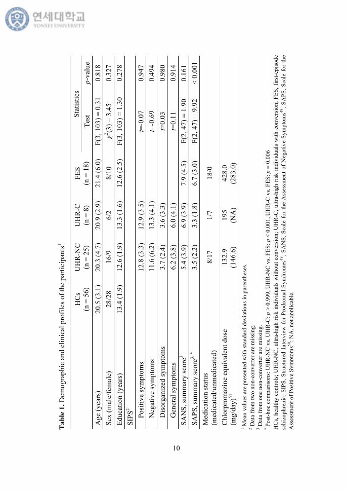
fMRI scanning. Table 1 shows the demographic and clinical profiles of the
participants.51
10
Tab
le 1
. Dem
ogra
phic
and
clin
ical
pro
files
of t
he p
artic
ipan
ts1
H
Cs
(n =
56)
U
HR
-NC
(n
= 2
5)
UH
R-C
(n
= 8
) FE
S (n
= 1
8)
Stat
istic
s Te
st
p-va
lue
Age
(yea
rs)
20.5
(3.1
) 20
.3 (4
.7)
20.9
(2.9
) 21
.4 (6
.0)
F(3,
103
) = 0
.31
0.81
8 Se
x (m
ale/
fem
ale)
28
/28
16/9
6/
2 8/
10
𝜒2 (3) =
3.4
5 0.
327
Educ
atio
n (y
ears
) 13
.4 (1
.9)
12.6
(1.9
) 13
.3 (1
.6)
12.6
(2.5
) F(
3, 1
03) =
1.3
0 0.
278
SIPS
2
Posi
tive
sym
ptom
s
12.8
(3.3
) 12
.9 (3
.5)
t=
-0.0
7 0.
947
N
egat
ive
sym
ptom
s
11.6
(6.2
) 13
.3 (4
.1)
t=
-0.6
9 0.
494
D
isor
gani
zed
sym
ptom
s
3.7
(2.4
) 3.
6 (3
.3)
t=
0.03
0.
980
G
ener
al sy
mpt
oms
6.
2 (3
.8)
6.0
(4.1
)
t=0.
11
0.91
4 SA
NS,
sum
mar
y sc
ore3
5.
4 (3
.9)
6.9
(3.9
) 7.
9 (4
.5)
F(2,
47)
= 1
.90
0.16
1 SA
PS, s
umm
ary
scor
e3, *
3.5
(2.2
) 3.
3 (1
.8)
6.7
(3.0
) F(
2, 4
7) =
9.9
2 <
0.00
1 M
edic
atio
n st
atus
(m
edic
ated
/unm
edic
ated
)
8/17
1/
7 18
/0
Chl
orpr
omaz
ine
equi
vale
nt d
ose
(mg/
day)
51
13
2.9
(146
.6)
195
(NA
) 42
8.0
(283
.0)
1 Mea
n va
lues
are
pre
sent
ed w
ith st
anda
rd d
evia
tions
in p
aren
thes
es.
2 Dat
a fro
m tw
o no
n-co
nver
ter a
re m
issi
ng.
3 Dat
a fro
m o
ne n
on-c
onve
rter a
re m
issi
ng.
* Po
st-ho
c co
mpa
rison
s: U
HR
-NC
vs.
UH
R-C
: p >
0.9
99, U
HR
-NC
vs.
FES:
p <
0.0
01, U
HR
-C v
s. FE
S: p
= 0
.006
H
Cs,
heal
thy
cont
rols
; UH
R-N
C, u
ltra-
high
risk
indi
vidu
als
with
out c
onve
rsio
n; U
HR
-C, u
ltra-
high
risk
indi
vidu
als
with
con
vers
ion;
FES
, firs
t-epi
sode
sc
hizo
phre
nia;
SIP
S, S
truct
ured
Int
ervi
ew f
or P
rodr
omal
Syn
drom
es48
; SA
NS,
Sca
le f
or th
e A
sses
smen
t of N
egat
ive
Sym
ptom
s49; S
APS
, Sca
le f
or th
e A
sses
smen
t of P
ositi
ve S
ympt
oms50
; NA
, not
app
licab
le.
11
2. Assessment of ipseity disturbances
To assess the ipseity disturbances, all participants completed the eight-item
schizophrenia-specific subscale of the Frankfurt Complaint Questionnaire (FCQ-
S; see Appendix), which has been shown to be diagnostically specific to
schizophrenia.52 Although the FCQ is primarily utilized for assessing basic
symptoms, that is, the subtle and subjectively experienced disturbances in affect,
cognition, perception, and motor abilities,5 these basic symptoms largely overlap
with anomalous self-experiences and thus have been used to index the severity of
the ipseity disturbances in previous studies.8,53 The statements of the FCQ-S can
be conceptually divided into two categories: “sensomotoric irritations (items 11,
14, 63, and 81)” and “avoidance behavior (items 15, 90, 93, and 94).”52 The items
describing sensomotoric irritations may correspond to operative hyperreflexivity
which refers to a sudden conscious awareness of phenomena that normally
remain tacit and pre-reflective.6 On the other hand, the items on avoidance
behavior seem to be a behavioral counterpart of diminished self-presence, which
refers to a decreased feeling of being affected by the cognitive-perceptual world.6
Because such disturbances are no longer implicit in the experiential field, they
can be detected by self-reported ratings. Participants answered each item with
either yes (1) or no (0), and the total score was calculated as the sum of the eight
items, which ranged from 0 to 8. The internal consistency (Cronbach’s 𝛼) of the
FCQ-S was 0.737 in the present study.
3. Assessment of conversion to overt psychosis in UHR participants
UHR participants were re-assessed every month during a follow-up period
12
(median = 14.2 months, IQR = 7.1−28.9 months, range: 1−94.0 months) to
determine whether they had developed schizophrenia spectrum psychosis. There
were included six UHR participants, who were followed up for less than six
months (all non-converters). Conversion to overt psychosis was confirmed
according to the DSM-IV criteria for psychotic disorders (schizophrenia,
schizoaffective disorder, delusional disorder, and psychotic disorder not
otherwise specified [NOS]),47 which corresponded to the criteria of the Presence
of Psychotic Syndrome (POPS) from the SIPS.48
Eight out of 33 UHR participants (24.2%) were found to develop
schizophrenia spectrum psychosis. Five participants were diagnosed with
schizophrenia, one with delusional disorder, and the other two with psychotic
disorder NOS. Among the eight converters, four participants converted within 12
months of enrolment, three participants converted between 12 and 24 months
after enrolment, and one participant converted 27.1 months after enrolment. The
Kaplan-Meier estimates of conversion risk are 31.0% (95% confidence interval
[CI]: 11.4–50.6%) at two years and 38.7% (95% CI: 16.2–61.2%) at the end of
the last follow-up.
4. Neuroimaging data acquisition
Functional and structural MRI data were acquired using a three-Tesla
scanner (Intera Achieva; Philips Medical System, Best, The Netherlands). All
participants underwent a 5.5-min resting-state scan, during which they were
instructed to stay quietly with their eyes closed, without moving, sleeping, or
focusing on any specific thought. Blood oxygen level-dependent (BOLD) images
13
were obtained using a T2*-weighted gradient echo-planar imaging (EPI)
sequence (repetition time [TR] = 2000 ms; echo time [TE] = 30 ms; flip angle =
90˚; matrix = 80 × 80; voxel size = 2.75 × 2.75 × 4 mm3; field-of-view [FOV] =
220 mm; 31 interleaved slices without slice gap). High-resolution structural
images were subsequently acquired using a three-dimensional T1-weighted turbo
field echo (TFE) sequence (TR = 9.7 ms; TE = 4.6 ms; flip angle = 8˚; matrix =
256 × 256; voxel size = 0.859 × 0.859 × 1.2 mm3; FOV = 220 mm; 180 slices).
5. Image preprocessing
Resting-state functional images were preprocessed using Statistical
Parametric Mapping (SPM12; Wellcome Trust Center for Neuroimaging,
London, UK), implemented in MATLAB (Mathworks Inc., Natick, MA, USA).
The first five images of each participant were discarded to ensure steady-state
magnetization. The remaining images underwent standard preprocessing steps,
including correction of acquisition time delays between different slices,
correction for head motion by realigning all consecutive volumes to the first
image of the session, and co-registration of T1-weighted images to the first EPI
data using the non-linear registration algorithm. The nonlinear co-registration
algorithm was used to minimize image distortions in the EPI sequence by
maximizing normalized mutual information between the first EPI and T1-
weighted images over second-order B-spline basis functions. Co-registered T1-
images were used to spatially normalize functional EPI into Montreal
Neurological Institute (MNI) template space by using a non-linear transformation
in SPM12. The functional volumes were resampled to a voxel dimension of 2 ×
14
2 × 2 mm3.
The fMRI time courses were processed by (1) regressing out the effects of
six rigid motions and their derivatives, and three principal components from the
white matter and the cerebrospinal fluid mask, which were segmented using
SPM12; (2) despiking based on the median absolute deviation; and (3) high-pass
filtering up to 0.009 Hz.54-57 Since it is known that serious motion effects can be
a major confounder in the functional connectivity analysis,54,58-62 despiking, as
one of the censoring procedures,59 was performed to mitigate such motion effects.
To describe the amount of movements in our data, the framewise displacement
(FD) that measures the sum of the absolute values of the derivatives from the
translational and rotational motion estimates were calculated for each participant.
There was no significant difference in the FD values among the four groups (FES:
mean ± SD = 0.23 ± 0.18; UHR-C: mean ± SD = 0.22 ± 0.04; UHR-NC: mean ±
SD = 0.23 ± 0.09; HCs: mean ± SD = 0.22 ± 0.07; F[3, 103] = 0.11, p = 0.955).
Low-pass filtering was not applied since a growing number of studies have
reported information over the 0.1 Hz.63-66 All procedures were performed using
in-house multimodal brain network analysis software, MNET (Multimodal brain
NETwork analysis toolbox; Yonsei University, Seoul, Republic of Korea;
http://neuroimage.yonsei.ac.kr/mnet).
6. Connectivity analysis
A seed-based functional connectivity map was calculated for each
participants using cerebellar seeds from left (MNI: -32, -79, -31) and right Crus
I (MNI: 29, -78, -32), which have been found to be functionally connected to the
15
cerebral DMN in 1,000 healthy individuals.40 Seed regions were defined as 2-mm
radius spheres, and the mean time series of each region was extracted. Correlation
maps for each participant were created by calculating the Pearson’s correlation
coefficients between the time series of the seed regions and that of other voxels
in the brain. The resulting correlation maps were converted to z-values using
Fisher’s r-to-z transformation. To obtain an overall functional connectivity of the
cerebro-cerebellar DMN, z-maps from each cerebellar seed were averaged,
resulting in one mean connectivity map for each participant. Before the statistical
analyses, data were smoothed using a Gaussian filter with a 6-mm full-width at
half-maximum (FWHM).
One-sample t-tests were respectively conducted for HCs, UHR participants
with conversion (UHR-C) and without conversion (UHR-NC), and FES
participants to identify significant correlations with the cerebellar DMN seeds.
Explicit masks were created by applying a voxel-level family-wise error (FWE)
rate of p < 0.05 to correct for multiple comparisons over the whole brain. By
combining the masks of each group, one single explicit mask that included all of
the significant voxels from the four groups was created.
To examine significant between-group differences, the functional
connectivity map for each participant was entered into an analysis of covariance
(ANCOVA) model with age and sex as covariates. Statistical significance was
defined by the clusters surviving the voxel-level threshold of p < 0.005
(uncorrected) and the cluster-level extent threshold of p < 0.05 (cluster size ≥ 196
voxels) generated with Monte Carlo simulations using the 3dClustSim program
(January 2016 version), implemented in Analysis of Functional NeuroImages
16
software (AFNI; the National Institute of Mental Health, Bethesda, MD, USA;
https://afni.nimh.nih.gov/afni). To minimize the problems related to the
underestimation of the group smoothness,67 the group residuals from the general
linear model were used to calculate the group smoothness. Post-hoc tests for each
significant cluster were corrected for multiple comparisons using the Bonferroni
correction. To examine the association between the functional connectivity maps
and the FCQ-S scores, correlation analyses were performed for the UHR and FES
groups, respectively. Three UHR-NC and 2 FES participants were excluded from
these analyses owing to missing data for the FCQ-S. Age and sex were added as
covariates in the analyses. Statistical significance was set using the same methods
as mentioned above (single voxel p < 0.005, cluster size ≥ 277 voxels [two-
tailed]).
7. Statistical analysis
The demographic and clinical profiles of the HCs, UHR-NC, UHR-C, and
FES participants were compared using one-way analyses of variance,
independent t-tests, and chi-square tests. Post-hoc analyses were adjusted for
multiple comparisons with the Bonferroni correction. Statistical significance was
set at p < 0.05, and a p-value greater than 0.05 but less than 0.10 was accepted to
indicate a trend toward statistical significance. All statistical tests were performed
using the Statistical Package for the Social Sciences (SPSS23; IBM Corp.,
Armonk, NY, USA).
17
III. RESULTS
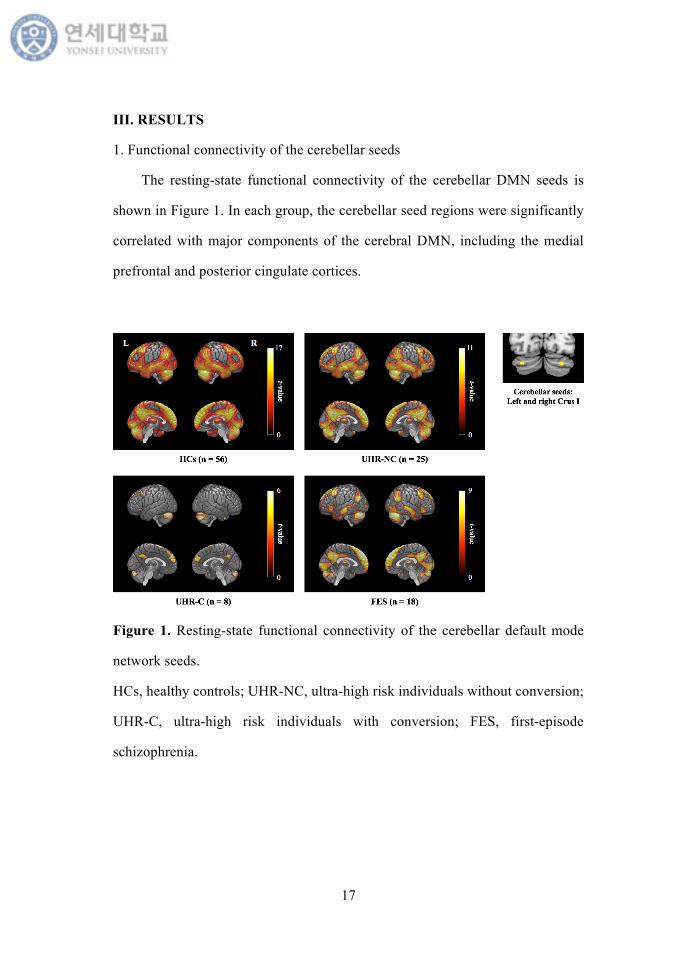
1. Functional connectivity of the cerebellar seeds
The resting-state functional connectivity of the cerebellar DMN seeds is
shown in Figure 1. In each group, the cerebellar seed regions were significantly
correlated with major components of the cerebral DMN, including the medial
prefrontal and posterior cingulate cortices.
Figure 1. Resting-state functional connectivity of the cerebellar default mode
network seeds.
HCs, healthy controls; UHR-NC, ultra-high risk individuals without conversion;
UHR-C, ultra-high risk individuals with conversion; FES, first-episode
schizophrenia.
18
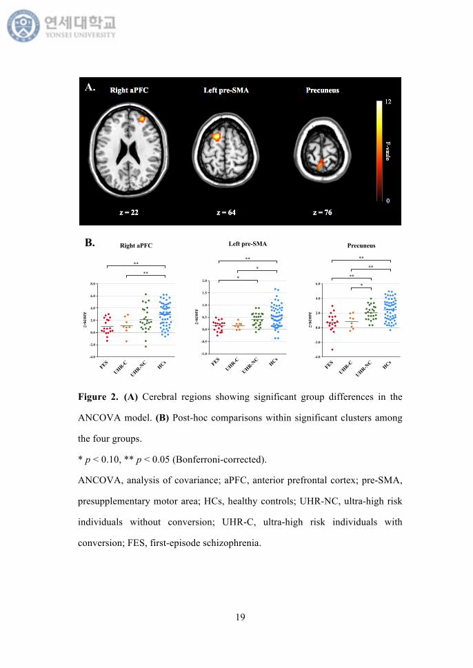
2. Group differences in the cerebro-cerebellar functional connectivity
An ANCOVA revealed significant differences in functional connectivity
between the cerebellum and several cerebral regions including the right anterior
prefrontal cortex (aPFC), left presupplementary motor area (pre-SMA), and
precuneus (Figure 2A). Post-hoc analyses of the regions identified in the
ANCOVA were shown in Figure 2B. A decreasing tendency in the cerebellar
functional connectivity with these regions was noted from HCs to UHR-NC, to
UHR-C, and then to FES participants.
Compared to HCs, significantly decreased cerebellar functional connectivity
with the right aPFC and precuneus was found in both UHR-C (right aPFC: p =
0.039; precuneus: p = 0.005) and FES participants (right aPFC: p < 0.001;
precuneus: p < 0.001). Compared to UHR-NC participants, FES participants also
showed significantly reduced cerebellar connectivity with the precuneus (p =
0.007), while UHR-C participants showed a trend-level reduction (p = 0.092).
The functional connectivity between the cerebellum and the left pre-SMA was
significantly decreased in FES participants, compared to that in HCs (p < 0.001).
Besides, there were trend-level differences between UHR-C participants and HCs
(p = 0.090), and between FES and UHR-NC participants (p = 0.067). Table 2 lists
the cerebral regions showing significant differences in functional connectivity
with the cerebellum among the four groups.
19
Figure 2. (A) Cerebral regions showing significant group differences in the
ANCOVA model. (B) Post-hoc comparisons within significant clusters among
the four groups.
* p < 0.10, ** p < 0.05 (Bonferroni-corrected).
ANCOVA, analysis of covariance; aPFC, anterior prefrontal cortex; pre-SMA,
presupplementary motor area; HCs, healthy controls; UHR-NC, ultra-high risk
individuals without conversion; UHR-C, ultra-high risk individuals with
conversion; FES, first-episode schizophrenia.
FES
UHR-C
UHR-NC
HCs-1.0
-0.5
0.0
0.5
1.0
1.5
2.0
z-sc
ore
Left pre-SMA
*
**
*
A.
B.
FES
UHR-C
UHR-NC
HCs-4.0
-2.0
0.0
2.0
4.0
6.0
8.0
z-sc
ore
Right aPFC
**
**
FES
UHR-C
UHR-NC
HCs-4.0
-2.0
0.0
2.0
4.0
6.0
z-sc
ore
Precuneus
**
**
**
*
20
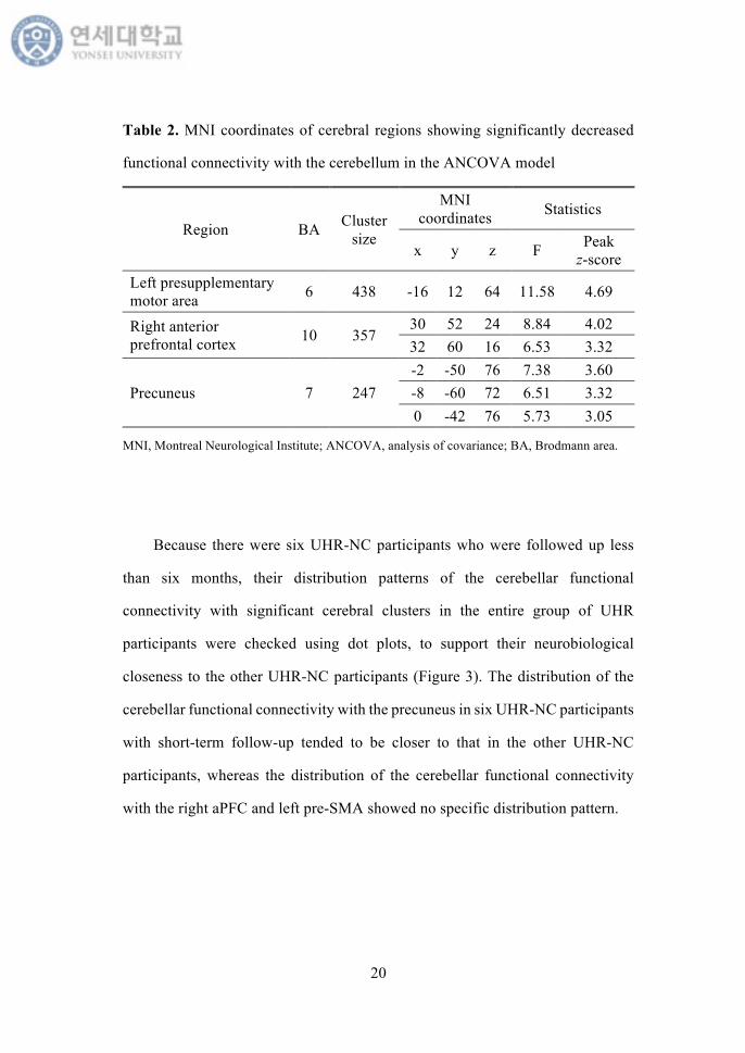
Table 2. MNI coordinates of cerebral regions showing significantly decreased
functional connectivity with the cerebellum in the ANCOVA model
Region BA Cluster size
MNI coordinates Statistics
x y z F Peak z-score
Left presupplementary motor area 6 438 -16 12 64 11.58 4.69
Right anterior prefrontal cortex 10 357
30 52 24 8.84 4.02 32 60 16 6.53 3.32
Precuneus 7 247 -2 -50 76 7.38 3.60 -8 -60 72 6.51 3.32 0 -42 76 5.73 3.05
MNI, Montreal Neurological Institute; ANCOVA, analysis of covariance; BA, Brodmann area..
Because there were six UHR-NC participants who were followed up less
than six months, their distribution patterns of the cerebellar functional
connectivity with significant cerebral clusters in the entire group of UHR
participants were checked using dot plots, to support their neurobiological
closeness to the other UHR-NC participants (Figure 3). The distribution of the
cerebellar functional connectivity with the precuneus in six UHR-NC participants
with short-term follow-up tended to be closer to that in the other UHR-NC
participants, whereas the distribution of the cerebellar functional connectivity
with the right aPFC and left pre-SMA showed no specific distribution pattern.

21
Figure 3. Dot plots of the cerebellar functional connectivity with significant
cerebral clusters in the entire group of UHR participants.
aPFC, anterior prefrontal cortex; pre-SMA, presupplementary motor area; UHR-
NC, ultra-high risk individuals without conversion; UHR-C, ultra-high risk
individuals with conversion.
22
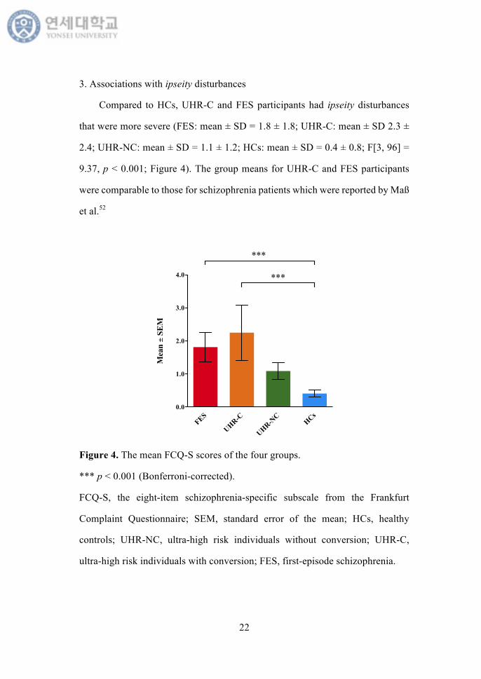
3. Associations with ipseity disturbances
Compared to HCs, UHR-C and FES participants had ipseity disturbances
that were more severe (FES: mean ± SD = 1.8 ± 1.8; UHR-C: mean ± SD 2.3 ±
2.4; UHR-NC: mean ± SD = 1.1 ± 1.2; HCs: mean ± SD = 0.4 ± 0.8; F[3, 96] =
9.37, p < 0.001; Figure 4). The group means for UHR-C and FES participants
were comparable to those for schizophrenia patients which were reported by Maß
et al.52
Figure 4. The mean FCQ-S scores of the four groups.
*** p < 0.001 (Bonferroni-corrected).
FCQ-S, the eight-item schizophrenia-specific subscale from the Frankfurt
Complaint Questionnaire; SEM, standard error of the mean; HCs, healthy
controls; UHR-NC, ultra-high risk individuals without conversion; UHR-C,
ultra-high risk individuals with conversion; FES, first-episode schizophrenia.
FES
UHR-C
UHR-NC
HCs0.0
1.0
2.0
3.0
4.0
Mea
n ±
SEM
***
***
23
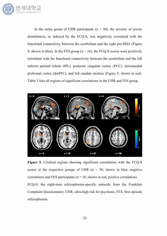
In the entire group of UHR participants (n = 30), the severity of ipseity
disturbances, as indexed by the FCQ-S, was negatively correlated with the
functional connectivity between the cerebellum and the right pre-SMA (Figure
4; shown in blue). In the FES group (n = 16), the FCQ-S scores were positively
correlated with the functional connectivity between the cerebellum and the left
inferior parietal lobule (IPL), posterior cingulate cortex (PCC), dorsomedial
prefrontal cortex (dmPFC), and left caudate nucleus (Figure 5; shown in red).
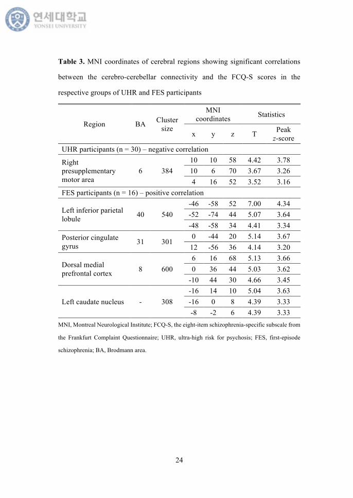
Table 3 lists all regions of significant correlations in the UHR and FES group.
Figure 5. Cerebral regions showing significant correlations with the FCQ-S
scores in the respective groups of UHR (n = 30; shown in blue; negative
correlation) and FES participants (n = 16; shown in red; positive correlation).
FCQ-S, the eight-item schizophrenia-specific subscale from the Frankfurt
Complaint Questionnaire; UHR, ultra-high risk for psychosis; FES, first-episode
schizophrenia.
24
Table 3. MNI coordinates of cerebral regions showing significant correlations
between the cerebro-cerebellar connectivity and the FCQ-S scores in the
respective groups of UHR and FES participants
Region BA Cluster size
MNI coordinates Statistics
x y z T Peak z-score
UHR participants (n = 30) – negative correlation
Right presupplementary motor area
6 384 10 10 58 4.42 3.78 10 6 70 3.67 3.26 4 16 52 3.52 3.16
FES participants (n = 16) – positive correlation
Left inferior parietal lobule 40 540
-46 -58 52 7.00 4.34 -52 -74 44 5.07 3.64 -48 -58 34 4.41 3.34
Posterior cingulate gyrus 31 301
0 -44 20 5.14 3.67 12 -56 36 4.14 3.20
Dorsal medial prefrontal cortex 8 600
6 16 68 5.13 3.66 0 36 44 5.03 3.62
-10 44 30 4.66 3.45
Left caudate nucleus - 308 -16 14 10 5.04 3.63 -16 0 8 4.39 3.33 -8 -2 6 4.39 3.33
MNI, Montreal Neurological Institute; FCQ-S, the eight-item schizophrenia-specific subscale from
the Frankfurt Complaint Questionnaire; UHR, ultra-high risk for psychosis; FES, first-episode
schizophrenia; BA, Brodmann area.
25
IV. DISCUSSION
To our best knowledge, this is the first study to investigate resting-state
functional connectivity of the cerebro-cerebellar DMN and its association with
ipseity disturbances in UHR and FES participants. Compared to HCs, UHR-C
and FES participants showed significantly weaker functional connectivity
between the cerebellum and the right aPFC, left pre-SMA, and precuneus,
whereas UHR-NC participants did not. Furthermore, the degree of the cerebellar
functional connectivity with several cerebral regions was significantly associated
with more-severe ipseity disturbances in the entire group of UHR participants and
FES group, respectively. These findings imply that aberrant cerebro-cerebellar
connectivity may be related to the fundamental pathogenesis of schizophrenia as
a disorder of ipseity.
Decreased functional connectivity between the cerebellar DMN seeds and
several cerebral regions including the right aPFC, left pre-SMA, and precuneus
was observed in UHR-C and FES participants compared to in HCs. This finding
is globally in line with previous studies showing functional disruptions between
the cerebellum and cerebral cortices in patients with schizophrenia.37,68-70 On the
other hand, our finding is partially inconsistent with previous studies showing not
only decreased, but also increased cerebro-cerebellar functional connectivity in
patients with first-episode schizophrenia.38,39 This discrepancy may be explained
by complex interactions between the developmental and pathogenetic dynamics
of the brain. Because the early pathogenesis of schizophrenia is closely
intertwined with the neurodevelopmental process of myelination and neural
pruning during adolescence and young adulthood,71,72 patterns of functional
26
connectivity in the prodromal and early stages of the disease may be
heterogeneous; however, reduced myelination and excessive pruning eventually
lead to disruptions of the functional organization of the brain in schizophrenia.73
It is noteworthy that UHR participants who later developed schizophrenia
spectrum psychosis showed a pattern of aberrant functional connectivity that was
similar to that identified in FES participants in the present study. There were
some reports of increased cerebral-cerebellar functional connectivity in healthy
siblings of patients with schizophrenia, which were proposed to reflect an
endophenotype of schizophrenia.38,39 However, it is also possible that these
findings of increased functional connectivity may reflect protective or
compensatory efforts for the underlying vulnerability of the brain to maintain its
normal function in genetic high risk individuals,74-76 given that a majority of
siblings never develop overt psychosis in life even with genetic vulnerabilities.
In addition, although one previous study reported increased cerebro-cerebellar
functional connectivity in UHR individuals,42 the functional connectivity of UHR
individuals with conversion and without conversion were not separately
investigated. Because it is known that there is a qualitative difference between
this two group of UHR individuals, our findings of decreased functional
connectivity between the cerebellum and the right aPFC, left pre-SMA, and
precuneus in UHR-C and FES participants may reflect the fundamental
pathogenetic changes, which is specifically related to the risk of schizophrenia.
Cerebellar functional connectivity with the precuneus was more decreased
in UHR-C and FES participants than it was in UHR-NC participants and HCs.
The precuneus, as a part of the posterior DMN, has been found to be involved in
27
various kinds of self-related processing including recollection of prior
experiences and attribution of them to oneself,77 self-related intentional causality
processing,78 and the experience of agency.79 Previous neuroimaging studies have
reported that both functional and structural abnormalities of the precuneus are
closely associated with lack of insight in patients with schizophrenia.80,81 Insight
in schizophrenia is conceptualized as consisting of two dimensions: awareness of
illness and attribution of illness.82 Based on previous findings in functional
neuroimaging studies,83,84 lack of insight into illness is mediated by the self-
reflection network which includes the precuneus as one of the core regions. In a
phenomenological context, it has been suggested that ipseity disturbances
destabilize those two dimensions of insight by distorting one’s first-person
perspective on experiences of oneself and its surroundings.85 Nonetheless, UHR
individuals usually hold full insight into their anomalous self-experiences, while
patients with schizophrenia do not.5 This implies that the impairment in
attributing one’s first-person experiences to oneself may facilitate the emergence
of overt psychotic symptoms. Then, the current finding of decreased functional
connectivity between the cerebellum and the precuneus in UHR-C and FES
participants may suggest that disruptions of the neurobiological mechanism
underlying insight accelerate psychotic elaborations of ipseity disturbances and
that the functional connectivity between the cerebellum and the precuneus
reflects a clinical risk state for schizophrenia.
The functional connectivity between the cerebellum and the left pre-SMA
was significantly decreased in FES participants, while UHR-C participants
showed a trend-level of decrease compared to HCs. Although this region is not a
28
major component of the DMN, the current finding is not surprising given the
functional association between the pre-SMA and the cerebellar Crus I.40 The pre-
SMA is known as a source of the “readiness potential,” which reflects the
preparation of a voluntary action prior to conscious intention.86 This readiness
signal contributes to the neural representation of the “desired state” of the self.27
On the other hand, the cerebellum helps produce error signals by comparing the
“desired state” and “predicted future state” from the internal model, and then
tacitly attributing agency to oneself when error signals are cancelled out.27,29,87
Through this series of processes, our intentions are linked to actual perceptual-
motor performance, thereby enhancing the integrated feeling of control.86 Hence,
the decreased cerebellar connectivity with the pre-SMA that was observed in FES
participants may implicate disruptions in this comparator system, perhaps
interrupting the ability to experience the self as one’s own agency in a pre-
reflective way.25-27
Decreased cerebellar connectivity with the right aPFC was found in UHR-C
and FES participants compared to in HCs. The aPFC is involved in higher-level
cognitive functions, particularly the explicit processing of internally generated
thoughts.88,89 Indeed, patients with schizophrenia exhibit difficulties in turning
their attention away from their own inner thoughts and feelings,18 although this
impairment has also found in other psychiatric disorders such as depressive
disorders.18 Thus, the current finding of decreased functional connectivity
between the cerebellum and the right aPFC in UHR-C and FES participants may
be a secondary reaction that occurs when people are engaged in introspection or
“reflective hyperreflexivity,” to cope with more fundamental anomalous self-
29
experiences.90 However, such efforts seem less effective and even maladaptive
for them, as reflected in the disrupted functional connectivity between the
cerebellum and the right aPFC.
The associations we identified between the cerebro-cerebellar functional
connectivity and the severity of ipseity disturbances in the respective groups of
UHR and FES participants further support the role of the cerebro-cerebellar
network in experiencing the pre-reflective aspect of the self. Correlation analyses
showed significant associations between the cerebro-cerebellar functional
connectivity and the FCQ-S scores in the UHR and FES groups respectively,
while the pattern of the associations was different according to the clinical state.
UHR participants showed a negative correlation between the FCQ-S scores and
the cerebellar functional connectivity with the right pre-SMA. This finding may
further support the functional link between the cerebellum and the pre-SMA in
experiencing the self as one’s own agency. On the other hand, FES participants
showed positive correlations between the FCQ-S scores and the cerebellar
functional connectivity with several cerebral regions including the dmPFC, PCC,
left IPL and left caudate nucleus. There has been some evidence that a functional
reorganization occurs after psychotic elaborations in patients with
schizophrenia.91-93 Although the cerebellar functional connectivity with those
regions was not significantly different in the between-group comparisons, it has
been widely known that those regions show functional aberrations in patients
with schizophrenia.18,34,94 In particular, the dmPFC and PCC are major functional
subdivisions of the cerebral DMN.19 They are mainly involved in a higher
cognitive level of self-related processing; the dmPFC is related to conflict

30
monitoring and response selection,95 while the PCC and adjacent precuneus are
engaged in autobiographical memory retrieval.96 The associations between the
FCQ-S scores and the cerebellar functional connectivity with the dmPFC and
PCC may directly reflect a maladaptive struggle to cope with ipseity disturbances
in FES participants. Unlike UHR participants who did not reach the threshold for
overt psychosis, FES participants seem to be engaged in hyper-reflection to find
out acceptable meanings of their anomalous experiences; such maladaptive
hyper-reflection may facilitate the emergence and formation of delusions and
hallucinations. Therefore, our findings on the associations between the cerebro-
cerebellar functional connectivity and ipseity disturbances in the respective
groups of UHR and FES participants may provide a link between the
phenomenology and neuropathogenesis of schizophrenia.
The present study has several limitations. First, because of the small sample
size of the UHR-C group (n = 8), a generalization of the current findings may be
limited. However, although a replication with more participants is required, the
present study provides a novel insight into the neural basis of ipseity disturbances
in schizophrenia. Second, the follow-up duration of UHR participants, which
ranged from 1 to 94.0 months, was various among them. Since some of the UHR-
NC participants (6 out of 25) were followed up for less than 6 months, there is a
chance that these individuals will convert to overt psychosis later. Nonetheless,
the cerebellar functional connectivity with the precuneus in UHR-NC
participants with short-term follow-up tended to be closer to that in the other
UHR-NC participants. Therefore, our main results would not be greatly affected
by the variation of follow-up periods, although it is necessary to be cautious when
31
interpreting the results. Third, some UHR (1 out of 8 UHR-C and 8 out of 25
UHR-NC) and all FES participants were taking atypical antipsychotic
medications. Some studies have suggested that antipsychotic medications may
reduce the functional connectivity of the brain in schizophrenia,97,98 while other
studies state that this is not always the case.99,100 Given the complex effects of
antipsychotic medications on the functional connectivity of the brain, it is unclear
how much neuronal activity is affected by the medications. Fourth, the instrument
used to assess the ipseity disturbances in this study was a self-report
questionnaire. Further studies using semi-structured, objective assessment
instruments, such as the Examination of Anomalous Self-Experience (EASE)41
may help reveal more comprehensive information about the neurobiological
aspects of ipseity disturbances. Finally, because the present study was designed
as a baseline comparison, the temporal trajectories of the functional connectivity
changes from the ultra-high risk state to overt psychosis could not be
investigated; thus, additional longitudinal studies are needed to examine this
issue. However, baseline findings are still meaningful, as they provide clues
about the conversion risk at the time of the initial evaluation in UHR individuals.
32
V. CONCLUSION
In conclusion, our findings of aberrant cerebro-cerebellar functional
connectivity in both converted UHR and FES individuals and its association with
ipseity disturbances support the notion that schizophrenia is a disorder of ipseity.
These findings also imply that the underlying neuropathological changes
associated with ipseity disturbances can be detected in UHR individuals who will
later develop schizophrenia spectrum psychosis. In the near future, the
neuromodulation strategies that target the cerebro-cerebellar circuits could
potentially be a novel method of treating schizophrenia and preventing
conversion to overt psychosis in UHR individuals.
33
REFERENCES
1. Andreasen NC. DSM and the death of phenomenology in America: an
example of unintended consequences. Schizophr Bull 2007;33:108-12.
2. Parnas J, Sass LA, Zahavi D. Rediscovering psychopathology: the
epistemology and phenomenology of the psychiatric object. Schizophr Bull
2013;39:270-7.
3. Sass LA, Parnas J. Schizophrenia, consciousness, and the self. Schizophr
Bull 2003;29:427-44.
4. Sass LA, Parnas J, Zahavi D. Phenomenological psychopathology and
schizophrenia: contemporary approaches and misunderstandings. Philos
Psychiatr Psychol 2011;18:1-23.
5. Schultze-Lutter F, Debbané M, Theodoridou A, Wood SJ, Raballo A,
Michel C, et al. Revisiting the basic symptom concept: toward translating
risk symptoms for psychosis into neurobiological targets. Front Psychiatry
2016;7:9.
6. Borda JP, Sass LA. Phenomenology and neurobiology of self disorder in
schizophrenia: primary factors. Schizophr Res 2015;169:464-73.
7. Nelson B, Whitford TJ, Lavoie S, Sass LA. What are the neurocognitive
correlates of basic self-disturbance in schizophrenia?: integrating
phenomenology and neurocognition, part 1 (source monitoring deficits).
Schizophr Res 2014;152:12-9.
8. Parnas J, Handest P. Phenomenology of anomalous self-experience in early
schizophrenia. Compr Psychiatry 2003;44:121-34.
9. Hur JW, Kwon JS, Lee TY, Park S. The crisis of minimal self-awareness in
34
schizophrenia: a meta-analytic review. Schizophr Res 2014;152:58-64.
10. Nordgaard J, Parnas J. Self-disorders and the schizophrenia spectrum: a
study of 100 first hospital admissions. Schizophr Bull 2014;40:1300-7.
11. Parnas J, Handest P, Jansson L, Saebye D. Anomalous subjective experience
among first-admitted schizophrenia spectrum patients: empirical
investigation. Psychopathology 2005;38:259-67.
12. Raballo A, Saebye D, Parnas J. Looking at the schizophrenia spectrum
through the prism of self-disorders: an empirical study. Schizophr Bull
2011;37:344-51.
13. Davidsen KA. Anomalous self-experience in adolescents at risk of
psychosis: clinical and conceptual elucidation. Psychopathology
2009;42:361-9.
14. Koren D, Reznik N, Adres M, Scheyer R, Apter A, Steinberg T, et al.
Disturbances of basic self and prodromal symptoms among non-psychotic
help-seeking adolescents. Psychol Med 2013;43:1365-76.
15. Nelson B, Thompson A, Yung AR. Basic self-disturbance predicts psychosis
onset in the ultra high risk for psychosis "prodromal" population. Schizophr
Bull 2012;38:1277-87.
16. Parnas J, Carter J, Nordgaard J. Premorbid self-disorders and lifetime
diagnosis in the schizophrenia spectrum: a prospective high-risk study.
Early Interv Psychiatry 2016;10:45-53.
17. Northoff G, Heinzel A, de Greck M, Bermpohl F, Dobrowolny H, Panksepp
J. Self-referential processing in our brain: a meta-analysis of imaging
studies on the self. Neuroimage 2006;31:440-57.
35
18. Whitfield-Gabrieli S, Ford JM. Default mode network activity and
connectivity in psychopathology. Annu Rev Clin Psychol 2012;8:49-76.
19. Raichle ME. The brain's default mode network. Annu Rev Neurosci
2015;38:433-47.
20. Buckner RL, Andrews-Hanna JR, Schacter DL. The brain's default network:
anatomy, function, and relevance to disease. Ann N Y Acad Sci
2008;1124:1-38.
21. Spreng RN, Grady CL. Patterns of brain activity supporting
autobiographical memory, prospection, and theory of mind, and their
relationship to the default mode network. J Cogn Neurosci 2010;22:1112-
23.
22. Uddin LQ, Iacoboni M, Lange C, Keenan JP. The self and social cognition:
the role of cortical midline structures and mirror neurons. Trends Cogn Sci
2007;11:153-7.
23. D'Argembeau A, Ruby P, Collette F, Degueldre C, Balteau E, Luxen A, et
al. Distinct regions of the medial prefrontal cortex are associated with self-
referential processing and perspective taking. J Cogn Neurosci
2007;19:935-44.
24. Nelson B, Fornito A, Harrison BJ, Yücel M, Sass LA, Yung AR, et al. A
disturbed sense of self in the psychosis prodrome: linking phenomenology
and neurobiology. Neurosci Biobehav Rev 2009;33:807-17.
25. Gallagher S. Philosophical conceptions of the self: implications for
cognitive science. Trends Cogn Sci 2000;4:14-21.
26. Blakemore SJ, Frith CD. Self-awareness and action. Curr Opin Neurobiol
36
2003;13:219-24.
27. Frith CD, Blakemore SJ, Wolpert DM. Abnormalities in the awareness and
control of action. Philos Trans R Soc Lond B Biol Sci 2000;355:1771-88.
28. Blakemore SJ, Sirigu A. Action prediction in the cerebellum and in the
parietal lobe. Exp Brain Res 2003;153:239-45.
29. Blakemore SJ, Wolpert DM, Frith CD. The cerebellum contributes to
somatosensory cortical activity during self-produced tactile stimulation.
Neuroimage 1999;10:448-59.
30. Blakemore SJ. Monitoring the self in schizophrenia: the role of internal
models. In: Zahavi D, editor. Exploring the self: philosophical and
psychopathological perspectives on self-experience. Amsterdam: John
Benjamins Publishing Co; 2000. p.185-202.
31. Ito M. Control of mental activities by internal models in the cerebellum. Nat
Rev Neurosci 2008;9:304-13.
32. Ito M. 'Nurturing the brain' as an emerging research field involving child
neurology. Brain Dev 2004;26:429-33.
33. Ceylan ME, Dönmez A, Ülsalver BÖ. The contribution of the cerebellum in
the hierarchial development of the self. Cerebellum 2015;14:711-21.
34. Karbasforoushan H, Woodward ND. Resting-state networks in
schizophrenia. Curr Top Med Chem 2012;12:2404-14.
35. Camchong J, MacDonald AWr, Bell C, Mueller BA, Lim KO. Altered
functional and anatomical connectivity in schizophrenia. Schizophr Bull
2011;37:640-50.
36. Pomarol-Clotet E, Salvador R, Sarro S, Gomar J, Vila F, Martinez A, et al.
37
Failure to deactivate in the prefrontal cortex in schizophrenia: dysfunction
of the default mode network? Psychol Med 2008;38:1185-93.
37. Wang L, Zou F, Shao Y, Ye E, Jin X, Tan S, et al. Disruptive changes of
cerebellar functional connectivity with the default mode network in
schizophrenia. Schizophr Res 2014;160:67-72.
38. Guo W, Liu F, Chen J, Wu R, Zhang Z, Yu M, et al. Resting-state cerebellar-
cerebral networks are differently affected in first-episode, drug-naive
schizophrenia patients and unaffected siblings. Sci Rep 2015;5:17275.
39. Guo W, Liu F, Zhang Z, Liu G, Liu J, Yu L, et al. Increased cerebellar
functional connectivity with the default-mode network in unaffected
siblings of schizophrenia patients at rest. Schizophr Bull 2015;41:1317-25.
40. Buckner RL, Krienen FM, Castellanos A, Diaz JC, Yeo BT. The
organization of the human cerebellum estimated by intrinsic functional
connectivity. J Neurophysiol 2011;106:2322-45.
41. Parnas J, Moller P, Kircher T, Thalbitzer J, Jansson L, Handest P, et al. EASE:
examination of anomalous self-experience. Psychopathology 2005;38:236-
58.
42. Wang H, Guo W, Liu F, Wang G, Lyu H, Wu R, et al. Patients with first-
episode, drug-naive schizophrenia and subjects at ultra-high risk of
psychosis shared increased cerebellar-default mode network connectivity at
rest. Sci Rep 2016;6:26124.
43. An SK, Kang JI, Park JY, Kim KR, Lee SY, Lee E. Attribution bias in ultra-
high risk for psychosis and first-episode schizophrenia. Schizophr Res
2010;118:54-61.
38
44. Kang JI, Park HJ, Kim SJ, Kim KR, Lee SY, Lee E, et al. Reduced binding
potential of GABA-A/benzodiazepine receptors in individuals at ultra-high
risk for psychosis: an [18F]-fluoroflumazenil positron emission tomography
study. Schizophr Bull 2014;40:548-57.
45. First MB, Spitzer RL, Gibbon M, Williams JBW. Structured clinical
interview for DSM-IV Axis I disorders: patient edition (SCID-I/P), version
2. New York: New York State Psychiatric Institute, Biometrics Research;
1996.
46. First MB, Gibbon, M, Spitzer, RL, Williams, JBW. Structured clinical
interview for DSM-IV Axis I disorders: non-patients edition (SCID-I/PS),
version 2. New York.: New York State Psychiatric Institute, Biometric
Research; 1996.
47. American Psychiatric Association. Diagnostic and statistical manual of
mental disorders: DSM-IV-TR (text revision). 4th ed. Washington, DC:
American Psychiatric Association; 2000.
48. McGlashan T, Miller T, Woods S, Rosen J, Hoffman R, Davidson L.
Structured interview for prodromal syndromes, version 4.0. New Haven, CT:
PRIME Research Clinic, Yale School of Medicine; 2003.
49. Andreasen NC. Scale for the assessment of negative symptoms (SANS).
Iowa City, IA: University of Iowa; 1984.
50. Andreasen NC. Scale for the assessment of positive symptoms (SAPS).
Iowa City, IA: University of Iowa; 1984.
51. Kroken RA, Johnsen E, Ruud T, Wentzel-Larsen T, Jorgensen HA.
Treatment of schizophrenia with antipsychotics in Norwegian emergency
39
wards: a cross-sectional national study. BMC Psychiatry 2009;9:24.
52. Maß R, Weigel S, Schneider S, Klepsch R. Schizophrenia-specific basic
symptoms: a successful replication. Psychopathology 1998;31:113-9.
53. Parnas J, Handest P, Saebye D, Jansson L. Anomalies of subjective
experience in schizophrenia and psychotic bipolar illness. Acta Psychiatr
Scand 2003;108:126-33.
54. Power JD, Barnes KA, Snyder AZ, Schlaggar BL, Petersen SE. Spurious
but systematic correlations in functional connectivity MRI networks arise
from subject motion. Neuroimage 2012;59:2142-54.
55. Taylor JS, Rastle K, Davis MH. Interpreting response time effects in
functional imaging studies. Neuroimage 2014;99:419-33.
56. Thomas JB, Brier MR, Bateman RJ, Snyder AZ, Benzinger TL, Xiong C, et
al. Functional connectivity in autosomal dominant and late-onset Alzheimer
disease. JAMA Neurol 2014;71:1111-22.
57. Weissenbacher A, Kasess C, Gerstl F, Lanzenberger R, Moser E,
Windischberger C. Correlations and anticorrelations in resting-state
functional connectivity MRI: a quantitative comparison of preprocessing
strategies. Neuroimage 2009;47:1408-16.
58. Ciric R, Wolf DH, Power JD, Roalf DR, Baum G, Ruparel K, et al.
Benchmarking confound regression strategies for the control of motion
artifact in studies of functional connectivity. arXiv:1608.03616 [q-
bio.NC]; 2016.
59. Power JD, Mitra A, Laumann TO, Snyder AZ, Schlaggar BL, Petersen SE.
Methods to detect, characterize, and remove motion artifact in resting state

40
fMRI. Neuroimage 2014;84:320-41.
60. Power JD, Schlaggar BL, Petersen SE. Recent progress and outstanding
issues in motion correction in resting state fMRI. Neuroimage
2015;105:536-51.
61. Satterthwaite TD, Elliott MA, Gerraty RT, Ruparel K, Loughead J, Calkins
ME, et al. An improved framework for confound regression and filtering for
control of motion artifact in the preprocessing of resting-state functional
connectivity data. Neuroimage 2013;64:240-56.
62. Siegel JS, Mitra A, Laumann TO, Seitzman BA, Raichle M, Corbetta M, et
al. Data quality influences observed links between functional connectivity
and behavior. Cereb Cortex 2016; doi:10.1093/cercor/bhw253.
63. Chen JE, Glover GH. BOLD fractional contribution to resting-state
functional connectivity above 0.1 Hz. Neuroimage 2015;107:207-18.
64. De Domenico M, Sasai S, Arenas A. Mapping multiplex hubs in human
functional brain networks. Front Neurosci 2016;10:326.
65. Kalcher K, Boubela RN, Huf W, Bartova L, Kronnerwetter C, Derntl B, et
al. The spectral diversity of resting-state fluctuations in the human brain.
PLoS One 2014;9:e93375.
66. Sasai S, Homae F, Watanabe H, Sasaki AT, Tanabe HC, Sadato N, et al.
Frequency-specific network topologies in the resting human brain. Front
Hum Neurosci 2014;8:1022.
67. Eklund A, Nichols TE, Knutsson H. Cluster failure: Why fMRI inferences
for spatial extent have inflated false-positive rates. Proc Natl Acad Sci U S
A 2016;113:7900-5.
41
68. Collin G, Hulshoff Pol HE, Haijma SV, Cahn W, Kahn RS, van den Heuvel
MP. Impaired cerebellar functional connectivity in schizophrenia patients
and their healthy siblings. Front Psychiatry 2011;2:73.
69. Liu H, Fan G, Xu K, Wang F. Changes in cerebellar functional connectivity
and anatomical connectivity in schizophrenia: a combined resting-state
functional MRI and diffusion tensor imaging study. J Magn Reson Imaging
2011;34:1430-8.
70. Repovs G, Csernansky JG, Barch DM. Brain network connectivity in
individuals with schizophrenia and their siblings. Biol Psychiatry
2011;69:967-73.
71. Thompson PM, Vidal C, Giedd JN, Gochman P, Blumenthal J, Nicolson R,
et al. Mapping adolescent brain change reveals dynamic wave of accelerated
gray matter loss in very early-onset schizophrenia. Proc Natl Acad Sci U S
A 2001;98:11650-5.
72. Insel TR. Rethinking schizophrenia. Nature 2010;468:187-93.
73. Karlsgodt KH, Sun D, Jimenez AM, Lutkenhoff ES, Willhite R, van Erp TG,
et al. Developmental disruptions in neural connectivity in the
pathophysiology of schizophrenia. Dev Psychopathol 2008;20:1297-327.
74. Choi JS, Park JY, Jung MH, Jang JH, Kang DH, Jung WH, et al. Phase-
specific brain change of spatial working memory processing in genetic and
ultra-high risk groups of schizophrenia. Schizophr Bull 2012;38:1189-99.
75. Schlosser R, Gesierich T, Kaufmann B, Vucurevic G, Hunsche S, Gawehn
J, et al. Altered effective connectivity during working memory performance
in schizophrenia: a study with fMRI and structural equation modeling.
42
Neuroimage 2003;19:751-63.
76. Whalley HC, Simonotto E, Marshall I, Owens DG, Goddard NH, Johnstone
EC, et al. Functional disconnectivity in subjects at high genetic risk of
schizophrenia. Brain 2005;128:2097-108.
77. Cavanna AE, Trimble MR. The precuneus: a review of its functional
anatomy and behavioural correlates. Brain 2006;129:564-83.
78. den Ouden HE, Frith U, Frith C, Blakemore SJ. Thinking about intentions.
Neuroimage 2005;28:787-96.
79. Farrer C, Frith CD. Experiencing oneself vs. another person as being the
cause of an action: the neural correlates of the experience of agency.
Neuroimage 2002;15:596-603.
80. Cooke MA, Fannon D, Kuipers E, Peters E, Williams SC, Kumari V.
Neurological basis of poor insight in psychosis: a voxel-based MRI study.
Schizophr Res 2008;103:40-51.
81. Faget-Agius C, Boyer L, Padovani R, Richieri R, Mundler O, Lancon C, et
al. Schizophrenia with preserved insight is associated with increased
perfusion of the precuneus. J Psychiatry Neurosci 2012;37:297-304.
82. Amador XF, Flaum M, Andreasen NC, Strauss DH, Yale SA, Clark SC, et
al. Awareness of illness in schizophrenia and schizoaffective and mood
disorders. Arch Gen Psychiatry 1994;51:826-36.
83. van der Meer L, Costafreda S, Aleman A, David AS. Self-reflection and the
brain: a theoretical review and meta-analysis of neuroimaging studies with
implications for schizophrenia. Neurosci Biobehav Rev 2010;34:935-46.
84. van der Meer L, de Vos AE, Stiekema AP, Pijnenborg GH, van Tol MJ,
43
Nolen WA, et al. Insight in schizophrenia: involvement of self-reflection
networks? Schizophr Bull 2013;39:1288-95.
85. Henriksen MG, Parnas J. Self-disorders and schizophrenia: a
phenomenological reappraisal of poor insight and noncompliance.
Schizophr Bull 2014;40:542-7.
86. Haggard P. Human volition: towards a neuroscience of will. Nat Rev
Neurosci 2008;9:934-46.
87. David N. Functional anatomy of the sense of agency: past evidence and
future directions. In: Balconi M, editor. Neuropsychology of the Sense of
Agency: Springer Science & Business Media; 2010. p.69-80.
88. Christoff K, Ream JM, Geddes LP, Gabrieli JD. Evaluating self-generated
information: anterior prefrontal contributions to human cognition. Behav
Neurosci 2003;117:1161-8.
89. Ramnani N, Owen AM. Anterior prefrontal cortex: insights into function
from anatomy and neuroimaging. Nat Rev Neurosci 2004;5:184-94.
90. Sass LA, Borda JP. Phenomenology and neurobiology of self disorder in
schizophrenia: secondary factors. Schizophr Res 2015;169:474-82.
91. Faget-Agius C, Boyer L, Lancon C, Richieri R, Fassio E, Soulier E, et al.
Structural and functional reorganization of working memory system during
the first decade in schizophrenia: a cross-sectional study. Schizophr Res
2013;151:48-60.
92. Guo S, Palaniyappan L, Liddle PF, Feng J. Dynamic cerebral reorganization
in the pathophysiology of schizophrenia: a MRI-derived cortical thickness
study. Psychol Med 2016;46:2201-14.
44
93. Skudlarski P, Jagannathan K, Anderson K, Stevens MC, Calhoun VD,
Skudlarska BA, et al. Brain connectivity is not only lower but different in
schizophrenia: a combined anatomical and functional approach. Biol
Psychiatry 2010;68:61-9.
94. Duan M, Chen X, He H, Jiang Y, Jiang S, Xie Q, et al. Altered basal ganglia
network integration in schizophrenia. Front Hum Neurosci 2015;9:561.
95. Amodio DM, Frith CD. Meeting of minds: the medial frontal cortex and
social cognition. Nat Rev Neurosci 2006;7:268-77.
96. Maddock RJ, Garrett AS, Buonocore MH. Remembering familiar people:
the posterior cingulate cortex and autobiographical memory retrieval.
Neuroscience 2001;104:667-76.
97. Achard S, Bullmore E. Efficiency and cost of economical brain functional
networks. PLoS Comput Biol 2007;3:e17.
98. Lui S, Li T, Deng W, Jiang L, Wu Q, Tang H, et al. Short-term effects of
antipsychotic treatment on cerebral function in drug-naive first-episode
schizophrenia revealed by "resting state" functional magnetic resonance
imaging. Arch Gen Psychiatry 2010;67:783-92.
99. Abbott CC, Jaramillo A, Wilcox CE, Hamilton DA. Antipsychotic drug
effects in schizophrenia: a review of longitudinal fMRI investigations and
neural interpretations. Curr Med Chem 2013;20:428-37.
100. Sambataro F, Blasi G, Fazio L, Caforio G, Taurisano P, Romano R, et al.
Treatment with olanzapine is associated with modulation of the default
mode network in patients with Schizophrenia. Neuropsychopharmacology
2010;35:904-12.

45
APPENDIX
The eight-item schizophrenia-specific subscale of the Frankfurt Complaint
Questionnaire (FCQ-S)
11 When walking, I am sometimes conscious of every step.
14 It happened that people’s faces looked unusual, almost distorted or displaced to me.
15 My sexual needs have diminished.
63 It often happens that I don’t see things as a whole, but only parts, e.g., of a face, a row of houses.
81 Sometimes a movement goes on by itself, and I cannot stop at once.
90 I am reluctant to read, because I have so much trouble to grasp the meaning correctly.
93 I withdraw from people, because I have so much trouble to follow conversations.
94 If someone uses long sentences, it is particularly difficult for me to grasp the meaning.

46
ABSTRACT (IN KOREAN)
정신증 고위험군 및 초발 조현병 환자에서의
대뇌-소뇌 간 기능적 연결성 손상 및 현상학적 징후
<지도교수 안 석 균>
연세대학교 대학원 의학과
방 민 지
현상학적 관점에서 바라본 조현병의 근본적인 정신병리는 ipseity
의 손상일 것이라고 여겨지나, 아직까지 이에 대한 신경학적 기전은
아직 밝혀지지 않았다. 이 연구에서는 정신증 고위험군 및 초발 조현
병 환자를 대상으로 휴지기에서 대뇌-소뇌 디폴트 모드 네트워크
(default mode network; DMN)의 기능적 연결성과 ipseity 손상 사이의 관
련성을 규명하고자 하였다. 8명의 정신증 이환군을 포함한 33명의 정
신증 고위험군, 18명의 초발 조현병 환자군 및 56명의 정상 대조군을
대상으로 기저 시점에서 휴지기 기능적 뇌자기공명영상을 촬영하였으
며, 정신증 고위험군에서 정신증 이환 여부를 확인하기 위해 추적관
47
찰 기간 동안 1개월 간격으로 임상적 평가를 시행하였다. 이후 기저
시점의 휴지기 기능적 뇌자기공명영상에서 소뇌 DMN 영역을 시드
(seed)로 설정하여 대뇌와의 기능적 연결성 분석을 시행하고, 소뇌
DMN 영역과 유의한 연결성을 보인 대뇌 영역을 중심으로 각 그룹의
대뇌-소뇌 기능적 연결성을 비교하였다. 정신증 고위험군 및 초발 조
현병 환자군 각각에 대해서는 대뇌-소뇌의 기능적 연결성과 자가 보
고를 통해 측정한 ipseity 손상의 심각도 간 상관 분석을 시행하였다.
연구 결과, 정신증에 이환된 고위험군 및 초발 조현병 환자군에서는
오른쪽 앞 이마앞엽 피질(anterior prefrontal cortex), 왼쪽 전보조운동영
역(presupplementary motor area), 그리고 쐐기앞소엽(precuneus)과 소뇌와
의 기능적 연결성이 정상 대조군에 비해 저하된 소견을 보였다. 이에
반해, 정신증에 이환되지 않은 고위험군은 정상 대조군과 비교하여
대뇌-소뇌의 기능적 연결성에 차이가 없는 것으로 나타났다. 상관 분
석에서는 정신증 고위험군 및 초발 조현병 환자군 각각에서 몇몇 영
역의 대뇌-소뇌 간 기능적 연결성 및 ipseity 손상의 심각한 정도 사이
에 유의한 상관성이 관찰되었다. 이 연구의 결과는 조현병이 ipseity의
장애라는 사실을 뒷받침하며, 그 기저에는 대뇌-소뇌의 기능적 연결성
손상이 있음을 보여준다. 또한 ipseity 손상과 관련된 신경병리적 변화
는 조현병-스펙트럼 장애로 이환되기 이전의 고위험군 단계에서 미리
발견될 수 있음을 시사한다. 이러한 대뇌-소뇌 네트워크의 이상은 정
신증 고위험군에서 미래의 정신증 이환 여부를 예측하는데 도움을 줄

48
수 있으며, 조현병-스펙트럼 장애로의 이환을 막는 조기 개입을 가능
하게 할 것으로 기대된다.
------------------------------------------------------------------------------------------------ 핵심되는 말: 조현병, 정신증 고위험군, 소뇌, 디폴트 모드 네트워크,
ipseity, 현상학