2014 Identification of _-fodrin as an autoantigen in experimental coronavirus retinopathy (ECOR)

Edward A. Phelps,1 Chiara Cianciaruso,1,2 Iacovos P. Michael,3 Miriella Pasquier,1
Jamil Kanaani,4 Rita Nano,5 Vanessa Lavallard,6 Nils Billestrup,7 Jeffrey A. Hubbell,1,2,8
and Steinunn Baekkeskov1,2,4
Aberrant Accumulation of the DiabetesAutoantigen GAD65 in GolgiMembranes in Conditions of ER Stressand AutoimmunityDiabetes 2016;65:2686–2699 | DOI: 10.2337/db16-0180
Pancreatic islet b-cells are particularly susceptible to en-doplasmic reticulum (ER) stress, which is implicated inb-cell dysfunction and loss during the pathogenesis oftype 1 diabetes (T1D). The peripheral membrane proteinGAD65 is an autoantigen in human T1D. GAD65 synthe-sizes g-aminobutyric acid, an important autocrine andparacrine signaling molecule and a survival factor in islets.We show that ER stress in primary b-cells perturbs thepalmitoylation cycle controlling GAD65 endomembranedistribution, resulting in aberrant accumulation of the pal-mitoylated form in trans-Golgi membranes. The palmitoy-lated form has heightened immunogenicity, exhibitingincreased uptake by antigen-presenting cells and T-cellstimulation compared with the nonpalmitoylated form.Similar accumulation of GAD65 in Golgi membranes isobserved in human b-cells in pancreatic sections fromGAD65 autoantibody-positive individuals who have not yetprogressed to clinical onset of T1D and from patients withT1Dwith residual b-cell mass and ongoing T-cell infiltrationof islets. We propose that aberrant accumulation of immu-nogenic GAD65 in Golgi membranes facilitates inappropri-ate presentation to the immune system after release fromstressed and/or damaged b-cells, triggering autoimmunity.
The g-aminobutyric acid (GABA)–synthesizing enzymeGAD exists in two isoforms, GAD65 and GAD67, encodedby different genes (1). The smaller isoform, GAD65, is an
early target of autoimmunity in 70–80% of patients whodevelop type 1 diabetes (T1D) (2–4). Rat and humanb-cells primarily express GAD65, but no GAD65 is de-tected at the protein level in mouse b-cells (5,6). Ourunderstanding of how GAD65 becomes an autoantigenin T1D is limited due to the lack of this protein in b-cellsof mice, and therefore, an apparent lack of a significant rolein the pathogenesis of diabetes in the highly studied NODmouse model of T1D (7).
The GAD65 enzyme is synthesized in the cytosol as ahydrophilic cytosolic molecule that undergoes hydro-phobic posttranslational modifications in the N-terminaldomain to become membrane anchored (8–11). The firststep of hydrophobic modifications is irreversible and re-sults in a hydrophobic form that targets specifically to thecytosolic face of endoplasmic reticulum (ER) and cis-Golgimembranes, establishing an equilibrium between membraneand cytosolic pools (12). The second step of modifications,which include stabilization of membrane anchoring, fol-lowed by a reversible double palmitoylation of cysteines30 and 45 (10) by a Golgi localized protein acyl transferaseDHHC17 (also known as HIP14) (13), result in trapping ofGAD65 in Golgi membranes, sorting to the trans-Golginetwork (TGN) and targeting to an axonal vesicular path-way in route to synaptic vesicles in presynaptic clusters in
1Institute of Bioengineering, School of Life Sciences, École Polytechnique Fédéralede Lausanne, Lausanne, Switzerland2Graduate Program in Biotechnology and Bioengineering, School of Life Sciences,École Polytechnique Fédérale de Lausanne, Lausanne, Switzerland3Swiss Institute for Experimental Cancer Research (ISREC), School of Life Sciences,École Polytechnique Fédérale de Lausanne, Lausanne, Switzerland4Departments of Medicine, Microbiology and Immunology and Diabetes Center,University of California San Francisco, San Francisco, CA5Diabetes Research Institute, IRCCS, Pancreatic Islet Processing Facility, IRCCS SanRaffaele Scientific Institute, Milan, Italy6Cell Isolation and Transplantation Center, Faculty of Medicine, Department ofSurgery, Geneva University Hospitals and University of Geneva, Geneva, Switzerland
7Section of Cellular and Metabolic Research, Department of Biomedical Sciences,University of Copenhagen, Copenhagen, Denmark8Institute for Molecular Engineering, University of Chicago, Chicago, IL
Corresponding author: Steinunn Baekkeskov, [email protected].
Received 5 February 2016 and accepted 27 May 2016.
This article contains Supplementary Data online at http://diabetes.diabetesjournals.org/lookup/suppl/doi:10.2337/db16-0180/-/DC1.
© 2016 by the American Diabetes Association. Readers may use this article aslong as the work is properly cited, the use is educational and not for profit, andthe work is not altered. More information is available at http://diabetesjournals.org/site/license.
2686 Diabetes Volume 65, September 2016
ISLETSTUDIES

neurons and to peripheral vesicles in b-cells (12,14,15).Palmitoylation is not required for anchoring of GAD65 toGolgi membranes but is critical for anterograde targeting ofGAD65 from cis-Golgi to TGN membranes and post-Golgiperipheral vesicles (12). A depalmitoylation step by an acylprotein thioesterase can release GAD65 from peripheralvesicle membranes and/or TGN membranes, mediating ret-rograde trafficking back to Golgi membranes by a nonve-sicular pathway. The protein can then enter a cycle ofrepalmitoylation and depalmitoylation (12). Palmitoylationis suggested to serve a critical function in regulating therate of GABA synthesis in the presynaptic compartment ofneurons (16,17).
Pancreatic b-cells have a well-developed, extensive, andhighly active ER, reflecting their role in synthesizing andsecreting large amounts of insulin. When the protein syn-thesis and secretion machinery becomes overloaded, dueto a high physiological demand on a limited number ofcells or from exogenous stressors, such as inflammationor a diet high in fatty acids, the accumulation of unfoldedand improperly folded proteins transiting through the ERresults in the cell experiencing a state of ER stress (18).b-Cells are highly sensitive to ER stress, which is impli-cated in the pathogenesis of T1D (19–22). It has beenproposed that the initiation of autoimmunity againstthe b-cell follows an initial period of prolonged b-cellER stress and apoptosis, induced by inflammatory cyto-kines secreted by early invading immune cells as well as bythe b-cells themselves (19,23).
Here we report a dramatic effect of ER stress on thesubcellular distribution of the T1D autoantigen GAD65 inprimary rat and human b-cells and accumulation of amore highly immunogenic palmitoylated form in Golgimembranes. A similar accumulation is detected in pancre-atic sections of human GAD65 autoantibody-positive in-dividuals and patients with T1D.
RESEARCH DESIGN AND METHODS
Cell CulturesINS-1E rat insulinoma cells (24) were cultured in RPMI1640 with GlutaMAX, 10% FBS, 1% penicillin/streptomycin(P/S), 1 mmol/L sodium pyruvate, 10 mmol/L HEPES,and 50 mmol/L b-mercaptoethanol. The DR4 (DRA1*0101,DRB1*0401)-positive human Epstein Barr virus–transformedB-cell line Priess (25) was cultured in Iscove’s ModifiedDulbecco’s Medium GlutaMAX medium with 10% FBS,and 1% P/S. A DR4 (DRA1*0101, DRB1*0401) restrictedmouse T-cell hybridoma cell line T33.1, recognizing theGAD65 aa 274-286 epitope (GAD65274-286) (26), was cul-tured in RPMI 1640 with GlutaMAX, 10% FBS, 1% P/S, and0.1% b-mercaptoethanol. Primary rat hippocampal neu-rons were prepared from 2- to 3-day-old Sprague-Dawleyrats, as previously described by Codazzi et al. (27).
Islet CultureRat islets isolated from P5 Sprague-Dawley rats as previ-ously described (15) were cultured in RPMI 1640, 10% FBS,
and 1% P/S at 37°C in 5% CO2. Human islets were culturedin Connaught Medical Research Laboratories 1066 with2 mmol/L L-glutamine, 25 mmol/L HEPES, 10% FBS,and 1% P/S at 25°C in 5% CO2.
Islet Single-Cell CulturesRat or human islets dissociated into single cells by digestionwith trypsin-EDTA were cultured at 50,000 cells/well onlaminin-coated Thermanox coverslips (Nunc) or 100,000cells/well on Fluorodishes (World Precision Instruments) inminimum essential medium, 11 mmol/L glucose, 5% FBS,1 mmol/L sodium pyruvate, 10 mmol/L HEPES, 13 B-27(Gibco), and 1% P/S at 37°C in 5% CO2.
Human Pancreatic SectionsHuman pancreatic sections were obtained from the JDRFNetwork for Pancreatic Organ Donors with Diabetes(nPOD) tissue bank (28,29). Sections were obtained fromeight autoantibody-negative healthy donors with normalislets (Supplementary Table 1), eight individuals whowere potentially prediabetic and positive for GAD65autoantibodies (GADA+) (Supplementary Table 2), andeight autoantibody-positive patients with T1D, includingsix who were GADA+ (Supplementary Table 3). Donorswere selected to have remaining insulin-positive b-cellsto enable analyses of the subcellular localization ofGAD65. Furthermore, we sought to include individualswho were reported by nPOD as positive for CD3+ T-cellinfiltration and/or insulitis (Supplementary Tables 2 and3). Insulitis is defined by nPOD as the presence of six ormore CD3+ T cells immediately adjacent to or withinthree or more islets of a defined minimum size in pan-creatic sections (30).
Immunofluorescence StainingHuman pancreas sections were deparaffinized, followedby acidic-pH heat–mediated antigen retrieval. Cultures ofsingle pancreatic islet cells were fixed with 4% parafor-maldehyde. Samples were blocked and permeabilized inPBS with 0.3% Triton X-100 and 10% goat or donkeyserum. Primary antibodies CHOP (1:100; sc-575, SantaCruz Biotechnology), GAD6 mouse monoclonal antibody(mAb) against C-terminus of GAD65 (31) (1:1,000); N-GAD65mouse mAb against the N-terminus of GAD65 (32) (1:300),giantin (1:1,000; ab24586, Abcam); insulin (1:10,000; 4011-01, Linco); insulin (1:2,000; ab14042, Abcam), and CD3(1:30; M7254, Dako) were incubated overnight at 4°C inPBS with 0.3% Triton X-100 and 1% goat or donkey serum.Alexa Fluor conjugated secondary antibodies (MolecularProbes) were incubated at 1:200 dilution in PBS with0.3% Triton X-100 for 30 min at room temperature.
Image Capture, Analysis, and QuantificationSamples were imaged on a Zeiss LSM700 confocal micro-scope with 633/1.40 NA Plan-Apochromat oil-immersionobjective for single islet cells and 403/1.30 NA Plan-Apochromat oil-immersion objective for pancreatic tis-sue sections. All images for quantification within a singleexperiment were captured with the same laser power and
diabetes.diabetesjournals.org Phelps and Associates 2687
detector gain. The ratio of GAD65 mean fluorescence inten-sity (MFI) in the Golgi compartment and post-Golgi vesiclescompared with the rest of the cytosol was calculated witha custom ImageJ macro. Individual b-cells in a given fieldof view were identified and outlined by hand. For eachcell, the macro automatically defined a region of inter-est (ROI) outlining the Golgi compartment, identifiedby giantin costain or by characteristic morphology andbrightness thresholding of GAD65 stain, and GAD65+
vesicles, identified by brightness thresholding of GAD65+
bright puncta. A second ROI defined the remainder of thecell, excluding the Golgi, GAD65+ vesicles, and nucleus.GAD65 Golgi accumulation was reported as the ratio ofMFI for the two ROIs.
SDS-PAGE and Western BlottingGel electrophoresis was performed with the NuPAGEsystem (Invitrogen) with transfer onto polyvinylidenefluoride membranes with the iBlot 2.0 (Life Technologies)device. Membranes were blocked with Odyssey BlockingBuffer (LI-COR Biosciences), incubated in primary anti-body GAD1701 (a custom antibody against C-terminus ofGAD67 that reacts equally well with GAD65 and GAD67[5], 1:5,000) overnight at 4°C, and detected with second-ary antibody IRDye 800CW (LI-COR Biosciences). Blotswere imaged on the LI-COR Odyssey scanner.
Treatment of Cells to Induce ER StressSodium palmitate (1 mmol/L) (Sigma-Aldrich) was conju-gated to fatty acid–free BSA (0.17 mmol/L) (Calbiochem)in 150 mmol/L NaCl (pH 7.4) for 1 h. Stock palmitate-BSA (1 mmol/L palmitate and 0.17 mmol/L BSA) wasadded to culture media to achieve a final palmitate con-centration of 0.1 mmol/L or 0.5 mmol/L. A stock solutionof BSA (0.17 mmol/L BSA) without palmitate was addedto the control (untreated) wells. Thapsigargin (Invitrogen)was used at a final concentration of 2 mmol/L. Ratinterferon-g and rat interleukin (IL)-1b (R&D Systems)were used at a final concentration of 10 units/mL for each.
Fluorescence Recovery After Photobleaching Imagingand AnalysisFluorescence recovery after photobleaching (FRAP) exper-iments were performed on a Zeiss LSM700 confocal mi-croscope with environmental stage at 512 3 512 pixelresolution, 1% laser power, and 1.94-s frame interval.GAD65-GFP fluorescence in the Golgi compartment oftransfected cells, treated or not to induce ER stress, wasbleached with 50% laser power. Time-stacks of GAD65-GFP fluorescence recovery in the Golgi compartment weredouble normalized for percentage of initial intensity andwhole-cell photobleaching in ImageJ. Two-phase andsingle-phase association curves were plotted in GraphPadPrism software to obtain half-time of recovery.
S-Acylation Resin-Assisted Capture Palmitoylation AssayS-Acylation resin-assisted capture (Acyl-RAC) on recombi-nant human GAD65 (rhGAD65; FIRS Laboratories, RSR)was performed as described by Forrester et al. (33).
Uptake of GAD65-488 by Priess CellsrhGAD65 was depalmitoylated overnight by treatmentwith 200 mmol/L hydroxylamine (HA) and labeled withDyLight 488 NHS Ester (Thermo Fisher Scientific). Palmi-toylated or depalmitoylated GAD65-488 was incubated at10 mg/mL with 50,000 Priess cells/well in 96-well plates.Cells were stained with LIVE/DEAD Aqua (MolecularProbes) and analyzed by Cyan Flow Cytometer (BeckmanCoulter) and FlowJo software.
Activation of GAD65-Specific T CellsUnmodified rhGAD65 containing native palmitate modi-fications or HA-treated depalmitoylated GAD65 were in-cubated overnight with 50,000 Priess cells/well at 2 mg/mLin 96-well plates. GAD65 loaded Priess cells were then in-cubated for 24 h with 30,000 T33.1 T cells. IL-2 secretionwas analyzed by ELISA kit (eBioscience).
PlasmidsGeneration of GAD65-GFP and GAD65(C30,45A)-GFPwas described previously (11,12,14). INS-1E and primaryrat islet cells were transfected by Lipofectamine 2000(Invitrogen).
StatisticsMeans among three or more groups were compared byANOVA in GraphPad Prism 6 software. If deemed signif-icant, Tukey post hoc pairwise comparisons were per-formed. Means between two groups were compared usingthe Student t test. A 95% CI was considered significant.
Ethical ApprovalAnimals were used under École Polytechnique Fédérale deLausanne (EPFL) animal regulation guidelines and a pro-tocol approved by the Institutional Animal Care and UseCommittee. Human islets were received from the Univer-sity Hospital of Geneva and San Raffaele Scientific Insti-tute, Milan, through the European Consortium for IsletTransplantation (ECIT) islets for basic research programand were approved by the University Hospital of GenevaInstitutional Review Board (CER No. 05-028) and by theSan Raffaele Scientific Institute of Milan Ethics Commit-tee (IPF002-2014). Human pancreatic sections obtainedfrom the nPOD tissue bank, University of Florida, Gainesville,FL, were harvested from cadaveric organ donors by certifiedorgan procurement organizations partnering with nPOD inaccordance with organ donation laws and regulations andwere classified as “nonhuman subjects” by the University ofFlorida Institutional Review Board (28,29). EPFL grantspermit for the use of human material as long as the pro-vider can certify that the samples were obtained accordingto local laws and regulations as well as good practices in thecountry where they were collected.
RESULTS
ER Stress Results in Accumulation of GAD65in the Golgi CompartmentPrimary rat and human b-cells derived from dissociatedwhole islets were cultured as monolayers on coverslips to
2688 ER Stress–Induced Golgi Accumulation of GAD65 Diabetes Volume 65, September 2016

allow for high-resolution confocal microscopy. The mono-layer islet cells were fixed and immunostained for insulin,GAD65, and the Golgi marker giantin (Fig. 1). As previ-ously reported (15), GAD65 is detected diffuse in thecytosol as well as in Golgi membranes and post-Golgivesicles in b-cells. Insulin is detected in distinct largedense core vesicles.
To induce mild ER stress, primary islet cell monolayerswere subjected to 48 h of low levels (10 units/mL) of theinflammatory cytokines IL-1b and interferon-g or over-night treatment with the saturated fatty acid palmitate(500 mmol/L) (Fig. 2), both of which have been previously
shown to trigger ER stress in b-cells (34–36). Cells werealso treated for 8 h with thapsigargin, which is a potentER stress–inducing agent (37) (Fig. 2). We monitored ERstress by nuclear translocation of CHOP, a multifunc-tional transcription factor in the ER stress response(Fig. 2).
The different treatment conditions resulted in varyingdegrees of ER stress, as shown by the time frame untilnuclear translocation of CHOP, indicating that the differenttreatment conditions affected the cells with variable strengthand specificity. Although incubation with thapsigarginor palmitate induced CHOP activation at 8 and 18 h,
Figure 1—Confocal analyses of GAD65 localization in primary b-cells. A: Primary rat b-cells immunostained for GAD65 and giantin (Golgi)show a subcellular distribution of GAD65 in the cytosol and the Golgi compartment. Apart from the Golgi compartment, GAD65 is localizedto vesicles, which are distinct from insulin granules. The confocal images are representative of similar analyses of seven independentisolations of rat islets. Scale bar: 10 mm. B: Immunostaining of primary human b-cells for GAD65, giantin, and insulin reveals localization ofGAD65 in the Golgi compartment as well as in peripheral vesicles distinct from insulin-containing vesicles. The confocal images arerepresentative of similar analyses of independent isolations of human islets from four donors. Scale bar: 10 mm.
diabetes.diabetesjournals.org Phelps and Associates 2689

respectively, CHOP activation was detected at 48 h(but not 24 h) of cytokine treatment (Fig. 2). Notably,all three treatment conditions resulted in a markedincrease in the Golgi-localization of GAD65 (Fig. 2).Accumulation of GAD65 in Golgi membranes duringpalmitate-induced ER stress was also observed in pri-mary human b-cells and in another cell type expressingGAD65, primary rat hippocampal GABA-ergic neurons(Supplementary Fig. 1).
Accumulation of GAD65 in the Golgi compartmentduring induction of ER stress by palmitate or thapsigarginwas confirmed by costaining for the Golgi marker giantin(Fig. 3A and Supplementary Fig. 1A–C), but Golgi struc-tures are also clearly identifiable by GAD65 localization inb-cells and neurons (12,15,38). Quantification after a 2-hincubation with palmitate or thapsigargin revealed a sig-nificant accumulation of GAD65 in the Golgi compart-ment (Fig. 3B and Supplementary Fig. 1D).
The time course of accumulation of GAD65 in Golgimembranes during palmitate-induced ER stress was stud-ied further. Rat islet cell monolayers were treated with100 or 500 mmol/L palmitate for 10 min, 2 h, and 18 hand immunostained for CHOP, GAD65, and insulin (Fig.4 and Supplementary Fig. 2). Golgi accumulation ofGAD65 occurred almost immediately upon addition ofpalmitate and was clearly visible at 10 min (Fig. 4Aand Supplementary Fig. 2A). The strong Golgi accumu-lation of GAD65 persisted at 2 h and was still visible at18 h, although the intensity began to fade coinciding withactivation of CHOP (Fig. 4C and D). Quantification of
GAD65 membrane accumulation showed a statistically sig-nificant increase in the ratio of MFI for GAD65 in the Golgicompartment for cells incubated for 10 min and 2 h in100 or 500 mmol/L palmitate (Fig. 4B). Across all condi-tions and times, there were no notable changes in the sub-cellular localization of insulin (Supplementary Fig. 2B).Taken together, the results indicate that induction of ERstress results in aberrant accumulation of GAD65 in theGolgi compartment.
Palmitoylation Is Required for GAD65 Accumulationin the Golgi Compartment During ER StressThe distribution of GAD65 between ER/cis-Golgi and TGN/vesicular membranes is controlled by its palmitoylation–depalmitoylation–repalmitoylation cycle. Although palmi-toylation of cysteines 30 and 45 is not required for firmanchoring of GAD65 to Golgi membranes, it is criticalfor its anterograde trafficking from cis-Golgi to TGNmembranes and targeting to post-Golgi vesicles (12). Weassessed whether a palmitoylation-deficient mutant ofGAD65 was capable of accumulating in the Golgi compart-ment in b-cells undergoing ER stress. Primary rat b-cellswere transfected with wild-type (WT) GAD65-GFP or pal-mitoylation-deficient GAD65(C30,45A)-GFP, and treatedwith palmitate or thapsigargin for 1 h to induce ER stress.Cells were imaged live by confocal microscopy, and theimages were measured for MFI of GAD65-GFP in the Golgicompartment (Fig. 5). ER stress induced by palmitate orthapsigargin significantly increased the fraction of WTGAD65-GFP in Golgi and vesicle membranes, but palmitoy-lation-deficient GAD65(C30,45)-GFP was unaffected (Fig. 5).
Figure 2—Activation of ER stress pathways in b-cells by thapsigargin, palmitate, or inflammatory cytokines. Primary rat b-cells weretreated with palmitate (500 mmol/L, 18 h), thapsigargin (2 mmol/L, 8 h), or cytokines (10 units/mL, 8, 24, or 48 h) to induce ER stress andimmunostained for GAD65 (top two panels) and CHOP (bottom panel). GAD65-positive Golgi compartments are indicated by arrowheads.For all three treatment modules, GAD65 expression in the Golgi compartment becomes noticeably brighter. The middle panels showincreased magnification of framed regions in the top panels. The confocal images are representative of similar analyses of six independentisolations of rat islets. Scale bars: 10 mm.
2690 ER Stress–Induced Golgi Accumulation of GAD65 Diabetes Volume 65, September 2016

Thus, Golgi accumulation of GAD65 in response to ER stressis restricted to palmitoylation-competent GAD65.
Recovery of WT but Not Palmitoylation-DeficientGAD65 in the Golgi Compartment AfterPhotobleaching Is Inhibited During ER StressWe next assessed the effect of ER stress on the kineticsof replenishment of WT GAD65-GFP as well as thepalmitoylation-deficient mutant GAD65(C30,45A)-GFP intoGolgi membranes after irreversible photobleaching. Wepreviously reported that the FRAP of WT GAD65-GFP inGolgi membranes involves two pools of the protein, arapid pool and a slow pool (12). The rapid Golgi replen-ishment pool represents the nonpalmitoylated form of
GAD65, which has undergone the first step of hydropho-bic modifications resulting in weak on/off membraneassociation. The second and slower replenishment poolrepresents palmitoylation-competent GAD65, which, af-ter anterograde vesicular trafficking to the TGN and pe-riphery, can undergo depalmitoylation and nonvesicularretrograde trafficking back to Golgi membranes. INS-1Ecells (Fig. 6A–D) and primary rat islet cells (SupplementaryFigs. 3 and 4) were subjected to irreversible photobleach-ing of the Golgi compartment. Recovery of GAD65-GFPin Golgi membranes was recorded for untreated cells andfor cells pretreated with palmitate (Fig. 6 and Supple-mentary Fig. 3) or thapsigargin (Supplementary Fig. 4)
Figure 3—Treatment of b-cells with palmitate or thapsigargin promotes Golgi accumulation of GAD65. A: Primary rat b-cells were treatedwith palmitate (500 mmol/L, 2 h) or thapsigargin (2 mmol/L, 2 h) to induce ER stress and immunostained for GAD65 and giantin. Scale bars:10 mm. B: Image quantification of GAD65 accumulation in the Golgi compartment reported as the ratio of MFI for GAD65 in the Golgicompartment and GAD65-positive vesicles to MFI for GAD65 in the rest of the cell, excluding the nucleus. Results are presented as mean6SEM (n = 39–43 b-cells from eight image fields analyzed per condition). Cells positive for giantin and negative for GAD65 represent non–b-islet cells. Data were analyzed using one-way ANOVA, followed by the Tukey multiple comparisons test. ***P < 0.001. Analyses ofaccumulation of GAD65 in Golgi membranes in rat b-cells, human b-cells, and rat hippocampal neurons incubated with palmitate for2 h are shown in Supplementary Fig. 1.
diabetes.diabetesjournals.org Phelps and Associates 2691

for 1 h to induce early-stage ER stress. Analysis of thedata were performed using nonlinear regression, assum-ing one or multiple pools of replenishing protein. In-duction of ER stress significantly impaired the Golgireplenishment kinetics of WT GAD65-GFP in INS-1E cellstreated with palmitate (Fig. 6C and D) and in primary islet
cells treated with palmitate (Supplementary Fig. 3) orthapsigargin (Supplementary Fig. 4). In contrast, theGolgi replenishment kinetics of palmitoylation-deficientGAD65(C30,45A)-GFP was similar for untreated andtreated cells (Fig. 6C and D and Supplementary Figs.3 and 4). Calculations of half-time of recovery of the rapid
Figure 4—Time course of Golgi accumulation of GAD65 upon palmitate treatment. A: Confocal analyses of monolayer cultures of primaryrat b-cells treated or not with 100 mmol/L or 500 mmol/L palmitate for 10 min, 2 h, and 18 h, respectively. Fixed cells were immunostainedfor GAD65 and CHOP. Scale bar: 10 mm. B: Image quantification of GAD65 accumulation in the Golgi compartment reported as the ratio ofMFI for GAD65 in the Golgi and GAD65-positive vesicles to MFI for GAD65 in the rest of the cell, excluding the nucleus. Results arepresented as mean 6 SEM (n = 28–45 cells from 4–5 image fields analyzed per condition). Data were analyzed using two-way ANOVA,followed by the Tukey multiple comparisons test. *P < 0.05, **P < 0.01, and ***P < 0.001. C: Image quantification of CHOP-positive nucleias a percentage of total nuclei. Results are presented as mean 6 SEM (n = 4–10 image fields analyzed per condition). Data were analyzedusing two-way ANOVA, followed by the Tukey multiple comparisons test. **P < 0.01 and ***P < 0.001; ns, not significant. Correspondinglower magnification images of primary rat b-cells stained for GAD65 and insulin are shown in Supplementary Fig. 2. hr, hour.
2692 ER Stress–Induced Golgi Accumulation of GAD65 Diabetes Volume 65, September 2016

and slow pools of WT GAD65-GFP revealed that althoughthe half-time of recovery of the rapid pool replenishingthe Golgi was minimally or not affected, the half-time ofrecovery of the slow pool was increased three- to fourfold(Fig. 6C and D and Supplementary Figs. 3 and 4). Theresults of these experiments indicate that ER stress,whether induced by palmitate or thapsigargin, inhibitsand perturbs the palmitoylation cycle of WT GAD65-GFP.
Uptake and Processing of GAD65 by Antigen-Presenting B Lymphocytes Is Enhanced byPalmitoylationGiven that modification of peptides and proteins bypalmitoylation may enhance their immunogenicity (39),we assessed whether the palmitoylation state of GAD65has an effect on its uptake by antigen-presenting cells(APCs) and/or activation of T cells in an APC/T-cell cocultureassay. rhGAD65 produced in yeast was depalmitoylated bythiol-acyl cleavage with HA, and depalmitoylation was con-firmed biochemically by the Acyl-RAC assay (33) (Fig. 7A).
Analyses of uptake of DyLight-488–labeled palmitoylatedand nonpalmitoylated GAD65 by the HLA-DR4 (DRB1*0401)–positive human B-cell line Priess revealed a 2.5-fold in-crease in uptake of palmitoylated GAD65 compared withthe depalmitoylated GAD65 (Fig. 7B). Furthermore, Priesscells were loaded with palmitoylated or depalmitoylatedGAD65 and used as APCs to stimulate the GAD65-specificHLA-DR4 (DRB1*0401)–restricted murine T-cell hybrid-oma line T33.1 that recognizes the GAD65274-286 epitope.In this APC/T-cell coculture assay, presentation of pal-mitoylated GAD65 induced a threefold higher secretionof IL-2 by T33.1 cells compared with the depalmitoylatedprotein (Fig. 7C). Taken together, the results indicatethat palmitoylation facilitates uptake and processingof GAD65 by APCs, resulting in increased antigen-specific stimulation of T cells. Thus the palmitoylatedform of GAD65 that accumulates in Golgi during ERstress has higher immunogenicity, consistent with apossible role of ER stress in activating autoreactive Tcells in T1D.
Figure 5—Palmitoylation is required for Golgi accumulation of GAD65. A: Primary rat b-cells were transfected with WT GAD65-GFP or thepalmitoylation-deficient mutant GAD65(C30,45A)-GFP and imaged live after a 1-h treatment with palmitate (500 mmol/L) or thapsigargin(2 mmol/L) to induce ER stress. Images are also displayed with a heatmap to highlight the increase in Golgi and vesicle brightness. Golgiregions are indicated with a dashed outline. Scale bar: 10 mm. B: Image quantification of GAD65 accumulation in the Golgi compartmentexpressed as the ratio of MFI for GAD65 in the Golgi and GAD65-positive vesicles to MFI for GAD65 in the rest of the cell, excluding thenucleus. Results are presented as mean 6 SEM (n = 6–10 cells analyzed per condition). Data were analyzed using one-way ANOVA,followed by the Tukey multiple comparisons test. *P < 0.05; ns, not significant.
diabetes.diabetesjournals.org Phelps and Associates 2693
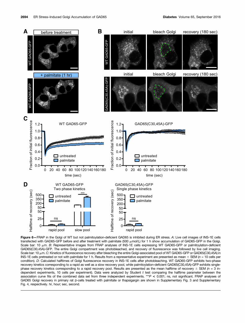
Figure 6—FRAP in the Golgi of WT but not palmitoylation-deficient GAD65 is inhibited during ER stress. A: Live cell images of INS-1E cellstransfected with GAD65-GFP before and after treatment with palmitate (500 mmol/L) for 1 h show accumulation of GAD65-GFP in the Golgi.Scale bar: 10 mm. B: Representative images from FRAP analyses of INS-1E cells expressing WT GAD65-GFP or palmitoylation-deficientGAD65(C30,45A)-GFP. The entire Golgi compartment was photobleached, and recovery of fluorescence was followed by live cell imaging.Scale bar: 10 mm.C: Kinetics of fluorescence recovery after bleaching the entire Golgi-associated pool of WTGAD65-GFP or GAD65(C30,45A) inINS-1E cells pretreated or not with palmitate for 1 h. Results from a representative experiment are presented as mean 6 SEM (n = 10 cells percondition). D: Calculated halftimes of Golgi fluorescence recovery in INS-1E cells after photobleaching. WT GAD65-GFP exhibits two-phaserecovery kinetics corresponding to a rapid as well as a slow recovery pool, while palmitoylation-deficient GAD65(C30,45A)-GFP exhibits single-phase recovery kinetics corresponding to a rapid recovery pool. Results are presented as the mean halftime of recovery 6 SEM (n = 3 in-dependent experiments, 10 cells per experiment). Data were analyzed by Student t test comparing the halftime parameter between theassociation curve fits of the combined data set from three independent experiments. ***P < 0.001; ns, not significant. FRAP analyses ofGAD65 Golgi recovery in primary rat b-cells treated with palmitate or thapsigargin are shown in Supplementary Fig. 3 and SupplementaryFig. 4, respectively. hr, hour; sec, second.
2694 ER Stress–Induced Golgi Accumulation of GAD65 Diabetes Volume 65, September 2016

GAD65 Accumulates in the Golgi Compartmentin Human b-Cells During Progression of T1DAutoimmunityWe addressed the question of whether the experimentalaccumulation of GAD65 in the Golgi compartment inducedby treatment with ER stressors in vitro is of relevance forhuman diabetes. Human pancreatic sections provided bythe nPOD tissue bank (28,29) representing eight healthydonors (Supplementary Table 1), eight individuals withpotential prediabetes and GADA+ (Supplementary Table 2),and eight patients with T1D with residual b-cell mass (Sup-plementary Table 3) were immunostained for GAD65,giantin, insulin, glucagon, and the pan–T-cell marker CD3(Fig. 8A–D). In five of eight GADA+ individuals and sevenof eight patients with T1D, the GAD65 signal in b-cellGolgi membranes compared with cytosol was higher thanone SD above the mean for healthy control individualswhere GAD65 immunostaining was mostly uniform (Fig.8E and Supplementary Tables 1–3, last column). Stainingof serial sections for the pan–T-cell marker, CD3 (Fig. 8A),confirmed peri- and intraislet infiltrating T cells in isletsfrom three of eight GADA+ individuals and in six of eightpatients with T1D (Fig. 8A and results not shown). Exam-ination of the patient information (Supplementary Tables1–3) for individual donors provided by nPOD revealed thatthe highest Golgi accumulation observed in the GADA+
group with prediabetes was from a single autoantibody-positive (GADA+) 2.2-year-old child expressing the HLA-classII T1D susceptibility haplotype DR4, DQ8 (SupplementaryTable 2) showing infiltrates of CD3+ T cells in and aroundislets (Fig. 8A, nPOD 6090) but not meeting the nPOD
insulitis criteria (Supplementary Table 2). This child mayrepresent a case of a young, genetically susceptible indi-vidual in the early stages of autoimmunity associatedwith development of T1D. The lowest Golgi accumula-tion in the GADA+ group was observed in a 31-year-old,single-autoantibody–positive (GADA+) individual withnormal islet morphology and no insulitis (Supplemen-tary Table 2, nPOD 6181). The highest Golgi accumula-tion observed in the group with T1D was observed in adouble-autoantibody–positive (GADA+ and microinsulinautoantibody assay+) 11-year-old individual expressingthe T1D HLA-class II susceptibility haplotypes DR3,DQ2 and DR4, DQ8 and with ongoing insulitis (Sup-plementary Table 3) (30) as confirmed by our CD3immunostaining (Fig. 8A, nPOD 6265). Taken together,the results suggest that aberrant accumulation of GAD65in Golgi membranes may correlate with active islet auto-immunity in human T1D.
DISCUSSION
Accumulation of GAD65 in Golgi Membranes in b-CellsExperiencing ER Stress and/or Active AutoimmunityIn this study, we show that treatment of islet cell culturesby three separate regimens known to induce ER stress inb-cells results in accumulation of GAD65 in Golgi mem-branes. Importantly, analysis of human pancreatic tissuesections revealed significant accumulation of GAD65 inb-cell Golgi membranes in GADA+ individuals with po-tential prediabetes and in patients with T1D with resid-ual b-cell mass. Thus, aberrant accumulation of GAD65in Golgi membranes is observed in b-cells in culture
Figure 7—Palmitoylation confers increased immunogenicity upon GAD65. A: Recombinant human GAD65 produced in yeast was shown tobe palmitoylated by the Acyl-RAC assay. Free thiols in recombinant GAD65 were capped with methyl methanethiosulfonate, followed bycleavage of palmitate-thiol modifications with HA, pulldown with a thiol-reactive pyridyl-disulfide agarose resin, and detection by Westernblot. Lanes: i, input, positive control of unmodified recombinant GAD65; –HA, negative control without HA, resulting in no palmitatecleavage and no protein pulldown; +HA, HA added with palmitate groups cleaved and protein recovered by thiol-reactive pulldown. B:Time course of uptake of equimolar amounts of DyLight-488–labeled palmitoylated or depalmitoylated (HA cleaved) recombinant GAD65 byMHC-class II DR4–positive human Priess B-cell APCs measured by flow cytometry. Results are presented as mean 6 SEM (n = 3 exper-imental replicates). Data were analyzed by two-way ANOVA, followed by the Tukey multiple comparisons test. ***P < 0.001. C: Priess cellswere loaded with equimolar amounts of palmitoylated or depalmitoylated GAD65 (HA cleaved) and tested for stimulation of the DR4restricted GAD65-specific T33.1 T-cell hybridoma cells by IL-2 secretion measured by ELISA. Peptide, GAD65274-286 epitope. Resultsare presented as mean 6 SEM (n = 3 experimental replicates). Data were analyzed by two-way ANOVA, followed by the Tukey multiplecomparisons test. ***P < 0.001; ns, not significant.
diabetes.diabetesjournals.org Phelps and Associates 2695

undergoing experimentally induced ER stress as well as inb-cells of individuals experiencing active b-cell autoimmunity.
GAD65 in Pancreatic b-Cells Undergoes aPalmitoylation–Depalmitoylation–RepalmitoylationCycle That Is Perturbed During ER Stress Resulting inAccumulation of the Protein in Golgi MembranesWe have shown earlier that palmitoylation of GAD65results in anterograde trafficking from cis-Golgi to TGNmembranes (12). The evidence presented in this studyshows that only WT GAD65, but not palmitoylation-deficient GAD65(C30,45A), accumulates in Golgi mem-branes during ER stress. This increase of GAD65 in Golgi
membranes is consistent with the accumulated proteinrepresenting the palmitoylated form of GAD65 in TGNmembranes, suggesting that the palmitoylation cycle ofGAD65 significantly slows down during induction of ERstress. This retardation in transport likely affects thecontrol of GAD65 distribution between peripheral vesi-cle membranes, where most of the GAD65 enzymaticfunctional activity is believed to take place, and Golgimembranes, which may serve as a sorting station forthis protein. We therefore propose that perturbation inthe regulation of GAD65 membrane distribution by ERstress may have negative consequences for GABA synthesis
Figure 8—Confocal analyses of islets in paraffin sections of human pancreas from healthy donors, donors who were GAD65 autoantibody-positive but without diabetes, or donors with T1D with residual b-cell mass. A: Immunostaining of human pancreatic sections for insulin,glucagon, or CD3 from the indicated nPOD case numbers. The arrowheads indicate CD3+ T cells. Scale bar: 50 mm. B: The same humanislet from a serial section, immunostained for GAD65 and giantin. Scale bar: 50 mm. C: Increased magnification of GAD65 staining in theframed region shown in B. Scale bar: 10 mm. D: Thresholded binary images of the same region in C show the colocalization (Coloc.) betweengiantin (Golgi) and the bright GAD65 signal. Scale bar: 10 mm. E: Image quantification of GAD65 accumulation in the Golgi compartment inthree representative islets from eight individuals per donor category, reported as the ratio of MFI for GAD65 in the Golgi of b-cells to MFI forGAD65 in the rest of the cell, excluding the nucleus. Results are presented as scatter dot plots overlaying the mean 6 SEM (n = 24 isletsfrom eight donors). Data were analyzed by one-way ANOVA, followed by the Tukey multiple comparisons test. *P < 0.05, **P < 0.01. Theaverage GAD65 Golgi accumulation calculated per individual and demographic information for healthy, GADA+, and donors with T1D arelisted in Supplementary Tables 1–3.
2696 ER Stress–Induced Golgi Accumulation of GAD65 Diabetes Volume 65, September 2016

and secretion. GABA serves as an important signaling mol-ecule and survival/growth factor in islets of Langerhans(40).
The mechanisms by which cytokines, palmitate, andthapsigargin induce ER stress in b-cells may differ (34).One commonality between the three different treatmentsis that each is implicated in dysregulation of Ca2+ homeo-stasis (41). Inflammatory cytokines and thapsigargin haveboth been shown to induce b-cell ER stress throughdownregulation or inhibition of the sarcoplasmic/ER Ca2+
ATPase, resulting in elevation of cytosolic Ca2+ concentra-tions and depletion of ER Ca2+ stores (42). Palmitate isreported to induce ER stress through lipotoxic signalingpathways mediating a block in ER uptake of Ca2+ and asustained depletion of ER Ca2+ stores (35,43). The ideathat dysregulation of ER/cytosolic Ca2+ concentrations isinvolved in Golgi accumulation of GAD65 is supported bythe fact that a similar effect could be achieved by stronglydepolarizing cells by abruptly elevating extracellular glu-cose or KCl concentration (E.A.P., C.C., and S.B., unpub-lished results). Although how a change in Ca2+ homeostasiscould affect membrane trafficking of GAD65 is currentlyunknown, it is of note that earlier studies have suggestedmodulation of the affinity of GAD65 to liposome mem-branes by Ca2+ ion concentration (44). Interestingly, thereis evidence to suggest that palmitate treatment of insuli-noma cells alters the lipid composition of endomem-branes and specifically disrupts lipid raft microdomains(45), a dynamic hub for palmitoylated proteins (46). It isof note that distribution of H-Ras into lipid rafts isregulated by its palmitoylation–depalmitoylation cycle(47), which shares similarities with the acylation cycleof GAD65 (16). Thus, it is possible that alterations in lipidraft composition are part of the mechanism involved inperturbation of GAD65 trafficking and accumulation inGolgi membranes.
Accumulation of Palmitoylated GAD65 in GolgiMembranes May Have Implications for Initiation ofAutoimmunityParallel to the important role of GAD65 as the highlyregulated and the only GABA-synthesizing enzyme inhuman b-cells, it can assume a detrimental role as amajor target of autoimmunity associated with pancreatichuman b-cell destruction and development of T1D ingenetically susceptible individuals (2). The autoantige-nicity of GAD65 is in stark contrast to the highly homol-ogous GAD67 isoform, which is not an independentautoantigen. GAD67 primarily differs from GAD65 inthe N-terminal domain (48) that mediates hydrophobicposttranslational modifications, membrane anchoring,palmitoylation, and trafficking of GAD65 (12), suggest-ing that this region is integral to the susceptibility ofGAD65 to become a pathogenic autoantigen. GAD67does not undergo hydrophobic modifications (49) andcan only be targeted to membranes by piggybackingonto other proteins (15,38).
Our data show that compared with depalmitoylatedGAD65, palmitoylated GAD65 induces a significantlystronger effector T-cell response by the T-cell hybrid-oma T33.1, which recognizes GAD65274-286 in the contextof DR4, a T1D MHC-class II susceptibility haplotype.Palmitoylation of peptide epitopes has been shown toenhance immunogenicity of autoimmune epitopes in ex-perimental autoimmune encephalomyelitis (50) and insynthetic peptide vaccines (51). Palmitoylation of a pro-tein increases its avidity for binding to membranes. Theuptake of palmitoylated GAD65 by the DR4-positive hu-man B-cell line Priess was significantly enhanced com-pared with nonpalmitoylated GAD65. We posit that theincrease in uptake, conferred by palmitoylation, may in-volve enhanced binding to the surface of Priess cellsresulting in increased uptake by endocytosis and en-hanced targeting to late endosomes for proteolytic pro-cessing and presentation to T cells in the context ofMHC-class II antigens (reviewed by Blum et al. [52]).Palmitoylation of GAD65 may also affect antigen unfold-ing and proteolysis and alter the hierarchy of peptidesdisplayed to CD4+ T cells. The T33.1 hybridoma reportercell line is clonally restricted to a single MHC-class IIbinding epitope, GAD65274-286, which is distant fromthe palmitoylated cysteine residues in GAD65, aa 30 and45. Therefore, the possibility of palmitoylation increasingthe MHC-class II binding affinity of this epitope can beexcluded in our experimental system.
Rather, we suggest that increased stimulation of T33.1cells reflects a quantitative increase in the levels of theGAD65274-286 epitope available for binding to DR4 in lateendosomes and elevated expression on the surface of Pri-ess cells. If palmitoylated GAD65, which has accumulatedin TGN during ER stress, is released by distressed or dyingb-cells and encountered by APCs, its heightened immu-nogenicity compared with nonpalmitoylated GAD65 maystimulate autoimmunity in genetically predisposed indi-viduals. An important agenda for future studies will be toelucidate the mechanisms by which palmitoylated, immu-nogenic GAD65 is released from b-cells undergoing ERstress, as well as the determinants of that stress, whichthe results presented here suggest could be key factors inthe mechanism that results in autoimmunity to GAD65associated with development of T1D.
Acknowledgments. The authors thank Drs. Lorenzo Piemonti, SanRaffaele Scientific Research Institute, and Domenico Bosco and Thierry Berney,University of Geneva, for help in procuring human islets; Gisou van der Goot andLaurence Abrami, EPFL, for insightful suggestions and donation of reagents;Janice Blum of Indiana University (Priess cells, T33.1 cells), Bloomington, IN,Linda Wicker of Cambridge Institute for Medical Research (T33.1 cells),Cambridge, U.K., Christiana Hämpe of University of Washington (N-GAD65 mAb),Seattle, WA, David Gottlieb, Washington University of St. Louis (GAD6 antibody),St. Louis, MO, and Pierre Maechler, University of Geneva (INS-1E cells), forgenerously donating cells and reagents.Funding. This study was supported by a Whitaker International ProgramPostdoctoral Scholarship (E.A.P.), by a JDRF Advanced Postdoctoral Fellowship
diabetes.diabetesjournals.org Phelps and Associates 2697
(3-APF-2014-208-A-N) (E.A.P.), by the Nora Eccles Treadwell Foundation(J.K., S.B.), by an International Network Program from the Danish Ministry ofScience, Innovation and Higher Education (INP-2010-0102) (N.B., S.B.), by theIntramural Research Program of EPFLs School of Life Sciences (J.A.H., S.B.), by aJDRF award to the ECIT Islets for Basic Research Program (31-2008-416), by theEPFL Bioimaging and Optics Core Facility and EPFL Flow Cytometry Core Facility,by a National Institutes of Health Diabetes Education and Research Center grant(P30-DK-063720) funded University of California, San Francisco, Diabetes CenterMicroscopy Core, and by the nPOD, a collaborative T1D research projectsponsored by JDRF. Organ Procurement Organizations partnering with nPOD toprovide research resources are listed at www.jdrfnpod.org/our-partners.php.Duality of Interest. No potential conflicts of interest relevant to this articlewere reported.Author Contributions. E.A.P., C.C., J.K., and S.B. developed theconcept. E.A.P., C.C., and M.P. conducted all experiments and collected data.E.A.P., C.C., and S.B. designed the study and analyzed data. E.A.P. and S.B.wrote the manuscript with input from the other authors. I.P.M. contributed criticalreagents and technical expertise and assisted in experiments related to GAD65-GFP expression. J.K. and N.B. assisted with developing primary b-cell single-cellculture and transfection techniques. R.N. and V.L. isolated the human isletsprovided through the ECIT islet distribution network. J.A.H. contributed criticalreagents and expertise. S.B. is the guarantor of this work and, as such, had fullaccess to all of the data in the study and takes responsibility for the integrity ofthe data and the accuracy of the data analysis.Prior Presentation. Part of this work was presented at the BiomedicalEngineering Society Annual Scientific Meeting, Tampa, FL, 7–10 October 2015,and at the Keystone Symposia, Islet Biology: From Cell Birth to Death, Keystone,CO, 13–17 March 2016.
References1. Erlander MG, Tillakaratne NJK, Feldblum S, Patel N, Tobin AJ. Two genesencode distinct glutamate decarboxylases. Neuron 1991;7:91–1002. Baekkeskov S, Aanstoot HJ, Christgau S, et al. Identification of the 64Kautoantigen in insulin-dependent diabetes as the GABA-synthesizing enzymeglutamic acid decarboxylase. Nature 1990;347:151–1563. Baekkeskov S, Landin M, Kristensen JK, et al. Antibodies to a 64,000 Mrhuman islet cell antigen precede the clinical onset of insulin-dependent diabetes.J Clin Invest 1987;79:926–9344. Atkinson MA, Maclaren NK, Scharp DW, Lacy PE, Riley WJ. 64,000 Mrautoantibodies as predictors of insulin-dependent diabetes. Lancet 1990;335:1357–13605. Kim J, Richter W, Aanstoot HJ, et al. Differential expression of GAD65 andGAD67 in human, rat, and mouse pancreatic islets. Diabetes 1993;42:1799–18086. Petersen JS, Russel S, Marshall MO, et al. Differential expression of glu-tamic acid decarboxylase in rat and human islets. Diabetes 1993;42:484–4957. Kash SF, Condie BG, Baekkeskov S. Glutamate decarboxylase and GABA inpancreatic islets: lessons from knock-out mice. Horm Metab Res 1999;31:340–3448. Christgau S, Schierbeck H, Aanstoot HJ, et al. Pancreatic beta cells expresstwo autoantigenic forms of glutamic acid decarboxylase, a 65-kDa hydrophilicform and a 64-kDa amphiphilic form which can be both membrane-bound andsoluble. J Biol Chem 1991;266:235169. Christgau S, Aanstoot HJ, Schierbeck H, et al. Membrane anchoring of theautoantigen GAD65 to microvesicles in pancreatic beta-cells by palmitoylation inthe NH2-terminal domain. J Cell Biol 1992;118:309–32010. Shi Y, Veit B, Baekkeskov S. Amino acid residues 24-31 but not palmitoylationof cysteines 30 and 45 are required for membrane anchoring of glutamic aciddecarboxylase, GAD65. J Cell Biol 1994;124:927–93411. Kanaani J, el-Husseini Ael-D, Aguilera-Moreno A, Diacovo JM, Bredt DS,Baekkeskov S. A combination of three distinct trafficking signals mediatesaxonal targeting and presynaptic clustering of GAD65. J Cell Biol 2002;158:1229–1238
12. Kanaani J, Patterson G, Schaufele F, Lippincott-Schwartz J, Baekkeskov S.A palmitoylation cycle dynamically regulates partitioning of the GABA-synthesizingenzyme GAD65 between ER-Golgi and post-Golgi membranes. J Cell Sci 2008;121:437–44913. Huang K, Yanai A, Kang R, et al. Huntingtin-interacting protein HIP14 is apalmitoyl transferase involved in palmitoylation and trafficking of multiple neu-ronal proteins. Neuron 2004;44:977–98614. Kanaani J, Diacovo MJ, El-Husseini Ael-D, Bredt DS, Baekkeskov S.Palmitoylation controls trafficking of GAD65 from Golgi membranes to axon-specific endosomes and a Rab5a-dependent pathway to presynaptic clusters. J CellSci 2004;117:2001–201315. Kanaani J, Cianciaruso C, Phelps EA, et al. Compartmentalization of GABAsynthesis by GAD67 differs between pancreatic beta cells and neurons. PLoS One2015;10:e011713016. Baekkeskov S, Kanaani J. Palmitoylation cycles and regulation of proteinfunction (review). Mol Membr Biol 2009;26:42–5417. Fukata Y, Fukata M. Protein palmitoylation in neuronal development andsynaptic plasticity. Nat Rev Neurosci 2010;11:161–17518. Oakes SA, Papa FR. The role of endoplasmic reticulum stress in humanpathology. Annu Rev Pathol 2015;10:173–19419. Eizirik DL, Cardozo AK, Cnop M. The role for endoplasmic reticulum stressin diabetes mellitus. Endocr Rev 2008;29:42–6120. Marhfour I, Lopez XM, Lefkaditis D, et al. Expression of endoplasmic re-ticulum stress markers in the islets of patients with type 1 diabetes. Diabetologia2012;55:2417–242021. Engin F, Yermalovich A, Nguyen T, et al. Restoration of the unfolded proteinresponse in pancreatic b cells protects mice against type 1 diabetes. Sci TranslMed 2013;5:211ra15622. Song B, Scheuner D, Ron D, Pennathur S, Kaufman RJ. Chop deletion re-duces oxidative stress, improves beta cell function, and promotes cell survival inmultiple mouse models of diabetes. J Clin Invest 2008;118:3378–338923. Eizirik DL, Colli ML, Ortis F. The role of inflammation in insulitis and beta-cell loss in type 1 diabetes. Nat Rev Endocrinol 2009;5:219–22624. Merglen A, Theander S, Rubi B, Chaffard G, Wollheim CB, Maechler P.Glucose sensitivity and metabolism-secretion coupling studied during two-yearcontinuous culture in INS-1E insulinoma cells. Endocrinology 2004;145:667–67825. Jaume JC, Parry SL, Madec AM, Sønderstrup G, Baekkeskov S. Suppressiveeffect of glutamic acid decarboxylase 65-specific autoimmune B lymphocytes onprocessing of T cell determinants located within the antibody epitope. J Immunol2002;169:665–67226. Wicker LS, Chen SL, Nepom GT, et al. Naturally processed T cell epitopesfrom human glutamic acid decarboxylase identified using mice transgenic for thetype 1 diabetes-associated human MHC class II allele, DRB1*0401. J Clin Invest1996;98:2597–260327. Codazzi F, Di Cesare A, Chiulli N, et al. Synergistic control of protein kinaseCgamma activity by ionotropic and metabotropic glutamate receptor inputs inhippocampal neurons. J Neurosci 2006;26:3404–341128. Campbell-Thompson M, Wasserfall C, Kaddis J, et al. Network for Pan-creatic Organ Donors with Diabetes (nPOD): developing a tissue biobank fortype 1 diabetes. Diabetes Metab Res Rev 2012;28:608–61729. Pugliese A, Yang M, Kusmarteva I, et al. The Juvenile Diabetes ResearchFoundation Network for Pancreatic Organ Donors with Diabetes (nPOD) Program:goals, operational model and emerging findings. Pediatr Diabetes 2014;15:1–930. Campbell-Thompson M, Fu A, Kaddis JS, et al. Insulitis and b-cell mass inthe natural history of type 1 diabetes. Diabetes 2016;65:719–73131. Chang YC, Gottlieb DI. Characterization of the proteins purified with mono-clonal antibodies to glutamic acid decarboxylase. J Neurosci 1988;8:2123–213032. Hampe CS, Lundgren P, Daniels TL, Hammerle LP, Marcovina SM, Lernmark A.A novel monoclonal antibody specific for the N-terminal end of GAD65.J Neuroimmunol 2001;113:63–7133. Forrester MT, Hess DT, Thompson JW, et al. Site-specific analysis of proteinS-acylation by resin-assisted capture. J Lipid Res 2011;52:393–398
2698 ER Stress–Induced Golgi Accumulation of GAD65 Diabetes Volume 65, September 2016

34. Kharroubi I, Ladrière L, Cardozo AK, Dogusan Z, Cnop M, Eizirik DL. Freefatty acids and cytokines induce pancreatic beta-cell apoptosis by differentmechanisms: role of nuclear factor-kappaB and endoplasmic reticulum stress.Endocrinology 2004;145:5087–509635. Cunha DA, Hekerman P, Ladrière L, et al. Initiation and execution of lipotoxicER stress in pancreatic beta-cells. J Cell Sci 2008;121:2308–231836. Brozzi F, Nardelli TR, Lopes M, et al. Cytokines induce endoplasmic re-ticulum stress in human, rat and mouse beta cells via different mechanisms.Diabetologia 2015;58:2307–231637. Bertolotti A, Zhang Y, Hendershot LM, Harding HP, Ron D. Dynamic in-teraction of BiP and ER stress transducers in the unfolded-protein response. NatCell Biol 2000;2:326–33238. Kanaani J, Kolibachuk J, Martinez H, Baekkeskov S. Two distinct mecha-nisms target GAD67 to vesicular pathways and presynaptic clusters. J Cell Biol2010;190:911–92539. Pfender NA, Grosch S, Roussel G, Koch M, Trifilieff E, Greer JM. Route ofuptake of palmitoylated encephalitogenic peptides of myelin proteolipidprotein by antigen-presenting cells: importance of the type of bond betweenlipid chain and peptide and relevance to autoimmunity. J Immunol 2008;180:1398–140440. Soltani N, Qiu H, Aleksic M, et al. GABA exerts protective and regenerativeeffects on islet beta cells and reverses diabetes. Proc Natl Acad Sci U S A 2011;108:11692–1169741. Marré ML, James EA, Piganelli JD. b cell ER stress and the implications forimmunogenicity in type 1 diabetes. Front Cell Dev Biol 2015;3:6742. Cnop M, Welsh N, Jonas JC, Jörns A, Lenzen S, Eizirik DL. Mechanisms ofpancreatic beta-cell death in type 1 and type 2 diabetes: many differences, fewsimilarities. Diabetes 2005;54(Suppl. 2):S97–S107
43. Marmugi A, Parnis J, Chen X, et al. Sorcin links pancreatic b-cell lipotoxicityto ER Ca2+ stores. Diabetes 2016;65:1009–102144. Covarrubias M, Tapia R. Calcium-dependent binding of brain glutamatedecarboxylase to phospholipid vesicles. J Neurochem 1978;31:1209–121445. Boslem E, Weir JM, MacIntosh G, et al. Alteration of endoplasmic reticulumlipid rafts contributes to lipotoxicity in pancreatic b-cells. J Biol Chem 2013;288:26569–2658246. Levental I, Lingwood D, Grzybek M, Coskun U, Simons K. Palmitoylationregulates raft affinity for the majority of integral raft proteins. Proc Natl Acad SciU S A 2010;107:22050–2205447. Agudo-Ibáñez L, Herrero A, Barbacid M, Crespo P. H-ras distribution andsignaling in plasma membrane microdomains are regulated by acylation anddeacylation events. Mol Cell Biol 2015;35:1898–191448. Bu DF, Erlander MG, Hitz BC, et al. Two human glutamate decarboxylases,65-kDa GAD and 67-kDa GAD, are each encoded by a single gene. Proc NatlAcad Sci U S A 1992;89:2115–211949. Kanaani J, Lissin D, Kash SF, Baekkeskov S. The hydrophilic isoform ofglutamate decarboxylase, GAD67, is targeted to membranes and nerve terminalsindependent of dimerization with the hydrophobic membrane-anchored isoform,GAD65. J Biol Chem 1999;274:37200–3720950. Greer JM, Denis B, Sobel RA, Trifilieff E. Thiopalmitoylation of myelinproteolipid protein epitopes enhances immunogenicity and encephalitogenicity.J Immunol 2001;166:6907–691351. Beekman NJ, Schaaper WM, Tesser GI, et al. Synthetic peptide vaccines:palmitoylation of peptide antigens by a thioester bond increases immunogenicity.J Pept Res 1997;50:357–36452. Blum JS, Wearsch PA, Cresswell P. Pathways of antigen processing. AnnuRev Immunol 2013;31:443–473
diabetes.diabetesjournals.org Phelps and Associates 2699