Abdominal Tumour Recurrence
196
Influence of Peritoneal Trauma on Postoperative Adhesion Formation and Intra- Abdominal Tumour Recurrence
Transcript of Abdominal Tumour Recurrence

Influence of Peritoneal Trauma on
Postoperative Adhesion Formation and Intra
Abdominal Tumour Recurrence

ISBN 90-77017-16-X
© 2001 M.P. van den Tal
All rights reserved. No part of this thesis may be reproduced, stored in a retrieval system of
any nature, or transmitted in any form by any means, electronic, mechanical, photocopying,
recording or otherwise, included a complete or partial transcription, without the permission of
the author.
Printed by Optima Grafische Communicatie, Rotterdam, The Netherlands
Influence of Peritoneal Trauma on
Postoperative Adhesion Formation and Intra
Abdominal Tumour Recurrence
lnvloed van Peritoneaal Trauma op
Postoperatieve Adhesie Vorming en lntra
Abdominale Tumor Recidivering
PROEFSCHRIFT
Ter verkrijging van de graad van doctor
aan de Erasmus Universiteit Rotterdam
op gezag van de Rector Magnificus
Prof.dr.ir. J.H. van Bemmel
en volgens besluit van het College voor Promoties
De openbare verdediging zal plaatsvinden op
woensdag 21 november 2001 om 9.45 uur
door
Monique Petrousjka van den Tol
geboren te Paramaribo

Promotiecomissie
Promotor:
Overige !eden:
Co-promotor:
Prof.dr. J. Jeekel
Prof.dr. R.P. Bleichrodt
Prof.dr. H.J. Bonjer
Prof.dr. HA Verbrugh
Dr. RL Marquet

Aan opa en oma Bakema
Aan Lida
Aan Chris


Contents
Part I General introduction
II
Part II
Ill
IV
Part Ill
v
VI
Part IV
VII
General introduction
Aims of the thesis
Surgical peritoneal trauma and adhesion formation
Reduction of intra-peritoneal adhesion formation by use of non
abrasive gauze.
BrJ Surg 1997; 84: 1410-1415
Peroperative abdominal lavage promotes intra-peritoneal adhesion
formation in the rat.
Eur Surg Res 1999; 31(2): 196-201
Surgical peritoneal trauma and intra~abdominal tumour recurrence
Reduction of peritoneal trauma by using non-surgical gauze leads
to less implantation metastasis of spilled tumour cells.
Ann Surg 1998; 227(2): 242-248
Glove powder promotes adhesion formation and facilitates intra
peritoneal tumour cell adhesion and growth.
BrJ Surg 2001; 88:1258-1263
11
23
41
57
69
83
Pathways of adhesion formation and intra-abdominal tumour recurrence
after surgical peritoneal trauma; possible methods for prevention
The inflammatory sequelae of surgery provoke enhanced tumour
recurrence; a crucial role for neutrophils and cytokines.
Submitted for publication
99

VIII
IX
X
Part V
XI
XII
XIII
Appendices
Preventing intra-abdominal influx of neutrophils after surgical
peritoneal trauma reduces adhesion formation and local tumour
recurrence.
Submitted for publication
Hyaluronate-based coating solution for prevention of surgical
adhesions has no major effect on adhesion and growth of intra
peritoneal tumour cells.
Eur J Surg 1999; 165: 791-795
lcodextrin® is a potent inhibitor of postoperative adhesion formation
in rats but does not affect intra-peritoneal tumour cell adhesion and
growth.
Submitted for publication
General discussion and summary
General discussion
Summary and conclusions
Summary in Dutch
List of abbreviations
Publications and awards
Acknowledgements
Curriculum vitae auctoris
115
133
145
161
173
179
187
189
191
195

PART I
General Introduction and Aims of the Thesis


Chapter I
General Introduction
Adhesions
Definition, incidence and complications
Intra-abdominal adhesions are abnormal unions between different peritoneal surfaces. The
most common cause of intra-abdominal adhesion formation is prior abdominal surgery which
accounts for 70-90% of all adhesions. 1-3 Remaining adhesions are due to inflammation or
endometriosis or are congenital (18% and 2.8-6% respectively). 1•
3 Postoperative intra
abdominal adhesion formation is a major, up till now unavoidable complication of any kind of
abdominal surgery. Postoperative adhesions occur in 55-100% of patients undergoing
abdominal surgery, with an average of approximately 85%.2· 3· s-? These adhesions can
eventuate in bowel, fertility and abdominal syndromes which frequently require further
surgery. Adhesion formation is the cause of 30% of all bowel obstructions,8-10 of 15-20% of
infertility in women 11"15 and of 13-26% of chronic pelvic pain in women.16
' 17 The cumulative
risk of adhesive small-bowel obstruction after (sub)total colectomy is 11% within 1 year,
increasing to 30% at 10 years.18 After open abdominal or pelvic surgery in 29,790 patients
studied by Ellis et al, 34.6% were readmitted a mean of 2.1 times in the subsequent 1 0
years for a disorder directly or possibly related to adhesions. Of these readmissions 22.1%
occurred in the first year after initial surgery, but readmissions continued steadily throughout
the 10 year study period.19 About 1% of all surgical admissions and 3% of all laparotomies
take place for intestinal obstruction due to adhesions? The mortality rate from intestinal
obstruction as a result of adhesions is 6-13%. 1·
4·
20 Adhesions also increase the technical
difficulty and the risk of intraoperative complications at subsequent surgery depending on the
organs involved and severity of adhesions. One of five patients undergoing adhesiotomy
during reoperation suffers an inadvertent enterotomy, possibly resulting in extensive
postoperative morbidity and mortality. Morbidity involves bowel obstruction, anastomotic
leak, wound dehiscence, sepsis and pneumonia.21 Clearly in addition to the obvious
morbidity and mortality due to postoperative adhesion formation the costs of adhesion
related health care are significant.19'
20"
22"24 Until now there is no clinically relevant cost
effective method available to reduce or prevent postoperative intra-abdominal adhesion
formation.
11

Clinical diagnosis
Intra-abdominal adhesions cannot be shown by non-invasive modalities. The combination of
medical history and physical examination fail to adequately predict adhesion formation and
invasive as well as non-invasive imaging studies have poor sensitivity for detecting
adhesions. 25-27 In general, the presence of adhesions can only be confirmed by either
laparoscopy or laparotomy.
Aetiology and patholophysiology
Adhesion formation is a physiological consequence of peritoneal tissue repair. Surgical
trauma of the peritoneal surfaces induces a sequence of events which effectuates wound
healing but which can also ultimately lead to fibrous adhesions between different peritoneal
surfaces. Damage to the peritoneum causes desquamation of injured mesothelial cells leaving
a denuded area, and a local inflammatory reaction which leads to the formation of a
serosanguineous exudate.2e-31 The primary inflammatory response, the acute phase response,
is characterised by an increased vascular permeability and the migration of neutrophils (PMNs)
and subsequently macrophages and leucocytes to the site of the inflammation during the first
48 to 72 hours after trauma.32-34 Chemoattractants (IL-8, MCP-1), cytokines (TN F-a, IL-1~ and
IL-6) and growth factors (TGF-~, IGF-1, PDGF), produced and released by damaged
mesothelial cells and resident and invading inflammatory cells are the key mediators of this
inflammatory reaction. 35-
39 In the resulting serosanguineous exudate fibrin deposition will take
place through the activated complement- and coagulation systems and when two surfaces
contact each other during this process a fibrinous adhesion will appear between day 2 and 4. 30·
31·
40 Whether these adhesions are permanent of character or will eventually be lysed depends
upon the fibrinolytic capacity of the peritoneum. 30·
4144 1ntra-abdominally deposited fibrin has to
be lysed to free attached structures, restore the patency of the peritoneal cavity and open up
the way to adhesion free healing. When the peritoneum is slightly damaged and mesothelial
cells are mostly intact, there will be a dynamic balance between fibrinogenesis and fibrinolysis,
between tissue-type plasminogen activator (t-PA) and plasminogen activator inhibitors (PAls). 41
· 42
· 45
· 46 Adhesion-free healing may then take place by conversion of plasminogen into
plasmin effectuating the sufficient break-down of fibrin by day 4 to 5 followed by proliferation
and centripetal migration of residual mesothelial cells and proliferation and differentiation of
interspersing mesothelial stem cells.28·
32·
4749 Reepithelialisation occurs through continuous
simultaneous dividing of adjacent mesothelial cells and "islands" of multipotentia! sub
mesothelial cells until the surface of the entire site of injury is covered by new mesothelium 5 to
8 days after the initial peritoneal trauma.28·
32' 48 The remesothelialisation process appears to be
mostly the result of mediators released by the mesothelial cells themselves. 50
12

Loss of mesothelial integrity due to the initial peritoneal trauma and invading leucocytes, which
have been shown to enhance peritoneal injury by adhering to and damaging mesothelial cells
through the release of active oxygen species, 51-54 will expose the underlying connective tissue
(ECM) and normal mesothelial fibrinolytic activity will be lost for at least 48 hours post
trauma.43'
55'
56 Exposed endothelial cells, damaged mesothelial cells, as well as resident and
invading inflammatory cells, produce plasminogen activator inhibitor 1 and 2 (PAl 1 and 2),
factors mediating a reduced functional fibrinolytic activity by down-regulating t-PA activity and
thus plasminogen-plasmin conversion and fibrin demolishment.42·
57 Cytokines and growth
factors, also produced by damaged mesothelial cells and inflammatory cells, decrease t-PA
and increase PAl concentrations intra-peritoneally diminishing fibrinolytic activity of the
mesothelium likewise.58•
59 The autonomous plasminogen activator activity (PAA) of the
peritoneal exudates itself (result of local t-PA release and clearance) is also reduced during
several hours postoperatively which contributes even more to the disbalance between t-PA
and PAls and thus to decreased fibrinolytic activity.60•
61 Reduced plasminogen-plasmin
conversion i.e. low fibrinolytic activity declines fibrin degradation and allows the fibrinous
adhesion to organise to a fibrous permanent adhesion through invasion, proliferation and
differentiation of fibroblasts and endothelial cells. This is followed by capillary formation and
incorporation of collagen, all stimulated by cytokines, and growth factors (day 4 to 10).29. 62-6
7
Cytokines increase expression of integrins, cell surface receptors mediating fibroblast and
mesothelial cell adhesion to ECM.68 The mesothelial cells recover the exterior aspects of the
adhesion, isolating the ECM from the peritoneal cavity and reconstituting an intact peritoneal
mesothelial surface.69
The results of the inflammatory and fibrinogenetic response to peritoneal trauma eventuate in
ultimate resolution of the process, by triggering events that lead to cell regeneration and wound
healing. Normal intra-abdominal inflammatory and fibrinogenetic defences become detrimental
to the host if the balance between mediators of the inflammatory response and the balance
between fibrinogenesis and fibrinolysis is lost and two peritoneal surfaces are apposed (Figure
1.1 ).
Treatment
Relaparotomy or laparoscopy for adhesiolysis can be a technical challenge with elevated
peroperative complication risks and significant postoperative morbidity and even mortality.
Clinical studies involving second look operations show that operative adhesiolysis is also
complicated by a high recurrence rate of adhesions at the operated site (adhesion
reformation) or at other sites (de novo adhesions). 70-73 Laparoscopy does appear to be
associated with equal adhesion reformation as laparotomy, between 38 and 97%, whereas
de novo adhesions seem to be significantly less common after laparoscopy (12%) than
13

laparotomy (51%)?0-74 Recurrence rate of adhesions possibly depends on technique of
adhesiolysis and time between initial surgery and evaluation of reformation.72. 73 Furthermore
the degree of reformed adhesions is often more severe than that of the primary adhesions. 75
Adhesive bowel obstructions can be treated conservatively, by intravenous drip and
nasogastric suction, or by operation. The danger of conservative measures is of course that
a loop of obstructed bowel is strangulated and is going to progress to gangrene with eventual
perforation and subsequent increased mortality rate. Even for experienced surgeons the
distinction between simple and strangulated obstruction is extremely difficult since there are
no clinical signs, diagnostic laboratory tests or radiological features that will accurately
confirm or refute the diagnosis of bowel ischemia. There are no fixed rules regard'1ng
indications for or timing of operation and the decision is ultimately at the discretion of the
individual surgeon. Conservative management is attempted in 27 to 83% of the patients
while surgery rates also vary widely, from 22 to 54%. 4'
76-78 Non-operative treatment results in
a shorter hospital stay and similar recurrence and reoperation quantities, but a reduced
interval to reobstruction when compared with operative treatment 78 During adhesiolysis the
bowel resection rate ranges from 5 to 20% and mortality following resection varies from 15 to
20%.77 After an operation for an episode of adhesive bowel obstruction the risk of further
episodes of obstruction ranges from 11-21%.79·
80 The likelihood of reobstruction increases
and the time to reobstruction decreases with an increasing number of previous episodes of
obstruction. 78•
81 Patients with multiple dispersed adhesions have a greater recurrence rate
than those with band adhesions but strangulation occurs more frequently in patients with a
single obstructive band. 78'
82
Prevention of adhesion formation
Many chemical agents and procedures to prevent or reduce the formation of postoperative
adhesions have been studied. The purpose of all studies has been to interfere with one or
several of the pathogenetic steps in the dynamic process of peritoneal healing and adhesion
formation (Figure 1.2).
Reduction of the inflammatory reaction
The use of anti-inflammatory drugs like corticosteroids, nonsteroidal anti-inflammatory drugs
(NSAID's) and antihistamines can lead to a decreased permeability of vessels and to
reduced posttraumatic intra-peritoneal influx of inflammatory cells. Experimental nor clinical
studies could show significant adhesion reducing effects of these agents and various side
effects like impaired wound healing, immunosuppression, gastrointestinal bleeding and even
the occurrence of psychoses eliminated this treatment modality.83-86 The efficacy of antibiotic
peritoneal lavage (also quite an a-specific anti-inflammatory method) in the prevention of
14

adhesion formation has long been controversial and recent experimental studies even
revealed adhesion promoting qualities of lavage fluids and antibiotics.87· 88
Attempts at decreasing adhesion formation by specifically altering macrophage function have
also been considered. Results of experiments using calcium channel blockers have not
yielded benefit of sufficient magnitude in animal models to implement these in large scale
human trials.89•
90 "Simply" reducing the number of macrophages infiltrating the traumatised
peritoneal surface by using neutralising antibodies to monocyte chemotactic protein-1 (MCP-
1) diminished adhesion formation in an animal study. 91 It is too early to know whether these
experiments will lead to a treatment option in humans, but to focus on the cellular
constituents of the inflammatory reaction during wound healing appears to be a worthwhile
avenue of investigation.
Efforts to attenuate the inflammatory reaction by mastering the mediators of this process
have also been suggested as an adhesion abating alternative. An experimental study
showed a decrease of adhesion formation after the administration of antibodies against the
cytokines TNF-a and IL-1.92 Again, it is not certain that these results can be repeated in clinical
studies, but selective immunosuppression, at a molecular level, might be part of future
solutions to the problem of postoperative adhesion formation.
Prevention of fibrin deposition
In the absence of a coagulated serosanguineous exudate or a fibrinous adhesion,
organisation of adhesions may not occur. To prevent coagulation of the inflammatory
exudates, dilution and neutralisation of the coagulation factors (comprising prothombin,
calcium, trombokinase, fibrinogen and trombin) has been effectuated by sodium citrate,
heparin, dicoumarol and dextran. Experimental as well as clinical trials demonstrated that
these agents also had inconclusive effects on postoperative adhesion formation while side
effects like intra-abdominal haemorrhage were imposing.93. 94 This treatment modality was
abandoned because it simply had too many side effects without a clear demonstration of a
sufficient efficacy.
Removal of fibrinous exudates
Fibrin is a prerequisite for normal tissue repair; on the other hand the removal of fibrin is
needed to restore preoperative conditions and guarantee adhesion free peritoneal healing.
The earliest methods to forestall adhesion formation were methods removing fibrin
depositions. Removal of fibrinous exudates by peritoneal lavages theoretically erases the
source of fibrous adhesions but experimental results showed no adhesion reducing effect, on
the contrary, peritoneal lavage even seemed to induce adhesion formation in some studies. 95·
96
15

Stimulation of fibrinolysis
The disturbed dynamic balance between fibrinogenesis and fibrinolysis to the detriment of
fibrinolysis prohibits adhesion free healing. The actual cause of adhesions is the excess of
f1brin that is not broken down fast enough as result of an insufficient fibrinolytic capacity.
Experimental and clinical reports on the intra-peritoneal use of digestive ferments with
supposed natural fibrinolytic activity like pepsin, trypsin and papain did not offer any
acceptable evidence for its effectiveness in adhesion prevention. The instability of the
preparation, the neutralisation of the solutions by the peritoneal fluid and the occurrence of
peritonitis did not support the usage of these materials. 97~100 Animal studies in which the
fibrinolytic activity was enhanced by streptok'1nase and urokinase did also show conflicting
results while carrying the risk of bleeding. 101·
102 The rate-limiting factor of the fibrinolysis is t
PA which converts plasminogen into plasmin that in turn will stimulate demolishment of fibrin.
Eliminating the intra-abdominal posttraumatic t-PA deficiency by adding recombinant tissue
plasminogen activator (rt-PA) might increase the fibrinolytic activity and reduce adhesion
formation. Experimental studies demonstrated that rt-PA decreased postoperative
adhesions 103.105 but that the levels of rt-PA required to prevent adhesion formation also
produced a significant impairment of the early phase of wound healing, as measured by the
wound content of hydroxyproline and bursting strength of the wound. 106
Inhibition of fibroblast proliferation and organisation of fibrinous adhesion
Through blocking of integrins, and thus binding of fibroblasts to the fibrinous adhesion/ECM
components, fibroblast invasion and reorganisation of fibrinous into fibrous adhesions may
be prohibited and adhesion formation decreased. Administration of viscous agents containing
arginine-glycine-aspartic acid (RGD) peptides (blockers of integrin-ligand interactions) at the
end of surgery significantly reduced adhesion formation in animals.107 Whether this treatment
modality will proof to be effective in humans remains to be investigated, but given the
complexity of the cell adhesion apparatus, blocking of one single receptor is not likely to
prohibit adhesion through another and therefore this pathway may prove difficult for the
clinical situation.
Inhibition of fibroblast proliferation by the use of steroids and 5-fluorouracil was not effective in
adhesion prevention and these chemotherapeutics had side-effects that made them
inappropriate as anti-adhesive treatment modality.108•
109
Averting the production of procollagen by invading fibroblasts might forestall the formation of
collagenous fibers and thereby prohibit transformation of fibrinous into fibrous adhesions. An
in vitro inhibitor of procollagen (cianidanol) substantially inhibited adhesion formation but was
toxic in low concentrations to animals and consequently not suitable for further therapeutic
research in humans.110
16

Mesenchymal stem cell seeding
Mesothelial damage is the primary event in adhesion formation. Mesothelial stem cells
differentiate into mesothelial cells and hypothetically, adhesiogenesis could be reduced by
seeding mesothelial stem cells onto peritoneal defects. One experimental study
demonstrated this effect in a dose-dependent manner in rats but clinical trials have yet to be
carried out.111
Adhesion prevention barriers
Theoretically with the barrier technique two, damaged peritoneal surfaces are mechanically
separated during mesothelial regeneration, thereby preventing adhesion formation. Different
animal and clinical studies have indicated that placement of an absorbable barrier like
expanded polytetraflouroethylene (Preclude® Surgical Membrane), polaxamer 407, oxidised
regenerated cellulose (INTERCEED•), or hyaluronic acid/carboxymethylcellulose
(Seprafilm ™) between injured peritoneal sites or the peroperative intra-abdominal application
of a viscous solution such as ionically cross-linked 0.5% hyaluronic acid (lntergel™l, 32%
dextran 70 (Hyskon®) or low viscosity 0.04% hyaluronic acid (Sepracoat™) can reduce
postoperative adhesion formation. 112-129 In our prospective clinical randomised multicenter
trial performed by Vrijland et al, the effectiveness of bioresorbable Seprafilm TM membrane in
the prevention of postoperative adhesion formation was assessed in patients requiring a
Hartmann's procedure for either sigmoid diverticulitis or obstructing sigmoid carcinoma. The
intra-abdominal placement of Seprafilm TM at the midline abdominal incision and in the pelvis
was effective in significantly abating the severity of the formed adhesions, but did not
diminish the incidence of adhesion formation. No results are available yet about the effect of
this on the postoperative incidence of small bowel obstruction, infertility and chronic
abdominal pain but it seemed to facilitate abdominal re-exploration. 130 Nevertheless major
disadvantages of the site-specific adjuvants are that the surgeon must predict adhesion
formation sites to determine barrier placement and that the materials in itself may be
injurious to the peritoneum. 131 Non site-specific adjuvants show doubtful adhesion
diminishing qualities and are associated with undesirable local and systemic side effects like
oedema, an increase in body weight and central venous pressure, transient liver function
disturbances and anaphylactic shock.132-135 Furthermore, most barrier materials are in
practice difficult to handle and apply. 130 Despite the great variety of agents employed, there is
no standard treatment for adhesions yet. The ideal anti-adhesive barrier would be composed
of a material that (1) does not injure the peritoneum, (2) does not elicit or activate peritoneal
neutrophils, macrophages or mesothelial cells, (3) does not impede the natural fibrinolytic
activity of the peritoneal surface (4) is removed from the peritoneum by simple absorption
17

and not by degradation, (5) is not trombogenic, (6) covers all peritoneal surfaces, (7) remains
intact and in situ for 5 to 7 days and (8) can be used at both laparotomy and laparoscopy.
Other drugs
Nitric oxide has been shown to modulate a number of inflammatory and vascular disease
processes with effect on macrophages and endothelial cells. Its natural precursor, L-arginine,
is used to increase levels of nitric oxide in tissues and turned out to diminish adhesions in
rats after intra-peritoneal administration.136
Because oxygen free radicals are believed to play a role in adhesion induction through
damaging of the mesothelial cells, intra-abdominal adhesion formation may be reduced by
limiting the intra-peritoneal levels of reactive oxygen. Drugs that can function as free radical
scavengers indeed have been shown to decrease adhesion formation in animals. Further
research has to be executed to estimate the clinical value of these kind of pharmaceutics. 137·
138
Minimalisation of peroperative peritoneal trauma
The only consensus concerning the prevention of postoperative adhesions, that has
remained valid over time, comprehends the avoidance of peroperative damage to the
peritoneum through evading procedures with known adhesion-provoking consequences. 13s-
141 Forestalling of unnecessary drying, handling, clamping or suturing (ischaemia) of tissue
and leaving behind as little foreign material as possible is the sole clinically available option
to reduce postoperative adhesion formation. 142·
143 Universally accepted agreement also
exists about the fact that closure of the peritoneum is superfluous, since the peritoneum
regenerates rapidly without reapproximation. 28•
32•
48·
56 Non-closure even reduced non
adhesion related postoperative complications, 144-147 and is desired because of its adhesion
preventing effect. 148-141
Laparoscopic surgical procedures with their minimal access to the abdominal cavity, and
thus minimal parietal peritoneal trauma, are likely to be associated with fewer postoperative
adhesions compared to open surgery. 152·
153
18

Intra-Abdominal Tumour Recurrence
Incidence and aetiology
Peritoneal dissemination is a common cause for post-surgical tumour recurrence after
potentially curative resection of gastro-intestinal adenocarcinomas and represents terminal
stage of the disease.154"157 Loco-regional recurrence incidence may amount up to 45% for
colorectal carcinoma 15a-161 and lies around 50% for gastric carcinoma.162 Distribution patterns of
first peritoneal recurrence show that the resection site is preferential, and combined
recurrence on peritoneal surfaces and resection site is common. 154·
159·
163·
164
Early preoperative tumour cell seeding and peroperative intra-abdominal shedding of tumour
cells, due to handling the tumour and leakage from dissected lymphatic channels, are the
most likely causes of peritoneal carcinosis. 154'
155'
165.167 With current techniques,
disseminated cancer cells are detectable in the peritoneal cavity in patients with colorectal,
gastric and pancreatic cancer.147'
168·
169 Previous experimental data have demonstrated that
the proliferative and metastatic potentials of these spilled tumour cells are very well
preserved. 165·
17° Consequently exfoliated carcinoma cells may undergo further division and give
rise to implantation of metastases. Furthermore, the peroperative occurrence of tumour cells in
the peritoneal cavity has been shown to correlate with the postoperative survival rate. Positive
peritoneal (tumour) cytology in abdominal washings of patients with colorectal, gastric and
pancreatic cancer is associated with poor prognosis.157·
17H
73 The process of haematogenic or
lymphogenic cancer metastasis consists of a series of sequential, interrelated steps including
invasion, embolism and transport, arrest in organs, adherence and growth. 174 In case of
peritoneal carcinosis due to pre- or peroperatively seeded tumour cells, the metastatic
process only depends on adherence and subsequent growth of these cells on the peritoneal
surfaces. Prevention of peritoneal tumour recurrence by impeding these steps offers a
promising mode of improving disease free survival.
Mechanism of action in peritoneal tumour recurrence
Several theories speculate on the mechanisms of intra-peritoneal tumour recurrence as a
consequence of peroperatively spilled intra-abdominal tumour cells. Presumed implantation
strategies of tumour cells include the theory of metastatic efficiency and the tumour cell
entrapment hypothesis.154 According to the theory of metastatic efficiency the implantation of
spilled tumour cells on raw tissue surtaces is very efficient as opposed to the inefficient
implantation on intact surtaces. The tumour cell entrapment hypothesis proposes that the
fibrinous exudate, formed as an initial response to surgical trauma of the peritoneum,
facilitates implantation of cancer cells onto raw tissue due to entrapment of the spilled tumour
cells in the fibrin. This would secure tumour emboli at surgically injured peritoneal sites
19

particularly. These trapped tumour cells may also be protected from natural host defences
and systemic chemotherapy by their coating of fibrin. During peritoneal wound healing after
peritoneal trauma, exposed endothelial cells, damaged mesothelial cells, as well as resident
and invading inflammatory cells produce an abundance of chemoattractants (11-8, MCP-1 ),
cytokines (TNF-a, IL-1~ and IL-6) and growth factors (TGF-~, IGF-1, PDGF).35·39 The micro
environment of the peritoneal wound hereby contains a plethora of factors creating a
prerequisite area for cell proliferation and differentiation. Accordingly surgical peritoneal
damage induces the release of substances that not only participate in the local healing
process but that also, unfortunately, could be beneficial for proliferation and growth of spilled
and trapped tumour cells. This should make the surgical wound in particular, a fertile soH for
tumour growth and could result in augmented growth of an intra-abdominal tumour cell
deposit at traumatised peritoneal sites. Indeed previously described clinical and experimental
studies showed that surgical trauma may promote intra-abdominal tumour recurrence and in
clinical situations, it appears that peritoneal tumour implants may recur within a fibrous adhesion
resulting from surgical trauma. 175-179 Apart from stimulated growth, tumour cells may profit from
the enriched wound micro-environment by enhancing the process of cell adhesion. 170-172 This
would explain tumour recurrence at not specifically traumatised peritoneal surfaces. Most
likely, metastatic cells respond to physiologic signals produced when homeostasis is
disturbed. Tumour cells that either originate from or have an affinity for growth in a particular
organ can also respond to these signals. 174
Recapitulating, the dynamic cascade of peritoneal healing, induced by peritoneal damage,
sometimes leading to adhesion formation also seems to be important in the process of intra
peritoneal adhesion and growth of free intra-abdominal tumour cells. Consequently the
trauma ensuing tissue regeneration may affect tumour recurrence in various ways (Figure
1.1 ).
20

ADHESION FORMATION AND TUMOUR CELL ADHESION AND GROWTH
+ I PERITONEAL TRAUMA
A) Damaged mesothelial cells __,. activated mesothelial cell,.----1> Fibrinolysis inhibitors
B) Loss of mesothelial integrity -texposure extra cellular matrix *Macrophages *Fibroblasts *Collagen *Endothelial cells
C) Local inflammatory reaction-t vessel permeabi!ityt, influx *PMN
PAI1 PAI2
Growth factors TGF-a IGFI PDGF FGF EGF
pro-inflammatory cytokine TN F-a IL-1B IL-6
Chemoattractant IL-8l '---±u_ ______ _,MCP-1
_ ROS (H20 2, NO,-)G--02 radical production
+
I Fiaure 1.1
PAI-1 PAI-2 PAI-3
+++
Serosanguineous exudate +
Activation adhesion molecules on PMN (CD11, CD18)
+ Activation adhesion molecules on
endothelial cells (ICAM)
Fibrin deposition in serosanguineous exudates
I~ Contact 2 suriaces---t Contact suriace
:jrotease nexin a-2-~nrlasmin fibrinous ad!hesion and ~mlour cell
Plasmin ~ Fibrinolysis ~~-..=J•"f-------•~
r (Fibrin degradation)
u-PA + r;=c=======ii r;==:=====ii t-PA Fibroblast ingrowth+ Tumour cell
Capillary ingrowth + adhesion and Plasminogen Collagen deposition growth
21
FIBROUS ADHESION
+
TUMOUR RECURRENCE
+

MEASURES TO PREVENT ADHESION FORMATI
PERITONEAL TRAUMA Minimal invasive surgery: *no touch
~--·-·-·-·-·-·-·-+
' A) Damaged mesothelial cells -t activated mesothelial cell
• - .----------------~ + i.. . _. ~ Mesenchvmal stem cell seedina I B) Loss of mesothelial integrity -texposure extra cellular rna
*Macrophages } *Fibroblasts
L *Collagen L-arainine r ::+ *Endothelial cell
+
C) Local inflammatory reaction-t vessel permeabilityl , i 4 4 *PMN
I I
*no foreign body *minimal ischaemia
Fibrinolysis inhibitors PA11 PAI2
TGF-a IGFI PDGF FGF EGF
pro-inflammatory cytokine
Corticosteroids NSAID's Antihitamines Antibiotics
Ca channel blocker MCP-1 antibody TNF-a antibody IL-1 antibody
*Macrophag *Leucocytes
TN F-a IL-113 IL-6
Chemoattractant IL-8 L---~L------------ MCP-
ROS (H202, N02.) 2 radical production ..
! _ I
I
+
PAI-1 PAI-2 PAI-3 Protease nexin
u-PA _ _,_ _ __. t-PA
{
Serosanguineous exudate
Activation adhesio~ molecules on PMN (CD11, CD18)
+ Activation adhesion molecules on
endothelial cells (ICAM)
Heparin Sodium citrate Dicoumarol Dextran
::_._.,. Fibrin deposition in '--------------' serosanguineous exudate
Trypsin
1
Barriers v Papain Streptokinase Urokinase ~-:-- ·-~ontact 2 surfaces
. = fibrinous adhesion
a-2-antiplasmin
Contact surface tumour cell
r-----..,-·- ·! ·-·-• Fibr?blas! ingrowth+ rt-PA I r·-·· Capillary Ingrowth+
Tumour cell adhesion and
growth '---- - -' Plasmmogen ! Collagen depos1t1on
Figure 1.2
Fluorouracil Corticosteroids
RGD-peptides Cianidanol
22
~ -!
I
I
FIBROUS ADHESION
+
TUMOUR RECURRENCE
~
1

Chapter II
Aims of the Thesis
Surgical peritoneal trauma and adhesion formation
Adhesions may be regarded as the most frequent complication of surgery, with a high impact
on health-care. 19'
20·
22-24 Adhesion formation produces burden to the patient and a clinical
challenge for general surgeons due to complications of adhesions, prolonged incision time
and substantial time necessary for divisiort of adhesions at abdominal re-operations and
inadvertent enterotomy during difficult re-interventions. 21•
183 Adhesions were considered to
be a natural occurring phenomenon by most surgeons. It is only recently that surgeons,
realising that there might be precautions that may be useful at the time of surgery, are
becoming interested in identifying the magnitude of adhesion-related problems. A wide
variety of therapeutic modalities to reduce the incidence of postoperative adhesion formation
have been evaluated in clinical and experimental work.83.139 The results achieved with the
different treatment modalities are inconsistent and associated with many side effects. At the
present time no agent seems to be overwhelmingly suitable for general use in humans.
Accordingly, it is the surgeon herself who seems to play the most important role by operating
in a non-traumatising fashion to prevent peritoneal injury. 139.151 Studies, in which peritoneal
trauma was proven to be a cause of adhesion formation did not mention whether there was a
connection between the degree of trauma, and the extent of formed adhesions.28·
29·
55
Therefore one of the aims of this thesis was to analyse whether the extent and type of post
surgical adhesion formation correlates with the degree of peritoneal damage. Furthermore we
tried to identify whether agents like surgical gauze, lavage solutions and glove powder, not
yet generally considered as traumatic for the peritoneum, and commonly used in general
surgical practice, indeed were traumatic and could consequently cause adhesion formation.
and whether there were less traumatic and feasible alternatives for these agents. In order to
achieve these aims we first developed a reproducible rat adhesion model allowing
semiquantitative and qualitative scoring of adhesions.
23

Surgical peritoneal trauma and intra-abdominal tumour recurrence
Pre- and peroperative!y seeded tumour cells influence the prognosis of patients with gastro
intestinal malignancies dramatica!ly.147·
161-163 Tumour cells disseminated during surgical
dissection will not be treated using the conventional therapeutic modalities. Peroperative
lavage will remove some of the spilled cells, but sufficient irrigation of the peritoneal cavity is
not possible. In addition several experimental approaches to control the local recurrence
have been initiated. C1"1nical trials are investigating the possibility of intra-peritoneal
chemotherapy. 184·
185 Lavaging the abdominal cavity with chemotherapeutic agents will
deteriorate the micro milieu for residual tumour cells but may have adverse side~effects on
wound healing and peritoneal adhesion formation. 186·
187 Photodynamic therapy (PDT) may
be another promising approach to attack manifest peritoneal tumour recurrence. PDT is a
surface oriented, locally cytotoxic intervention. Defined tumour foci are specifically
eradicated, hereby only traumatising restricted areas of the peritoneum.188-190 The above
mentioned therapies are mainly aimed at annulling manifest peritoneal tumour foci but do not
focus on prohibiting the postoperative arising of peritoneal tumour recurrence. A profound
understanding of the pathophysiology of tumour recurrence may lead to more spec'lf1c tools
to confront the initial process of tumour cell implantation. One hypothesis, trying to clarify the
pattern of surgical treatment failure after potentially curative resection of gastro~intestinal
adenocarcinoma, suggests the ·implantation of free tumour cells is most efficient on damaged
tissue while the coating of these trapped cells with reactive fibrin protects them from natural
host defences.154 The fact that the resection site and surgical wounds are predestined sites
for recurrence, 191 indeed indicates there may be a mechanistic relation between surgical
tissue trauma and tumour recurrence. The dynamic cascade of peritoneal healing, induced
by peritoneal damage, sometimes leading to adhesion formation also seems to be important
in the process of intra~peritoneal adhesion and growth of free intra-abdominal tumour cells.
This thesis tries to demonstrate the similarities between the process of adhesion formation
and tumour recurrence by evaluating whether there was a relationship between degree of
peritoneal trauma and the extent of tumour cell adhesion and growth and if this presumed
relationship was merely a local phenomenon or whether systemic effects might also be
involved. Furthermore we tried to identify whether exposure of the peritoneum to surgical
gauze and glove powder known to cause adhesion formation also could induce more tumour
recurrence. Next we investigated whether the less traumatising alternatives were valid in
reducing tumour recurrence as well. To fulfil these tasks we developed a reproducible rat
tumour adhesion and growth model, analogous to the rat adhesion model, allowing
semiquantitative scoring of tumour load.
24

Pathways of adhesion formation and intra-abdominal tumour recurrence
Adjuncts to good surgical techniques are needed for adhesion and tumour recurrence
prevention. Better understanding of the underlying pathogenesis of both processes is a
prerequisite to rational prophylaxis and therapy of both surgical complications. Further
experiments described in this thesis aim at unravelling pathways of enhanced tumour cell
adhesion and growth after surgical peritoneal trauma. Soluble and cellular constituents of the
peritoneal cavity are believed to both reflect the events occurring during peritoneal healing
and the potential to modulate the outcome of tissue response to injury. We analysed the
effect of cellular and non-cellular soluble inflammatory components using our in vivo and in
vitro reproducible tumour adhesion and growth models.
Results of the analysis of the inflammatory cells in these experiments demonstrated a
trauma-related influx of neutrophils (PMN) in the abdominal cavity. We evaluated the role of
these PMN in postoperative adhesion formation and peritoneal tumour recurrence by treating
rats intra-peritoneally with anti-neutrophil serum (ANS}.
Adjuvant measures to prevent adhesion formation and tumour recurrence
Since peroperative peritoneal trauma cannot entirely be prevented in practice, the need for
supplementary measures to reduce adhesions and tumour recurrence remains urgent.
Accordingly, we performed experiments evaluating the capacity of a new glucose polymer
solution, icodextrin®, to reduce adhesion formation and whether icodextrin® might reduce or
possibly promote the adhesion and growth of intra-peritoneally injected tumour cells.
The results obtained from these experiments will lead to feasible measures that may
ultimately support the surgeon to prevent peritoneal injury and thereby reduce postoperative
adhesion formation and intra-abdominal tumour recurrence.
A more thorough understanding of the impact of neutrophils (PMN}, macrophages, cytokines
and integrins on the wound healing process and its derangement will undoubtedly help in the
development of more specific therapeutic strategies to reduce postoperative adhesion
formation and local tumour recurrence without causing serious complications.
25

REFERENCES 1. Perry JF, Smith GD, Yonehiro EG. Intestinal obstruction caused by adhesions; a review of 388
cases. Ann Surg 1955; 142: 810-816.
2. Weibel MA, Majno G. Peritoneal adhesions and thek relation to abdominal surgery. Am J Surg
1973; 126: 345-353.
3. Nemir P Jr. Intestinal obstruction: ten year survey at the Hospital of the University of
Pennsylvania. Ann Surg 1952; 135: 367-375.
4. Menzies D, Parker M, Hoare R, Knight A. Small bowel obstruction due to postoperative
adhesions: treatment patterns and associated costs in 110 hospital admissions. Ann R Coli
Surg Eng/2001; 83: 40-46.
5. Ell'ls H. Internal overheating: The problem of intra-peritoneal adhesions. Wrfd J Surg 1980; 4:
303-306.
6. Diamond MP. Surgical aspects of infertil'lty. In Sciarra JJ, ed. Gynecology and Obstetrics, Vol
5. Philadelphia: Harper & Row, 1995: 1-26.
7. Menzies D, Ellis H. Intestinal obstruction from adhesions-how big is the problem? Ann R Coli
Surg Eng/1990; 72: 60-63.
8. Ellis H. The causes and prevention of intestinal adhesions. Br J Surg 1982; 69: 241-243.
9. McEntee G, Pender D, Mulvin 0, McCullough M, Naeeder S, Farah S, Badurdeen MS, Ferraro
V, Cham C, Gillham N. Current spectrum of intestinal obstruction. Br J Surg 1987; 74: 976-
980.
10. Menzies D. Peritoneal adhesions. Incidence, cause and prevention. Surg Annu 1992; 24: 27-
45.
11. Drake TS, Grunert GM. The unsuspected pelvic factor in the infertility investigation. Br J Surg
1980; 50: 10-16.
12. PfefferWH. Adjuvants in tubal surgery. Fertil Sterif 1980; 33:245-256.
13. Gamel V. An odyssey through the evident. Fertil Steri/1983; 39: 144-156.
14. Fayez JA. An assessment of the role of operative laparoscopy in tuboplasty. Fertil Steri/1983;
39: 476-479.
15. Hershlag A, Diamond MP, DeCherney AH. Adhesiolysis. Cfin Obstet Gyneco/1991; 34:395-
302.
16. Goldstein DP, DeCholnoky C, Emans SJ, Leventhal JM. Laparoscopy in the diagnosis and
management of pelvic pain in adolescents. J Reprod Med 1980; 24:251-256.
17. Rapkin AJ. Adhesions and pelvic pain: a retrospective study. Obstet Gyneco/1986; 68: 13-15.
18. Nieuwenhuijzen M, Reijnen MM, Kuijpers JH, Goor van H. Small bowel obstruction after total
or subtotal colectomy: a 1 0-year retrospective review. Br J Surg 1998; 85: 1242-1245.
19. Ellis H, Moran BJ, Thompson JN, Parker MC, Wilson MS, Menzies D, McGuire A, Lower AM,
Hawthorn RJ, 'OBrien F, Buchan S, Crowe AM. Adhesion-related hospital readmissions after
abdominal and pelvic surgery: A retrospective cohort study. Lancet 1999; 353: 1476-1480.
20. Jeekel J. Cost implication of adhesions as highlighted in a European study. Eur J Surg 1997;
579 (suppl): 43-45.
26

21. Krabben van der AA, Dijkstra FR, Nieuwenhuijzen M, Reijnen MM, Schaapveld M, Goor van
H. Morbidity and mortality of inadvertent enterotomy during adhesiotomy. Br J Surg 2000; 87:
467-471.
22. Ray NF, Larsen JW, Stillman RJ. Economical impact of hospitalisations for lower abdominal
adhesiolysis in the United States in 1988. Surg Gynecol Obstet 1993; 176: 271-276.
23. Holmdahl L, Risberg B. Adhesions: prevention and complications in general surgery. Eur J
Surg 1997; 163: 169-173.
24. Beck DE, Opelka FG, Bailey HR, Rauch SM, Pashas CL. Incidence of small bowel obstruction
and adhesiolysis after colorectal and general surgery. Dis. Colon Rectum 1999; 42: 241-248.
25. Stovall TG, Elders RF, Ling FW. Predictors of pelvic adhesions. J Reprod Med 1989; 34: 345-
348.
26. Swart P, Mol BW, Veen van der F, Beurden van M, Redekop WK, Bossuyt PM. The accuracy
of hysterosalpingography in the diagnosis of tubal pathology: a meta-analysis. Fertil Steril
1995; 64: 486-491.
27. Ubaldi F, Wisanto A, Camus M, Tournaye H, Clasen K, Devroey P. The role of transvaginal
ultrasonography in the detection of pelvic pathologies in the infertility workup. Human Reprod
(Ox0 1998; 13: 330-333.
28. Ellis H. The healing of peritoneum under normal and pathological conditions. Br J Surg 1965;
52: 471-476.
29. Milligan OW, Raftery AT. Observations on the pathogenesis of peritoneal adhesions: a light
and electron microscopical study. Br J Surg 197 4; 61: 27 4-280.
30. Thompson JN, Whawell SA. Pathogenesis and prevention of adhesion formation. Br J Surg
1995; 82: 3-5.
31. Monz FJ, Shimanuki T, diZerega GS. Post-surgical mesothelial reepithelia\isation. In:
DeCherney AH, Polan ML eds. Reproductive Surgery, Year Book Medical Publishes. 1987:
31-47.
32. Raftery AT. Regeneration of parietal and visceral peritoneum. A light microscopical study. Br J
Surg 1973; 60: 293-299.
33. Nagy JA, Jackman R. Response of the peritoneal membrane to injury. Semin Dialysis 1998;
11; 242-248.
34. Ward PA, Lentsch AB. The acute inflammatory response and its regulation. Arch Surg 1999;
134: 666-669.
35. Lanfrancone L, Boraschi 0, Ghiara P, Falini B, Grignani F, Peri G, Mantovani A, Pelicci PG.
Human peritoneal mesothelial cells produce many cytokines {granulocyte colony-stimulating
factor {CSF), granulocyte-monocyte-CSF, Macrophage-CSF, interleukin-1 {11-1) and 11-6) and
are activated and stimulated to grow by !1-1. Blood 1992; 80: 2835-2842.
36. Tsudaka K, Katoh H, Shiojima M, Suzuki T, Takenoshita S, Nagamachi Y. Concentrations of
cytokines in the peritoneal fluid after abdominal surgery. Eur J Surg 1993; 159: 475-479.
37. Tapley N. The cytokine network controlling peritoneal inflammation. Peril Dial fnt 1995; 15:
835-839.
27

38. Badia JM, Whawe!l SA, Scott-Coombes OM, Abel PO, Williamson RC, Thompson JN.
Peritoneal and systemic cytokine response to laparotomy. Br J Surg 1996; 83: 347-348.
39. Tapley N, Liberek T, Davenpot A, Li FK, Fear H, Wiliams JD. Activation of inflammation and
leukocyte recruitment into the peritoneal cavity. Kidney lnt Supp/1996·, 56·. S17-S21.
40. Holz G. Prevention and management of peritoneal adhesions. Fertil Steril1984; 41:497-507.
41. Buckman RF, Woods M, Sargent L, Gervin AS. A unifying pathogenetic mechanism in the
aetiology of intra-peritoneal adhesions. J Surg Res 1976; 20: 1-5.
42. Vipond MN, Whawell SA, Thompson JN, Dudley HA. Peritoneal fibrinolytic activity and intra
abdominal adhesions. Lancet 1990; 335: 1120-1122.
43. Raftery AT. Effect of peritoneal trauma on peritoneal fibrinolytic activity and intra-peritoneal
adhesion formation. EurJ Res 1981; 13:397-401.
44. Goor van H, Graaf FJ, Grand J, Sluiter WJ, Meer J, Born VJJ, BJeichrodt RP. Fibrinlytic activity
in the abdominal cavity of rats with faecal peritonitis. Br J Surg 1994; 81: 1046-1049.
45. Myrhe-Jensen 0, Larsen SB, Astrup T. Fibrinolytic activity in serosal and synovial
membranes. Arch Patho/1969; 88: 623-630.
46. D'Angelo A, Kluft C, Verheijen JH, Rijken DC, Mozzi E. Fibrinolytic shutdown after surgery:
impairment of the balance between tissue type plasminogen activator and its specific
inhibitors. Eur J C/in Invest 1985; 15: 308-312.
47. Sherry S. The fibrinolytic system and its pharmacologic activation for trombolysis. Cardia! Clin
1987; 5:1-11.
48. Watters WB, Buck RC. Scanning electron microscopy of mesothelial regeneration in the rat.
Lab Invest 1972; 13: 48-54.
49. Bolen JW, Hammer SP, McNutt MA. Serosal tissue: reactive tissue as a model for
understanding mesotheliomas. Ultrastruct Patho/1987; 11: 251-262.
50. Yung S, Davies M. Response of the human peritoneal mesothelial cell to injury: an in vitro
model of peritoneal wound healing. Kidney Jnt 1998; 54:2160-2169.
51. Weiss SJ, Young J, LoBuglio AF, Slivka A, Nimeh NF. Role of hydrogen peroxide in neutrophil
mediated destruction of cultured mesothelial cells. J C/in Invest 1981; 68: 714-721.
52. Weiss SJ. Tissue destruction by neutrophils. N Eng! J Med 1989; 320:365-376.
53. Anderson 80, Brown JM, Harken AH. Mechanism of neutrophil-mediated tissue injury. J Surg
Res 1991; 51:170-179.
54. Adreoll SP, Mallett C, Williams K, McAteer JA, Rothlein R, Doerschuk CM. Mechanisms of
polymorphoneclear leukocyte mediated peritoneal mesothelial cell injury. Kidney tnt 1994; 46:
1100-1109.
55. Graeme BR, Grobety J, Majno G. Mesothelial injury and recovery. Am J Pathol 1973; 71:93-
102.
56. Raftery AT. Regeneration of peritoneum: a fibrinolytic study. J Anat 1979; 129: 659-664.
57. Whawell SA, Wang Y, Fleming KA, Thompson EM, Thompson JN. Localisation of
plasminogen activator inhibitor-1 production in inflamed appendix by in situ mRNA
hybridisation. J Patho/1993; 169:67-71.
28

58. Holmdahl L, lvarsson ML. The role of cytokines, coagulation, and fibrinolysis in peritoneal
tissue repair. EurJ Surg 1999; 165:1012-1019.
59. Tietze L, Elbrecht A, Schauerte C, Klosterhalfen B, Amo Takyi B, Gehlen J, Winkeltau G,
Mittermayer C, Handt S. Modulation of pro- and anti-fibrinolytic properties of human peritoneal
mesothelial cells by transforming growth factor beta (TGF-beta), tumour necrosis factor alpha
(TNF-alpha) and interleukin 1 beta (11-1 beta). Tromb Haemostasis 1998; 79: 362-370.
60. Scott-Coombes ON, Whawell SA, Vipond MN, Thompson JN. The human intra-peritoneal
fibrinolytic response to elective surgery. Br J Surg 1995; 82: 414-417.
61. Holmdahl L, Eriksson E, Eriksson B, Risberg B. Depression of peritoneal fibrinolysis during
operation is a local response to trauma. Surgery 1998; 123:539-544.
62. Schade OS, Williamson JR. The pathogenesis of peritoneal adhesions; an ultrastructural
study. Ann Surg 1968; 167: 500.
63. Seppa H, Grotendorst G, Seppae S, Schiffmann E, Martin GR. Platelet derived growth factor
is chemotactic for fibroblasts. J Cell Bio/1982; 92 ; 584-588.
64. Postlethwaite AE, Lachman LB, Mainadri CL, Kang AH. lnterleukin-1 stimulation of
collagenase production by cultured fibroblasts. J Exp Med 1983; 157: 801-806.
65. Ignatz RA, Massaque J. Transforming growth factor [3 stimulates the expression of fibronectin
and collagen and their incorporation into the extra cellular matrix. J Bioi Chern 1986; 261:
4337-4345.
66. Pledgers WJ, Stiles CD, Antiniades HN. An ordered sequence of events is required before
BALB/c-3T3 cells have become committed to DNA synthesis. Proc Nat! Acad Sci USA 1987;
74: 2839-2848.
67. Varga J, Jimenez SA. Stimulation of normal human fibroblast collagen production and
processing by transforming growth factor-[3 and 12-0-tetradecanoyl phorbol-1 3-acetate. J Bioi
Chern 1987; 262:2283-2287.
68. Hynes RO. lntegrins: a family of cell surface receptors. Cel/1987; 48: 549-554.
69. Haney AF, Doty E. The formation of coalescing peritoneal adhesions requires injury to both
contacting peritoneal surfaces. Fertil Steri/1994; 61: 767-775.
70. Diamond MP, Daniell JF, Feste J, Surrey MW, Mclaughlin OS, Friedman S, Vaughn WK,
Martin DC. Adhesion reformation and de novo adhesion formation after reproductive pelvic
surgery. Fertil Steri/1987; 47: 864-866.
71. Nezhat CR, Nezhat FR, Mezger OA, Luciano AA. Adhesion reformation after reproductive
surgery by videolaparoscopy. Fertil Steri/1990; 53: 1008-1011.
72. Operative Laparoscopy Study Group. Postoperative adhesion development after operative
laparoscopy: evaluation at early second look procedures. Fertil Steri/1991; 55: 700-704.
73. Trimbos-Kemper TCM, Trimbos JB, Hall van EV. Adhesion formation after tubal surgery:
results of the eight-day laparoscopy in 188 patients. Fertil Steri/1985; 43: 395-400.
74. Tittel A, Schippers E, Treutner KH, Anuroff M, Polivoda M, Ottinger A, Schumpelick V.
Laparoscopy versus laparotomy comparing adhesion formation in the dog. Langebecks Arch
Chir 1994; 379: 95-98.
29

75. Diamond MP, Seprafilm Adhesion Group. Reduction of adhesion formation after uterine
myomectomy by Sepreafilm™ membrane (HAL-F™): a blinded, prospective, randomised,
multicenter clinical study. Fertil Sterif 1996; 66: 904-910.
76. Seror D, Feigin E, Szold A, A!lweis TM, Carmon M, Nissan S, Freund HR. How conservatively
can postoperative small bowel obstruction be treated? Am J Surg 1993; 165: 121-125.
77. Meagher AP, Moiler C, Hoffmann DC. Non-operative treatment of small bowel obstruction
following appendicectomy or operation on the ovary or tube. Br J Surg 1993; 80 1310-1311.
78. Miller G, Boman J, Shrier I, Gordon PH. Natural history of patients with adhesive small bowel
obstruction. Br J Surg 2000; 87: 1240-1247.
79. Close MB, Christensen NM. Transmesenteric small bowel plication or intraluminal stenting.
Am J Surg 1979; 138: 89-96.
80. Sizer LS, Delaney HM, Gerut Z. Observations on recurrent intestinal obstruction and modern
non-operative management. Dig Surg 1986; 3: 229-231.
81. Barkan H, Webster S, Ozaran S. Factors predicting the recurrence of adhesive small bowel
obstruction. Am J Surg 1995; 170: 361-365.
82. Cox MR, Gunn IF, Eastman MC, Hunt RF, Heinz AW. The operative aetiology and types of
adhesions causing small bowel obstruction. Aust N Z J Surg 1993; 63: 848-852.
83. Horne RW Jr., Clyman M, Debrovner C, Griggs G, Kistner R, Kosasa T, Stevenson CS,
Taymor M. The prevention of postoperative adhesions following conservative operative
treatment for human infertility. lnt J Fert 1973; 18: 109-115.
84. Hackel M, Ott S, Siemann U, Kissell T. Prevention of peritoneal adhesions in the rat with
sustained intra-peritoneal dexamethason delivered by a novel therapeutic system. Ann Chir
Gyneco/1987; 76: 306-313.
85. Siegler AM, Kontopoulos V, Wang CE. Prevention of postoperative adhesions in rabbits with
Ibuprofen, a non-steroidal anti-inflammatory agent. Fertil Steri11980; 34: 46-49.
86. Rodgers KE. Non-steroidal anti-inflammatory drugs (NSAID's) in the treatment of post
surgical adhesion. In: Treatment of Postsurg Adh.Eds.: diZeraga GS, Russell Malinak L,
Diamond MP, Linsky CB, Willy-Liss, New York 1990; 35-44: 119-130.
87. Philips RK, Dudley HA. The effect of tetracycline lavage and trauma on visceral and parietal
ultrastructure and adhesion formation. Br J Surg 1984; 71: 537-539.
88. Rappaport WD, Holcomb M, Valente J, Chvapil M. Antibiotic irrigation and the formation of
intra-abdominal adhesions. Am J Surg 1989; 158: 435-437.
89. Steinleitner A, Lambert H, Kazensky C, Sanchez I, Sueldo C. Reduction of primary
postoperative adhesion formation under calcium channel blockade in the rabbit. J Surg Res
1990; 48: 42-54.
90. Dunn RC, Steinleitner AJ, Lambert H. Synergystic effect of intra-peritoneally administered
calcium channel blockers and recombinant tissue plasminogen activator to prevent adhesion
formation in an animal model. Am J Obstet Gynecol1991; 164: 1327-1330.
30

91. Zeyne!og!u HB, Se!i E, Senturk LM, Gutierrez LS, Olive DL, Arici A. The effect of monocyte
chemotactic protein-1 in intra-peritonea! adhesion formation in a mouse mode!. Am J Obstet
Gyneco/1998; 179:438-443.
92. Kaidi AA, Nazza! M, Gurchumelidze T, AliMA, Dawe EJ, Silva YJ. Preoperative administration
of antibodies against tumour necrosis factor-alpha (TNF-a!pha) and inter!eukin-1 (1!-1) and
their impact on peritonea! adhesion formation. Am Surg 1995; 61: 569-572.
93. Davidson MN. Systemic administration of heparin and dicoumaro! for postoperative
adhesions: an experimental study. Arch Surg 1949; 59: 300-325.
94. Janseno RPS. Failure of intra-peritoneal adjuncts to improve the outcome of pelvic operations
in young women. Am J Obstet Gyneco/1985; 153: 363-371.
95. Kappas AM, Fatouros M, Papadimitriou K, Katsouyannopoulos V, Cassioumis D. Effect of
intra-peritoneal saline at different temperatures on adhesion formation. Br J Surg 1988; 75:
854-856.
96. Treutner KH, Winkeltau G, Lerch MM, Stadel R, Schumpe!ick V. Prevention of postoperative
intra-peritoneal adhesions. A new study standardised in rats. Research in Surgery 1992; 4:
50-54.
97. Johnson HL Observations in the prevention of postoperative peritonitis and abdominal
adhesions. Surg Gyn Obst 1927; 45:612-619.
98. Kubota T. Peritoneal adhesions. Japan Med World 1922; 11: 226-229.
99. Donaldson JK. Abdominal adhesions and the use of papain. Arch Surg Chicago 1938; 36: 20-
27.
100. Ochnser A, Manson F. Prevention of peritoneal adhesions by use of vegetable ferments. Proc
Soc Exp Bioi Med 1928; 25: 524-525.
101. James DCO, E!!is H, Hugh TB. The effect of streptokinase on experimental adhesion
formation. J Pathol Bacterio/1965; 90:279-287.
102. Rivkind AI, Lieberman N, Durst AL. Urokinase does not prevent adhesion formation in rats.
EurJ Surg 1985; 17:254-258.
103. Doody KJ, Dunn RC, Buttram VC. Recombinant tissue plasminogen activator reduces
adhesion formation in a rabbit uterine horn model. Fertil Steri/1989; 51: 509-512.
104. Menzies D, Ellis H. Adhesion formation: the role of plasminogen activator. Surg Gyneco/1991;
172: 362-366.
105. Vipond MN, Whawe!! SA, Scott-Coombed OM, Thompson JN, Dudley HA. Experimental
adhesion prophylaxis with recombinant tissue plasminogen activator. Ann R Coli Surg Eng/
1994; 76:412-415.
106. Evans OM, McAree K, Guyton DP, Hawkins N, Stakleff K. Dose dependency and wound
healing aspects of tissue plasminogen activator on the prevention of intra-abdominal
adhesions. Am J Surg 1993; 165: 229-232.
107. Rodgers KE, Girgis W, Campeau J, diZerega GS. Reduction of adhesion formation by intra
peritoneal administration of Arg-Giy-Asp-containing peptides. Fertil Steril 1998; 70: 1131-
1138.
31

108. Holden M, Adams LB. lnh'1bitory effect of cortisone acetate and hydrocortisone on growth of
fibroblasts. Proc Soc Exp Bioi Med 1957; 96: 64.
109. Goldman U, Rosemond GP. 5-Fiuorouracil inhibition of experimental peritoneal adhesions.
Am J Surg 1967; 113: 491-493.
110. Rivkind AI, Marshood M, Durst AL, Becker Y. Cianidanol (+-cyanidanol-3) prevents the
development of abdominal adhesions in rats. Arch Surg 1983; 118: 1431-1433.
111. Lucas PA, Waejcka OJ, Zang LM, Newman WH, Young HE. Effect of rat mesenchymal stem
cells on development of abdominal adhesions after surgery. J Surg Res 1996; 62:229-232.
112. Boyers SP, Diamond MP, DeCheney AH. Reduction of postoperative pelvic adhesions in the
rabbit with Gore-Tex surgical membrane. Fertil Steri/1988; 49: 1066-1070.
113. Magro B, Mita P, Bracco GL, Coccia E, Scarselli G. Expanded polytetraethylene surgical
membrane in ovarian surgery on the rabbit. Biocompatibility, adhesion prevention properties
and ability to preserve reproductive capacity. J Reprod Med 1996; 41: 73-78.
114. Hellebrekers BW, Trimbos-Kemper GC, Blitterswijk van CA, Bakkum EA, Trimbos JB. Effects
of five different barrier materials on post-surgical adhesion formation in the rat. Hum Reprod
2000; 15: 1358-1363.
115. Leach RE, Henry RL. Reduction of postoperative adhesions in the rat uterine horn model with
poloxamar 407. Am J Obstet Gyneco/1990; 162: 1317-1319.
116. Stein Ieitner A, Lambert H, Kazensky C, Cantor B. Polaxamer 407 as an intra-peritoneal barrier
material for the prevention of post-surgical adhesion formation and reformation in rodent
models for reproductive surgery. Obstet Gyneco/1991; 77:48-52.
117. Adhesion Barrier Study Group (lnterceed, TC7). Prevention of post-surgical adhesions by
lnterceed (TC7), an absorbable adhesion barrier: a prospective randomised multicenter
clinical study. Fertil Steri/1989; 51: 933-938.
118. Franklin RR, Ovarian Adhesion Group. Reduction of ovarian adhesions by the use of
lnterceed. Obstet Gyneco/1995; 86: 335-340.
119. Becker JM, Dayton MT, Fazio V, Beck DE, Stryker SJ, Wexner SO, Wolff BG, Roberts PL,
Smith LE, Sweeney SA, Moore M. Prevention of postoperative abdominal adhesions by a
sodium hyaluronate-based bioresorbable membrane: a prospective, randomised, double blind
multicenter study. JAm Coli Surg 1996; 183: 297-306.
120. Burns JW, Colt MJ, Burgess LS, RoseR, Skinner KC. Preclinical evaluation of SeprafilmTM
bioresorbable membrane. Eur J Surg 1997; 163 (suppl 557): 40-48.
121. Beck DE. The role of Seprafilm ™ bioresorbable membrane in adhesion prevention. Eur J Surg
1997; 163 (suppl 557): 49-55.
122. Burns JW, Skinner K, Colt MJ, Burgess L, Rose R, Diamond MP. A hyaluronate based gel for
the prevention of post-surgical adhesions: evaluation in two animal species. Fertil Steri/1996;
55: 814-820.
123. Johns DB, Rodgers KE, Donahue WD, Kiorpes TC, diZerega GS. Reduction of adhesion
formation by postoperative administration of ionically cross-linked hyaluronic acid. Fertil Steril
1997; 68:37-42.
32

124. Thornton MH, Johns DB, Campeau JD, Hoehler F, diZerega GS. Clinical evaluation of 0.5%
ferric hyaluronate adhesion prevention gel for the reduction of adhesions following peritoneal
cavity surgery: open-label pilot study. Hum Reprod 1998; 13: 1480-1485.
125. diZerega GS, Hodger GO. Prevention of postoperative tubal adhesions. Comparative study of
commonly used agents. Am J Obstet Gyneco/1980; 136: 173-178.
126. Heidrick GW, Pippitt CH Jr, Morgan MA, Thurnau GR. Efficacy of intra-peritoneal sodium
carboxymethylcellulose in preventing postoperative adhesion formation. J Reprod Med 1994;
39: 575-580.
127. Urman B, Gamel V, Jetha N. Effect of hyaluronic acid on postoperative intra-peritoneal
adhesion formation in the rat model. Fertil Steri/1991; 56: 563-367.
128. Goldberg EP, Burns JW, Yaacobi Y. Prevention of postoperative adhesions by precoating
tissues with dilute sodium hyaluronic solutions. Prog Clin Bioi Res 1993; 381: 191-204.
129. Burns JW, Skinner K, Colt J, Sheidlin A, Bronson R, Yaacobi Y, Goldberg EP. Prevention of
tissue injury and post-surgical adhesions by precoating tissues with hyaluronic acid solutions.
J Surg Res 1995; 59: 644-652.
130. Vrijland WW, Tseng LNL, Eijkman HJM, Hop WCJ, Jakimowicz JJ, Leguit P, Stassen LPS,
Swank OJ, Haverlag R, Bonjer HJ, Jeekel J. Less intra-peritoneal adhesions due to hyaluronic
acid/carboxymethylcellulose membrane: a randomised clinical trial. Ann Surg 2001 in press.
131. Haney AF, Doty E. Comparison of the peritoneal cells elicited by oxidised regenerated
cellulose (lnterceed®) and expanded-polytetrafluoroethylene (Gore-Tex® Surgical Membrane)
in a murine model. Am J Obstet Gynecol1992; 166: 1137-1149.
132. Diamond MP, Michael P and the Sepracoat Adhesion study group. Reduction of de novo post
surgical adhesions by interoperative precoating with Sepracoat (HAL-C ™) solution: a
prospective, randomised, blinded, placeb-o-controlled multicenter study. Fertil Steri/1998; 69:
1067-1074.
133. Trimbos-Kemper TC, Veering BT. Anaphylactic shock from intracavitary 32% dextran-70
solution during hysteroscopy. Fertil Steri/1989; 51: 1053-1054.
134. Wei mans MJ, Kauer FM, Klompmaker IJ, Wijma J. Transient liver function disturbances after
intra-peritoneal use of 32% dextran 70 as adhesion prophylaxis in infertility surgery. Fertil
Steri/ 1990; 53: 159-161.
135. Gauwerky JF, Heinrich D, KubH F. Complications of intra-peritoneal dextran application for
prevention of adhesions. Bioi Res Pregnancy Perinato/1996; 7: 93-96.
136. Kaleli B, Ozden A, Aybek Z, Bostanci B. The effect of L-arginin and pentoxifylline on
postoperative adhesion formation. Acta Obstet Gynecol Scand 1998; 77: 377-380.
137. Portz OM, Elkins TE, WhiteR, Warren J, Adadevoh S, Randolph J. Oxygen free radicals and
pelvic adhesion formation. I. Blocking oxygen free radical toxicity to prevent adhesion
formation in an endometriosis model . lnt J Fertil1991; 36: 39-42.
138. Rodgers KE, Girgis W, St Amand K, Campeau J, diZerega GS. Reduction of adhesion
formation in rabbits by intra-peritoneal administration of lazaroid formulations. Hum Reprod
(Ox~ 1998; 13: 2443-2451.
33

139. Siegler AM, Kontoupoulos V. An analysis of macrosurgica! and microsurgical techniques in the
management of the tuboperitoneal factor in infertility. Fertil Steri/1 979; 32: 377-383.
140. Thompson J. Pathogenesis and prevention of adhesion formation. Dig Surg 1998; 15:153-157.
141. Holmdahl L, Risberg B, Beck DE, Burns JW, Chegini N, diZerega GS, Ellis H. Adhesions:
pathogenesis and prevention-panel discussion and summary. Eur J Surg Supp/1997: 56-62.
142. Elkins TE, Stovall TG, Warren J, Ling FW, Meyer NK. A histologic evaluation of peritoneal
injury and repair: Implications for adhesion formation. Obstet Gyn 1987; 70: 377-383.
143. luijendijk RW, Lange de DC, Wauters CCAP, Hop WC, Duron JJ, Pailler JC, Camprodon BR,
Holmdahl LE, Geldrop van HJ, Jeekel J. Foreign material in postoperative adhesions. Ann
Surg 1996; 223: 242-248.
144. Ellis H, Heddle R. Does the peritoneum need to be closed at laparotomy? Br J Surg 1977; 64:
733-736.
145. Gilbert JM, Ellis H, Foweraker S. Peritoneal closure after lateral paramedian incision. Br J
Surg 1987; 74: 113-115.
146. Hugh TB, Nankivell C, Meagher AP, LiB. Is closure of the peritoneal layer necessary in the
repair of the midline surgical abdominal wounds? World J Surg 1 990; 14: 231-234.
147. Irion 0, Luzuy F, Seguin F. Non-closure of the visceral and parietal peritoneum at caesarean
section: a randomised controlled trial. Br J Obstet Gynaeco/1 996; 103: 690-694.
148. Kyzer S, Bayer I, Turani H, Chaimhoff C. The influence of peritoneal closure on formation of
intra-peritoneal adhesions: an experimental study. lnt J Tissue React 1 986; 8: 355-359.
149. Holmdahl L, AI-Jabreen M, Risberg B. Experimental models for quantitative studies on
adhesion formation in rats and rabbits. Eur Surg Res 1 994; 26: 248-256.
150. diZerega GS. Contemporary adhesion prevention. Fertil Steri/1 994; 61: 219-235.
151. Duffy OM, diZerega GS. Is peritoneal closure necessary? Obstet Gynecol SuN 1994; 49:817-
822.
152. Schafer M, Krahenb HL, Buchler MW. Comparison of adhesion formation in open and
laparoscopic surgery. Dig Surg 1998; 15: 148-152.
153. Duron JJ, Hay JM, Msika S, Gaschard D, Domergue J, Gainant A, Fingerhut A. Prevalence
and mechanism of small intestinal obstruction following laparoscopic abdominal surgery: a
retrospective multicenter study. French Association for Surgical Research. Arch Surg 2000;
135: 208-212.
154. Sugarbaker PH. A perspective on cllnical research strategies in carcinoma of the large bowel.
World J Surg 1991; 15: 609-616.
155. Abulafi AM, Williams NS. Local recurrence of colorectal cancer: the problem, mechanisms,
management and adjuvant therapy [see comments]. Br J Surg 1994; 81: 7-19.
156. Delpero JR, Pol B, Le Treut P, Bardou VJ, Moutardier V, Hardwigsen J, Granger F,
Houvenaeghel G. Surgical resection of locally recurrent colorectal adenocarcinoma. Br J Surg
1998; 85: 372-379.
34

157. Schott A, Vogel I, Krueger U, Kalthoff A, Schreiber HW, Schmiegel W, Henne-Burns D,
Kremer B, Juhl H. Isolated tumour cells are frequently detectable in the peritoneal cavity of
gastric and colorectal cancer patients and serve as a new prognostic marker. Ann Surg 1998;
227: 372-379.
158. Rich T, Gunderson LL, Lew R, Ga!dibini JJ, Cohen AM, Donaldson G. Patterns of recurrence of
rectal cancer after potentially curative surgery. Cancer 1983; 52: 1317-1329.
159. McDermott FT, Hughes ES, Pihl E, Johnson WR, Price AS. Local recurrence after potentially
curative resection for rectal cancer in a series of 1008 patients. Br J Surg 1985; 72: 34-37.
160. Long RT, Edwards RH. Implantation metastasis as a cause of local recurrence of colorectal
carcinoma. Am J Surg 1989; 157: 194-201.
161. Brodsky JT, Cohen AM. Peritoneal seeding following potentially curative resection of colonic
carcinoma: Implications for adjuvant therapy. Dis Colon Rectum 1991; 34: 723-727.
162. Sugarbaker PH, Cunliffe WJ, Belliveau J, Bruijn de EA, Graves T, Mullins RE, Schlag P.
Rationale for integrating early postoperative intra-peritoneal chemotherapy into the surgical
treatment of gastrointestinal cancer. Semin Oncol1989; 16: 83-97.
163. Cunliffe WJ, Sugarbaker PH. Gastrointestinal malignancy: rationale for adjuvant therapy using
early postoperative intra-peritoneal chemotherapy. Br J Surg 1989; 76: 1082-1090.
164. Galandiuk S, Wienand HS, Moertel CG, Cha SS, Fitzgibbons RJ Jr, Pemberton JH, Wolff BG.
Patterns of recurrence after curative resection of carcinoma of the colon and rectum. Surg
Gynecol Obstet 1992; 174:27-32.
165. Umpleby HC, Fermor 8, Symes MO, Williamson RCN. Viability of exfoliated colorecta! carcinoma
cells. Br J Surg 1984; 71: 659-663.
166. Skipper D, Cooper AJ, Marston JE, Taylor !. Exfoliated cells and in vitro growth in colorectal
carcinoma. Br J Surg 1987; 74: 1049-1052.
167. Catterall JB, Gardner MJ, Jones LM, Thompson GA, Turner GA. A precise, rapid and sensitive in
vitro assay to measure the adhesion of ovarian tumour cells to peritoneal mesothelial cells.
Cancer Lett 1994; 87: 199-203.
168. Juhl H, Stritzel M, Wroblewski A, Henne-Bruns D, Kremer B, Schmiegel W, Neumaier M,
Wagener C, Schreiber HW, Kalthoff H. lmmunocytological detection of micrometastatic cells:
comparative evaluation of findings in the peritoneal cavity and the bone marrow of gastric,
colorectal and pancreatic cancer patients. fnt J Cancer 1994; 57: 330-335.
169. Leather AJ, Kocjan G, Savage F, Hu W, Yiu CY, Boulos PB, Northover JM, Phillips RK.
Detection of free malignant cells in the peritoneal cavity before and after resection of
colorectal cancer. Dis Colon Rectum 1994; 37: 814-819.
170. Fermer B, Umpleby HC, Lever JV, Symes MO, Williamson RCN. Proliferative and metastatic
potential of exfoliated colorectal cancer cells. J Nat! Cancer 1986; 76: 347-349.
171. Bonenkamp JJ, Songun I, Hermans J, Velde van de CJ. Prognostic value of positive cytology
findings from abdominal washings in patients with gastric cancer. Br J Surg 1996; 83: 672-
674.
35

172. Burke EC, Karpeh MSJ, Conlon KC, Brennan MF. Peritoneal lavage cytology in gastric
cancer; an independent predictor of outcome. Ann Surg Onco/1998; 5:411-415.
173. Eijck van CHJ, Muller SOL, Meyer DCM, Toorenbergen van AW, Hop WCJ, Dam van JH,
Jeekel J. A prospective study on the prognostic value of cytology and tumour markers (CEA
and CA 1 9-9) in peritoneal washings of patients with periampullary tumours. HPS 2000; 2: 75-
81.
174. Gutman M, Fidler IJ. Biology of human colon cancer metastasis. World J Surg 1995; 19: 226-
234.
175. Eggermont AMM, Steller EP, Sugarbaker PH. Laparotomy enhances intra-peritoneal tumour
growth and abrogates the anti-tumour effects of interleukin-2 and lymphokine-activated killer cells.
Surgel)'1987; 102:71-78.
176. Busch OR, Hop WC, Hoynck van Papendrecht MA, Marquet RL, Jeekel J. Blood transfusion
and prognosis in colorectal cancer. N Eng/ J Med 1993; 328: 1372-1376.
177. Lee YT. Local and regional recurrence of carcinoma of the colon and rectum: II. Factors relating
to operative technique. Surg Onco/1996; 5: 1-13.
178. Bouvy ND, Marquet RL, Hamming JF, Jeekel J, Bonjer HJ. Laparoscopic surgery in the rat:
beneficial effect on body weight and tumour take. Surg Endosc 1996; 10: 490-494.
179. Jacobi CA, Ordemann J, Zieren HU, Volk HD, Bauhofer A, Halle E, Muller JM. Increased
systemic inflammation after laparotomy vs laparoscopy in an animal model of peritonitis. Arch
Surg 1998; 133:258-262.
180. Rice GE, Gimbrone MAJ, Bevilacqua MP. Tumour cell-endothelial interactions. Increased
adhesion of human melanoma cells to activated vascular endothelium. Am J Pathol 1988;
133:204-210.
181. Menger MD, Vollmar B. Adhesion molecules as determinants of disease: from molecular
biology to surgical research. Br J Surg 1996; 83: 588-601.
182. Meager A. Cytokine regulation of cellular adhesion molecule expression in inflammation.
Cytokine Growth Factor Rev 1999; 10: 27-39.
183. Coleman MG, Mclain AO, Moran BJ. Impact of previous surgery on the time taken for incision
and division of adhesions during laparotomy. Dis Colon Rectum 2000; 43: 1297-1299.
184. Sugerbaker PH. Successful management of microscopic residual disease in large bowel
cancer. Cancer Chemother Pharmaco/1999; 43 (suppl): S15-S25.
185. Esquivel J, Vidal-Jove J, Steves MA, Sugarbaker PH. Morbidity and mortality of cytoreductive
surgery and intra-peritoneal chemotherapy. Surgery 1993; 113: 631-636.
186. Drake DB, Oishi SN. Wound healing considerations in chemotherapy and radiation therapy.
Clin Plast Surg 1995; 22: 31-37.
187. Jacquet P, Sugarbaker PH. Effects of postoperative intra-peritoneal chemotherapy on
peritoneal wound healing and adhesion formation. Cancer Treat Res 1996; 82: 327-335.
188. Pass HI, Donington JS. Use of photodynamic therapy for the management of pleural
malignancies. Semin Surg Onco/1995; 11: 360-367.
36

189. Veenhuizen RB, Reuvekamp MC, Oppe!aar H, He!merhorst T J, Kenemans PJ, Stewart FA.
Foscan-mediated photodynamic therapy for a peritoneal-cancer mode!: drug distribution and
efficacy studies. tnt J Cancer 1 997; 73: 230-235.
190. Gahlen J, Prosst RL, Pietschmann M, Rheinwald M, Haase T, Hertarth C. Spectometry
supports fluorescence staging laparoscopy after intra-peritoneal aminoleavulinic acid lavage
for gastro-intestinal tumours. J Photochem Photobiof 8 1999; 52: 131-135.
191. Hofer SO, Molema G, Hermens RA, Wanebo HJ, Reichner JS, Hoekstra HJ. The effect of
surgical wounding on tumour development. Eur J Surg Onco/1999; 25: 231-243.
37


PART II
Surgical Peritoneal Trauma and Adhesion Formation


Chapter Ill
Reduction of Intra-Peritoneal Adhesion Formation by
Use of Non-Abrasive Gauze Adapted from the original publication in the British Journal of Surgery 1997; 84: 1410-1415
Adhesion formation is potentially harmful. Surgical swabs may contribute to adhesions by
traumatising the peritoneum. The purpose of this study was to evaluate whether standard
surgical gauze (Medipres) has an adhesion promoting effect, and to determine whether a soft
textile (Fastsorb), used in the electronics industry, might be less traumatic and therefore lead
to less adhesion formation.
A reproducible rat model allowing semiquantitative scoring of adhesion formation was used.
Three different adhesion models representing increasing degrees of peritoneal trauma
(minimal, moderate and severe) were employed. The model inflicting minimal peritoneal
trauma was combined with standardised rubbing of the peritoneum with surgical gauze or
non-surgical textile.
Minimal peritoneal trauma resulted in a significantly lower adhesion percentage (21%) than
moderate (44%) or severe (60%) peritoneal trauma (p s 0.005). Rubbing of the peritoneum
with surgical gauze after infliction of minimal peritoneal trauma did induce significantly extra
adhesion formation (58% versus 23%, p < 0.0001). After inflicUon of minimal peritoneal
trauma, rubbing with surgical gauze did produce significantly more adhesions than rubbing
with non-surgical textile (63% versus 19%, p < 0.0001). Moreover, rubbing the peritoneum
with non-surgical textile after infliction of minimal peritoneal trauma did not induce additional
adhesion formation at all (35% versus 24%, p = 0. 23).
The extent of adhesion formation correlates significantly with the inflicted degree of
peritoneal damage. Standard surgical gauze is traumatising to the peritoneum and promotes
adhesion formation whereas a less abrasive non-surgical textile does not.
41
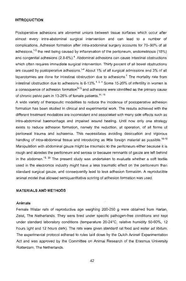
INTRODUCTION
Postoperative adhesions are abnormal unions between tissue surfaces which occur after
almost every intra-abdominal surgical intervention and can lead to a number of
complications. Adhesion formation after intra-abdominal surgery accounts for 70-90% of all
adhesions, 1•3 the rest being caused by inflammation of the peritoneum, endometriosis (18%)
and congenital adhesions (2.8-6%). 4 Abdominal adhesions can cause intestinal obstructions
which often requires immediate surgical intervention. Thirty percent of all bowel obstructions
are caused by postoperative adhesions. 5-8 About 1% of all surgical admissions and 3% of all
laparotomies are done for intest'1nal obstruction due to adhesions.7 The mortality rate from
intestinal obstruction due to adhesions is 6-13%. 1·
2·
4 Some 15-20% of infertility in women is
a consequence of adhesion formation9-13 and adhesions were identified as the primary cause
of chronic pelvic pain in 13-26% of female patients. 14·
15
A wide variety of therapeutic modalities to reduce the incidence of postoperative adhesion
formation has been studied in clinical and experimental work. The results achieved with the
different treatment modalities are inconsistent and associated with many side effects such as
intra-abdominal haemorrhage and impaired wound healing. Until now only one strategy
exists to reduce adhesion formation, namely the reduction, at operation, of all forms of
peritoneal trauma and ischaemia. This necessitates avoiding desiccation and vigorous
handling of intra-abdominal tissue and introducing as little foreign material as possib!e.16-18
Manipulation with abdominal gauze might be traumatic to the peritoneum either because it is
rough and abrades the peritoneum and serosa or because remnants of gauze are left behind
in the abdomen. 19·
20 The present study was undertaken to evaluate whether a soft textile
used in the electronics industry might have a less traumatic effect on the peritoneum than
standard surgical gauze, and consequently lead to less adhesion formation. A reproducible
animal model that allowed semiquantitative scoring of adhesion formation was used.
MATERIALS AND METHODS
Animals
Female Wistar rats of reproductive age weighing 200-250 g were obtained from Harlan,
Zeist, The Netherlands. They were bred under spec"1f1c pathogen-free conditions and kept
under standard laboratory conditions (temperature 20-24°C, relative humidity 50-60%, 12
hours light and 12 hours dark). The rats were given standard rat food and water ad libitum.
The experimental protocol adhered to rules laid down by the Dutch Animal Experimentation
Act and was approved by the Committee on Animal Research of the Erasmus University
Rotterdam, The Netherlands.
42

Gauzes
The evaluated "gauzes" were surgical Medipres gauze (van Heek Medical, Lasser, The
Netherlands), consisting of 100% cotton and commonly used in abdominal surgery, and non·
surgical Fastsorb cleanroom wiper (Berkshire Corporation, Great Barrington, Massachusetts,
USA), used in the electronics industry (Figure 3.1 ). Fastsorb is a rayon-polyester blend which
possesses strength and softness combined with a high absorbing capacity. It has low particle
generation, minimal extractable matter and a low ion content. Fastsorb is used on abrasion
sensitive surfaces.
Absorbing capacity
The absorbing capacity of both materials was evaluated using the following method. Ten 5 x
5 em-pieces of surgical Medipres gauze (double layer) and non-surgical Fastsorb textile
(monolayer) were saturated by immersion in water. After saturation, the materials were
drained for 30 seconds. The pieces were weighed before and after saturation; results were
expressed in millllitres of water absorbed per gram gauze.
Surgical techniques
The three adhesion models employed in this study were derived from a model described by
van Bakkum et al.21
Model 1. Under ether anaesthesia and aseptic but not sterile conditions, using Biogel sterile
gloves (Regent Hospital Products, London, UK), the abdomen was shaved and cleaned with
alcohol. Laparotomy was performed using a lower midline incision of 5 em. In the lateral
abdominal wall, 1.5 em downwards from the abdominal incision, a standard area of 1.5 x 1.2
em, 3 mm deep, which contained the mesothelial as well as both underlying muscular layers,
was excised with an oval shaped punch. This severe peritoneal defect was then closed with
three 5-0 Surgilene sutures (Braun, Melsungen, Germany). Subsequently the uterus horn
was sutured to the lateral peritoneum, both proximally and distally from the closed peritoneal
defect, with 6-0 Surgilene. All knots were double and fastened tightly to ensure ischaemia.
The abdomen was closed in two layers with 5-0 polyglycolic acid and 2-0 silk sutures.
Model 2. The same type of operation was performed with only one difference: the damage
inflicted to the lateral peritoneum was of moderate severity and consisted only of an incision
of 1.5 em long, 3 mm deep, which was also closed with three 5-0 Surgilene sutures.
Model 3. In this model minimal trauma was applied to the lateral peritoneum. No wound was
inflicted. The trauma was ischaemic only and was brought about by three 5-0 Surgilene
sutures at 0. 7 em intervals.
43

Figure 3.1
On the left surgical Medipres gauze and on the right non-surgical Fastsorb texti le.
Figure 3.2
Rubbing device used to apply standardised trauma to the uterine horn. A spring attached to
the handle enables rubbing under a certain pressure. The device is constructed in such a
manner that a pressure of 120 g/cm2 is applied when the indicator needle (above the spring)
touches the blue roll. The gauze is wrapped and fixed around a hooked injection needle (1.1
x 40 mm Luer-Lock; Braun, Melsungn, Germany).
44

Rubbing with surgical gauze and non-surgical textile was performed using a device enabling
the application of a constant pressure of 120 g/cm2 (Figure 3.2). The uterus horn was rubbed
ten times over its total length.
Experimental design
Adhesion formation after different amounts of peritoneal trauma.
In 15 rats one lateral abdominal wall side was operated on according to adhesion model
(severe peritoneal trauma). In nine of these animals the other side was treated according to
adhesion model 2 (moderate peritoneal trauma) and six were treated according to adhesion
model 3 (minimal peritoneal trauma).
Adhesion formation in model 3 after rubbing with surgical gauze or non-surgical
textile.
In 30 rats minimal peritoneal trauma was inflicted to both lateral peritoneal sides (model 3).
Both uterine horns were exposed and sutured to the lateral peritoneum. In ten rats one
uterine horn was rubbed with surgical Medipres gauze whereas the other was not touched
(group I); in ten animals one uterine horn was rubbed surgical Medipres gauze and the other
one with non-surgical Fastsorb textile (group II); and in the last ten rats one uterine horn was
rubbed with Fastsorb textile while the other was not touched (group Ill). In all cases
randomisation was used to determine which rubbing model was to be carried out on which
side.
Evaluation of adhesion formation.
Some 14 days after operation the rats were sacrificed by a humane method for assessment
of post-surgical adhesion formation. Macroscopically the adhesions were scored according to
their extent (quantity) and type (quality) by two independent observers. The extent of
adhesion formation was quantified by dividing the area to be scored into eight by means of
the three 5-0 sutures by which the defect was closed (Figure 3.3). The presence or absence
of adhesions in the eight demarcated areas was scored. If there were adhesions in an area
this accounted for 12.5% adhesions; a maximum of 100% adhesions could be scored. In
each rat two lateral peritoneal sides were assessed. The type of adhesions formed was
classified macroscopically using the ZUhlke classification (Table 3.1). 22
45

Evaluation of peritoneal damage
Microscopic analysis of peritoneal reaction after rubbing ten times with surgical Medipres
gauze and non-surgical Fastsorb textile was performed. Peritoneal reaction was evaluated
directly after, and 2 and 4 hours after rubbing the uterus horn. For each time point one-third of
the four rubbed uterus horns (two rubbed with Medipres and two with Fastsorb) were excised
and fixed in formalin. The samples were stained with haematoxylin and eosin. The appearance
of the mesothelial and underlying muscular layers and the infiltration of leucocytes were
observed and compared.
Statistical analysis
Statistical analysis was performed using the t-test for paired samples. In two instances the
unpaired Student's ttest for independent samples was used, as indicated in the text. Statistical
significance was defined as p < 0.05. Data were expressed as mean adhesion percentage ±
so.
RESULTS
Gauzes: absorbing capacity
The mean absorbing capacity of non-surgical Fastsorb textile (n = 10) was 8.4 ml per g gauze
(SO: 0.2) and that of surgical Medipres gauze (n = 10) was 6.6 ml per g gauze (SO: 0.6). This
difference is statistically significant (p < 0.0001, Student's t test for independent samples).
Adhesion formation after inflicting different amounts of peritoneal trauma.
On almost all peritoneal defects adhesion formation was found after 2 weeks. No pathological
conditions as bowel obstruction, peritonitis or abscesses were found.
Table 3.2 shows that a significantly more adhesions were found at sites where severe (model
1) trauma had been applied, compared with moderate (model 2) and minimal (model 3)
peritoneal trauma (p ::; 0.05). Adhesions formed after severe and moderate trauma of the
peritoneum were dense and thick and could be classified as Zuhlke type 2-3 and Zuhlke type
2. The pelvic fat and the uterine horn took part in the adhesion formation. The adhesions
formed after minimal trauma of the peritoneum were filmy and could be classified as ZUhlke
type 1-2. Only pelvic fat took part in the adhesion formation.
46

sut re 3
12.5% 12.5%
12.5% 12.5% 12.5% 12.5%
Figure 3.3
The extent of adhesion formation was quantified by dividing the "defect" area to be scored into
eight areas of 12.5% by means of the three sutures used to close the defect.
Score Characteristics
Filmy adhesion, easy to separate by blunt dissection
2 Stronger adhesion: blunt dissection possible, partly sharp dissection necessary;
beginning of vascularisation
3 Strong adhesion: lysis possible by sharp dissection only; clear vascularisation
4 Very strong adhesion: lysis possible by sharp dissection only; organs strongly
attached with severe adhesions: damage of organs hardly preventable
Table 3.1
Macroscopic (morphological) classification of abdominal adhesions according to Zuhlke et
al.zz
47

Adhesion formation in model 3 after rubbing with surgical gauze or non~surgical textile.
Table 3.3 shows that rubbing the uterine horn with surgical Medipres gauze after infliction of
minimal peritoneal trauma gave rise to a significantly higher mean adhesion percentage than
infliction of minimal peritoneal trauma alone (p ~ 0.0001 ). The adhesions in the latter group
were often filmy (Zuhlke type 1-2) involving only pelvic fat. Adhesions at srtes also rubbed by
Medipres gauze were denser (ZUhlke type 2-3); pelvic fat, uterine horn and the bowel were
involved in these adhesions.
Table 3.3 shows that, after infliction of minimal peritoneal trauma, rubbing the uterus horn with
surgical Medipres gauze induced a significantly higher mean adhesion percentage than
rubbing the (opposite) uterus horn with non-surgical Fastsorb textile (P ~ 0.0001). The
adhesions formed after rubbing with Medipres gauze were more dense (ZUhlke type 2-3) than
those formed after rubbing with Fastsorb textile (Zuhlke type 1-2). In the first group the uterine
horn, the bowel and pelvic fat were involved in adhesion formation, whereas in the Fastsorb
group the bowel was never involved and the uterine horn rarely.
The mean adhesion percentage found after infliction of minimal peritoneal trauma did not
significantly differ from that observed after combining this minimal trauma with rubbing the
uterus with non-surgical Fastsorb textile (p = 0.23). The adhesions found were all filmy, Zuhlke
type 1 in the first group and type 1-2 in the second group; only pelvic was involved in adhesion
formation.
Light microscopy
In all cases the surface of the uterine peritoneum was more damaged after rubbing with
surgical Medipres gauze than after rubbing with non-surgical Fastsorb textile. After rubbing
with Medipres gauze, the mesothelial layer had disappeared and the underlying muscular layer
was partly lost and looked frayed. After rubbing with Fastsorb textile the mesothelial layer had
disappeared for the most part but the underlying muscular layer looked smooth and was intact.
At 2 and 4 hours after causing peritoneal damage with Medipres there were more leucocytes
infiltrating the severely damaged area than after rubbing with Fastsorb (Figure 3.4 ).
48

Abdominal trauma n Percentage adhesion Zuhlke p1 p2 p3
formation (SD) score
Model1 15 60 (12.7) 2-3 0.011
Model2 9 44 (15.5) 2 0.004
Model3 6 21 (6.5) 1-2 0.005
Table 3.2
Mean adhesion percentages (SD), and ZUhlke classification of found adhesions, after inflicting
severe (model 1 ), moderate (model 2) and minimal (model 3) peritoneal trauma. Fifteen rats
underwent an operation according to adhesion model 1 at one lateral abdominal wall side. In 9
of these rats the other, opposite, side was operated according to adhesion model 2 and in 6 of
these rats according to adhesion model 3. Therefore, n is the number of peritoneal sites
(uterine horns) assessed. Statistics p1 and p3: t test for paired samples (model 1 versus
model 2 and model 1 versus model 3). Statistics p2: Student's t test for independent
samples (model 2 versus model 3 ).
Uterine horns n Percentage adhesion Zuhlke pl p2 p3
fonnation (SD) score
I". No touch 10 23 (9.9) 1-2
Ib Medipres rubbing 10 58 (10.5) 2-3 < 0.0001
II'. Medipres rubbing 10 63 (11.8) 2-3
lib Fastsorb rubbing 10 19 (12.2) 1-2 < 0.0001
III'. No touch 10 24(11.6) I
Illb. Fastsorb rubbing 10 35 (24.3) 1-2 ns
Table 3.3
Mean adhesion percentages (SD), and ZUhlke classification of found adhesions, after inflicting
minimal peritoneal trauma alone (the uterine horn was not touched) and after rubbing the
uterine horn with Medipres gauze or Fastsorb textile following the infliction of minimal
peritoneal trauma. N is the number of defected peritonea! sites (uterus horns) assessed. Statis
tics p1 (I" versus I b), p2 (II' versus II b) and p3 (Ill' versus Ill b): !-test for paired samples (95%
Cl = 95% confidence interval).
49

A
B
Figure 3.4
Light micrograph of uterus horn surfaces after rubbing with Medipres gauze (A) and Fastsorb
textile (B). A: the mesothelial layer has disappeared, the underlying muscular layer is partly
lost and frayed. Reactive leucocyte cells infiltrate the severely damaged surface area. 8: The
mesothelial layer has disappeared for the most part but the underlying muscular layer is
intact. Magnification 1 Ox.
50

DISCUSSION
Numerous attempts in clinical and animal studies to prevent or reduce the incidence of intra
peritoneal adhesions have yielded limited success.
Adhesion formation is a dynamic process consisting of several consecutive stages following
damage to the parietal or visceral peritoneum. Different studies, in which peritoneal trauma
was proven to be a cause of adhesion formation, 23•
24·
32 did not mention whether there was an
association between the degree of trauma inflicted, and the extent of formed adhesions. The
present study shows that the extent and type of post-surgical adhesion formation correlates
significantly with the degree of peritoneal damage. At sites where severe, moderate or minimal
peritoneal trauma was inflicted, mean adhesion percentages of 60%, 44% and 21%
respectively were found.
Damage to the peritoneum causes a local inflammatory reaction which leads to the formation
of a serosanguineous exudate.23-26 In the exudate fibrin deposition will take place,25
-27 and
when two surfaces contact each other during this process a fibrinous adhesion will appear.
Whether these adhesions are permanent of character or will eventually be lysed is dependent
on the fibrinolytic capacity of the peritoneum. 25·
28-
31 When the peritoneum is slightly damaged
and mesothelial cells are mostly intact, there will be a dynamic balance between fibrinogenesis
and fibrinolysis, and adhesion-free healing may take place. When the peritoneum is more
severely damaged the underlying connective tissue will be exposed and normal serosal
fibrinolytic activity will be lost.32 Exposed endothelial and inflammatory cells, as well as
damaged mesothelial cells, may produce plasminogen activator inhibitor 1 and 2, factors
mediating a reduced functional fibrinolytic activity. 29 Low fibrinolytic activity allows the fibrinous
adhesion to organise to a fibrous permanent adhesion. 24 Therapeutic modalities, focusing on
the different stages in adhesion formation include the use of anticoagulants and anti
inflammatory agents,33-35 removal of fibrinous exudates by peritoneal lavages,36 the
augmentation of fibrinolysis by use of polysorb, saccharose, vitamin E or recombinant tissue
plasminogen activator (t-PA), 37 and the inhibition of fibroblast proliferation by use of steroids
and 5-fluorouracil. 38 Proving clinical effectiveness remains difficult and there may be severe
side-effects.
Recent animal and clinical studies evaluating mechanical separation during reperitonealisation
of two adjacent anatomical structures are more promising.39-4
2 Non-resorbable and resorbable
barriers have up to now been used mainly in experimental settings, leaving minimalisation of
peritoneal trauma as the main therapeutic modality in prevention of postoperative adhesion
formation.
Limitation of peritoneal injury should include attempts to reduce trauma inflicted by surgical
gauze. Down et al19·
20 showed in two experimental studies that the abrasive effect of gauzes
51
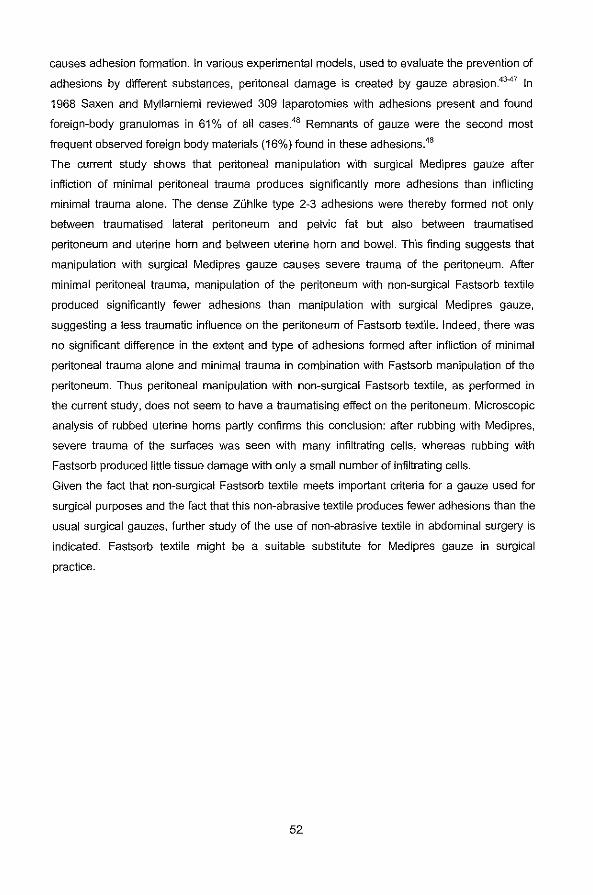
causes adhesion formation. In various experimental models, used to evaluate the prevention of
adhesions by different substances, peritoneal damage is created by gauze abrasion.4347 In
1968 Saxen and Myllamiemi reviewed 309 laparotomies with adhesions present and found
foreign-body granulomas in 61% of all cases.48 Remnants of gauze were the second most
frequent observed foreign body materials (16%) found in these adhesions.48
The current study shows that peritoneal manipulation with surgical Medipres gauze after
infliction of minimal peritoneal trauma produces significantly more adhesions than inflicting
minimal trauma alone. The dense ZUhlke type 2-3 adhesions were thereby formed not only
between traumatised lateral peritoneum and pelvic fat but also between traumatised
peritoneum and uterine horn and between uterine horn and bowel. This finding suggests that
manipulation with surgical Medipres gauze causes severe trauma of the peritoneum. After
minimal peritoneal trauma, manipulation of the peritoneum with non-surgical Fastsorb textile
produced significantly fewer adhesions than manipulation with surgical Medipres gauze,
suggesting a less traumatic influence on the peritoneum of Fastsorb textile. Indeed, there was
no significant difference in the extent and type of adhesions formed after infliction of minimal
peritoneal trauma alone and minimal trauma in combination with Fastsorb manipulation of the
peritoneum. Thus peritoneal manipulation with non-surgical Fastsorb textile, as performed in
the current study, does not seem to have a traumatising effect on the peritoneum. Microscopic
analysis of rubbed uterine horns partly confirms this conclusion: after rubbing with Medipres,
severe trauma of the surfaces was seen with many infiltrating cells, whereas rubbing with
Fastsorb produced little tissue damage with only a small number of infiltrating cells.
Given the fact that non-surgical Fastsorb textile meets important criteria for a gauze used for
surgical purposes and the fact that this non-abrasive textile produces fewer adhesions than the
usual surgical gauzes, further study of the use of non-abrasive textile in abdominal surgery is
indicated. Fastsorb textile might be a suitable substitute for Medipres gauze in surgical
practice.
52

REFERENCES 1. Perry JF, Smith GO, Yonehiro EG. Intestinal obstruction caused by adhesions; a review of 388
cases. Ann Surg 1955; 142:810-816.
2. Raf LE. Causes of small intestinal obstructions. A study covering the Stockholm area. Acta Chir
Scand 1969; 135:67-72.
3. Weibel MA, Majno G. Peritoneal adhesions and their relation to abdominal surgery. Am J Surg
1973; 126:345-353.
4. Nemir P Jr. Intestinal obstruction: ten year survey at the Hospital of the University of
Pennsylvania. Ann Surg 1952; 135:367-375.
5. Ellis H. The causes and prevention of intestinal adhesions. Br J Surg 1982; 69: 241-243.
6. McEntee G, Pender D, Mulvin D, McCullough M, Naeeder S, Farah S, Baburdeen MS, Ferraro
V, Cham C, Gillham N. Current spectrum of intestinal obstruction. Br J Surg 1987; 74: 976-980.
7. Menzies D, El!is H. Intestinal obstruction from adhesions-how big is the problem? Ann R Col!
Surg Eng! 1990; 72: 60-63.
8. Menzies D. Peritoneal adhesions. Incidence, cause and prevention. Surg Annu 1992; 24: 27-45.
9. Drake TS, Grunert GM. The unsuspected pelvic factor in the infertility investigation. Br J Surg
1980; 50: 10-16.
10. Pfeffer WH. Adjuvants in tubal surgery. Fertil Steril 1980; 33: 245-256.
11. Gamel V. An odyssey through the evident. Fertil Steri11983; 39: 144-156.
12. Fayez JA. An assessment of the role of operative !aparoscopy in tubop!asty. Fertil Steri/1983;
39:476-479.
13. Hershlag A, Diamond MP, DeCherney AH. Adhesiolysis. C/in Obstet Gynecol 1991; 34: 395-
302.
14. Goldstein DP, DeCholnoky C, Emans SJ, Leventhal JM. Laparoscopy in the diagnosis and
management of pelvic pain in adolescents. J Reprod Med 1980; 24: 251-256.
15. Rapkin AJ. Adhesions and pelvic pain: a retrospective study. Obstet Gyneco/1986; 68: 13-15.
16. Ward GG. Prevention of postoperative adhesions of the peritoneum. Am J Obstet 1901; 43: 753-
771.
17. Siegler AM, Kontopou!os V. An analysis of macrosurgical and microsurgical techniques in the
management of the tuboperitoneal factor in infertility. Fertil Steri/1979; 32: 377-383.
18. Luijendijk RW, Lange de DC, Wauters CCAP, Hop WC, Duron JJ, Pailler JC, Camprodon BR,
Holmdahl LE, Geldrop van HJ, Jeekel J. Foreign material in postoperative adhesions. Ann
Surg 1996; 223: 242-248.
19. Down RHL, Whitehead R, Watts JMcK. Do surgical packs cause peritoneal adhesions? Aust N
Z J Surg 1979; 49: 379-382.
20. Down RHL, Whitehead R, Watts JMcK. Why do surgical packs cause peritoneal adhesions?
Aust N Z J Surg 1980; 50: 83-85.
21. Bakkum EA, van Blitterswijk CA, Dalmeijer RAJ, Trimbos JB. A semiquantitative rat model for
postoperative adhesion formation. Gynecol Obstet Invest 1994; 37: 99-105.
53

22. ZUhlke HV, Lorenz EMP, Straub EM, Savvas V. Pathophysiologie und Klassifikation von
Adh8.sionen. Langenbecks Arch Chir 1 990; 345: 1009-1016.
23. Ellis H. The healing of peritoneum under normal and pathological conditions. Br J Surg 1965; 52:
471-476.
24. Milligan OW, Raftery AT. Observations on the pathogenesis of peritoneal adhesions: a light and
electron microscopical study. Br J Surg 1974; 61:274-280.
25. Thompson JN, Whawell SA. Pathogenesis and prevention of adhesion formation. Br J Surg
1995; 82: 3-5.
26. Monz FJ, Shimanuki T, diZerega GS. Post-surgical mesothelial reepithelialisation. In:
DeCherney AH, Polan MLeds. Reproductive Surgery, Year Book Medical Publishes. 1987: 31-
47.
27. Holz G. Prevention and management of peritoneal adhesions. Fertil Steri/1984; 41: 497-507.
28. Buckman RF, Woods M, Sargent L, Gervin AS. A unifying pathogenetic mechanism in the
aetiology of intra-peritoneal adhesions. J Surg Res 1976; 20: 1-5.
29. Vipond MN, Whawell SA, Thompson JN, Dudley HA. Peritoneal fibrinolytic activity and intra
abdominal adhesions. Lancet 1990; 335: 1120-1122.
30. Raftery AT. Effect of peritoneal trauma on peritoneal fibrinolytic activity and intra-peritoneal
adhesion formation. Eur J Res 1981; 13: 397-401.
31. Goor van H, Graaf FJ, Grand J, Sluiter WJ, Meer J, Born VJJ, Bleichrodt RP. Fibrinlytic activity
in the abdominal cavity of rats with faecal peritonitis. Brit J Surg 1994; 81: 1046-1049.
32. Graeme BR, Grobety J, Majno G. Mesothelial injury and recovery. Am J Patho/1973; 71: 93-
102.
33. Davidson MN. Systemic administration of heparin and dicumarol for postoperative adhesions: an
experimental study. Arch Surg 1949; 59: 300-325.
34. Rodgers KE. Non-steroidal anti-inflammatory drugs (NSAID's) in the treatment of post-surgical
adhesion. In: Treatment of Postsurg Adh. Eds.: GS diZerega, L Russeii-Malinak, MP Diamond,
CB Linsky, Willy-Liss, 1990: 119-130.
35. Hackel M, Ott S, Siemann U, Kissell T. Prevention of peritoneal adhesions in the rat with
sustained intra-peritoneal dexamethason delivered by a novel therapeutic system. Ann Chir
Gynecof 1987; 76:306-313.
36. Treutner KH, Winkeltau G, Lerch MM, Stadel R, Schumpelick V. Prevention of postoperative
intra-peritoneal adhesions. A new study standardised in rats. Research in Surgery 1992; 4: 50-
54.
37. Doody KJ, Dunn RC, Buttram VC. Recombinant tissue plasminogen activator reduces adhesion
formation in a rabbit uterine horn model. Fertif Steri/1989; 51:509-512.
38. Holden M, Adams LB. Inhibitory effect of cortisone acetate and hydrocortisone on growth of
fibroblasts. Proc Soc Exp Bioi Med 1957; 96: 64.
39. Adhesion Barrier Study Group (lnterceed, TC7). Prevention of post-surgical adhesions by
lnterceed. An absorbable adhesion barrier: a prospective randomised multicenter clinical study.
Fertif Sterif 1989; 51:933-938.
54

40. Steinleitner A, Lambert H, Kazensky C, Cantor B. Polaxamer 407 as an intra-peritoneal barrier
material for the prevention of post-surgical adhesion formation and reformation in rodent models
for reproductive surgety. Obstet Gynecof 1991; 77: 48-52.
41. Goldberg EP, Burns JW, Yaacobi Y, Genzyme Corporation, Cambridge, MA. Prevention of
postoperative adhesions by precoating tissues with dilute sodium hyaluronic solutions.
Gynaecologic Surgery and adhesion prevention, 1993 Wiley-Uss, Inc. pages 191-204.
42. Becker JM, Dayton MT, Fazio V. Evaluation of the safety and Effectiveness of HAL™
bioresorbable membrane in limiting postoperative adhesion formation to serosal tissue. Clinical
Congress of the American College of Surgeons 1995.
43. Yaacobi Y, Goldberg EP, Habal MB. Effects of Ringer's lactate irrigation on the formation of
postoperative abdominal adhesions. J Invest Surg 1991; 4: 31-36.
44. Moll HD, Schumacher J, Wright JC, Spano JS. Evaluation of sodium carboxymethyl cellulose for
prevention of experimentally induced abdominal adhesions in pony's. Am J Vet Res 1991; 52:
88-91.
45. Hemadeh 0, Chilukuri S, Bonet V, Hussein S, Chaudry IH. Prevention of peritoneal adhesions
by administration of sodium carboxylmethyl cellulose and oral vitamin E. Surgery 1993; 114:
907-910.
46. Wurster SH, Bonet V, Mayberry A, Hoddinott M, Williams T, Chaudry IH. Intra-peritoneal sodium
carboxymethylcellulose administration prevents reformation of peritoneal adhesions following
surgical lysis. J Surg Res 1995; 59:97-102.
47. Harris SH, Morgan FM, Rodeheaver GT. Analysis of the kinetics of peritoneal adhesion
formation in the rat and evaluation of potential anti-adhesive agents. Surgery 1995; 117: 663-
669.
48. Saxen L, Myllarniemi H. Foreign materials and postoperative adhesions. New Eng J Med 1968;
279: 200-202.
55


Chapter IV
Peroperative Abdominal lavage Promotes Intra
Abdominal Adhesion Formation in the Rat Adapted from the original publication in the European Surgical Research 1999; 31: 196-201
Intra-abdominal adhesions continue to pose a serious postoperative clinical problem. Per
operative peritoneal trauma can lead to postoperative adhesion formation. In vitro studies
show that exposure of the peritoneal surface to lavage solutions enhances peritoneal activity
and provokes an inflammatory response. This study was designed to evaluate the in vivo
effect of lavage solutions on the peritoneum and post-surgical adhesion formation.
A reproducible rat model allowing semiquantitative scoring of adhesions was used to study
adhesion formation after lavage of the abdominal cavity with RPM! medium, NaG! 0.9%,
polyvinylpyrrolidone iodine 1% (PVP-1), Viaspan and chlorhexidine 0.02% in dilution. All
solutions were applied during a laparotomy in which standardised minimal peritoneal trauma
was inflicted.
Peritoneal lavage of the abdominal cavity after the infliction of minimal peritoneal trauma
induced significantly more adhesions than the infliction of minimal peritoneal trauma alone (p
= 0.0001). Aggressive as well as non-aggressive lavage solutions caused extra adhesion
formation.
The results found in the present study correlate with observations from in vitro experiments;
exposure of peritoneal cavity to lavage solutions traumatises the peritoneum, enhances
peritoneal activation and thus promotes intra-abdominal adhesion formation. During surgery
in non-contaminated abdominal cavities peritoneal lavage should not be performed. Per
operative lavage of contaminated abdominal cavities should be viewed with caution.
57
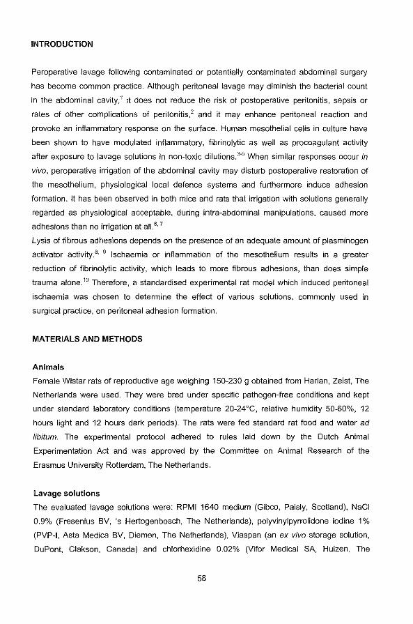
INTRODUCTION
Peroperative lavage following contaminated or potentially contaminated abdominal surgery
has become common practice. Although peritoneal lavage may diminish the bacterial count
in the abdominal cavity, 1 it does not reduce the risk of postoperative peritonitis, sepsis or
rates of other complications of peritonitis, 2 and it may enhance peritoneal reaction and
provoke an inflammatory response on the surface. Human mesothelial cells in culture have
been shown to have modulated inflammatory, fibrinolytic as well as procoagulant activity
after exposure to lavage solutions in non-toxic dilutions.3-5 When similar responses occur in
vivo, peroperative irrigation of the abdominal cavity may disturb postoperative restoration of
the mesothelium, physiological local defence systems and furthermore induce adhesion
formation. lt has been observed in both mice and rats that irrigation with solutions generally
regarded as physiological acceptable, during intra-abdominal manipulations, caused more
adhesions than no irrigation at all. 6·
7
Lysis of fibrous adhesions depends on the presence of an adequate amount of plasminogen
activator activity.8·
9 Ischaemia or inflammation of the mesothelium results in a greater
reduction of fibrinolytic activity, which leads to more fibrous adhesions, than does simple
trauma alone. 10 Therefore, a standardised experimental rat model which induced peritoneal
ischaemia was chosen to determine the effect of various solutions, commonly used in
surgical practice, on peritoneal adhesion formation.
MATERIALS AND METHODS
Animals
Female Wistar rats of reproductive age weighing 150-230 g obtained from Harlan, Zeist, The
Netherlands were used. They were bred under specific pathogen-free conditions and kept
under standard laboratory conditions (temperature 20-24°C, relative humidity 50-60%, 12
hours light and 12 hours dark periods). The rats were fed standard rat food and water ad
libitum. The experimental protocol adhered to rules laid down by the Dutch Animal
Experimentation Act and was approved by the Committee on Animal Research of the
Erasmus University Rotterdam, The Netherlands.
lavage solutions
The evaluated lavage solutions were: RPMI 1640 medium (Gibco, Paisly, Scotland), NaCI
0.9% (Fresenius BV, 's Hertogenbosch, The Netherlands), polyvinylpyrrolidone iodine 1%
(PVP-1, Asta Medica BV, Diemen, The Netherlands), Viaspan (an ex vivo storage solution,
DuPont, Clakson, Canada) and chlorhexidine 0.02% (Vifor Medical SA, Huizen, The
58

Netherlands). Dilutions of the lavage solutions were prepared in medium (RPM!) for optimal
pH (7.2-7.4) and osmolarity (280-300 mosmol/kg). All solutions were prepared under
pathogen-free conditions. The analysed solutions were divided into non-aggressive and
aggressive lavage solutions. Non-aggressive solutions caused only moderate activation of
mesothelial functional properties in previous in vitro experiments. 2-5 Aggressive solutions
provoked major mesothelial cytokine production and other functional activation of cultured
human mesothelial cells. 3·5
Operative procedures
To study the effect of the different solutions on adhesion formation in rats our previously
described reproducible rat adhesion model was used. 11 Briefly, under ether anaesthesia and
aseptic conditions a laparotomy was performed using a lower midline incision of 5 em. Three
Surgilene 5-0 sutures (Braun, Melsungen, Germany) were applied to both lateral peritoneal
sides 0.7 em from each other and 1.5 em downwards from the abdominal incision. All knots
were double knots fastened tightly to ensure local ischaemia. Both uterine horns were
exposed and sutured to the lateral peritoneum with Surgilene 6-0 (Braun) proximally and
distally from the three 5-0 sutures. In this way a standardised amount of minimal peritoneal
trauma was inflicted. Subsequently the abdominal cavity was exposed to 10 ml of lavage
solutions (25°C) for 10 minutes. After 10 minutes the fluids were suctioned out carefully. At
least 5 ml of the lavage solutions remained within the peritoneal cavity. The abdomen was
closed in two layers with 5-0 polyglycolic acid and 2-0 silk sutures.
Experimental design
Adhesion formation after peritoneal lavage with non~aggressive solutions.
In 15 rats, standardised minimal peritoneal trauma was inflicted to both lateral peritoneal
sides by applying 3 sutures in, and fixating the uterine horns to the lateral peritoneum. In five
rats (group I) no peritoneal lavage was performed. The other ten rats were treated with non
aggressive solutions. Five rats (group II) underwent lavage with 10 ml NaCI 0.9% and five
(group Ill) with 10 ml povidone-iodine (PVP-1 diluted 1:100 in RPMI).
Adhesion formation after peritoneal lavage with aggressive solutions.
Again minimal peritoneal trauma was inflicted to both lateral peritoneal sides in 18 rats. Six
rats (group IV) underwent a lavage with the non-aggressive solution RPM!, six rats (group V)
underwent a lavage with Viaspan and six (group VI) with the aggressive lavage solution
chlorhexidine 0.02% (diluted in 1:10 in culture medium).
59

Evaluation of adhesion formation.
Fourteen days postoperatively the rats were sacrificed for assessment of post-surgical
adhesion formation. Macroscopically the adhesions were scored according to their extent
(quantity) and type (quality) by three independent observers. Quantification was assessed by
dividing the area to be scored into eight by means of the three 5-0 sutures in the lateral
peritoneum (Figure 4.1 ). The presence or absence of adhesions in the eight demarcated
areas was scored. If there were adhesions in an area this accounted for 12.5% adhesions; a
maximum of 100% adhesions could be scored. In each rat two lateral peritoneal sides were
assessed. The type of adhesions formed was classified macroscopically using the ZUhlke
classification (Table 4.1 ).12
suture 1 su ure 2 sut re 3
12.5% 12.5% 12.5% 12.5%
12.5% 12.5% 12.5% 12.5%
Figure 4.1
The extent of adhesion formation was quantified by dividing the "defect" area to be scored into
eight areas of 12.5% by means of the three sutures.
Score Characteristics
Filmy adhesion, easy to separate by blunt dissection
2 Stronger adhesion: blunt dissection possible, partly sharp dissection necessary;
beginning of vascularization
3 Strong adhesion: lysis possible by sharp dissection only; clear vascularization
4 Very strong adhesion: lysis possible by sharp dissection only; organs strongly
attached with severe adhesions: damage of organs hardly preventable
Table 4.1
Macroscopic (morphological) classification of abdominal adhesions according to ZUhlke et
aiY
60

Statistical analysis
Data were expressed as mean adhesion percentage ± SO. Statistical analysis was pertormed
using the one-way ANOVA test to determine overall differences followed by a Bonferroni post
hoc test to analyse differences between groups if the ANOVA test showed significance.
Statistical significance was defined asp< 0.05
RESULTS
In almost all peritoneal cavities, adhesions were found after 2 weeks. No pathological
conditions as bowel obstruction, peritonitis or abscesses had occurred.
Table 4.1 and 4.2 show that infliction of minimal (ischaemic) peritoneal trauma alone induced a
mean adhesion percentage of 22.5% and that additional peritoneal lavage with all five lavage
solutions gave rise to significantly higher mean adhesion percentages.
Adhesion formation after peritoneal lavage with non-aggressive solutions
Table 4.2 shows that peritoneal lavage with non-aggressive solutions as NaCI 0.9% or
povidone-iodine (PVP-1) after infliction of minimal peritoneal trauma caused over 150% more
adhesions than infliction of minimal peritoneal trauma alone (p = 0.0001). The mean adhesion
percentage found after peritoneal lavage with NaCI 0.9% did not significantly differ from that
observed after lavage with povidone-iodine (p = 0.61 ). Adhesions found after minimal
ischaemic peritoneal trauma were all filmy (ZUhlke type 1 ). The adhesions formed after
peritoneal lavage with NaCI 0.9% or povidone-iodine were more dense and could be classified
as ZUhlke type 1-2. In all groups only pelvic fat took part in the adhesion formation.
Adhesion formation after peritoneal lavage with aggressive solutions.
Tables 4.2 and 4.3 show (again) that peritoneal lavage with non-aggressive solutions as RPMl
or Viaspan after infliction of minimal peritoneal trauma induced about 100% more adhesion
formation than infliction of minimal peritoneal trauma alone (p ~ 0.01 ). Table 4.3 also shows
that peritoneal lavage of a minimally traumatised peritoneal cavity with chlorhexidine (0.02%;
1:10), an aggressive solution for mesothelial cells in culture, induced significantly more
adhesion formation than lavage of minimally traumatised peritoneum with RPMI or Viaspan (p
~ 0.012). The adhesions found after peritoneal lavage with RPMI or Viaspan were dense
(Zuhlke type 2) and involved pelvic fat and seldom the uterine horn. Adhesions found after
lavage with chlorhexidine were more dense (ZUhlke type 3) and the uterine horn, the bowel
and pelvic fat were involved in adhesion formation.
61

Lavage solution n Percentage adhesion Zuhlke p1 p2 p3
formation (SO) score
i, I. No lavage 5 22.5 (8.4) 1 0.0001
Ill NaCI 0.9% 5 60.0 (11.4) 1-2 ns
Ill. PVP-11% 5 63.8 (11.2) 1-2 0.0001 ll
Table 4.2
Mean adhesion percentages (SD) and ZUhlke classification of adhesions found after minimal
peritoneal trauma alone (group I) and after peritoneal lavage with NaCI 0.9% (group II) or
povidone-iodine 1% 1:100 (group Ill) following infliction of minimal peritoneal trauma. For each
rat, the individual data concerning the 2 lateral abdominal wall sites (uterine horns) were
averaged; n is the number of data (::::rats) per group used for analysis. Statistics p1 (I versus
II), p2 (II versus Ill) and p3 (I versus Ill): one-way ANOVA test, with Bonferroni post hoc test.
Lavage solution n Percentage adhesion Zuhlke p1 p2 p3
formation (SO) ' score
IV.RPMI 6 47.7 (12.2) 1-2 ns
V. Vispan 6 40.6 (13.0) 2 0.012
VI. Chlorhexidine 6 70.8 (14.1) 2 0.003 .
Table 4.3
Mean adhesion percentages (SD) and ZUhlke classification of adhesions found after peritoneal
lavage with RPMI (group IV), Viaspan (group V) or chlorhexidine 0.02% 1:10 (group VI)
following infliction of minimal peritoneal trauma. For each rat, the individual data concerning
the 2 lateral abdominal wall sites (uterine horns) were averaged; n is the number of data(::::
rats) per group used for analysis. Statistics: p1 (IV versus V), p2 (V versus VI) and p3 (IV
versus VI): one-way AN OVA test, with Bonferroni post hoc test.
62

DISCUSSION
Intra-peritoneal postoperative adhesion formation creates a major challenge to abdominal
surgery, as it is the main cause of mechanical bowel obstruction, unexplained abdominal pain
and female infertility. Furthermore the presence of dense adhesions makes reoperation
technically difficult. By far the most common cause of intra-peritoneal adhesions is previous
surgical intervention.13 Intra-abdominal surgery will, by traumatising of the peritoneum, alter
mesothelial activity and inflammatory responses of the peritoneum and disturb the fibrinolytic
homeostasis.14'
15 Previous in vitro studies show that exposure of the peritoneum to different
lavage solutions enhances mesothelial functional features and provokes an inflammatory
response.2·5
We have assessed the effect of peroperative peritoneal lavage, with non-aggressive and
aggressive solutions, in vivo on postoperative adhesion formation. We used an established
reproducible rat adhesion model in which standardised peritoneal trauma, which mimics
peritoneal injury induced in patients during abdominal surgery, was inflicted before peritoneal
irrigation took place. As in our previous studies the infliction of standardised minimal ischaemic
peritoneal trauma caused a mean adhesion percentage of 22%. The present study shows that
peritoneal lavage of areas with ischaemic injury is associated with increased adhesion
formation. Aggressive as well as non-aggressive solutions caused significantly more
adhesions. Peritoneal lavage with aggressive solutions increased adhesion formation
significantly more than lavage with non-aggressive solutions. Irrigation with aggressive fluids
gave rise to adhesions that were more dense and contained more organs than the adhesions
formed after irrigation with non-aggressive solutions.
Possible explanations for the induction of adhesions by lavage solutions include cellular injury,
increased peritoneal permeability and peritoneal activation.16 We have studied cellular injury by
measuring the release of an intracellular enzyme (LDH) in vitro and in peritoneal fluid of
patients undergoing elective colonic surgery and noted increased levels of LDH after exposure
to lavage solutions (unpublished data). Furthermore, permeability studies with a
macromolecule (inulin) after treatment of mesothelial monolayers with lavage solutions showed
disturbance of mesothelial integrity with increase permeability. This facilitated passage for
macromolecules may cause increasing fibrous exudation to the abdominal cavity and thereby
induce adhesion formation. In vitro studies also show an increased pro-inflammatory peritoneal
cytokine response after exposure to various solutions. In vivo these increased cytokine
concentrations may lead to an enhanced inflammatory response and increased adhesion
formation.2·
5 Moreover, procoagulant and fibrinolytic properties of human mesothelial
monolayers were also affected by various solutions in earlier in vitro experiments.5
Notwithstanding an overall increase of responses of human mesothelial cells in culture to
63

various kinds of clinically used lavage solutions, not all solutions induced an equal effect. In the
current in vivo study no significant difference in the extent of postsurgical adhesion formation
was observed between exposure of the peritonea! cavity to NaC! 0.9% or povidone-iodine 1%,
1:100. This discrepancy between in vitro and in vivo results may be explained as follows.
Firstly, by observations indicating that the cytokine profile and peritoneal fibrinolytic activity
differ in different species. 17-19 In humans, intra-peritoneal responses to stimuli may differ and be
more complicated compared with responses in the rat. Secondly, povidone-iodine as a highly
viscous solution reduced mesothelial cell activation in vitro probably by coating the cells. In vivo
the coating effect over the entire peritoneal surface may not be as effective as in vitro.
Studies at our laboratories have emphasised the use of scavengers to minimise human
mesothelial cell damage and activation during oxidative stress. The trauma induced in this rat
model was mainly ischaemic injury. Therefore, we also tested the organ preservation fluid
Viaspan, regarded as more biocompatible as it contains lactate and oxigen radical scavengers
(glutathione and allopurinol). Nevertheless peroperative lavage with Viaspan also induced a
significant increase of adhesion formation. However the effect of antioxidants on mechanisms
underlying the initiation of fibrin appeared also to be species specific in another experimental
study.20
In conclusion, this study is in accordance with previous in vitro experiments with human
mesothelial cells. This study shows that exposure of the abdominal cavity to peritoneal lavage
solutions may lead to more postoperative adhesion formation. Therefore, peroperative
irrigation with any kind of lavage solution should be discouraged especially during surgery in
non-contaminated abdominal cavities. Further studies to establish the composition of an ideal
lavage solution which does not traumatise the peritoneum and which might even protect the
peritoneum from the effects of trauma seems to be indicated.
64

REFERENCES 1. Ambrose NS, Donovan lA, Deerges S, Minervini S, Harding LK. The efficacy of peritoneal lavage
at elective abdominal operations. Br J Surg 1982; 69: 143-144.
2. Minervini S, Bently S, Youngs D, Williams JA, Burdon D, Keighley MRB. Prophylactic saline
peritoneal lavage in elective colorectal operations. Dis Col & Rect 1980; 23: 292-294.
3. Westreenen van M, Mul FPJ, Prank A, Hoynck van Papendrecht AAGM, Diepersloot RJ, Roos
D, Legu!t P. Influence of peroperative lavage solutions on peritoneal defence mechanisms in
vitro. Eur J Surg 1999; 165: 1066-1071.
4. Sendt W, Schaffel U, Schantz JT. Peritoneal cytokine respons: In vitro study on interleukin (II) 1 b
and 11-6 release in two different models (abstract). Br J Surg 1996; 83: 856.
5. Westreenen van M, Pronk A, Hoynck van Papendrecht AAGM, Groot de PhG, Leguit P. Effect of
perioperative lavage solutions on coagulant and fibrinolytic properties of mesothelial cells.
Submitted for publication.
6. Philips RKS, Dudley HAF. The effect of tetracycline lavage and trauma on visceral and parietal
peritoneal ultrastructure and adhesion formation. Br J Surg 1984; 71: 537-539.
7. Yaacobi Y, Habal MB, Goldberg EP. Formation of postoperative adhesions following irrigation
with Ringer's lactate solutions. J Invest Surg 1991; 4: 31-36.
8. Buckman RF, Buckman PO, Hufnagel HV, Gervin AS. A physiologic basis for adhesion-free
healing of de-peritonealised surfaces. J Surg Res 1976; 21: 76.
9. Thompson JN, Paterson-Brown S, Harbourne T, Whawell SA, Kalodiki E, Dudley HA. Reduced
human peritoneal plasminogen activity: Possible mechanism of adhesion formation. Br J Surg
1989; 76: 382-384.
10. Ellis H. The aetiology of postoperative abdominal adhesions. An experimental study. Br J Surg
962; 50: 10.
11. Tal van den MP, Stijn van I, Bondhuis F, Marquet RL, Jeekel J. Reduction of intra-peritoneal
adhesion formation by use of non-abrasive gauze. Br J Surg 1997; 84: 1410-1415.
12. ZUhlke HV, Lorenz EMP, Straub EM, Savvas V. Pathophysiologie und Klassifikation von
Adhasionen. Langenbecks Arch Chir 1990; 345: 1009-1016.
13. Ellis H. The causes and prevention of postoperative intra-peritoneal adhesion formation. Surg
Gynecol Obstet 1971; 133: 497-511.
14. Holmdahl L, Eriksson EE, AI-Jabreen M, Risberg B. Fibrinolysis in human peritoneum during
operation. Surg 1996; 119:701-704.
15. Watson RWG, Redmond HP, McCarthy J, Burke PE, Bouchier-Hayes D. Exposure of the
peritoneal cavity to air regulates to early inflammatory responses to surgery in a murine mode!.
Br J Surg 1995; 82: 1060-1665.
16. Treutner KH, Schumpelick V (eds): Peritoneal Adhesions. New York, Springer, 1997: 111-120.
17. Holmes WE, Lee J, Kuang WJ, Rice GC, Wood WI. Structure and functional expression of a
human interleukin-8 receptor. Science 1991; 253: 1278-1290.
18. Wahl RC, Sinio L, Summaria L, Robbins KC. Comparative activation kinetics of mammalian
plaminogens. Biochem Biophys Acta 1983; 745: 20-31.
65

19. Myhre-Jensen 0, Bergmann-Larsen S, Astrup T. Fibrinolytic activity in serosal and synovial
membranes. Arch Patho/1969; 88: 623-630.
20. Brisseau GF, Dackiw AP, Cheung PY, Chritie N, Rotstein OD. Post-transcriptional regulation of
macrophage tissue factor expression by antioxidants. Blood 1995; 85: 1025-1035.
66

PART Ill
Surgical Peritoneal Trauma and Intra-Abdominal
Tumour Recurrence


Chapter V
Reduction of Peritoneal Trauma by Using Non
Surgical Gauze leads to less Implantation
Metastasis of Spilled Tumour Cells Adapted from the original publication in the Annals of Surgery 1998; 227: 242-248
After potentially curative resection of colorectal carcinoma, the most common site for recurrence
is loco-regional. We previously demonstrated that surgical trauma induces a cascade of events
leading to adhesion formation. The same mechanisms may be responsible for improved tumour
cell adherence and growth facilitation in early local recurrence. The objective of this study was to
evaluate whether surgical peritoneal trauma affects tumour recurrence.
A reproducible rat model, in which peritoneal damage was inflicted by standardised rubbing of
the peritoneum with surgical gauzes of different texture, was used to assess tumour ceff
adherence and growth at traumatised and non-traumatised peritoneal sites. In an analogue rat
model the effect of peritoneal trauma on "ectopic" tumour growth was investigated. The final
experiment evaluated how soon after peritoneal trauma tumour cell adhesion and growth
promoting factors were active and whether they could be passively transferred to naive non
traumatised abdominal cavities.
A significant correlation between the amount of peritoneal trauma and degree of tumour load at
damaged peritoneal surfaces was found (p:::; 0.018). Tumour load at not directly traumatised
remote peritoneal sites was significantly higher after severe trauma than following moderate
trauma of the peritoneum (p ,o.005). In addition, a significant correlation between the degree of
peritoneal trauma and the growth of "ectopic" tumours, situated under the renal capsule, was
observed (p :::; 0.009). Within a few hours after infliction of peritoneal trauma tumour cell
adhesion and growth promoting effects could be passively transferred to naive recipients.
Surgical trauma is an important factor in the promotion of local tumour recurrence. The
enhancing effect of trauma is not restricted to the inflicted site but rather has a generalised
character. Avoidance of unnecessary surgical trauma by employing gentle techniques and
materials is therefore indicated.
69

INTRODUCTION
Loco-regional recurrence of colorectal adenocarcinoma remains an important complication after
potentially curative surgical resection. Its incidence varies between 0 and 45%.14 Ways to
prevent these loco-regional recurrences are the subject of various clinical and experimental
studies. 5-9 The most common site for colorectal adenocarcinoma to recur is the site of the
primary tumour; the second is the peritoneal surface.2'
6'
10·
11 The tumour cell entrapment
hypothesis might explain this pattern of surgical treatment failure resulting in loco-regional
recurrence. When a tumour is removed, tumour cells can leak from transacted lymphatics into
the abdominal cavity.10 Experimental studies have shown that the proliferative and metastatic
potentials of these spilled cells are very well preserved. Consequently exfoliated carcinoma cells
may undergo further division and give rise to implantation of metastases.12'
13 However,
implantation of spilled tumour cells on surfaces with intact basement membranes is an inefficient
process, whereas implantation on damaged surfaces, resulting from intra-abdominal
manipulation, is very efficient.10'
14 The dynamic process of peritoneal healing following
ischaemic damage of the peritoneal surfaces, sometimes leading to adhesion formation, also
seems to be important in the process of adhesion and growth of spilled tumour cells to the
peritoneum.15 In clinical situations, it appears that peritoneal tumour implants may recur within a
fibrous adhesion resulting from surgical trauma. 10
In a rat model we recently showed that surgical trauma evoked by standard surgical gauze led to
marked adhesion formation, which could significantly be reduced by using non-abrasive textile. 16
The present study was undertaken to evaluate whether the intra-abdominal use of this less
traumatic non-surgical textile would also lead to less intra-peritoneal tumour cell adhesion and
tumour growth of spilled carc'1noma cells. In addition, exper'1ments were performed to evaluate
whether the relationship between degree of trauma and tumour growth was merely a local
phenomenon, or whether systemic effects might also be involved.
MATERIALS AND METHODS
Animals
Female inbred WAG rats of reproductive age weighing 115-170 g (Harlan-CPB, Austerlitz, The
Netherlands) were used. They were bred under specific pathogen-free conditions, kept under
standard laboratory conditions (temperature 20-24°C, relative humidity 50-60%, 12 hours light/
12 hours dark) and fed with standard rat food and water ad libitum. The experimental protocol
adhered to rules laid down by the Dutch Animal Experimentation Act and was approved by the
Committee on Animal Research of Erasmus University, Rotterdam, The Netherlands. Before
70

performing any new experiments we made sure that the model used in our adhesion studies, 16
executed on Wistar rats, were also valid in WAG rats.
Gauzes
The used "gauzes" were surgical Medipres gauze, consisting of 100% cotton, commonly used in
abdominal surgery (van Heek Medical, Lasser, The Netherlands), and non-surgical Fastsorb
cleanroom wiper, used in the electronics industry on abrasion-sensitive surfaces (Berkshire
Corporation, Great Barrington, Massachussets, USA). Fastsorb is a rayon-polyester blend which
possesses strength and softness combined with a high absorbing capacity. In previous
experiments we demonstrated that non-surgical Fastsorb textile was less traumatic for the
peritoneum and caused less adhesion formation after intra-abdominal manipulation than surgical
Medipres gauze.16
Tumour
Tumour CC531 is a moderately differentiated, weakly immunogenic colonic adenocarcinoma
induced in WAG rats by 1 ,2-dimethylhydrazine. 17 1t is transplantable in syngeneic WAG rats. The
tumour is maintained as a cell culture in RPM I 1640 medium (Gibco, Paisley, UK) supplemented
with 5% foetal calf serum (virus- and Mycoplasma-screened), 1% penicillin (5000 U/ml), 1%
streptomycin (5000 U/ml) and 1% L-glutamin (200 mM). Before use in vivo tumour cells were
harvested from stationary cultures by gentle trypsinisation (5 minutes, 3rC), centrifugation (5
minutes, 700 g) and re-suspension in RPM! 1640, providing cell suspensions with a viability
greater than 95%. CC531 is relatively insensitive to chemotherapy, but sensitive to the effects of
biological response modifiers. To grow solid tumour, 1 x 10⁸ tumour cells were injected into the
right flank of a syngeneic WAG rat. After 6 weeks, a tumour mass with a volume of 2.5 cm3 had
grown and could be aseptically isolated from the outer membrane of the main lesion with a
scalpeL The harvested tumour was cut into 1 mm3 cubes (weighing 5.8- 7.2 mg) and immersed
in a culture solution stored at 4°C. Within 1 to 4 hours after collection of the solid CC531 tumour,
the cubes were implanted sub-rena!!y in syngeneic WAG rats.
Operative procedures
Under ether anaesthesia the abdomen was shaved and cleansed with alcohol 70%. Laparotomy
was performed using a lower midline incision of 5 em. Both horns of the uterus were exposed,
and rubbed either with severely traumatising surgical Medipres gauze, or less traumatising non
surgical Fastsorb textile, or not manipulated at all by any gauze. Rubbing took place using a
device enabling the application of a constant pressure of 120 gr/cm2.16 The uterus horn was
rubbed 10 times over its total length. Thus, three different peritoneal traumas could be inflicted.
71

After performing one of these 3 procedures, the uterus horn was subsequently sutured to the
lateral peritoneum, both proximally and distally, using single Surgilene 6-0 sutures (B Braun,
Melsungen AG, Germany). The abdomen was closed in 2 layers with Dexon 5-0 and silk 2-0
sutures (B Braun).
Experimental design
Effect of uterus horn manipulation on intra-peritoneal tumour cell adhesion and growth
Ten rats (group I) underwent an operation in which one uterus horn was rubbed with surgical
gauze and the other was not manipulated. In 10 rats (group II), one uterus horn was rubbed with
non-surgical textile and the other was not touched. In 9 rats (group Ill), one uterus horn was
rubbed with surgical gauze and the other with non-surgical textile. Directly after manipulation of
the peritoneum and before closing the abdomen, 0.5 x 10⁶ CC531 tumour cells, in 1 ml RPMI
1640, were injected intra-peritoneally (0.5 ml along the left and 0.5 ml along the right abdominal
wall). Three weeks after surgery, the rats were sacrificed and intra-peritoneal tumour load was
scored semiquantitatively at the following sites: right uterus horn, left uterus horn,
subcutaneously (at the site of the operative scar), parietal peritoneum (at the lateral abdominal
wall sides where no uterus horns were fixed), kidney, liver, retroperitoneum and omentum. The
scoring was performed by 2 independent observers and ranged from 0 to 5 per site according to
the peritoneal cancer index derived from an index described by Steller.18 A score of 0 meant
there was no tumour growth, a score of 1 indicated an estimated tumour diameter less than 0.5
em, a score of 2 a tumour diameter between 0.5 and 1 em, a score of 3 a tumour diameter
between 1 and 2 em, a score of 4 a tumour diameter between 2 and 3 em, and a score of 5 a
tumour diameter of more than 3 em (Figure 5.1 )- For each rat the score at all peritoneal sites,
except for the uterus horns, were summarised, from which a mean total tumour load per rat
could be estimated. In the present study the term tumour recurrence is sometimes used to
illustrate tumour load, which is the assumed net result of tumour cell adhesion and tumour
growth, because we presume intra-peritoneal injecting of tumour cells resembles the clinical
situation of tumour cell spill during tumour resection.
Effect of uterus horn manipulation on established "ectopic" tumour growth
On day 1, 30 rats underwent a laparotomy using a midline incision of 2.5 em. A solid cube of
CC531 colon tumour weighing about 6 mg was placed under the capsule of both exposed
kidneys under microscopic vision. Thereafter, the abdomen was closed in one layer. On day 3,
all 30 rats were operated on again. Both uterus horns and 5 em of the small bowel were rubbed,
in 10 rats (group IV) with surgical gauze, and in 10 rats (group V) with non-surgical textile. Group
72

VI (n = 10) underwent a laparotomy only; neither the left nor the right uterus horn nor the small
bowel were touched. Ten days after tumour implantation, the rats were sacrificed and growth of
the sub-capsular tumours was measured by weighing the 60 e-nucleated lumps. (for each rat,
the 2 individual data were averaged, 10 data per group were used for statistical analysis).
Passive transfer experiments
To evaluate whether the tumour-promoting effect of surgical trauma of the peritoneum could be
passively transferred to naive non-traumatised rats, the following procedure was employed.
Three rats were operated on. Two animals underwent a laparotomy, during which both uterus
horns and a 5 em long part of small intestine were rubbed with either surgical gauze (rat 1) or
non-surgical textile (rat 2). The third rat only underwent a laparotomy. The abdomen was closed
in one layer. After 5 hours, these rats underwent a second laparotomy during which the
abdominal cavity was rinsed 5 times per rat with 5 ml RPMI1640. Each time, 1 ml of the injected
irrigant was collected.
Subsequently, 15 naive rats were treated. In 5 rats (group VII), 1 ml of irrigant collected from rat
1 (peritoneal manipulation with surgical gauze) and 0.5 x 10⁶ CC531 tumour cells in 1 ml RPMI
1640 were injected intra-peritoneally along the inner left and right abdominal walls. In 5 rats
(group VIII), the same procedure was performed with the irrigants collected from rat 2 (peritoneal
manipulation with non-surgical textile). In the last group (group IX), irrigants collected from rat 3
(no peritoneal manipulation) were used. To ensure that all fluids were injected intra-peritoneally,
the drop test was performed. In this test a drop of saline solution is placed within the open lumen
of the injecting needle from which it should disappear as soon as the needle enters the
peritoneal cavity because of its relative negative pressure.
After 3 weeks, the rats were sacrificed and tumour load was scored semiquantitatively at the
sites depicted in Table 5.4.
Statistical analysis
The median and range of intra-peritoneal tumour load at each site, of the total tumour load and
the means and standard deviations (SO) of the sub-renal tumour weights were calculated per
group. Statistical analysis was performed using the Wilcoxon matched pairs test if two groups
were compared and the non-parametric Kruskai-Wallis test if three groups were compared. If the
latter overall test indicated significance, comparisons between groups were made using the
Mann-Whitney U test. Statistical significance was defined as p < 0.05.
73
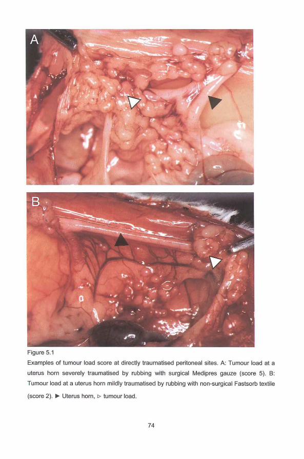
Figure 5.1
Examples of tumour load score at directly traumatised peritoneal sites. A: Tumour load at a
uterus horn severely traumatised by rubbing with surgical Medipres gauze (score 5). B:
Tumour load at a uterus horn mildly traumatised by rubbing with non-surgical Fastsorb textile
(score 2) . ...,. Uterus horn, t> tumour load.
74

RESULTS
Effect of uterus horn manipulation on intra-peritoneal tumour cell adhesion and growth
Table 5.1 summarises the results observed at the site of the uterus horns. In three different
experiments, a significant correlation was found between tumour load and the degree of
peritoneal trauma imposed by rubbing. Rubbing with severely traumatising surgical gauze
produced the highest tumour load, whereas no rubbing resulted in the lowest (group I; p =
0.005). Rubbing with mildly traumatising non-surgical textile evoked a low degree of tumour load,
but it was still significantly more than when no rubbing had taken place (group II; p = 0.018). At
the site of the uterus horns, the tumours were often located in adhesions. Table 5.2 shows the
tumour load at the non-manipulated remote peritoneal sites in rats from groups I, II and Ill. It
shows significant differences in tumour load at 2 abdominal sites (the retroperitoneum (p ::;; 0.01)
and the omentum (p ~ 0.01 )) between group II (non-surgical textile) and groups I and Ill (surgical
gauze). A significant difference in total tumour load between the same groups (II versus I and II
versus Ill) was found (p::;; 0.005). Differences in tumour load at the other 3 peritoneal sites were
not significant.
Effect of uterus horn manipulation on established "ectopic" tumour growth
The mean weight of the sub-renal tumours was measured 10 days after tumour implantation, 7
days after manipulation with surgical gauze or non-surgical textile. Significant differences in
mean tumour weight between the 3 groups were found (Table 5.3). Again, a significant
correlation between degree of peritoneal trauma and tumour growth was observed: the mean
weight of the "ectopic" tumour was the highest in rats that were rubbed with surgical gauze
(group IV) and significantly lower in rats rubbed with non-surgical textile (group V) (p = 0.009).
When the peritoneum was not touched (group VI), the lowest mean tumour weight was found,
significantly lower than after rubbing with non-surgical textile (p = 0.002) or surgical gauze (p <
0.0001 ).
Passive transfer experiments
The median total peritoneal tumour load in rats injected with irrigants collected from abdominal
cavities, manipulated with surgical gauze or non-surgical textile or not differed significantly from
each other (Table 5.4, p::;; 0.016). These differences were mainly due to differences at the site of
the omentum and the kidney. As in the previous experiments, a decreasing gradient of tumour
load was found, from surgical (group VII) to non-surgical (group VIII) to non-traumatised (group
IX) abdominal cavities.
75

'
Uterus horns n Median tumour load (range) p1 p2 p3
1'. No touch 10 0.0 (0-2)
lb. Medipres rubbing 10 4.5 (3-5) 0.005
I Ia. No touch 10 0.0 (0-2)
II'- Fastsorb rubbing 10 2.0 (0-3) 0.018
Ill'. Medipres rubbing 9 5.0 (4-5)
Ill'- Fastsorb rubbing 9 1.0 (0-3) 0.008
Table 5.1
Median tumour load (range) at uterus horns severely traumatised by rubbing with surgical gauze
(group lb and Ill'), at uterus hams mildly traumatised by rubbing with non-surgical textile (group
lib and I lib) and at not directly traumatised uterus horns (group II' and Ill'). N is the number of
uterus horns assessed. Statistics p1 (I' versus lb), p2 (II' versus lib) and p3 (Ill' versus lllb ):
Wilcoxon Matched pairs test.
Abdominal sites Tumour load Tumour load Tumour load p1 p2 p3
I. Medipres II. Fastsorb Ill. M& F
n = 10 n = 10 n=9
Subcutis 0 (1-3) 2 (0-3) 1 (0-4) ns ns ns
Parietal peritoneum 0 (0-2) 0 (0-1) 0 (0-1) ns ns ns
Kidney 1 (0-2) 1 (0-2) 1 (0-2) ns ns ns
Liver 2 (0-3) 1 (0-2) 2 (1-3) ns ns ns
Retroperitoneum 2 (1-3) 1 (0-2) 2 (1-3) 0.007 0.01 ns
Omentum 2 (0-3) 1 (0-1) 2 (2-4) 0.01 0.0001 ns
Total 1.5 (0-3) 1.0 (0-3) 1.5(0-4) 0.005 0.001 ns
Table 5.2
Median tumour load (range) at different not directly traumatised peritoneal sites in rats having
been intra-abdominally manipulated by severely traumatising surgical gauze (group 1), mildly
traumatising non-surgical textile (group II) or a combination of both materials (group Ill). N is the
number of treated rats. Statistics p1 (I versus II), p2 (II versus Ill) and p3 (I versus Ill): Kruskai
Wallis test, with a Mann-Whitney U post-hoc test.
76

Uterus horns n Mean tumour weight (mg) p1 p2 p3
(SO)
IV. Medipres rubbing 10 34.14 (7.2) 0.009
V. Fastsorb rubbing 10 28.01 (6.5) <0.0001
VI. No touch 10 17.80 (6.1) 0.002
Table 5.3
Mean tumour weight (SO) of the sub-renal tumours after intra-abdominal rubbing with severely
traumatising surgical gauze (group IV), with mildly traumatising non-surgical textile (group V) and
after no rubbing of the peritoneum at a!! (group VI). N is the number of operated rats, per rat the
mean weight of two sub-renal tumours were assessed and used for analysis. Statistics p1 (IV
versus V), p2 (V versus VI) and p3 (IV versus VI): Kruskai-Wallis test, with a Mann-Whitney U
post-hoc test.
Abdominal sites Tumour load Tumour load Tumour load p1 p2 p3
VII. Medipres VIII. Fastsorb IX. No touch
n = 10 n = 10 n = 10
Left uterus horn 0 (0-0) 0 (0-0) 0 (0-0) ns ns ns
Right uterus horn 0 (0-0) 0 (0-0) 0 (0-0) ns ns ns
Subcutis 0 (0-0) 0 (0-0) 0 (0-0) ns ns ns
Parietal peritoneum 0 (0-0) 0 (0-0) 0 (0-0) ns ns ns
Kidney 2 (0-3) 1 (0-2) 0 (0-0) ns 0.03 0.03
1
Liver 2 (1-3) 1 (0-2) 0 (0-2) ns ns ns I Retroperitoneum 1 (0-3) 1 (0-2) 1 (0-2) ns ns ns
Omentum 3 (2-5) 1 (0-3) 0 (0-2) 0.03 ns 0.008
Total 0 (0-5) 0 (0-3) 0 (0-2) 0.02 0.002 0.008
Table 5.4
: I
Median tumour load (range) at different peritoneal sites in rats intra-abdominally injected with
irrigant obtained from rats that underwent rubbing of the peritoneum with severely traumatising
surgical gauze (group VII), mildly traumatising non-surgical textile (group VIII), or no rubbing at
all (group IX). N is the number of treated rats. Statistics: p1 (VII versus VIII), p2 (VIII versus IX)
and p3 (VII versus IX): Kruskai-Wallis test, with a Mann-Whitney U post-hoc test.
77
DISCUSSION
Experimental and clinical studies suggest that surgical trauma promotes tumour cell adherence
and tumour growth.15'
19-22 The mechanism by which surgical trauma promotes these processes
is not completely understood but is probably multi-factorial, because tumour cell adherence as
well as local and regional tumour growth can be enhanced. It seems that trauma leads to a
process during which locally and regionally active tumour promoting agents are produced.15·
19
We recently demonstrated that surgical Medipres gauze was more traumatising to the
peritoneum than non-surgical Fastsorb textile, leading to significantly more adhesion formation. 16
Our current data suggest that the factors responsible for the formation of post-surgical
adhesions also play a role in the adhesion and growth of tumour cells to the peritoneum. The
most impressive tumour load was observed at sites where abrasion of the mesothelium was
most severe. The degree of tumour recurrence at traumatised sites was highly correlated with
the degree of trauma; abrasion with surgical gauze produced the highest tumour load whereas
untouched peritoneum showed the lowest tumour burden and surfaces traumatised by non
surgical textile presented intermediate tumour encumbrance. The finding that traumatised
surfaces are privileged sites for tumour cells has been demonstrated before.15' n 24 It is
conceivable that the process of enhanced tumour recurrence in traumatised tissue is biphasic.
Firstly, trauma of the peritoneum and the ensuing inflammatory response will lead to
upregulation of adhesion molecules, thus promoting the anchoring of tumour cells. Second, the
subsequent healing of the peritoneum leads to growth promotion of the adhered tumour cells
through the action of locally produced growth factors.
Using the same tumour model as in the present study, we recently demonstrated that the
phenomenon of enhanced tumour recurrence as it relates to trauma and healing also occurs in
other experimental settings. It was found that laparoscopic removal of a bowel segment led to
less adherence and growth of intra-peritoneal tumour cells than when conventional surgery was
performed, again indicating that the degree of surgical trauma was proportional to the extent of
tumour recurrence.22 In addition, we observed that the growth of a regenerating liver following
partial hepatectomy led to a marked propagation of intra-hepatic tumour growth.25· 26
Interestingly, our present results indicate that the sequelae of peritoneal trauma with regard to
tumour recurrence are not confined to the inflicted site itself, but appear to have a generalised
character. We showed that trauma led to more tumour at the traumatised site and also at non
traumatised peritoneum. Again, the amount of tumour at these loco-regional sites correlated with
the severity of the inflicted trauma. This clear correlation was also found in the experiment in
which we studied the effect of peritoneal trauma on tumour growth under the renal capsule.
Even in this "ectopic" tumour model, the consequences of the intra-abdominal trauma were
78

demonstrable. Because promotion of adherence was irrelevant in this model, this experiment
also revealed that trauma could evoke enhancement of the growth of an established tumour.
Gutman et al made comparable observations, finding that a regenerating liver induced enhanced
tumour growth not only in the liver but also at distant sites. 27
Our final experiment, in which we demonstrated that within a few hours after infliction of
peritoneal trauma, the effects on tumour recurrence could be passively transferred to naive
recipients, supports the notion that trauma per se has a marked effect, most likely on tumour cell
adhesion.
Taken together, the current experiments suggest that both tumour cell adherence and tumour
growth are modified by surgical trauma. It is clear that the present model provides unique
possibilities to further unravel the similarities and differences between the processes of adhesion
formation and tumour cell adhesion and tumour growth. Variables such as kinetics of adhesion
molecule expression with regard to inflammatory cytokines and growth factors and the role of
mesothelial hyaluronic acid and CD44 are currently being investigated. These studies may lead
to sophisticated tools to prevent the unwanted side effects of surgery. On the other hand, the
present study clearly indicates that these unwanted side effects already can partly be omitted by
employing delicate surgery, using non-abrasive gauze material.
79

REFERENCES
1. Rich T, Gunderson LL, Lew R, Galdibini JJ, Cohen AM, Donaldson G. Patterns of recurrence of
rectal cancer after potentially curative surgery. Cancer 1983; 52: 1317-1329.
2. McDermott FT, Hughes ES, Pihl E, Johnson WR, Price AB. Local recurrence after potentially
curative resection for rectal cancer in a series of 1008 patients. Br J Surg 1985; 72: 34-37.
3. Long RT, Edwards RH. Implantation metastasis as a cause of local recurrence of colorectal
carcinoma. Am J Surg 1989; 157: 194-201.
4. Brodsky JT, Cohen AM. Peritoneal seeding following potentially curative resection of colonic
carcinoma: Implications for adjuvant therapy. Dis Colon Rectum 1991; 34:723-727.
5. Eggermont AMM, Steller EP, Marquet RL, Jeekel J, Sugarbaker PH. Local regional promotion of
tumour growth after abdominal surgery is dominant over immunotherapy with interleukin-2 and
!ymphokine activated killer cells. Cancer Detect Prev 1988; 12:421-429.
6. Cunliffe WJ, Sugarbaker PH. Gastrointestinal malignancy: rationale for adjuvant therapy using
early postoperative intra-peritoneal chemotherapy. Br J Surg 1989; 76: 1082-1090.
7. Eggermont AMM, Sugarbaker PH. lmmunogenicity of the tumour determines the outcome of the
immunotherapy with interleukin-2, ABPP, and cyclo-phosphamide of micro- and macroscopic
intra-peritoneal tumour. Cancer Detect Prev 1990; 14:483-490.
8. Hubens G, Willems G. Factors influencing the implantation of colon cancer cells on the colonic
suture line in rats. Eur Surg Res 1990; 22: 356-364.
9. Reinbach 0, McGregor JR, O'Dwyer OJ. Effect of suture material on tumour cell adherence at
sites of colonic injury. Br J Surg 1993; 80: 774-776.
10 Sugarbaker PH. A perspective on clinical research strategies in carcinoma of the large bowel.
World J Surg 1991; 15: 609-616.
11. Galandiuk S, Wienand HS, Moertel CG, Cha SS, Fitzgibbons RJ Jr, Pemberton JH, Wolff BG.
Patterns of recurrence after curative resection of carcinoma of the colon and rectum. Surg
Gynecol Obstet 1992; 174: 27-32.
12. Umpleby HC, Fermor B, Symes MO, Williamson RCN. Viability of exfoliated colorectal carcinoma
cells. Br J Surg 1984; 71: 659-663.
13. Fermor B, Umpleby HC, Lever JV, Symes MO, Williamson RCN. Proliferative and metastatic
potential of exfoliated colorectal cancer cells. J Nat! Cancer 1986; 76: 347-349.
14. Fisher B, Fisher ER, Fedustea N. Trauma and localisation of the tumour cells. Cancer 1967; 20:
23-30.
15. Eggermont AMM, Steller EP, Sugarbaker PH. Laparotomy enhances intra-peritoneal tumour
growth and abrogates the anti-tumour effects interleukin-2 and lymphokine-activated killer cells.
Surgery 1987; 102: 71-78.
16. Tol van den MP, Stijn I, Bonthuis F, Marquet RC, Jeekel J. Reduction of intra-peritoneal adhesion
formation by use of nonabrasive gauze. Br J Surg 1997; 84: 1410-1415.
17. Marquet RL, Westbroek DL, Jeekel J. Interferon treatment of a transplantable colon
adenocarcinoma, importance of tumour site. lnt J Cancer 1984; 33: 688-692.
80

18. Steller EP, Ottow RT, Eggermont AMM, Marquet RL, Sugarbaker PH. Local conditions in the
host influence immunotherapy with interleukin-2 and LAK cells. Cancer Detect. Prev. 1988;
12: 81-90.
19. Weese JL, Ottery FD, Emoto SE. Do operations facilitate tumour growth? An experimental model
in rats. Surgery 1986; 100:273-277.
20. Ratajczak HV, Lange RW, Sothern RB, Hagen KL, Vescei P, Wu J, Halberg F, Thomas PT.
Surgical influence on murine immunity and tumour growth: relationship of body temperature and
hormones with splenocytes. Proc Soc Exp Bioi Med 1992; 199: 432-440.
21. Busch OR, Hop WC, Hoynck van Papendrecht MA, Marquet RL, Jeekel J. Blood transfusion and
prognosis in colorectal cancer. N Eng! J Med 1993; 328: 1372-1376.
22. Bouvy NO, Marquet RL, Hamming JF, Jeekel J, Bonjer HJ. Laparoscopic surgery in the rat
beneficial effect on body weight and tumour take. Surg Endosc 1996; 10: 490-494.
23. Murthy SM, Goldschmidt RA, Rao LN, Ammirati M, Buchmann T, Scanlon EF. The influence of
surgical trauma on experimental metastasis. Cancer 1989; 64: 2035-2044.
24. Lee JY, Murphy SM, Scanlon EF. Effect of trauma on implantation of metastatic tumour in bone in
mice. J Surg Onco/1994; 56: 178-184.
25. Jong de KP, Lont HE, Bijma AM, Brouwers MAM, Vries de EGE, Veen van ML, Marquet RL,
Slooff MJH, Terpstra OT. The effect of partial hepatectomy on tumour growth in rats: In vivo and in
vitro studies. Hepatology 1995; 22: 1263-1272.
26. Slooter GO, Marquet RL, Jeekel J, IJzermans JNM. Tumour growth stimulation after partial
hepatectomy can be reduced by treatment with tumour necrosis factor a. Br J Surg 1995; 82: 129-
132.
27. Gutman M, Sing RK, Price JE, Fan D, Fidler IJ. Accelerated growth of human colon cancer cells
in nude mice undergoing liver regeneration. Invasion Metastasis 1994-1995; 14: 362-371.
81


Chapter VI
Glove Powder Promotes Adhesion Formation and
Facilitates Intra-Peritoneal Tumour Cell Adhesion
and Growth Adapted from the original publication in the British Journal of Surgery 2001; 88: 1258-1263
The presence of foreign material in the abdominal cavity during surgery irritates the
peritoneal surface leading to an inflammatory reaction of the peritoneum. This defensive
mechanism can provoke adhesion formation. The same peritoneal defence cascade is
thought to play a role in the process of intra-abdominal tumour recurrence. The aim of this
study was to evaluate whether the adhesion provoking effect of glove powder could be
reproduced in our rat adhesion model and to evaluate whether glove powder also promotes
intra-abdominal tumour recurrence in our rat tumour cell adhesion and growth model.
Reproducible rat models allowing semiquantitative scoring of adhesion formation and tumour
load were used to observe adhesion formation and tumour recurrence in three different
groups of rats. One group was treated by intra-abdominal application of powder obtained
from starch-powdered gloves, one by application of pure starch and in one group no
application of powder took place.
Local application of glove powder or pure starch on minimally and severely traumatised
peritoneum gave rise to a, comparable, significantly higher percentage of adhesion formation
than infliction of peritoneal trauma alone (p < 0.0001). Peritoneal application of glove powder
or pure starch induced significantly more intra-abdominal tumour load than when no powder
was applied (p s-0.002)
Starch induced peritoneal trauma not only leads to more adhesion formation but also to
increased adhesion and growth of free intra-abdominal tumour cells. This finding adds to the
already existing evidence that intra-abdominal contamination with starch from starch
powdered gloves should be avoided. Since good powder-free alternatives are available there
is no longer any justification for the use of powdered gloves during intra-abdominal surgery
83

INTRODUCTION
Postoperative adhesions are abnormal unions between peritoneal surfaces which occur after
almost every intra-abdominal surgical intervention and can lead to a number of
complications. A variety of therapeutic modalities to reduce postoperative adhesion formation
has been studied but clinical useful therapy is currently not available. The sole available
strategy to reduce postoperative adhesions is prevention by limiting peroperative trauma of
the peritoneum. Over a period of 45 years, several studies have shown that exposure of
peritoneum to starch powder present on surgical gloves leads to an inflammatory response
of the peritoneum and contributes to adhesion formation. 1-6 Exposure of already injured
peritoneal surfaces to starch enhances the intensity of the peritoneal reaction and gives rise
to more and stronger adhesions than peritoneal trauma or starch alone.7'
8 In previously
described rat studies we showed that surgical trauma of the peritoneum promotes adhesion
formation and that the intensity of inflicted trauma correlates with the extent of adhesion
formation. 9 One aim of the present study was to evaluate whether the experimentally and
clinically observed effect of glove powder on peritoneal adhesion formation could be
reproduced in our rat adhesion model and to differentiate whether starch powder alone or
also other glove contents are responsible for peritoneal damage and subsequent adhesions.
Following potentially curative resection of gastro-intestinal carcinoma, local recurrence and
peritoneal dissemination are common in tumour recurrence. 10 Distribution patterns of first
peritoneal recurrence show that the resection site is preferential, and combined recurrence
on peritoneal surfaces and resection site is common. 11 Early preoperative tumour cell
seeding and peroperative shedding of tumour cells, due to handling the tumour and leakage
from dissected lymphatic channels, are the most likely causes of tumour recurrence. 11 The
tumour cell entrapment theory proposes that the fibrinous exudate, formed as an initial
response to surgical trauma of the peritoneum, facilitates implantation of cancer cells onto
raw tissue. 11 The dynamic cascade of peritoneal healing, induced by peritoneal damage,
leading to adhesion formation also seems to be important in the process of intra-peritoneal
adhesion and growth of tumour cells. Indeed, previously described clinical and experimental
studies showed that surgical trauma may promote intra-abdominal tumour recurrence. 12"14
The degree of inflicted trauma correlated with the extent of intra-abdominal tumour !oad. 14
The secondary aim of this study was, therefore, to analyse whether the traumatic effect of
glove powder on peritoneum also promotes intra-abdominal tumour recurrence.
84

MATERIALS AND METHODS
Animals
Female inbred WAG rats of reproductive age weighing 120-160 g were obtained from
Harlan-CPS, Austerlitz, The Netherlands. They were bred under specific pathogen-free
conditions, kept under standard laboratory conditions (temperature 20-24 oc, relative humidity
50-60%, 12 hours light and 12 hours dark cycles) and fed with standard rat food and water
ad libitum. The experimental protocol adhered to rules laid down by the Dutch Animal
Experimentation Act and was approved by the Committee on Animal Research of the
Erasmus University Rotterdam, The Netherlands.
Surgical gloves; isolation of glove powder
Starch-powdered gloves (Baxter, Utrecht, The Netherlands) were cut into pieces of 1 x 1cm.
The glove parts were washed in phosphate buffered saline (PBS) by gentle stirring during 24
hours. Subsequently the suspension was filtered over a stericup filter with a pore size of 0.22
~m (Millipore, Ettenleur, The Netherlands). The particles left behind on the filter were
centrifuged (2500 rpm, 10 minutes). The supernatant was removed and the pellets were left
to dry under sterile conditions for 24 hours. The obtained powder was used for further
experiments.
Pure starch
It might be possible that enforcing the described process of isolating powder from starch
powdered gloves accidentally extricated other glove contents. To investigate whether starch
alone or in combination with other glove contents were responsible for any effects on
adhesion formation and tumour cell adhesion and growth all experiments were also
performed with pure starch obtained from Regent Hospital Products, London, UK.
Three mg of glove powder or pure starch was used in subsequent experiments. This dose is
large compared to the amount of glove powder that might be left in the peritoneal cavity
during laparotomy in humans. However laparotomy in humans lasts longer than in rats so
glove powder will obviously irritate the peritoneum for a longer period.
Tumour
Tumour CC531 is a moderately differentiated, weakly immunogenic colonic adenocarcinoma
induced in WAG rats by 1 ,2-dimethylhydrazine.15 It is transplantable in syngeneic WAG rats.
The tumour is maintained as a cell culture in RPMI 1640 medium supplemented with 5%
foetal calf serum (virus- and Mycoplasma-screened), 1% penicillin (5000 U/ml), 1%
streptomycin (5000 U/ml) and 1% L-glutamin (200 mM). Medium and all supplements were
85

obtained from Life Technologicals BV, Breda, The Netherlands. Cells were passaged once a
week using trypsin (0.05%) and EDTA (0.02%). Before use in vivo tumour cells were
harvested from stationary cultures by gentle trypsinisation (5 minutes, 3T'C), centrifugation
(5 minutes, 700 g) and re-suspension in RPM! 1640, providing cell suspensions with a
viability greater than 95%. CC531 is relatively insensitive to chemotherapy but is sensitive to
the effects of biologic response modifiers.
Operative procedures
To study the effect of starch-powdered glove particles on adhesion formation in rats our
previously described reproduc·lble rat adhesion model was used? Briefly, under ether
anaesthesia and aseptic conditions a laparotomy was pertormed using a lower midline
incision of 5 em. Three Surgilene 5-0 sutures (Braun, Melsungen, Germany) were applied to
both lateral peritoneal sides 0.7 em apart and 1.5 em away from the abdominal incision. All
knots were double, fastened tightly to ensure local ischaemia. Both uterus horns were
exposed, not handled and sutured to the lateral peritoneum with Surgilene 6-0 (Braun)
proximally and distally from the three 5-0 sutures. In this way a standardised amount of
minimal peritoneal trauma was inflicted. Standardised severe peritoneal trauma was inflicted
by additively rubbing the exposed uterus horns with severely traumatising surgical Medipres
gauze (van Heek Medical, Lasser, The Netherlands) before suturing them to the lateral
abdominal wall. Rubbing was pertormed with a device that enabled the application of a
constant pressure of 120 g/cm2. The uterus horns were rubbed 10 times over their total
length.
Minimally and severely traumatised peritoneal sides were (further) treated locally by
application of 3 mg of powder obtained from the starch-powdered gloves, by application of 3
mg of pure starch (Regent Hospital Products) or were not exposed to any kind of powder at
all. The abdomen was closed in two layers with 5-0 polyglycolic acid and 2-0 silk sutures
(Braun).
To study the effect of glove powder and pure starch on local tumour recurrence our
previously designed reproducible tumour adhesion and growth model was used.14 Under
ether anaesthesia and aseptic conditions a laparotomy was pertormed using a lower midline
incision of 5 em. Both uterus horns were exposed and sutured to the lateral peritoneum both
proximally and distally, using Surgilene 6-0 sutures. Peritoneum of the uterus horn and
lateral abdominal wall side was treated locally by application of 3 mg of glove powder, by
application of 3 mg of pure starch or was not exposed to any kind of powder at all. The
abdomen was closed in two layers with Dexon 5-0 and 2-0 silk sutures.
86

Experimental design
Adhesion formation after infliction of minimal peritoneal trauma and additive
application of glove powder or starch
In 20 rats standardised minimal peritoneal trauma was inflicted to both lateral peritoneal
sides by applying the three sutures and fixating the uterus horns to the lateral peritoneum. In
10 rats (group I) glove powder was applied on one uterus horn while the other uterus horn
was not exposed. In the other 10 rats (group II) pure starch was applied on one uterus horn
while the other one was not exposed.
Adhesion formation after infliction of severe peritoneal trauma and additive
application of glove powder or starch
In 19 rats standardised severe peritoneal trauma was inflicted to both lateral peritoneal sides
by means of the three sutures in the lateral peritoneum and rubbing of the uterus horns. In 9
rats (group Ill) glove powder and in 10 rats (group IV) pure starch was applied on one uterus
horn while in both groups the other uterus horn was not exposed to powder or starch.
Effect of glove powder and starch on intra~peritoneal tumour cell adhesion and growth
Nine rats (group V) underwent an operation during which both uterus horns were exposed
and sutured to the lateral peritoneum. No powder was applied. Before closing the abdomen,
0.5 x 10⁶ CC531 tumour cells, in 1 ml RPMI 1640, were injected intra-peritoneally (0.5 ml
along the left and 0.5 ml along the right abdominal wall). Nine rats (group VI) underwent the
same operative procedure with the additional application of 3 mg of glove powder on each
uterus horn. Nine rats (group VII) underwent the same procedure during which 3 mg of pure
starch was applied on both uterus horns.
Evaluation of adhesion formation
Fourteen days after laparotomy the rats were sacrificed for assessment of intra-abdominal
adhesion formation. Macroscopically the adhesions were scored according to their extent
(quantity) and type (quality) by two independent observers. The extent of adhesion formation
was quantified by dividing the area to be scored into eight by means of the three 5-0 sutures
in the lateral peritoneum (Figure 3.3, Chapter 3). The presence or absence of adhesions in
the eight demarcated areas was scored. Adhesions in an area accounted for 12.5%
adhesions; thus a maximum of 100% adhesions could be scored. In each rat two lateral
peritoneal sides were assessed. The type of adhesions formed was classified
macroscopically using the Zuhlke classification (Table 3.1, Chapter 3). 16
87

Evaluation of intra-peritoneal tumour cell adhesion and growth
Twenty-one days after surgery all rats were sacrificed and intra-peritoneal tumour load was
scored semiquantitatively at the following sites: right uterus horn, left uterus horn,
subcutaneously (at the site of the operative scar), parietal peritoneum (at the lateral abdominal
wall sides where no uterus horns were fixed), kidney, liver, retroperitoneum and omentum. The
scoring was performed by two independent observers and ranged from 0 to 5 per site (Table
6.1 ). For each rat the score at peritoneal sites, except for the uterus horns, was added and
averaged, from which a mean total tumour load per rat could be estimated. In the present
study the term tumour recurrence is used to illustrate tumour load, which is the assumed net
result of tumour cell adhesion and tumour growth, because we presume intra-peritoneal
injecting of tumour cells resembles the clinical situation of tumour cell spill during tumour
resection.
Score Characteristics
0 No tumour growth
Estimated tumour diameter of less than 0.5 em
2 Estimated tumour diameter between 0.5 and 1 em
3 Estimated tumour diameter between 1 and 2 em
4 Estimated tumour diameter between 2 and 3 em
5 Estimated tumour diameter more than 3 em
Table 6.1
Tumour scoring system derived from the peritoneal cancer index described by Steller. 17
Statistical analysis
The mean adhesion percentage and standard deviation was calculated per group. Statistical
analysis was performed with a t test for paired samples where dependent samples were
concerned and an unpaired Student's ttest where independent samples were concerned.
The median and range of intra-peritoneal tumour load at each site and of the total tumour
load were calculated per group. Statistical analysis was performed using the non-parametric
Kruskall Wallis test to determine overall differences followed by the non-parametric Mann
Whitney U test to compare differences between groups. Statistical significance was defined
asp< 0.05.
88

RESULTS
None of the rats were found to have adhesions at the initial operation. There were no
postoperative complications e.g. bowel obstructions, peritonitis or tumour overgrowth.
Adhesion formation after infliction of minimal peritoneal trauma and additive
application of glove powder or starch
Table 6.2 shows that application of glove powder, after minimal peritoneal trauma, led to
significantly more adhesion formation than minimal peritoneal trauma alone (p < 0.0001 ).
Local application of pure starch also induced a significantly higher mean adhesion
percentage than minimal peritoneal trauma alone (p < 0.0001 ). The mean adhesion
percentage found in the glove powder group did not differ significantly from that of the pure
starch group (p = 0.112). Adhesions formed after minimal peritoneal trauma were filmy
(Zuhlke type 1-2) involving only pelvic fat. Adhesions found after local application of glove
powder or pure starch were stronger (ZUhlke type 2-3) and involved pelvic fat, uterus horn
and small bowel.
Adhesion formation after infliction of severe peritoneal trauma and additive
application of glove powder or starch
Table 6.3 shows that local application of powder obtained from starch-powdered gloves or
pure starch induced additional adhesion formation, even in the presence of severe trauma to
the peritoneal surface. Both gave rise to a significantly higher mean adhesion percentage
than severe peritoneal trauma alone (p < 0.0001 ). As in the previous experiment there was
no significant difference in the adhesion inducing capacity of glove powder and pure starch
(P = 0.47). Adhesions found after Medipres rubbing of the uterus horn were dense and thick
(ZUhlke type 2-3} while application of glove powder or pure starch on severely traumatised
peritoneum lead to even stronger adhesions (Zuhlke type 3).
Effect of glove powder and starch on intraNperitoneal tumour cell adhesion and growth
Table 6.4 summarises results observed at peritoneal sites where glove powder or pure
starch were applied (uterus horns) in comparison to controls. Glove powder as well as pure
starch evoked a significant increase in tumour load at the uterus horns (p < 0.0001 ).
Application of glove powder or pure starch gave rise to similar results (p = 0.395}.
The median total peritoneal tumour load in rats treated with glove powder or pure starch,
measured at peritoneal sites other than the uterus horns, differed significantly from the
median total peritoneal tumour load in controls (Table 6.5, p = 0.002 and p = 0.001
89

respectively). These differences were mainly due to differences at the site of the subcutis
and the omentum. Again treatment with glove powder or pure starch did not induce a
significant different extent of total tumour load (p = 0.588).
Uterus horns n Percentage adhesion Zuhlke p1 p2 p3
formation (SD) score
1'. No powder 10 33 (10.5) 1-2
lb. Glove powder 10 84 (13.3) 2-3 <0.0001
II'. No starch 10 35 (9.9) 1-2
lib Pure starch 10 81 (8.8) 2-3 <0.0001 ns
Table 6.2
Mean adhesion percentage (SO), and ZUhlke classification of found adhesions, after inflicting
minimal peritoneal trauma alone and after application of glove powder or pure starch
following the infliction of minimal peritoneal trauma. N is the number of defected peritoneal
sites (uterus horns) assessed. Statistics p1 (I' versus lb ) and p2 (II' versus lib): t test for
paired samples. Statistics p3: unpaired Student's t test (lb versus lib).
Uterus horns n Percentage adhesion Zuhlke p1 p2 p3
formation (SD) score
lila. Medipres rubbing 9 72 (10.4) 2-3
Ill b. Medipres rubbing 9 97 (5.5) 3 <0.0001
& Glove powder
IVa_ Medipres rubbing 10 71 (10.3) 2
IVb Medipres rubbing 10 96 (6.08) 3 <0.0001 ns
& Pure starch '
Table 6.3
Mean adhesion percentage (SO), and ZUhlke classification of found adhesions, after inflicting
severe peritoneal trauma alone and after application of glove powder or pure starch following
the infliction of severe peritoneal trauma. N is the number of defected peritoneal sites (uterus
horns) assessed. Statistics p1 (Ill' versus lllb) and p2 (IV' versus IVb): t test for paired
samples. Statistics p3: unpaired Student's t test (lllb versus IVb ).
90

Uterus horns n Median tumour load (range) p1 p2 p3
V. No powder 9 0 (0-1) <0.0001
VI. Glove powder 9 4 (3-5) ns
VII. Pure starch 9 4 (3-5) <0.0001
Table 6.4
Median tumour load (range) at uterus horns which were only fixed to the lateral peritoneum
(group V), at uterus horns which were treated with glove powder after fixation (group VI) and
at uterus horns treated with pure starch after fixation (group VII). For each rat, the individual
data concerning the 2 uterus horn sites were averaged; n is the number of data (= rats) per
group used for analysis. Statistics p1 (V versus VI), p2 (VI versus VII) and p3 (V versus VII):
Kruskall Wallis test with Mann-Whitney U post hoc test.
Abdominal sites Tumour load Tumour load Tumour load p1 p2 p3
V. No powder VI. Glove powder VII. Pure starch
n=9 n=9 n=9
Subcutis 1 (0-1) 1 (1-4) 1 (1-3) 0.006 ns 0.006
Parietal peritoneum 1 (0-1) 1 (0-2) 1 (0-2) ns ns 0.039
Kidney 1 (0-2) 1 (1-2) 1 (0-2) ns ns ns
Liver 0 (0-2) 1 (0-2) 1 (0-2) ns ns ns
Retroperitoneum 1 (0-3) 1 (1-2) 1 (1-3) ns ns ns
Omentum 1 (0-2) 2 (1-4) 2 (1-3) 0.037 ns 0.037
Total 0.8 (0-2) 1.3 (1-2) 1.3(1-2) 0.002 ns 0~
Table 6.5
Median tumour load (range) at different peritoneal sites in rats having undergone a
laparotomy followed by fixation of the uterus only (group V), or laparotomy followed by
fixation of the uterus horn and additive application of glove powder (group VI) or pure starch
(group VII). N is number of treated rats. Statistics p1 (V versus VI), p2 (VI versus VII) and p3
(V versus VII): Kruskall Wallis test with Mann-Whitney U post hoc test.
91
DISCUSSION
Since the introduction of surgical gloves in 1896, several components and additives have
been used in the glove industry to facilitate manufacturing and to Jessen the hazards
associated with glove use.4 Modern gloves are made of two main components: rubber and
glove lubricants. Glove lubricants have been associated with a number of iatrogenic
problems in surgical patients. 4 Especially starch powder in surgical gloves can lead to
serious complications such as granulomatous peritonitis, adhesion formation and infection
potentiation. 18 These conditions themselves give rise to serious, sometimes even lethal,
complications such as intestinal obstruction, infertility, chronic pelvic pain, complicated and
technically difficult re-operations. The exact pathogenic mechanisms behind the undesirable
reactions to starch granules are not clear and may be diverse. 19 A possible (host defence)
cascade by which starch increases the propensity of tissues to form adhesions starts with
irritation of the peritoneum by starch particles which leads to reduced fibrinolysis and
activation of neutrophils and marophages.20·
21 Activated macrophages produce oxygen free
radicals, prostaglandin E2 , thromboxane 82 , and various cytokines which are part of the
fibrotic processY Our rat adhesion model previously showed that the degree of peritoneal
trauma correlated with the extent of adhesion formation. 9 In the currently described
experiments using the same rat model powder obtained from starch-powdered gloves as well
as pure starch significantly increased adhesion formation. There was no difference in the
adhesion inducing capacity of glove powder or pure starch. These results fortify the
hypothesis that starch is the main component of powdered gloves that provokes adhesion
formation due to a traumatising effect on the peritoneum.
We previously suggested that the peritoneal defence mechanism triggered by surgical
trauma to the peritoneum not only promotes adhesion formation but also stimulates tumour
recurrence. 14 Several theories speculate on the underlying mechanisms of the adhesion and
growth of spilled tumour cells. According to the theory of metastatic efficiency, implantation of
tumour cells onto raw tissue surfaces is an efficient process as opposed to inefficient
implantation on intact surfaces. 11 The fibrin entrapment hypothesis proposes that tumour
cells are trapped in fibrin at the resection site and abraded peritoneal surfaces, hereby
providing protection from host defence mechanisms.22 In peritoneal wound healing
inflammatory and mesothelial cells produce an abundance of cytokines and growth factors
which might also be beneficial for tumour cell adhesion and tumour growth. 23"25 Indeed a
significant correlation between the intensity of surgical trauma to the peritoneum and the
degree of tumour recurrence could be demonstrated in a rat tumour adhesion and growth
model.14 In the current experiments, using the same experimental model, the traumatising
effect of glove powder and pure starch promoted adhesion and growth of free intra-
92

abdominal tumour cells. The most impressive tumour load was found at directly traumatised
peritoneal sites (sites on which glove powder or starch was applied). However the tumour
cell adhesion and growth promoting effects of glove powder and starch were not confined to
the traumatised sites, as tumour load also was significantly higher at remote peritoneal sites.
Despite the evidence in support of starch induced complications, a considerable number of
general surgeons continues to wear starch powdered gloves.26 It has been shown that
careful washing of powdered gloves with saline fails to remove all the starch and even results
in clumping of the residual starch granules.7 Cleansing with povidone-iodine and sterile water
did reduce the amount of starch particles, 27 but our previous study in rats showed that
povidone-iodine itself induces adhesion formation.28 This study shows that starch induced
peritoneal trauma not only leads to adhesion formation but also to adhesion and growth of
free intra-abdominally tumour cells. Providing substantial reason to avoid peroperative intra
abdominal contamination with starch from powdered gloves. Since good powder-free
alternatives are available there is no longer any justification for the use of powdered gloves
during intra-abdominal surgery.
93

REFERENCES 1. Sneierson H, Woo ZP. Starch powder granuloma. A report of two cases. Ann Surg 1955; 142:
1045-1050.
2. Cooke SAR, Hamilton DG. The significance of starch powder contamination in the aef1ology of
peritoneal adhesions. Br J Surg 1977; 64: 410-412.
3. McEntee GP, Stuart RC, Byrne PJ, Leen E, Hennessy TP. Experimental study of starch
induced intra-peritoneal adhesions. Br J Surg 1990; 77: 1113-1114.
4. Ellis H. The hazards of surgical glove dusting powders. Surg Gynecol Obstet 1990; 171: 521-
527.
5. Kamffer WJ, Jooste EV, Nel JT, Wet de Jl. Surgical glove powder and intra-peritoneal
adhesion formation. An appeal for the use of powder-free surgical gloves. South African Med J
1992; 81: 158-159.
6. Luijendijk RW, Lange de DC, Wauters CCAP, Hop WC, Duron JJ, Pail!er JC, Camprodon BR,
Holmdahl LE, Geldrop van HJ, Jeekel J. Foreign material in postoperative adhesions. Ann
Surg 1996; 223: 242-248.
7. Jagelman DG, Ellis H. Starch and intra-peritoneal adhesion formation. Br J Surg 1973; 60:
111-114.
8. Walker EM. Effects of blood, bile and starch in the peritoneal cavity of the rat. J Anat 1978;
126: 495-507.
9. To! van den MP, Stijn van I, Bonthuis F, Marquet RL, Jeekel J. Reduction of intra-peritoneal
adhesion formation by use of non-abrasive gauze. Br J Surg 1997; 84: 1410-1415.
10. Abulafi AM, Williams NS. Local recurrence of colorectal cancer: the problem, mechanisms,
management and adjuvant therapy [see comments]. Br J Surg 1994; 81: 7-19.
11. Sugarbaker PH. A prospective on clinical research strategies in carcinoma of the large bowel.
World J Surg 1991; 15: 609-616.
12. Busch OR, Hop WC, Hoynck van Papendrecht MA, Marquet RL, Jeekel J. Blood transfusion
and prognosis in colorectal cancer. N Eng! J Med 1993; 328: 1372-1376.
13. Bouvy NO, Marquet RL, Hamming JF, Jeekel J, Bonjer HJ. Laparoscopic surgery in the rat:
beneficial effect on body weight and tumour take. Surg Endosc 1996; 10: 490-494.
14. Tot van den MP, Rossen van MEE, Eijck van CH, Bonthuis F, Marquet RL, Jeekel J.
Reduction of peritoneal trauma by using non-surgical gauze leads to less implantation
metastasis of spilled tumour cells. Ann Surg 1998; 227(2): 242-248.
15. Marquet RL, Westbroek DL, Jeekel J. Interferon treatment of a transplantable colon
adenocarcinoma, importance of tumour site. tnt J Cancer 1984; 33: 688-692.
16. ZUhlke HV, Lorenz EMP, Straub EM, Savvas V. Pathophysiologie und Klassifikation von
Adh8sionen. Langenbecks Arch Chir Supplll Verh Dtsch Ges Chir 1990; 345: 1009-1016.
17. Steller EP, Ottow RT, Eggermont AMM, Marquet RL, Sugarbaker PH. Local conditions in the
host influence immunotherapy with interleukin-2 and LAK cells. Cancer Detect. Prev. 1988; 12:
81-90.
18. Osman MO, Jensen SL. Surgical gloves: current problems. World J Surg 1999; 23: 630-637.
94

19 Hunt TK, Slavin JP, Goodson WH. Starctl powder contamination of surgical wounds. Arch
Surg 1994; 129: 825-827.
20. Norgen L. Excessive foreign body reaction following abdominal surgery. Acta Chir Scand
1978; 144:121-122
21. Renz H, Schmidt A, Hofmann P, Amann S, Gemsa D. Tumour necrosis factor-alpha,
interleukin-1, eicosanoid and hydrogen peroxide release from macrophages exposed to glove
starch particles. Cfin fmmunollmmunopathof 1993; 68:21-28.
22. Cunliffe WJ, Sugerbaker PH. Gastrointestinal malignancy: rationale for adjuvant therapy using
early postoperative intra-peritoneal chemotherapy. Br J Surg 1989; 76: 1082-1090.
23. Baker DG, Masterson TM, Pace R, Constable WC, Wanebo H. The influence of the surgical
wound on local tumour recurrence. Surgery 1989, 106: 525-532.
24. Gutman M, Fidler IJ. Biology of human colon cancer metastasis. World J Surg 1995; 19: 226-
234.
25. Hofer SO, Molema G, Hermens RA, Wanebo HJ, Reichner JS, Hoekstra HJ. The effect of
surgical wounding on tumour development. Eur J Surg Oncof 1999; 25: 231-243.
26. Scott-Coombes OM, Vipond MN, Thompson JN. General surgeon's attitude to the treatment
and prevention of abdominal adhesions. Ann R Golf Surg Eng! 1993; 75: 123-128.
27. Fraser I. Simple and effective method of removing starch powder from surgical gloves. Br Med
J (Ciin Res Ed) 1982; 284: 1835.
28. Westreenen van M, Tal van den MP, Prank A, Marquet RL, Jeekel J, Leguit P. Perioperative
lavage promotes intra-peritoneal adhesion in the rat. Eur Surg Res 1999; 31 (2): 196-201.
95


PART IV
Pathways of Adhesion Formation and Intra
Abdominal Tumour Recurrence after Surgical
Peritoneal Trauma; Possible Methods for Prevention


Chapter VII
The Inflammatory Sequelae of Surgery Provoke
Enhanced Tumour Recurrence; a Crucial Role for
Neutrophils and Cytokines Submitted for publication
After potentially curat;ve resection of colorectal carcinoma, the most common site for recurrence
is loco-regional. In rat studies we previously demonstrated that surgical peritoneal trauma is an
important factor in the promotion of local tumour recurrence and that within a few hours after
peritoneal trauma, surgical related factors in the abdominal cavity could be captivated in a
lavage fluid and enhance tumour recurrence in naive recipients. The objective of this study was
to evaluate the role of different individual inflammatory mediators, produced after abdominal
surgery and captivated in a lavage fluid, in loco-regional tumour recurrence.
Using our reproducible rat model post trauma lavage fluid was collected and separated in a
cellular and supernatant component, the latter containing soluble factors. Tumour recurrence
was determined after injection of naive recipients with either component of the lavage fluid. The
effect of soluble factors in the lavage fluid on tumour cell adhesion to a mesothelial monolayer
was investigated in an in vitro model. In an additional experiment, different intensities of surgical
peritoneal trauma were inflicted to identify the cell compilation in the traumatised abdominal
cavity. Cytokine and growth factors concentrations were determined in the supernatant.
Intra-peritoneal injection of naive recipients with both components of the post trauma collected
lavage fluid resulted in statistically significant more tumour recurrence than injection with RPM/
(p <; 0.01). The cellular component produced the highest tumour load. In vitro tumour cell
adhesion to the mesothelium was not affected by soluble factors in the lavage fluid Analysis of
the lavage fluid, gathered after minimal or severe peritoneal trauma, demonstrated a significant
influx of neutrophils (PMN) after infliction of severe peritoneal trauma (p < 0.0001). The acute
phase cytokines IL-1{3, IL-6 and TNF-a were present irrespective of trauma-intensity. Statistically
significant more IGF-1 was detected in the lavage fluids of severely traumatised rats (p <
0.0001).
It seems in vivo tumour recurrence is mainly promoted by the cellular component of the post
surgical inflammatory process. Cytokines also enhance in vivo recurrence, but play an inferior
role. IGF-1 may facilitate the growth of lodged tumour cells.
99

INTRODUCTION
Surgical treatment of gastro-intestinal malignancies is often complicated by loco-regional
recurrence. 1 Regardless of this detrimental adversity, surgery remains the best treatment option.
In a clinical trial, Busch et al put forward an association between recurrent disease and the
extent of surgical injury.2 In experimental models, post-surgically produced factors have been
shown to augment local and remote tumour growth. Experimental and clinical studies have
demonstrated that enhanced tumour cell adherence and tumour growth are inevitable
repercussions of surgical trauma. 1· H We previously demonstrated that within a few hours after
infliction of peritoneal trauma, surgically related factors in the abdominal cavity could be
captivated in a lavage fluid and enhance tumour recurrence in naive, non-operated recipients.7
Surgery is an inflammatory stimulus that activates the body's immune response. The ensuing
inftux of polymorph nuclear leukocytes (PMN) and mononuclear cells to the surgically
traumatised site is mediated through chemotactic factors like IL-8 (CINC in rats) and pro
infllammatory cytokines such as ll-1 13 or TN F-a and is the first line of defence. a-.-10
Insights in the host defence mechanisms of the peritoneum have demonstrated that peritoneal
lymphocytes, sub-mesothelial monocytes, PMN and mesothelial cells act in an orchestrated
response under the control of locally expressed cytokines, chemokines and adhesion molecules.
The peritoneal membrane and the mesothelium in particular, as a site for the production of
mediators, play a pivotal role in the activation and control of inflammation.11' 12
Inflammatory products produced after abdominal surgery might participate directly or indirectly in
effective tumour recurrence. In this study, we focus our attention on the individual capacity of
the, by peritoneal trauma released, inflammatory peritoneal cells and soluble factors to ascertain
which element is responsible for enhanced tumour recurrence. In an experimental model post
surgically produced tumour enhancing factors are captivated in a lavage fluid, and passively
transferred to naive recipients. The in vivo and in vitro studies presented in this paper investigate
a possible correlation between the inflammatory cells and the cell free soluble factors present in
the abdominal cavity after surgical trauma and postoperative tumour cell adhesion and growth.
MATERIALS AND METHODS
Animals
Female inbred rats of the WAG strain, weighing 155-200 g, were obtained from Harlan-CPB,
Zeist, The Netherlands. The rats were bred under specific pathogen-free conditions, kept under
standard laboratory conditions (temperature 20-24'C, relative humidity 50-60%, 12 hours light/
12 hours dark) and fed with laboratory diet and water ad libitum. The experimental protocol
100

adhered to rules laid down by the Dutch Animal Experimentation Act and was approved by the
Committee on Animal Research of Erasmus University, Rotterdam, The Netherlands.
Tumour
Tumour CC531 is a moderately differentiated, weakly immunogenic colonic adenocarcinoma
induced in the WAG/Rij rat by 1 ,2-dimethylhydrazine. It is transplantable in syngeneic WAG rats.
A cell line was established from this carcinoma and maintained by serial passage after
trypsinization in culture medium. 13 CC531 tumour cells were cultured in RPMI 1640 medium
supplemented with 5% foetal calf serum (virus- and Mycoplasma-screened), L-glutamin (2 mM)
and penicillin (105 U/L). Medium and all supplements were obtained from Life Technologicals
BV, Breda, The Netherlands. Cells were passaged once a week using trypsin (0.05%) and
EDTA (0.02%). Viability was measured by trypan blue exclusion and always exceeded 90%.
Mesothelial cell culture
Mesothelial cells (MC) were isolated from the small bowel mesentery of rats as described
before-" Mesothelial monolayers were established in 96 well plates (Greiner, The Netherlands)
precoated with collagen type I (15 ~em', Boehringer Mannheim, Mannheim, Germany). The
plates were incubated at 3rC, in a humidified atmosphere of 5% C02 in air. Medium consisted
of RPMI enriched with 10% FCS, glutamin (2 mM), penicillin (105 U/L) and fungizone (1.25
mg/L) and was replaced daily with fresh medium. Monolayers reached confluence in 2 days as
determined by microscopic evaluation.
Tumour cell labelling
The dye solution, calcein-AM, used to quantify tumour cell adhesion was prepared by dissolving
50 ~g calcein (Molecular Probes, Leiden, The Netherlands) in 5 ftl anhydrous dimethyl
sulphoxide and adding this solution to 5 ml of RPMI medium supplemented with 0.5% bovine
serum albumin (RPMI/0.5%BSA). Trypsinized CC531 cells (1 x 10⁶ cells/ml) were incubated in
this solution at 37°C for 45 minutes with occasional mixing. Before adding to the mesothelial
monolayers, the labelled cells were washed twice with RPMI/0.5%BSA to remove free dye.
Operative procedures
Under ether anaesthesia, of 14 rats the abdomen was shaved and cleansed with alcohol 70%.
Laparotomy was performed using a lower midline incision of 5 em; exposure and rubbing of both
uterus hams and a 5 em long part of the smal~ intestine with surgical Medipres gauze inflicted
subsequent trauma to the peritoneum. Rubbing was performed with a device enabling the
application of a constant pressure of 120 gr/cm2.7 The uterus hom was rubbed 10 times over its
101

total length. In this way a standardised amount of peritoneal trauma can be inflicted. The
abdomen was closed in one layer with silk 2-0 sutures (Braun, Melsungen AG, Germany). After
5 hours a second laparotomy was performed during which the abdominal cavity was lavaged
with 5 ml RPMI 1640 medium. After massaging the abdomen the remaining fluid was aspired,
pooled and kept on ice until further processing.
Experimental design
Effect of inflammatory cells and cell free soluble factors on intra-peritoneal tumour cell
adhesion and growth; in vivo experiment
The collected post trauma lavage fluid was centrifuged, the cell pellet re-suspended to original
volume with RPM I and thus divided into a "cellular'' component containing the different cell types
present in the abdominal cavity after surgical trauma and a "supernatant" containing soluble
components produced after surgical peritoneal trauma.
Subsequently 24 rats were divided into three groups. Group I served as a control group
receiving RPMI 1640 medium. Group II was acceptor for the cellular component of the post
trauma lavage fluid and group Ill for the supernatant. Of all three components, 3 ml was injected
intra-peritoneally together with 0.5 million CC531 cells (in 0.5 ml RPMI) without opening the
abdominal cavity. In this way, the factors contained by the different components represented the
mediators after surgical abdominal trauma, without inflicting additional trauma.
Evaluation of intra peritoneal tumour cell adhesion and growth
Tumour scoring took place three weeks after tumour injection. The rats were sacrificed and intra
peritoneal tumour load was scored semiquantitatively at the following peritoneal sites: parietal
peritoneum, kidney, liver, retroperitoneum, omentum and mesentery. The scoring was
performed by two independent observers using a tumour scoring system derived from the
peritoneal cancer index described by Steller and ranging from 0 to 5 per abdominal site
(Table 6.1, Chapter 6). 15 For each rat the score at all peritoneal sites was summarised, from
which a mean total tumour load per rat could be estimated. In the present study the term
tumour recurrence is sometimes used to illustrate tumour load, which is the net result of
tumour cell adhesion and tumour growth, because we presume that intra-peritoneal injecting
of tumour cells resembles the clinical situation of tumour cell spill during tumour resection.
Tumour cell adhesion assay; in vitro experiment
To demonstrate an effect of surgery related soluble factors on tumour cell adhesion to the
mesothelial cells of the peritoneum, a standardised tumour cell adhesion model was used.16
102

After confluence, overnight pre-incubation of the mesothelial monolayers took place with 200 ~I
of the supernatant samples of lavage fluids collected directly after a midline laparotomy (minimal
peritoneal trauma, n = 1 0), 5 hours after a midline laparotomy during which the peritoneum was
traumatised by rubbing with surgical gauze (severe peritoneal trauma, n = 14) or RPMI 1640
medium enriched with 10% FCS. Non pre-incubated monolayers in mesothelial cell culture
medium served as standardised control.
Medium was removed from the monolayers and 200 ~I RPMI/0.5%BSA containing 30,000
calcein labelled tumour cells was added. Plates were centrifuged for 1 minute at 80 g on a
Heraeus centrifuge and incubated for 1 hour at 3rC to allow cell adhesion. After this, the
medium of each well was removed and washed twice with 200 J..ll RPMI/0.5%BSA. Fluorescence
of adherent cells was measured on a Perkin Elmer plate reader using 485 excitation and 530
emission filters. On each plate a standard curve was prepared by adding different numbers of
labelled tumour cells to the wells. The amount of tumour cells adhered was determined by
calibrating the measured fluorescence of the experimental wells on the standard.
Cell differentiation and ELISA
Lavage ftuid was collected directly after performing a midline laparotomy without handling of
intra-abdominal structures (minimal peritoneal trauma, n = 1 0) and 5 hours after a midline
laparotomy during which the peritoneum was traumatised by rubbing with surgical gauze (severe
peritoneal trauma, n = 14). The collected lavage fluid samples were kept individually on ice and
were separated in a cellular component and a supernatant.
The cellular component was re-suspended in RPMI medium, total cell amount was determined
and HE stained cytocentrifuge slides were made for cell differentiation. Under a light microscope
at a magnification of 1 OOx, 100 cells were counted in duplicate and classified into granulocytes
(eosinophils, neutrophils and mast cells) and lymphoid cells (mononuclear phagocytes and
lymphocytes). Cell classification was done on duplicate slides.
The supernatants were filtered over a low binding 0.45 ).lm filter and stored at -Booc for further
analysis. The presence of the acute phase cytokines IL-1~, IL-6 and TNF-a in addition to the
growth factor IGF-1 were determined by ELISA (Biomedical Diagnostics, Brugge, Belgium) and
RIA (Biosource Europe, Fleurus, Belgium).
Statistical analysis
The median and range of intra-peritoneal tumour load at each site, of the total tumour load, of
cell counts in lavage fluids and of different soluble factors in the supernatant of lavage fluids
were calculated per group. Statistical analysis was performed using the non-parametric Kruskal
Wallis analysis of variance to determine overall differences If the latter overall test indicated
103

significance, comparisons between groups were made using the Mann-Whitney U test. The
mean and standard deviation of in vitro tumour cell adhesion was calculated per group. Data
were statistically analysed using analysis of variance (ANOVA) to determine overall differences,
followed by the Newman-Keuls post hoc test to compare between groups. Statistical significance
was defined asp< 0.05.
RESULTS
Effect of inflammatory cells and cell free soluble factors on intra-peritoneal tumour cell
adhesion and growth; in vivo experiment
After intra-peritoneal injection of the lavage fluid samples collected after surgical trauma, diffuse
peritoneal tumour load was found in all groups. However, injection with the cellular component of
the lavage fluid resulted in the highest amount of recurrence. When compared to the control
group (RPMI) both the cellular factors and the supernatant caused significantly enhanced
tumour recurrence (p ;;;; 0.01 ). Injection of tumour cells with RPM I alone resulted in a total of 31%
tumour load whereas injection with the cells or supernatant resulted in 79% and 67% total
tumour load (Figure 7.1 ).
Impressive differences in peritoneal tumour deposits were observed between the groups
receiving the cellular part and the supernatant of the lavage fluid. When taking the tumour size
into account an obvious shift towards larger tumours is evident in the first group. Whereas no
tumours larger than 2 em were found in the control or supernatant injected group, 38% of the
tumours in the cellular injected group were scored in this range (Figure 7.2). Table 7.1 shows the
distribution of tumour deposits at the different peritoneal sites. Statistically significant differences
in tumour load were scored in liver, retroperitoneum, omentum, and mesentery between
supernatant and cellular fraction (p.; 0.01 ).
104

100 , "' .2 ~
::i 0 E ::i -"' -0 -:>: 0
RPM! cells suoernatant
Figure 7.1
Differences in median percentages of total peritoneal tumour load (range) after passive transfer
of RPMI only, the cellular fraction or the supernatant of lavage fluid collected after infliction of
severe peritoneal trauma. The percentage total tumour load was defined after scoring the
presence or absence of a tumour irrespective of tumour size. P1 (I versus II) = 0.001, p2 (II
versus Ill) = 0.003 and p3 (I versus Ill) = O.G1. Statistics: Kruskai-Wallis test, with a Mann
Whitney U post-hoc test.
... 75 0 ~
Q)
.c E 1/)
50 ::l ~
t: ::i
iii 0 E - ::i 0 --... 25 0
:>: 0
0 0.0 <0.5 0.5-1.0 1-2 2-3 >3
Figure 7.2
Comparison of median percentages of different tumour sizes (range) after passive transfer of
RPMI only, the cellular fraction or the supernatant of lavage fluid collected after infliction of
severe peritoneal trauma. Open bars represent RPMI, cross-hatched bars the cellular fraction
and the filled bars the supernatant fraction. Statistics: Kruskai-Wallis test, with a Mann-Whitney
U post-hoc test.
105

1 Abdominal sites Tumour load Tumour load Tumour load p1 p2 p3
I.RPMI II. Cells Ill. Supernatant
n=8 n=S n=S
Parietal peritoneum 0 (0-0) 0.0 (0-0) 0.0 (0-0) ns ns ns
Kidney 0 (0-3) 2.5(0-4) 1.0 (0-2) ns ns ns
Liver 1 (0-1) 4.0 (2-5) 2.5 (0-3) 0.001 0.01 0.01
Retroperitoneum 0 (0-2) 3.5 (3-5) 2.5 (0-3) 0.000 0.007 0.007
Omentum 0 (0-2) 4.5 (4-5) 3.0 (0-5) 0.000 0.005 0.004
Mesentery 0 (0-2) 2.0 (1-3) 0.5 (0-2) 0.007 0.03 ns
Total 0 0 (0-3) 3.0 (0-5) 1.0 (0-5) 0.001 0.003 0.01
Table 7.1
Median tumour load (range) at different abdominal sites in rats having been injected with RPM I
medium (group 1), the cellular fraction of the lavage fluid (group II), or the supernatant of the
lavage fluid (group Ill). N is the number of treated rats. The p-value 1 represents differences
between groups I and II; p-value 2 represents differences between group II and Ill; p-value 3
represents differences between groups I and Ill. Statistics: Kruskai-Wallis test, with a Mann
Whitney U post-hoc test.
150
" .~
"' "' .<: 100 , "' "' " :,!1 50 •
0 RPMI minimal trauma severe trauma
Figure 7.3
Percentage tumour cell adhesion versus control after pre-incubation of mesothelial cells with
supernatant of lavage fluids gathered after infliction of different intensities of surgical peritoneal
trauma. Mean values (SO) are shown (n = 6 per fluid sample). There was no difference in cell
adhesion between groups. Statistics: AN OVA test with Newman-Keuls post hoc test.
106

Tumour cell adhesion assay; in vitro experiment
Figure 7.3 shows the percentage of tumour cell adhesion versus the standardised control of
adhesion assay, consisting of mesothelial cell culture medium. Control cell adhesion consisted
of 23% from the total amount of cells added (data not shown). RPM I did not affect tumour cell
adhesion, nor did lavage fluid samples from both surgically traumatised groups.
Effect of surgical trauma on peritoneal cell compilation
There was no change in the total cell amount between minimally (laparotomy only) and severely
(laparotomy and rubbing) traumatised rats (Figure 7.4). In the cell differentiation however, a
significant reversal was seen regarding the ratio of granulocytes and lymphoid cells, with a 22-
78% ratio of granulocyte-lymphoid cells in the minimally traumatised group versus a 78-22%
ration in the severely traumatised group (p < 0.0001) (Figure 7.5). The granulocyte component
consisted for 99.5% out of PMN cells.
Effect of surgical trauma on cytokine and growth factor production
IL-1 13 was detected in 7 out of 14 lavage fluid samples of the severely traumatised rats ranging
from 20-72 pg/ml and in 2 out of 10 of the minimally traumatised rats ranging from 37 to 69
pg/ml. IL-6 was present in 13 samples of the severely traumatised (range 54-848 pg/ml) and all
samples of the minimally traumatised group (range 72-1194 pg/ml), TNF-a in 2 and 5 samples of
the minimally and severely traumatised rats (67 and 170 pg/ml and ranging from 35 to 110
pg/ml) respectively. IGF-1 was present in all samples, however statistically significant more IGF-1
was detected in lavage ftuid from the severely traumatised rats (p < 0.0001) (Figure 7.5).
107

" 0 15
~ . . _§. . ~
10 . . , 0 E • 5 = • 0
0 Mild trauma Severe trauma
Figure 7.4
Differences in total cell amount in lavage fluids gathered from minimally and severely
traumatised peritoneal cavities. Median and range are shown. Statistics: Mann-Whitney U test.
= • '-'
100
Figure 7.5 minimal trauma severe trauma
Differentiation of cellular fraction of lavage fluids taken from minimally and severely traumatised
abdominal cavities. Open bars represent the median percentage lymphoid cells and filled bars
the median percentage granulocytes. There was a significant shift towards granulocyte ration
after severe trauma from 22 to 78%, p = 0.0001. Statistics: Mann-Whitney U test.
7.5
...J 5.0 · . ~ 0
2.5 •.. ...
o.oc_-~~c-----=----,----Mild trauma Severe trauma
Figure 7.6
Median and range of IGF-1 concentration in the supernatant fraction of lavage fluids of differently
traumatised rats. Statistically significant more IGF-1 was found in abdominal cavities of severely
than in cavities of minimally traumatised rats, p < 0.0001. No IGF-1 was found in RPM I. Statistics:
Kruskai-Wallis test, with a Mann-Whitney U post-hoc test.
108

DISCUSSION
The success of surgical treatment in patients with gastro-intestinal cancer is often limited due to
local recurrence or peritoneal carcinosis by peroperatively seeded tumour cells. 1 In this study, a
cell seeding model was used to mimic the clinical situation of free intra-peritoneal tumour cells
and associates the combination of cell adhesion and growth ultimately leading to manifest
tumour recurrence. The presented results give evidence that the inflammatory sequelae of
surgery enhance peritoneal tumour recurrence. In an experimental rat model, we previously
demonstrated that components produced after surgical peritoneal trauma, which can be
captivated in a lavage fluid collected from peritoneally traumatised abdominal cavities, could
enhance tumour recurrence in naive recipients. 7 The current study demonstrates that separated
components of lavage fluid collected after infliction of peritoneal trauma i.e. inflammatory cells
and soluble factors, each lead to enhanced tumour recurrence.
The cellular fraction however, led to the greatest tumour load manifested by large tumours. More
detailed analysis of the cellular fraction revealed a trauma related influx of granulocytes i.e. PMN
into the abdominal cavity. Similar shifts in cell differentiation have also been shown in other
animal models following peritoneal trauma.9•
10'
17 The shift of primarily monocytes to PMN will
evidently affect the homeostatic milieu of the peritoneum. PMN generate reactive oxygen
metabolites and discharge contents of granular organelles into either phagocytic vacuoles or the
local environment to ingest foreign particles or microorganisms. Both oxygen-dependent and
oxygen-independent processes participate in the killing of bacteria and also in damage to host
tissue.10'
18•
19 In vitro increased adhesion of activated PMN to a mesothelial monolayer has been
shown to induce retraction, gap formation and detachment ending with substantial mesothelial
cell injury.20 Mesothelial cell injury leads to exposure of underlying extra cellular matrix
components. Experimental studies have demonstrated a preferential adhesion of tumour cells to
these denuded areas.21 Effective inhibition of tissue injury by PMN has been achieved by
blocking of cell adhesion molecules used to enter the inflamed tissue20·
22 and scavenging of
reactive oxygen species. 23'24
A relation has been demonstrated between the extent of tissue trauma and tumour recurrence.
In order to diminish tissue trauma and tumour recurrence, minimal invasive surgery is
promoted.3•
7'
21 Diminished tumour recurrence after laparoscopic surgery as compared to
conventional surgery has attributed to this phenomenon.3·
21 In addition, laparoscopic surgery
appears to impact on the cellular components of the immune response less than laparotomy.5·
25
Carbon dioxide pneumoperitoneum has been shown to impair peritoneal macrophage cytokine
production (IL-1, TNT-a) and coincides with diminished neutrophil superoxide anion release and
chemotaxis.26
109

In a peritonitis model, Pruimboom et al also showed a non-significant increase in cells in the
peritoneal cavity. In the peritoneal cell compilation a pronounced increase of PMN was seen on
the first day followed by an influx of macrophages from day 1 until seven days after induction of
peritonitis. PMN produced inflammatory mediators, however the capacity of these cells was very
low in comparison with peritoneal macrophages. 9' 10 In this study, the supernatant fraction of the
lavage fluid, containing inflammatory factors produced after surgical trauma also enhanced
tumour recurrence in vivo. In vitro, no difference was seen in the cell adhesion between lavage
fluid taken from differently traumatised rats versus control.
The presence of the acute phase cytokines IL-1~, IL-6 and TNF-a was detected in the ftuid
although no significant differences in the concentration were observed between minimally and
severely traumatised rats. IL -1 ~ and TN F-a have been shown to upregulate cell adhesion
molecules on mesothelial cells, which are used by PMN for adhesion.20 In addition, cytokine
activated mesothelial cells produce chemoattractant cytokines such as IL-8 (CINC in rats),
required for PMN recruitment. 12'
27 In this way, the mesothelium and inflammatory cells in the
abdominal cavity may perpetuate a cytokine loop, resulting in extreme activation of the
inflammatory process. However, when omitting inflammatory cells, as done in vitro, the sole
effect of cytokines present in the peritoneal fluid does not affect cell adhesion. The in vivo effect
may therefore be direct, based on the additional trauma inflicted by inflammatory cells. High
concentrations of IL-1 f) and TN F-a have been demonstrated to upregulate adhesion molecule
expression and therefore enhance the possibility of cell adhesion.12' 27 Taking this into account a
direct cytokine effect may occur locally, at the site where cytokines are produced. In this study
this effect may be missed as the absolute concentration of cytokines is diluted by lavaging with
excess fluid.
Growth factors play a vital role in post surgical wound healing. Insulin like growth factor-1 (IGF-1)
is released during the first stage of wound healing.28 Previous in vitro experiments demonstrated
IGF-1 does not affect tumour cell adhesion but was a potent growth stimulant for tumour and
mesothelial cells.29 Clinical and experimental studies have demonstrated that surgery is followed
by a rapid decrease of IGF-1 in serum. 30'
31 This apparent suppression of IGF-1 system is thought
to be caused by an increased efflux of IGF-1 from the blood to the peripheral tissues.30 In this
study, significantly higher IGF-1 concentrations were found in the abdominal lavage fluids from
severely traumatised rats. The origin of IGF-1 could be the proposed efflux from the blood or
could be the result of local production by mesothelial cells. 14 IGF-1 stimulation and over
production has been shown in several types of carcinoma and may support an autocrine growth
loop of the tumour cells. IGF-1 is also potently capable of priming PMN for an enhanced
respiratory burst that may lead to additional peritoneal tissue damage.32 In this way, tumour cells
can use the hosts immune response in wound healing for their own benefit.
110

In conclusion, the studies brought forward in this paper provide evidence that the intra
abdominal release of inflammatory sequelae after surgery promote local tumour recurrence and
that this effect is mainly based on the cellular component of the inflammatory process.
Extrapolation of the presented results to the clinical situation is limited as the inflammatory
response in animals deviates from humans. There is, however, limited species variation in the
first line of defence after surgical peritoneal traumatisation when taking PMN influx and
macrophage activation into account.10 Preventing tissue damage by inflammatory cells therefore
may provide a novel strategy to defeat progression and metastasis of cancer. Manipulation of
the intricate cytokine network may cause unwanted side effects in the wound healing process.
Tackling of reactive oxygen products, however, seems a feasible way of preventing cellular
tissue damage ultimately resulting in diminished tumour recurrence.
111
REFERENCES 1. Sugarbaker PH. A perspective on clinical research strategies in carcinoma of the large bowel.
World J Surg 1991; 15: 609-616.
2. Busch OR, Hop WC, Hoynck van Papendrecht MA, Marquet RL, Jeekel J. Blood transfusion and
prognosis in colorectal cancer. N Eng/ J Med 1993; 328: 1372-1376.
3. Bouvy NO, Marquet RL, Jeekel J, Bonjer HJ. Laparoscopic surgery is associated with less tumour
growth stimulation than conventional surgery: an experimental study [see comments]. Br J Surg
1997; 84:358-361.
4. Eggermont AMM, Steller EP, Sugarbaker PH. Laparotomy enhances intra-peritoneal tumour
growth and abrogates the anti-tumour effects of interleukin-2 and lymphokine-activated killer cells.
Surgel]f 1987; 102: 71-78.
5. Jacobi CA, Ordemann J, Zieren HU, Vol HD, Bauhofer A, Halle E, Muller JM. Increased systemic
inflammation after laparotomy vs laparoscopy in an animal model of peritonitis. Arch Surg 1998;
133: 258-262.
6. Lee YT. Local and regional recurrence of carcinoma of the colon and rectum: 1!. Factors relating to
operative technique. Surg Onco/1996; 5: 1-13.
7. To! van den MP, Rossen van MEE, Eijck van CH, Bonthuis F, Marquet RL, Jeekel J. Reduction of
peritoneal trauma by using non-surgical gauze leads to less implantation metastasis of spilled
tumour cells. Ann Surg 1998; 227: 242-248.
8. Williams MA, Cave CM, Quaid G, Solomkin JS. Chemokine regulation of neutrophil function ln
surgical inflammation. Arch Surg 1999; 134: 1360-1366.
9. Pruimboom W, Verdoold A, Tak C et al. Changes in eicosanoid and tumour necrosis factor a
production by rat macrophages during carrageenin-'1nduced peritonitis. Mediators lnflamm 1994;
3: 335-340.
10. Styrt B. Species variation in neutrophil biochemistry and function. J Leukoc Bio/1989; 46: 63-74.
11. Douvdevani A, Rapoport J, Konforty A, Argov S, Ovnat A, Chaimovitz C. Human peritoneal
mesothelial cells synthesize 11-1 alpha and beta. Kidney lnt 1994; 46: 993-1001.
12. Tapley N, Williams JD. Effect of peritoneal dialysis on cytokine production by peritoneal cells.
Blood Purif1996; 14: 188-197.
13. Marquet RC, Westbroek DL, Jeekel J. Interferon treatment of a transplantable colon
adenocarcinoma, importance of tumour site. Jnt J Cancer 1984; 33: 688-692.
14. Wal van der BC, Hofland LJ, Marquet RL, Koetsve!d van PM, Rossen van MEE, Eijck van CHJ.
Paracrine interactions between mesothelial and colon-carcinoma cells in a rat model. fnt J Cancer
1997; 73; 885-890.
15. Steller EP, Ottow RT, Eggermont AMM, Marquet RL, Sugarbaker PH. Local conditions in the
host influence immunotherapy with interleukin-2 and LAK cells. Cancer Detect. Prev. 1988;
12: 81-90.
16. Catterall JB, Gardner MJ, Jones LM, Thompson GA, Turner GA A precise, rapid and sensitive in
vitro assay to measure the adhesion of ovarian tumour cells to peritoneal mesothelial cells.
112

Cancer Left 1994; 87: 199-203.
17. Ho DPM, Krediet RT. Inflammatory changes in vivo during CAPO: what can the effluent tell us?
Kidney lnt Supp/1996; 56: 512-516.
18. Heinzelmann M, Simmen HP, Battaglia H, Friedl HP, Trentz 0. Inflammatory response after
abdominal trauma, infection, or intestinal obstruction measured by oxygen radical production in
peritoneal fluid. Am J Surg 1997; 174: 445-447.
19. Weiss SJ. Tissue destruction by neutrophi!s [see comments]. N Eng/ J Med 1989; 320: 365-376.
20. Andreoli SP, Malett C, Williams K, McAteer JA, Rothlein R, Doerschuk CM. Mechanisms of
polymorphonuclear leukocyte mediated peritoneal mesothelial cell injury. Kidney lnt 1994; 46:
1100-1109.
21. Bouvy NO, Marquet RL, Jeekel J, Bonjer HJ. Impact of gas(!ess) laparoscopy and laparotomy on
peritonea! tumour growth and abdominal waH metastases see [Comments]. Ann Surg 1996; 224:
694-700.
22. Wa!zog B, Scharffetter-Kochanek K, Gaehtgens P. Impairment of neutrophil emigration in CD18-
null mice. Am J Physio/1999; 276: G1125-G1130.
23. Shandall AA, Williams GT, Hallett MB, Young HL. Colonic healing: a role for polymorphonuclear
!eucocytes and oxygen radical production. Br J Surg 1986; 73: 225-228.
24. Wang P, Chen H, Qin H, Sankarapandi S, Becher MW, Wong PC, Zweier JL. Overexperssion of
human copper zinc-superoxide dismutase (SOD1) prevents postischemic injury. Proc Nat! Acad
Sci US A 1998; 95:4556-4560.
25. Griffith JP, Everitt NJ, Lancaster F, Boylston A, Richards SJ, Scott CS, Benson EA, Sue-Ling HM,
McMahon MJ. Influence of laparscopic and conventional cholecystectomy upon cell-mediated
immunity. Br J Surg 1995; 82: 677-680.
26. Vittimberga FJJ, Foley DP, Meyers WC Callery MP. Laparoscopic surgery and the systemic
immune response. Ann Surg 1998; 227: 326-334.
27. Meager A. Cytokine regulation of cellular adhesion molecule expression in inflammation. Cytokine
Growth Factor Rev 1999; 10: 27-39.
28. Hofer SO, Molema G, Hermens RA, Wanebo HJ, Reichner JS, Hoekstra HJ. The effect of surgical
wounding on tumour development. Eur J Surg Oncof 1999; 25: 231-243.
29. Rossen van MEE, Hofland LJ Tol van den MP, Koetsveld PM, Jeekel J, Marquet RL, Eijck van
CHJ. Effect of inflammatory cytkines and growth factors on tumour cell adhesion to the
peritoneum. J Path 2001; 193: 530-537.
30. Bouvy NO, Marquet RL, Tseng LN, Jeekel J, Bonjer HJ. Laparoscopic vs conventional bowel
resection in the rat. Eartier restoration of serum insulin-like growth factor 1 levels. Surg Endosc
1998; 12:412-415.
31. Skjaerbaek C, Frystyk J, Orskov H, Kissmeyer-Nielsen P, Jensen MB, Lauerberg S, Mallen V,
Flyvbjerg A. Differential changes in free and total insulin-like growth factor 1 after major, elective
abdominal surgery: the possible role of insulin-like growth factor binding protein-3 proteolysis. J
Cfin Endocrinof Metab 1998; 83: 2445-2449.
113
32 Fu YK, Arkins S, Wang BS, Kelley KW. A novel of growth hormone and insulin-like growth factor I.
Priming neutrophils for superoxide anion secretion. J /mmuno/1991; 146: 1602-1608.
114

Chapter XIII
Preventing Intra-Abdominal Influx of Neutrophils
after Surgical Peritoneal Trauma Reduces Adhesion
Formation and Local Tumour Recurrence Submitted for publication
Peritoneal trauma activates a cascade of peritoneal defence mechanisms responsible for
postoperative adhesion formation. The same cascade is thought to play a key role in the
process of intra-abdominal tumour recurrence. We previously demonstrated that within a few
hours after peritoneal trauma, surgery related factors in the abdominal cavity could be
captivated in a lavage fluid and enhance tumour recurrence in naive recipients. The
inflammatory cells contained by the lavage fluid proved to have the strongest tumour
recurrence promoting effect. FAGS analysis of the inflammatory cells demonstrated a trauma
related influx of neutrophils (PMN) in the abdominal cavity. We evaluated if intra-peritoneal
injection of anti-neutrophil serum (ANS) could reduce post-traumatic intra-abdominal PMN
influx, and if so, whether this influenced adhesion formation and tumour recurrence.
Total cell amount and differentiation of cellular parts of post-traumatic lavage fluids were
estimated in four groups of rats. Reproducible rat models allowing semiquantitative scoring of
adhesion formation and intra-abdominal tumour load were used to observe adhesions and
tumour recurrence in the same four groups. In one group minimal peritoneal trauma was
inflicted {day 0). The other groups underwent severe peritoneal trauma (day 0) without or
with intra-peritoneal injections of 3 doses (day-1, 0, +1) or 1 dose ANS (day -1).
Severe peritoneal trauma provoked a significant intra-abdominal PMN influx, this influx could
be prevented by ANS treatment (3 and 1 dose) (p <:"0.02). Treatment with 3 doses ANS also
significantly decreased blood lymphocyte, monocyte and PMN counts (p <:" 0.02) while
treatment with 1 dose only affected blood PMN counts (P = 0.009). Treatment of severely
traumatised rats with 3 doses ANS reduced adhesion formation (p = 0.0001) and induced
tumour load significantly (p :5 0.002), whereas treatment with 1 dose ANS caused statistically
significant tess tumour recurrence (p < 0.0001).
Intra-abdominal influx of PMN after surgical peritoneal trauma plays a crucial role in
postoperative adhesion formation and in the process of adhesion and growth of spilled
tumour cells. A well-balanced prevention of post-traumatic intra-abdominal PMN influx
reduces adhesion formation and local tumour recurrence.
115

INTRODUCTION
Postoperative adhesion formation as well as loco-regional tumour recurrence of colorectal
carcinomas remain important complications of potentially curative surgical intra-abdominal
interventions. For neither problem clinically relevant curative treatment modalities are
available yet. The pathogenesis of the processes responsible for postoperative adhesion
formation and intra-abdominal tumour recurrence is only partly clarified. We previously
suggested that the dynamic cascade of peritoneal healing, following peritoneal damage, not
only plays an important role in postoperative adhesion formation but also in the process of
intra-abdominal tumour recurrence,1 which is in agreement with the tumour cell entrapment
hypothesis.2 A clinical trial performed at our institute indeed demonstrated an association
between recurrent tumour disease and the extent of surgical injury.3 It has also become
evident from experimental studies that enhanced tumour cell adherence and tumour growth
are inevitable repercussions of surgical peritoneal trauma. 1•
4-6 We demonstrated earlier that
within a few hours after infliction of peritoneal trauma, factors in the abdominal cavity could
be captivated in a lavage fluid and enhance tumour recurrence in naive, non-operated
recipients. 1 Separated components of these lavage fluids i.e. inflammatory cells and soluble
factors, could each enhance tumour recurrence, however the cellular fraction led to the
greatest tumour load. More detailed analysis of the cellular fraction revealed a peritoneal
trauma related influx of polymorph nuclear leucocytes (PMN) in the abdominal cavity. 7
Similar shifts in cell differentiation following peritoneal trauma have been demonstrated in
other animal models. 8• 9
· 10 A role of PMN in the pathophysiological cascade leading to
adhesion formation has also been suggested earlier. 11·
12·
13
Assuming that the observed post-traumatic intra-abdominal influx of PMN is an important
factor in the communal dynamic cascade of peritoneal defence, responsible for both
adhesion formation and local tumour recurrence, prevention of PMN influx might influence
both processes and open up the way to novel therapeutic strategies. The present study was
performed to evaluate whether post-traumatic intra-abdominal PMN influx could be reduced
by treatment with anti-neutrophil serum (ANS), and if so, whether this reduction could
influence postoperative adhesion formation and local tumour recurrence.
MATERIALS AND METHODS
Animals
Female inbred WAG rats of reproductive age weighing 140-180 g were obtained from
Harlan-CPS, Austerlitz, The Netherlands. They were bred under specific pathogen-free
conditions, kept under standard laboratory conditions (temperature 20-24°C, relative humidity
116

50-60%, 12 hours light and 12 hours dark cycles), and fed with standard rat food and water
ad libitum. The experimental protocol adhered to rules laid down by the Dutch Animal
Experimentation Act and was approved by the Committee on Animal Research of the
Erasmus University Rotterdam, The Netherlands.
Rabbit anti-rat neutrophil serum
Polyclonal rabbit anti-rat neutrophil serum (ANS) was purchased from Accurate, Westbury,
NY, USA ANS can deplete blood neutrophils by 99.9% when administered intra-peritoneally
in a dose of 2 ml per kg bodyweight. 14·
15 The number of blood neutrophils remains at this low
level until administration of ANS is stopped. In this dose ANS is not specific for neutrophils
only, because the number of blood monocytes, lymphocytes and, to a lesser extent, the
platelets decrease as well, i.e. by 100%, 80% and 25% respectively. 15
In our experiments rats were either given a single dose of 1 ml ANS per kg bodyweight by
intra-peritoneal injection 1 day before laparotomy (day -1) or received two additional intra
peritoneal injections of 0.5 ml ANS per kg for 2 consecutive days (day 0 and +1 ).
Tumour
Tumour CC531 is a moderately differentiated, weakly immunogenic colonic adenocarcinoma
induced in WAG rats by 1 ,2-dimethylhydrazine. 16 It is transplantable in syngeneic WAG rats.
The tumour is maintained as a cell culture in RPMI 1640 medium supplemented with 5%
foetal calf serum (virus- and Mycoplasma-screened), 1% penicillin (5000 U/ml), 1%
streptomycin (5000 U/ml), and 1% L-glutamine (200 mmol). Medium and all supplements
were obtained from Life Technologicals BV, Breda, The Netherlands. Cells were passaged
once a week using trypsin (0.05%) and EDTA (0.02%). Before use in vivo, tumour cells were
harvested from stationary cultures by gentle trypsinisation (5 minutes, 3TC), centrifugation
(5 minutes, 700 g), and re-suspension in RPMI 1640, providing cell suspensions with a
viability greater than 90%. CC531 is relatively insensitive to chemotherapy but is sensitive to
the effects of biologic response modifiers.
Operative procedures
To investigate whether treatment with ANS interferes with adhesion formation our previously
described reproducible rat adhesion model was used. 17 Briefly, under ether anaesthesia and
aseptic conditions a laparotomy was performed using a lower midline incision of 5 em. Three
Surgilene 5-0 sutures (Braun, Melsungen, Germany) were applied to both lateral peritoneal
sides 0.7 em from each other and 1.5 em downwards from the abdominal incision. All knots
were double knots fastened tightly to ensure local ischaemia. Both uterus horns were
exposed, not touched and sutured to the lateral peritoneum with Surgilene 6-0 (Braun)
117

proximally and distally from the three 5-0 sutures. In this way a standardised amount of
minimal peritoneal trauma was inflicted. Severe peritoneal trauma was inflicted by additively
rubbing the exposed uterus horns with surgical Medipres gauze (van Heek Medical, Lasser,
The Netherlands) before suturing them to the lateral abdominal wall. Rubbing was performed
with a device enabling the application of a constant pressure of 120 g/cm2. The uterus horns
were rubbed 10 times over their total length. The abdomen was closed in two !ayers with 5-0
polyglycolic acid and 2-0 silk sutures (Braun).
To study the effect of treatment with ANS on local tumour recurrence our reproducible
tumour adhesion and growth mode! was used.1 Under ether anaesthesia and aseptic
conditions a laparotomy was performed using a lower midline incision of 5 em. Both uterus
horns were exposed, not touched or rubbed with surgical Medipres gauze, and sutured to the
lateral peritoneum both proximally and distally using Surgilene 6-0 sutures. In this way a
standardised amount of minimal (only two sutures, no rubbing) or severe (rubbing and two
sutures) peritoneal trauma was inflicted. Before closing the abdomen, 0.5 x 10⁶ CC531
tumour cells, in 1 ml RPM! 1640, were injected intra-peritoneally. The abdomen was closed
in two layers with 5-0 polyglycolic acid and 2-0 silk sutures.
Experimental design
Effect of ANS treatment on cell content in peritoneal cavity and blood
To investigate the influence of ANS treatment on intra-abdominal neutrophil cell count and on
the rat immune system the following procedures were performed. Under ether anaesthesia
eighty-five rats underwent a laparotomy. In 10 rats (group A) standardised minimal peritoneal
trauma was inflicted by exposing both uterus horns without rubbing them. In 25 rats (group
B) standardised severe peritoneal trauma was inflicted by rubbing both exposed uterus horns
and a 5 em long part of the small intestine with surgical Medipres gauze. In 25 rats (group C)
severe peritoneal trauma was inflicted in rats treated with 3 intra-peritoneal doses of ANS, on
day -1, 0 and +1. In 25 rats (group D) severe peritoneal trauma was inflicted after a single
intra-peritoneal injection of ANSon day -1. After 5 hours (t1), 72 hours (t2), 96 hours(!,), 144
hours (t,) and 192 hours (ts) 5 rats of each group were operated for the second time. During
this second laparotomy the abdominal cavity was lavaged with 5 ml RPMI 1640 medium.
After massaging the abdomen the remaining fluid was aspirated and individually kept on ice
until further processing.
Blood samples were obtained by cardiac puncture.
118

Adhesion formation after treatment with 3 doses of ANS
In 10 rats (group I) standardised minimal peritoneal trauma was inflicted to both lateral
peritoneal sides. In 10 rats (group II) standardised severe peritoneal trauma was inflicted to
both lateral peritoneal sides. In 10 rats (group Ill) severe peritoneal trauma was inflicted
during treatment with intra-peritoneal injections of ANSon days -1, 0, and +1 peri operatively.
lntra~peritoneal tumour cell adhesion and growth after treatment with ANS
Nine rats (group IV) underwent minimal peritoneal trauma and nine (group V) severe
peritoneal trauma. Nine rats (group VI) underwent severe peritoneal trauma and intra
peritoneal injection of ANSon days -1, 0 and +1 perioperatively. Before closing the abdomen
0.5 x 10⁶ CC531 tumour cells, in 1 ml RPMI 1640, were injected intra-peritoneally, 0.5 ml
along the left and 0.5 ml along the right abdominal wall.
Minimal or severe peritoneal trauma was inflicted in 2 groups of 10 rats (group VII and VIII).
In 10 rats (group IX) severe peritoneal trauma was inflicted after a single intra-peritoneal
ANS injection on day -1. Before closing the abdomen 0.5 x 10⁶ CC531 tumour cells were
injected into the abdominal cavity.
Evaluation of cell content in peritoneal cavity and blood
The collected lavage fluid samples were separated in a supernatant and a cellular
component by centrifugation (1500 rpm, 5 minutes). The cellular component was re
suspended in RPM! medium, total cell amount was determined and HE stained
cytocentrifuge slides were made for cell differentiation. At a magniftcation of 100 x, 100 cells
were counted in duplicate and classified into granulocytes (neutrophils, eosinophils,
basophils and mast cells) and lymphoid cells (mononuclear phagocytes and lymphocy1es).
Total blood leukocyte counts were determined with a micro cell counter, and duplicate
differential counts were carried out on May-Grl.inwald and Giemsa-stained blood smears.
Evaluation of adhesion formation
Two weeks after surgery, the rats were sacrificed for assessment of intra-abdominal
adhesion formation. Macroscopically the adhesions were scored according to their extent
(quantity) and type (quality) by two independent observers. The ex1ent of adhesion formation
was quantified by dividing the area to be scored into eight by means of the three 5-0 sutures
in the lateral peritoneum (Figure 3.3, Chapter 3). The presence or absence of adhesions in
the eight demarcated areas was scored. If there were adhesions in an area this accounted
for 12.5% adhesions; a maximum of 100% adhesions could be scored. In each rat two lateral
peritoneal sides were assessed. The type of adhesions formed was classified
macroscopically using the ZUhlke classification (Table 3.1, Chapter 3).18
119

Evaluation of intra-peritoneal tumour cell adhesion and growth
Three weeks after laparotomy all rats were sacrificed and intra-peritoneal tumour load was
scored semiquantitatively at the following peritoneal sites: right uterus horn, left uterus horn,
subcutaniously (at the site of the operative scar), parietal peritoneum (at the lateral
abdominal wall sides where no uterus horns were fixed), kidney, liver, retroperitoneum, and
omentum. The scoring was performed by two independent observers using a tumour scoring
system derived from the peritoneal cancer index described by Steller and ranging from 0 to 5
per abdominal site (Table 6.1, Chapter 6).19 For each rat the score at all peritoneal sites,
except for the uterus horns, was summarised, from which a mean total tumour load per rat
could be estimated. In the present study the term tumour recurrence is sometimes used to
illustrate tumour load, which is the net result of tumour cell adhesion and tumour growth,
because we presume that intra-peritoneal injecting of tumour cells resembles the clinical
situation of tumour cell spill during tumour resection.
Statistical analysis
Median and range from different cell counts were estimated per group. Statistical analysis
was performed using the non-parametric Kruskall Wallis test to determine overall differences
followed by the non-parametric Mann-Whitney U test to compare differences between groups
The mean adhesion percentage and standard deviation was calculated per group. Data were
statistically analysed using the one-way ANOVA test to determine overall differences. If the
ANOVA test was significant on a 5% level, the Student Newman Keuls post hoc test was
carried out to make a comparison between groups.
The median and range of intra-peritoneal tumour load at each scored abdominal site and of
the total tumour load were calculated per group. Statistical analysis was performed the
Kruskall Wallis test followed by the Mann-Whitney U post hoc test. Statistical significance
was defined asp< 0.05.
120
RESUlTS
Effect of ANS treatment on cell content in peritoneal cavity and blood
There was a significantly higher total intra-abdominal cell count after infliction of severe
peritoneal trauma than after infliction of minimal peritoneal trauma until 72 hours after the
operation (p = 0.009) (Figure 8.1 ). Figure 8.1 also shows that intra-peritoneal administration
of 3 doses of ANS as well as 1 dose of ANS significantly decreased the total intra-abdominal
cell count after infliction of severe peritoneal trauma for at least 96 hours postoperatively (p <
0.05). After treatment with 1 dose of ANS the total cell count seemed to increase earlier and
faster than after treatment with 3 doses, but differences were not significant at any time point
(p ~ 0.386) (Figure 8.1 ). A reversal was seen in the differential cell counts of lavage fluids
regarding the granulocyte-lymphocyte ratio, with a 43-57% ratio in the mildly traumatised
group (A) versus an 81-19% ratio in the severely traumaf1sed group (B) (p = 0.009). This
reversal was seen till 96 hours postoperatively (Figure 8.2). Treating the rats with 3 doses
(group C) as well as 1 dose (group D) of ANS did annul this reversal: a 23-77% ratio was
found in both groups (t1-t3 , p,; 0.01) (Figure 8.2) at all time points. The total number of PMN's
found in abdominal lavage fluids at different time points are depicted in Figure 8.3. Up till 96
hours after laparotomy severely traumatised peritoneal cavities contained significantly higher
numbers of neutrophils than mildly traumatised cavities or severely traumatised peritoneal
cavities treated with of ANS (p 5 0.02).
Figure 8.4 summarises the results of blood differential cell counts in the 4 groups at different
time points. Lymphocyte, monocyte and neutrophil counts did not differ significantly in
severely or mildly peritoneally traumatised rats. Treatment with 3 doses of ANS significantly
decreased these cell counts for a period of at least 96 hours (p ::; 0.02). This effect was not
seen after treatment with 1 dose of ANS.
121

=No touch
- Medipres rubbing
- 3 ip doses ANS Eillilliil]t ip dose ANS
Figure 8.1
Median total cell count (range) in abdominal lavage fluids after infliction of minimal (group A,
open bars) or severe (group B, filled bars) peritoneal trauma and after infliction of severe
peritoneal trauma in rats treated with 3 (group C, chequered bars) or 1 (group D, cross
hatched bars) intra-peritoneal doses of ANS. Fluids were collected 5 (t,), 72 (t2), 96 (t3), 144
(4), and 192 hours (t5) postoperatively. Statistics p1 (A versus B), p2 (A versus C), p3 (A
versus D), p4 (B versus C), p5 (B versus D), p6 (C versus D): Kruskall Wallis test with Mann
Whitney U post hoc test. T1; p1-p5 = 0.009, p6 ns. T,; p1, p4, p5 = 0.009, p2, p3 = 0.014, p6
ns. T3; p4 = 0.021, p5 = 0.048, p6 ns. T4; p4-p6 ns. T5; p4-p6 ns.
=No touch
- Medipres rubbing -3 ipdosesANS
Eillilliil] 1 ip dose ANS
Figure 8.2
Median percentage of neutrophils (range} in lavage fluids taken from abdominal cavities after
minimal (group A, open bars) or severe (group B, filled bars) peritoneal trauma and after
severe peritoneal trauma in rats treated with 3 (group C, chequered bars) or 1 (group D,
cross-hatched bars) intra-peritoneal doses of ANS. Fluids were collected 5 (t1), 72 (t2), 96
(t,), 144 (4), and 192 hours (t5) postoperatively. Statistics p1 (A versus B), p2 (A versus C),
p3 (A versus D), p4 (B versus C), p5 (B versus D), p6 (C versus D): Kruskall Wallis test with
Mann-Whitney U post hoc test. T1; p1-p5 = 0.009, p6 ns. T,; p1, p4, p5 = 0.009, p2 = 0.03,
p3 = 0.004, p6 ns. T,; p4, p5 = 0.01, p6 ns. T,; p4-p6 ns. T5; p4-p6 ns.
122

7.5
~- =No touch 0 1/a Medipres rubbing ~
" 5.0 - 3 ip doses ANS ~
8 l!liill!ll1 ip dose ANS
:2 a. 0
2.5 .g " z
Figure 8.3
Median neutrophil granulocyt count (range) in abdominal lavage fluids after infliction of
minimal (group A, open bars) or severe (group B, filled bars) peritoneal trauma and after
infliction of severe peritoneal trauma in rats treated with 3 (group C, chequered bars) or 1
(group D, cross-hatched bars) intra-peritoneal doses of ANS. Fluids were collected 5 (t1), 72
(t,), 96 (t,), 144 (t.), and 192 hours (t5) postoperatively. Statistics p1 (A versus B), p2 (A
versus C), p3 (A versus D), p4 (B versus C), p5 (B versus D), p6 (C versus D): Kruskall
Wallis test with Mann-Whitney U post hoc test. T1; p1, p4, p5 = 0.009, p2 = 0.02. p3 = 0.014,
p6 ns. T,; p1-p5 = 0.009, p6 ns. T3 ; p4, p5 = 0.021, p6 ns. T4; p4-p6 ns. T5; p4-p6 ns.
Figure 8.4
=No touch
1/a Medipres rubbing
ll!l!!ll!ll 3 i p doses ANS
l!liill!ll 1 ip dose ANS
Median blood lymphocyte count after infliction of minimal (group A, open bars) or severe
(group B, filled bars) peritoneal trauma and after infliction of severe peritoneal trauma in rats
treated with 3 (group C, chequered bars) or 1 (group D, cross-hatched bars) intra-peritoneal
doses of ANS. Blood samples were obtained 5 (t1), 72 (t,), 96 (!,}, 144 (t.). and 192 hours (t5)
postoperatively. Statistics p1 (A versus B), p2 (A versus C), p3 (A versus D), p4 (B versus
C), p5 (B versus D), p6 (C versus D): Kruskall Wallis test with Mann-Whitney U post hoc test.
T,; p1, p3, p5 ns, p2, p4 = 0.009. p6 = 0.018. T2; p1, p3, p5 ns, p2 = 0.014, p4 = 0.009, p6 = 0.027. T3 ; p4 = 0.021, p5 ns, p6 = 0.043. T4; p4-p6 ns. T5 ; p4-p6 ns.
123

Adhesion formation after treatment with 3 doses of ANS
Part of table 8.1 shows that the infliction of severe peritoneal trauma induced significantly
more adhesions than infliction of minimal peritoneal trauma {p = 0.0001 ). It also shows that
injection of 3 doses of ANS significantly reduced adhesion formation after infliction of severe
peritoneal trauma {p = 0.0001 ), although the mean adhesion percentage was still significantly
higher than after infliction of minimal trauma (p = 0.0001 ).
Intra-peritoneal tumour cell adhesion and growth after treatment with 3 doses of ANS
Table 8.2 shows that the median tumour load at severely traumatised peritoneal sites was
significantly higher than the median tumour load at minimally traumatised peritoneal sites (p
< 0.0001 ). Surprisingly, intra-peritoneal injection of 3 doses of ANS induced significantly
more tumour load at severely traumatised peritoneal sites (p = 0.002), but also at all other
not directly traumatised sites of the peritoneum (p = 0.001 ), than in controls (Table 8.2). The
median total tumour load after infliction of minimal peritoneal trauma was always significantly
lower than after infliction of severe peritoneal trauma irrespective of ANS treatment or not (p
< 0.0001 ). These differences were due to significant differences at subcutis, kidney and
omentum (p s 0.037) (Table 8.2).
Intra-peritoneal tumour cell adhesion and growth after treatment with 1 dose of ANS
Table 8.3 shows that, treatment with a single dose of ANS significantly reduced median
tumour load at severely traumatised peritoneal sites (p < 0.001 ), and at remote peritoneal
sites in severely traumatised abdominal cavities (p < 0.0001) when compared to median
tumour load found at these sites in severely traumatised rats not treated with ANS.
124

'Uterus horns n Percentage adhesion Zuhlke p1 p2 ! p3
formation (SO) score
I. No touch 10 22.5 (7.9) 1 0.0001
II. Medipres rubbing 10 76.3 (7.1) 2-3 0.0001
Ill. Medipres rubbing 10 46.9 (10.6) 1-2 0.0001
& 3xANS ip
Table 8.1
Mean adhesion percentages (SO), and ZUhlke classification of found adhesions, after infliction
of minimal peritoneal trauma (group 1), severe peritoneal trauma (group II) and after infliction of
severe peritoneal trauma and treatment with 3 intra-peritoneal doses of ANS (group Ill). For
each rat, the individual data concerning the 2 lateral abdominal wall sites (uterus horns) were
averaged; n is the number of data(::::: rats) per group used for analysis. Statistics p1 (I versus
II), p2 (II versus Ill) and p3 (I versus Ill): one-way ANOVA test, with Student Newman Keuls
post hoc test
Abdominal sites Tumour load Tumour load Tumour load p1 p2 p3
IV. No touch V. Medipres VI. 3xANS
n=9 n=9 n=9
erus horns 0 (0-0) 4.2 (4-5) 5 (5-5) <0.0001 0.002 <0.0001
Subcutis 1 (1-2) 3 (1-4) 5 (4-5) 0.01 <0.0001 <0.0001
1 Parietal peritoneum 0 (0-0) 0.5(0-1) 3 (3-4) ns <0.0001 <0.0001
!Kidney 1 (0-2) 2.5 (2-3) 4 (3-4) 0.001 <0.0001 <0.0001
Liver 0.5 (0-2) 1 (0-2) 3 (2-4) ns 0.001 0.001
Retroperitoneum 0 (0-0) 0 (0-1) 2 (1-3) ns <0.0001 <0.0001
Omentum 1 (0-2) 2 (1-3) 5 (4-5) 0.037 <0.0001 <0.0001
Total 0.8 (0-2) 1.8 (1-2) 3.7 (3-5) <0.0001 0.001 <0.0001
Table 8.2
Median tumour load (range) at none traumatised uterus horns (group VI), at uterus horns
severely traumatised by rubbing with surgical Medipres gauze (group V) and at uterus horns
severely traumatised by rubbing with surgical gauze during treatment w'1th 3 intra-peritoneal
doses of ANS (group VI). For each rat, the individual data concerning the 2 uterus horn sites
were averaged. Median tumour load (range) at different not directly traumatised peritoneal
sites and median total tumour load following laparotomy (group IV), laparotomy and severe
125
I
! i
I i '

peritoneal traumatasation (group V) and laparotomy, severe traumatisation and treatment
with 3 intra-peritoneal doses of ANS (group VI). N is the number of treated rats. Statistics p1
(IV versus V), p2 (V versus VI) and p3 (IV versus VI): Kruskall Wallis test with Mann-Whitney
U post hoc test.
Abdominal sites Tumour load Tumour load Tumour load p1 p2 p3
VII. No touch VIII. Medipres IX. 1x ANS
n = 10 n = 10 n = 10
Uterus horns 0 (0-2) 5 (4-5) 2.2 (1-4) <0.0001 <0.0001 <0.0001
Subcutis 1 (1-2) 3 (2-5) 2 (1-3) <0.0001 0.002 0.007
Parietal peritoneum 0 (0-0) 0 (0-1) 0 (0-1) ns ns ns
Kidney 1 (0-2) 2 (0-3) 1 (0-2) 0.002 0.002 ns
Liver 0 (0-2) 0.5 (0-2) 0 (0-1) ns ns ns
Retroperitoneum 0 (0-0) 0 (0-1) 0 (0-1) ns ns ns
Omentum 1 (1-3) 3 (2-4) 1.5(1-3) 0.001 0.001 ns
Total 0.6 (0-2) 1.6(1-2) 0.8 (0-2) <0.0001 <0.0001 ns
Table 8.3
Median tumour load (range) at none traumatised uterus horns (group VII), at uterus horns
severely traumatised by rubbing with surgical Medipres gauze (group VIII) and at uterus
horns severely traumatised by rubbing with surgical gauze after treatment with 1 intra
peritoneal dose of ANS (group IX). For each rat, the individual data concerning the 2 uterus
horn sites were averaged. Median tumour load (range) at different not directly traumatised
peritoneal sites and median total tumour load following laparotomy (group VII), laparotomy
and severe peritoneal traumatisation (group VIII) and laparotomy, severe traumatisation and
treatment with 1 intra-peritoneal dose of ANS (group IX). N is the number of treated rats.
Statistics p1 (VII versus VIII), p2 (VIII versus IX) and p3 (VII versus IX): Kruskall Wallis test
with Mann-Whitney U post hoc test.
126

DISCUSSION
We and others showed earlier that peritoneal trauma promotes postoperative adhesion
formation, induces local tumour recurrence at traumatised peritoneal sites and even
increases tumour growth at extra-peritoneal sites. 1· 3
· 4
· 5
· 6
·17
· 20
· 21
The pathogenesis of the processes leading to postoperative adhesion formation and intra
abdominal tumour recurrence is multifactorial and only partly clarified. We previously
suggested that one common cascade of reactfons following peritoneal damage, is responsible
for both pathological conditions. 1 Following peritoneal trauma, a variety of cytokines and
other inflammatory mediators are produced by activated mesothelial cells and by stampeded
inflammatory cells.22·
23 The production of mesothelial and inflammatory cell derived
chemokines such as IL-8 (CINC in rats), MCP-1 and IL-1~ will cause post-traumatic
migration of PMN and monocytes to the injured peritoneal cavity in order to promote the
peritoneal healing process.24"28 However, these mediators and recruited cells not only serve
peritoneal healing, but are believed to be responsible for adhesion formation and tumour cell
adhesion and growth as well.7·
12·
29 We showed that lavage fluids collected after infliction of
peritoneal trauma could enhance tumour recurrence in nai've, non-operated recipients. 1
Detailed analysis of the cellular fraction of these lavage fluids revealed a trauma related
influx of PMN in the abdominal cavity.7 PMN's generate reactive oxygen metabolites and
discharge contents of granular organelles into either phagocytic vacuoles or the local
environment to ingest foreign particles or microorganisms. Both oxygen-dependent and
oxygen-independent processes participate in the killing of bacteria but also may (further)
damage surrounding host tissue. 8·
30·
31 The coincidence of post-traumatic intra-abdomina!
PMN influx with adhesion formation and tumour eel! adhesion and growth is no solid prove
for the role of PMN in these pathogenetic processes. Effective inhibition of tissue injury by
PMN has been achieved by neutralising PMN chemoattractants, 32 blocking of PMN adhesion
molecules33·
34 and scavenging of reactive oxygen species, 35'
36 but whether this affected
adhesion formation or tumour recurrence has not been investigated. For pathogenetic
disorders of other organ systems, the role of PMN could be demonstrated by blocking of
PMN adhesion molecules which resulted in attenuated inflammatory tissue injury and
decreased organ failure.37-40
The present study shows that infliction of severe peritoneal trauma provokes a significant
intra-abdominal PMN influx during a period of at least 96 hours. This influx could be
prevented by treatment with 3 doses of ANS. Averting the post-traumatic intra-abdominal
PMN influx by intra-peritoneal injection of 3 doses of ANS significantly reduced adhesion
formation which indicates that neutrophils play a crucial role in the dynamic cascade of
adhesion formation. However, intra-peritoneal injection of 3 doses of ANS did not decrease
127

local total tumour load but significantly increased tumour recurrence. Treatment with 3 doses
of ANS also significantly decreased blood lymphocyte, monocyte and PMN counts, thereby
seriously compromising the rat immune system. It is conceivable that this
immunosuppression does not have an effect on adhesion formation but does promote
tumour growth. The effect of ANSon growth of the weakly immunogenic CC531 tumour may
be twofold: inhibitory by reducing PMN influx and stimulatory by the additional
immunosuppression. We observed earlier that immunosuppression leads to enhanced
growth of the tumour used in the current experiments.41 Treatment of severely peritoneally
traumatised rats with 1 dose of ANS did still annul the post-traumatic PMN influx, but more
selectively than 3 doses, because it did not influence blood lymphocyte and monocyte count
The last experiments showed that a more selective reduction of post-traumatic PMN influx,
without causing immunosuppression, was possible and indeed significantly lowered tumour
cell adhesion and growth.
In conclusion, these studies demonstrated the adhesion and tumour promoting effect of
PMN's. Preventing tissue damage by reduction of inflammatory cells like PMN's or by
prohibiting their intra-abdominal influx might provide a novel strategy in averting adhesion
formation and tumour recurrence. Wound healing processes rely on the same biological
mechanisms as adhesion formation and possibly, tumour recurrence, thus manipulation of
PMN has to be done selectively and in moderation to prevent unwanted side effects. Our rat
models may be helpful in further unravelling the cascade of both processes to find the perfect
balance between wanted and unwanted effects of manipulation of the post-traumatic
inflammatory response.
128

REFERENCES 1. Tal van den MP, Rossen van MEE, Eijck van CH, Bonthuis F, Marquet RL, Jeekel J.
Reduction of peritoneal trauma by using non-surgical gauze leads to less implantation
metastasis of spi!led tumour cells. Ann Surg 1998; 227(2): 242-248.
2. Sugarbaker PH. A perspective on clinical research strategies in carcinoma of the large bowel.
World J Surg 1991; 15: 609-616.
3. Busch OR, Hop WC, Hoynck van Papendrecht MA, Marquet RL, Jeekel J. Blood transfusion
and prognosis in co!orectal cancer. N Eng! J Med 1993; 328: 1372-1376.
4. Eggermont AMM, Steller EP, Sugarbaker PH. Laparotomy enhances intra-peritoneal tumour
growth and abrogates the anti-tumor effects of interleukin-2 and lymphokine-activated killer
cells. Surgef)f 1987; 102: 71-78.
5. Bouvy NO, Marquet RL, Jeekel J, Bonjer HJ. Laparoscopic surgery is associated with less
tumour growth stimulation than conventional surgery: an experimental study. Br J Surg 1997;
84: 358-316.
6. Jacobi CA, Ordemann J, Zieren HU, Volk HD, Bauhofer A, Halle E, Muller JM. Increased
systemic inflammation after laparotomy vs laparoscopy in an animal model of peritonitis. Arch
Surg 1998; 133: 258-262.
7. Rossen van MEE, To! van den MP, Aalbers AGJ, Hofland LJ, Koetsve!d van PM, Bonthuis F,
Jeekel J, Marquet RL, Eijck van CHJ. The inflammatory sequelae of surgery provoke
enhanced tumour recurrence; a crucial role for neutrophils and cytokines. Submitted for
publication.
8. Styrt B. Species variation in neutrophil biochemistry and function. J Leukoc Bio/1989; 46: 63-
74.
9. Pruimboom W, Verdoold A, Tak C et al. Changes in eicosanoid and tumour necrosis factor a
production by rat peritoneal macrophages during carrageenin-induced peritonitis. Mediators
lnflamm 1994:3: 335-340.
10. Ho DPM, Krediet RT. Inflammatory changes in vivo during CAPO: what can the effluent tell
us? Kidney lnt Supp/1996; 174: 445-447.
11. Bars low L. Neutrophilia, oxygen free radicals, and abdominal adhesions. Arch Surg 1992; 127:
747.
12. Ar'Rajab A, Mileski W, Sentementes JT, Sikes P, Harris RB, Dawidson IJA. The role of
neutrophi!s in peritoneal adhesion formation. J of Surg Res 1996; 61: 143-146.
13. Vural B, CantUrk NZ, Esen N, Solakoglu S, Canturk Z, Kirkali G, SOkmensUer C. The role of
neutrophils in the formation of peritoneal adhesions. Human Reproduction 1999; 14: 49-54.
14. Sandier H, HOgstorp H, Lundberg C, Gerdin B. Antiserum-induced neutropenia in the rat:
characterisation of a rabbit anti-rat neutrophil serum. Br J Exp Path 1987; 68: 71-80.
15. Vree WJ, Essers MC, Bruijn HS, Star WM, Koster JF, Sluiter W. Evidence for an important
role of neutrophils in the efficacy of photodynamic therapy in vivo. Cancer Res 1996; 56: 2908-
2911.
129

16. Marquet RL, Westbroek DL, Jeekel J. Interferon treatment of a transplantable colon
adenocarcinoma, importance of tumour site. lnt J Cancer 1984; 33: 688-692.
17. Tol van den MP, Stijn van I, Bonthuis F, Marquet RL, Jeekel J. Reduction of intra-peritoneal
adhesion formation by use of non-abrasive gauze. Br J Surg 1997; 84: 1410-1415.
18. ZUhlke HV, Lorenz EMP, Straub EM, Savvas V. Pathophysiologie und Klassifikation von
Adhi3.sionen. Langen becks Arch Chir Supp/11 Verh Dtsch Ges Chir 1990; 345: 1009-1016.
19. Steller EP, Ottow RT, Eggermont AMM, Marquet RL, Sugarbaker PH. Local conditions in the
host influence immunotherapy with interleukin-2 and LAK cells. Cancer Detect. Prev. 1988; 12:
81-90.
20. Ellis H. The healing of peritoneum under normal and pathological conditions. Br J Surg 1965; 52:
471-476.
21. Graeme BR, Grobety J, Majno G. Mesothelial injury and recovery. Am J Patho/1973; 71: 93-
102.
22. Nagy JA. Peritoneal membrane morphology and function. Kidney lnt Supp/1996; 56: S2-S11.
23. Holmdahl L, lvarsson ML. The role of cytokines, coagulation, and fibrinolysis in peritoneal
tissue repair. Eur J Surg 1999; 165: 1012-1019.
24. Ribeiro RA, Flores CA, Cunha FQ, Ferreira SH. IL-8 causes in vivo neutrophil migration by a
cell-dependent mechanism. Immunology 1991; 73: 472-477.
25. Kuraoka S, Campeau JD, Nakamura RM, diZerega GS. Modulation of post-surgical
macrophage function by early post-surgical polymorphonuclear leukocytes. J Surg Res 1992;
53: 245-250.
26. Rodgers KE, diZerega GS. Modulation of peritoneal reepitheliatisation by post-surgical
macrophages. J Surg Res 1992; 53: 542-548.
27. Slavin J. The role of cytokines in wound healing. J Path 1996; 178: 5-10.
28. Rollins BJ, Waf ZA, Baggiolini M. Recombinant human MCP-1/JE induces chemotaxis,
calcium flux and respiratory burst in human monocytes. Blood 1991; 78: 1112-1116.
29. Rossen van MEE, Hofland LJ, Tol van den MP, Koetsveld van PM, Jeekel J, Marquet RL,
Eijck van CHJ. Effect of inflammatory cytokines and growth factors on tumour cell adhesion to
the peritoneum. J Patho/2001; 193: 530-537.
30. Heinzelmann M, Simmen HP, Battaglia H, Friedl HP, Trentz 0. Inflammatory response after
abdominal trauma, infection, or intestinal obstruction measured by oxygen radical production
in peritoneal fluid. Am J Surg 1997; 174: 445-447.
31. Weiss SJ. Tissue destruction by neutrophils [see comments]. N Eng! J Med 1989; 320:365-
376.
32 Zagorski J, Wahl SM. Inhibition of acute peritoneal inflammation in rats by a cytokine-induced
neutrophil chemoattractant receptor antagonist. J /mmuno/1997; 159: 1059-1062.
33. Andreoli SP, Malett C, Williams K, McAteer JA, Rothlein R, Doerschuk CM. Mechanisms of
polymorphonuclear leukocyte mediated peritoneal mesothelial cell injury. Kidney tnt 1994; 46:
1100-1109.
130

34. Walzog 8, Scharffetter-Kochanek K, Gaehtgens P. Impairment of neutrophil emigration in
CD18-null mice. Am J Physio/1999; 276: G1125-G1130.
35. Shandall AA, Williams GT, Hallett MB, Young HL Colonic healing: a role for
polymorphonuclear leucocytes and oxygen radical production. Br J Surg 1986; 73: 225-228.
36. Wang P, Chen H, Qin H, Sakarapandi S, Becher MW, Wong PC, Zweier JL. Overexpression
of human copper zinc-superoxide dismutase (8001) prevents post-ischaemic injury. Proc Nat/
A cad Sci US A 1998; 95: 4556-4560.
37. Jaeschke H, Farhood A, Bautista AP, Spo!arics Z, Spitzer JJ, Smith CW. Functional
inactivation of neutrophils with a Mac-1 (CD11/CD18) monoclonal antibody protects against
ischaemia-reperfusion injury in rat liver. Hepathology 1993; 17: 915-923.
38. Yamazaki T, Seko Y, Tamatani T, Miyasaka M, Yagita H, Okumura K, Nagai R, Yazaki Y.
Expression of intracellular adhesion molecule-1 in rat heart with ischaemia/reperfusion and
limitation of infarct size by treatment with antibodies against cell adhesion molecules. AJP
1993; 143:410-418.
39. Palmen MJHJ, Dijkstra MB, Ende van der MB, Pena AS, Rees van EP. Anti CD11/CD18
antibodies reduce inflammation in acute colitis in rats. Clin Exp /mmuno/1995; 101: 351-356.
40. Yamasawa H, Ishii Y, Kitamura S. Cytokine-induced neutrophil chemoattractant in a rat model
of lipopolysacharide-induced acute lung injury. Inflammation 1999; 23: 263-274.
41. Vrie van de W, Marquet RL, Eggermont AMM. Cyclosporin A enhances loco-regional
metastasis of the rat CC531 rat colon tumour. J Cancer Res Clin Oncol1997; 123: 21-24.
131


Chapter IX
Hyaluronic-Based Coating Solution for Prevention of
Surgical Adhesions Has no Major Effect on
Adhesion and Growth of Intra-Peritoneal Tumour
Cells Adapted from the original publication in the European Journal of Surgery 1999; 165: 791-795
We recently demonstrated that surgical peritoneal trauma induces a cascade of defensive
events that can lead to adhesion formation and adhesion and growth of peroperatively spilled
tumour cells. Placement of an absorbable barrier of hyaluronic acid/carboxymethylcellu/ose
(Seprafilm™) or low viscosity 0.4% HA (Sepracoat™) bet\Neen injured peritoneal sites during
abdominal surgery can reduce postoperative adhesion formation. The objective of our study
was to find out whether the use of the HA containing solutions might effect the adhesion and
growth of free intra-abdominal tumour cells.
Rat mesothelial cells were cultured in mono/ayers and adhesion of CC531 tumour cells was
measured after pre-incubation of the mono/ayers with Sepracoat™ or PBS. Reproducible rat
models allowing semiquantitative scoring of tumour load were used to observe the effect of
Sepracoat™ on local tumour cell adhesion and grovvth at minimally and severely traumatised
and remote, not directly traumatised, peritoneal sites.
Sepracoat™ had a small but significant inhibitory effect on the adhesion of CC531 tumour cells
to mesothelial cell mono/ayers in vitro (p < 0. 05). However, we were unable to repeat these
effects in our in vivo rat models, the peroperative inoculation of Sepracoat™ did not reduce
postoperative tumour load at directly minimally or severely, or indirectly traumatised peritoneal
surfaces.
Sepracoat™ may inhibit adhesion of tumour cells to the mesothelium in culture but in the
currently used dose it had no appreciable effect on intra-abdominal tumour cell adhesion and
grovvth in rats.
133

INTRODUCTION
Peritoneal trauma is, as we recently demonstrated, not only is a causal factor for adhesion
formation, but also for the enhanced adhesion and growth of tumour cells.1·
2 Using a
reproducible rat model, a significant correlation was found between the amount of peritoneal
trauma and the degree of tumour recurrence. Remarkably, this was not only observed at the
traumatised site, but also distantly at non-abraded peritoneal surfaces.
A new development in the field of adhesion prevention is the use of bioabsorbable formulations
based on hyaluronic acid (HA). Seprafilm ™ is a membrane composed of hyaluronic acid and
carboxymethylcellulose with proven efficacy against adhesion formation in animals and
humans.3-5 SepracoatTM is a more manageable coating solution, that contains 0.4% of sodium
hyaluronate in phosphate buffered saline (PBS). Precursor formulations of the latter product
have been demonstrated to prevent adhesion formation in various animal models.6' 7
Hyaluronic acid (HA) is a major component of the extra-cellular matrix (ECM) and is also
secreted by mesothelial cells. ECM and mesothelial cells, either exposed or activated by
surgery, play major roles in the dynamic process of tumour cell adhesion. It has been
demonstrated that the presence of a HA-containing coat on mesothelial celts interferes with the
adhesion of tumour cells in vitro. 8
The aim of the present study was to investigate whether Sepracoat™ might interfere with intra
peritoneal tumour cell adhesion. We investigated this in vitro by assessing the adherence of
tumour cells to cultured rat mesothelial cell monolayers, and in vivo in a well-defined
reproducible tumour adhesion and growth model. 1
MATERIALS AND METHODS
Animals
Female inbred WAG rats of reproductive age weighing 200-250 g were obtained from Harlan,
Zeist, The Netherlands. They were bred under specific pathogen-free conditions and kept
under standard laboratory conditions (temperature 20-24°C, relative humidity 50-60%, 12 hours
light and 12 hours dark cycles). The rats were given standard food and water ad libitum. The
experimental protocol adhered to rules laid down by the Dutch Animal Experimentation Act and
was approved by the Committee on Animal Research of the Erasmus University Rotterdam.
Sepracoat TM
Sepracoat™ coating solution is a sterile-filtered, non-pyrogenic 0.4% (w/w) solution of sodium
hyaluronate in phosphate buffered saline (PBS). It was provided by the manufacturer
134

(Genzyme Corporation, Boston, Massachussets, USA}.
Tumour
Tumour CC531 is a 1,2 dimethylhydrazine~induced weakly immunogenic, moderately
differentiated colon adenocarcinoma, transplantable in syngeneic WAG rats.9 The tumour is
maintained in cell culture in RPMI-1640 medium supplemented with 5% foetal calf serum
(virus- and Mycoplasma-screened), 1% penicillin (5000 U/ml}, 1% streptomycin (5000 U/mL},
and 1% L-glutammine (200 mM}. All supplements were obtained from Gibco, Paisley, UK. Cells
were passaged once a week using trypsin (0.05%) and EDTA (0.02%). Before their use, cells
were trypsinised (5 minutes, 3rC}, centrifuged (5 minutes, 700g}, re-suspended in RPMI-1640
and counted. Viability was measured by trypan blue exclusion and always exceeded 95%.
Tumour cells were injected within 4 hours after being obtained. CC531 is relatively insensitive
to chemotherapy but is sensitive to the effect of biologic response modifiers.
Mesothelial cell culture
Mesothelial cells were isolated from the small bowel mesentery of male WAG rats as described
previously. 10 In brief, during a laparotomy under ether anaesthesia window like transparent
triangular sheets of mesentery were isolated and collected in Hank's Balanced Salt Solution
(HBSS} containing 5% human serum albumin (CLB, Amsterdam, The Netherlands}, penicillin
(5000 U/mL}, 1% L-glutammine (200 mM} and fungizone (1.25 mg/L}. After washing the sheets
twice in this medium, they were incubated in a mixture of collagenase (1g/L) and dispase (2.4 x
103 U/L} (Boehringer, Mannheim, Germany}. Following incubation during 15 minutes at 37'C
and continuous gentle shaking, the detached mesothelial cells were pelleted by centrifugation
at 300 g for 5 minutes. Cell viability was determined by trypan blue and always exceeded 95%.
The isolated mesothelial cells were re-suspended in culture medium consisting of RPMI 1640
supplemented with 10% foetal call serum, penicillin (5000 U/ml), 1% L-glutamin (200 mM} and
fungizone (1.25 mg/L}. Medium and al supplements were obtained from Life Technologicals
BV, Breda, The Netherlands. The mesentery derived cells grew forming a mesothelial
monolayer in 48 well plates (Greiner, The Netherlands}, pre-coated with collagen type I (15
~glcm' collagen S} (cell biology Roche Diagnostics, Almere, The Netherlands}. The identity of
mesothelial cells was illustrated by positive immunohistochemical staining for vimentin and
keratin, and the absence of von Willebrand factor-staining, as reported previously. 1° Cells for
staining were harvested from confluent mesothelial cell monolayers with the typical
cobblestone appearance. For all experiments, primary cell cultures were used. Isolated
mesenteries from at least three different rats were used.
135

Operative procedures
To study the effect of Sepracoat™ on tumour cell adhesion and growth in minimally traumatised
abdominal cavities a laparotomy mode! allowing semiquantitative scoring of tumour load was
used. Under ether anaesthesia the abdomen was opened using a lower midline incision of 5
em. Both uterus horns were left untouched, thus not damaged. Laparotomy was the only
minimal surgical injury inflicted to the peritoneum. Sepracoat™ or PBS was instilled into the
abdominal cavity. Half a million CC531 tumour cells, in 1 ml RPMI 1640, were left behind in the
abdominal cavity and the abdomen was closed in two layers with Dexon 5-0 and silk 2-0
sutures (Braun, Melsungen, Germany).
To study the effect of Sepracoat™ on tumour cell adhesion and growth at minimally and
severely traumatised peritoneal sites our reproducible tumour adhesion and growth model was
used.2 Briefly, under ether anaesthesia a laparotomy was performed using a lower midline
incision of 5 em. Sepracoat™ or PBS was instilled into the abdominal cavity. One uterus horn
was exposed, not touched and sutured to the lateral peritoneum both proximally and distally
with Surgilene 6-0 (Braun). In this way a standardised amount of minimal peritoneal trauma
was inflicted. The other uterus horn was exposed and rubbed with severely traumatising
surgical Medipres gauze (van Heek Medical, Lasser The Netherlands) before suturing it to the
lateral abdominal wall. Rubbing was performed with a device enabling the application of a
constant pressure of 120 g/cm2. The uterus horn was rubbed 10 times over its total length. A
standardised amount of severe peritoneal trauma was inflicted this way. Half a million CC531
tumour cells, in 1 ml RPM I 1640, were injected intra-peritoneally and the abdomen was closed
in two layers with Dexon 5-0 and silk 2-0 sutures (Braun).
Experimental design
Effect of Sepracoat™ on tumour cell adhesion to cultured mesothelial cell monolayers
(in vitro)
After mesothelial monolayers, established in 48 well plates pre-coated with collagen, reached
confluence (usually after 2 days as determined by light microscopy), culture medium was
aspired and an arbitrarily chosen amount of 200 JJI of Sepracoat™ per well was added. This
was left to settle for 3 minutes after which 5 x 10⁴ CC531 cells in 1 ml of culture medium were
added. Non-treated mesothelial monolayers served as controls. Tumour cell adhesion to the
monolayer was assessed at 1, 2, 4 and 8 hours by measuring the DNA content of the cells per
well, after washing away superfluous non-adherent cells, as described previously.11 Specific
adherence was calculated by substraction of the DNA content of control monolayers.
Mesothelial monolayers and cultures of CC531 were also cultured separately with Sepracoatru
136

to investigate its possible interference with norma! cell growth in both circumstances.
Effect of Sepracoat™ on tumour cell adhesion and growth in minimally traumatised
abdominal cavities (in vivo)
Eighteen rats underwent a laparotomy during which both uterus horns were left untouched. In 9
rats (group I) 3 ml of Sepracoat™ and in 9 rats (group II) 3 ml of PBS was instilled in the
abdominal cavity. Before closing the abdomen, 0.5 x 10⁶ CC531 tumour cells, in 1 ml RPM I
1640, were injected intra-peritoneally (0.5 ml along the left and 0.5 ml along the right
abdominal wall).
Effect of Sepracoat™ on tumour cell adhesion and growth at minimally and severely
traumatised peritoneal sites (in vivo)
In 5 rats (group Ill) 3 ml of Sepracoat™ and in 4 rats (group IV) 3 ml of PBS was inoculated
after opening the abdominal cavity. Subsequently, in all 9 rats both uterus horns were exposed
and sutured to the lateral peritoneum. All right uterus horns were rubbed with severely
traumatising Medipres gauze before fixation and all left uterus horns remained untouched.
Before closing the abdomen, 0.5 x 10⁶ CC531 tumour cells, in 1 ml RPMI 1640, were
inoculated intra-peritoneally (0.5 ml along the left and 0.5 ml along the right abdominal wall).
Evaluation of intraRperitoneal tumour cell adhesion and growth
Three weeks postoperatively the rats were sacrificed and tumour load was scored
semiquantitatively at the following sites: right uterus hom, left uterus hom, subcutaneously (at
the site of the operative scar), parietal peritoneum (at the lateral abdominal wall sides where no
uterus horns were fixated), kidney, liver, retroperitoneum and omentum. The scoring was
performed by 2 independent observers using a tumour scoring system derived from the
peritoneal cancer index described by Steller and ranging from 0 to 5 per abdominal site (Table
6.1, Chapter 6).12 For each rat the score at all peritoneal sites, except for the uterus horns, were
summarised, from which a mean total tumour load per rat could be estimated. In the present
study the term tumour recurrence is sometimes used to illustrate tumour load, which is the
assumed in vivo net result of tumour cell adhesion and tumour growth, because we presume
intra-peritoneal injecting of tumour cells resembles the clinical situation of tumour cell spill
during tumour resection.
Statistical analysis
The mean and standard deviation of in vitro tumour cell adhesion was calculated per group.
Data were statistically analysed using analysis of variance (ANOVA) to determine overall
137

differences, followed by the Newman-Keuls post hoc test to compare between groups. Statistical
significance was defined as p < 0.05. The median and range of intra-peritoneal tumour load at
each site and of the total tumour load were calculated per group. Statistical analysis was
performed using the non-parametric Mann-Whitney U test. Statistical significance was
def1ned as p < 0.05.
RESULTS
Effect of Sepracoat™ on tumour cell adhesion to cultured mesothelial cell monolayers
(in vitro)
SepracoatTM had no effect on growth or survival of mesothelial cells and CC531 tumour cells
{data not shown). The effect of Sepracoat™ on the adhesion of tumour cells to mesothelial cell
monolayers is shown in figure 9.1. Although the differences were small, tumour cell adhesion in
the presence of Sepracoat™ was significantly less than in controls at all time points (p < 0.05)
Effect of Sepracoat™ on tumour cell adhesion and growth in minimally traumatised
abdominal cavities (in vivo)
Table 9.1 shows that intra-abdominal injection of Sepracoat™ after infliction of minimal
peritoneal trauma did not significantly decrease the median total tumour load '1n comparison to
the median total tumour load found in abdominal cavities injected with PBS (p = 0.6). Only at
one peritoneal site (the parietal peritoneum) the median tumour load was significantly lower in
the group injected with SepracoatrM than in the control group injected with PBS (p = 0.03).
Effect of Sepracoat™ on tumour cell adhesion and growth at minimally and severely
traumatised peritoneal sites {in vivo)
Table 9.2 shows that neither at the severely nor at the minimally traumatised peritoneum of the
uterus horn the median tumour load could be significantly impaired by intra-abdominal
inoculation of SepracoatTM before peritoneal traumatisation (p:;.:: 0.8).
The median tumour load at not directly traumatised peritoneal sites and the median total
tumour load did not differ significantly in groups intra-abdominally treated with SepracoatTM or
PBS (p ~ 0.05) (Table 9.3).
138

3000-
"0 Q) ~
2000 " "' "' ~ <t z 1000 c
0
Figure 9.1
1.0 2.0 4.0 L__
8.0
Time (hours)
Time dependent adhesion of CC531 tumour cells to mesothelial monolayers in the presence
(filled bars) or absence (open bars) of Sepracoat'"- P < 0.05 at all time points. The number of
cells adhering was estimated by DNA measurements (ng of DNA per well). Data were
expressed as the mean and SO. Statistics: ANOVA test with Newman-Keuls post hoc test.
Abdominal sites Tumour load Tumour load p
I. Sepracoat TM II. PBS
n= 9 n=9
Uterus horns 0 (0-0) 0 (0-0) ns
Subcutis 3 (0-4) 3 (1-3) ns
Parietal peritoneum 0 (0-1) 1 (0-3) 0.03
Kidney 2 (0-4) 2 (0-3) ns
Liver 1 (1-3) 1 (0-3) ns
Retroperitoneum 1 (0-3) 1 (0-3) ns
Omentum 2 (1-4) 2 (1-4) ns
Total 2 (0-4) 2 (0-4) ns
Table 9.1
Median tumour load (range) at different not directly traumatised peritoneal sites in rats having
undergone a laparotomy followed by injection of 3 ml Sepracoat'M (group I) or 3 ml PBS (group
II). For each rat, the individual data concerning the 2 uterus horn sites were averaged·, n is the
number of data(= rats) per group used for analysis. Statistics: Mann-Whitney U test.
139

Abdominal sites Tumour load Tumour load p
Ill. Sepracoat™ IV. PBS
n=5 n=4
Medipres rubbing 4 (2-4) 4 (2-4) ns
No touch 4 (2-4) 3 (2-4) ns
Table 9.2
Median tumour load {range) at uterus horns severely traumatised by rubbing with Medipres
gauze and at not directly traumatised uterus horns in rats inoculated with 3 ml Sepracoat™
(group Ill) or 3 ml PBS (group IV). N is the number of data (uterus horns) per group used for
analysis. Statistics: Mann-Whitney U test.
Abdominal sites Tumour load Tumour load p
Ill. Sepracoat™ IV. PBS
n=5 n=4
Subcutis 3 (0-4) 3 (1-3) ns
Parietal peritoneum 0 (0-0) 0 (0-0) ns
Kidney 1 (0-2) 1 (0-2) ns
Liver 1 (0-2) 1 (0-3) ns
Retroperitoneum 1 (0-3) 1 (0-3) ns
Omentum 2 (1-4) 2 (2-4) ns
Total 2 (0-4) 1.5(0-4) ns
Table 9.3
Median tumour load (range) at different not directly traumatised peritoneal sites in rats having
undergone a laparotomy followed by inoculation of 3 ml SepracoatTM (group Ill) or 3 ml PBS
(group IV). In all rats at least one uterus hom was rubbed with severely traumatising Medipres
gauze. N is the number of data(= rats) per group used for analysis. Statistics: Mann-Whitney U
test.
140
DISCUSSION
Sheets and solutions containing HA have clinically and experimentally shown to be effective in
preventing surgery-induced adhesion formation. 3.8 HA is an important component of the extra
cellular matrix (ECM) and mesothelial cells, both of which play pivotal roles in tumour cell
adhesion. HA is a principal ligand for CD44 which is an ubiquitous, multistructural and
multifunctional surface adhesion molecule regulating cell-cell and cell-matrix interactions.13
CD44 and its variants are present on many cancer cell types and have found to be involved in
local tumour progression and metastasis. 14·
15 C044 is also present on tumour CC531.16
Therefore, a major objective of the current study was to investigate whether peroperative
application of a HA containing solution, i.e. Sepracoat™, might affect the local adhesion and
growth of "spilled" tumour cells. We anticipated that pre-treatment with Sepracoat™ might
reduce the severity of peritoneal trauma by protecting the mesothelial cell lining, and thus
prevent activation of the mesothelium and exposure of ECM. Consequently, tumour cell
adhesion might have been decreased, especially at traumatised sites. The results obtained in
the tumour adhesion and growth model indicate that this was not the case. If anything,
Sepracoat™ seemed to enhance tumour cell adhesion to traumatised peritoneal sites, resulting
in a higher score at the uterine horns and subcutis, however the differences were not
statistically significant. It is noteworthy that there was no difference between the tumour load
scores at the rubbed and unabraided uterus horns. A difference might have been expected
because of the recently demonstrated relationship between degree of trauma and tumour
load.2 The explanation for this seemingly discrepancy is that local intra-peritoneal trauma does
affect the whole peritoneal cavity. The existence of this emanation phenomenon has currently
been demonstrated by van den To! et al.2 In the laparotomy model the inflicted surgical trauma
was relatively mild and consisted of laparotomy only. We previously demonstrated that this
procedure suffices to enhance the intra-peritoneal adhesion and growth of CC531 tumour cells
considerably, as compared to the minimal outgrowth occurring in rats that did not undergo
surgery. 17 Sepracoat™ applied in this mild surgical setting may, theoretically, act in two
opposite directions. Firstly, it may decrease mesothelial activation and consequently tumour
cell adhesion by preventing dissication. On the other hand, Sepracoat™ applied on an intact
mesothelial surface may improve tumour cell adhesion by providing numerous sites for HA
CD44 interaction. The results in our in vitro studies do not support the hypothesis that
Sepracoat™ may improve tumour cell adhesion; on the contrary the adhesion of tumour cells to
mesothelial monolayers was inhibited. Our in vivo results in the laparotomy model were
obvious; except for one site, there were no significant differences in tumour load scores
between the two groups. Possibly this was the net result of the two opposing mechanisms
141
described above.
In conclusion, although the in vitro results indicate that Sepracoat™ may inhibit the adhesion of
tumour cells to mesothelial surfaces, this effect had no impact in vivo, when it had neither a
beneficial nor a detrimental effect on intra-abdominal tumour cell adhesion and growth in the
models used.
142

REFERENCES 1. Tal van den MP, Stijn van I, Bonthuis F, Marquet RL, Jeekel J. Reduction of intra-peritoneal
adhesion formation by use of non-abrasive gauze. Br J Surg 1997; 84: 1410-1415.
2. Tal van den MP, Rossen van MEE, Eijck van CHJ, Bonthuis F, Marquet RL, Jeekel J. Reduction
of peritoneal trauma by using non-surgical gauze leads to less implantation metastasis of spil!led
tumour cells. Ann Surg 1998; 227: 242-248.
3. Beck DE. The role of Seprafilm TM bioresorbable membrane in adhesion prevention. Eur J Surg
1997; 163 (suppl557): 49-55.
4. Burns JW, Colt MJ, Burgess LS, Rose R, Skinner KC. Preclinical evaluation of Seprafilm ™
bioresorbable membrane. Eur J Surg 1997; 163 (suppl557): 40-48.
5. Mitchell JD, Lee R, Neya K, Vlahakes GJ. Reduction in experimental pericardia! adhesion using
a hyaluronic acid bioabsorbab!e membrane. Eur J Cardiothorac Surg 1994; 8: 149-152.
6. Urman 8, Gamel V, Jetha N. Effect of hyaluronic acid on postoperative intra-peritoneal adhesion
formation in the rat model. Fertil Steri/1991; 56:563-567.
7. Bums JW, Skinner K, Colt MJ, Burgess L, Rose R, Diamond MP. A hyaluronate based gel for
the prevention of post-surgical adhesions: evaluation in two animal species. Fertil Steri/1996;
55: 814-820.
8. Jones LMH, Gardner MJ, Catterall, Turner GA. Hyaluronic acid secreted by mesothelial cells: a
natural barrier to ovarian cancer cell adhesion. Clin Exp Metastasis 1995; 13: 373-380.
9. Marquet RL, Westbroek DL, Jeekel J. Interferon treatment of a transplantable colon
adenocarcinoma, importance of tumour site. tnt J Cancer 1984; 33: 688-692.
10. Wal van der BCH, Hofland LJ, Marquet RL, Koetsve!d van PM, Rossen van MEE, Eijck van
CHJ. Paracrine interactions between mesothelial and colon-carcinoma cells in rats. fnt J Cancer
1997; 73: 885-890.
11. Hofland LJ, Koetsveld van PM, Lamberts SWJ. Percell density gradient centrifugation of rat
pituitary tumour cells: a study of functional heterogeneity within and between tumours with
respect to growth rates, prolactin production and responsiveness to the somatostatin analogue
SMS. Eur J Cancer 1990; 26: 37-44.
12. Steller EP, Ottow RT, Eggermont AMM, Marquet RL, Sugarbaker PH. Local conditions in the
host influence immunotherapy with interleukin-2 and LAK cells. Cancer Detect. Prev. 1988;
12: 81-90.
13. Naot 0, Sionov RV, Ish-Shalom D. CD44: structure, function, and association with the malignant
process. Adv Cancer Res 1997; 71: 241-319.
14. Gardner MJ, Catterall! JB, Jones LM, Turner GA. Human ovarian tumour cells can bind
hyaluronic acid via membrane CD44: a possible step in peritoneal metastasis. Clin Exp
Metastasis 1996; 14: 325-334.
15. Goebe!er M, Kaufmann 0, Brocker EB, Klein CE. Migration of highly aggressive melanoma cells
on hyaluronic acid is associated with functional changes, increased turnover and shedding of
C044 receptors. J Cell Sci 1996; 109: 1957-1964.
143

16. Rossen van MEE, Hofland LJ, Tal van den MP, Koetsveld van PM, Jeekel J, Marquet RL, Eijck
van CHJ. Effect of inflammatory cytokines and growth factors on tumour cell adhesion to the
peritoneum. J Path 2001; 193:530-537.
17. Bouvy ND, Marquet RL, Hamm"1ng JF, Jeekel J, Bonjer HJ. Laparoscopic surgery in the rat:
beneficial effect on body weight and tumour take. Surg Endosc 1996; 10:490-494.
144

Chapter X
lcodextrin® Is a Potent Inhibitor of Postoperative
Adhesion Formation in Rats but Does not Affect
Intra-Peritoneal Tumour Cell Adhesion and Growth Submitted for publication
Postoperative adhesion formation and intra-abdominal tumour recurrence are significant
clinical problems causing severe morbidity and mortality. Peroperative peritoneal trauma
activates a cascade of peritoneal defence mechanisms responsible for postoperative
adhesion formation. The same cascade may, according to our previous studies, play a key
role in the process of intra-abdominal tumour recurrence after "curative" resection of
co/orectal carcinomas. The purpose of the present study was to evaluate the influence of a
new glucose polymer solution, which physically separates peroperatively injured peritoneal
surfaces (icodextrin®), on postoperative intra-abdominal adhesion formation and tumour
recurrence.
Reproducible rat models allowing semiquantitative scoring of adhesion formation or tumour
load were used in three different groups of rats. After the infliction of severe peritoneal
trauma one group was treated by peroperative intra-abdominal instillation of a 7.5%
icodextrin® solution, one by instillation of RPM/ (placebo) and in one group there was no
instil/ate (controls).
Treatment of severely peritoneally traumatised rats with icodextrin® caused a more than 60%
reduction in postoperative adhesion formation (p < 0.0001), while RPM/ treatment had no
significant effect. Peroperative intra-abdominal treatment with icodextrin® did not affect intra
peritoneal tumour cell adhesion and growth of free intra-abdominal tumour cells in severely
traumatised peritoneal cavities.
/codextrin® was found to be a well tolerated, safe and easy to use solution that effectively
reduces extent and severity of postoperative adhesion formation in rats without promoting or
inhibiting tumour recurrence.
145

INTRODUCTION
Postoperative adhesion formation is a major, up till now unavoidable complication of any kind
of abdominal surgery. Postoperative adhesions occur in 55-100% of patients undergoing
surgery, with an average of approximately 85%.w These adhesions can result in bowel,
fertility and abdominal syndromes. Adhesions are the cause of 30% of all bowel
obstructions, 4-6 of 15-20% of infertility in women7
• 8 and of 13-26% of chronic pelvic pain in
women. 9·
10 Adhesions also increase the technical difficulty and the risk of intraoperative
complications at subsequent surgery. 11 Clearly, in addition to morbidity and mortality
adhesion formation also has financial consequences.12-14 A simple cost effective method to
reduce or prevent adhesion formation is therefore needed.
Different animal and clinical studies have indicated that placement of absorbable barriers like
expanded polytetraflouroethylene (Preclude®), polaxamer 407, oxidized regenerated
cellulose (INTERCEED®), or hyaluronic acid/carboxymethylcellulose (Seprafllm™) between
injured peritoneal sites, or the peroperative intra-abdominal application of a viscous solution
such as ionically cross-linked 0.5% hyaluronic acid (lntergel™), 32% dextran 70 (Hyskon®) or
low viscosity 0.04% hyaluronic acid (Sepracoat™), can reduce postoperative adhesion
formation.1S-
23 A major disadvantage of the site-specific adjuvants is that the surgeon must
augur adhesion formation sites to determine barrier placement Non site-specific adjuvants
show doubtful adhesion preventive qualities and are associated with undesirable local and
systemic side effects like oedema, body weight and central venous pressure increase, and
transient liver function disturbances.24•26 To overcome these problems, a glucose polymer
solution already successfully and safely used in peritonea! dialysis was recently further
developed into a fluid that is absorbed only slowly, allowing prolonged "hydroflotation" of the
peritoneal cavity.27"
28 One aim of this study was to further evaluate the adhesion reducing
effect of this new glucose polymer solution, icodextrin®, in a well-defined rat adhesion model.
Despite intentionally curative resection for gastro-intestinal carcinoma, local recurrence and
peritoneal dissemination are a common cause for post-surgical tumour recurrence. 29
Distribution patterns of first peritoneal recurrence show that the resection site is preferential,
and combined recurrence on peritoneal surfaces and resection site is common.30 We and
others previously suggested that the dynamic cascade of peritoneal healing, induced by
peritoneal damage, leading to adhesion formation also seems to be important in the process
of intra-peritoneal adhesion and growth of tumour cells. 30'
31 Indeed previously described
clinical and experimental studies showed that surgical trauma may promote intra-abdominal
tumour recurrence. 31.33 The degree of inflicted peritoneal trauma correlated with the extent of
intra-abdominal tumour load. 31 The second aim of this study was to analyse the effect of
icodextrin® on tumour recurrence. Using a reproducible rat tumour adhesion and growth
146

model we analysed whether icodextrin® might reduce or possibly promote the adhesion and
growth of intra-peritoneally injected tumour cells.
MATERIALS AND METHODS
Animals
Female inbred WAG rats of reproductive age weighing 145-190 g were obtained from
Harlan-CPS, Austerlitz, The Netherlands. The rats were bred under specific pathogen-free
conditions. The animals were kept under standard laboratory conditions (temperature 20-
24"C, relative humidity 50-60%, 12 hours light and 12 hours dark cycles), fed with standard
rat food and water ad libitum and quarantined in our University facilities for at least two days
prior to use. The experimental protocol adhered to rules laid down by the Dutch Animal
Experimentation Act and was approved by the Committee on Animal Research of the
Erasmus University Rotterdam, The Netherlands.
lcodextrin®
We used the 7.5 % solution of icodextrin, which is regularly used for peritoneal dialysis
(Extraneal™, Baxter Healthcare Inc.). lcodextrin is a biodegradable, biocompatible, a-1,4
linked glucose polymer. The large icodextrin molecule is not digested intra-abdominally but
is, via the lymphatic system, gradually absorbed into the bloodstream where it is partitioned
sequentially by the enzymes a-amylase and maltase to maltose and glucose. Previous
experimental and clinical studies assessed the safety, tolerability and preliminary
effectiveness with regard to reducing postoperative adhesion formation. A volume of 20 ml
per kg bodyweight has been indicated as the optimal applicable volume. 27·
28
Tumour
Tumour CC531 is a 1 ,2-dimethylhydrazine-induced, moderately differentiated, weakly
immunogenic colonic adenocarcinoma transplantable in syngeneic WAG rats. 34 The tumour
is maintained as a cell culture in RPM! 1640 medium supplemented with 5% foetal calf
serum (virus- and Mycoplasma-screened), 1% penicillin (5000 U/mL), 1% streptomycin (5000
U/mL) and 1% L-glutamine (200 mmol). Medium and all supplements were obtained from
Life Technologicals BV, Breda, The Netherlands. Cells were passaged once a week using
trypsin (0.05%) and EDTA (0.02%). Before use in vivo, tumour cells were harvested from
stationary cultures by gentle trypsinisation (5 minutes, 3rC), centrifugation (5 minutes, 700
g) and re-suspension in RPM I 1640, providing cell suspensions with a viability greater than
90%. CC531 is relatively insensitive to chemotherapy but is sensitive to the effects of biologic
response modifiers.
147

Operative procedures
To study the effect of icodextrin® on adhesion formation our previously described
reproducible rat adhesion model was used.35 Briefly, under ether anaesthesia and aseptic
conditions a laparotomy was performed using a lower midline incision of 5 em. Three
Surgilene 5-0 sutures (Braun, Melsungen, Germany) were applied to both lateral peritoneal
sides 0.7 em from each other and 1.5 em downwards from the abdominal ·Incision. All knots
were double knots fastened tightly to ensure local ischemia. Both uterus horns were
exposed, rubbed with surgical Medipres gauze (van Heek Medical, Lasser, The Netherlands)
and sutured to the lateral peritoneum with Surgilene 6-0 (Braun) proximally and distally from
the three 5-0 sutures. Rubbing was performed with a device enabling the application of a
constant pressure of 120 g/cm2. The uterus horns were rubbed 10 times over their total
length. In this way a standardised amount of severe peritoneal trauma was inflicted. Before
closing the abdominal cavity the rats were treated by intra-abdominal instillation of
icodextr"1n®, RPMI (placebo) or received no further treatment (controls). The abdomen was
closed in two layers with 5-0 polyglycolic acid and 2-0 silk sutures (Braun).
To study the effect of icodextrin® on local tumour recurrence our previously designed
reproducible tumour adhesion and growth model was used. 31 Under ether anaesthesia and
aseptic conditions a laparotomy was performed using a !ower midline ·Incision of 5 em. Both
uterus horns were exposed, rubbed 10 times with surgical Medipres gauze and sutured to
the lateral peritoneum both proximally and distally, using Surgilene 6-0 sutures. In this way a
standardised amount of severe peritoneal trauma was inflicted. After traumatising the
peritoneum icodextrin®, RPMI (placebo) or nothing (control) was administered intra
abdominally. Before closing the abdomen, 0.5 x 10⁶ CC531 tumour cells, in 1 ml RPM I 1640,
were injected intra-peritoneally. The abdomen was closed in two layers with 5-0 polyglycolic
acid and 2-0 silk sutures.
Experimental design
Effect of icodextrin® on adhesion formation
In 30 rats standardised severe peritonea! trauma was inflicted to both lateral peritoneal sides
by applying 3 sutures in, and fixating the rubbed uterus horn to the lateral peritoneum. At the
end of surgery in 10 rats (group I) 3 ml icodextrin® and in 10 rats (group II) 3 ml RPMI
(placebo) was instilled intra-abdominally prior to closure of the abdomen. In 1 0 rats (group Ill)
there was no instillate (controls).
148

Effect of icodextrin® on tumour cell adhesion and growth
In 30 rats standardised severe peritoneal trauma was inflicted by rubbing the exposed uterus
horns and fixating them to the lateral peritoneum. In 10 rats (group IV) 3 ml icodextrin® and in
10 rats (group V) 3 ml RPM I (placebo) was administered intra-abdominally after traumatising
the peritoneum. Ten rats (group VI) received no treatment (controls). Before closing he
abdomen the CC531 tumour cells were injected intra-peritoneally in all groups.
Evaluation of adhesion formation
Fourteen days after laparotomy all rats were sacrificed for assessment of intra-abdominal
adhesion formation. Macroscopically the adhesions were scored according to their extent
(quantity) and type (quality) by two independent observers. The extent of adhesion formation
was quantified by dividing the area to be scored into eight segments marked by the three 5-0
sutures in the lateral peritoneum (Figure 3.3, Chapter 3). The presence or absence of
adhesions in the eight demarcated areas was scored. If there were adhesions in an area this
accounted for 12.5% adhesions; a maximum of 100% adhesions could be scored. In each rat
two lateral peritoneal sides were assessed. The type of adhesions formed was classified
macroscopically using the Zuhlke classification (Table 3.1, Chapter 3). 36
Evaluation of intra-peritoneal tumour cell adhesion and growth
Twenty-one days after surgery all rats were sacrificed and intra-peritoneal tumour load was
scored semiquantitatively at the following peritoneal sites: right uterus horn, left uterus horn,
subcutaniously (at the site of the operative scar), parietal peritoneum (at the lateral
abdominal wall sides where no uterus horns were fixated), kidney, liver, retroperitoneum, and
omentum. The scoring was performed by two independent observers using a tumour scoring
system derived from the peritoneal cancer index described by Steller and ranging from 0 to 5
per abdominal site (Table 6.1, Chapter 6).37 For each rat the score at all peritoneal sites,
except for the uterus horns, was summarised, from which a mean total tumour load per rat
could be estimated. In the present study the term tumour recurrence is sometimes used to
illustrate tumour load, which is the net result of tumour cell adhesion and tumour growth,
because we presume that intra-peritoneal injection of tumour cells resembles the clinical
situation of tumour cell spill during tumour resection.
Statistical analysis
The mean adhesion percentage and standard deviation was calculated per group. Data were
statistically analysed using the one-way ANOVA test to determine overall differences. If the
ANOVA test was significant on a 5% level, the Student Newman Keuls post hoc test was
carried out to make a comparison between groups.
149

The median and range of intra-peritoneal tumour load at each scored abdominal site and of
the total tumour load were calculated per group. Statistical analysis was performed using the
non-parametric Kruskall Wallis test to determine overall differences followed by the Mann
Whitney U test to compare differences between groups. Statistical significance was defined
asp< 0.05.
RESULTS
None of the rats were found to have adhesions at the initial operation. There were no
postoperative complications e.g. bowel obstructions, peritonitis or tumour overgrowth. No
leaking of fluids from the abdominal wounds and no postoperative bulging of abdomens was
observed. No remnant fluids were found at necropsy at day 14 or day 21.
Effect of icodextrin® on adhesion formation
Table 10.1 shows that icodextrin® significantly reduced postoperative adhesion formation
after severe peritoneal trauma (p < 0.0001 ). The mean adhesion percentage found after
peroperative instillation of RPMI did not differ significantly from that of controls. The
adhesions formed in the icodextrin® group were filmy (Zuhlke type 1-2) involving only pelvic
fat. Adhesions found after administration of RPM I and in controls were stronger (ZUhlke type
2-3), involving pelvic fat, uterine horn and small boweL
Effect of icodextrin® on tumour cell adhesion and growth
Table 10.2 shows the tumour scores at directly traumatised peritoneal sites, at remote non
traumatised peritoneal sites and also depicts the total tumour load. Treatment with
icodextrin® or RPM I did not significantly affect tumour load on severely traumatised uterus
horns. The median total tumour scores (at not directly traumatised peritoneal sites) were
similar in the three experimental groups. At one peritoneal site (retroperitoneum) the tumour
score in the icodextrin® group was significantly lower than in the control group (p = 0.007).
150
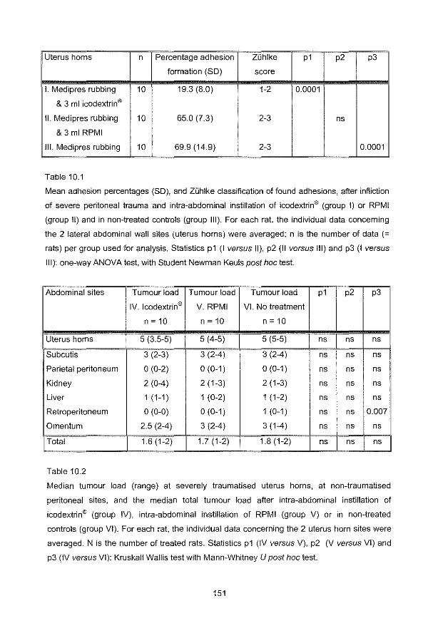
Uterus horns n Percentage adhesion Zuhlke p1 p2 p3
formation (SD) score
I. Medipres rubbing 10 19.3 (8.0) 1-2 0.0001
& 3 ml icodextrin®
II. Medipres rubbing 10 65.0 (7.3) 2-3 ns
&3ml RPMI
Ill. Medipres rubbing 10 69.9 (14.9) 2-3 0.0001
Table 10.1
Mean adhesion percentages (SO), and ZUhlke classification of found adhesions, after infliction
of severe peritoneal trauma and intra-abdominal instillation of icodextrin® (group I) or RPMI
(group II) and in non-treated controls (group Ill). For each rat, the individual data concerning
the 2 lateral abdominal wall sites (uterus horns) were averaged; n is the number of data (=
rats) per group used for analysis. Statistics p1 (I versus II), p2 (II versus Ill) and p3 (I versus
Ill): one-way ANOVA test, with Student Newman Keuls post hoc test.
Abdominal sites Tumour load Tumour load Tumour load p1 p2 p3
IV. lcodextrin® V. RPMI VI. No treatment
n = 10 n = 10 n = 10
Uterus horns 5 (3.5-5) 5 (4-5) 5 (5-5) ns ns ns
Subcutis 3 (2-3) 3 (2-4) 3 (2-4) ns ns ns
Parietal peritoneum 0 (0-2) 0 (0-1) 0 (0-1) ns ns ns
Kidney 2 (0-4) 2 (1-3) 2 (1-3) ns ns ns
Liver 1 (1-1) 1 (0-2) 1 (1-2) ns ns ns
Retroperitoneum 0 (0-0) 0 (0-1) 1 (0-1) ns ns 0.007
Omentum 2.5 (2-4) 3 (2-4) 3 (1-4) ns ns ns
Total 1.6(1-2) 1.7 (1-2) 1.8(1-2) ns ns ns
Table 10.2
!
Median tumour load (range) at severely traumatised uterus horns, at non-traumatised
peritoneal sites, and the median total tumour load after intra-abdomina! instillation of
icodextrin® (group IV), intra-abdominal instillation of RPMI (group V) or in non-treated
controls (group VI). For each rat, the individual data concerning the 2 uterus horn sites were
averaged. N is the number of treated rats. Statistics p1 (IV versus V), p2 (V versus VI) and
p3 (IV versus VI): Kruskall Wallis test with Mann-Whitney U post hoc test.
151

DISCUSSION
The aetiology of adhesion formation is only partly understood but the process is initiated by
traumatising the peritoneal surface either by surgical action, by endometriosis, or by
infection.35·
38"40 The formation of an adhesion commences with injury to two layers of the
peritoneum, one opposite the other, and the resulting exudation contributes to the deposit of
fibrin and adhesion between the two membranes.41'
42 Whether such adhesions are
permanent or will eventually be lysed, is assumed to be dependent on the fibrinolytic
capacity of the peritoneum.4244 A wide variety of therapeutic modalities affecting different
levels in the cascade of f1binogenesis and fibrinolysis have been studied experimentally and
clinically.45-4
8 Results are inconsistent and associated with many side effects such as intra
abdominal haemorrhage and impaired wound healing. Avoiding per- and postoperative
contact between traumatised peritoneal surfaces might bring us closer to reducing
postoperative adhesion formation. Increasing the physiological liquid interface between two
peritoneal surfaces is liable to reduce the initial adhesion phenomena at its origin. Indeed in
a rat model, postoperative peritoneal dialysis significantly reduced adhesion formation,
presumably due to a flotation effect and dilution of the fibrin exudates.49 Obviously,
postoperative peritoneal dialysis is not feasible in the clinical setting but these results
stimulated further investigations. Various liquids have been installed peroperatively in the
peritoneal cavity with the hope that "hydroflotation" would enable traumatised surfaces to be
kept separate for a while. 21-23 Unfortunately these liquids were absorbed too quickly to bring
about clinical successful adhesion prevention50 or caused unwanted side effects.25·
26
Separating peritoneal surfaces by means of slow absorption solutions, allowing prolonged
postoperative "hydrofoliation" of the peritoneal cavity might have a more efficient anti
adhesive effect. lcodextrin is a large a-1,4 linked glucose polymer broken down sequentially
by the enzymes a-amylase and maltase to maltose and glucose. Amylase is widely
distributed throughout the body but '1s not or hardly present in the peritoneal cavity of humans
and rats. When administered peroperatively, icodextrin is largely retained within the
abdominal cavity for up to five days during which the polymer is gradually absorbed through
the lymphatic system into the systemic circulation. 51 lcodextrin 7.5%, although iso-osmolar,
induces ultrafiltration through colloid osmosis and the presence of the polymer is presumed
to create a constant fluid layer between peritoneal surtaces.52 Theoretically, the fluid acts to
reduce the formation of adhesions by a 5-day period of "hydroflotation" of the total abdominal
cavity, the time of maximum risk of adhesion formation. 53·
54 This study showed that after
severe surgical peritoneal trauma icodextrin® significantly reduced the extent and severity of
adhesion formation.
152

We have previously suggested that the peritoneal defence mechanism triggered by surgical
trauma to the peritoneum not only promotes adhesion formation but also stimulates tumour
recurrence. 31 Several theories speculate on the underlying mechanisms of the adhesion and
growth of spilled tumour cells. Implantation of tumour cells onto raw tissue surfaces is an
efficient process as opposed to implantation on intact surfaces. 30 The fibrin entrapment
hypothesis proposes that tumour cells are trapped in fibrin at the resection site and abraded
peritoneal surfaces, hereby providing protection from host defence mechanisms. 55 Whether
postoperative "hydroflotation", could also keep free intra-abdominal tumour cells from
adhering to damaged peritoneum was doubtful since icodextrin® does not prohibit disruption
of the mesothelial cell lining and exposure of the underlying extracellular matrix playing a
pivotal in adhesion of tumour cells. Results obtained in our tumour adhesion and growth
model showed that tumour load at traumatised peritoneal surfaces indeed was not reduced
by peroperative intra-abdominal icodextrin® treatment. This does not mean that there is no
analogy in the processes of adhesion formation and tumour recurrence. Apparently it is not
possible to keep free floating tumour cells from an injured peritoneal surface by a liquid
barrier in which tumour cells suspend and stay alive. Physically, icodextrin® does not form a
barrier between tumour cells and damaged peritoneal surfaces as it does between two
injured peritoneal surfaces. lcodextrin® might be a medium in which tumour cells can
proliferate and augment motility, increasing their metastatic ability. The results of this study
did not support this hypothesis since total peritoneal tumour load at traumatised and remote
peritoneal sites was not increased by icodextrin®, on the contrary tumour load at one remote
peritoneal site was even s'1gnificantly reduced in the icodextrin® treated group.
In conclusion the current study showed that the novel glucose polymer solution, icodextrin®,
used as an intraoperative intra-abdominal instillate was well tolerated, safe, easy to use and
significantly reduced postoperative adhesion formation without promoting or inhibiting tumour
recurrence. lcodextrin® therefore seems to be a good modality to prevent intra-peritoneal
adhesion formation and may be used safely in oncological surgery.
153

REFERENCES
1. Weibel MA, Majno G. Peritoneal adhesions and their relations to abdominal surgery. Am J
Surg 1973; 126: 345-353.
2. Ellis H. Internal overheating: The problem of intra-peritoneal adhesions. Wrld J Surg 1980; 4:
303-306.
3. Diamond MP. Surgical aspects of inferti!ity./n Sciarra JJ, ed. Gynecology and Obstetrics, Vol
5. Philadelphia: Harper & Row, 1995: 1-26.
4. Ellis H. The causes and prevention of intestinal adhesions. Br J Surg 1982; 69: 241-243.
5. McEntee G, Pender D, Mulvin D, Me Cough M, Naeeder S, Farah S, Badurdeen MS, Ferraro
V, Cham C, Gillhamn N. Current spectrum of intestinal obstruction. Br J Surg 1987; 74: 976-
980.
6. Menzies D, Ellis H. Intestinal obstruction from adhesions-how big is the problem? Ann R Colt
Surg Eng/ 1990; 72: 60-63.
7. Drake TS, Grunert GM. The unsuspected pelvic factor in the infertility investigation. Br J Surg
1980; 50: 10-16.
8. Fayez JA. An assessment of the role of operative laparoscopy in tubop!asty. Fertif Sterif 1983;
39: 476-479.
9. Goldstein DP, DeCholnoky C, Emans SJ, Leventhal JM. Laparoscopy in the diagnosis and
management of pelvic pain in adolescents. J Reprod Med 1980; 24:251-256.
10. Rapkin AJ. Adhesions and pelvic pain: a retrospective study. Obstet Gyneco/1986; 68: 13-15.
11. Krabben van der AA, Dijkstra FR, Nieuwenhuijzen M, Reijnen MM, Schaapveld M, Goor van
H. Morbidity and mortality of inadvertent enterotomy during adhesiotomy. Br J Surg 2000; 87:
467-471.
12. Ray NF, Larsen JW, Stillman RJ. Economical impact of hospitalisations for lower abdominal
adhesiolysis in the United States in 1988. Surg Gynecol Obstet 1993; 176:271-276.
13. Jeekel J. Cost implications of adhesions highlighted in a European study. Eur J Surg 1997;
Suppl 579: 43-45.
14. Ellis H, Moran BJ, Thompson JN, Parker MC, Wilson MS, Menzies D, McGuire A, Lower AM,
Hawthorn RJ, 'OBrien F, Buchan S, Crowe AM. Adhesion-related hospital readmissions after
abdominal and pelvic surgery: A retrospective cohort study. Lancet 1999; 353: 1476-1480.
15. Boyers SP, Diamond MP, DeCheney AH. Reduction of postoperative pelvic adhesions in the
rabbit with Gore-Tex surgical membrane. Fertif Steri/1988; 49: 1066-1070.
16. Hel!ebrekers BW, Trimbos-Kemper GC, Blitterswijk van CA, Bakkum EA, Trimbos JB. Effects
of five different barrier materials on postsurgical adhesion formation in the rat. Hum Reprod
2000; 15: 1358-1363.
17. Leach RE, Henry RL. Reduction of postoperative adhesions in the rat uterine horn model with
poloxamar 407. Am J Obstet Gyneco/1990; 162: 1317-1319.
18. Adhesion Barrier Study Group (lnterceed, TC7). Prevention of post-surgical adhesions by
lnterceed (TC7), an absorbable adhesion barrier: a prospective randomised multicenter
clinical study. Fertil Steri/1989; 51: 933-938.
154

19. Becker JM, Dayton MT, Fazio V, Beck DE, Stryker SJ, Wexner SO, Wolff BG, Roberts PL,
Smith LE, Sweeney SA, Moore M. Prevention of postoperative abdominal adhesions by a
sodium hyaluronate-based bioresorbable membrane: a prospective, randomised, double blind
multicenter study. JAm Coli Surg 1996; 183·. 297-306.
20. Beck DE. The role of Seprafilm ™ bioresorbable membrane in adhesion prevention. Eur J Surg
1997; 163 (suppl557); 49-55.
21. Burns JW, Skinner K, Colt MJ, Burgess L, RoseR, Diamond MP. A hyaluronate based gel for
the prevention of post-surgical adhesions: evaluation in two animal species. Fertil Steri11996;
55: 814-820.
22. Heidrick GW, Pippitt CH Jr, Morgan MA, Thurnau GR. Efficacy of intra -peritoneal sodium
carboxymethylcel!ulose in preventing postoperative adhesion formation. J Reprod Med 1994;
39: 575-580.
23. Burns JW, Skinner K, Colt J, Sheidlin A, Bronson R, Yaacobi Y, Goldberg EP. Prevention of
tissue injury and postsurgical adhesions by precoating tissues with hyaluronic acid solutions. J
Surg Res 1995; 59: 644-652.
24. Diamond MP, Michael P and the Sepracoat Adhesion study group. Reduction of de novo
postsurgical adhesions by interoperative precoating with Sepracoat (HAL-C™) solution: a
prospective, randomised, blinded, placebo-controlled multicenter study. Fertil Sterif 1998; 69:
1067-1074.
25. Weimans MJ, Kauer FM, Klompmaker IJ, Wijma J. Transient liver function disturbances after
intra-peritoneal use of 32% dextran 70 as adhesion prophylaxis in infertility surgery. Fertil
Sleri/1990; 53: 159-161.
26. Gauwerky JF, Heinrich D, Kubli F. Complications of intra -peritoneal dextran application for
prevention of adhesions. Bioi Res Pregnancy Perinato/1996; 7:93-96.
27. Verco SJS, Peers EM, Brown CB, Rodgers KE, Roda N, diZerega G. Development of a novel
glucose polymer solution (icodextrin®) for adhesion prevention: pre-clinical studies. Hum
Reprod 2000; 15: 1764-1772.
28. Verco SJ, diZerega G and the CLASSIC Study Group. Tolerability and effectiveness of 4%
icodextrin solution in the reduction of post-surgical adhesions following laparoscopic surgery: a
multicenter, pilot, clinical study. J Reprod Fertil Abstr Ser 2000; 25: 62.
29. Abulafi AM, Williams NS. Local recurrence of colorectal cancer: the problem, mechanisms,
management and adjuvanttherapy [see comments]. Br J Surg 1994; 81: 7-19.
30. Sugarbaker PH. A prospective on clinical research strategies in carcinoma of the large bowel.
World J Surg 1991; 15: 609-616.
31. Tol van den MP, Rossen van MEE, Eijck van CH, Bonthuis F, Marquet RL, Jeekel J.
Reduction of peritoneal trauma by using non -surgical gauze leads to less implantation
metastasis of spilled tumour cells. Ann Surg 1998; 227: 242-248.
32. Busch OR, Hop WC, Hoynck van Papendrecht MA, Marquet RL, Jeekel J. Blood transfusion
and prognosis in colorectal cancer. N Eng! J Med 1993; 328: 1372-1376.
155

33. Bouvy NO, Marquet RL, Hamming JF, Jeeke! J, Bonjer HJ. Laparoscopic surgery in the rat:
beneficial effect on body weight and tumor take. Surg Endosc 1996; 10: 490-494.
34. Marquet RL, Westbroek DL, Jeekel J. interferon treatment of a transplantable colon
adenocarcinoma, importance of tumor site. lnt J Cancer 1984·, 33: 688-692.
35. Tal van den MP, Stijn van I, Bonthuis F, Marquet RL, Jeekel J. Reduction of intra-peritoneal
adhesion formation by use of non-abrasive gauze. Br J Surg 1997; 84: 1410-1415.
36. ZUhlke HV, Lorenz EMP, Straub EM, Savvas V. Pathophysiologie und Klassifikation von
Adh8sionen. Langenbecks Arch Chir Suppl fl Verh Dtsch Ges Chir 1990; 345: 1009-1016.
37. Steller EP, Ottow RT, Eggermont AMM, Marquet RL, Sugarbaker PH. Local conditions in the
host influence immunotherapy with interleukin-2 and LAK cells. Cancer Detect Prev 1988; 12:
81-90.
38. Ellis H. The healing of peritoneum under normal and pathological conditions. Br J Surg 1965;
52:471-476.
39. Graeme BR, Grobety, Majno G. Mesothelial injury and recovery. Am J Patho/1973; 71: 93-
102.
40. Milligan OW, Raftery AT. Observations on the pathogenesis of peritoneal adhesions: a light
and electron microscopical study. BrJ Surg 1974; 61:274-280.
41. Monz FJ, Shimanuki T, diZerega GS. Post-surgical mesothelial reepithelialisation. In:
DeCherney AH, Polan Mleds. Reproductive Surgery, Year Book medical Publishes. 1987: 31-
47.
42. Thompson JN, Whawell SA. Pathogenesis and prevention of adhesion formation. Br J Surg
1995; 82: 3-5.
43. Raftery AT. Effect of peritoneal trauma on peritoneal fibrinolytic activity and intra-peritoneal
adhesion formation. Eur J Res 1981·, 13: 397-401.
44. Vipond MN, Whawell SA, Thompson JN, Dudley HA. Peritoneal fibrinolytic activity and intra
abdominal adhesions. Lancet 1990; 335: 1120-1122.
45. Hackel M, Ott S, Siemann U, Kissell T. Prevention of peritoneal adhesions in the rat with
sustained intra-peritoneal dexamethason delivered by a novel therapeutic system. Ann Chir
Gynecol 1987; 76: 306-313.
46. Rodgers KE. Non-steroidal anti-inflammatory drugs (NSAID's) in the treatment of post-surgical
adhesion. In: Treatment of Postsurg Adh. Eds.: G.S. diZerega, L. Russell Malinak, M.P.
Diamond, G.B. Linsky, Wi!ly-Uss, 1990: 119-130.
47. Treutner KH, Winkeltau G, Lerch MM, Stadel R, Schumpelick V. Prevention of postoperative
intra-peritoneal adhesions. A new study standardised in rats. Research in Surgery 1992; 4: 50-
54.
48. Doody KJ, Dunn RC, Buttram VC. Recombinant tissue plasminogen activator reduces
adhesion formation in a rabbit uterine horn model. Fertil Steri/1989; 51: 509-512.
49. Lindenberg S, Moesgaard F, Vangsted P, Lykkegaard Nielsen M. Peritoneal dialysis reduces
intra-peritoneal adhesion formation. An experimental study in rats. Eur Surg Res 1982; 14:
274-278.
156

50. Hart R, Magos A. Laparoscopically instilled fluid: the rate of absorption and the effects on
patient discomfort and fluid balance. Gynaecol Endos 1996; 5: 287-291.
51. Gilbert JA, Peers EM, Brown CB et al. Intra-peritoneal fluid dynamics of 4% icodextrin in non
ESRD patients. Peril Diallnt 1999; 19: (suppl 1) S79.
52. Mistry CD, Gokal R. The use of glucose polymer (icodextrin°) in peritoneal dialysis: an
overview. Perit Diallnt 1994; 14: S158-S161.
53. Raftery A. Regeneration of parietal and visceral peritoneum: an electron microscopical study.
J Anal 1973; 115: 375-392.
54. Harris ES, Morgan RF, Rodeheaver GT. Analysis of the kinetics of peritoneal adhesion
formation in the rat and the evaluation of potential antiadhesive agents. Surgery 1995; 117:
663-669.
55. Cunliffe WJ, Sugerbaker PH. Gastrointestinal malignancy: rationale for adjuvant therapy using
early postoperative intra-peritoneal chemotherapy. Br J Surg 1989; 76: 1082-90.
157


PARTV
General Discussion and Summary

Chapter XI
General Discussion
Surgical peritoneal trauma and postoperative adhesions
Peritoneal trauma causes a disturbance of the normal intra-abdominal inflammatory and
fibrinogenetic defences leading to a disrupted equilibrium between mediators of the
inflammatory response and between fibrinigenesis and fibrinolysis and thus induces post
surgical adhesions. Due to a lack of complete understanding of the dynamic process of
peritoneal healing and its derailment, literature abounds on all kinds of methods to prevent
adhesion formation. The results achieved with the different treatment modalities are
inconsistent and associated with many side effects such as impaired wound healing,
reduction of the immune system, intra-abdominal haemorrhage and gastrointestinal bleeding.
Until today, only one technique is effective in reducing the occurrence of post-surgical
adhesions to some extent. This technique aims to avoid manipulations which are known to
provoke adhesion formation. 1·5 That includes preventing unnecessary desiccation, handling,
clamping or suturing (ischaemia) of tissue and spilling of foreign material during operative
intervention.4'
5 Also, general agreement exists that it is unnecessary and even unwanted to
close the peritoneum postoperatively. 6-17 However, although peritoneal trauma was proven to
be a cause of adhesion formation, a link between the degree of peritoneal trauma and the
extent of formed adhesions was never demonstrated. 6· 18
· 19 Therefore, the first aim of the
experiments described in this thesis was to analyse whether the extent and type of post
surgical adhesion formation correlates with the degree of peritoneal damage inflicted. In order
to evaluate this, we developed a reproducible rat model, allowing semiquantitative and
qualitative scoring of adhesions in vivo in which the clinical situation of post-surgical
adhesion formation was imitated. Results of the first experiments showed that the extent of
adhesion formation correlated significantly with the inflicted degree of peritoneal damage.
Furthermore one of the aims of studies portrayed in this thesis was to identify whether agents
like surgical gauze, lavage solutions and glove powder, not yet generally considered as
traumatic for the peritoneum and commonly used in general surgical practice, indeed were
traumatic and could consequently cause postoperative adhesions. We also investigated less
161

traumatic alternatives for these agents. Experimental results demonstrated that intra
abdominal manipulation with surgical Medipres gauze was very traumatic to the peritoneum
and induced significantly more adhesion formation than manipulation of the peritoneum with
less traumatic non-surgical Fastsorb textile. Moreover, there was no significant difference in
the extent and type of adhesions formed after infliction of minimal peritoneal trauma alone or in
combination with Fastsorb manipulation, suggesting Fastsorb is hardly traumatic for the
peritoneum. Given these results and the fact that non-surgical Fastsorb textile meets important
criteria for surgical abdominal swabs, this non-abrasive textile might be a suitable substitute for
Medipres gauze in surgical practice. Further experiments showed that exposure of the
abdominal cavity to lavage solutions, often used in the end stage of abdominal interventions,
also traumatised the peritoneum and engendered significant adhesions formation. During
surgery in non-contaminated abdominal cavities peritoneal lavage should not be performed
and per-operative lavage of contaminated abdominal cavities should be viewed with caution.
Last but not least, because powdered gloves are still universally used in abdominal surgery,
we performed the studies demonstrating that local application of glove powder on minimally
and severely traumatised peritoneum gave rise to a significantly higher percentage of
adhesion formation than infliction of peritoneal trauma alone. This finding adds to the already
existing evidence20-25 that intra-abdominal contamination with starch by the use of starch
powdered gloves should be avoided.
Surgical peritoneal trauma and tumour recurrence
Most attempts at treating intra-abdominal tumour recurrence, like local or regional chemo
and radiotherapy and photodynamic therapy,27-30 are aimed at annulling manifest peritoneal
tumour foci but do not focus on prohibiting the postoperative arising of local tumour
recurrence. A different approach to diminishing post-surgical intra-abdominal tumour
recurrence is to interfere with the primary implantation and growth process of intra
abdominally spilled free tumour cells. This necessitates a profound understanding of the
underlying pathophysiologic mechanisms leading to local tumour recurrence. Subsequent
studies described in this thesis were aimed at unravelling the !inks between surgical
peritoneal trauma and the process of postoperative adhesion formation and intra-abdominal
tumour recurrence and analysing the similarities amongst the latter.
The observed preference of tumours to recur at surgically traumatised sites26•
31-35 and the
theory of metastatic efficiency and the tumour cell entrapment hypothesis26 brings up the
question whether the dynamic peritoneal healing process, which when not in balance can
lead to adhesion formation, also plays a role in the process of local tumour recurrence. This
ascertainment and theoretic perceptions led us to investigate if a correlation existed between
surgical peritoneal trauma and intra-abdominal tumour recurrence.
162

In order to meet this purpose and evaluate patterns of tumour recurrence in vivo, different rat
models were used. We developed a reproducible rat tumour adhesion and growth model,
analogous to the rat adhesion model, allowing semiquantitative scoring of tumour load in
which the clinical situation of free intra-abdominal tumour cells was mimicked. This model
associates the combination of cell adhesion and growth, ultimately leading to manifest
tumour load. The amount of tumour recurrence at directly and not directly traumatised
peritoneal sites appeared to be highly correlated with the degree of peritoneal trauma. The most
impressive tumour load was observed at severely traumatised peritoneal sites. However,
enhanced tumour recurrence was also seen at not directly traumatised peritoneal sites and the
amount of tumour at these loco-regional sites also correlated with the severity of the trauma.
This indicates that the sequelae of peritoneal trauma with regard to tumour recurrence are not
confined to the inflicted site itself, but appear to have a generalised character. In a previously
developed sub-renal capsule assay, the effect of surgical peritoneal trauma on the growth of
extra-peritoneal tumours could be reproduced. Because promotion of adherence was
irrelevant in this model it represents the systemic effects on tumour growth that may occur
after surgical injury. After placing a solid tumour in the extra-peritoneal space, the intensity of
surgical trauma correlated with the extra-peritoneal tumour growth, suggesting that systemic
factors produced after surgical peritoneal trauma indeed play an important role in enhanced
tumour growth. The apparent generalised character of tumour recurrence led to the
hypothesis that factors produced after peritoneal trauma, may influence loco-regional and
distant aspects of tumour recurrence. When these factors were captivated in a peritoneal
lavage fluid and passively transferred to naive recipients, this hypothesis was confirmed. To
study merely the effect of tumour cell adhesion an in vitro model was designed. Culturing a
monolayer of mesothelial cells on a matrix coating mimicked the peritoneum. Tumour cell
adhesion to mesothelial cells could be studied after pre-incubating the mesothelial monolayer
with different substances. In experiments using this model, described by van Rossen et al, 36
cytokines and growth factors, produced by exposed endothelial cells, damaged mesothelial
cells and inflammatory cells during peritoneal wound healing,3741 proved to be significant
promoting factors in tumour cell adhesion to mesothelium. Supported by these results the
initial impetus to separate the generalised term "tumour recurrence" into the more elementary
mechanisms of tumour cell adhesion and growth was made. Both components may
contribute evenly to successful intra-abdominal tumour recurrence and both are modified by
surgical trauma.
As mentioned before, previous experiments showed that exposure of peritoneal cavity to
surgical gauzes, lavage solutions and glove powder traumatised the peritoneum and
promoted postoperative adhesion formation. Since subsequent data suggest that the factors
responsible for the formation of post-surgical adhesions also play a role in the adhesion and
163

growth of tumour cells to the peritoneum we investigated whether surgical gauze and glove
powder also could induce more tumour recurrence. The studies demonstrated that surgical
Medipres gauze induced significantly more tumour load than non-surgical Fastsorb textile and
peritoneal application of glove powder or pure starch induced significantly more intra
abdominal tumour load than when no powder was applied. Avoiding unnecessary
peroperative peritoneal trauma by employing gentle surgical techniques and materials is
therefore not only indicated to reduce postoperative adhesion formation but also to prevent
intra-abdominal tumour recurrence. In accordance with this goal and given the fact that non
surgical Fastsorb textile meets important criteria for surgical purposes we again point out that
this non-abrasive textile seems a suitable substitute for surgical gauze. Intra-peritoneal
contamination with starch from starch-powdered gloves should also be avoided to prevent
local tumour recurrence. Since good powder-free alternatives are available there is no longer
any justification for the use of powdered gloves during intra-abdominal surgery.
Pathways of adhesion formation and intra~abdominal tumour recurrence
Since peroperative peritoneal trauma cannot entirely be prevented in practice, the need for
supplementary prophylactic measures remains urgent. Better understanding of the
underlying pathogenesis of postoperative intra-abdominal adhesion formation and peritoneal
tumour cell adhesion and growth will undoubtedly help in the development of balanced and
more effective therapeutic strategies. Cellular and soluble constituents of the peritoneal
cavity are believed to both reflect the events arising during wound healing in the peritoneal
cavity and have the potential to modulate the outcome of tissue response to injury.18·
37-52 As
cited before, preceding experiments showed that within a few hours after infliction of peritoneal
trauma tumour cell adhesion and growth promoting factors could be captivated in a peritoneal
lavage fluid and passively transferred to naive recipients. Further analysis of the fragments in
the captured lavage fluid brought forward the presence of a cellular and soluble fraction. When
these fractions were individually injected into pristine abdominal cavities, without any form of
additional surgical trauma, both induced significantly more peritoneal tumour recurrence.
However, the effect of the soluble factors was inferior to that of the cellular fraction. More
detailed unravelling of the cellular fraction demonstrated that peritoneal trauma induces
oscillation in intra-abdominal cells. Apart from an augmented absolute cell amount, the ration
changes from primarily monocytes to mainly neutrophil granulocytes (PMN). This observation
was made during the acute phase after surgery and most likely represents the PMN influx in
response to chemoattractant factors. The coincidence of posttraumatic intra-abdominal PMN
influx with adhesion formation and tumour cell adhesion and growth is no solid prove for the
role of PMN in these pathogenetic processes. Therefore, studies were performed to
investigate whether we were able to prohibit posttraumatic intra-abdominal PMN influx. Intra-
164

peritoneal injection of anti-neutrophil serum (ANS) indeed foiled this PMN influx in rats.
Moreover a well-balanced prevention of posttraumatic intra-abdominal PMN influx by ANS
reduces adhesion formation and local tumour recurrence significantly. Accordingly, intra
abdom·lnal influx of PMN after surgical peritoneal trauma plays a crucial role in postoperative
adhesion formation and in the process of adhesion and growth of spilled tumour cells. This
knowledge opens up the way to develop of novel therapeutic strategies acting subtle, in
order to prevent unwanted side effects, upon the (dis)balance of the peritoneal healing
process.
The adhesion formation and tumour recurrence promoting effect of PMN's may be based on
several characteristics of these cells. First, through PMN-cytokine production an upregulation
of cell adhesion molecules may be induced,53·
54 hereby facilitating adhesion of tumour cells.
In addition, cytokine activated mesothelial cells produce chemoattractants like IL-8, required
for PMN recruitment. 55·
56 In this way, the mesothelium and inflammatory cells in the
abdominal cavity may perpetuate a cytokine loop, resulting in extreme activation of the
inflammatory process. Secondly, mesothelial damage may be aggravated by the
degranulation of organelles, but most likely through the production of reactive oxygen
species (ROS). 57 Obviously, affecting one or more of these PMN involving steps in the
cascade of derailed peritoneal healing may lead to the evolvement of new adhesion and
tumour recurrence preventive formulas.
Adjuvant measures to prevent adhesion formation and tumour recurrence
Developing novel prophylactic measures through unravelling the pathophysiologic
mechanisms of the peritoneal healing cascade is a time consuming process. In the mean
time the need to treat today's patients as good as possible remains and on that account we
analysed the effect of a known adhesion reducing fluid (Sepracoat™),58"61 with also known
trammels, on tumour recurrence in vivo and tumour cell adhesion in vitro. Hyaluronic acid is a
principal ligand for CD44 which is an ubiquitous, multistructural and multifunctional surface
adhesion molecule regulating cell-cell and cell-matrix interactions.62 CD44 and its variants are
present on many cancer cell types and have found to be involved in local tumour progression
and metastasis.63'
64 Therefore, hyaluronic acid containing solutions theoretically may affect
local adhesion and growth of free inrta-abdominal tumour cells. The results of our experiments
demonstrate that Sepracoat™ had a small but significant inhibitory effect on the adhesion of
tumour cells to mesothelial cell monolayers in vitro. However, we were unable to repeat these
effects in our in vivo rat models; the peroperative inoculation of Sepracoat™ did not reduce nor
promote postoperative tumour recurrence.
To overcome a number of major disadvantages of issued absorbable barriers and viscous
barrier solutions60·
61·
65-74 a glucose polymer solution already successfully and safely used in
165

peritoneal dialysis was recently developed into a fluid that is absorbed only slowly, allowing
prolonged "hydroflotation" of the peritoneal cavity.75•
76 With this "water" barrier technique two
damaged peritoneal suliaces are mechanically separated during the period of mesothelial
regeneration, thereby in theory enabling the prevention of adhesion formation. In our hands
icodextrin® was found to be a well tolerated, safe and easy to use solution that effectively
reduces extent and severity of postoperative adhesion formation in rats without promoting or
inhibiting intra-abdominal tumour recurrence. Apparently it is not possible to keep free
floating tumour cells from an injured peritoneal surtace by a liquid barrier in which tumour
cells suspend and stay alive. Physically, icodextrin® does not form a barrier between tumour
cells and damaged peritoneal suliaces as it does between two injured peritoneal suliaces
during the peritoneal healing process. lcodextrin® seems a very promising development in
the avenue of future resaerch because next to working as an adhesion reducing adjuvant by
itself, it could also function as a long lasting carrier for other adhesion and tumour inhibiting
substances. Whether this treatment modality will proof to be effective in human remains to be
investigated but experimental studies look promising.
Considerations for clinical applications.
Any form of peritoneal trauma is likely to cause postoperative adhes'1on formation and
enhance intra-abdominal tumour recurrence. The operating skill of the surgeon therefore is a
relevant factor in adhesion and tumour prognosis. Avoiding of unnecessary desiccation,
handling, clamping or suturing (ischaemia) of tissue and leaving behind as little foreign
material as possible during operative intervention are generally accepted measures for
peliorming surgery in a non-traumatising fashion. 1-5 In addition, we advice surgeons to
prohibit unnecessary peroperative intra-abdominal use of surgical gauzes and if frequent or
profound use is inevitable to choose a non-abrasive textile as a substitute. Furthermore during
surgery in non-contaminated abdominal cavities peritoneal lavage should not be peliormed.
And moreover since good powder-free alternatives are available powdered gloves should not
be used during intra-abdominal surgery. Minimal invasive surgery may also diminish the
extent of peroperative peritoneal injury.
Still intra-abdominal surgery will always bring about peroperative peritoneal trauma in some
quantity at some point during operation. Adjuvant measures associated with surgical
intervention seem a logical step not only to prevent postoperative adhesions but also to avert
loco-regional tumour recurrence. At the present time, despite the great variety of agents
employed, none of them seems to be overwhelmingly suitable for effective and save general
use in the humans. Although it is difficult to manoeuvre, presently the clinically most
successful and save anti-adhesive measure seems to be hyaluronic acid
/carboxymethylcellulose. Since it effectively reduces the severity of postoperative adhesions
166

in humans and, as this thesis elaborates by animal studies, did not influence tumour
recurrence it seems recommendable to use Sepreafilm ™ at least during abdomina!
interventions which will be followed by planned relaparotomy or second look operations.
Experiments described in this thesis showed that an easy to handle new glucose polymer
solution, icodextrin®, could markedly reduce the extent and severity of postoperative
adhesion formation in rats, (unfortunately) without affecting intra-abdominal tumour
recurrence. It is too early to know whether these results can be repeated in humans but
further research, in the form of clinical trials, has to be executed to estimate the clinical value
of this barrier solution.
In this thesis, one major step in the pathway of adhesion formation and peritoneal tumour
recurrence has been brought into perspective. Post-traumatic intra-abdominal PMN influx
proved to play a major role in the processes of postoperative adhesion formation and intra
abdomina! tumour recurrence. Efforts to dim the inflammatory reaction by mastering the
mediators of this process has already been suggested as adhesion and tumour recurrence
abating alternatives and these experimental results contribute to this idea. In the
development of such substrates one always has to keep in mind that the peritoneal healing
processes rely on the same biological me-chanisms as adhesion formation and tumour
recurrence, thus manipulation of steps in the derailed cascade has to be done selectively
and in moderation to prevent unwanted side effects.
167

REFERENCES
1. Siegler AM, Kontoupoulos V. An analysis of macrosurgical and microsurgical techniques in the
management of the tuboperitoneal factor in infertility. Fertil Steri/1979; 32: 377-383.
2. Thompson J. Pathogenesis and prevention of adhesion formation. Dig Surg 1998; 15: 153-
157.
3. Holmdahl L, Risberg 8, Beck DE, Burns JW, Chegini N, diZerega GS, Ellis H. Adhesions:
pathogenesis and prevention-panel discussion and summery. EurJ Surg Supp/1997: 56-62.
4. Elkins TE, Stovall TG, Warren J, Ling FW, Meyer NK. A histologic evaluation of peritoneal
injury and repair: Implications for adhesion formation. Obstet Gyn 1987; 70: 377-383.
5. Luijendijk RW, Lange de DC, Wauters CCAP, Hop WC, Duron JJ, Pailler JC, Camprodon BR,
Holmdah! LE, Ge!drop van HJ, Jeeke! J. Foreign materia! in postoperative adhesions. Ann
Surg 1996; 223: 242-248.
6. Ems H. The healing of peritoneum under norma! and pathological conditions. Br J Surg 1965; 52:
471-476.
7. Watters WB, Buck RC. Scanning electron microscopy of mesothelial regeneration in the rat. Lab
Invest 1972; 13:48-54.
8. Raftery AT. Regeneration of parietal and visceral peritoneum. A light microscopical study. Br J
Surg 1973; 60: 293-299.
9. Ellis H, Heddle R. Does the peritoneum need to be closed at laparotomy? Br J Surg 1977; 64:
733-736.
10. Raftery AT. Regeneration of peritoneum: a fibrinolytic study. J Anat 1979; 129: 659-664.
11. Kyzer S, Bayer I, Turani H, Chaimhoff C. The influence of peritonea! closure on formation of
intra-peritoneal adhesions: an experimental study./nt J Tissue React 1986; 8: 355-359.
12. Gilbert JM, Ellis H, Foweraker S. Peritoneal closure after lateral paramedian incision. Br J
Surg 1987; 74: 113-115.
13. Hugh TB, Nankivell C, Meagher AP, Ll B. Is closure of the peritoneal layer necessary in the
repair of the midline surgical abdominal wounds? World J Surg 1990; 14: 231-234.
14. Holmdahl L, AI-Jabreen M, Risberg B. Experimental models for quantitative studies on
adhesion formation in rats and rabbits. Eur Surg Res 1994; 26: 248-256.
15. diZerega GS. Contemporary adhesion prevention. Fertil Steril 1994; 61: 219-235.
16. Duffy OM, diZerega GS. Is peritoneal closure necessary? Obstet Gynecol Surv 1994; 49: 817-
822.
17. Irion 0, Luzuy F, Beguin F. Non-closure of the visceral and parietal peritoneum at caesarean
section: a randomised controlled trial. Br J Obstet Gynaeco/1996; 103: 690-694.
18. Milligan OW, Raftery AT. Observations on the pathogenesis of peritoneal adhesions: a light and
electron microscopical study. Br J Surg 1974; 61: 274-280.
19. Graeme BR, Grobety J, Majno G. Mesothelial injury and recovery. Am J Patho/1973; 71: 93-
102.
20. Sneierson H, Woo ZP. Starch powder granuloma. A report of two cases. Ann Surg 1955; 142:
1045-1050.
168

21. Cooke SAR, Hamilton DG. The significance of starch powder contamination in the aetiology of
peritoneal adhesions. Br J Surg 1977; 64: 410-412.
22. McEntee GP, Stuart RC, Byrne PJ, Leen E, Hennessy TP. Experimental study of starch
induced intra-peritoneal adhesions. Br J Surg 1990; 77: 1113-1114.
23. Ellis H. The hazards of surgical glove dusting powders. Surg Gynecol Obstet 1990; 171: 521-
527.
24. Kamffer WJ, Jooste EV, Nel JT, Wet de Jl. Surgical glove powder and intra-peritoneal
adhesion formation. An appeal for the use of powder-free surgical gloves. South African Med J
1992; 81; 158-159.
25. Luijendijk RW, Lange de DC, Wauters CCAP, Hop WC, Duron JJ, Pai!!er JC, Camprodon BR,
Holmdahl LE, Geldrop van HJ, Jeekel J. Foreign materia! in postoperative adhesions. Ann
Surg 1996; 223: 242-248.
26. Sugarbaker PH. A perspective on clinical research strategies in carcinoma of the large boweL
World J Surg 1991; 15: 609-616.
27. Sugerbaker PH. Successful management of microscopic residual disease in large bowel
cancer. Cancer Chemother Pharmacol1999; 43 (suppl): S15-S25.
28. Esquivel J, Vidal-Jove J, Steves MA, Sugarbaker PH. Morbidity and mortality of cytoreductive
surgery and intra-peritoneal chemotherapy. Surgery 1993; 113: 631-636.
29. Veenhuizen RB, Reuvekamp MC, Oppelaar H, Helmerhorst T J, Kenemans PJ, Stewart FA.
Foscan-mediated photodynamic therapy for a peritoneal-cancer model: drug distribution and
efficacy studies. lnt J Cancer 1997; 73: 230-235.
30. Eijck van CHJ, Link KH, Rossen van MEE, Jeekel J. (Neo)adjuvant treatment in pancreatic
cancer; the need for future trials. Eur J Surg On col 199; 25: 132-137.
31. Eggermont AMM, Stetler EP, Sugarbaker PH. Laparotomy enhances intra-peritoneal tumour
growth and abrogates the anti-tumour effects of interleukin-2 and lymphokine-activated killer cells.
Surgery 1987; 102: 71-78.
32. Busch OR, Hop WC, Hoynck van Papendrecht MA, Marquet RL, Jeekel J. Blood transfusion
and prognosis in colorecta! cancer. N Eng/ J Med 1993; 328: 1372-1376.
33. Lee YT. Local and regional recurrence of carcinoma of the colon and rectum: II. Factors relating
to operative technique. Surg Oneal 1996; 5: 1-13.
34. Bouvy NO, Marquet RL, Hamming JF, Jeekel J, Bonjer HJ. Laparoscopic surgery in the rat:
beneficial effect on body weight and tumour take. Surg Endosc 1996; 10: 490-494.
35. Jacobi CA, Ordemann J, Zieren HU, Volk HD, Bauhofer A, Halle E, Muller JM. Increased
systemic inflammation after laparotomy vs laparoscopy in an animal model of peritonitis. Arch
Surg 1998; 133:258-262.
36. Rossen van MEE, Hofland LJ, Tal van den MP, Koetsveld van PM, Jeekel J, Marquet RL,
Eijck van CHJ. Effect of inflammatory cytokines and growth factors on tumour cell adhesion to
the peritoneum. J Pathol 2001; 193: 530-537.
37. Lanfrancone L, Boraschi D, Ghiara P, Falini B, Grignani F, Peri G, Mantovani A, Pelicci PG.
Human peritoneal mesothelial cells produce many cytokines (granulocyte colony-stimulating
169

factor (CSF), granu!ocyte-monocyte-CSF, Macrophage-CSF, inter!eukin-1 (ll-1) and IL-6) and
are activated and stimulated to grow by IL-1. Blood 1992; 80:2835-2842.
38. Tsudaka K, Katoh H, Shiojima M, Suzuki T, Takenoshita S, Nagamachi Y. Concentrations of
cytokines in the peritoneal fluid after abdominal surgery. Eur J Surg 1993; 159:475-479.
39. Tapley N. The cytokine network controlling peritoneal inflammation. Perit Diallnt 1995; 15: S35-
S39.
40. Badia JM, Whawe!l SA, Scott-Coombes OM, Abel PD, Williamson RC, Thompson JN. Peritoneal
and systemic cytokine response to laparotomy. Br J Surg 1996; 83: 347-348.
41. Tapley N, Liberek T, Davenpot A, Li FK, Fear H, Wiliams JD. Activation of inflammation and
leukocyte recruitment into the peritoneal cavity. Kidney lnt Supp/1996; 56: S17-S21.
42. Schade DS, Williamson JR. The pathogenesis of peritoneal adhesions; an ultrastructural
study. Ann Surg 1968; 167: 500.
43. Raftery AT. Regeneration of parietal and visceral peritoneum. A light microscopical study. Br J
Surg 1973; 60: 293-299.
44. Seppa H, Grotendorst G, Seppae S, Schiffmann E, Martin GR. Platelet derived growth factor
is chemotactic for fibroblast. J Cell Bio/1982; 92; 584-588.
45. Postlethwaite AE, Lachman LB, Mainadri CL, Kang AH. lnterleukin-1 stimulation of
collagenase production by cultured fibroblasts. J Exp Med 1983; 157: 801-806.
46. Ignatz RA, Massaque J. Transforming growth factor ~ stimulates the expression of fibronectin
and collagen and their incorporation into the extracellular matrix. J Bioi Chern 1986; 261:
4337-4345.
47. Pledgers WJ, Stiles CD, Antiniades HN. An ordered sequence of events is required before
BALB/c-3T3 cells have become committed to DNA synthesis. Proc Nat! Acad Sci USA 1987;
74: 2839-2848.
48. Varga J, Jimenez SA. Stimulation of normal human fibroblast collagen production and
processing by transforming growth factor-~ and 12-0-tetradecanoyl phorbol-1 3-acetate. J Bioi
Chern 1987; 262:2283-2287.
49. Vipond MN, Whawel! SA, Thompson JN, Dudley HA Peritoneal fibrinolytic activity and intra
abdominal adhesions. Lancet 1990; 335: 1120-1122.
50. Whawell SA, Wang Y, Fleming KA, Thompson EM, Thompson JN. Localisation of plasminogen
activator inhibitor-1 production in inflamed appendix by in situ mRNA hybridisation. J Pathol
1993; 169:67-71.
51. Nagy JA, Jackman R. Response of the peritoneal membrane to injury. Semin Dialysis 1998; 11:
242-248.
52. Ward PA, Lentsch AB. The acute inflammatory response and its regulation. Arch Surg 1999;
134: 666-669.
53. Jonjic N, Peri G, Bernasconi S, Sciacca FL, Colotta F, Pelicci P, Lanfrancone L, Mantovani A.
Expression of adhesion molecules and chemotactic cytokines in human mesothelial cells. J Exp
Med 1992; 176:1165-1174.
170

54. Klein CL, Bittinger F, Skarke CC, Wagner M, Kohler H, Walgenbach S, Kirkpatrick CJ. Effects of
cytokines on the expression of cell adhesion molecules by cultured human omental mesothelial
cells. Pathobiology 1995; 63: 204-212.
55. Tapley N, Williams JD. Effect of peritoneal dialysis on cytokine production by peritoneal cells.
Blood Purif1996; 14: 188-197.
56. Meager A. Cytokine regulation of cellular adhesion molecule expression in inflammation.
Cytokine Growth Factor Rev 1999; 10: 27-39.
57. Weiss SJ. Tissue destruction by neutrophils. N Eng J Med 1989; 320: 365-376.
58. Urman B, Gamel V, Jetha N. Effect of hyaluronic acid on postoperative intra-peritoneal adhesion
formation in the rat model. Fertil Sterif 1991; 56:563-567.
59. Goldberg EP, Burns JW, Yaacobi Y. Prevention of postoperative adhesions by precoating
tissues with dilute sodium hyaluronic solutions. Prog Clin Bioi Res 1993; 381: 191-204.
60. Burns JW, Skinner K, Colt J Sheidlin A, Bronson R, Yaacobi Y, Goldberg EP. Prevention of
tissue injury and post-surgical adhesions by precoating tissues with hyaluronic acid solutions.
J Surg Res 1995; 59: 644-652.
61. Bums JW, Skinner K, Colt MJ, Burgess L, RoseR, Diamond MP. A hyaluronate based gel for
the prevention of post-surgical adhesions: evaluation in two animal species. Fertif Steri/1996;
55: 814-820.
62. Naot D, Sionov RV, Ish-Shalom D. CD44: structure, function, and association with the malignant
process. Adv Cancer Res 1997; 71: 241-319.
63. Gardner MJ, Catterall! JB, Jones LM, Turner GA. Human ovarian tumour cells can bind
hyaluronic acid via membrane CD44: a possible step in peritoneal metastasis. Clin Exp
Metastasis 1996; 14: 325-334.
64. Goebeler M, Kaufmann D, Brocker EB, Klein CE. Migration of highly aggressive melanoma cel!s
on hyaluronic acid is associated with functional changes, increased turnover and shedding of
CD44 receptors. J Cell Sci 1996; 109: 1957-1964.
65. Boyers SP, Diamond MP, DeCheney AH. Reduction of postoperative pelvic adhesions in the
rabbit with Gore-Tex surgical membrane. Fertil Steri/1988; 49: 1066-1070.
66. Adhesion Barrier Study Group (lnterceed, TC7). Prevention of post-surgical adhesions by
lnterceed {TC7), an absorbable adhesion barrier: a prospective randomised multicenter
clinical study. Fertil Steri/1989; 51: 933-938.
67. Leach RE, Henry RL Reduction of postoperative adhesions in the rat uterine horn model with
poloxamar 407. Am J Obstet Gyneco/1990; 162: 1317-1319.
68. Weimans MJ, Kauer FM, Klompmaker IJ, Wijma J. Transient liver function disturbances after
intra-peritoneal use of 32% dextran 70 as adhesion prophylaxis in infertility surgery. Fertil
Steri/ 1990; 53: 159-161.
69. Heidrick GW, Pippitt CH Jr, Morgan MA, Thurnau GR. Efficacy of intra-peritoneal sodium
carboxymethylcellulose in preventing postoperative adhesion formation. J Reprod Med 1994;
39: 575-580.
171

70. Gauwerky JF, Heinrich D, Kubli F. Complications of intra-peritoneal dextran application for
prevention of adhesions. Bioi Res Pregnancy Perinatol 1996; 7: 93-96.
71. Becker JM, Dayton MT, Fazio V, Beck DE, Stryker SJ, Wexner SO, Wolff BG, Roberts PL,
Smith LE, Sweeney SA, Moore M. Prevention of postoperative abdominal adhes'1ons by a
sodium hyaluronate-based bioresorbable membrane: a prospective, randomised, double blind
multicenter study. JAm Golf Surg 1996; 183: 297-306.
72. Beck DE. The role of Seprafi!m TM bioresorbable membrane in adhesion prevention. Eur J Surg
1997; 163 (suppl557): 49-55.
73. Diamond MP, Michael P and the Sepracoat Adhesion study group. Reduction of de novo post
surgical adhesions by interoperative precoating with Sepracoat (HAL-C)™ solution: a
prospective, randomised, blinded, placebo-controlled multicenter study. Fertil Steril 1998; 69:
1067-1074
74. Hellebrekers BW, Trimbos-Kemper GC, Blitterswijk van CA, Bakkum EA, Trimbos JB. Effects
of five different barrier materials on postsurgical adhesion formation in the rat. Hum Reprod
2000; 15: 1358-1363.
75. Verco SJS, Peers EM, Brown CB, Rodgers KE, Roda N, diZerega G. Development of a novel
glucose polymer solution (icodextrin®) for adhesion prevention: pre-clinical studies. Hum
Reprod2000; 15:1764-1772.
76. Verco SJ, diZerega G and the CLASSIC Study Group. Tolerability and effectiveness of 4%
icodextrin® solution in the reduction of post-surgical adhesions following laparoscopic surgery:
a multicenter, pilot, clinical study. J Reprod Fertil Abstr Ser 2000; 25: 62.
172

Chapter XII
Summary and Conclusions
Postoperative adhesion formation as well as loco-regional tumour recurrence remain serious
complications of potentially curative surgical intra-abdominal interventions. For neither
problem clinically relevant curative treatment modalities are available yet. Although
understanding of the pathogenesis of adhesions and local tumour recurrence has improved
recently, the molecular mechanisms involved still need to be delineated. Adhesion formation
and adhesion-free reepithelialisation are alternative pathways of the cascade of the
peritoneal defence after peritoneal trauma and we assume the same cascade plays a role in
the process of tumour recurrence. The studies described in this thesis were aimed at
unravelling the correlation between surgical peritoneal trauma and the process of
postoperative adhesion formation and intra-abdominal tumour recurrence.
The only consensus concerning the prevention of postoperative adhesions is the avoidance
of peroperative damage to the peritoneum through the use of meticulous surgical techniques.
Subsequently, we also tried to identify whether, besides generally accepted traumatic
elements and actions, agents like surgical gauze, lavage solutions and glove powder, not yet
generally considered as traumatic for the peritoneum and commonly used in surgical
practice, were traumatic and could consequently cause adhesion formation and local tumour
recurrence. Better understanding of the underlying pathogenesis of both processes is a
prerequisite to rational prophylaxis and therapy of both surgical complications.
Further experiments laid down in this thesis aimed at studying which surgery related factors
affected adhesion formation, tumour cell adhesion and/or growth and consequently tumour
recurrence. One aspect of the peritoneal response to surgical trauma was highlighted with
the intention to discover whether this part of the cascade could be used for specific
therapeutical alternatives. The last experiments appointed in this thesis aimed at evaluating
the capacity of a new glucose polymer solution, icodextrin®, in reducing adhesion formation
and the capacity of Sepracoat™ and icodextrin® to modify adhesion and growth of free intra
abdominal tumour cells.
173

Chapter 1 describes postoperative adhesions as a long term and unpredictable
problem causing impressive morbidity and even mortality, with great impact in surgical
workload and hospital resources, resulting in considerable health care expenditures. An
overview is given of the pathophysiology of the local peritoneal defence cascade following
peritoneal trauma, sometimes leading to adhesion formation, and of the literature on
adhesion preventive measures interfering with one or several steps in this dynamic process
of peritoneal healing. Peritoneal dissemination is a common cause of post-surgical tumour
recurrence after potentially curative resection of gastro-intestinal adenocarcinomas. The
theoretical similarities in the pathogenesis of the process of intra-peritoneal tumour
recurrence and the process of adhesion formation are discussed.
In Chapter 2, the aims of this dissertation are presented.
Chapter 3 describes the development of a reproducible rat model allowing
semiquantitative and qualitative scoring of adhesions. Experiments using this model
demonstrated that the infliction of minimal peritoneal trauma resulted in a significantly lower
adhesion percentage than the infliction of moderate or severe peritoneal trauma. Adhesions
formed after severe and moderate trauma of the peritoneum were dense and thick and could
be classified as ZUhlke type 2-3, while adhesions formed after minimal trauma of the
peritoneum were filmy and could be classified as ZUhlke type 1-2. Rubbing of the peritoneum
with surgical gauze after infliction of minimal peritoneal trauma did induce significantly extra
adhesion formation. After infliction of minimal peritoneal trauma, rubbing with surgical gauze
did produce significantly more adhesions than rubbing with non-surgical textile. Moreover,
rubbing the peritoneum with non-surgical textile after infliction of minimal peritoneal trauma
did not cause additional adhesion formation at ail.
Chapter 4 presents the results of studies evaluating the influence of peroperative
lavage of the abdominal cavity with RPMI medium, NaCI 0.9%, polyvinylpyrrolidone iodine
1% (PVP-1), Vias pan and chlorhexidine 0.02% on postoperative adhesion formation. They
show that peritoneal lavage of areas with ischaemic injury was associated with significantly
increased adhesion formation. Aggressive (chlorhexidine 0.02%) as well as non-aggressive
(RPMI, NaCI 0.9%, PVP-1 and Viaspan) solutions engendered adhesions. Peritoneal lavage
with aggressive solutions promoted adhesion formation significantly more than lavage with
non-aggressive solutions.
Chapter 5 portrays the development of a reproducible rat tumour adhesion and growth
model, analogous to the rat adhesion model, allowing semiquantitative scoring of tumour
174
load. This model was employed to test the metastatic efficiency and the tumour cell
entrapment hypothesis. The displayed results clearly point out that a similar correlation exists
between local tumour recurrence and peritoneal trauma as between adhesion formation and
peritoneal trauma. Tumour load at directly and not directly traumatised remote peritoneal sites
was significantly higher after severe trauma than following moderate or minimal trauma of the
peritoneum. In addition, a significant correlation between the degree of peritoneal trauma and
the growth of "ectopic" tumours, situated under the renal capsule, was observed during
experiments using a previously developed sub-renal capsule assay. Moreover, within a few
hours after infliction of peritoneal trauma tumour cell adhesion and growth promoting effects
could be captivated in a lavage fluid and be passively transferred to na·ive recipients. In these
experiments severe peritoneal trauma was inflicted by rubbing the peritoneal surface with
surgical Medipres gauze.
The experiments laid down in Chapter 6 investigate whether the well-known
adhesion provoking effect of glove powder could be reproduced in our rat adhesion model
and whether glove powder also promotes intra-abdominal tumour recurrence in our rat
tumour cell adhesion and growth model. Local application of glove powder or pure starch on
minimally and severely traumatised peritoneum gave rise to a significantly higher percentage
of adhesion formation than infliction of peritonea! trauma alone. Peritoneal application of
glove powder or pure starch induced significantly more intra-abdominal tumour load than
when no powder was applied.
The studies in Chapter 7 focussed on further analysing the surgery related factors in
the abdominal cavity that could be captivated in a lavage fluid and enhance tumour recurrence
in naive recipients. Lavage fluids collected after trauma could be separated in a cellular and
supernatant component, the latter containing soluble factors. Intra-peritoneal injection of naive
recipients with both components resulted in statistically significant more tumour recurrence than
injection with RPM!. The cellular component of the post-surgical inflammatory process was the
most potent stimulator of tumour recurrence. Cytokines and growth factors also enhanced
tumour load but played an inferior role. In vitro tumour cell adhesion to the mesothelium was not
affected by soluble factors in the lavage ftuid. Analysis of the cellular part of the lavage ftuid,
gathered after minimal or severe peritoneal trauma, demonstrated a significant influx of
neutrophils (PMN) after infliction of severe peritoneal trauma. No differences in intra-abdominal
cytokine concentrations were detected in mildly or severely traumatised rats. However, elevated
concentrations of IGF-1 were detected in the abdomens of severely traumatised animals.
175
In Chapter 8 the question is addressed whether the observed coincidence of post
traumatic intra-abdominal PMN influx and the processes of adhesion formation and local tumour
recurrence are causally related. Experimental results again exposed that infliction of
peritoneal trauma provokes a significant intra-abdominal PMN influx during a period of at
least 96 hours post-trauma. This influx could be prevented by treatment with anti-neutrophil
serum (ANS, 3 and 1 dose). Averting the post-traumatic intra-abdominal PMN influx by
injection of 3 doses of ANS significantly reduced adhesion formation. Yet, treatment with 3
doses of ANS did not decrease local tumour recurrence, on the contrary it significantly
increased tumour recurrence. This was probably the consequence of immunosuppression,
induced by 3 doses of ANS decreasing the blood lymphocyte, monocyte and PMN counts,
which does not have an effect on adhesion formation but conceivably does promote tumour
growth. Therapy with 1 dose of ANS did annul the post-traumatic PMN influx more selectively
than 3 doses since it only affected blood PMN counts and did not influence blood lymphocyte
and monocyte count. More selective prevention of post-traumatic PMN influx significantly
lowered tumour cell adhesion and growth.
Chapter 9 elaborates on the issue of Sepracoat™ and postoperative intra-abdominal
adhesion and growth of free intra-peritoneal tumour cells. Sepracoat™ had a small but
significant inhibitory effect on the adhesion of CC531 tumour cells to mesothelial cell
monolayers in vitro. However, we were unable to repeat these results in our in vivo rat models,
the peroperative inoculation of Sepracoat™ did not reduce postoperative tumour load at directly
or indirectly traumatised peritoneal surfaces. If anything, SepracoatTM seemed to enhance
tumour cell adhesion to traumatised peritoneal sites, however these differences were not
statistically significant.
Chapter 10 deals with the way a new glucose polymer solution, icodextrin®, sways
the processes of postoperative adhesion formation and local tumour recurrence.
Peroperative intra-abdominal administration of icodextrin® after infliction of surgical peritoneal
trauma significantly decreased the extent and severity of found adhesions. Treatment with
icodextrin® did not prevent nor promote intra-peritoneal tumour eel! adhesion and growth of
free intra-abdominal tumour cells.
Chapter 11 includes the general discussion of this thesis. The results of the
presented studies and the putative explanations for the findings are debated and possible
considerations for clinical implications are postulated.
176

Conclusions
The degree of peroperative peritoneal trauma correlates with the extent of
postoperative adhesion formation. (Chapter 3)
The degree of peroperative peritoneal trauma correlates with the extent of intra
abdominal tumour cell adhesion and growth. (Chapter 5)
Standard surgical gauze is traumatic to the peritoneum and promotes adhesion
formation and local tumour cell adhesion and growth. (Chapter 3 and 5)
The non-abrasive textile Fastsorb meets important criteria for surgical swabs, is hardly
traumatic to the peritoneum and consequently induces less adhesions and tumour cell
adhesion and growth than standard surgical gauze. (Chapter 3 and 5)
Lavage solutions are traumatic to the peritoneum and promote adhesion formation.
(Chapter 4)
Glove powder is traumatic to the peritoneum and promotes adhesion formation and
tumour cell adhesion and growth. (Chapter 6)
Inflammatory cells (mainly PMN), present in the abdominal cavity after surgical
peritoneal trauma, promote tumour cell adhesion and growth. (Chapter 7)
Cytokines and growth factors, present in the abdominal cavity after surgical peritoneal
trauma, promote tumour cell adhesion and growth. (Chapter 7)
Post-traumatic intra-abdominal neutrophil (PMN) influx is an important factor in the
derailment of the dynamic cascade of peritoneal defence and preventing this influx
reduces postoperative adhesion formation and tumour cell adhesion and growth.
(Chapter 8)
The hyaluronic acid solution Sepracoat™ reduces tumour cell adhesion to
mesothelium in culture but has no effect on intra-abdominal tumour cell adhesion and
growth. (Chapter 9)
A new glucose polymer, icodextrin®, reduces postoperative adhesion formation very
effectively but does not affect intra-abdominal tumour cell adhesion and growth.
(Chapter 10)
177


Chapter XIII
Nederlandse Samenvatting
In dit proefschrift wordt nader ingegaan op het verschijnsel van postoperatieve
adhesievorming en het lokaal in de buik (intra-abdominaal} terugkomen (recidiveren) van
verwijderde kwaadaardige maagdarm tumoren. Adhesies zijn verklevingen tussen twee
buikvlies oppervlakken welke normaliter niet met elkaar verbonden zijn. Adhesievorming
ontstaat na bijna iedere buikoperatie en veroorzaakt meestal geen klachten. Tach kunnen
adhesies leiden tot een aantal ernstige complicatles zoals darmobstructies, welke vaak acuut
opnieuw operatief ingrijpen vereisen, onvruchtbaarheid bij vrouwen en chronische
buikpijnklachten. Deze complicaties hebben niet aileen klinische- maar oak grate financi8!e
gevolgen.
Postoperatieve adhesievorming is een dynamisch proces, een cascade van opeenvolgende
gebeurtenissen, wat optreedt na beschadiging (trauma) van het buikvlies (peritoneum).
Alhoewel we deze cascade nag niet volledig begrijpen, is wei bekend dat het een
ontregeling betreft van het normale verdedigingsmechanisme dat zorgt voor wondheling van
beschadigde peritoneale oppervlakken. Een groat aantal therapeutische mogelijkheden, dat
aangrijpt op verschillende niveaus in de cascade van adhesievorming, heeft zijn klinische
relevantie tot nu toe nag niet bewezen. Er bestaat dus nag geen algemeen geaccepteerde
behande!ing ter voorkoming van adhesies en hun complicaties.
Operatieve behandeling van tumoren in het maagdarmkanaal wordt vaak gecompliceerd
door het terugkomen van kanker in de buik (loco-regionale tumor recidivering). De "tumor cell
entrapment hypothese" zou het patroon van falen van de chirurgische therapie, leidend tot
deze locale recidieven, kunnen verk!aren. Bij deze hypothese zouden tijdens de operatie
tumorcellen in de buikholte lekken uit de door de chirurg doorgesneden lymfebanen van het
uitgenomen weefsel. De vrijgekomen tumorcellen zouden vervolgens op het peritoneum
aanhechten en uitgroeien. De aanhechting (adhesie) van tumorcellen zou gemakkelijker
plaatsvinden op beschadigde peritoneale oppervlakken. Theoretisch zou het proces van
aanhechting van gespilde tumorcellen kunnen worden gestimuleerd door dezelfde cascade
als het hierboven beschreven proces van adhesievorming van tijdens de operatie
(peroperatief) beschadigde peritoneale oppervlakken. De studies beschreven in d"1t
179

proefschrift hebben het doel de mogelijke correlatie tussen chirurgisch peritoneaal trauma en
de processen van adhesievorming en tumor recidivering te analyseren.
De enige effectieve therapeutische mogelijkheid ter voorkoming c.q. vermindering van
postoperatieve adhesievorming is het zoveel mogelijk voorkomen van peroperatief
peritoneaal trauma. Het Iaten uitdrogen van het peritoneum, onnodig plaatsen van klemmen
en hechtingen en het achterlaten van vreemd lichaamsmateriaal wordt algemeen beschouwd
als traumatisch voor het peritoneum. Dit dient dus zoveel mogelijk te worden vermeden
tijdens buikoperaties. Voor dit proefschrift zijn experimenten opgezet die onderzoeken of
vee! bij operatie gebruikte en niet algemeen als traumatisch voor het peritoneum geachte
materialen zoals chirurgische gazen, spoelvloeistoffen en handschoenenpoeder wellicht ook
adhesievorming en tumorcel adhesie en groei kunnen bevorderen.
In dit proefschrift wordt verder bestudeerd welke factoren in de buik geproduceerd worden na
beschadiging van het peritoneum, en welke van deze factoren een belangrijke rol spelen bij
het ontstaan van postoperative adhesies en tumor recidivering. Op een facet van deze
peritoneale respons op trauma wordt dieper ingegaan. Het is de bedoeling om te ontdekken
of door ingrijpen op dit niveau van de helingscascade, adhesievorming en tumor recidivering
kunnnen worden voorkomen. Dit ingrijpen dient dan wei te gebeuren zonder het peritoneale
genezingsproces te be"lnvloeden omdat anders ernstige bijwerkingen zouden kunnen
optreden.
De in dit proefschrift a!s laatste beschreven experimenten evalueren de preventieve
capaciteit van een nieuwe glucose polymeer op!ossing, icodextrin® ten aanzien van
adhesievorming. Voorts worden de mogelijkheden bestudeerd om met Seprecoat™ en
icodextrin® de adhesie en groei van vrije in de buikholte aanwezige tumorcellen te
voorkomen.
Hoofdstuk 1 geeft een samenvatting van literatuur gegevens over de incidentie, de
klinische- en financi81e consequenties, de eathiologie, de pathophysiologie, de behandeling
en de preventie van postoperatieve adhesievorming en locoregionale tumor recidivering.
In Hoofdstuk 2 worden de doelstellingen van dit proefschrift gepresenteerd.
Hoofdstuk 3 beschrijft de ontwikkeling van een reproduceerbaar proefdier model
waarin semi-kwantitatieve en kwalitatieve beoordeling van adhesievorming mogelijk is. Met
behulp van dit model kon worden aangetoond dat er een relatie bestaat tussen de intensiteit
van peritoneaal trauma en de mate van adhesievorming; aanbrengen van ernstig trauma
resulteerde in een significant hager adhesie percentage dan aanrichten van minimaal
trauma. Adhesies die ontstaan na ernstig trauma waren dikker en steviger dan adhesies die
180

ontstaan na minimaal trauma. In hetzelfde proefdier model werd vervolgens oak aangetoond
dat intra-abdominaal gebruik van ruw chirurgisch gaas schadelijker is voor het peritoneum en
tot significant meer adhesievorming leidt dan gebruik van een zachter gaas afkomstig uit de
elektronica industrie. Het bleek zelfs dat minimaal peritoneaal trauma dat werd veroorzaakt
door schuren met het zachtere gaas, helemaal niet leidde tot additionele adhesievorming.
Hoofdstuk 4 laat de resultaten zien van experimenten die het effect van
verschillende spoelvloeistoffen op het peritoneum onderzoeken. Peroperatief spoelen van de
buikholte met RPMI, NaCI 0.9%, polyvinylpyrrolidone iodine 1% (PVP-1), Viaspan en
chloorhexidine 0.02% beschadigt het peritoneum zodanig dat het postoperatieve
adhesievorming bevordert. Peritoneale spoeling van minimaal getraumatiseerde buikholtes
met agressieve oplossingen bracht significant meer, dikkere en steviger adhesies teweeg
dan lavage van minimaal getraumatiseerde buikholtes met minder agressieve oplossingen.
Hoofdstuk 5 beschrijft de ontwikkeling van een tweede reproduceerbaar proefdier
model, analoog aan het ratten adhesiemodel. In dlt model is een seml-kwantitatieve
beoordeling van intra-abdominale tumor deposities mogelijk. Hiermee werd de correlatie
tussen de intensiteit van peritoneaal trauma en de mate van tumor recidivering
gedemonstreerd; op de plaatsen waar het peritoneum het meest werd beschadigd, werd een
significant grotere tumor depositie aangetroffen dan op peritoneale oppervlakken die minder
ernstig werden beschadigd. Nag opmerkelijker was dater oak op de niet direct beschadigde
peritoneale oppervlakken meer tumor depositie werd gevonden in ernstig getraumatiseerde
buikholtes dan in minimaal getraumatiseerde buikholtes.
Met behulp van een eerder ontwikkeld reproduceerbaar proefdier model, waarin
kwantitatieve beoordeling van de groei van buiten de buikholte gelegen tumoren mogelijk is,
kon worden aangetoond dat de mate van groei van deze tumoren correleert met de
intensiteit van aangebracht peritoneaal trauma. Het lijkt dan oak dat factoren die
geproduceerd worden na een chirurgisch trauma van het peritoneum niet aileen locaal (in de
buik), maar oak op afstand een tumor stimulerend effect hebben. Resultaten uit volgende
experimenten toonden aan dat deze in de buikholte vrijkomende factoren konden worden
gevangen in een vloeistof waarmee men de buik na het aanbrengen van peritoneaal trauma
spoelde. Wanneer deze vloeistof nu samen met tumorcellen in buiken van niet geopereerde
naTeve proefdieren werd ingespoten, stimuleerde dit tumorcel adhesie en groei op de niet
beschadigde peritoneale oppervlakken. De mate van tumor depositie in deze niet
geopereerde proefdieren hing zelfs samen met de intensiteit waarmee het peritoneum van
het "donordier" werd beschadigd.
181

In studies vastgelegd in Hoofdstuk 6 is onderzocht of het "bekende" adhesie
provocerende effect van handschoenpoeder kon worden gereproduceerd in ons proefdier
adhesiemodel en of handschoenpoeder ook recidivering van intra-abdominale tumoren
veroorzaakt. Locale applicatie van handschoenpoeder of puur zetmeel op minimaal- en
ernstig getraumatiseerd peritoneum deed een significant hager percentage adhesies
ontstaan dan aanbrengen van peritoneaal trauma aileen. Het aanbrengen van
handschoenpoeder of puur zetmeel op het peritoneum induceerde ook een significant
hogere tumor recidivering in de buikholte dan wanneer geen poeder was achtergelaten.
Experimenten beschreven in Hoofdstuk 7 zijn gericht op verdere analyse van
chirugie-gerelateerde factoren die na peritoneaal trauma in de buikholte geproduceerd
worden. Het bleek dat niet aileen de spoel-oplossing in zijn geheel maar ook zijn gescheiden
componenten tumor recidivering kon bevorderen. Oplosbare factoren en ontstekings
(inflammatoire) celten konden ieder individueel, wanneer zij samen met tumorcellen in de
buikholtes van niet geopereerde ontvangers werden ingespoten, significant meer intra
abdominale tumor deposities veroorzaken dan wanneer aileen tumorcellen werden
ge'lnjecteerd. De cellulaire component stimuleerde de tumorcel adhesie en groei significant
meer dan de vloeistof die aileen oplosbare factoren bevat. Analyse van de cellulaire
component liet zien dat peritoneaal trauma een significante influx van neutrofielen
granulocyten (PMN) in de buikholte veroorzaakt. In het oplosbare deel van de spoel
oplossing waren acute fase pro-inflammatoire cytokines, chemokines en groeifactoren
waarneembaar.
In Hoofdstuk 8 wordt de vraag gesteld of de geobserveerde co'lncidentie van post
traumatische influx van neutrofielen in de buikholte en post-traumatische adhesievorming en
tumor recidivering ook betekent dat neutrofile granulocyten een hoofdrol spelen in het proces
van adhesievorming en van adhesie en groei van tumorcellen. Experimentele resultaten
lieten opnieuw zien dat peritoneale schade, gedurende minstens 96 uur na het trauma, een
hoog aantal intra-abdominale neutrofielen bewerkstelligt. Deze post-traumatische PMN influx
kon worden voorkomen door het intra-abdominaal toedienen van selectieve antilichamen
tegen PMN (ANS). Verhindering van post-traumatische PMN influx door inspuiten van 3
doses ANS verminderde postoperatieve adhesievorming significant. Daarentegen
reduceerde een behandeling met 3 doses ANS de tumor recidivering niet. Het bleek zelfs dat
de behandeling met 3 doses ANS de tumor recidivering significant induceerde. Dit was
waarschijnlijk de consequentie van immuunsuppressie omdat injectie van 3 doses ANS
gepaard ging met een significante daling van het aantal lymfocyten, monocyten en
neutrofielen in het bloed. Behandeting met een dosis ANS voorkwam de post traumatische
182

PMN influx meer selectief. Hierdoor daalden aileen de neutrofielen in het bloed nag Iicht.
Deze meer se!ectieve preventie van post-traumatische PMN influx ging gepaard met een
significante vermindering van post-traumatische tumorcel adhesie en groei.
In Hoofdstuk 9 wordt de adhesiepreventieve gel Sepracoat™ getest op haar
vermogen om tumorcel adhesie en groei na operatief ingrijpen te beperken. Sepracoat™ had
een minimaal maar significant remmend effect op de aanhechting van tumorcellen op
mesotheelcellen (buikvliescel!en) in kweek. Echter, dit effect van Sepracoat™ kon niet
worden gestaafd met resultaten verkregen uit in vivo studies; wanneer Sepracoat™
peroperatief in de buikholte werd aangebracht had dit geen invloed op postoperatieve tumor
depositie op direct en indirect getraumatiseerd peritoneum.
In Hoofdstuk 10 worden de resultaten gepresenteerd van experimenten die het
effect van een nieuwe glucose polymeer op!ossing op adhesievorming en tumor recidivering
evalueren. Na aanbrengen van ernstig peritoneaal trauma reduceerde het in de buikholte
achterlaten van icodextrin® de omvang en de ernst van postoperatieve adhesies significant.
Behandeling van ernstig getraumatiseerde buikholtes met icodextrin® had noch een
remmend noch een stimulerend effect op tumorcel adhesie en groei van vrij in de buikholte
aanwezige tumorcellen.
Met de algemene discussie in Hoofdstuk 11 wordt dit proefschrift besloten. Het
geeft een overzicht van de resultaten van de in deze dissertatie gepresenteerde studies,
bediscussieert deze resultaten tegen het Iicht van andere in de literatuur beschreven data
over dit onderwerp, bespreekt de mogelijke klinische gevolgen en toepasbaarheid van de
resultaten en geeft suggesties voor verder onderzoek. Het is belangrijk dat, zolang nag geen
klinisch toepasbare preventieve mogelijkheden zonder bijwerkingen bestaan voor
postoperatieve adhesievorming en tumor recidivering, het experimenteel onderzoek wordt
voortgezet en de resultaten hiervan getest worden in prospectief gerandomiseerde klinische
trials.
Conclusies
De intensiteit van peroperatief aangericht peritoneaal trauma correleert met de mate en
ernst van postoperatieve adhesievorming.
De intensiteit van peroperatief aangericht peritoneaal trauma correleert met de mate van
intra-abdominale tumorcel adhesie en groei en dus met de mate van locale tumor
recidive ring.
183

Standaard chirurgisch buikgaas is traumatisch voor het peritoneum en stimuleert
postoperatieve adhesievorming en intra-abdominale tumor recidivering.
Zacht textiel afkomstig uit de elektronica industria voldoet aan belangrijke eisen voor
chirurgisch buikgaas, is nauwelijks traumatisch voor het peritoneum en induceert dus
minder adhesievorming en tumor recidivering dan standaard chirurgisch gaas.
Spoelvloeistoffen zijn traumatisch voor het peritoneum en stimuleren postoperatieve
adhesievorming en intra-abdominale tumor recidivering.
Handschoenpoeder is traumatisch voor het peritoneum en stimuleert postoperatieve
adhesievorming en intra-abdominale tumor recidivering.
Neutrofiele granulocyten, geproduceerd in de buikholte na peritoneaal trauma, stimuleren
tumorcel adhesie en groei.
Cytokines en groei factoren geproduceerd in de buikholte na peritoneaal trauma,
stimuleren tumorcel adhesie en groei.
Post-traumatische intra-abdominale neutrofiele granulocyten influx speelt een belangrijke
rol bij de ontsporing van de dynamische peritoneale verdedigings cascade en het
verijdelen van deze influx reduceert postoperatieve adhesievorming en tumorcel adhesie
en groei.
De hyaluronzuur oplossing Sepracoat™ reduceert tumorcel adhesie op mesotheelcellen
in kweek, maar heeft geen invloed op intra-abdominale tumorcel adhesie en groei.
Een nieuwe glucose polymeer oplossing, icodextrin®, reduceert postoperatieve
adhesievorming maar heeft geen invloed op intra-abdominale tumorcel adhesie en groei.
184

APPENDICES

LIST OF ABBREVIATIONS
ANOVA
ANS
bFGF
BSA
CAPD
CD
ECM
EDTA
EGF
ELISA
FCS
HA HE
I CAM
lg
IGF
ll
LDH
MCP
NSAID's
PAA
PAl
PBS
PDGF
PMN
PG
PVP
ROS
RPM I
SD
TGF
TNF
t-PA
rt-PA
u-PA
analysis of variance
anti-neutrophil serum
basic fibroblast growth factor
bovine serum albumin
continuous ambulatory peritoneal dialysis
cluster of differentiation
extra cellular matrix
ethylenediaminetetraacetic
epidermal growth factor
enzyme linked immune sorbent assay
foetal calf serum
hyaluronic acid
hematoxilin eosin
inter cellular adhesion molecule
immunoglobulin
insulin-like growth factor
interleukin
lactate dehydrogenase
monocyte chemotactic protein
nonsteroidal anti-inflammatory drugs
plasminogen activator activity
plasminogen activator inhibitor
phophate buffered saline
platelet derived growth factor
polymorph neuclear leucocytes
prostaglandins
polyvinylpyrolidine iodine
reactive oxygen species
Rosewall Park Memorial Institute medium
standard deviation
transforming growth factor
tumour necrosis factor
tissue-type plasminogen activator
recombinant tissue-type plasminogen activator
urokinase-type plasminogen activator
187


PUBLICATIONS AND AWARDS
Publications related to this dissertation
Tal van den MP, Stijn van I, Bonthuis F, Marque\ RL, Jeekel J. Reduction of intra-peritoneal
adhesion formation by use of non-abrasive gauze. Br J Surg 1997; 84: 1410-1415.
Tal van den MP, Rossen van MEE, Eijck van CHJ, Bonthuis F, Marque! RL, Jeekel J.
Reduction of peritoneal trauma by using non-surgical gauze leads to less implantation
metastasis of spilled tumour cells. Ann Surg 1998; 227(2): 242-248.
Marque! RL, Rossen van MEE, Tal van den MP, Bonthuis F, Haverlag R, Jeekel J. Can
exposure to latex cause adhesion formation? Allergy 1998; 53: 1229-1230.
Westreenen van M, Tal van den MP, Pronk A, Marquet RL, Jeekel J, Leguit P. Peroperative
lavage promotes intra-peritoneal adhesion formation in the rat. Eur Surg Res 1999; 31 (2):
196-201.
Haverlag R, Rossen van MEE, Tal van den MP, Bonthuis F, Marque\ RL, Jeekel J.
Hyaluronate-based coating solution for prevention of surgical adhesions has no major effect
on adhesion and growth of intra-peritoneal tumour cells. Eur J Surg 1999; 165: 791-795.
Rossen van MEE, Hoftand LJ, Tal van den MP, Koetsveld van PM, Jeekel J, Marque! RL, Eijck
van CHJ. Effect of inflammatory cytokines and growth factors on tumour cell adhesion to the
peritoneum. J Path 2001; 193: 530-537.
Tal van den MP, Haverlag R, Rossen van MEE, Bonthuis F, Marquet RL, Jeekel J. Glove
powder not only provokes adhesion formation but also facilitates tumour cell adhesion and
growth. Br J Surg 2001; 88: 1258-1263.
Tal van den MP, Marque! RL, Rossen van MEE, Grevenstein van WMU, Eijck van CHJ,
Jeekel J. Remming van intra-abdominale neutrofielen influx na peritoneaal trauma reduceert
adhesievorming en tumor recidivering. Ned Tijdschr Heelk in press.
Rossen van MEE, Tal van den MP, Aalbers AGJ, Hoftand LJ, Koetsveld van PM, Bonthuis F,
Jeekel J, Marquet RL, Eijck van CHJ. The inflammatory sequelae of surgery provoke enhanced
tumour recurrence; a crucial role for neutrophils and cytokines. Submitted for publication.
189

Tol van den MP, Rossen van MEE, Grevenstein van WMU, Eijck van CHJ, Marquet RL,
Jeekel J. Reduction of intra-abdominal influx of neutrophils after surgical peritoneal trauma
inhibits adhesion formation and local tumour recurrence. Submitted for publication.
Tal van den MP, Grevenstein van WMU, Marquet RL, Jeekel J. lcodextrin® is a potent
inhibitor of postoperative adhesion formation in rats but does not affect intra-abdominal
tumour cell adhesion and growth. Submitted for publication.
Awards related to this dissertation
1996 Nomination excellent paper session by the Dutch Society for Surgery.
2001 The best poster award by the Dutch Society for Surgery.
Other publications
Marquet RL, Stijn van I, Tal van den MP, Bonthuis F, Yannoutsos N, IJzermans JNM. Human
C046 but not C059 in transgenic mice protects against hyperacute rejection evoked by ex
vivo perfusion with human serum. Transplant Proceed 1997; 29: 931-932.
Luijendijk RW, Hop WCJ, Tal van den MP, Lange de DCD, Braaksma MMJ, IJzermans JNM,
Boelhouwer RU, Vries de BC, Salu MKM, Wereldsma JCJ, Bruijninckx CMA, Jeekel J. A
comparison of suture repair with mesh repair for incisional hernia. N Eng/ J Med 2000; 343: 392-
398.
Luijendijk RW, Riet van 't M, Hop WCJ, Tal van den MP, Lange de DCD, Braaksma MMJ,
IJzermans JNM, Boelhouwer RU, Vries de BC, Salu MKM, Wereldsma JCJ, Bruijninckx
CMA, Jeekel J. Littekenbreukcorrectie met mat de nieuwe gouden standaard? Ned Tijdschr
Geneeskd2001; 145 (34): 1642-1646.
Vrijland WW, Tol van den MP, Hop WCJ, Busschbach JJV, Lange de DCD, Luijendijk RW,
Geldere van 0, Rattier AB, Vegt PA, lJzermans JNM, Jeekel J. A randomised clinical trial of
non-mesh versus mesh repair of primary inguinal hernias. Br J Surg in press.
190

DANKWOORD
T enslotte zou ik mijn dank willen uitspreken aan allen die de realisatie van dit
wetenschappelijk werk mogelijk hebben gemaakt. Zander de illusie te hebben volledig te
kunnen zijn, wil ik de volgende personen in het bijzonder noemen:
Prof.dr. J. Jeekel: U bent de intellectuele inspirator achter dit proefschrift. Uw optimisme,
originaliteit, enthousiasme, vertrouwen en directe begeleiding creeerden de
randvoorwaarden binnen welke ik op mijn eigen manier, zelfstandig en met heel veel plezier
aan deze dissertatie heb kunnen werken. Gelukkig verloor u dit vertrouwen niet toen mijn
onderzoeksactiviteiten tijdens mijn perifere opleidingsjaren op een laag pitje kwamen te
staan en kreeg ik bij terugkomst in Rotterdam volop de gelegenheid om het onderzoek
opnieuw op te pakken en af te ronden. lk heb zowel op wetenschappelijk als op diplomatiek
gebied veel geleerd van onze, vaak "multiculturele", werkbesprekingen. Hopelijk stoorde ik u
nooit echt als ik u kwam storen. Het is een voorrecht u als mijn promotor te mogen hebben.
Dr. R.L. Marquet: Richard, zonder jou had ik er niet over gedacht om mijn wetenschappelijke
activiteiten werkelijk te Iaten resulteren in een dissertatie. Het verrichten van experimenten
en organiseren van klinische trials was voor mlj lange tijd het leukste, en enige wat ik op
onderzoeksgebied dacht te kunnen. Jij overtuigde mij ervan dat ik oak kon schrijven, in de
loop van de tijd ging dat zelfs steeds beter en werd het leuker! Ook het houden van
presentaties heb ik dankzij jou onder de knie gekregen. lk vind het echter nog steeds moeilijk
als jij niet ln de zaal zit voor de "mental support". Je ideeEm, je steun, je geduld, je niet
aflatende interesse voor mijn (onderzoeks) werk en de dingen daaromheen, je luisterend oar
en ons eindeloos geklets over wetenschap, chirurgie en niet gerelateerde zaken waardeer ik
zeer en ik hoop dat de voltooiing van dit proefschrift niet betekent dat dat er straks allemaal
niet meer zal zijn. lk voel me vereerd dat ik je laatste "co-promotor project" mocht zijn.
Fred Bonthuis: Je leerde lise van Stijn en mij de kneepjes van in vivo research en het
microchirurgisch opereren. Bedankt voor het mee-opereren van de honderden ratten en de
heerlijke lunches. Ratten zijn banger voor mij dan ik voor hen.
Dr. W.C.J. Hop: Dank voor uw adviezen betreffende de statistische bewerking.
Ann eke van Duuren: Je was niet aileen de beste datamanager die ik me had kunnen wen sen
bij het cobrdineren van de klinische trials, je was ook een onontbeerlijke leermeester op
computergebied. Zander jouw had ik de statistische adviezen van dr. Hop niet in de praktijk
kunnen toepassen.
Aile medewerkers en collegae op het Laboratorium voor Experimentele Chirurgie en met
name mijn kamergenoten Mirjam de Jong, Eric Hazebroek, Martijne van 't Riet en Conny
191

Vollebregt: de goede steer en vele koppen- koffie creeerden- een zeer stimulerende
werkomgeving. Mirjam, computers en ander technisch vernuft ziin inderdaad echt leuk.
De chirurgische staf en assistenten van het Academisch Ziekenhuis Dijkzigt wil ik bedanken
voor hun medewerking in de periods dat de afronding van mijn dissertatie parallel liep met
mijn klinische werkzaamheden.
Prof.dr. H.J. Bonjer, Prof.dr. H.A. Verbrugh en Prof.dr. R.P. Bleichrodt wil ik bedanken voor
hun grondige, kritische en snelle beoordeling van het manuscript. Prof.dr. R Benner, Prof.
dr. J.M.B.Z. Trimbos en dr. P. Leguit dank ik voor het plaatsnemen in de promotiecommissie.
Elma van Rossen en Helma van Grevenstein: Veel van het in dit proefschrift beschreven
werk hebben we samen uitgevoerd. Jullie kennen als geen ander de frustraties die het Ieven
a!s onderzoeker 66k kent, en juist onze gezamenlijke inspanningen en discussies brachten
oplossingen. Elma, ik vraag me nog steeds af wie van ons de grootste "bitch" is. Helma, ik
verwacht dat je het schrijven van een dissertatie (de derde in deze onderzoekslijn) tot een
goed einde zult brengen. lk ben blij dat jullie niet aileen col!ega's en vriendinnen maar ook
mijn paranymphen willen zijn en hoop dat we in de toekomst als collega-chirurgen opnieuw
samen zullen werken.
Lida Bakema: Lieve Lida, je hebt me geleerd altijd weloverwogen zelf mijn eigen keuzes te
maken en volledige verantwoordelijkheid voor die keuzes te dragen. Hierdoor heb je mij het
vertrouwen en inzicht gegeven om zelfstandig te kunnen en willen functioneren binnen en
buiten mijn werk. Gelukkig heb ik ook nog wat van die grijze cellen van je ge€rid, anders was
het me nog nooit gelukt om mijn onvermoede capaciteiten in mijn voordeel te benutten en
(onder andere) deze dissertatie te schrijven. Dankzij de vrijheid die je me gaf en de
onvoorwaardelijke steun die ik van je kreeg kon ik worden wat en wie ik zelf wilde zijn. Het
zal altijd zo blijven-; mijn moeder is mijn beste vriendin.
Opa en oma Bakema: Lieve opa en oma dank voor jullie continue liefde, steun, interesse en
verzorging. In Bergen was het altijd vakantie in eigen land, kon ik in de zon komen uitrusten
en inspiratie opdoen. Oma, sorry dat dit niet op tijd af was. Opa, bedankt voor je beoordeling
van de Nederlandse samenvatting.
Schoonfamilie, met name de collegae aan de andere zijde van het greene doek Toine, Ay
Yin en Han, zie je wei dat chirurgie best interessant kan zijn en dat ook wij kunnen
nadenkenl
192

Chris Lim: Lieve Chris, ondanks dat je, tegen een ieders wens in, absoluut geen arts wilde
worden, ben je er we! mee getrouwd en word je alsnog medisch onderbouwd. Je eeuwige
geduld wordt beloond, het proefschrift is af, je kan weer achter je eigen computer
plaatsnemen als je daar zin in hebt. Je vrolijkheid, je aanwezigheid achter en voor de
schermen en je afleidingsmanoeuvres zorgden steeds weer voor een heldere kijk op de
zaken en voor het nodige relatlveringsvermogen. We kunnen eindelijk weer met vakantie.
De publicatie van deze dissertatie werd financieel ondersteund door de Stichting Erasmus
Heelkundig Kankeronderzoek, ML Laboratories, Regent Medical, Genzyme, Ethicon, Hope
Farms, Braun Medical B.V., Greiner bio-one en Mediprof Medical Products (retractor pad,
novacol).
193


CURRICULUM VITAE AUCTORIS
Monique Petrousjka van den Tal was born on May 18th, 1968 in Paramaribo, Surinam. After
graduating from high school at the Stedelijk Lyceum in Zutphen in 1986, she attended
medical school at the Faculty of Medicine of the Erasmus University in Rotterdam. During her
undergraduate years in medical school she worked as a student assistant at the Obstetrics
Department of the Sint Franciscus Gasthuis, Rotterdam, The Netherlands and performed
clinical sub internships at the Department of General Surgery (1990, Prof.dr. R. Adar) and
the Department of Plastic and Reconstructive Surgery (1991, Prof.dr. H. Tsur) at the Chaim
Sheba Medical Center in Tel Hashomer, Israel. In 1993, she obtained her medical degree
cum laude. In 1993 and 1994 she worked as a surgical intern at the Departments of General
Surgery of the Albert Schweitzer Hospital, Dordrecht, The Netherlands (dr. J. de Gruyl) and
the University Hospital V.U., Amsterdam, The Netherlands (Prof.dr. H.J.Th.M. Haarman). In
1994 she was offered a research position at the Department of General Surgery of the
University Hospital Dijkzigt, Rotterdam, The Netherlands by Prof.dr. J. Jeekel and dr. R.L.
Marquet. In 1997 she started her training in Surgery at the Department of General Surgery of
the Reinier de Graaf Gasthuis, Delft, The Netherlands (dr. P.W. de Graaf and dr. L.P.S.
Stassen) and in 2000 she continued her surgical residency at the Department of General
Surgery of the University Hospital Dijkzigt, Rotterdam, The Netherlands (Prof.dr. H.J. Bonjer
and Prof.dr. J. Jeekel).
195