Abdominal Pain in Children - University of New Mexico · Abdominal Pain in Children Maureen...
31
Abdominal Pain in Children Maureen McCollough, MD, MPH a, T , Ghazala Q. Sharieff, MD b a Pediatric Emergency Medicine, Keck USC School of Medicine, 755 Woodward Boulevard, Pasadena, CA 91107, USA b Children’s Hospital and Health Center, University of California, San Diego, 3030 Children’s Way, San Diego, CA 92123, USA Abdominal pain and gastrointestinal (GI) symptoms, such as vomiting or di- arrhea, are common chief complaints in young children presenting in emergency departments (ED). It is the emergency physician’s role to differentiate between a self-limited process such as viral gastroenteritis or constipation and more life- threatening surgical emergencies. Extra-abdominal conditions such as pneumonia or pharyngitis caused by streptococcal infection also can present with abdominal pain and must be considered (Box 1). Considering the difficulties inherent in the pediatric examination, it is not surprising that the diagnoses of appendicitis, intussusception, or malrotation with volvulus continue to be among the most elusive diagnoses for the emergency physician (EP). This article reviews self- limited and more benign gastrointestinal conditions such as viral gastroenteritis or constipation and emergency surgical conditions that may present such as appendicitis or intussusception. General approach to the child who has abdominal pain Important information often can be elicited even before speaking to the par- ents or laying hands on a child. Infants and young toddlers are usually afraid of strangers. Older children may associate a clinic environment or a ‘‘man in a white coat’’ with immunizations and pain. The difficulty of physical examination increases when the physician enters the examination room and the child bursts 0031-3955/06/$ – see front matter D 2006 Elsevier Inc. All rights reserved. doi:10.1016/j.pcl.2005.09.009 pediatric.theclinics.com T Corresponding author. E-mail address: [email protected] (M. McCollough). Pediatr Clin N Am 53 (2006) 107 – 137
Transcript of Abdominal Pain in Children - University of New Mexico · Abdominal Pain in Children Maureen...
Pediatr Clin N Am 53 (2006) 107–137
Abdominal Pain in Children
Maureen McCollough, MD, MPHa,T,Ghazala Q. Sharieff, MDb
aPediatric Emergency Medicine, Keck USC School of Medicine, 755 Woodward Boulevard,
Pasadena, CA 91107, USAbChildren’s Hospital and Health Center, University of California, San Diego, 3030 Children’s Way,
San Diego, CA 92123, USA
Abdominal pain and gastrointestinal (GI) symptoms, such as vomiting or di-
arrhea, are common chief complaints in young children presenting in emergency
departments (ED). It is the emergency physician’s role to differentiate between
a self-limited process such as viral gastroenteritis or constipation and more life-
threatening surgical emergencies. Extra-abdominal conditions such as pneumonia
or pharyngitis caused by streptococcal infection also can present with abdominal
pain and must be considered (Box 1). Considering the difficulties inherent in the
pediatric examination, it is not surprising that the diagnoses of appendicitis,
intussusception, or malrotation with volvulus continue to be among the most
elusive diagnoses for the emergency physician (EP). This article reviews self-
limited and more benign gastrointestinal conditions such as viral gastroenteritis or
constipation and emergency surgical conditions that may present such as
appendicitis or intussusception.
General approach to the child who has abdominal pain
Important information often can be elicited even before speaking to the par-
ents or laying hands on a child. Infants and young toddlers are usually afraid of
strangers. Older children may associate a clinic environment or a ‘‘man in a white
coat’’ with immunizations and pain. The difficulty of physical examination
increases when the physician enters the examination room and the child bursts
0031-3955/06/$ – see front matter D 2006 Elsevier Inc. All rights reserved.
doi:10.1016/j.pcl.2005.09.009 pediatric.theclinics.com
T Corresponding author.
E-mail address: [email protected] (M. McCollough).
Box 1. Extra-abdominal causes of gastrointestinal distress
Abdominal epilepsyAbdominal migraineBlack widow spider biteHemolytic uremic syndromeHenoch-Schoenlein purpuraIngestions (eg, iron)Pharyngitis (especially induced by streptococcal infection)PneumoniaSepsis
mccollough & sharieff108
into tears. Observing the child’s behavior before any interaction may reveal the
reassuring signs of a young child ambulating comfortably around the ED or of
an older infant sitting up on a gurney, interested in the surroundings. An older
child who walks slowly down a corridor in the ED holding his right lower
quadrant similarly has given the examiner a great deal of information. Once the
child is approached, use of a nonthreatening manner may pay dividends during
the assessment; for example, a position sitting down or kneeling will bring the
examiner closer to the child’s eye level and is less intimidating.
If a child is found to be poorly responsive or displays other signs of shock,
the ongoing assessment of the abdomen will need to occur simultaneously with
the immediate priorities of resuscitation. A patent and secure airway must be
ensured. Ventilation should be assisted if necessary and supplemental oxygen
delivered. Vascular access should be achieved using the intravenous or
intraosseous routes, and fluid boluses of normal saline should be administered
as necessary. The child should be placed on a cardiac monitor. Immediate
bedside tests should include a blood glucose test and hemoglobin determina-
tion. The delivery of intravenous antibiotics should not be delayed if there is a
reasonable suspicion of underlying sepsis.
Children and parents are often poor historians. Trying to elicit the chronol-
ogy of symptoms with questions such as ‘‘did the pain start before the vomiting
or visa versa?’’ may be difficult. Parents of young infants may only describe
their child as irritable and not realize that the abdomen is the source of pain.
Adolescents may be embarrassed to talk about bodily functions or sexual issues,
especially with physicians of the opposite sex. It is important also to question
adolescents about their medical history separately from their parents because they
may be more forthcoming when assured of their privacy.
Attempting to bond with the child or using a toy as a distraction before
auscultation or palpation can often improve the reliability of the abdominal
examination. Infants may be distracted by a set of car keys. Hand and finger
puppets also can be used for this purpose. Allowing the child to remain in a
parent’s arms or lap for as long as possible is also helpful. For older children,
examining the mother first may show the child that the examination is nothing

abdominal pain in children 109
to be feared. An older child also can be allowed to place his or her hand on top of
the examiner’s and simultaneously apply pressure and can be questioned about
school or play activities.
Before touching the patient’s abdomen, the examiner should look for any
obvious abnormalities such as distension, masses, or peristaltic waves. If a child
is crying, it should be remembered that the abdomen is relatively soft during the
child’s’ inhalation. This may be the best time to detect masses. To elicit areas of
tenderness or peritoneal signs, a quieter, calm child is helpful. If the examiner
has difficulty, in some cases it may be possible to have the mother gently push on
different areas of the abdomen, while the examiner merely observes the child’s
response. Another technique is to have the mother hold the child over her shoul-
der with the child facing away. The examiner can then stand behind the child
and slip a hand between the mother and child to palpate the abdomen. Peritoneal
signs may also be elicited by having the mother bounce the child up and down on
her lap. Fussiness or crying while this maneuver is performed raises the suspicion
of peritonitis. Older children can be asked simply to jump up and down.
Rectal examinations are not imperative in a child presenting with abdominal
pain. In particular, rectal examinations have not been shown to be helpful in the
diagnosis of appendicitis. Rectal examinations, however, can aid in the diagnosis
of gastrointestinal bleeding, intussusception, rectal abscess, or impaction. If a
rectal examination is necessary, it can be performed by partially introducing a
small finger. Inspection of the genitalia may reveal a hair tourniquet, hernia, or
signs of testicular torsion and is an important part of the examination.
A thorough extra-abdominal examination is indicated in most children with
abdominal pain. For example, failure to examine the throat may lead to a missed
diagnosis of pharyngitis, which may be associated with abdominal pain. Lower
lobe pneumonias also can mimic an abdominal emergency. The general exami-
nation also includes an assessment of the child’s hydration status. Classic signs
and symptoms of dehydration in infants and young children are dry mucous
membranes, decreased tearing, sunken eyes and fontanelles, decreased skin tur-
gor, prolonged capillary refill, and decreased urine output. Interestingly, most of
these signs have not been well studied, and some may not be reliable.
For the surgical disease processes discussed in this article, pain is typically the
chief complaint. Management of the child’s pain during the evaluation is of paramount
importance. The use of pain medication in children with abdominal pain does not
appear to increase the risk of misdiagnosis [1]. In fact, often a better physical
examination can be accomplished once the patient’s pain has been addressed.
Gastroenteritis
Epidemiology
Acute gastroenteritis (AGE) is the most common gastrointestinal inflamma-
tory process in children. The cause is usually viral, and rotavirus is the most
mccollough & sharieff110
common virus. In the United States, 200,000 children are hospitalized every
year, and 300 to 400 deaths are caused by diarrheal disease. Costs to health care
are estimated at $2 billion per year. Rotavirus is the most significant cause of
severe diarrhea in childhood, with a peak incidence between 4 and 23 months
of age. A rotavirus vaccine was to be part of the routine immunization schedule
recommended by the American Academy of Pediatrics; however, the Centers for
Disease Control and Prevention does not recommend the vaccine as of 1999
because of the significant number of bowel obstructions and intussusception
cases that occurred after the first vaccines were administered. Further studies are
underway and may be promising for a new vaccine. Norwalk virus is responsi-
ble for up to 40% of diarrheal disease in older children. Campylobacter is the
leading cause of bacterial diarrhea in the United States.
Presentation
Vomiting usually precedes the diarrhea by as much as 12 to 24 hours. A low-
grade fever may or may not be associated with AGE. When the parent states that
the child is vomiting ‘‘everything,’’ clarify how much the child is taking in at
one time (many times a child will drink too much at one time and then vomit).
Children who are mildly dehydrated may not manifest clinical signs. Decreased
urine output can be a late sign of dehydration. The children who are more at
risk for dehydration include those who are younger than 12 months old; those
with frequent stools (more than 8 per day); those with frequent vomiting (more
than twice per day); and those who are severely undernourished. Examination of
the abdomen usually reveals a nondistended soft abdomen with no localized
tenderness (may be diffusely, mildly tender), and usually there is minimal to no
guarding. AGE may cause an ileus in severe cases.
Viral diarrhea will target the small bowel, resulting in midabdominal cramping
and large volumes of watery diarrhea. Bacterial diarrhea will target the large
bowel, resulting in lower abdominal pain and smaller volumes of bloody mucoid
diarrhea. A bacterial cause should be considered in any child who has a history
of travel, has been exposed to an epidemic in daycare, or has higher fevers,
bloody stools, or severe cramping. Other diagnoses to consider when a child
presents with vomiting include urinary tract infection, appendicitis, inborn errors
of metabolism, or volvulus, especially in very young infants, diabetic keto-
acidosis, and hemolytic uremic syndrome (the appearance of illness in children
usually is preceded by diarrhea).
The assessment of dehydration can be based on the known pre-illness weight
in kilograms. The problem in the ED is that parents rarely know the exact weight
of a child, especially in kilograms, and scales may vary slightly. If the pre-illness
weight is known, then every kilogram of weight lost corresponds to a loss of 1 L
of body fluid. The assessment of dehydration also can be based on clinical
findings. The percentage of dehydration based on clinical findings such as dry
mucous membranes or decreased urine output can vary slightly from reference
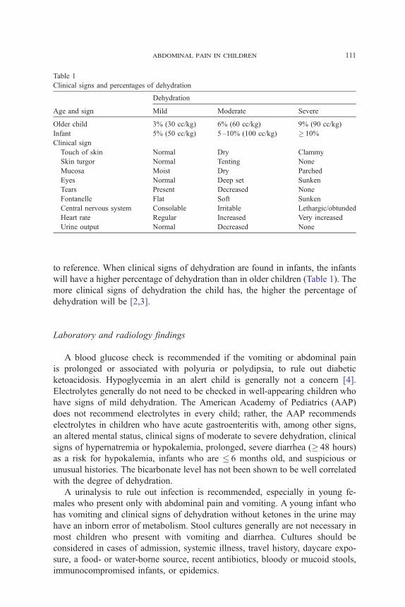
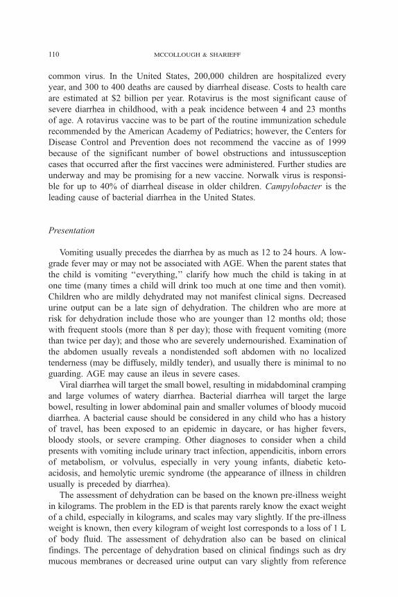
Table 1
Clinical signs and percentages of dehydration
Age and sign
Dehydration
Mild Moderate Severe
Older child 3% (30 cc/kg) 6% (60 cc/kg) 9% (90 cc/kg)
Infant 5% (50 cc/kg) 5 –10% (100 cc/kg) � 10%
Clinical sign
Touch of skin Normal Dry Clammy
Skin turgor Normal Tenting None
Mucosa Moist Dry Parched
Eyes Normal Deep set Sunken
Tears Present Decreased None
Fontanelle Flat Soft Sunken
Central nervous system Consolable Irritable Lethargic/obtunded
Heart rate Regular Increased Very increased
Urine output Normal Decreased None
abdominal pain in children 111
to reference. When clinical signs of dehydration are found in infants, the infants
will have a higher percentage of dehydration than in older children (Table 1). The
more clinical signs of dehydration the child has, the higher the percentage of
dehydration will be [2,3].
Laboratory and radiology findings
A blood glucose check is recommended if the vomiting or abdominal pain
is prolonged or associated with polyuria or polydipsia, to rule out diabetic
ketoacidosis. Hypoglycemia in an alert child is generally not a concern [4].
Electrolytes generally do not need to be checked in well-appearing children who
have signs of mild dehydration. The American Academy of Pediatrics (AAP)
does not recommend electrolytes in every child; rather, the AAP recommends
electrolytes in children who have acute gastroenteritis with, among other signs,
an altered mental status, clinical signs of moderate to severe dehydration, clinical
signs of hypernatremia or hypokalemia, prolonged, severe diarrhea (� 48 hours)
as a risk for hypokalemia, infants who are � 6 months old, and suspicious or
unusual histories. The bicarbonate level has not been shown to be well correlated
with the degree of dehydration.
A urinalysis to rule out infection is recommended, especially in young fe-
males who present only with abdominal pain and vomiting. A young infant who
has vomiting and clinical signs of dehydration without ketones in the urine may
have an inborn error of metabolism. Stool cultures generally are not necessary in
most children who present with vomiting and diarrhea. Cultures should be
considered in cases of admission, systemic illness, travel history, daycare expo-
sure, a food- or water-borne source, recent antibiotics, bloody or mucoid stools,
immunocompromised infants, or epidemics.

mccollough & sharieff112
Management
Children with clinical signs of dehydration need rehydration. Rehydration
can be administered either orally or through a nasogastric tube or an intravenous
line. If a child has signs of severe dehydration, altered mental status, or evidence
of an ileus, then rehydration should occur through an intravenous line.
An oral challenge is not oral rehydration. If a child is going to be orally
rehydrated, then parents need to be instructed on the proper techniques of oral
rehydration. Oral rehydration is very effective but is more labor intensive for
children and parents [5]. However, when surveyed, parents actually prefer oral
rehydration compared with intravenous rehydration. The commercially available
rehydration solutions (eg, Pedialyte or Rehydralyte) are fairly close to delivering
the optimal amount of sodium and glucose recommended by the World Health
Organization (sodium plus 60–90 mEq and dextrose 2.0%). Many homemade
recipes (eg, a water, salt, and sugar mixture) for oral rehydration solution can be
found on the Internet.
The key to successful oral rehydration in children who present with vomiting
is to offer small amounts at a time; for example, 5 cc (sips) for young children or
15 cc (tablespoon) for older children every 2 minutes. A syringe or a 5-F feeding
tube attached to a syringe can be used to help facilitate the oral rehydration. The
parent also can drip the solution slowly into the child’s mouth or through the
nares into the posterior pharynx. If vomiting occurs, wait 10 to 15 minutes and try
again. The child should receive either 50 cc/kg orally for mild to moderate
dehydration or 100 cc/kg for moderate to severe dehydration, over 3 to 4 hours.
Children who experience ongoing losses, such as by continued diarrhea, should
receive an additional 10 cc/kg of rehydration.
Breast feeding infants can be rehydrated using more frequent, shorter feeds.
Another option for oral rehydration is the use of frozen rehydration popsicles
such as Revital-ICE. Placing a nasogastric tube is an alternative route for hy-
dration in the child who refuses absolutely to take anything by mouth or in whom
an intravenous line cannot be established but is clinically stable [6].
Intravenous rehydration should be used for any child who fails oral re-
hydration or has signs of severe dehydration, an ileus, or an altered mental
status. Many experts recommend a minimum of 30 to 40 cc/kg in cases of mild
to moderate dehydration, which will correct dehydration of 3% to 4%. Anti-
emetics and antidiarrheal medications are not recommended currently by the
AAP because of limited literature to support their use [7]. Prochlorperazine,
promethazine, and metoclopramide have been shown to be of some benefit but
have some increased risk of sedation and an increased risk of dystonic reaction
in children. However, many emergency and pediatric emergency physicians
understand that is cruel to allow a child to remain nauseous and vomiting in
the ED. Ondansetron (Zofran), a 5-hydroxytryptamine-3 receptor antagonist,
which has been used for years as chemotherapy for pediatric nausea, has now
been studied in emergency departments for children with acute gastroenteritis
[8,9]. More literature is needed to support the use of antidiarrheal agents in
abdominal pain in children 113
children, but many emergency physicians use these medications in otherwise
healthy older children who are presumed to have viral diarrhea.
Live bacterial cultures, such as Lactobacillus in yogurt, have been shown to
help treat infectious diarrhea and to help prevent diarrhea associated with
antibiotics [10]. Antibiotics are not recommended for most children who are
presumed to have viral AGE. In children with confirmed bacterial diarrhea, the
role of antibiotics in treating infections by Campylobacter jejuni, Escherichia
coli, and Yersinia is unclear. Nontyphoid Salmonella infection is self-limiting
and may have prolonged excretion with antibiotic therapy. However, the
treatment for Salmonella is indicated in infants less than 3 months of age, who
have a history of immunodeficiency or hemoglobinopathy. Antibiotic therapy
should not be initiated unless E coli 0157:H7 has been excluded because
patients may develop hemolytic uremic syndrome from empiric antibiotic use.
Shigella infection may be treated with trimethoprim-sulfamethoxazole, 8 mg/kg/d,
divided twice per day, or erythromycin, 40 mg/kg/d, divided four times per day,
for 5 to 7 days. Erythromycin is the drug of choice for treating Campylobacter
infection. Giardia may be treated with metronidazole, 15 mg/kg/d, divided three
times per day, for 7 days. Clostridium difficile infections may be treated with
oral vancomycin, 50 mg/kg/d, divided four times per day, or metronidazole for
7 days.
Resuming formula feeding in young infants or solids in older children as soon
as possible should also be encouraged. Transient lactose intolerance may de-
velop, especially during AGE caused by rotavirus, but is transient. Most chil-
dren can return to eating milk products or formula. Occasionally, lactose
intolerance persists and may be a cause of post-AGE diarrhea. If persistent
diarrhea occurs after the reintroduction of milk products or if the stool is acidic
and contains more than 0.5% reducing substances, a lactose-free formula should
be considered.
Constipation
Parents often worry that their infant or child is constipated, particularly
because it is common for infants to strain and turn red in the face during bowel
movements. Unfortunately, a uniform definition of constipation has yet to be
determined. The best way is to define constipation is not by the frequency of
the stool but by the difficulty or painful passage of large or hard stools. Newborns
typically have a meconium stool in the first 48 hours of life and then can range
from zero to 12 stools per day for the first week of life. The stools of breast-fed
infants are very soft and pale yellow and often occur after each feeding. However,
bottle-fed infants tend to have firm, formed, yellow stools one to four times
per day. When infants are 3 to 4 months of age, stool frequency decreases, with
some bottle-fed infants passing one stool every other day. Most children develop
the adult pattern of having a mean of 1.2 stools per day by 4 years of age.
mccollough & sharieff114
Causes
The more common serious causes of constipation in the newborn and infant
are imperforate anus, anal stenosis, meconium plug syndrome, meconium ileus,
Hirschsprung’s disease, volvulus, anal fissure, infant botulism, hypocalcemia,
hypercalcemia, and hypothyroidism. Constipation in the older infant or child is
related commonly to changes in diet, especially from breast milk to formula or
advancement to solid baby foods. Inadequate fluid intake is another common
cause of constipation. The school-aged child may present with constipation
caused by high carbohydrate diets and a hesitance to go to the bathroom at
school. The child who has rectal retention and encopresis has fecal soiling of the
underpants and may paradoxically complain of diarrhea. A lower abdominal
mass may be found by palpation, and fecal impaction may be found on rectal
examination. Older children may present with abdominal pain, which may be in
the right lower quadrant and mimic appendicitis.
Presentation
Pertinent history that should be obtained from the caregiver includes the time
after birth of the first bowel movement, frequency of bowel movements,
consistency and size of stools, presence of pain with bowel movements, and
associated systemic findings such as fever, weight loss, and vomiting. Dietary
habits should be a particular focus, and a medication history should also be
obtained. A complete physical examination should be performed, including an
abdominal palpation for masses, inspection of the perineum and perianal area for
fissures, and imperforate anus or stenosis. A plain abdominal radiograph is
helpful in confirming the diagnosis when the history or physical examination is
confusing or inconclusive.
Laboratory and radiologic findings
Laboratory tests and radiologic studies generally are unnecessary in the
diagnosis and management of constipation in young children. An abdominal
series or flat-plate radiograph of the abdomen can confirm that the colon has a
significant amount of stool present.
Management
If fecal impaction is present, disimpaction is necessary. Oral medications
include mineral oil, 1 to 4 mL/kg/dose, once or twice per day (contraindicated
in infants and in children at risk for aspiration); lactulose, 1 to 2 mL/kg/dose,
once or twice per day; milk of magnesia, 1 to 3 mL/kg/dose, once or twice per day,
or with medications containing polyethylene glycol (PEG); or sorbitol, senna, or
bisacodyl [11]. A tasteless, commercially available electrolyte-free PEG solution
(MiraLax) can be mixed with any clear liquid beverage [12]. It is prepared by
abdominal pain in children 115
dissolving 1 capful (17 g) of powder in 8 oz of liquid and giving the child 10 to
14 mL/kg/d in two divided doses. Rectal disimpaction also can be performed.
However, hypertonic phosphate enemas have been associated with severe, acute
hypocalcemia and cardiac arrest in infants [13]. Tap water enemas have been
associated with acute hyponatremia, seizures, and death [14]. In the older infant
and toddler, milk of magnesia, mineral oil, or lactulose can be used. Docusate
(Colace), 5 to 10 mg/kg/d or senna extract (Senokot), 5 to 10 mL daily, can be
safely used in older children.
Maintenance therapy of constipation is most appropriately managed by a
primary care clinician. Dietary management includes increasing fluid intake and
adding fiber and fruits such as prunes, pears, or plums to the diet. A barley extract
(Maltsupex) or Karo syrup can be recommended safely for infants in a dosage
of 1 to 2 teaspoons two to four times daily, added to formula, juice, or food.
Behavioral modification for the older child includes regular toilet sitting, stool
diaries, and reward systems. If an anal fissure is discovered, management in-
cludes frequent, gentle, thorough cleansing of the anus and liberal lubrication
with petroleum jelly. A stool softener must be used, and a topical anesthetic
ointment may be helpful to avoid a pattern of pain and stool retention.
Appendicitis
Causes
Appendicitis is the abdominal pain most commonly treated surgically in
childhood, affecting four of every 1000 children. Appendicitis is the cause of
pain in 2.3% of all the children with abdominal pain seen in ambulatory clinics or
EDs. Of all the children admitted to the hospital with abdominal pain, 82% are
diagnosed with appendicitis [15]. Because of the difficulty in evaluating young
children with abdominal pain, perforation rates for appendicitis are higher than
in the general adult population (30%–65%). Moreover, because the omentum is
less developed in children, perforations are less likely to be ‘‘walled off’’ or
localized, leading to generalized peritonitis.
Presentation
The classic presentation, consisting of generalized abdominal pain migrating
to the right lower quadrant, associated with nausea, vomiting, and fever, is seen
less often in the pediatric patient [16]. In addition, children often present earlier
in their clinical course than adults do, when only mild or less specific symptoms
are present. However, limited data appear to indicate that individual signs such as
rebound tenderness and Rovsing’s sign have a high sensitivity and specificity in
children [4].
The most common findings of appendicitis in children are right lower quad-
rant pain, abdominal tenderness, guarding, and vomiting [17]. If available, a his-
mccollough & sharieff116
tory of abdominal pain preceded by vomiting can be helpful in distinguishing
appendicitis from acute gastroenteritis. Very young children commonly have
diarrhea as the presenting symptom [18]. Bearing in mind the special techniques
discussed above for eliciting peritoneal irritation, the EP should also remember
that the position of the appendix can vary greatly, and tenderness may be found in
locations other than the classic McBurney point. Although the rectal examination
is not usually helpful in making a diagnosis of appendicitis [19], some authors
advocate a rectal examination in infants, in whom there may be a palpable rectal
mass in up to 30% of cases [20]. Changes in skin temperature over the area of the
appendix have not been shown to be helpful in the diagnosis of appendicitis [21].
Differential diagnosis
Gastroenteritis is the most common diagnosis in cases of missed appendici-
tis. Although enteritis caused by Y enterocolitica and Y pseudotuberculosis has
been termed the ‘‘great imitator’’ of appendicitis, in reality, the amount of
diarrhea in gastroenteritis is usually more pronounced. Appendicitis is also fre-
quently mistaken for a urinary tract infection (UTI), which may also present with
abdominal pain and vomiting. A study reported by Reynolds [22] in 1993
showed that missed cases of appendicitis were more likely to have diarrhea, to
not be anorexic, and to be afebrile.
Laboratory evaluation
No laboratory test is 100% sensitive and specific for appendicitis. The white
blood cell count (WBC) can be helpful in the diagnosis, although, by itself, it
is neither specific nor sensitive for appendicitis and therefore cannot be used
alone to rule in or rule out the disease [23]. The WBC, however, can be used as
an adjunct, after the clinical suspicion of appendicitis is estimated. If clinical
suspicion is low before any laboratory or other investigations (for example, in a
child who has vomiting and diarrhea but minimal abdominal tenderness) and
the WBC is normal, the likelihood of appendicitis becomes very low. If the
WBC is high, the likelihood of appendicitis is raised sufficiently to warrant
further tests or observation.
A urinalysis should be performed; however, caution must be exercised in its
interpretation, because mild pyuria, hematuria, and bacteriuria can all be present
if an inflamed appendix is located adjacent to a ureter. The presence of C-reactive
protein also has been studied as a marker for appendicitis [24–27], but it is not
significantly more sensitive or specific than the WBC.
Diagnostic radiology
Plain film abdominal series typically have nonspecific findings and are of
low yield in cases of appendicitis [15]. Appendicoliths are present only in
approximately 10% of true appendicitis cases. Barium enemas have also been
abdominal pain in children 117
used, with the principle that an inflamed appendix will fail to fill and will not be
visualized. Unfortunately, 10% to 30% of normal appendices are not visualized
with barium studies, creating a high number of false-positive results [28].
Ultrasonography is considered by many experts to be the imaging test of
choice in children. Ultrasonography is noninvasive, rapid, and can be performed
at the bedside. It does not require oral contrast, which is an advantage for patients
who may require surgery. It also spares the pediatric patient exposure to radiation.
The normal appendix in pediatric patients is visualized readily by ultra-
sonography because there is usually less abdominal wall fat than in adults.
Graded compression of the appendix is used to determine the presence or absence
of inflammation. An inflamed appendix is usually aperistaltic, difficult to
compress, and measures � 6 mm in diameter. It is important for the ultra-
sonographer to visualize the entire appendix to avoid a false-negative reading
because sometimes only the distal tip of the appendix is inflamed. The mucosal
lining may be intact or poorly defined, and a fecolith may or may not be present.
A periappendiceal fluid collection may indicate an early perforation but may
result simply from inflammation. Experienced ultrasonographers can achieve
sensitivities of 85% to 90% and specificities of 95% to 100% in acute ap-
pendicitis [29–37]. However, studies have not shown an improvement in outcome
measures such as a decrease in negative laparotomies or time to the operating
room [38,39]. Color flow Doppler ultrasonography is now being added to in-
crease the accuracy of the sonographic examinations. Doppler measurement
demonstrates an increase in blood flow to the area of an inflamed appendix [40].
In recent years, CT has become the test of choice for pediatric surgeons when
ultrasonography fails to give a definitive diagnosis [41]. Every variation, from
triple-contrast (intravenous, oral, and rectal) CT scanning to noncontrast, un-
enhanced CT, has been used [42,43]. CT offers the advantage of greater accuracy,
the ability to identify alternative diagnoses, and in some studies, lower negative
laparotomy rates [44]. Although CT appears to be better than ultrasonography in
making the diagnosis of appendicitis in children [45], it is slower, requires oral
contrast in most centers, and exposes the young child to significant radiation. If
the child is vomiting, keeping the oral contrast in the gastrointestinal tract can be
a challenge, and antiemetics may be required.
Leukocyte imaging studies [46] and technetium scans [47] have been used for
equivocal cases of abdominal pain in children. The overall sensitivity, specificity,
and accuracy, however, are lower than with CT. Magnetic resonance imaging is
also superior in its ability to diagnose appendicitis in children [48], but it may
not be available or practical. No study can be relied on for 100% accuracy. If
clinical suspicion is high and imaging studies are negative, the child should be
hospitalized for observation and serial examinations.
Management
When the clinical suspicion for appendicitis is high, consultation with a
surgeon is warranted before any radiologic study. Nonetheless, many surgeons
mccollough & sharieff118
will request a diagnostic study to decrease the likelihood of a negative lapa-
rotomy. When the diagnosis of appendicitis is made, then preparing the child
for the operating room is essential. Usually the oral intake of these children has
been limited during the day or days before presentation, and intravenous fluids
are necessary. Electrolyte imbalances should also be addressed, although sig-
nificant abnormalities are not common in children with appendicitis.
If there are clinical or radiologic signs of perforation, antibiotics with gram-
negative and anaerobic coverage should be started in the ED [49]. A few studies
have shown a benefit to antibiotic therapy in decreasing infectious complica-
tions in children with uncomplicated, nonperforated appendicitis as well [50].
Diagnosing appendicitis early is the key to a better outcome. Any child who is
evaluated in the ED with a chief complaint of abdominal pain and who is
considered well enough to go home but in whom the diagnosis of appendici-
tis has not been ruled out should be asked to return to the ED within 8 hours for
another evaluation of the abdomen.
Intussusception
Pathophysiology
Intussusception was first described over 300 years ago. It is the prolapse of
one part of the intestine into the lumen of an immediately distal adjoining part.
The most common type is ileocolic invagination. During the invagination, the
mesentery is dragged along into the distal lumen, and venous return is obstructed.
This leads to edema, bleeding of the mucosa, increased pressure in the area, and
eventually obstruction to arterial flow. Gangrene and perforation result.
Causes
Intussusception is seen most frequently between the ages of 3 months and
5 years, with 60% of cases occurring in the first year and a peak incidence at 6 to
11 months of age. The disorder, which appears predominantly in males, was once
believed to occur more often in the spring and autumn, although now it appears
it has no seasonality [51,52]. Although it is usually idiopathic in the younger age
groups, children older than 5 years often have a pathologic ‘‘lead point’’ for
intussusception, such as polyps, lymphoma, Meckel’s diverticulum, or Henoch-
Schfenlein purpura and require a work-up to determine the underlying cause.
Presentation
The classic triad of intermittent colicky abdominal pain, vomiting, and bloody
mucous stools is encountered in only 20% to 40% of cases. At least two of these
findings will be present in approximately 60% of patients. The vomiting is not
abdominal pain in children 119
necessarily bilious because the level of obstruction is low in the ileocecal area.
A palpable abdominal mass in the right upper or lower quadrant is an uncommon
finding [53].
Abdominal pain associated with intussusception is colicky, lasts for ap-
proximately 1 to 5 minutes at a time, and then abates for 5 to 20 minutes. During
episodes of pain, the child cries and may draw the knees upward toward the chest.
Although the child often looks better between episodes, he or she still usually
appears ill, quiet, or exhausted. Gradually, irritability increases and vomiting
becomes more frequent and sometimes bilious. Fever may also develop at this
point as the child deteriorates.
If a colicky episode is not witnessed by the ED staff, the EP should ask the
parents to describe or demonstrate what the child was doing during the episodes.
Most parents of a child who has gastroenteritis do not indicate that their child
is in pain. Parents of a child who presents with intussusception usually believe
that the child is in pain before or during episodes of vomiting. Intussusception
also can present with lethargy, pallor, and unresponsiveness. It is important to
keep this diagnosis in mind when dealing with an infant who has an altered
mental status [54].
The abdomen may be distended and tender, but usually the pain appears to
be out of proportion to the physical examination. There may be an elongated
mass in the right upper or lower quadrants. Any type of blood in the stool may be
caused by intussusception. Rectal examination may reveal either occult blood or
frankly bloody, foul-smelling stool, classically described as ‘‘currant jelly’’ [55].
However, frank rectal bleeding is a late and unreliable sign; its absence should
not deter the EP in the pursuit of the diagnosis. It should also be noted that what
appears to be blood in a child’s stool may be something else, such as red fruit
punch or Jell-O, therefore, guaiac testing may prevent this error when there is
some question. A period of observation in the ED for the recurrence of a pain
episode is helpful in equivocal cases. Specifically noting the absence of such
episodes during ED observation is good practice and should be documented in
the clinical record.
Differential diagnosis
Gastroenteritis presents typically with more diarrhea than intussusception,
and the child usually has ill contacts. The presence of any degree of blood in
the stool should also raise suspicion for a more serious condition. Bleeding
from a Meckel’s diverticulum usually is painless, unless the diverticulum be-
comes inflamed.
An incarcerated hernia or testicular or ovarian torsion may also present with
sudden abdominal pain and vomiting. Inspection of the genitalia, especially in
males, is vital. With torsion, the rectal examination does not show occult or frank
blood. Renal colic presenting with pain and vomiting generally is not seen in
young children.
mccollough & sharieff120
Laboratory tests
No laboratory test reliably rules in or out the diagnosis of intussusception.
If the bowel has become ischemic or necrotic, acidosis may be present.
Diagnostic imaging
Unfortunately, plain abdominal films are neither sensitive nor specific for
intussusception [56,57]. Plain films initially may appear normal. As the disease
progresses, a variety of abnormalities may be seen, including a visible abdomi-
nal mass, abnormal distribution of gas and fecal contents, air fluid levels, and
dilated loops of small intestine. A ‘‘target sign’’ on plain film consists of
concentric circles of fat density, similar in appearance to a doughnut, visualized to
the right of the spine. This sign is caused by layers of peritoneal fat surrounding
and within the intussusception alternating with layers of mucosa and muscle.
Less commonly, the soft tissue mass of the intussusception (leading edge) can
be seen projecting into the colon. Large areas of gas with the head of the
intussuscepted bowel may take the shape of a crescent, although other patterns
may be seen.
Ultrasonography is used in some institutions to diagnose intussusception and
to confirm reduction after treatment [58]. Sonographic findings in intussusception
include the target sign, a single hypoechoic ring with a hyperechoic center and
the ‘‘pseudokidney’’ sign, superimposed hypo- and hyperechoic areas represent-
ing the edematous walls of the intussusceptum and layers of compressed mucosa.
Doppler flow may be used to identify bowel ischemia. If signs of intussusception
are not identified by ultrasonography in cases in which the diagnosis is suspected
clinically, proceeding with a barium or air enema should still be considered.
Management
The main focus in the management of a child who has intussusception is
emergent reduction of the obstructed bowel. Classically, this reduction is
accomplished by a barium enema, which acts as both a diagnostic and therapeutic
radiologic study. The barium enema has been the gold standard for both the
diagnosis and treatment of intussusception for decades [59]. Saline enemas have
also been used successfully [60,61], and newer modalities such as air enemas and
ultrasonographically guided enemas have emerged.
Many centers in the United States are now moving toward air enemas [62–67].
This modality was first introduced to the Western world at the American Pediat-
ric Surgical Association meeting in 1985, with the presentation of a series of
6396 successfully treated patients [68]. Air enemas offer several advantages over
barium enemas. They are easier to administer, and in most studies, they have a
higher rate of successful reduction. Air enemas using fluoroscopic guidance
deliver much less radiation than barium studies, and if ultrasonography guidance
is used, there is no exposure. Limiting radiation exposure is important to consider
abdominal pain in children 121
when dealing with infants and their susceptible reproductive organs; and if a
perforation occurs during these investigations, air is much less dangerous to the
peritoneum and abdominal contents than barium is.
Visualization of the entire colon to the terminal ileum is mandatory to rule
out ileocolic intussusception. Ileo–ileo intussusception can be much harder to
diagnose and much harder to reduce. Spontaneous reduction of intussuscepted
bowel has been reported, although in a patient with significant symptoms,
therapeutic intervention should not be delayed [69].
Not every child who has intussusception should undergo bowel reduction by
enema. Clinical signs of peritonitis, perforation, or hypovolemic shock are clear
contraindications to enemas. These signs mandate surgical exploration. Relative
contraindications to enemas include prolonged symptoms (� 24 hours), evidence
of obstruction such as air fluid levels on plain abdominal films, and ultra-
sonography findings of intestinal ischemia or trapped fluid.
Even in well-selected patients, enemas may cause the reduction of necrotic
bowel, perforation, and sepsis. After a successful reduction, the child should
be admitted for observation. A small percentage of patients (0.5% –15%) will
have a recurrence of the intussusception, usually within 24 hours but sometimes
after days or weeks. Even after reduction by laparotomy, the recurrence rate is
2% to 5% [52].
Small bowel obstruction
Pathophysiology
Small bowel obstruction may result from intrinsic, extrinsic, or intraluminal
disease. Although the most common causes of small bowel obstruction are
adhesions from previous abdominal surgery and incarceration of a hernia [70],
intussusception, appendicitis, Meckel’s diverticulum, malrotation with midgut
volvulus, and tumors also should be considered as possible causes. In addition
to inguinal hernias, umbilical, obturator, and femoral canal hernias may also lead
to small bowel obstruction [56].
Presentation
As obstruction develops, decreased oral intake occurs and vomiting ensues,
often becoming bilious in nature. This is followed by obstipation. Abdominal
distension and tenderness occur, and the abdomen may be tympanic to per-
cussion. If the small bowel obstruction is caused by mechanical compression,
high-pitched bowel sounds with ‘‘rushes’’ may be heard. When intraluminal
pressure becomes higher than the venous and arterial pressures, ischemia
develops in the bowel, and hematochezia may be seen. As with most abdominal
emergencies in children, hematochezia is a late finding. Sepsis is another late
finding because bacteria from the ischemic bowel enter the blood.
mccollough & sharieff122
Differential diagnosis
Abdominal pain and vomiting also can be seen with other processes such as
appendicitis. As time passes, a bowel obstruction will develop more abdominal
distension than is seen typically in other processes. The lack of stool or gas
passage points toward bowel obstruction. It is important to remember that the
underlying cause of the obstruction may be as important to recognize as the
obstruction itself.
Laboratory tests
No laboratory test is diagnostic of a bowel obstruction. Elevated levels of
blood urea nitrogen, creatinine, and hematocrit may signify dehydration.
Diagnostic radiology
Plain abdominal films should be obtained when obstruction is suspected. A
paucity of air in the abdomen is the most common finding in young children
with bowel obstruction. Distended loops of bowel may be seen; however, smooth
bowel walls are more common than distended bowel in small children. Multiple
air–fluid levels also are seen commonly with small bowel obstruction. In later
presentations, the bowel may resemble a tangle of hoses or sausages. An upright
or lateral decubitus film will help to determine whether free air is present, caused
by perforation. Further study with ultrasonography, CT, an upper-GI series, or
an enema should be performed when there is suspicion of underlying pathologies
such as appendicitis, midgut volvulus, and intussusception.
Management
Immediate surgical consultation is indicated when a bowel obstruction is
seen on plain radiographs. Morbidity and mortality are increased if the obstruc-
tion is not treated within 24 hours [71]. The patient should be aggressively
hydrated with normal saline boluses, and a nasogastric tube should be placed for
gastric decompression. Broad-spectrum antibiotics are indicated, particularly if
peritonitis is suspected.
Incarcerated hernia
Causes
Inguinal hernias occur in 1% to 4% of the population, more often in males
(6:1), and more often on the right side (2:1). Premature infants are at a higher risk
for hernias (30%), and 60% of incarcerated inguinal hernias occur during the first
abdominal pain in children 123
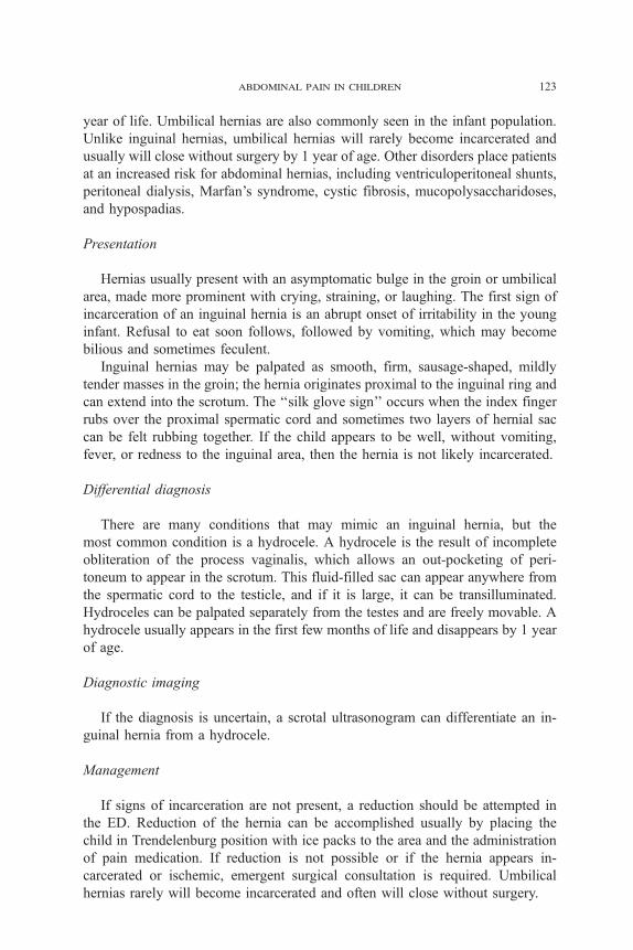
year of life. Umbilical hernias are also commonly seen in the infant population.
Unlike inguinal hernias, umbilical hernias will rarely become incarcerated and
usually will close without surgery by 1 year of age. Other disorders place patients
at an increased risk for abdominal hernias, including ventriculoperitoneal shunts,
peritoneal dialysis, Marfan’s syndrome, cystic fibrosis, mucopolysaccharidoses,
and hypospadias.
Presentation
Hernias usually present with an asymptomatic bulge in the groin or umbilical
area, made more prominent with crying, straining, or laughing. The first sign of
incarceration of an inguinal hernia is an abrupt onset of irritability in the young
infant. Refusal to eat soon follows, followed by vomiting, which may become
bilious and sometimes feculent.
Inguinal hernias may be palpated as smooth, firm, sausage-shaped, mildly
tender masses in the groin; the hernia originates proximal to the inguinal ring and
can extend into the scrotum. The ‘‘silk glove sign’’ occurs when the index finger
rubs over the proximal spermatic cord and sometimes two layers of hernial sac
can be felt rubbing together. If the child appears to be well, without vomiting,
fever, or redness to the inguinal area, then the hernia is not likely incarcerated.
Differential diagnosis
There are many conditions that may mimic an inguinal hernia, but the
most common condition is a hydrocele. A hydrocele is the result of incomplete
obliteration of the process vaginalis, which allows an out-pocketing of peri-
toneum to appear in the scrotum. This fluid-filled sac can appear anywhere from
the spermatic cord to the testicle, and if it is large, it can be transilluminated.
Hydroceles can be palpated separately from the testes and are freely movable. A
hydrocele usually appears in the first few months of life and disappears by 1 year
of age.
Diagnostic imaging
If the diagnosis is uncertain, a scrotal ultrasonogram can differentiate an in-
guinal hernia from a hydrocele.
Management
If signs of incarceration are not present, a reduction should be attempted in
the ED. Reduction of the hernia can be accomplished usually by placing the
child in Trendelenburg position with ice packs to the area and the administration
of pain medication. If reduction is not possible or if the hernia appears in-
carcerated or ischemic, emergent surgical consultation is required. Umbilical
hernias rarely will become incarcerated and often will close without surgery.
mccollough & sharieff124
Meckel’s diverticulum
Pathophysiology and causes
Meckel’s diverticulum is the most common congenital abnormality of the
small intestine. Meckel’s diverticulum is a remnant of the omphalomesenteric
(vitelline) duct that disappears normally by the seventh week of gestation. It is
a true diverticulum, containing all layers of the bowel wall. Up to 60% of
these diverticuli containing heterotopic gastric tissue and heterotopic pancreatic,
endometrial, and duodenal mucosa have also been reported [72,73]. The features
of Meckel’s diverticulum are commonly described by ‘‘the rule of 2s’’ [70]: it is
present in approximately 2% of the population with only 2% of affected patients
becoming symptomatic. Forty-five percent of symptomatic patients are less than
2 years of age [74]. The most common location is 2 feet (40–100 cm) from the
ileocecal valve, and the diverticulum typically is 2 inches long.
Clinical presentation
The classic presentation of Meckel’s diverticulum is painless or minimally
painful rectal bleeding. Isolated, red rectal bleeding is common, particularly in
boys less than 5 years of age [75]. Such painless bleeding is a result of het-
erotopic gastric tissue in the diverticulum or in the adjacent ileum. Abdominal
pain, distension, and vomiting may occur if obstruction has occurred, and the
presentation may mimic appendicitis or diverticulitis. Meckel’s diverticulum may
also ulcerate and perforate, presenting as a bowel perforation, or act as a lead
point, resulting in intussusception.
Differential diagnosis
The differential diagnosis includes both painful and nonpainful conditions.
Rectal bleeding associated with abdominal pain may be caused by peptic ulcer
disease, intussusception, and volvulus. Nonpainful rectal bleeding may be caused
by polyps, arteriovenous malformations, and tumors.
Laboratory tests
Although no laboratory test is diagnostic of Meckel’s diverticulum, children
with gastrointestinal bleeding should undergo screening laboratory tests such as
a complete blood count, coagulation profile, and a type and screen.
Diagnostic radiology
Abdominal films may show signs of obstruction such as dilated loops of
bowel or a paucity of bowel gas. Scanning Meckel’s diverticulum involves an
intravenous injection of technetium-pertechnetate. This test relies on the presence
abdominal pain in children 125
of gastric mucosa in or near the diverticulum that has an affinity for the
radionucleotide. A scan of Meckel’s diverticulum can detect the presence of
gastric mucosa within the diverticulum with up to 85% accuracy [76]. Mesenteric
arteriography can detect the site of active bleeding if bleeding is profuse.
Management
As carried out in any patient with active bleeding, fluid resuscitation is
warranted, starting with boluses of normal saline, 20 cc/kg. A blood transfusion
may be necessary, with a packed red blood cell increment of 10 cc/kg. The
patient should have nothing by mouth, and a nasogastric tube should be placed.
Antibiotic therapy must be initiated if there are peritoneal signs. Surgical
consultation should be obtained emergently. Surgical intervention may involve
a diverticulectomy or a more extensive small bowel segmental resection if there
is irreversible bowel ischemia.
Very young infants
Very young infants, those less than a few months old, also have unique
gastrointestinal conditions. Colic should be considered a diagnosis of exclusion.
Hypertrophic pyloric stenosis is a common presentation, and surgical correction
does not need to be immediate. Volvulus caused by congenital malrotation is a
true surgical emergency, and consultation with a pediatric surgeon should be
immediate once the diagnosis is considered. Fortunately, necrotizing entero-
colitis, another gastrointestinal condition of newborns with serious sequelae, is
usually seen by pediatric colleagues in the newborn nursery or neonatal intensive
care unit.
Colic
Colic affects 1 in 6 families and is more likely to be reported by older
mothers with longer full-time education and nonmanual occupations. To this
day, the cause of colic remains unclear but is believed to be related to increased
gas production in the infant’s intestines and, possibly, to neurologic or psy-
chologic reasons. Other experts consider colic to be part of the normal dis-
tribution of crying.
Presentation
Colic appears usually during the second week of life and is characterized by
screaming episodes and a distended or tight abdomen; some infants will draw up
mccollough & sharieff126
their legs, pass gas, cry, and act miserable for hours. Episodes may last min-
utes to hours, occurring usually in the evening. One common definition used is
3 hours per day, 3 days per week, and at least 3 weeks in duration. Severity
can increase around 4 to 8 weeks of age and will usually resolve around
12 weeks of age.
Growth and development remain unchanged, and the physical examination
is unremarkable. No vomiting, diarrhea, fever, or weight loss occurs with colic.
For any inconsolable crying infant, other correctable causes must be considered
(Box 2). Parents may become overwhelmed and frustrated with a constantly
crying young infant; look for signs that a parent is not coping before it becomes a
child abuse case. This diagnosis occurs early in life; a suddenly irritable or poorly
feeding 8-week-old who was previously healthy is less likely to have colic.
Treatment
There are no medications or treatments that have proven to be very effective
and yet safe. Anticholinergic medications work but have too many side effects,
such as seizures, respiratory trouble, syncope, and coma; therefore, they are not
recommended. Simethicone has not been found to reduce colic. Switching to soy-
or whey-based formulas has not been proven definitely to work [77]. Techniques
such as swaddling the infant, using a pacifier or the rocking motion of car ride,
or placing the infant in a car seat on top of a moving clothes dryer (watch car seat
does not fall off dryer) also may work to calm the infant. Reassuring parents that
episodes of colic will pass is the best antidote. Encourage parents to allow
themselves ‘‘time outs’’ from the child, allowing someone else they trust to care
for the child during a crying episode.
Box 2. The inconsolable, crying young infant
Anal fissuresCorneal abrasionsDiaper pinsFormula intoleranceFracturesHair tourniquetsHematomaHerniasInfections (eg, UTI or meningitis)IntussusceptionOtitis mediaReactions to medications such as decongestants
abdominal pain in children 127
Hypertrophic pyloric stenosis
Pathophysiology
Hypertrophic pyloric stenosis (HPS) is a narrowing of the pyloric canal caused
by hypertrophy of the musculature. The cause of this condition remains un-
clear, but some experts theorize that HPS is caused by Helicobacter pylori, the
same bacteria associated with peptic ulcer disease. This theory is based on non-
specific evidence, such as the temporal distribution, seasonality, and familial
clustering of HPS, along with the pathologic finding of leukocytic infiltrates, and
the increased incidence seen in association with bottle-feeding [78].
Causes
HPS occurs in 1 of every 250 births and appears predominantly in males (male
to female ratio of 4:1). The condition also has racial variation. It is observed to be
more common in whites than in African Americans and is rare in Asians.
Originally, first-born males were believed to be affected more often, but it is
now known that birth order is not a factor. A child of an affected parent has
an increased chance of HPS, with the risk being higher if the mother was af-
fected [79].
Presentation
HPS usually presents during the third to fifth week of life. Symptoms begin
rather benignly, with occasional vomiting at the end of feeding or soon there-
after. This is when HPS is often confused with a viral syndrome, gastro-
esophageal (GE) reflux, or milk intolerance. Emesis is nonbilious because the
stenosis is proximal to the duodenum. As the disease progresses, the incidence
of vomiting increases, now following every feed, and can become projectile.
Comparing birth weight to current weight is a key element in the evaluation of a
neonate with vomiting. After the first week, healthy neonates should gain
approximately 20 to 30 g (1 ounce) per day. Healthy normal infants who ‘‘spit
up’’ (regurgitate) will continue to gain weight and grow well. Infants with HPS
will continue to be hungry but, because of repeated vomiting, may reach a plateau
or even lose weight. An infant with HPS may also become constipated as the
result of dehydration and decreased intake.
On examination, the neonate with HPS may appear normal but hungry, or
the may have signs of dehydration. Dehydration may lead to the appearance of
jaundice. Peristaltic waves moving from left to right may be seen in the left upper
quadrant after feeding. A palpable ‘‘olive’’ or small mass in the right upper or
middle quadrant, at the lateral margin of the right rectus muscle just below the
liver edge, may also be detected during physical examination. Decompressing the
stomach with a nasogastric tube first and using a lubricant on the fingertips may
improve the ability to palpate this ‘‘olive.’’ Clinicians’ ability to palpate the
mccollough & sharieff128
pyloric ‘‘olive’’ has decreased over the years, probably because of the addition
of ultrasonography in confirming the diagnosis. In 1999, Abbas and colleagues
[80] reported that many infants with HPS who have palpable masses on exami-
nation still undergo one or more unnecessary and redundant tests. This situation
is associated with a delay in diagnosis, increased costs, and possibly adverse
clinical health problems.
Differential diagnosis
The differential diagnosis for a vomiting neonate includes the life-threatening
disorder of volvulus with or without associated malrotation of the intestine.
Infants with volvulus deteriorate rapidly, and the vomiting will be bilious,
eventually with signs of sepsis and bowel necrosis. Incarcerated hernias also can
present similarly, as well as intussusception (although less commonly in the
neonatal period). Viral gastroenteritis can occur in the neonate, but caution is
advised when making this diagnosis in infants less than 6 weeks old. At a
minimum, significant diarrhea and the presence of ill contacts should both be
present before considering viral gastroenteritis.
GE reflux is much more common than pyloric stenosis, and vomiting in the
neonatal period is often attributed to GE reflux when other diagnoses should be
considered. Vomiting caused by GE reflux usually occurs during feeds or
immediately afterwards. The amount of vomitus is smaller, and the neonate
will continue to gain weight. Infections, especially in the urinary tract, also can
present with vomiting as a chief complaint and an examination of the genitalia
and urine is imperative in any infant who presents with vomiting.
Laboratory tests
Prolonged vomiting in HPS causes the infant to lose large quantities of gas-
tric secretions rich in H+ and Cl� ions. As a result of dehydration, the kidney
attempts to conserve Na+ ions by exchanging them for K+ ions. The net result is a
loss of both H+ and K+ ions. Therefore, the infant with HPS will initially
demonstrate a hypokalemic, hypochloremic, metabolic alkalosis [81]. If the in-
fant remains dehydrated for a long period, this alkalosis may eventually turn
to acidosis.
Imaging studies
If no small mass or ‘‘olive’’ is palpable in the right upper or middle quadrant
of a young infant with a clinical picture suggestive of HPS, further studies are
warranted. Ultrasonography measures the thickness of the pyloric wall (normally
� 2.0 mm but in HPS is � 4.0 mm) and the length of the pyloric canal (normally
� 10.0 mm but in HPS is � 14–16 mm), leading to a diagnosis of HPS.
Ultrasonography has been shown to have a sensitivity and specificity as high
as 100% [82,83]. A false-negative result may occur if the ultrasonographer
abdominal pain in children 129
measures through the distal stomach or antrum and not through the pylorus itself.
A false-positive results if pyloric spasm is present and not pyloric stenosis.
If ultrasonography is nondiagnostic and HPS remains a concern, the next
radiologic test of choice is an upper-GI series. The upper GI will show the classic
‘‘string sign’’ as contrast flows through the narrowed pyloric lumen. There
will also be delayed gastric emptying. As with ultrasonography, false-positive
results may occur because of pyloric spasm, which also gives the appearance of
a string sign. Endoscopy also can be used to diagnose HPS but is not used
commonly [84].
Management
Once HPS has been diagnosed, admission to the hospital is indicated. Often
these infants are dehydrated and therefore hydration and correction of any
electrolyte abnormalities should be started in the ED. The surgical procedure
required to correct the stenosis is the Ramstedt procedure, which involves incis-
ing and separating the hypertrophic muscle fibers of the pylorus.
In Japan, intravenous atropine has been used to decrease the spasm of the
pylorus as an alternative to surgery. It is then administered orally for several
weeks until the child ‘‘outgrows’’ the stenosis. Surgery has been avoided in
many cases [85]; however, surgery remains the standard treatment in the
United States.
Malrotation with midgut volvulus
Pathophysiology
Congenital malrotation of the midgut portion of the intestine is often the cause
of volvulus in the neonatal period. Malrotation occurs during the fifth to eighth
week in embryonic life when the intestine projects out of the abdominal cavity,
rotates 2708, and then returns into the abdomen. If the rotation is not correct,
the intestine will not be ‘‘fixed down’’ correctly at the mesentery, and the vascular
mesentery will appear more stalk-like in its structure and is at risk later for
twisting, called volvulus. Volvulus is the twisting of a loop of bowel about its
mesenteric base stalk attachment; ischemia subsequently develops, and this con-
stitutes a true surgical emergency because bowel necrosis can occur within hours.
The entire small bowel is at risk for ischemia and necrosis.
Causes
The incidence of volvulus peaks during the first month of life but can present
anytime in childhood. The male to female ratio is 2:1, and this is rarely a familial
disorder. The exact frequency of midgut volvulus is not known because it is
frequently asymptomatic. Congenital adhesions, called Ladd’s bands, extending
mccollough & sharieff130
from the cecum to the liver, are associated with congenital malrotation. These
adhesions may cause external compression of the duodenum and obstruction.
This condition is not generally considered a surgical emergency, but it eventually
requires surgical intervention to lyse these bands.
Presentation
Volvulus may present in one of three ways: (1) as a sudden onset of bilious
vomiting and abdominal pain in a neonate; (2) as a history of ‘‘feeding problems’’
with bilious vomiting that now appears like a bowel obstruction; and (3) although
less commonly, as a failure to thrive with severe feeding intolerance [86]. Bilious
vomiting in a neonate is always worrisome and is a surgical emergency until
proven otherwise. If the bowel is already ischemic or necrotic, the neonate may
present with a pale complexion and grunting. The abdomen may or may not be
distended depending on the location of the volvulus. If the obstruction is
proximal, there may be no distension. The abdominal wall may appear ‘‘blue’’ if
the bowel is already ischemic or necrotic. The pain is constant, not intermittent,
and the neonate will appear irritable. Jaundice also may be present. Hematochezia
is a late sign and indicates intestinal necrosis. Neonates who have volvulus will
gradually deteriorate if bowel remains ischemic.
Differential diagnosis
As stated earlier, bilious vomiting in a neonate is considered a surgical emer-
gency until proven otherwise. However, in the early presentation of volvulus,
vomitus may be nonbilious, and a misdiagnosis of acute gastroenteritis may
result. As in the discussion of pyloric stenosis, the acute gastroenteritis should be
diagnosed cautiously in young infants. In pyloric stenosis, vomitus is always
nonbilious. The duration of symptoms with pyloric stenosis is usually longer, and
the child usually appears well, although possibly dehydrated and hungry.
Incarcerated hernias may also present with bilious vomiting. It is therefore
imperative to thoroughly examine a vomiting neonate for signs of a hernia. Rarer
causes of bilious vomiting include duodenal or ileal atresia, although this is
discovered typically in the newborn nursery or soon after. With intestinal atresia,
the neonate will not be as ill appearing as with volvulus. Necrotizing enterocolitis
also can rarely appear in term neonates. Intestinal hematomas may occur in cases
of child abuse.
Congenital adrenal hyperplasia (CAH) can cause bilious vomiting without
anatomical obstruction. It may present in the first few weeks of life. CAH results
in adrenal insufficiency with decreased cortisol levels and salt wasting. Infants
will present with hypotension and electrolyte imbalance (low Na+ and high K+ ).
It is more likely that CAH will be seen in male infants who present in the ED.
Female newborns who have this condition are less commonly missed in the
newborn nursery because the accumulation of androgenic compounds affects
the external genitalia to a greater extent. Hirschsprung’s disease or congenital
abdominal pain in children 131
intestinal aganglionosis also may also present with bilious vomiting. In this
condition, there should also be a history of decreased stool output since birth.
Laboratory tests
Laboratory tests are nonspecific for volvulus. Typically, blood tests will show
signs of dehydration and acidosis.
Diagnostic imaging
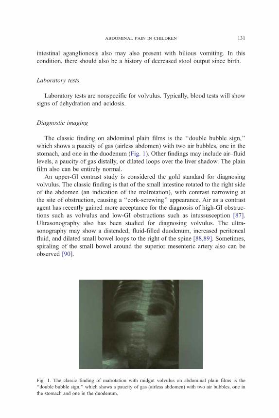
The classic finding on abdominal plain films is the ‘‘double bubble sign,’’
which shows a paucity of gas (airless abdomen) with two air bubbles, one in the
stomach, and one in the duodenum (Fig. 1). Other findings may include air–fluid
levels, a paucity of gas distally, or dilated loops over the liver shadow. The plain
film also can be entirely normal.
An upper-GI contrast study is considered the gold standard for diagnosing
volvulus. The classic finding is that of the small intestine rotated to the right side
of the abdomen (an indication of the malrotation), with contrast narrowing at
the site of obstruction, causing a ‘‘cork-screwing’’ appearance. Air as a contrast
agent has recently gained more acceptance for the diagnosis of high-GI obstruc-
tions such as volvulus and low-GI obstructions such as intussusception [87].
Ultrasonography also has been studied for diagnosing volvulus. The ultra-
sonography may show a distended, fluid-filled duodenum, increased peritoneal
fluid, and dilated small bowel loops to the right of the spine [88,89]. Sometimes,
spiraling of the small bowel around the superior mesenteric artery also can be
observed [90].
Fig. 1. The classic finding of malrotation with midgut volvulus on abdominal plain films is the
‘‘double bubble sign,’’ which shows a paucity of gas (airless abdomen) with two air bubbles, one in
the stomach and one in the duodenum.
mccollough & sharieff132
Management
Because of the risk of bowel necrosis and resulting sepsis, diagnosing this life-
threatening condition as early as possible is imperative. Once malrotation with
midgut volvulus has been diagnosed, aggressive resuscitation using boluses of
normal saline, 20 cc/kg, and the placement of a nasogastric tube should occur.
Antibiotics should be administered to cover gram-positive, gram-negative, and
anaerobic flora (eg, ampicillin, gentamicin, and clindamycin). Consultation with a
pediatric surgeon should not be delayed for diagnostic studies. The sooner the child
is admitted to the operating room, the lower the morbidity and mortality of this
condition. Some pediatric surgeons will take an ill-appearing neonate with bilious
vomiting directly to the operating room without any additional diagnostic tests.
Necrotizing enterocolitis
Causes
Necrotizing enterocolitis (NEC) is seen typically in the neonatal intensive
care unit, occurring in premature infants in their first few weeks of life. Occa-
sionally, it is encountered in the term infant, usually within the first 10 days after
birth. The cause of NEC is unknown, but a history of an anoxic episode at birth
and other neonatal stressors are associated with the diagnosis [91,92].
Pathophysiology
The pathologic finding of NEC is that of a necrotic segment of bowel with gas
accumulation in the submucosa. Necrosis can lead to perforation, sepsis, and
death. The distal ileum and proximal colon are most commonly involved.
Clostridium spp, E coli, Staphylococcus epidermidis, and rotavirus are the patho-
gens recovered most commonly [72,73].
Presentation
Infants who have NEC will present typically as appearing quite ill, with
lethargy, irritability, decreased oral intake, distended abdomen, and bloody stools.
Symptoms may present in a range from fairly mild, with only occult-blood
positive stools, to a much more critically ill presentation. Because this condition is
diagnosed typically in the neonatal intensive care unit, it still must be considered
in a term infant who has experienced significant stress, such as anoxia, at birth.
Radiologic studies
The plain abdominal film finding of pneumatosis intestinalis, caused by gas
in the intestinal wall, is diagnostic of NEC.
abdominal pain in children 133
Management
Management includes fluid resuscitation, bowel rest, and broad-spectrum anti-
biotic coverage. Early surgical consultation is imperative.
Summary
Abdominal pain or gastrointestinal symptoms are common complaints in
young children. It is the emergency physician’s duty to understand current rec-
ommendations regarding the evaluation and management of more benign con-
ditions such as gastroenteritis and also be able to differentiate a true surgical
condition such as appendicitis.
References
[1] Kim M, Strait RT, Sato TT, et al. A randomized clinical trial of analgesia in children with
acute abdominal pain. Acad Emerg Med 2002;9(4):281–7.
[2] Gorelick M. Validity and reliability of clinical signs in the diagnosis of dehydration in chil-
dren. Pediatrics 1997;99(5):e6.
[3] Duggan C, Refat M, Hashem M, et al. How valid are clinical signs of dehydration in infants?
J Pediatr Gastroenterol Nutr 1996;22:56–61.
[4] Lee PH, Bank DE, Flomenbaum N. Hypoglycemia and ABC’s (sugar). Ann Emerg Med 2000;
36(3):278–9.
[5] Atherly-John YC, Cunningham SJ, Crain EF. A randomized trial of oral vs intravenous
rehydration in a pediatric emergency department. Arch Peds Adol Med 2002;156:1240–3.
[6] Nager A, Wang VJ. Comparison of nasogastric and intravenous methods of rehydration in
pediatric patients with acute dehydration. Pediatrics 2002;109(4):566–72.
[7] American Academy of Pediatrics. Subcommittee on Acute Gastroenteritis Practice parameter:
the management of acute gastroenteritis in young children. Pediatrics 1996;97(3):424–35.
[8] Reeves J, Shannon M, Fleisher G. Ondansetron decreases vomiting associated with acute
gastroenteritis: a randomized, controlled trial. Pediatrics 2002;109(4):e62.
[9] Ramsook C, Sahagun-Carreon L, Kozinetz C, et al. A randomized clinical trial comparing
ondansetron with placebo in children with vomiting from acute gastroenteritis. Ann Emerg Med
2002;39(4):397–403.
[10] Van Niel CW, Feudtner C, Garrison MM, et al. Meta-analysis of Lactobacillus therapy. Pedi-
atrics 2002;109:678–84.
[11] Tolia V, Lin CH, Elitsur Y. A prospective randomized study with mineral oil and oral la-
vage solution for treatment of faecal impaction in children. Aliment Pharmacol Ther 1993;7:
523–9.
[12] Bishop WP. Miracle laxative? J Pediatr Gastroenterol Nutr 2001;32:514–5.
[13] Reedy J, Zwiren J. Enema-induced hypocalcemia and hyperphosphatemia leading to car-
diac arrest during induction of anesthesia in an outpatient surgery center. Anesthesiology 1983;
59:578.
[14] Ziskind A, Gellis SS. Water intoxication following tap water enemas. AMA J Dis Child 1958;
96:699–704.
[15] Wagner JM, McKinner WP, Carpenter JL. Does this patient have appendicitis? JAMA 1996;
276(19):1589–94.
mccollough & sharieff134
[16] Williams A, Bello M. Perforation rates relates to delayed presentation in childhood acute ap-
pendicitis. J R Coll Surg Edinb 1998;43:101–2.
[17] Saidi RF, Ghasemi M. Role of Alvarado score in diagnosis and treatment of suspected
acute appendicitis. Am J Emerg Med 2000;18(2):230–1.
[18] Horwitz JR, Gursoy M, Jaksic T, et al. Importance of diarrhea as a presenting symptom of
appendicitis in very young children. Am J Surg 1997;173(2):80–2.
[19] Scholer SJ, Pituch K, Orr DP, et al. Use of the rectal examination on children with acute
abdominal pain. Pediatr Clin N Am 1998;37(5):311–6.
[20] Rothrock SG, Pagane J. Acute appendicitis in children: emergency department diagnosis and
management. Ann Emerg Med 2000;36(1):39–51.
[21] Emery M, Jones J, Brown M. Clinical application of infrared thermography in the diagnosis of
appendicitis. Am J Emerg Med 1994;42(1):48–50.
[22] Reynolds SL. Missed appendicitis in a pediatric emergency department. Pediatr Emerg Care
1993;9(1):1–3.
[23] Coleman C, Thompson JE, Bennion RS, et al. White blood cell count is poor predictor of
severity of disease in the diagnosis of appendicitis. Am Surg 1998;64(10):983–5.
[24] Chung JL, Kong MS, Lin SL, et al. Diagnostic value of C-reactive protein in children with
perforated appendicitis. Eur J Pediatr 1996;155(7):529–31.
[25] Paajanen H, Mansikka A, Laato M, et al. Are serum inflammatory markers age dependent
in acute appendicitis? J Am Coll Surg 1997;184(3):303–8.
[26] Hallan S, Asberg A, Edna TH. Additional value of biochemical tests in suspected acute ap-
pendicitis. Eur J Surg 1997;163(7):533–8.
[27] Andersson RE, Hugander AP, Ghazi SH, et al. Diagnostic value of disease history, clini-
cal presentation, and inflammatory parameters in appendicitis. World J Surg 1999;23(2):133–40.
[28] Albiston E. The role of radiological imaging in the diagnosis of acute appendicitis. Can J Gas-
troenterol 2002;16(7):451–63.
[29] Orr RK, Porter D, Hartman D. Ultrasonography to evaluate adults for appendicitis: decision mak-
ing based on meta-analysis and probabilistic reasoning. Acad Emerg Med 1995;2(7):644–50.
[30] Wong ML, Casey SO, Leonidas JC, et al. Sonographic diagnosis of acute appendicitis in chil-
dren. J Pediatr Surg 1994;29(10):1356–60.
[31] Ramachandran P, Sivit CJ, Newman KD, et al. Ultrasonography as an adjunct in the diag-
nosis of acute appendicitis: a 4-year experience. J Pediatr Surg 1996;31(1):164–7 [disussion:
167–9].
[32] Crady SK, Jones JS, Wyn T, et al. Clinical validity of ultrasound in children with suspected
appendicitis. Ann Emerg Med 1993;22(7):1125–9.
[33] Zaki AM, MacMahon RA, Gray AR. Acute appendicitis in children: when does ultrasound
help? Aust N Z J Surg 1994;64(10):695–8.
[34] Hahn HB, Hoepner FU, Kalle T, et al. Sonography of acute appendicitis in children: 7 years
experience. Pediatr Radiol 1998;28(3):147–51.
[35] Carrico CW, Fenton LZ, Taylor GA, et al. Impact of sonography on the diagnosis and treat-
ment of acute lower abdominal pain in children and young adults. Am J Roentgenol 1999;
172(2):513–6.
[36] Lessin MS, Chan M, Catallozzi M, et al. Selective use of ultrasonography for acute appendicitis
in children. Am J Surg 1999;177(3):193–6.
[37] Rice HE, Arbesman M, Martin DJ, et al. Does early ultrasonography affect management of
pediatric appendicitis? a prospective analysis. J Pediatr Surg 1999;34(5):754–9.
[38] Rice HE, Arbesman M, Martin DJ, et al. Does early ultrasonography affect management of
pediatric appendicitis? a prospective analysis. J Pediatr Surg 1999;34(5):754–9 [discussion:
758–9].
[39] Roosevelt GE, Reynolds SL. Does the use of ultrasonography improve the outcome of chil-
dren with appendicitis? Acad Emerg Med 1998;5(11):1071–5.
[40] Quillin SP, Siegel MJ. Diagnosis of appendiceal abscess in children with acute appendicitis:
value of color Doppler sonography. Am J Roentgenol 1995;164(5):1251–4.
abdominal pain in children 135
[41] Funaki B, Grosskreutz SR, Funaki CN. Using unenhanced helical CT with enteric contrast ma-
terial for suspected appendicitis in patients treated at a community hospital. Am J Roentgenol
1998;171(4):997–1001.
[42] Garcia Pena BM, Mandl KD, Kraus SJ, et al. Ultrasonography and limited computed to-
mography in the diagnosis and management of appendicitis in children. JAMA 1999;282(11):
1041–6.
[43] Pena BM, Taylor GA, Lund DP, et al. Effect of computed tomography on patient manage-
ment and costs in children with suspected appendicitis. Pediatrics 1999;104(3 Pt 1):440–6.
[44] Rao PM, Rhea JT, Rattner DW. Introduction of appendiceal CT: impact on negative ap-
pendectomy and appendiceal perforation rates. Ann Surg 1999;229(3):344–9.
[45] Jabra AA, Shalaby-Rana EI, Fishman EK. CT of appendicitis in children. J Comput Assist
Tomogr 1997;21(4):661–6.
[46] Kanegaye JT, Vance CW, Parisi M, et al. Failure of technetium-99m hexamethylpropylene
amine oxime leukocyte scintigraphy in the evaluation of children with suspected appendicitis.
Ped Emerg Care 1995;11(5):285–90.
[47] Turan C, Tutus A, Ozokutan BH, et al. The evaluation of technetium 99m-citrate scintigraphy
in children with suspected appendicitis. J Pediatr Surg 1999;34(8):1272–5.
[48] Hormann M, Paya K, Eibenberger K, et al. MR imaging in children with nonperforated
acute appendicitis: value of unenhanced MR imaging in sonographically selected cases. Am J
Roentgenol 1998;171(2):467–70.
[49] Banani SA, Talei A. Can oral metronidazole substitute parenteral drug therapy in acute ap-
pendicitis? a new policy in the management of simple or complicated appendicitis with localized
peritonitis: a randomized controlled clinical trial. Am Surg 1999;65(5):411–6.
[50] Soderquist-Elinder C, Hirsch K, Bergdahl S, et al. Prophylactic antibiotics in uncomplicated
appendicitis during childhood–a prospective randomised study. Eur J Pediatr Surg 1995;5(5):
282–5.
[51] Parashar UD, Holman RC, Cummings KD, et al. Trends in intussusception-associated hos-
pitalizations and deaths among US infants. Pediatrics 2000;106(6):1413–21.
[52] Ugwu BT, Legbo JN, Dakum NK, et al. Childhood intussusception: a 9-year review. Ann
Trop Paediatr 2000;20(2):131–5.
[53] Kuppermann N, O’Dea T, Pinckney L, et al. Predictors of intussusception in young children.
Arch Pediatr Adolesc Med 2000;154(3):250–5.
[54] Heldrich FJ. Lethargy as a presenting symptom in patients with intussusception. Clin Pediatr
1986;25(7):363–5.
[55] Yamamoto LG, Morita SY, Boychuk R, et al. Stool appearance in intussusception: assessing
the value of the term ‘‘currant jelly.’’ Am J Emerg Med 1997;15(3):293–8.
[56] Smith DS, Bonadio WA, Losek JD, et al. The role of abdominal x-rays in the diagnosis and
management of intussusception. Pediatr Emerg Care 1992;8(6):325–7.
[57] Yang ST, Tsai CH, Chen JA, et al. Differential diagnosis between intussusception and gastro-
enteritis by plain film. Acta Paediatr Taiwan 1995;36(3):170–5.
[58] Bhisitkul DM, Listernick R, Shkolnik A, et al. Clinical application of ultrasonography in the
diagnosis of intussusception. J Pediatr 1992;121(2):182–6.
[59] Campbell JB. Contrast media in intussusception. Pediatr Radiol 1989;19(5):293–6.
[60] Gonzalez-Spinola J, Del Pozo G, Tejedor D, et al. Intussusception: the accuracy of ultrasound-
guided saline enema and the usefulness of a delayed attempt at reduction. J Pediatr Surg 1999;
34(6):1016–20.
[61] Rohrschneider WK, Troger J. Hydrostatic reduction of intussusception under US guidance.
Pediatr Radiol 1995;25(7):530–4.
[62] Wang G, Liu XG, Zitsman JL. Nonfluoroscopic reduction of intussusception by air enema.
World J Surg 1995;19(3):435–8.
[63] Daneman A, Alton DJ, Ein S, et al. Perforation during attempted intussusception reduc-
tion in children: a comparison of perforation with barium and air. Pediatr Radiol 1995;25(2):
81–8.
mccollough & sharieff136
[64] Gu L, Alton DJ, Daneman A, et al. John Caffey Award: intussusception reduction in children
by rectal insufflation of air. Am J Roentgenol 1988;150(6):1345–8.
[65] Schmit P, Rohrschneider WK, Christmann D. Intestinal intussusception survey about diagnos-
tic and nonsurgical therapeutic procedures. Pediatr Radiol 1999;29(10):752–61.
[66] Sandler AD, Ein SH, Connolly B. Unsuccessful air-enema reduction of intussusception: is a
second attempt worthwhile? Pediatr Surg Int 1999;15(3–4):214–6.
[67] Lui KW, Wong HF, Cheung YC, et al. Air enema for diagnosis and reduction of intussus-
ception in children: clinical experience and fluoroscopy time correlation. J Pediatr Surg 2001;
36(3):479–81.
[68] Guo JZ, Ma XY, Zhou QH. Results of air pressure enema reduction of intussusception:
6,396 cases in 13 years. J Pediatr Surg 1986;21(12):1201–3.
[69] Kornecki A, Daneman A, Navarro O, et al. Spontaneous reduction of intussusception: clini-
cal spectrum, management and outcome. Pediatr Radiol 2000;30(1):58–63.
[70] Vicario S, Price T. Intestinal obstruction. In: Tintinelli J, Kelen G, Stapczynski J, editors.
Emergency medicine: a comprehensive study guide. New York7McGraw-Hill; 2000. p. 539–43.
[71] Brolin RE, Krasna MJ, Mast B. Use of tubes and radiographs in bowel obstruction. Ann Surg
1987;206:126.
[72] Kusamoto H, Yoshida M, Takahashi I, et al. Complications and diagnosis of Meckel’s
diverticulum in 776 patients. Am J Surg 1992;164:382.
[73] Murali VP, Divaker D, Thachil MV, et al. Meckel’s diverticulum in adults. J Indian Med Assoc
1989;87:116.
[74] Wyllie R. Intestinal duplications, meckel diverticulum, and other remnants of the ompha-
lomesentric duct. In: Behrman R, Kliegman R, Jenson H, editors. Nelson textbook of pediatrics.
17th edition. Philadelphia7 Elsevier; 2004. p. 1236–7.
[75] Hostetler M, Bracikowski A. Pediatric gastrointestinal disorders. In: Marx J, Hockberger R,
Walls R, editors. Rosen’s emergency medicine concepts and clinical practice. 5th edition.
St Louis (MO)7 Mosby; 2002. p. 2296–314.
[76] St-Vil D, Brandt ML, Panic S, et al. Meckel’s diverticulum in children: a 20 year review.
J Pediatr Surg 1991;26(11):1289–92.
[77] Wade S. ‘‘Infantile colic’’: review of therapeutic trials for colic. BMJ 2001;323:437–40.
[78] Paulozzi LJ. Is Helicobacter pylori a cause of infantile hypertrophic pyloric stenosis? Med
Hypotheses 2000;55(2):119–25.
[79] Spicer RD. Infantile hypertrophic pyloric stenosis: a review. Br J Surg 1982;69(3):128–35.
[80] Abbas AE, Weiss SM, Alvear DT. Infantile hypertrophic pyloric stenosis: delays in diagnosis
and overutilization of imaging modalities. Am Surg 1999;65(1):73–6.
[81] Smith GA, Mihalov L, Shields BJ. Diagnostic aids in the differentiation of pyloric stenosis
from severe gastroesophageal reflux during early infancy: the utility of serum bicarbonate and
serum chloride. Am J Emerg Med 1999;17(1):28–31.
[82] Hernanz-Schulman M, Sells LL, Ambrosino MM, et al. Hypertrophic pyloric stenosis in the
infant without a palpable olive: accuracy of sonographic diagnosis. Radiology 1994;193(3):
771–6.
[83] Neilson D, Hollman AS. The ultrasonic diagnosis of infantile hypertrophic pyloric stenosis:
technique and accuracy. Clin Radiol 1994;49(4):246–7.
[84] De Backer A, Bove T, Vandenplas Y, et al. Contribution of endoscopy to early diagnosis of
hypertrophic pyloric stenosis. J Pediatr Gastroenterol Nutr 1994;18(1):78–81.
[85] Yamamoto A, Kino M, Sasaki T, et al. Ultrasonographic follow-up of the healing process
of medically treated hypertrophic pyloric stenosis. Pediatr Radiol 1998;28(3):177–8.
[86] Lin JN, Lou CC, Wang KL. Intestinal malrotation and midgut volvulus: a 15-year review.
J Formos Med Assoc 1995;94(4):178–81.
[87] Harrison RL, Set P, Brain AJ. Persistent value of air-augmented radiograph in neonatal
high gastrointestinal obstruction, despite more modern techniques. Acta Paediatr 1999;88(11):
1284–6.
[88] Shimanuki Y, Aihara T, Takano H, et al. Clockwise whirlpool sign at color Doppler US:
an objective and definite sign of midgut volvulus. Radiology 1996;199(1):261–4.
abdominal pain in children 137
[89] Weinberger E, Winters WD, Liddell RM, et al. Sonographic diagnosis of intestinal malrotation
in infants: importance of the relative positions of the superior mesenteric vein and artery. Am
J Roentgenol 1992;159(4):825–8.
[90] Zerin JM, DiPietro MA. Superior mesenteric vascular anatomy at ultrasound in patients with
surgically proved malrotation of the midgut. Radiology 1992;183(3):693–4.
[91] Kliegman R, Walsh M. Neonatal necrotizing enterocolitis: pathogenesis, classification, and
spectrum of illness. Curr Probl Pediatr 1987;27:215.
[92] Kulkarni A, Vigneswaran R. Necrotizing enterocolitis. Indian J Pediatr 2001;68(9):847–53.