
Abdominal Pain - BHS Education...
100
Abdominal Pain Dr Steve Costa Emergency Medicine Training Hub Ballarat & Grampians Region 13 th March 2014
Transcript of Abdominal Pain - BHS Education...
Abdominal Pain
Dr Steve Costa
Emergency Medicine Training Hub
Ballarat & Grampians Region
13th March 2014
Learning objectives
Systematic approach to abdominal pain
AXR, CT or USS ?
The elderly require different decision making processes
Children have a different frequency distribution of abdominal
pathology
Can we discharge patients who attend with abdominal pain
Pre-reading
Hughes T & Cruickshank J. Adult Emergency Medicine at a Glance.
Chichester, West Sussex, UK : John Wiley & Sons, 2011.
Refer to ED lecture series and self directed
workbooks
Acute abdomen
Definition
Pain of sudden onset
High severity of pain
Any severe abdominal pain that may require
urgent surgical intervention

The most important decision for a
surgeon to make is the ‘need to operate’

The next one is when it should be done . .
.

EPIDEMIOLOGY
5 to 10 percent of (ED) visits
Despite investigation
25 percent of patients discharged from the ED
35 and 41 percent for those admitted to the
hospital
Approximately 80 percent of patients discharged
resolve within two weeks
Older patients have 6-8x increase in
mortality
Abdominal pain
Surgical
O&G
Medical
Intra and extra-peritoneal
Referred
Spinal
Thoracic
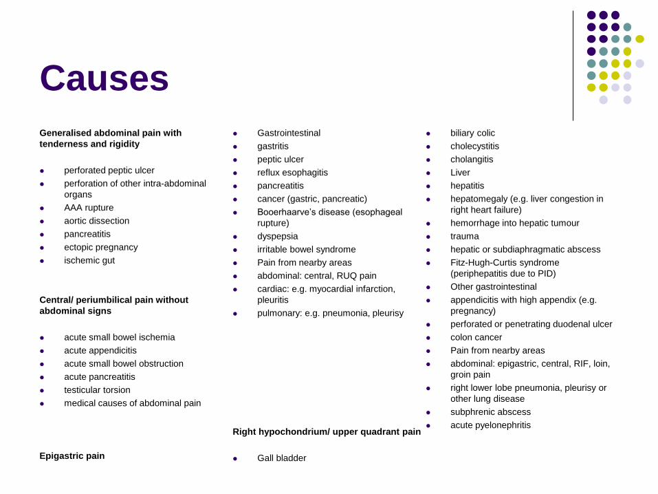
Causes
Generalised abdominal pain with
tenderness and rigidity
perforated peptic ulcer
perforation of other intra-abdominal
organs
AAA rupture
aortic dissection
pancreatitis
ectopic pregnancy
ischemic gut
Central/ periumbilical pain without
abdominal signs
acute small bowel ischemia
acute appendicitis
acute small bowel obstruction
acute pancreatitis
testicular torsion
medical causes of abdominal pain
Epigastric pain
Gastrointestinal
gastritis
peptic ulcer
reflux esophagitis
pancreatitis
cancer (gastric, pancreatic)
Booerhaarve’s disease (esophageal
rupture)
dyspepsia
irritable bowel syndrome
Pain from nearby areas
abdominal: central, RUQ pain
cardiac: e.g. myocardial infarction,
pleuritis
pulmonary: e.g. pneumonia, pleurisy
Right hypochondrium/ upper quadrant pain
Gall bladder
biliary colic
cholecystitis
cholangitis
Liver
hepatitis
hepatomegaly (e.g. liver congestion in
right heart failure)
hemorrhage into hepatic tumour
trauma
hepatic or subdiaphragmatic abscess
Fitz-Hugh-Curtis syndrome
(periphepatitis due to PID)
Other gastrointestinal
appendicitis with high appendix (e.g.
pregnancy)
perforated or penetrating duodenal ulcer
colon cancer
Pain from nearby areas
abdominal: epigastric, central, RIF, loin,
groin pain
right lower lobe pneumonia, pleurisy or
other lung disease
subphrenic abscess
acute pyelonephritis
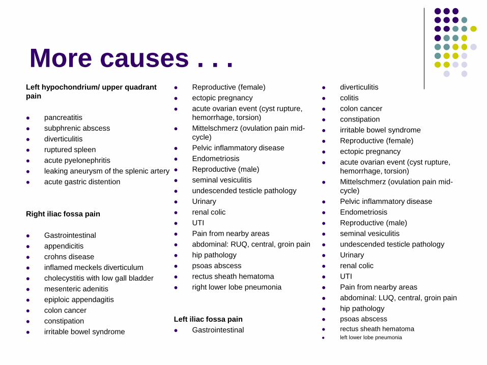
More causes . . . Left hypochondrium/ upper quadrant
pain
pancreatitis
subphrenic abscess
diverticulitis
ruptured spleen
acute pyelonephritis
leaking aneurysm of the splenic artery
acute gastric distention
Right iliac fossa pain
Gastrointestinal
appendicitis
crohns disease
inflamed meckels diverticulum
cholecystitis with low gall bladder
mesenteric adenitis
epiploic appendagitis
colon cancer
constipation
irritable bowel syndrome
Reproductive (female)
ectopic pregnancy
acute ovarian event (cyst rupture,
hemorrhage, torsion)
Mittelschmerz (ovulation pain mid-
cycle)
Pelvic inflammatory disease
Endometriosis
Reproductive (male)
seminal vesiculitis
undescended testicle pathology
Urinary
renal colic
UTI
Pain from nearby areas
abdominal: RUQ, central, groin pain
hip pathology
psoas abscess
rectus sheath hematoma
right lower lobe pneumonia
Left iliac fossa pain
Gastrointestinal
diverticulitis
colitis
colon cancer
constipation
irritable bowel syndrome
Reproductive (female)
ectopic pregnancy
acute ovarian event (cyst rupture,
hemorrhage, torsion)
Mittelschmerz (ovulation pain mid-
cycle)
Pelvic inflammatory disease
Endometriosis
Reproductive (male)
seminal vesiculitis
undescended testicle pathology
Urinary
renal colic
UTI
Pain from nearby areas
abdominal: LUQ, central, groin pain
hip pathology
psoas abscess
rectus sheath hematoma
left lower lobe pneumonia
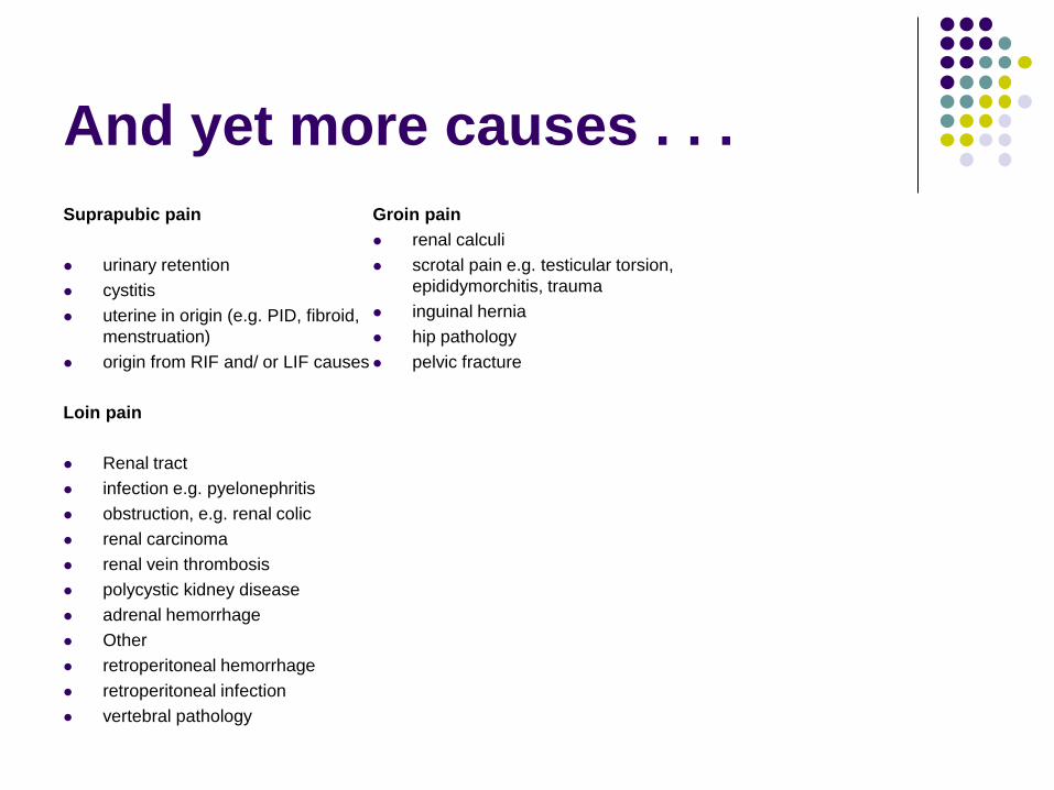
And yet more causes . . .
Suprapubic pain
urinary retention
cystitis
uterine in origin (e.g. PID, fibroid,
menstruation)
origin from RIF and/ or LIF causes
Loin pain
Renal tract
infection e.g. pyelonephritis
obstruction, e.g. renal colic
renal carcinoma
renal vein thrombosis
polycystic kidney disease
adrenal hemorrhage
Other
retroperitoneal hemorrhage
retroperitoneal infection
vertebral pathology
Groin pain
renal calculi
scrotal pain e.g. testicular torsion,
epididymorchitis, trauma
inguinal hernia
hip pathology
pelvic fracture
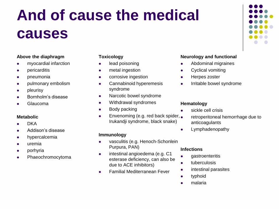
And of cause the medical
causes
Above the diaphragm
myocardial infarction
pericarditis
pneumonia
pulmonary embolism
pleurisy
Bornholm’s disease
Glaucoma
Metabolic
DKA
Addison’s disease
hypercalcemia
uremia
porhyria
Phaeochromocytoma
Toxicology
lead poisoning
metal ingestion
corrosive ingestion
Cannabinoid hyperemesis
syndrome
Narcotic bowel syndrome
Withdrawal syndromes
Body packing
Envenoming (e.g. red back spider,
Irukandji syndrome, black snake)
Immunology
vasculitis (e.g. Henoch-Schonlein
Purpura, PAN)
intestinal angioedema (e.g. C1
esterase deficiency, can also be
due to ACE inhibitors)
Familial Mediterranean Fever
Neurology and functional
Abdominal migraines
Cyclical vomiting
Herpes zoster
Irritable bowel syndrome
Hematology
sickle cell crisis
retroperitoneal hemorrhage due to
anticoagulants
Lymphadenopathy
Infections
gastroenteritis
tuberculosis
intestinal parasites
typhoid
malaria
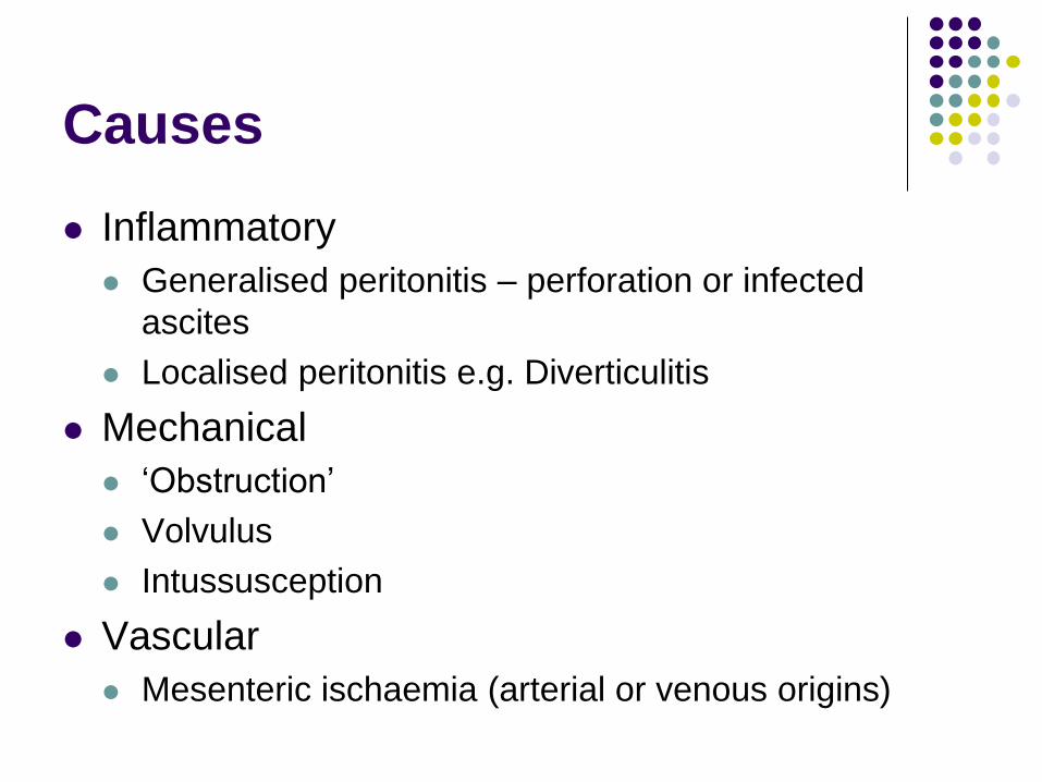
Causes
Inflammatory
Generalised peritonitis – perforation or infected
ascites
Localised peritonitis e.g. Diverticulitis
Mechanical
‘Obstruction’
Volvulus
Intussusception
Vascular
Mesenteric ischaemia (arterial or venous origins)

Assessment
What information do we need to know to
influence our diagnosis and management?

Assessment
What information do we need to know to
influence our diagnosis and management?
History
Examination
Amount of required analgesia
Physiological parameters
Radiological investigations
Haematological investigations
Discharge arrangements
Follow up
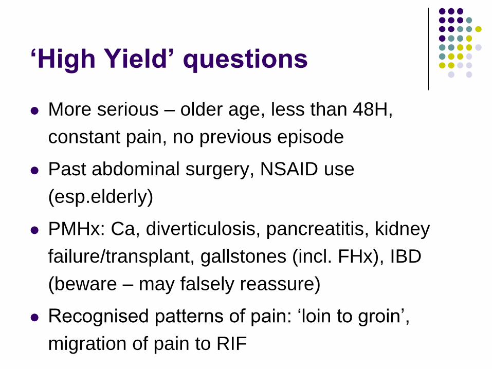
‘High Yield’ questions
More serious – older age, less than 48H,
constant pain, no previous episode
Past abdominal surgery, NSAID use
(esp.elderly)
PMHx: Ca, diverticulosis, pancreatitis, kidney
failure/transplant, gallstones (incl. FHx), IBD
(beware – may falsely reassure)
Recognised patterns of pain: ‘loin to groin’,
migration of pain to RIF
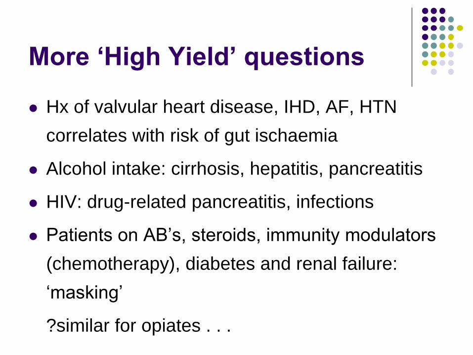
More ‘High Yield’ questions
Hx of valvular heart disease, IHD, AF, HTN
correlates with risk of gut ischaemia
Alcohol intake: cirrhosis, hepatitis, pancreatitis
HIV: drug-related pancreatitis, infections
Patients on AB’s, steroids, immunity modulators
(chemotherapy), diabetes and renal failure:
‘masking’
?similar for opiates . . .
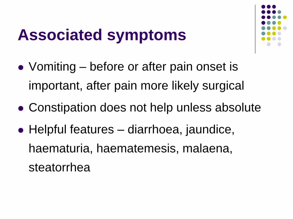
Associated symptoms
Vomiting – before or after pain onset is
important, after pain more likely surgical
Constipation does not help unless absolute
Helpful features – diarrhoea, jaundice,
haematuria, haematemesis, malaena,
steatorrhea
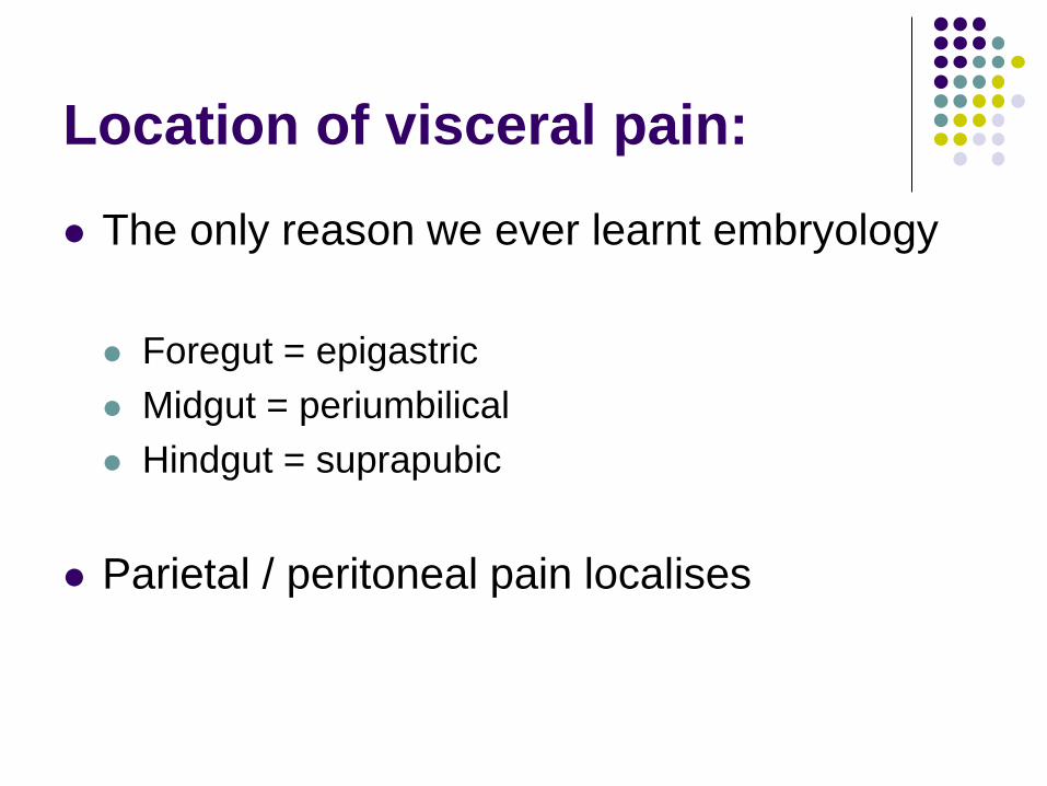
Location of visceral pain:
The only reason we ever learnt embryology
Foregut = epigastric
Midgut = periumbilical
Hindgut = suprapubic
Parietal / peritoneal pain localises

Epigastric pain – red flags
Age over 50
Weight loss
Persistent vomiting
Dysphagia
Anemia
Hematemesis
Palpable abdominal mass
Family history of upper gastrointestinal carcinoma
Previously identified pathology requiring reassessment,
or history of gastric surgery for pathology that could
recur
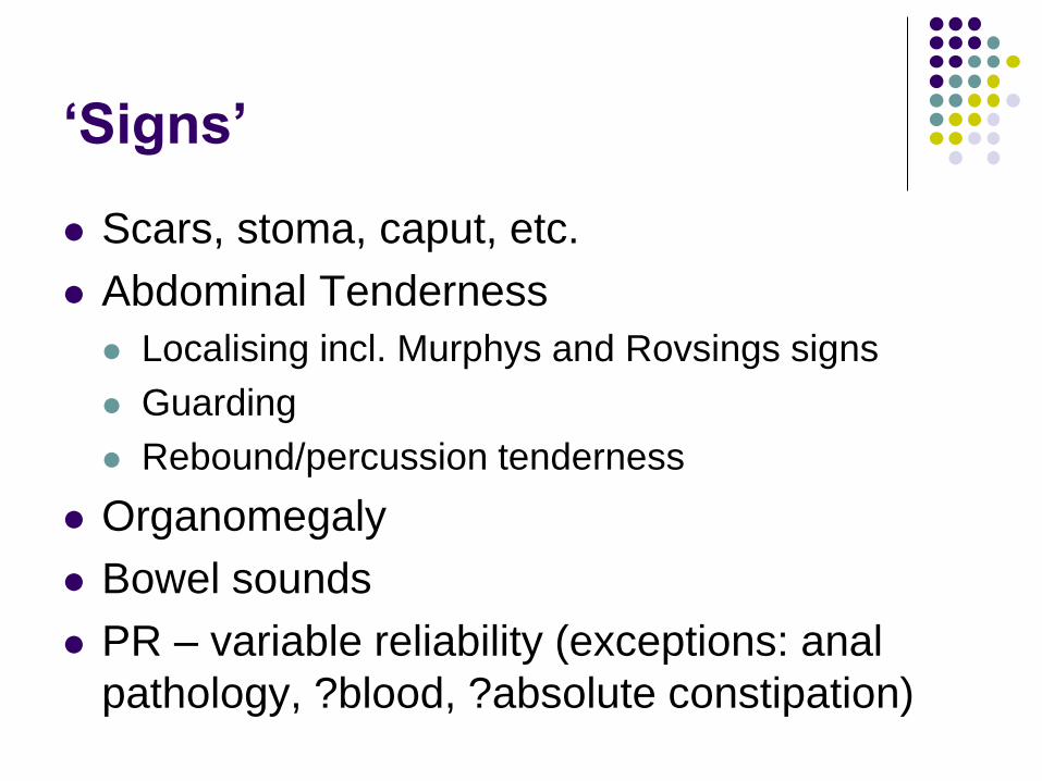
‘Signs’
Scars, stoma, caput, etc.
Abdominal Tenderness
Localising incl. Murphys and Rovsings signs
Guarding
Rebound/percussion tenderness
Organomegaly
Bowel sounds
PR – variable reliability (exceptions: anal
pathology, ?blood, ?absolute constipation)
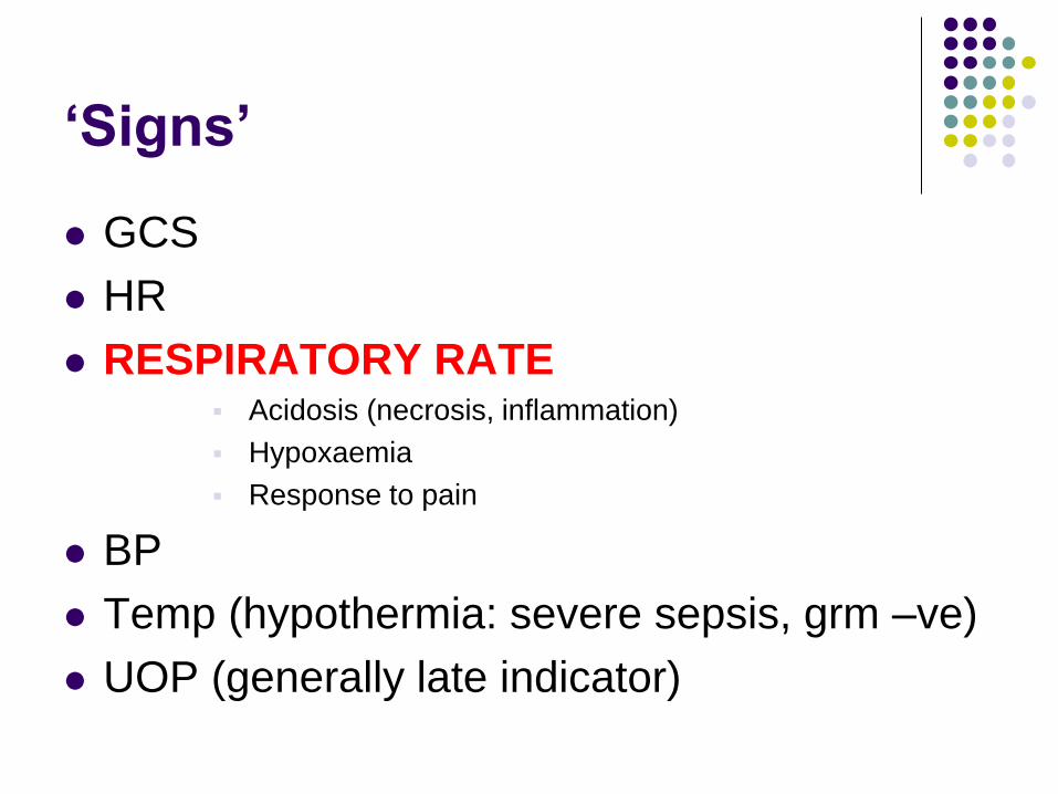
‘Signs’
GCS
HR
RESPIRATORY RATE Acidosis (necrosis, inflammation)
Hypoxaemia
Response to pain
BP
Temp (hypothermia: severe sepsis, grm –ve)
UOP (generally late indicator)
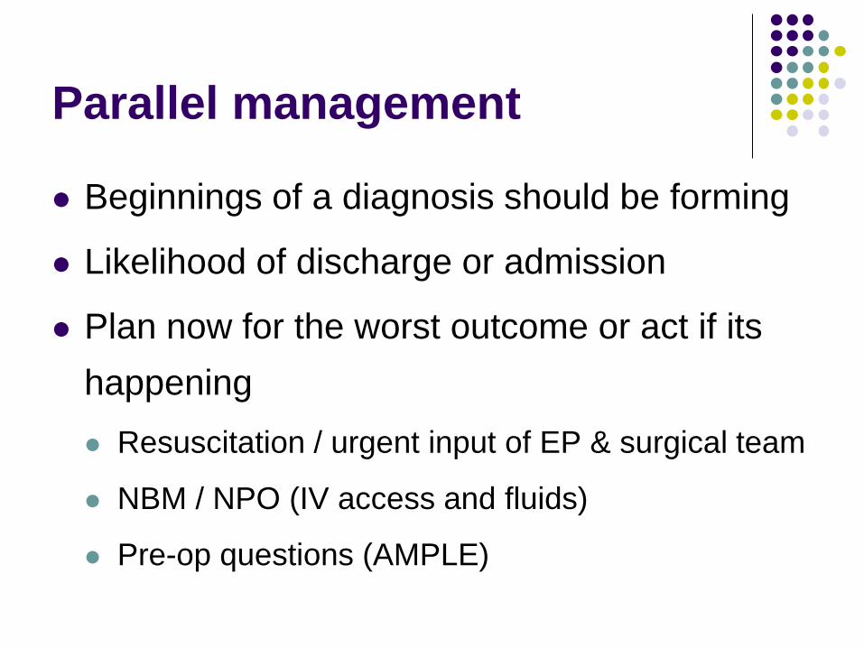
Parallel management
Beginnings of a diagnosis should be forming
Likelihood of discharge or admission
Plan now for the worst outcome or act if its
happening
Resuscitation / urgent input of EP & surgical team
NBM / NPO (IV access and fluids)
Pre-op questions (AMPLE)
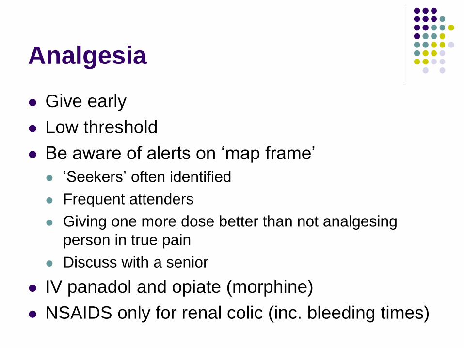
Analgesia
Give early
Low threshold
Be aware of alerts on ‘map frame’
‘Seekers’ often identified
Frequent attenders
Giving one more dose better than not analgesing
person in true pain
Discuss with a senior
IV panadol and opiate (morphine)
NSAIDS only for renal colic (inc. bleeding times)
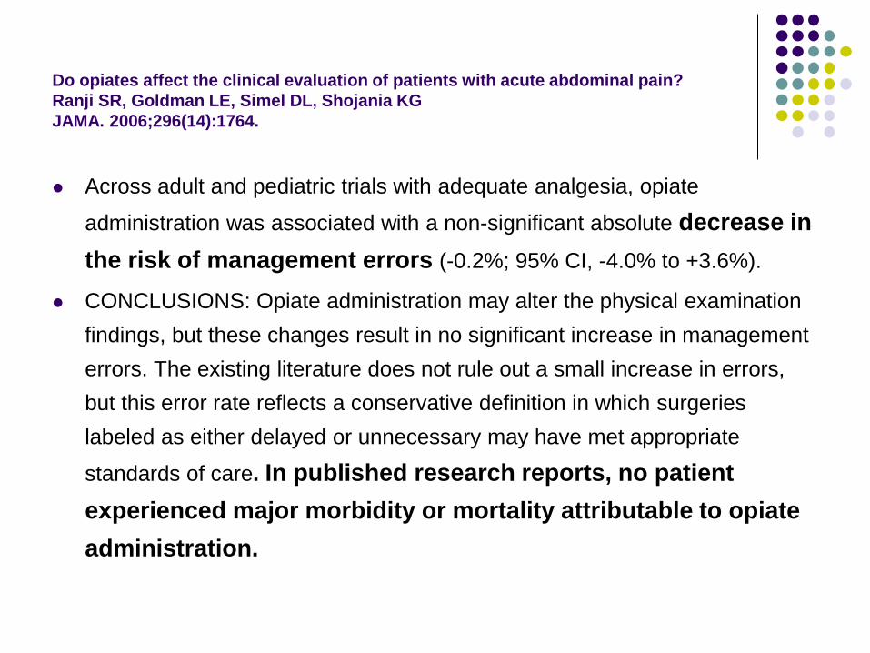
Do opiates affect the clinical evaluation of patients with acute abdominal pain?
Ranji SR, Goldman LE, Simel DL, Shojania KG
JAMA. 2006;296(14):1764.
Across adult and pediatric trials with adequate analgesia, opiate
administration was associated with a non-significant absolute decrease in
the risk of management errors (-0.2%; 95% CI, -4.0% to +3.6%).
CONCLUSIONS: Opiate administration may alter the physical examination
findings, but these changes result in no significant increase in management
errors. The existing literature does not rule out a small increase in errors,
but this error rate reflects a conservative definition in which surgeries
labeled as either delayed or unnecessary may have met appropriate
standards of care. In published research reports, no patient
experienced major morbidity or mortality attributable to opiate
administration.
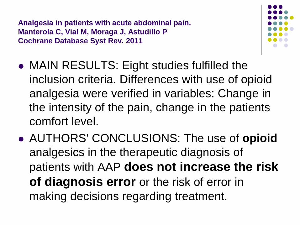
Analgesia in patients with acute abdominal pain.
Manterola C, Vial M, Moraga J, Astudillo P
Cochrane Database Syst Rev. 2011
MAIN RESULTS: Eight studies fulfilled the
inclusion criteria. Differences with use of opioid
analgesia were verified in variables: Change in
the intensity of the pain, change in the patients
comfort level.
AUTHORS' CONCLUSIONS: The use of opioid
analgesics in the therapeutic diagnosis of
patients with AAP does not increase the risk
of diagnosis error or the risk of error in
making decisions regarding treatment.
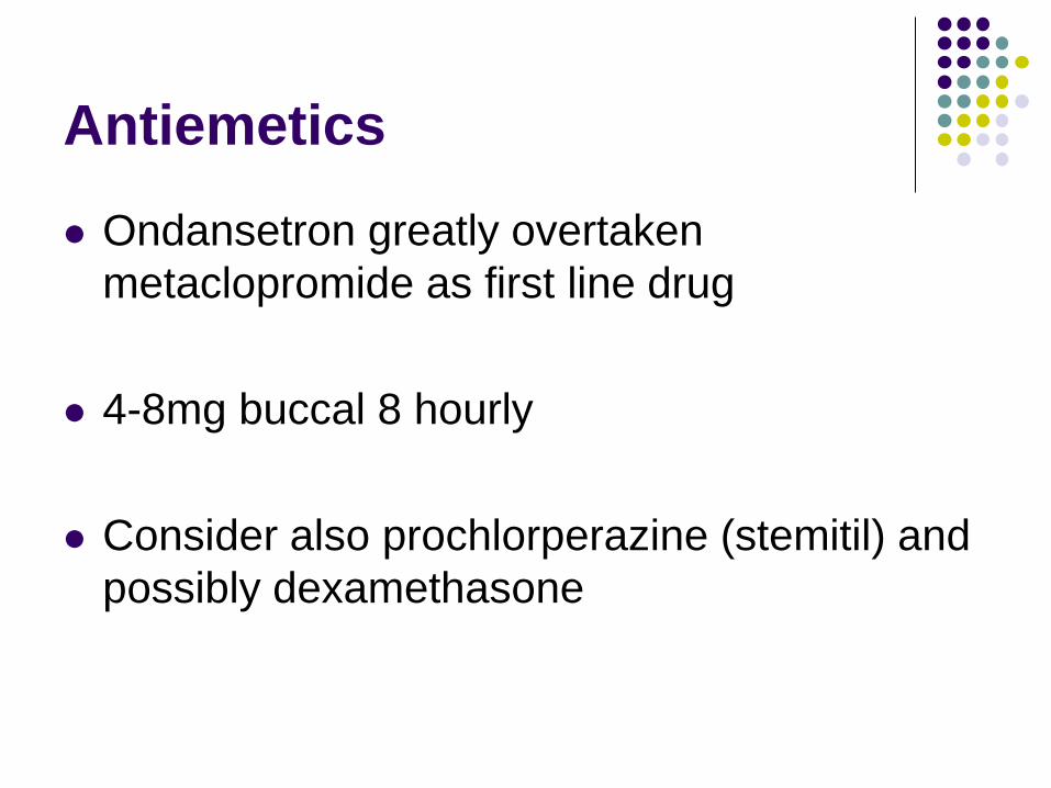
Antiemetics
Ondansetron greatly overtaken
metaclopromide as first line drug
4-8mg buccal 8 hourly
Consider also prochlorperazine (stemitil) and
possibly dexamethasone
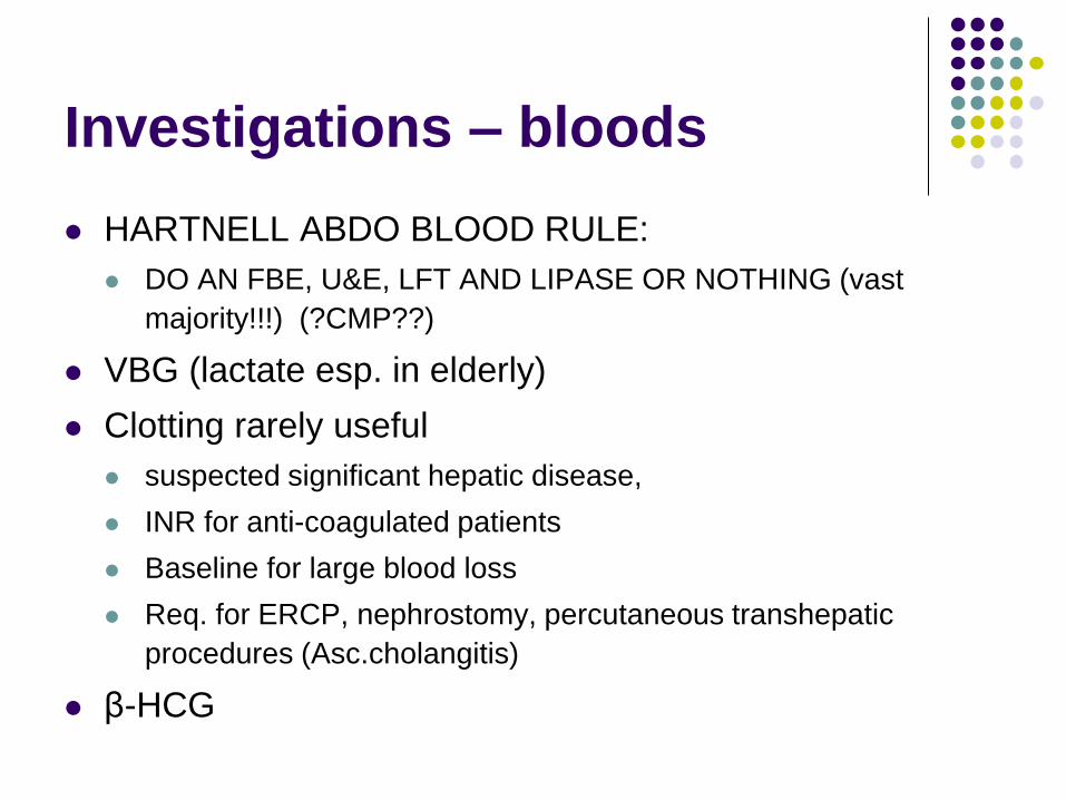
Investigations – bloods
HARTNELL ABDO BLOOD RULE:
DO AN FBE, U&E, LFT AND LIPASE OR NOTHING (vast
majority!!!) (?CMP??)
VBG (lactate esp. in elderly)
Clotting rarely useful
suspected significant hepatic disease,
INR for anti-coagulated patients
Baseline for large blood loss
Req. for ERCP, nephrostomy, percutaneous transhepatic
procedures (Asc.cholangitis)
β-HCG
Abdominal XR
Is it any good?
Abdominal XR
The role of abdominal radiography in the evaluation of the nontrauma
emergency patient. Kellow ZS, MacInnes M, Kurzencwyg D, Rawal S,
Jaffer R, Kovacina B, Stein LA. Radiology. 2008;248(3):887. [Pubmed]
Retrospective review of abdominal radiography of non-
traumatic abdominal pain presenting to an ED over a
period of 6 months
‘abdominal radiography helped confirm the suspected
diagnosis in 2%-8% of cases. In 37 (4%) of 874 patients,
abdominal radiography was possibly helpful in
changing patient treatment without a follow-up study’.

Abdominal XR
Many signs possible
FB esp. in children (what is it and is it below the
diaphragm) NB: don’t ask parents to search poo!
Volvulus
Inflammation, sentinel loop
Fluid levels (non-localising) suggesting
obstruction
& etc.
BUT poor returns esp. in undifferentiated pain

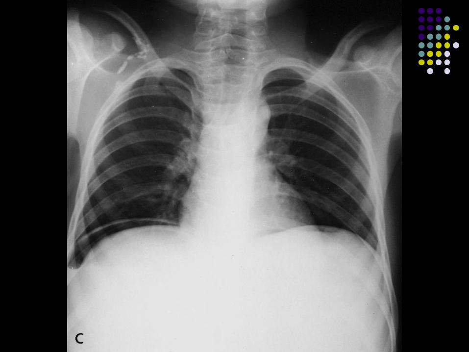
CXR
Pneumonia
Air under diaphragm
Mediastinal air
ARDS
CT abdomen
Requires ED consultant or Surgical reg/cons
approval
Non-contrast image of choice renal colic
Useful in pancreatitis (severity, some
planning of surg Mx, talk to them first!)
Intra-abdominal sepsis and trauma
Pre-operative for most patients except where
too unstable
Computed tomography (CT)
is the study of choice in the evaluation of
undifferentiated abdominal pain
Approximately 2/3 have a disease that can be
diagnosed by CT (up to 90% vs 76% by Hx
and Ex)
CT is particularly useful in the elderly
CT outperforms plain radiographs in the
diagnosis of non-traumatic abdominal pain
CT
Caution expressed over
overuse and over reliance (largely discredited)
Delay to operation in obstruction doubles mortality
Use out of hours in relatively stable patients
Cost
Resource rationalisation (esp.workforce)
Operative intervention at night increases mortality
radiation exposure
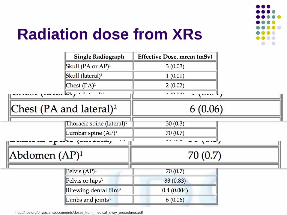
Radiation dose from XRs
http://hps.org/physicians/documents/doses_from_medical_x-ray_procedures.pdf
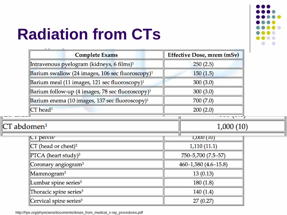
Radiation from CTs
http://hps.org/physicians/documents/doses_from_medical_x-ray_procedures.pdf
USS - no radiation!
RUQ pain (stones, cholecystitis, CBD block)
Obstructive uropathy (Not stones) e.g. pregnant
AAA – determines aortic size, not leak
Children (again, no radiation) finds:
pyloric stenosis, intussusception, appendicitis
Evaluating hernias
Looking for collections (good alternative to CT)
Some role in abdominal mass evaluation
Paediatric abdominal pain
Specific age groups
Volvulus among neonates
Bilious emesis and unwell (req. contrast study)
Pyloric stenosis 2-6/52
Failure to gain weight
Vomiting
Visible peristalisis +/- mass
Intussusception among older infants and
toddlers).
Pale and floppy with colicky pain (20mins between
pain) – req. USS (redcurrant jelly stool v. late sign)

Paediatric abdominal pain Red=time critical
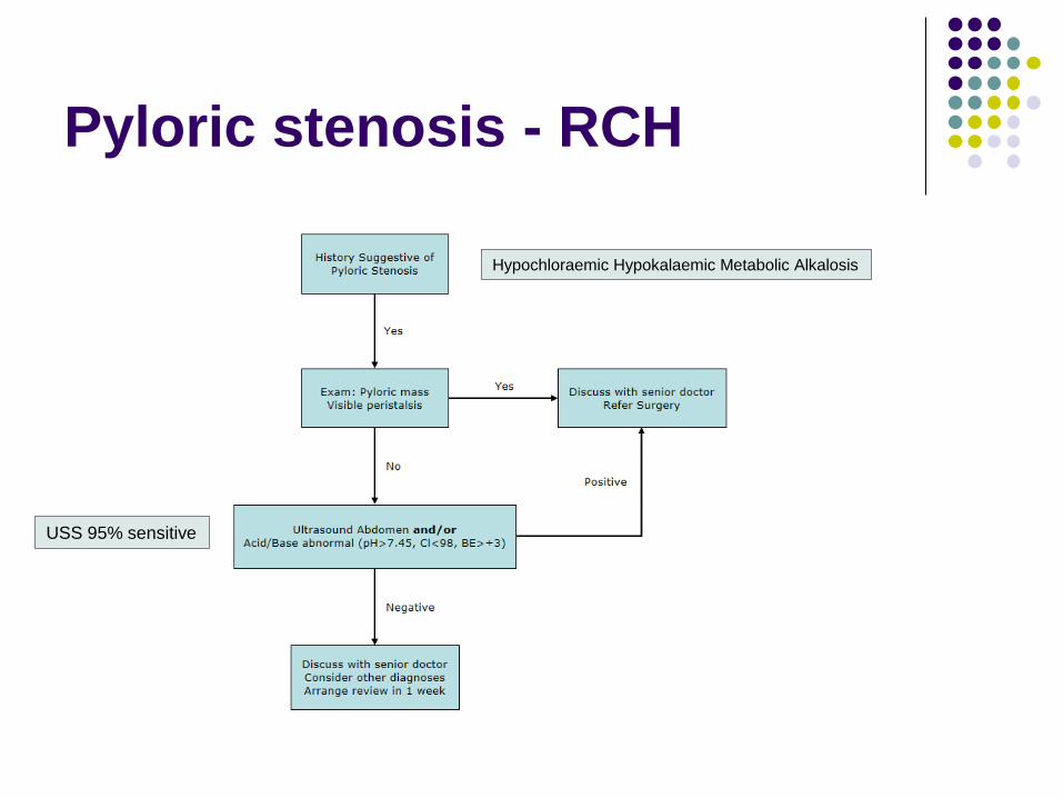
Pyloric stenosis - RCH
Hypochloraemic Hypokalaemic Metabolic Alkalosis
USS 95% sensitive
RCH guidelines - discharge
‘Children with abdominal pain who are
otherwise healthy, well-appearing, and have
normal physical examinations [and FWT]
typically do not require ancillary studies.
Those whose repeat examinations continue
to be unremarkable and who tolerate feeding
can usually be discharged with reliable
medical follow-up’.

My guidelines
Discuss all paediatric discharges – especially
from bays - with a consultant, please.

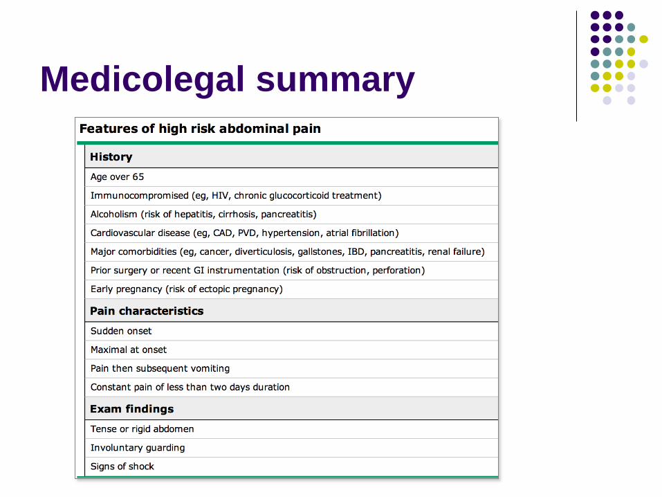
Medicolegal summary
Failure to examine pelvis and genitalia
Failure to appreciate ‘at risk’ patients
Over reliance on investigation results
Not appreciating variation in presentation in
elder population
Failure to arrange FU
Medicolegal summary
Medicolegal summary
Failure to examine pelvis and genitalia
Failure to appreciate ‘at risk’ patients
Over reliance on investigation results
Not appreciating variation in presentation in
elder population
Failure to arrange FU

My coffee is ready
See you 10 mins . . .

Elderly
Non-specific symptoms
Less severe physiological and
haematological signs (req. reference)
AF and mesenteric emboli – ischaemic gut
End of life decisions
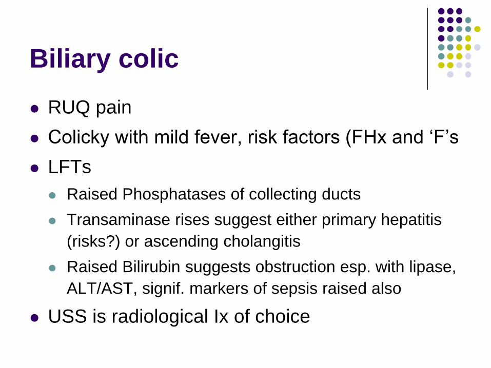
Biliary colic
RUQ pain
Colicky with mild fever, risk factors (FHx and ‘F’s
LFTs
Raised Phosphatases of collecting ducts
Transaminase rises suggest either primary hepatitis
(risks?) or ascending cholangitis
Raised Bilirubin suggests obstruction esp. with lipase,
ALT/AST, signif. markers of sepsis raised also
USS is radiological Ix of choice
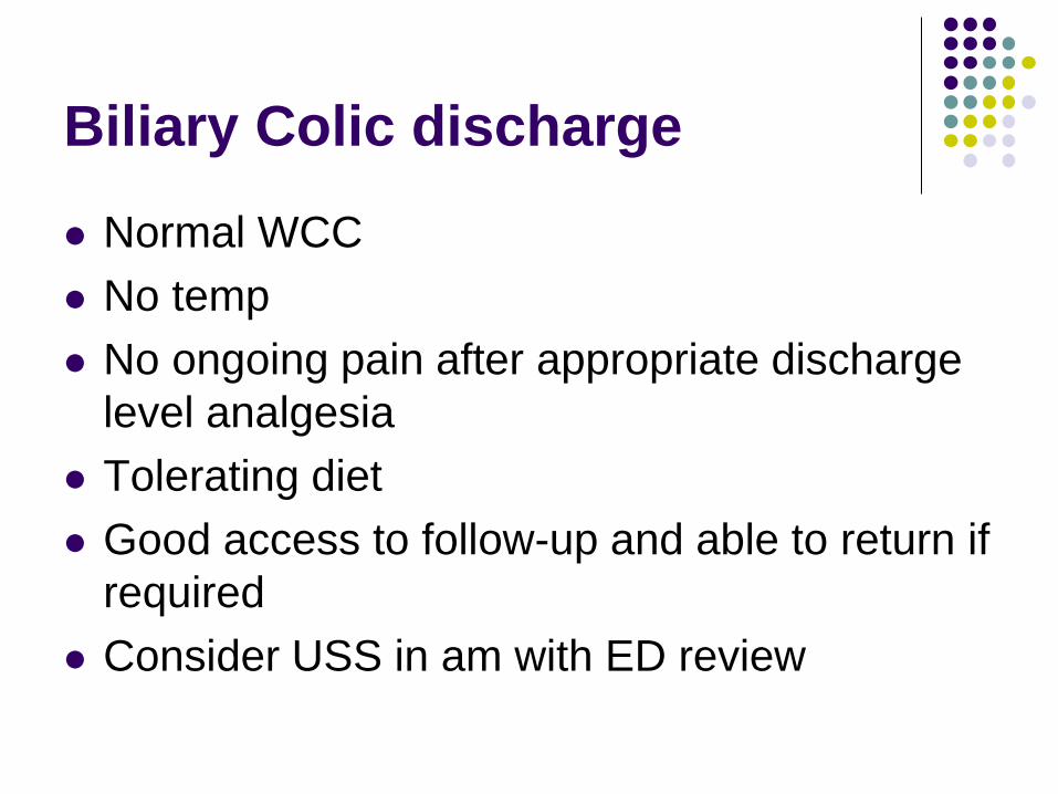
Biliary Colic discharge
Normal WCC
No temp
No ongoing pain after appropriate discharge
level analgesia
Tolerating diet
Good access to follow-up and able to return if
required
Consider USS in am with ED review
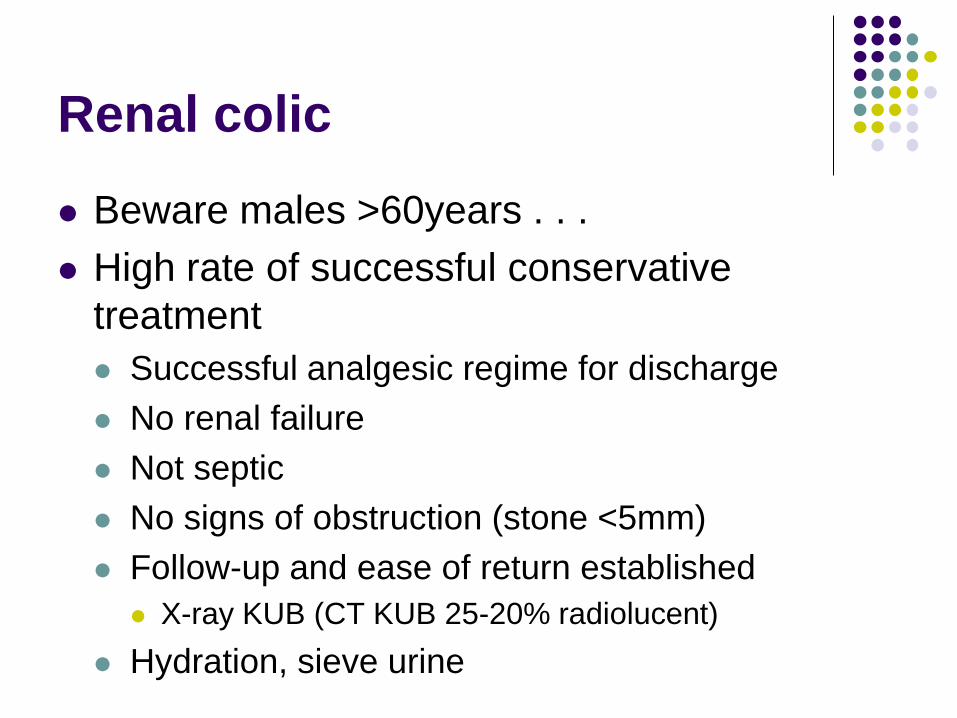
Renal colic
Beware males >60years . . .
High rate of successful conservative
treatment
Successful analgesic regime for discharge
No renal failure
Not septic
No signs of obstruction (stone <5mm)
Follow-up and ease of return established
X-ray KUB (CT KUB 25-20% radiolucent)
Hydration, sieve urine
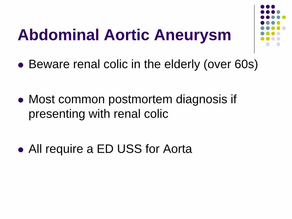
Abdominal Aortic Aneurysm
Beware renal colic in the elderly (over 60s)
Most common postmortem diagnosis if
presenting with renal colic
All require a ED USS for Aorta
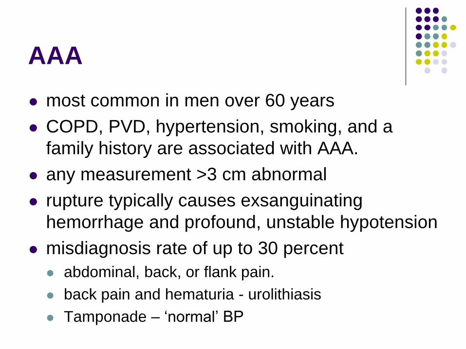
AAA
most common in men over 60 years
COPD, PVD, hypertension, smoking, and a
family history are associated with AAA.
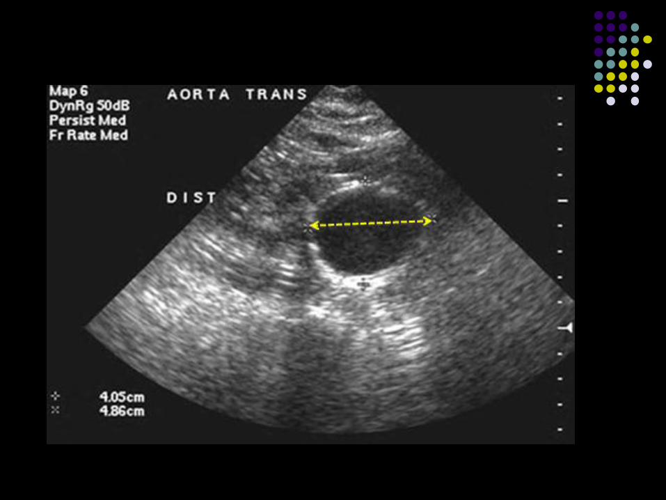
any measurement >3 cm abnormal
rupture typically causes exsanguinating
hemorrhage and profound, unstable hypotension
misdiagnosis rate of up to 30 percent
abdominal, back, or flank pain.
back pain and hematuria - urolithiasis
Tamponade – ‘normal’ BP
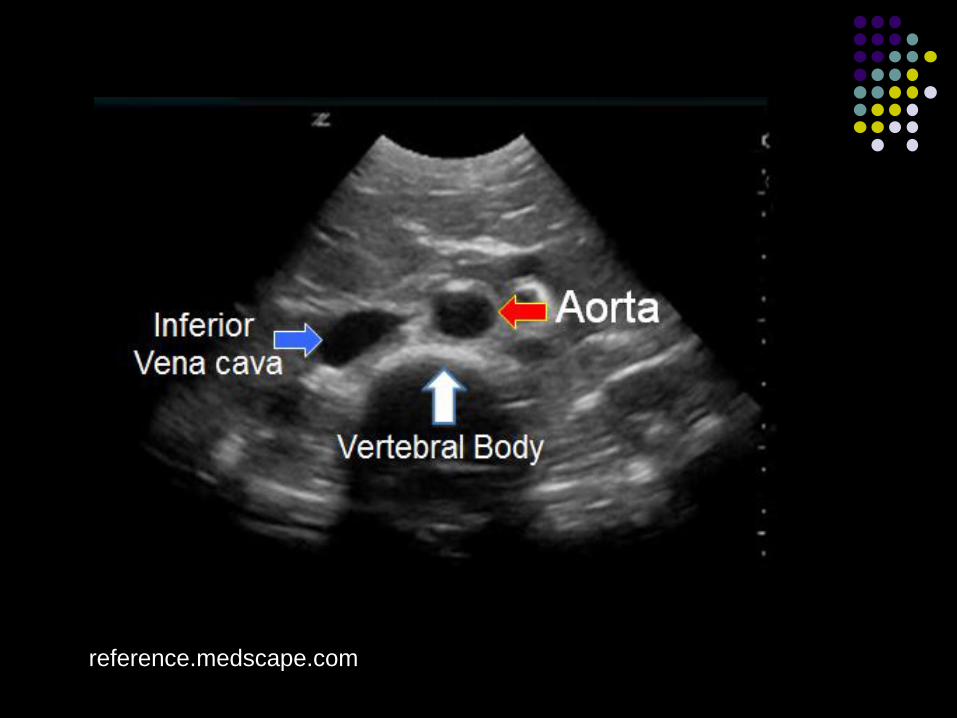
reference.medscape.com

Mesenteric ischaemia
Difficult diagnosis
Risks
Arterial disease: Atheroscl., vasculitis,
Embolism risk: AF, valvular disease, MI,
aneurysms
Reduced flow: Sepsis, severe dehydration,
vasoactive drugs
Venous occlusion intra-abdominal infection (with portal pyemia), hypercoagulable
states, portal hypertension/mass effect from tumours -> stasis,
trauma from surgery, band adhesions
Mesenteric Ischaemia
Initially can seem quite well, precipitous
deterioration
Pain out of proportion
Not as obvious in dementia
Possible obstructive signs
Distended abdomen
Examination
For risk factors
Esp. cardiac disease, hypercoagulability
Distended and tender abdomen
Severity of symptoms depends on area of gut
affected
Physiological stability and sepsis
Appropriate patient for surgery?
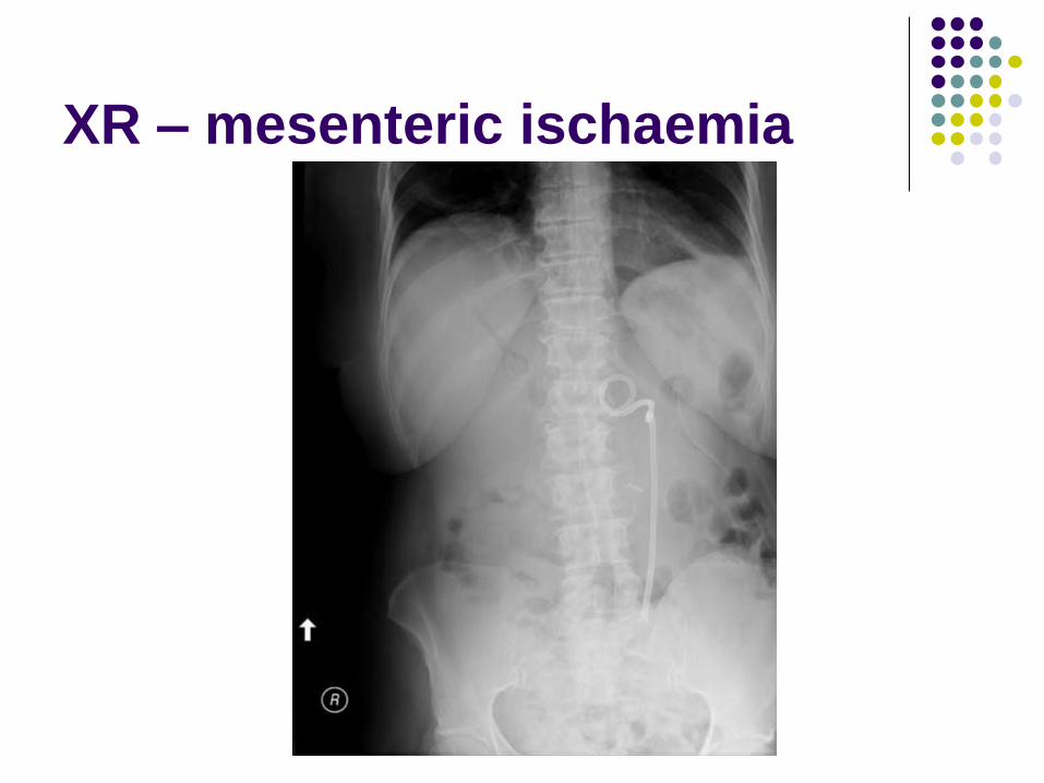
XR – mesenteric ischaemia

http://emcram.com/showimage.asp?ID=13

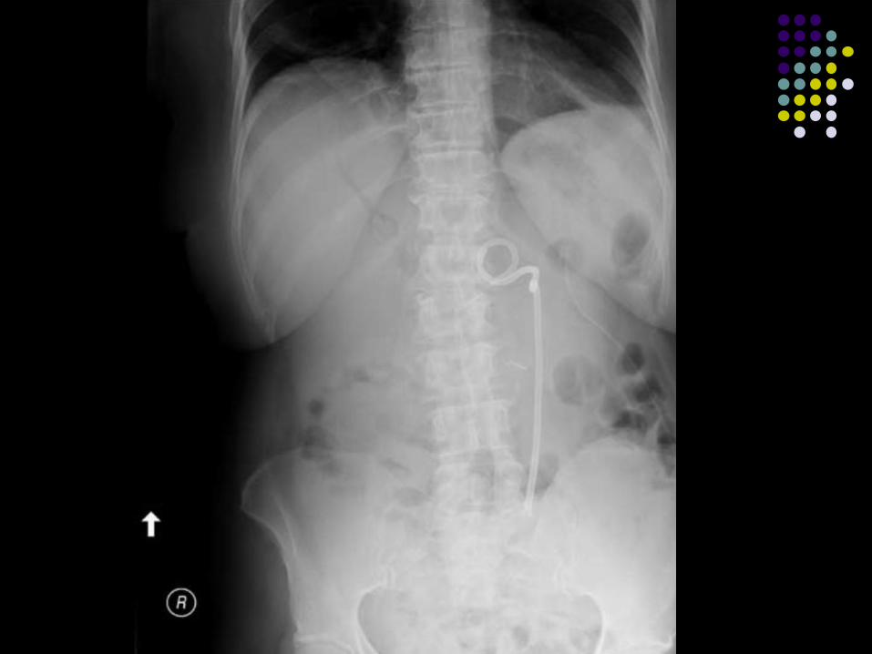
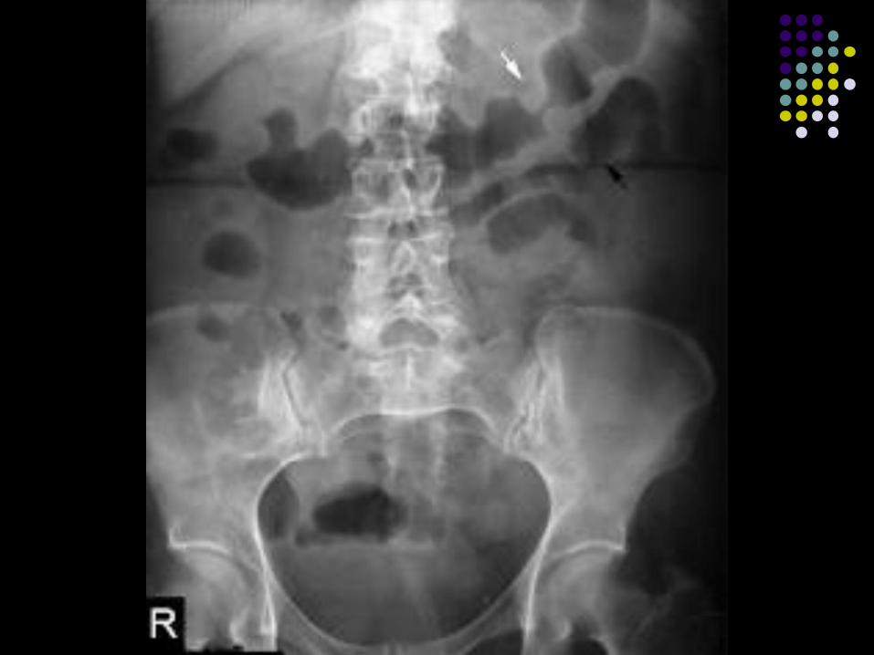

Thumb printing of thickened mucosa
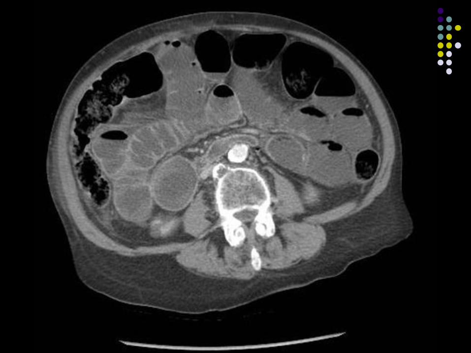
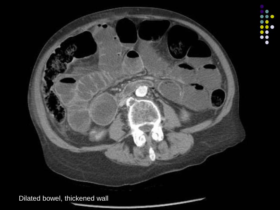
Dilated bowel, thickened wall

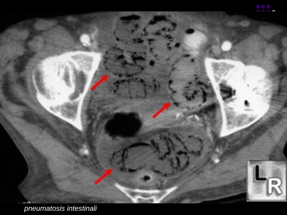
pneumatosis intestinali
Management
High suspicion, low threshold for investigation
Lactate is the most sensitive test
>70% mortality
If surgery indicated – get on with it!
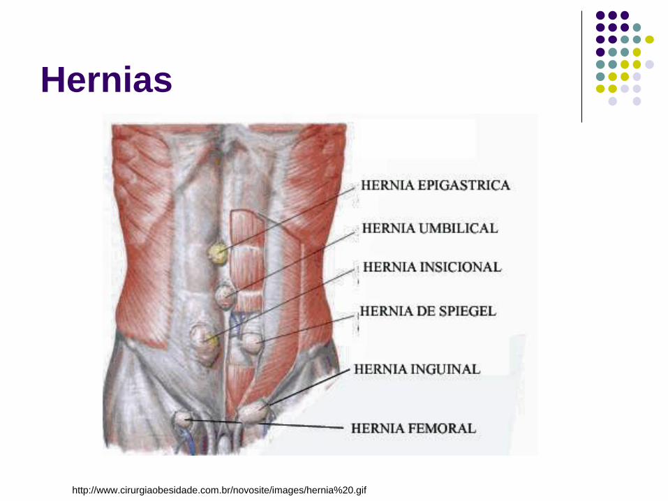
Hernias
http://www.cirurgiaobesidade.com.br/novosite/images/hernia%20.gif
Herniae
Occasionally intermittent pain
In ED likely to present acutely with lump
Fat and gut most likely contents
Look for them!
Attempt reduction
Long firm pressure with adequate analgesia
Diverticulitis/Diverticulosis
LIF pain
Differential includes gynae pathology
Mild change in BH
Blood PR
Spectrum of severity - ?admit/discharge
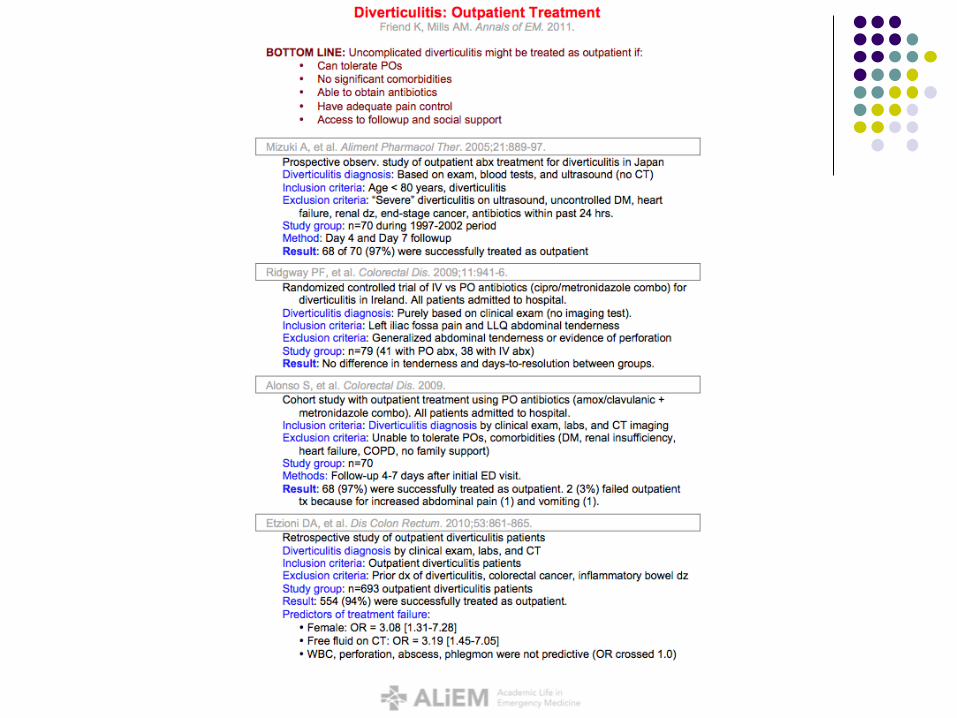
Discharge
Early/mild diverticulitis
Tolerating diet
With effective analgesia
With antibiotics
With advice to return if deteriorates
Follow up essential

Cases
Mr P.T.
Severe rapid onset of epigastric pain
2/7 of worsening pain
Unable to tolerate food, vomiting
intermittently
Mr P.T. 56 y.o. male
Severe rapid onset of epigastric pain
2/7 of worsening pain
Unable to tolerate food, vomiting
intermittently
Recent increase in alcohol to ?>60units/week
(double at least once?)
(°FHx of gallstones, no ERCP, no trauma)

What do you want to know?
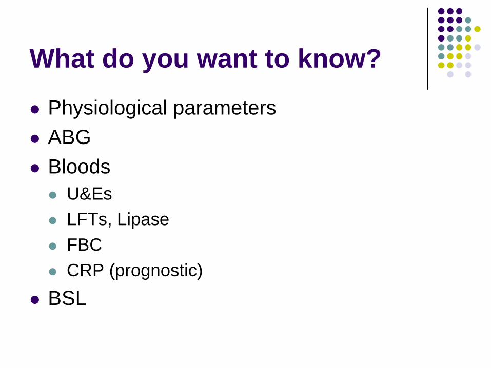
What do you want to know?
Physiological parameters
ABG
Bloods
U&Es
LFTs, Lipase
FBC
CRP (prognostic)
BSL
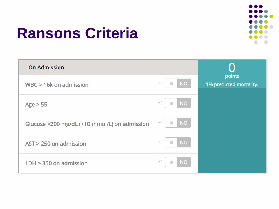
Ransons Criteria
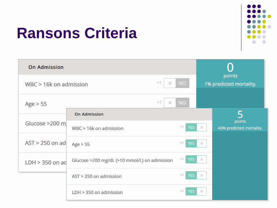
Ransons Criteria
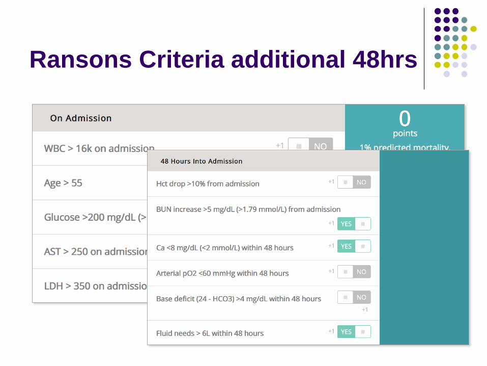
Ransons Criteria additional 48hrs
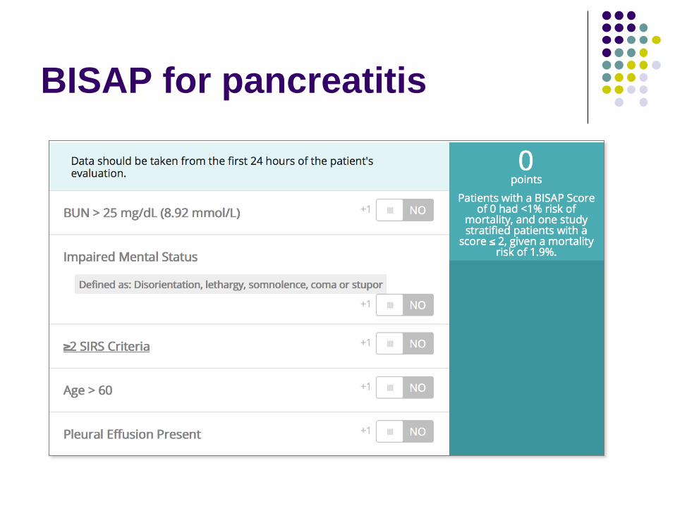
BISAP for pancreatitis

Further Mx
Resuscitation as required
NBM and IV fluids
Not for routine ABx
CXR (?effusion)
CT (pseudocysts, necrosis)
Don’t forget:
Alcohol withdrawal protocol (BZs and thiamine
replacement)

Mr S.P. 62 y.o.
Handed over to you:
Severe early Parkinsons Disease
Fully dependent for ADLs
Vomiting and ‘flat’
Some suggestion of vague abdominal pain
‘Better’ after fluids, Ondansetron and Morphine
All the tests are requested . . .
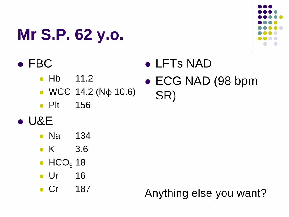
Mr S.P. 62 y.o.
FBC Hb 11.2
WCC 14.2 (Nϕ 10.6)
Plt 156
U&E Na 134
K 3.6
HCO3 18
Ur 16
Cr 187
LFTs NAD
ECG NAD (98 bpm
SR)
Anything else you want?
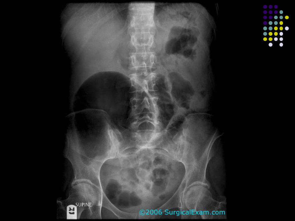
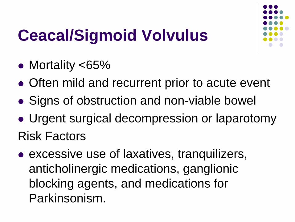
Ceacal/Sigmoid Volvulus
Mortality <65%
Often mild and recurrent prior to acute event
Signs of obstruction and non-viable bowel
Urgent surgical decompression or laparotomy
Risk Factors
excessive use of laxatives, tranquilizers,
anticholinergic medications, ganglionic
blocking agents, and medications for
Parkinsonism.
Mrs P.M.
76 y.o female from Jim Gay unit fro rehab
Recent L DHS for NOF ♯
PMHx
AF
Hypertensive
Gallstones and cholecystectomy
Collapse after c/o back and abdominal pain
Low BP - unrecordable
In ED
Grey and largely unresponsive
Fast AF
BP initially unrecordable
Improves to 90/40 with N saline, HR remains
120bpm irreg irreg
GCS to 12
In ED
Grey and largely unresponsive
Fast AF
BP initially unrecordable
Improves to 90/40 with N saline, HR remains
120bpm irreg irreg
GCS to 12
Exn
Generally tender, Dusky flanks noted
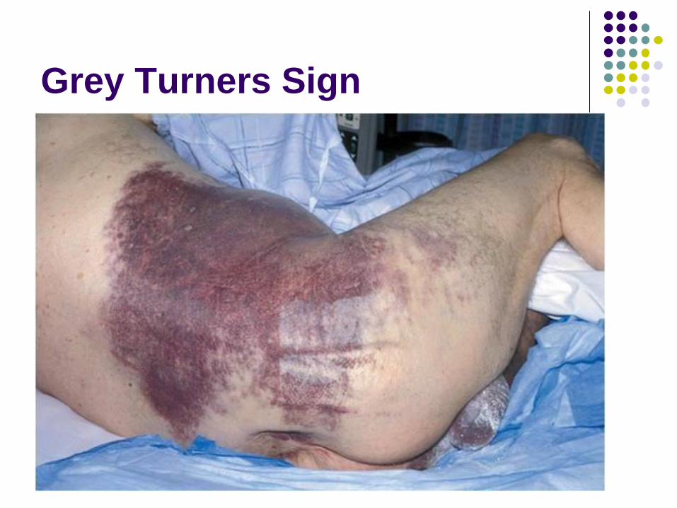
Grey Turners Sign
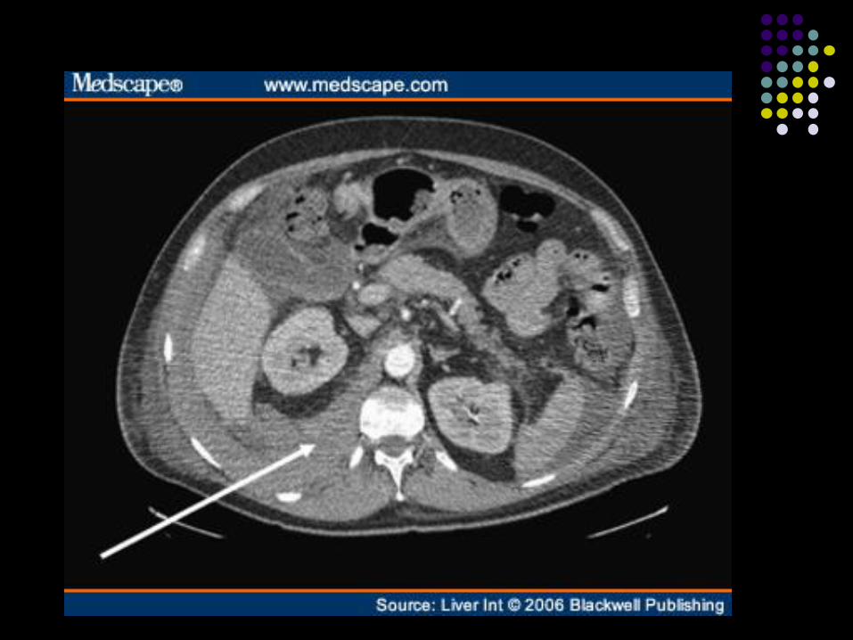
Further Mx
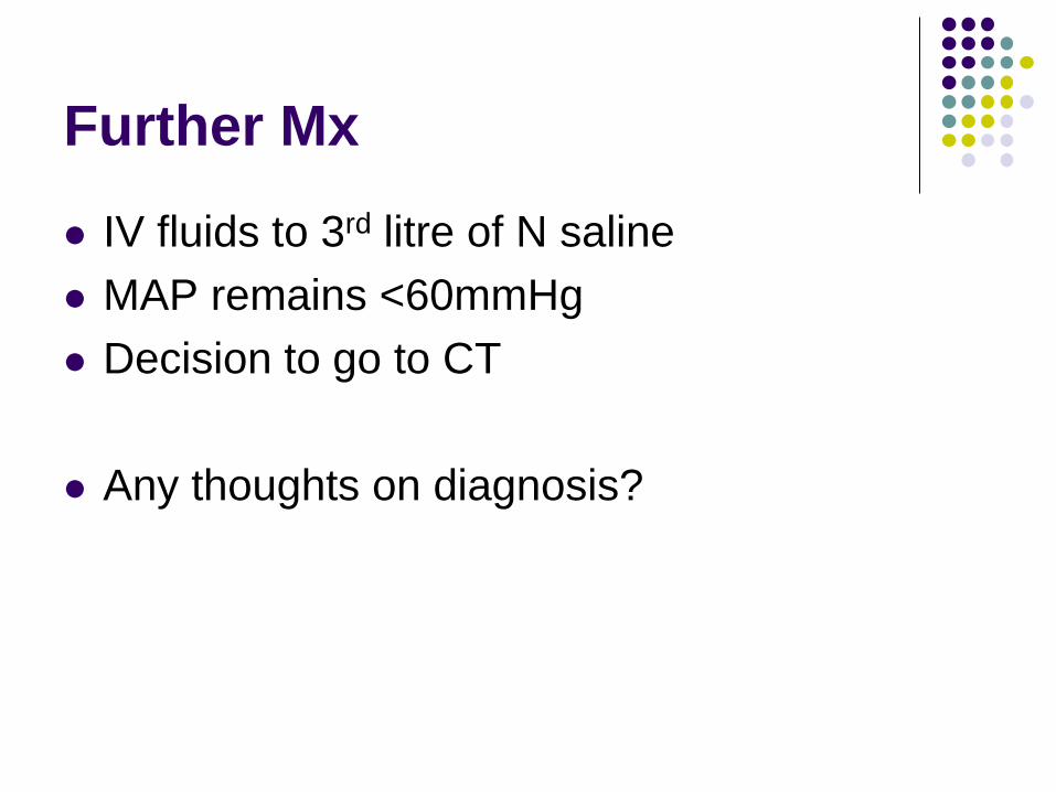
IV fluids to 3rd litre of N saline
MAP remains <60mmHg
Decision to go to CT
Any thoughts on diagnosis?
Further Mx
Reversal of anticoagulation – INR 6.5
Vit K 10mg IV
2 units FFP
Prothrombinex (contains factors II, IX and X)
(2’500 U initially)
Two visits to radiology for attempts at
embolisation unsuccessful
Pt died 3 days later

Questions?
Summary
Most abdominal pain will resolve
spontaneously – otherwise well patients are
likely to remain so
The very young and elderly require a high
index of suspicion and low threshold for
investigation
CT is the investigation of choice (use
appropriately)

Thankyou


