ABDOMINAL EMERGENCIES 1603 Multidetector CT...
15
1603 ABDOMINAL EMERGENCIES Jackson D. Hamilton, MD • Manickam Kumaravel, MBBS • Michael L. Censullo, MD • Alan M. Cohen, MD • Daniel S. Kievlan, BA • O. Clark West, MD Timely localization of a bleeding source can improve the efficacy of trauma management, and improvements in the technology of com- puted tomography (CT) have expedited the work-up of the traumatized patient. The classic pattern of active extravasation (ie, administered contrast agent that has escaped from injured arteries, veins, or urinary tract) at dual phase CT is a jet or focal area of hyperattenuation within a hematoma that fades into an enlarged, enhanced hematoma on delayed images. This finding indicates significant bleeding and must be quickly communicated to the clinician, since potentially lifesaving surgical or endovascular repair may be necessary. Active extravasation can be asso- ciated with other injuries to arteries, such as a hematoma or a pseudo- aneurysm. Both active extravasation and pseudoaneurysm (unlike bone fragments and dense foreign bodies) change in appearance on delayed images, compared with their characteristics on arterial images. Other clues to the location of vessel injury include lack of vascular enhance- ment (caused by occlusion or spasm), vessel irregularity, size change (such as occurs with pseudoaneurysm), and an intimal flap (which sig- nifies dissection). The sentinel clot sign is an important clue for locating the bleeding source when other more localizing findings of vessel injury are not present. Timely diagnosis, differentiation of vascular injuries from other findings of trauma, signs of depleted intravascular volume, and localization of vascular injury are important to convey to interven- tional radiologists or surgeons to improve trauma management. © RSNA, 2008 • radiographics.rsnajnls.org Multidetector CT Evaluation of Active Extravasation in Blunt Abdominal and Pelvic Trauma Patients 1 RadioGraphics 2008; 28:1603–1616 • Published online 10.1148/rg.286085522 • Content Codes: 1 From the Department of Diagnostic and Interventional Imaging, Memorial Hermann Hospital, University of Texas Houston School of Medicine, 6431 Fannin St, Houston, TX 77030-1503. Presented as an education exhibit at the 2007 RSNA Annual Meeting. Received March 18, 2008; revi- sion requested April 24; final revision received July 14; accepted July 16. J.D.H. holds stock in Merck; all other authors have no financial relationships to disclose. Address correspondence to J.D.H. (e-mail: [email protected]). © RSNA, 2008 See last page TEACHING POINTS Note: This copy is for your personal non-commercial use only. To order presentation-ready copies for distribution to your colleagues or clients, contact us at www.rsna.org/rsnarights.
Transcript of ABDOMINAL EMERGENCIES 1603 Multidetector CT...

1603ABDOMINAL EMERGENCIES
Jackson D. Hamilton, MD • Manickam Kumaravel, MBBS • Michael L. Censullo, MD • Alan M. Cohen, MD • Daniel S. Kievlan, BA • O. Clark West, MD
Timely localization of a bleeding source can improve the efficacy of trauma management, and improvements in the technology of com-puted tomography (CT) have expedited the work-up of the traumatized patient. The classic pattern of active extravasation (ie, administered contrast agent that has escaped from injured arteries, veins, or urinary tract) at dual phase CT is a jet or focal area of hyperattenuation within a hematoma that fades into an enlarged, enhanced hematoma on delayed images. This finding indicates significant bleeding and must be quickly communicated to the clinician, since potentially lifesaving surgical or endovascular repair may be necessary. Active extravasation can be asso-ciated with other injuries to arteries, such as a hematoma or a pseudo-aneurysm. Both active extravasation and pseudoaneurysm (unlike bone fragments and dense foreign bodies) change in appearance on delayed images, compared with their characteristics on arterial images. Other clues to the location of vessel injury include lack of vascular enhance-ment (caused by occlusion or spasm), vessel irregularity, size change (such as occurs with pseudoaneurysm), and an intimal flap (which sig-nifies dissection). The sentinel clot sign is an important clue for locating the bleeding source when other more localizing findings of vessel injury are not present. Timely diagnosis, differentiation of vascular injuries from other findings of trauma, signs of depleted intravascular volume, and localization of vascular injury are important to convey to interven-tional radiologists or surgeons to improve trauma management.©RSNA, 2008 • radiographics.rsnajnls.org
Multidetector CT Evaluation of Active Extravasation in Blunt Abdominal and Pelvic Trauma Patients1
RadioGraphics 2008; 28:1603–1616 • Published online 10.1148/rg.286085522 • Content Codes: 1From the Department of Diagnostic and Interventional Imaging, Memorial Hermann Hospital, University of Texas Houston School of Medicine, 6431 Fannin St, Houston, TX 77030-1503. Presented as an education exhibit at the 2007 RSNA Annual Meeting. Received March 18, 2008; revi-sion requested April 24; final revision received July 14; accepted July 16. J.D.H. holds stock in Merck; all other authors have no financial relationships to disclose. Address correspondence to J.D.H. (e-mail: [email protected]).
©RSNA, 2008
See last page
TEACHING POINTS
Note: This copy is for your personal non-commercial use only. To order presentation-ready copies for distribution to your colleagues or clients, contact us at www.rsna.org/rsnarights.

1604 October Special Issue 2008 RG ■ Volume 28 • Number 6
IntroductionImprovements in the technology of computed tomography (CT) and emergency medical ser-vices have expedited the work-up and triage of the traumatized patient. Timely localization of a bleeding source can improve the efficacy of patient management. The presence of active ex-travasation is an important indicator for morbid-ity and mortality in polytrauma patients because it denotes significant vessel or organ injury. Ac-tive extravasation refers to administered contrast agent that has escaped from injured arteries, veins, bowel, or urinary tract. Similar concepts apply to all types of extravasation, but herein we focus on vascular extravasation.
Active extravasation is seen in a minority of trauma patients in whom CT reveals a hema-toma in the abdomen or pelvis. Its detection with enhanced single phase CT, however, is lim-ited when nonspecific areas of hyperattenuation are encountered; active extravasation is better distinguished with a dual phase CT protocol. The classic pattern of active extravasation at dual phase CT is a jet or focal area of hyperattenu-ation within a hematoma on initial images that fades into an enlarged, enhanced hematoma on delayed images. This finding indicates significant bleeding and must be quickly communicated to the clinician, since potentially lifesaving surgical
or endovascular repair may be necessary. Early, dependable localization of the source of active extravasation is important for appropriate patient management, particularly in unstable patients, because mortality and morbidity are increased in this population (1).
The most appropriate management is chosen based on a variety of clinical and imaging factors that help determine which patients will require intervention. Interventions may range from intra-venous administration of fluids and transfusion of blood to endovascular embolization, surgical ligation of the bleeding vessel, or surgical resec-tion of an organ. An added benefit of using dual phase CT is the potential to reduce the time between diagnosis and treatment. For example, locating the most significant vessel injury by comparing the rate of change in size and attenua-tion of contrast material accumulated over a dual phase CT examination allows the interventional radiologist to subselectively address the suspected bleeding vessel and then perform nonselective angiography to look for additional injury. This approach prevents delay of the potentially lifesav-ing intervention.
In this article, we consider potential reasons for an apparent increase in the prevalence of abdomi-nal and pelvic vascular injuries diagnosed with multidetector CT, describe the differentiation of contained vascular injury from active extravasation of contrast material, discuss the precise localiza-
Figure 1. Splenic pseudoaneurysm. Axial CT images demonstrate a 5-mm round high-attenuation collection (ar-row) in the medial portion of the splenic parenchyma at the level of the hilum during the parenchymal enhancement phase. An adjacent low-attenuation area lacks enhancement and is most likely due to edema and injury. The high-attenuation collection represents a CT example of pseudoaneurysm. The image on the right (b) is a digitally altered, more blurred, and less contrast version of the image on the left (a). The postprocessing in b was performed to dem-onstrate how these findings might look on an image obtained with a single-detector CT scanner. Compared with more modern multidetector scanners, single-detector CT scanners have decreased temporal and spatial resolution, which causes some small injuries to become volume averaged and not apparent.
TeachingPoint
RG ■ Volume 28 • Number 6 Hamilton et al 1605
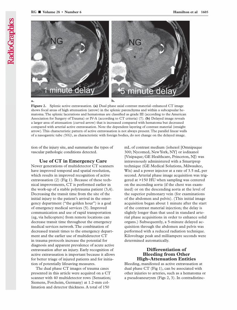
Figure 2. Splenic active extravasation. (a) Dual phase axial contrast material–enhanced CT image shows focal areas of high attenuation (arrow) in the splenic parenchyma and within a subcapsular he-matoma. The splenic lacerations and hematomas are classified as grade III (according to the American Association for Surgery of Trauma) or IV-A (according to CT criteria) (7). (b) Delayed image reveals a larger area of attenuation (curved arrow) that is increased compared with hematoma but decreased compared with arterial active extravasation. Note the dependent layering of contrast material (straight arrow). This characteristic pattern of active extravasation is not always present. The parallel linear walls of a nasogastric tube (NG), as characteristic with foreign bodies, do not change on the delayed image.
tion of the injury site, and summarize the types of vascular pathologic conditions detected.
Use of CT in Emergency CareNewer generations of multidetector CT scanners have improved temporal and spatial resolution, which results in improved recognition of active extravasation (2) (Fig 1). Because of these tech-nical improvements, CT is performed earlier in the work-up of a stable polytrauma patient (3,4). Decreasing the transit time from the site of the initial injury to the patient’s arrival in the emer-gency department (“the golden hour”) is a goal of emergency medical services (5). Improved communication and use of rapid transportation (eg, via helicopters) from remote locations can decrease transit time throughout the emergency medical services network. The combination of decreased transit times to the emergency depart-ment and the earlier use of multidetector CT in trauma protocols increase the potential for diagnosis and apparent prevalence of acute active extravasation after an injury. Early recognition of active extravasation is important because it allows for better triage of injured patients and for initia-tion of potentially lifesaving measures.
The dual phase CT images of trauma cases presented in this article were acquired on a CT scanner with 40 multidetector rows (Sensation; Siemens, Forcheim, Germany) at 1.2-mm col-limation and detector thickness. A total of 150
mL of contrast medium (iohexol [Omnipaque 300; Nycomed, New York, NY] or iodixanol [Visipaque; GE Healthcare, Princeton, NJ) was intravenously administered with a Smartprep technique (GE Medical Solutions, Milwaukee, Wis) and a power injector at a rate of 3.5 mL per second. Arterial phase image acquisition was trig-gered at >150 HU when sampling was centered on the ascending aorta (if the chest was exam-ined) or on the descending aorta at the level of the superior pulmonary vein (for examinations of the abdomen and pelvis). (This initial image acquisition began about 1 minute after the start of the contrast material injection; the delay is slightly longer than that used in standard arte-rial phase acquisitions in order to enhance solid organs.) Subsequently, a 5-minute delayed ac-quisition through the abdomen and pelvis was performed with a reduced radiation technique. Kilovoltage peak and milliampere seconds were determined automatically.
Differentiation of Bleeding from Other
High-Attenuation EntitiesBleeding, manifested as active extravasation at dual phase CT (Fig 1), can be associated with other injuries to arteries, such as a hematoma or a pseudoaneurysm (Figs 2, 3). In contradistinc-

1606 October Special Issue 2008 RG ■ Volume 28 • Number 6
Table 1 Characteristics That Distinguish Active Extravasation from Pseudoaneurysm
Distinguishing Characteristics Active Extravasation Pseudoaneurysm
Edges Ill-defined DefinedShape Commonly a jet (linear or layering);
may be a diffuse or focal area of hy-perattenuation
Often round or oval; possible neck that adjoins the finding to the adja-cent artery
Delayed appearance Increased attenuation or size of hema-toma; possible layering
Less apparent on delayed images; in isolation, no change in hematoma
Management Urgent embolization or surgery is re-quired if significant injury is present
Urgent or ambulatory embolization or surgery is required if significant injury is present
tion to active extravasation, an isolated pseudo-aneurysm is contained by connective tissue or the vessel wall (ie, the adventitia). Therefore, a pseudoaneurysm is likely to be adjacent to a vessel and does not enlarge or increase in at-tenuation as the contrast material washes out of the arterial system on 5-minute delayed images. The adventitia or connective tissue contains the contrast agent under pressure and forces the pool into a round or oval shape (1), which has a well-defined edge on CT images.
With vascular active extravasation, the con-trast-enhanced blood mixes with the fresh and clotted blood already present within the hema-
Figure 3. Retroperitoneal pseudoaneurysm. Dual phase axial contrast-enhanced CT images show a small round area of high attenuation (solid arrow) within a left retroperitoneal hematoma that fades on the delayed image (b). The high-attenuation area does not expand on the delayed image (cf Fig 2b), a pattern that is more suggestive of a pseudoaneurysm. Dashed arrow indicates delayed contrast material excretion in the left ureter.
toma. The mixing of these viscous fluids creates high-attenuation shapes that initially appear like a jet or fountain, with a tapered edge, or like spiraling eddy currents, with ill-defined edges. In one study, these findings were described as a jet (42% of cases), “diffuse density in hema-toma” (37%), and “focal density in hematoma” (21%) (6). The diffuse pattern represents spread of contrast material into the hematoma, which causes it to enhance and which may be more apparent on delayed images. The focal pattern represents either a settling of contrast material within focal dense layers of blood and contrast medium or a focal collection of contrast mate-rial that cannot diffuse because of clotted blood or limited space.

RG ■ Volume 28 • Number 6 Hamilton et al 1607
Active extravasation can be differentiated from other high -attenuation entities, such as bone frag-ments, foreign bodies, and other forms of vessel injury, on the basis of imaging characteristics seen at dual phase CT. Both pseudoaneurysm and ac-tive extravasation change in appearance on delayed images, compared with their characteristics on arterial images. Bone fragments or dense foreign bodies have high attenuation, but their appear-ance, unlike that of vessel injury, does not change
Figure 4. Combined hepatic pseudoaneurysm and active extravasation. (a, b) Initial 1-min-ute delayed image (a) shows a large nonenhancing area within the left hepatic lobe, a finding that represents a grade III laceration (7), and a central oval area of high attenuation (arrow), which represents a pseudoaneurysm. The high attenuation decreases on the 5-minute delayed image (b). (c) Coronal reformatted image reveals a small jet of active extravasation (curved arrow) inferior to the pseudoaneurysm (straight arrow). Both findings and the enhancement of the hematoma within the laceration are better seen with the coronal reformation from the initial image data after 1-minute delay compared with the 5-minute delayed image.
on delayed images. One must be careful not to
mistake volume averaging on thicker delayed im-ages or a change in patient position between the arterial and delayed acquisitions for a change in attenuation or size related to vascular injury.
Delayed imaging is an important tool for confirming vessel injury if it cannot be differen-tiated on the basis of shape, edge (ie, well- or ill-defined), location, and attenuation differences (Table 1). Delayed imaging is especially helpful in patients with multiple injuries (Fig 4).
The selection of treatment for both active extravasation and pseudoaneurysms depends on clinical factors and injury size, number, and location (Figs 5, 6). Not all injuries must be treated. Expectant management may be used for minor injuries such as small pseudoaneurysms or those amenable to treatment by direct pres-sure. However, when significant injuries are present (especially in solid organs such as the spleen), expectant management yields poor pa-tient outcome (2,7).
TeachingPoint

1608 October Special Issue 2008 RG ■ Volume 28 • Number 6
Figure 5. Splenic pseudoaneurysms with endovas-cular repair. (a–d) One-minute delayed axial CT im-ages of different levels of the spleen (a, b) and initial (c) and 10-second delayed (d) selective splenic an-teroposterior angiograms show multiple large periph-eral pseudoaneurysms (arrows) in a grade IV injury (7). Arrows in the axial CT images (solid in a; dashed in b) correspond to those in the angiogram (c). The delayed image (d) shows washout of the round peri-arterial collections, with no focal collection remaining (ie, there is no active extravasation). (e) Angiogram demonstrates technically successful coil embolization of the distal splenic artery.

RG ■ Volume 28 • Number 6 Hamilton et al 1609
Figure 6. Small splenic pseudoaneurysm. (a, b) Small single pseudoaneurysm (arrow) in a grade II splenic laceration is seen on the initial axial 1-minute delayed CT image (a) but is not seen on the 5-minute delayed image (b). (c) Subsequent anteroposterior subselective splenic angiogram demonstrates the small (4-mm) pseudoaneurysm (arrow) in the superior portion of the splenic parenchyma. In a case such as this one, some interventional radiologists may elect to monitor the patient for enlargement of the pseudoaneurysm (cf Fig 5, which illustrated a case that required mandatory treatment). However, because this particular patient worked in emergency services, participated in high-impact sports, and thus had higher than average risk of re-injury, he underwent subselective coil embolization. (d) Angiogram demonstrates technically successful coil embo-lization of the distal splenic artery.

1610 October Special Issue 2008 RG ■ Volume 28 • Number 6
Use of Attenuation to Determine Bleeding Source
Active extravasation is apparent when the attenu-ation is greater than that of clotted blood (70–90 HU). Most active extravasations will have attenu-ation values greater than 100 HU (7). The atten-uation differences (in Hounsfield units) between free fluid or blood and clotted blood are noted in Table 2. Very high attenuation measurements are usually from metal or calcium, but contrast mate-rial can also be concentrated in the urinary tract or in patients who are hypovolemic.
The area of highest attenuation on arterial phase CT images represents active extravasation, which is usually small compared with the larger hematoma of thrombus and unclotted blood mixed with contrast material. The attenuation of extravasated contrast material will often be simi-lar to that of its parent vessel (Fig 7). In addition, it is unlikely to have higher attenuation than that of the bleeding source because the contrast mate-rial is diluted in a fluid collection, such as a he-matoma or urinoma. Depending on the timing of image acquisition in relation to the bolus admin-istration, the attenuation values will vary for the arterial, venous, portal venous, and urinary tract systems. Table 3 gives idealized measurements of attenuation for various vascular compartments seen on arterial phase CT images. Attenuation
values may be inaccurate for differentiating the bleeding source for small injuries because of sam-pling issues or because of insufficient differences in attenuation between the various vascular and organ compartments. In practice, there are large
Figure 7. Bleeding into the extraperitoneal space of Retzius. (a) Axial pelvic CT image shows active extravasation (curved arrow) adjacent to the inguinal vessels, within the space of Retzius. (b) On the delayed image, the contrast material extravasation and hematoma expand (curved arrow). Based on location, the differential diagnosis includes iliac or inguinal vessel injury versus extraperitoneal bladder injury; however, no contrast material was seen in the bladder on the initial images. On the delayed image, volume averaging is unchanged for bone fragments (straight ar-row in a and b) of the right superior pubic ramus.
Table 2 Attenuation Measurements of Hematoma and Other CT Findings in Vascular Trauma
Fluid Type Attenuation (HU)
Free fluid 00–15Free blood 20–40Clotted blood or he-
matoma40–70
Active extravasation Often within 10 HU of adjacent major vessel source
Source.—Reference 6.
Table 3 Ideal Attenuation Measurements of Bleeding Vessels in Arterial Phase CT Images
Bleeding Source Attenuation (HU)
Hematoma with active extravasation
220 ± 5
Nearest artery 230 ± 5Inferior vena cava 180 ± 5Portal vein 150 ± 5

RG ■ Volume 28 • Number 6 Hamilton et al 1611
standard deviations of measurements, especially for small extravasations. Some extravasation may be apparent on the delayed images only because a hematoma or urinoma enhances.
There are other factors, such as fluid char-acteristics, that influence visualization of ac-tive extravasation. Flow—that is, the amount of contrast material extravasated over a given time—depends on physical properties of fluids. These properties include the pressure difference (blood pressure, potential space), the size of the
Figure 8. Liver injury and uterine artery injury ver-sus placental or uterine rupture. (a) Coronal CT image shows a grade IV liver injury (straight arrow) (7) and ac-tive extravasation (curved arrow) from a uterine artery injury. (b) Axial CT image also demonstrates the active extravasation (arrow). Use of multidetector CT allowed the uterine artery injury to be distinguished from a pos-sible placental or uterine rupture in a patient with a left vertex third trimester intrauterine pregnancy. In pregnant patients with significant blunt abdominal trauma, poten-tial lifesaving diagnostic studies should not be delayed because of potential radiation risk to the fetus.
disruption in the vessel wall (extent of injury, clotting factors), attenuation of contrast material (quality and timing of injection), and viscosity of blood (anemia or hypo- or hypercoaguable state). There are a host of other factors beyond the scope of this article to consider in the man-agement of a trauma patient, including stability of the patient, available resources, concomitant injuries, and medical condition, that also may affect whether a patient is imaged (Fig 8).
Types of Vessel InjuryUnderstanding the type of injury and its loca-tion can help determine which injuries are likely to rebleed. As the spatial and temporal resolu-tion of CT improves, its potential increases for depicting pathologic entities (eg, dissection, pseudoaneurysm, spasm, arteriovenous fistula, active extravasation, and multiple vessel injury) that are confirmed with surgery or other inter-ventions. Signs of vascular injury seen at CT include hematoma and contrast material that surrounds the injured vessel. Other clues to the location of vessel injury include lack of vascular enhancement (caused by occlusion or spasm), vessel irregularity, caliber change (such as oc-curs with pseudoaneurysm), and an intimal flap (which signifies dissection) (8). Some imaging findings are specific for a particular type of ves-sel injury, but most signs overlap with those of other types of vessel injury or pathologic condi-tions and cannot be used to exclude diagnoses. For instance, an intraluminal flap is pathogno-monic of dissection, but its presence could be oc-cult because of occlusion, spasm, or dissection, all of which would prevent contrast material from filling the occluded vessel.
The “contrast extravasation sign” seen at dual phase CT reportedly has greater than 95% ac-curacy, negative predictive value, and specificity with 80%–97% sensitivity and positive predictive
PointTeaching
PointTeaching

1612 October Special Issue 2008 RG ■ Volume 28 • Number 6
on the delay between examinations, it is antici-pated that some cases of active extravasation at CT may demonstrate other evidence of vascular injury but not contrast material extravasation at the time of angiography. Given the nonselective nature of vessel enhancement and the relatively long injection time used in dual phase CT, it is not possible to definitively diagnose the early draining vein seen in an arteriovenous fistula at CT (Fig 9). Ultimately, the role of CT may be to locate the bleeding vessel quickly in polytrauma patients. The final diagnosis as to the type of ves-sel injury may not be as critical to final patient management, as the latter may evolve with time. Treatment decisions will be based on findings from subsequent angiography or surgery.
In the triage of polytrauma patients, active extravasation represents current bleeding and is a better indicator of potential for continued bleeding or rebleeding than other forms of ves-sel injury that have not yet bled or have stopped bleeding.
Figure 9. Arteriovenous fistula. (a) Arterial phase axial CT image reveals a small pseudoaneurysm (dashed arrow) that is adjacent to the draining splenic vein (solid arrow). (b) Delayed image (ob-tained 2 cm inferior to a) shows active extravasation layering into a subcapsular hematoma (arrow). This finding was seen only on delayed images. (c) Sub-selective splenic arteriogram reveals the pseudoan-eurysm (dashed arrow) with early opacification of a draining vein (solid arrow), a finding that indicates an arteriovenous fistula. Although the proximity of the draining vein to the pseudoaneurysm suggests this diagnosis, it cannot be made with certainty from the CT findings. No active bleeding was seen at angiography.
value for identifying patients with pelvic fractures who require embolization to control bleeding (9,10). However, angiographic findings positive for vessel injury range from 43% to 64% and increase to only 56%–74% in patients with he-modynamic instability. These results mean that even in hemodynamically unstable patients with evidence of vessel injury at CT, no bleeding will be found at angiography in 26%–44% of patients with abdominal and pelvic trauma (1,11,12). In most cases, the lack of angiographically evi-dent bleeding can be attributed to the presence of venous or bone bleeding. In some cases, the findings vary because of inherent differences in the imaging modalities; the evaluation of vessel injury; and the timing of the performance of CT, angiography, and surgery. For example, a pseudo-aneurysm seen at CT may bleed before angiogra-phy is performed, leading to the finding of vaso-spasm and vessel truncation. Some patients who are actively bleeding will stop bleeding before a second procedure is performed because of the lifesaving assistance of blood transfusion, direct pressure, orthopedic fixation, or time. Depending

RG ■ Volume 28 • Number 6 Hamilton et al 1613
The spleen is a frequently injured vascular organ with a large adjacent potential space in the intraperitoneal cavity. Some treatment algo-rithms recommend use of follow-up CT to detect pseudoaneurysms not seen at initial imaging in patients with conservatively managed splenic in-jury. This follow-up may be deferred for younger individuals (<55 years old) with grade I injury. If a pseudoaneurysm (or evidence of other vessel injury) is demonstrated at any point, angiography is recommended (13). Management options for active extravasation from splenic blunt trauma include arteriography and possible selective or subselective splenic artery embolization versus surgical splenectomy. In a prospective study of polytrauma patients, the site of contrast material extravasation did not correlate with the eventual treatment modality, duration of intensive care hospitalization, or final patient outcome (14). Pa-tients with intraperitoneal extravasation required more aggressive transfusion with packed red blood cells and had a higher mortality rate in the first 24 hours (14). A multicenter study demon-strated that embolization or surgery was required in 83% of the patients with active extravasation in the spleen, compared with 30.3% for patients who had splenic injury but no active extravasa-tion (as seen at CT) (2). Active extravasation is a good predictor of the need for intervention in abdominal and pelvic trauma, and it is even more indicative in cases of splenic injury.
Figure 10. Demonstration of hemorrhage in potential spaces of the liver and abdomen. Anterior (a) and poste-rior (b) coronal 1-minute delayed CT images demonstrate extensive laceration of the right hepatic lobe with partial devascularization of segment VIII. Active extravasation is seen within the parenchyma (curved arrow in a), and it extends to the subcapsular area (arrowheads in b). Note the hematoma is also intraperitoneal (straight solid arrow in a) and perivascular (dashed arrow) with hypoattenuation on both sides of the portal triads.
Tissue Planes and LocationPressure gradient is an important concept that relates to anatomic planes (eg, serosal and peri-toneal). Understanding the anatomic planes and vascular variants is important because anatomy can be distorted by a large fluid collection. An example is illustrated in Figure 10, in which liver intraparenchymal hematomas will tamponade at a certain size because of compression of tissue unless it can decompress into a potential space. These potential spaces for the liver can be re-membered with the pneumonic RIPS: retroperi-toneal (bare area of segment VII), intraperitoneal, perivascular (second to portal triads, can be peri-portal and peribiliary), subcapsular.
Proximity of the vessel or organ injury to these potential spaces indicates more hemodynamic risk. Active extravasation from a vessel can occur only if the pressure in the vessel is greater than that of the surrounding tissues. A higher pressure vessel (ie, arterial) is at more risk for significant or recurrent bleeding (1).
The concept of pressure also applies to indi-vidual vessel injuries. In cases of aortic trauma, the use of antihypertensive medications has been shown to delay mortality (15). In cases of abdominal trauma, hypotension from hypovo-lemia and shock, rather than hypertension, is a more common problem. For blunt abdominal

1614 October Special Issue 2008 RG ■ Volume 28 • Number 6
directed embolization, can be performed if the patient continues to bleed or is unstable. The necessity of embolization or celiotomy for arte-rial bleeding after pelvic stabilization varies from 15% to 66% (2,10,12).
Extraperitoneal active extravasation (Fig 12) is unlike peritoneal and retroperitoneal bleeding be-cause it may be amenable to direct pressure to stop the bleeding. There is limited potential space be-tween the muscles of the thoracoabdominal wall. Similar to a hernia, a hematoma can dissect into a potential space such as inguinal, femoral, and spi-nal canals and cause significant hemorrhage.
Other Findings in Vascular Injury
Blood closest to the injury site has more time to retract, forming higher-density clotted blood. This phenomenon is known as the “sentinel clot
Figure 11. Pelvic active extravasation. (a, b) Initial 1-minute delayed CT image (a) shows a faint small area of high attenuation (arrow) that spreads into hematoma on the delayed image (b), a finding that indicates active extravasation. (c) Frontal selective right common iliac angiogram reveals no vessel injury. Other possible sources of bleeding include the right common iliac vein or bone bleeding from an unstable open book pelvic injury with a comminuted sacral fracture that required fixation. No further angiographic treatment is indicated.
trauma patients who are stable or who have been stabilized with 1–2 L of intravenous fluids, use of angiography is as efficacious as surgery (16). In some series, the spleen is reported as the most common location of active extravasa-tion; in other series, active extravasation has been observed more often in the liver and pelvis (1,2,14).
The patient in Figure 10 had extensive liver injury and was taken to surgery. In some severely injured patients who are not surgical candidates, angiographic embolization may be used to con-trol bleeding and has improved patient survival. However, liver-related complications of emboliza-tion can cause significant morbidity; these com-plications included hepatic necrosis, abscess, and bile leakage, which occurred in 58% of the cases in one series (17).
Pelvic bleeding often occurs secondary to unstable pelvic fractures and is more often ret-roperitoneal compared with bleeding that occurs in cases of stable pelvic fractures (10) (Fig 11). Treatment with pelvic fixation has a tampon-ade effect on the bleeding vessels, especially the veins. Further interventions, including catheter
sign” (Fig 13) (18). The sentinel clot sign is an
TeachingPoint

RG ■ Volume 28 • Number 6 Hamilton et al 1615
injury are not present. The sentinel clot sign is present in every patient with hemoperitoneum, whereas fewer injured patients demonstrate a positive sign of vascular injury, such as active ex-travasation or pseudoaneurysm.
It is also important to assess the intravascu-lar volume in patients who are bleeding, espe-cially younger patients. In these patients, blood pressure is not an accurate initial indicator of acute decompensation. Younger patients have greater vascular reserve, which helps maintain their blood pressure despite a low intravascular volume; thus, they may not demonstrate hy-potension until profound bleeding has occurred with subsequent acute decompensation. The only clinical manifestation may be tachycardia, which could also be attributed to pain. The im-aging findings may be helpful in those cases in which the need for intravenous fluids and blood transfusion is anticipated (13). The earliest sign of decreased vascular volume affects the low-pressure vessel and can manifest as a flattened contour of the inferior vena cava (Fig 13). A further decrease of vascular volume leads to de-velopment of a hypoperfusion complex, which is characterized by decreased enhancement of the spleen and pancreas and increased enhancement
important clue for locating the bleeding source when other more localizing findings of vessel
Figure 12. Extraperitoneal bleeding. Initial 1-minute delayed axial CT image (a) demonstrates a high-atten-uation active extravasation from the intercostal artery (arrow) that spreads on the 5-minute delayed image (b). The vascular injury is secondary to a fracture of the right tenth posterior rib. This extraperitoneal hematoma is contained in the chest wall and is amenable to treatment by direct pressure, often without further interven-tion. Extraperitoneal extravasation can still cause hemodynamically significant bleeding, especially in potential spaces of the extremities or nervous system.
Figure 13. Sentinel clot and decreased vascular volume. Axial CT image shows hemoperitoneum sur-rounding the liver and spleen. The finding of a flat-tened inferior vena cava (arrow) indicates the presence of decreased intravascular volume (especially since many trauma patients are already receiving intravenous fluids before the imaging evaluation begins). Note the differences in attenuation of the hemoperitoneum ad-jacent to the liver and the spleen. By using the sentinel clot sign, the radiologist localized the bleeding to the patient’s left side (image right side).

1616 October Special Issue 2008 RG ■ Volume 28 • Number 6
5. Thomas SH, Harrison TH, Buras WR, Ahmed W, Cheema F, Wedel SK. Helicopter transport and blunt trauma mortality: a multicenter trial. J Trauma 2002;52(1):136–145.
6. Willmann JK, Roos JE, Platz A, et al. Multidetector CT detection of active hemorrhage in patients with blunt abdominal trauma. AJR Am J Roentgenol 2002;179(2):437–444.
7. Mirvis S, Shanmuganathan K. Imaging in trauma and critical care. Philadelphia, Pa: Saunders, 2003.
8. LeBlang SD, Nunez DB Jr. Noninvasive imaging of cervical vascular injuries. AJR Am J Roentgenol 2000;174(5):1269–1278.
9. Pereira SJ, O’Brien DP, Luchette FA, et al. Dy-namic helical computed tomography scan accu-rately detects hemorrhage in patients with pelvic fracture. Surgery 2000;128(4):678–685.
10. Stephen DJ, Kreder HJ, Day AC, et al. Early detec-tion of arterial bleeding in acute pelvic trauma. J Trauma 1999;47(4):638–642.
11. Chiara O, Cimbanassi S, Castelli F, et al. Protocol-driven approach of bleeding abdominal and pelvic trauma. World J Emerg Surg 2006;1:1–7.
12. Agolini SF, Shah K, Jaffe J, Newcomb J, Rhodes M, Reed JF 3rd. Arterial embolization is a rapid and effective technique for controlling pelvic fracture hemorrhage. J Trauma 1997;43(3):395–399.
13. EMCrit.org. Abdominal trauma. http://emcrit.org /030–064/039-abd.trauma.htm. Accessed Septem-ber 1, 2008.
14. Lee K, Shin H. Prognosis of blunt abdominal trau-ma patients with contrast medium extravasation on computed tomography scan [abstr]. Crit Care 2007; 11(suppl 2):349.
15. Fabian TC, Davis KA, Gavant ML, et al. Prospec-tive study of blunt aortic injury: helical CT is diagnostic and antihypertensive therapy reduces rupture. Ann Surg 1998;227(5):666–676; discus-sion 676–677.
16. Hagiwara A, Murata A, Matsuda T, Matsuda H, Shimazaki S. The usefulness of transcatheter arte-rial embolization for patients with blunt polytrauma showing transient response to fluid resuscitation. J Trauma 2004;57(2):271–276; discussion 276–277.
17. Mohr AM, Lavery RF, Barone A, et al. Angiographic embolization for liver injuries: low mortality, high morbidity. J Trauma 2003;55(6):1077–1081; discus-sion 1081–1082.
18. Orwig D, Federle MP. Localized clotted blood as evidence of visceral trauma on CT: the sentinel clot sign. AJR Am J Roentgenol 1989;153(4): 747–749.
of the bowel wall, vasculature, kidneys, and pos-sibly the adrenal glands (7). Imaging findings can suggest a patient’s pending instability before it manifests clinically, thus further expediting the performance of a lifesaving intervention.
ConclusionsMultidetector CT with dual phase protocol is an important part of the patient work-up for blunt abdominal trauma. Timely diagnosis, dif-ferentiation of vascular injuries from other find-ings of trauma, signs of depleted intravascular volume, and localization of vascular injury are important to convey to the interventional ra-diologist or surgeons. More accurate diagnosis can be accomplished through attention to shape, attenuation, and evolution over time of areas of high attenuation, in addition to high-quality injections of contrast material and display tech-niques. Understanding the physical properties of bleeding may help radiologists predict those cir-cumstances that put patients at significant risk for rebleeding after vessel injury and thus aid in determining the necessity for interventional or surgical procedures.
References 1. Ryan MF, Hamilton PA, Chu P, Hanaghan J. Active
extravasation of arterial contrast agent on post-trau-matic abdominal computed tomography. Can Assoc Radiol J 2004;55(3):160–169.
2. Yao DC, Jeffrey RB Jr, Mirvis SE, et al. Using con-trast-enhanced helical CT to visualize arterial extra-vasation after blunt abdominal trauma: incidence and organ distribution. AJR Am J Roentgenol 2002; 178(1):17–20.
3. Sampson MA, Colquhoun KB, Hennessy NL. Computed tomography whole body imaging in multi-trauma: 7 years experience. Clin Radiol 2006;61(4):365–369.
4. Hessmann MH, Hofmann A, Kreitner KF, Lott C, Rommens PM. The benefit of multislice CT in the emergency room management of polytraumatized patients. Acta Chir Belg 2006;106(5):500–507.
RG Volume 28 • Volume 6 • October 2008 Hamilton et al
Multidetector CT Evaluation of Active Extravasation in Blunt Abdominal and Pelvic Trauma Patients Jackson D. Hamilton, MD, et al
Page 1604 The classic pattern of active extravasation at dual phase CT is a jet or focal area of hyperattenuation within a hematoma on initial images that fades into an enlarged, enhanced hematoma on delayed images. Page 1607 Bone fragments or dense foreign bodies have high attenuation, but their appearance, unlike that of vessel injury, does not change on delayed images. Page 1611 Some extravasation may be apparent on the delayed images only because a hematoma or urinoma enhances. Page 1611 Other clues to the location of vessel injury include lack of vascular enhancement (caused by occlusion or spasm), vessel irregularity, caliber change (such as occurs with pseudoaneurysm), and an intimal flap (which signifies dissection). Page 1614 Blood closest to the injury site has more time to retract, forming higher-density clotted blood. This phenomenon is known as the "sentinel clot sign."
RadioGraphics 2008; 28:1603–1616 • Published online 10.1148/rg.286085522 • Content Codes: