
Abdomen: Introduction · Antrolateral Abdominal Wall This extended from the thoracic cage to the...
41
Abdomen: Introduction Prof. Oluwadiya KS www.Oluwadiya.com
Transcript of Abdomen: Introduction · Antrolateral Abdominal Wall This extended from the thoracic cage to the...

Abdomen: Introduction
Prof. Oluwadiya KS
www.Oluwadiya.com
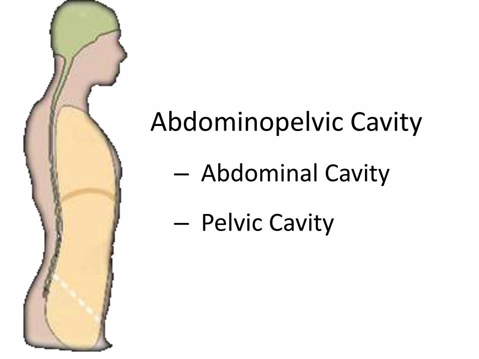
Abdominopelvic Cavity
– Abdominal Cavity
– Pelvic Cavity

Abdomen
• Extends from the inferior margin of the thorax to the superior margin of the pelvis and the lower limb

The abdominal wall
• Two parts
i. Bony
ii. Muscular

The skeletal abdominal wall
• Posteriorly: Five lumbar vertebrae and their intervening intervertebral discs
• Inferiorly: The superior expanded parts of the pelvic bones (Iliac wings)
• Superiorly: Bony components of the inferior thoracic wall including the costal margin, rib XII, the end of rib XI and the xiphoid process

The muscular abdominal wall
Posteriorly:
• Lateral to the vertebral column: Quadratus lumborum, psoas major, and iliacus muscles
Laterally:
• Transversus abdominis, internal oblique, and external oblique
Anteriorly:
• Rectus abdominis
• The muscles are reinforced by strong fascia and aponeurosis

Surface Anatomy

Antrolateral Abdominal Wall
This extended from the thoracic cage to the pelvis and bounded :
– Superiorly
• 7th through 10th costal cartilages and andxiphoid process
– Inferiorly
• Inguinal ligaments and the pelvic bones.
The wall consists of skin, subcutaneous tissues (fat), muscles, deep fascia and parietal peritoneum.

Abdominal wall
Anterolateral abdominal wall
Posterior abdominal wall

LAYERS
• Skin
• Superficial fascia
• Deep fascia
• Muscles
• Transversalis fascia
• Extraperitoneal fascia
• Peritoneum

Antrolateral Abdominal WallFascia & Subcutaneous Tissues
• The subcutaneous tissues over most of the wall consists of layer of connective tissues that contains a variable amount of fat.
• In the inferior part of the wall , the subcutaneous tissue is composed of two layers
– Fatty superficial layer (Camper’s fascia)– Membranous deep layer (Scarpa’s fascia)

Superficial fascia
• Camper’s fascia
• Scarpa's fascia

MUSCLES
Anterior Group Lateral Group
•Rectus Abdominis
•Pyramidalis
•External Oblique
•Internal Oblique
•Transversus
Two Vertical MusclesThree Flat Muscles with strong sheet-likeaponeurosis

External oblique
Table
Origin Insertion Innervation Function
Muscular slips
from the outer
surfaces of
the lower
eight ribs (ribs
5-12)
Lateral lip of
iliac crest;
aponeurosis
ending in
midline raphe
(linea alba)
Anterior rami of
lower six thoracic
spinal nerves (T7
to T12)
Compresses
abdominal
contents; both
muscles flex
trunk; each
muscle bends
trunk to same
side, turning
anterior part
of abdomen to
opposite side

External Oblique
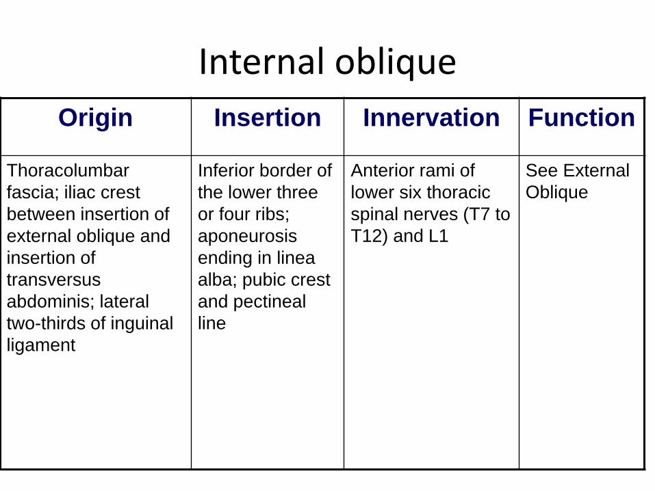
Internal oblique
Origin Insertion Innervation Function
Thoracolumbar
fascia; iliac crest
between insertion of
external oblique and
insertion of
transversus
abdominis; lateral
two-thirds of inguinal
ligament
Inferior border of
the lower three
or four ribs;
aponeurosis
ending in linea
alba; pubic crest
and pectineal
line
Anterior rami of
lower six thoracic
spinal nerves (T7 to
T12) and L1
See External
Oblique

Internal Oblique

Transverse abdominis
Origin Insertion Innervation Function
Thoracolumbar
fascia; medial
lip of iliac crest;
lateral one-third
of inguinal
ligament; costal
cartilages lower
six ribs (ribs 7-
12)
Aponeurosis
ending in linea
alba; pubic crest
and pectineal line
Anterior rami of
lower six thoracic
spinal nerves (T7
to T12) and L1
Compresses
abdominal
contents

Transversus Abdominis

Rectus abdominis
Origin Insertion Innervation Function
Pubic crest,
pubic tubercle,
and pubic
symphysis
Costal cartilages
of ribs 5-7;
xiphoid process
Anterior rami of
lower seven
thoracic spinal
nerves (T7 to T12)
Compress
abdominal
contents; flexes
vertebral
column; tenses
abdominal wall

RECTUS ABDOMINIS
• Tendinous Intersection (3)
• Linea Semilunaris

The rectus sheathArcuate line: is the lower border of the posterior aponeurotic part of the rectus sheath. The inferior epigastric artery and vein enter the sheath, pass upwards and anastomose withthe superior epigastric vessels.

SUPERFICIAL ARTERIES
• Lateral
– Posterior intercostal a.
– Subcostal a.
– Lumbar a.
• Median
– Epigastric a.
– hypogastric a.
• Inferior
– Superficial epigastric a.
– Superficial iliac a.

Superficial veins
subclavian
femoral
paraumbilical
Superficial epigastric
Superficial circumflex iliac
thoracoepigastric
lateral thoracic
portal


Caput Medusae(Medusa Head )

Lymphatic Drainage
Anterior →Intercostal Lymphatic Nodes Parasternal Lymphatic Nodes
Middle → Lumbar Lymphatic Nodes
Lower → External Iliac Lymphatic Nodes

INNERVATIONS
• Intercostal Nerves
T7-T11
• Subcostal nerve
(T12)
• L1

Innervations
• Intercostal n.
– Anterior cutaneous branch
– Lateral cutaneous branch

• T7-11: thoracoabdominal n.
• T12: Subcoastal n.
• Iliohypogastric n.
• Ilioinguinal n.
• Genitofemoral n L1,2.
L1

Regions of the Abdomen
• Dividing the abdomen into regions helps in localization of abdominal signs and symptoms
• Two methods of dividing the abdomen into regions:
1. Nine regions: By means of two vertical and two horizontal lines
2. Four Quadrants: By means of one vertical and one horizontal lines, both passing through the umblicus.

Regions of the Abdomen
• Nine regions
• Divided by two horizontal lines:
i. Transpyloric line
ii. Trans tubercular (Trans-iliac) line
• Two Vertical lines
i. Rt. & Lt. Midclavicular lines

Regions of the Abdomen II

Anterior Abdominal WallFunctions
• Forms a strong expandable support.• Protects the abdominal viscera from injury
such as low below in boxing• Compresses the abdominal content• Helps to maintain or increase the
intraabdominal pressure.• Moves the trunk and help to maintain
posture.

Abdominal Incisions
An incision is defined as a cut made with knife
for surgical purposes.
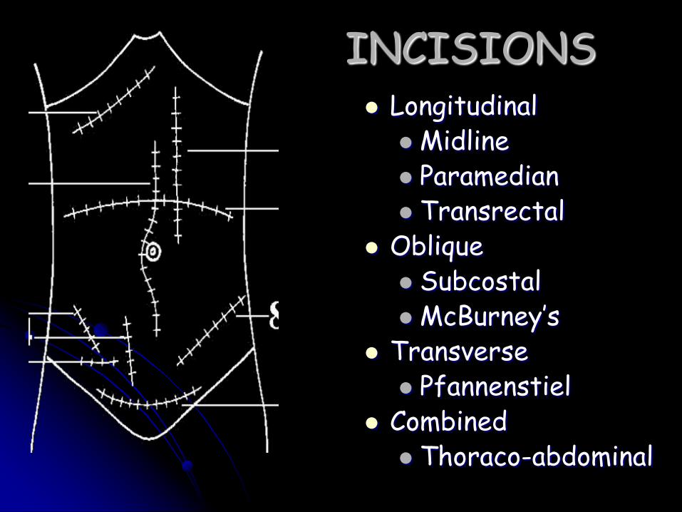
INCISIONS Longitudinal
Midline Paramedian Transrectal
Oblique SubcostalMcBurney’s
Transverse Pfannenstiel
Combined Thoraco-abdominal


Abdominal Hernia Orifices
• Hernia is defined as the protrusion of an organ through it’s containing wall.
• Abdominal hernias occurs because of weaknesses in the abdominal wall

Common Sites
• Inguinal Hernia
• Umbilical Hernia
• Femoral Hernia
• Incisional Hernia
Less common Herniao Epigastric Hernia
o Recurrent Hernia


The end


![Effects of diaphragm stretching on posterior chain muscle ... · xiphoid level was 2.48 [0.97 to 3.99], which shows significant differences in this outcome. The remaining between-group](https://static.fdocuments.net/doc/165x107/5f0501cd7e708231d410cbeb/effects-of-diaphragm-stretching-on-posterior-chain-muscle-xiphoid-level-was.jpg)


