ab139460 (Colorimetric) PDE Activity Assay Kit · Abcam PDE Activity Assay Kit (Colorimetric)...
28
Version 2 Last Updated 29 January 2019 ab139460 PDE Activity Assay Kit (Colorimetric) Instructions for Use For the screening of inhibitors and modulators of cyclic nucleotide phosphodiesterase activity. View kit datasheet: www.abcam.com/ab139460 (use www.abcam.cn/ab139460 for China, or www.abcam.co.jp/ab139460 for Japan) This product is for research use only and is not intended for diagnostic use.
Transcript of ab139460 (Colorimetric) PDE Activity Assay Kit · Abcam PDE Activity Assay Kit (Colorimetric)...

Version 2 Last Updated 29 January 2019
ab139460PDE Activity Assay Kit (Colorimetric)
Instructions for Use
For the screening of inhibitors and modulators of cyclic nucleotide phosphodiesterase activity.
View kit datasheet: www.abcam.com/ab139460(use www.abcam.cn/ab139460 for China, or www.abcam.co.jp/ab139460 for Japan)
This product is for research use only and is not intended for diagnostic use.

1

Table of Contents
1. Principle of the Assay 3
2. Protocol Summary 5
3. Materials Supplied 7
4. Storage and Stability 8
5. Materials Required, Not Supplied 9
6. Sample Preparation 10
7. Assay Protocol 13
8. Data Analysis 19
9. Troubleshooting 22
2
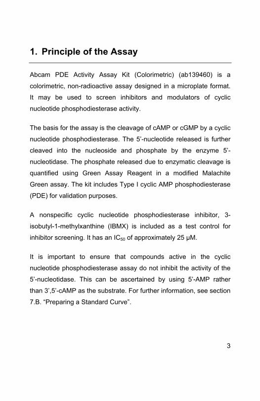
1. Principle of the Assay
Abcam PDE Activity Assay Kit (Colorimetric) (ab139460) is a
colorimetric, non-radioactive assay designed in a microplate format.
It may be used to screen inhibitors and modulators of cyclic
nucleotide phosphodiesterase activity.
The basis for the assay is the cleavage of cAMP or cGMP by a cyclic
nucleotide phosphodiesterase. The 5’-nucleotide released is further
cleaved into the nucleoside and phosphate by the enzyme 5’-
nucleotidase. The phosphate released due to enzymatic cleavage is
quantified using Green Assay Reagent in a modified Malachite
Green assay. The kit includes Type I cyclic AMP phosphodiesterase
(PDE) for validation purposes.
A nonspecific cyclic nucleotide phosphodiesterase inhibitor, 3-
isobutyl-1-methylxanthine (IBMX) is included as a test control for
inhibitor screening. It has an IC50 of approximately 25 μM.
It is important to ensure that compounds active in the cyclic
nucleotide phosphodiesterase assay do not inhibit the activity of the
5’-nucleotidase. This can be ascertained by using 5’-AMP rather
than 3’,5’-cAMP as the substrate. For further information, see section
7.B. “Preparing a Standard Curve”.
3

This kit is sold based on number of tests. A ‘test’ simply refers to a
single assay well. The number of wells that contain sample, control
or standard will vary by product. Review the protocol completely to
confirm this kit meets your requirements. Please contact our
Technical Support staff with any questions.
The assay offers the following advantages:
1) Non-radioactive
2) Convenient one step detection
3) Microplate format
4
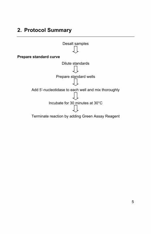
2. Protocol Summary
Desalt samples
Prepare standard curveDilute standards
Prepare standard wells
Add 5’-nucleotidase to each well and mix thoroughly
Incubate for 30 minutes at 30°C
Terminate reaction by adding Green Assay Reagent
5

Prepare time course/linearity assayDilute cAMP substrate
Add cAMP substrate to appropriate wells
Add assay buffer to each well
Add 5’-nucleotidase to each well and mix thoroughly
Dilute PDE enzyme
Add PDE enzyme to each well at 5 minute intervals
Terminate reaction by adding Green Assay Reagent
Prepare test sample/inhibitor assayPrepare samples containing PDE enzyme, substrate and test
compound. Include IBMX (optional)
Incubate samples at appropriate temperature and time
Terminate reaction by adding Green Assay Reagent
6

3. Materials Supplied
Item Quantity Storage
PDE Enzyme (from bovine brain) (lyophilized)
(4 U per vial)
5 vials -80°C
5’-Nucleotidase (5 kU/µL)
(from Crotalus atrox venom)
1 x 1 mL -80°C
3’,5’-cAMP Substrate (1mM) 1 x 2 mL -80°C
3’,5’-cGMP Substrate (1 mM) 1 x 2 mL -80°C
PDE Assay Buffer 1 x 40 mL -80°C
Green Assay Reagent 1 x 20 mL 4°C
5’-AMP Standard (100 µM) 1 x 1 mL -80°C
5’-GMP Standard (100 µM) 1 x 1 mL -80°C
PDE Inhibitor (IBMX) (200 µM) 1 x 200 µL -80°C
Desalting Column 1 4°C
Desalting Resin 1 x 1g 4°C
96-well Clear Microplate (½ Volume) 1 4°C
7

4. Storage and Stability
Please note that all components, with the exception of the Green
Assay Reagent, Desalting Column, Desalting Resin, PDE
Enzyme, and the 5’-nucleotidase can be stored at either -80°C
or -20°C.
The PDE Enzyme and 5’-Nucleotidase must be stored at -80°C.
Because its stability when frozen in solution is poor, the PDE
enzyme is provided as 5 lyophilized aliquots. For highest activity,
we recommend that each lyophilized aliquot be dissolved and
used for one day’s experiments (See Section 7.A. Reagent
Preparation).
The 5’-nucleotidase is used undiluted at 10 µL per well. It is
stable for up to 6 freeze/thaw cycles (snap freezing with liquid N2
or dry ice/ethanol), but it may be desirable to make several
aliquots if more numerous freeze/thaws are planned.
One U of 5’-nucleotidase will release 1 pmol phosphate per
minute from 200 μM 5’-AMP, at 30°C in a reaction buffer of
10 mM Tris-HCl, pH 7.4, 0.2 mM MgCl2.
One U of PDE enzyme = 1 nmol 3’ 5’-cAMP to 5’-AMP per
minute under the conditions of the linearity assay
8

The Green Assay Reagent is a highly sensitive phosphate
detection solution. Free phosphate present on labware and in
reagent solutions will greatly increase the background
absorbance of the assay. This is detected visually as a change
in color from yellow to green. Detergents used to clean labware
may contain high levels of phosphate. Use caution by either
rinsing labware with dH2O or employ unused plasticware.
5. Materials Required, Not Supplied
Microplate reader capable of measuring A620 to 3-decimal
accuracy.
Pipettes capable of pipetting 10-100 µL accurately.
Multi-channel pipetman capable of pipetting 100 µl (optional).
Ice bucket to keep reagents cold until use.
9
6. Sample Preparation
Note: The following procedures are intended only as a guideline. The
optimal experimental conditions will vary depending on the
parameters being investigated, and must be determined by the
individual user.
Desalting tissue samples by gel filtration
Note: This procedure is intended to remove excess phosphate and
nucleotides (which are slowly hydrolyzed to release free phosphate
in the presence of the Green Assay Reagent) in the high speed
supernatant (HSS) extract.
1. Rehydrate Desalting Resin in a 50 mL conical tube by adding
20 mL of phosphate free dH2O and vortexing briefly. Allow to set
for 4 hours at RT or overnight at 4°C.
2. Decant the dH2O carefully, then add fresh dH2O at a 1:1 ratio to
the rehydrated resin (~10 mL).
3. Add rehydrated resin to the Desalting Column to obtain a 5 mL
settled-bed volume (~5.5 cm bed height). Remove tip from
column and allow dH2O to drain by gravity.
4. Equilibrate column by adding 8 mL of assay buffer and allow to
drain by gravity.
10

5. Place column in a 15 mL centrifuge tube. Centrifuge at 800 x g
for 3 min at 4°C to displace column buffer. Discard flow-through
buffer.
6. Place column in a clean 15 mL centrifuge tube.
7. Add up to 350 μL sample to column.
8. Centrifuge at 800 x g for 3 min. Save extract flow-through. This
is the desalted cell lysate material to be tested for PDE activity
below.
9. Freeze sample immediately at -80°C.
Note: The effective removal of phosphate/nucleotides from the
extract should be tested qualitatively by adding 100 μL Green Assay
Reagent to 1 μL extract, and a separate sample of 1 μL dH2O. If no
phosphate/nucleotides are present, both samples should remain
yellow in color over a time period of 30 min @ RT. The development
of a visible green color indicates phosphate contamination, which
must be eliminated from the samples before proceeding further.
The rehydrated resin can be stored at 4°C until use. Please note that
the volume of Desalting Resin provided in this kit allows user to fill
the column twice (5mL settled-bed volume each). In theory, the
rehydrated resin can be reused for multiple times (or samples) *. The
column will lose efficiency in removing free phosphate gradually after
each use. The number of samples that can be desalted by the resin
will vary depending on the sample types and amount of phosphate
11

present in the samples. In general, an average of 5-7 samples can
be processed with each 5mL settled-bed volume of rehydrated resin,
ie an average of 10-14 samples can be desalted per kit.
*When reusing the rehydrated resin, the column should be rinsed
with 4-5 volumes of phosphate-free dH2O, and then equilibrated with
8 mL of a solution of 10mM Tris-hydrochloric acid, pH 7.4 before
loading the next sample.
12

7. Assay Protocol
A. Reagent Preparation1. Thaw assay buffer, 5’-nucleotidase, the 3’,5’-cAMP
substrate, IBMX inhibitor, 5’-AMP and/or 5’-GMP standard.
Store all on ice.
2. Prepare 20 U/mL solution of PDE by adding 200 µL of cold
assay buffer to one of the vials of lyophilized enzyme. Store
on ice. Note that each of the lyophilized PDE aliquots
provided are intended for use in one day’s assays only. The
enzyme solution may lose substantial activity from
freezing/thawing and frozen storage.
3. Warm Green Assay Reagent to room temperature.
B. Preparing a Standard Curve1. Prepare two dilutions in PDE Assay buffer, 75 μM and
50 μM, using either the 5’-AMP or 5’-GMP Standard. For
example, bring aliquots of 150 µL and 100 µL 5’-AMP
Standard (100 μM) and to 200 µL with assay buffer.
2. Using PDE Assay Buffer, prepare two sets of 1:1 serial
dilutions of the 75 μM and 50 μM 5’-AMP standard, plus an
assay buffer blank (40 µL per well). Concentrations of 75,
50, 37.5, 25, 18.75, 12.5, 6.25 μM correspond to 3, 2, 1.5,
1.0, 0.75, 0.50 and 0.25 nmol 5’-AMP or 5’-GMP (see
Table 1):
13

a) Add 80 µL of 75 μM 5’-AMP or 5’-GMP standard (Step 1) to well
A of assay plate and 80 µL of 50 μM standard to well B.
b) Add 40 µL 1X assay buffer to wells C through H.
c) Remove 40 µL from well A and add it to well C. Mix thoroughly
by pipetting up and down several times.
d) Remove 40 µL from well C and add it to well E. Mix well E
thoroughly and then remove 40 µL and discard.
e) Remove 40 µL from well B and add it to well D. Mix thoroughly
by pipetting up and down several times. Repeat this process
moving and mixing 40 µL from well D to F and then from F to G.
Remove and discard 40 µL from well G. DO NOT PROCEED TO
THE BLANK WELL ‘H’.
f) Add 10 µL of 5’-nucleotidase (undiluted, 5 kU/µL) to each well
and mix thoroughly.
g) Incubate at 30°C for 30 minutes.
h) Proceed to “Terminating Reactions” (Section 7.E).
14

Table 1. Example of standard curve and time course/linearity microplate samples.
Sample Well
5’-AMP/5’-GMP Standard Curve nmol
5’-AMP or 5’-GMP (Columns 1,2)
Time course Min. (Columns 3,4)
A 3.0 90
B 2.0 60
C 1.5 45
D 1.0 30
E 0.75 20
F 0.50 10
G 0.25 5
H 0 0
For highest accuracy, perform all samples in duplicate. See Figures 1 and 2 for example results.
15

C. Preparing a Time Course/Linearity Assay1. Dilute cAMP substrate to 0.5 mM with assay buffer.
2. Add 20 µL of cAMP substrate (0.5 mM) to appropriate wells.
The final substrate concentration will be 200 μM.
3. Add 15 µL of assay buffer to each well.
4. Add 10 µL of 5’-nucleotidase (undiluted, 5 kU/µL) per well.
5. Designate a reaction time to each well (e.g.: 90, 60, 45, 30,
20, 10, 5 and 0 min). See Table 1.
6. Equilibrate microplate to reaction temperature (e.g.: 30°C).
7. Prepare PDE enzyme at 20 U/mL (See Section 7.A. Reagent
Preparation). Dilute with assay buffer to 4 U/mL, making
enough for the assays planned. Each well will receive 5 µL.
Store dilution on ice.
8. Start reactions by addition of 5 µL of PDE enzyme. Total
PDE enzyme= 20 mU/well. Make the additions in the reverse
time order such that all incubations end at the same time
(e.g.: Add 90 min time pt. at t=0; add 5 min at t=85 min, etc.).
The total reaction volume=50 µL.
16

D. Preparing a Test Sample/Inhibitor Assay1. Prepare samples containing PDE enzyme, substrate and
test compound dissolved in PDE Assay Buffer as listed in
Table 2. Include the PDE Inhibitor if desired.
2. Incubate samples at appropriate temperature (e.g. 37°C)
and time (e.g.: 60 min).
Table 2: Example of Test Samples/Inhibitor Assay Microplate Samples
Substrate (0.5 mM)
PDE Assay Buffer
5’-Nase (5 kU/µL)
Test compound
PDE Enzyme
(4mU/ µL)
Control 20 µL 15 µL 10 µL 0 µL 5 µL
Test 20 µL 5 µL 10 µL 10 µL 5 µL
PDE Inhibitor
20 µL 5 µL 10 µL 10 µL 5 µL
3. To confirm that an apparent PDE inhibitor does not interfere
with the release of phosphate by 5’-nucleotidase, test
additional wells using 50 μM 5’-AMP or 5’-GMP standard
and the inhibitor(s) in question. Compare the results with the
50 μM standard curve well.
17
E. Terminating Reactions1. After incubating wells for desired duration, terminate
reactions by addition of 100 µL Green Assay Reagent.
Agitate plate or triturate wells gently to mix.
Note: Avoid production of air bubbles in the wells.
2. Allow color to develop for 20-30 minutes. Be careful to
assure samples spend approximately the same time with the
reagent before reading on the microplate reader.
3. Read OD620nm on microtiter-plate reader.
Note: Retain microtiter plate for future use of unused wells.
18

8. Data Analysis
A. 5’-AMP or 5’-GMP Standard Curve1. Plot standard curve data as OD620nm versus nmol 5’-AMP or
5’-GMP (see Fig. 1).
2. Fit a line to the plotted data using an appropriate linear
regression program.
3. Rearrange the equation for best-fit line to solve for nmol of
5’-AMP or 5’-GMP in terms of OD620nm.
5’-AMP released = (OD620nm – y-intercept)/slope
SAMPLE CALCULATION:Best-fit eqn.: OD620nm = 0.232(nmol 5'-AMP) + 0.0709
Rearranged eqn.: nmol 5’-AMP = (OD620nm – 0.0709)/ 0.232
Example: An unknown produces an OD620nm = 0.400
5’-AMP released = (0.400 – 0.0709)/0.232 = 1.42 nmol
4. Substitute OD620nm data obtained from experimental samples
(e.g. a PDE reaction) into the rearranged equation to obtain the
nmol of 5’-AMP or 5’GMP produced.
19

Figure 1. Standard Curve for 5’AMP. Duplicate wells of 5’-AMP dilutions were prepared as described (see Section 8.B). Phosphate was released from 5’-AMP by incubation with 5’-nucleotidase (50 kU/well, 30°C, 30 min.) and the reaction terminated by addition of Green Assay Reagent (100 µL/well). After 30 min., the phosphate-dependent color reaction was measured by reading OD620nm in a microplate-reading spectrophotometer.
B. Determining the linear range of a PDE reaction time courseAssays for inhibitor screening are most sensitive when the results
reflect the initial rate of the enzyme. It is therefore important to
choose an incubation time that lies within the initial, linear part of the
reaction progress curve. The time courses depicted in Figure 2,
remain linear over the course of 45 min. Reasons for the departure
from linearity late in a time course may include depletion of
substrate, product inhibition and instability of the enzyme. It should
also be noted that standard curves for cAMP or cGMP can
20

themselves become non-linear above 3 nmol (OD620nm > 0.8). PDE
incubation times that generate more than 3 nmol of cAMP or cGMP
should therefore be avoided.
Figure 2. Time Course of cAMP Hydrolysis by PDE, Inhibition by IBMX. PDE enzyme (20 mU/well) was incubated with cAMP (200 μM) and 5’-nucleotidase (50 kU/well) with or without the inhibitor IBMX (40 μM) at 30°C for the indicated times. Reactions were terminated by addition of 100 µL of Green Assay Reagent and OD620nm read 30 min. later. A cAMP standard curve (see Fig. 1) may be used to convert OD620nm data to nmol of 5’-AMP.
21

9. Troubleshooting
Problem Reason Solution
Assay buffer at
wrong temperature
Assay buffer must
not be chilled –
needs to be at RT
Protocol step missed Re-read and follow
the protocol exactly
Plate read at
incorrect wavelength
Ensure you are using
appropriate reader
and filter settings
Assay not working
Unsuitable microtiter
plate for assay
Fluorescence: Black
plates (clear
bottoms);
Luminescence:
White plates;
Colorimetry: Clear
plates.
If critical, protocol will
indicate whether to
use flat- or U-shaped
wells
22

Problem Reason Solution
Measured at wrong
wavelength
Ensure you are using
appropriate reader
and filter settings
Unsuitable sample
type
Refer to datasheet
for details about
incompatible
samples
Unexpected
results
Sample readings are
outside linear range
Concentrate/ dilute
samples to be in
linear range
Unsuitable sample
type
Refer to datasheet
for details about
incompatible
samples
Samples with
inconsistent
readings
Too many
freeze/thaw cycles
Aliquot samples to
reduce the number
of freeze/thaw cycles
23

Samples are too old
or incorrectly stored
Use fresh made
samples and store at
recommended
temperature until use
Problem Reason Solution
Not fully thawed kit
components
Wait for components
to thaw completely
and gently m ix prior
use
Reagents sitting for
extended periods on
ice
Try to prepare a
fresh reaction mix
prior to each use
Lower/higher
readings in
samples and
standards
Incorrect amounts
used
Check pipette is
calibrated correctly
(always use smallest
volume pipette that
can pipette entire
volume)
24

Problem Reason Solution
Not fully thawed kit
components
Wait for components
to thaw completely
and gently m ix prior
use
Pipetting errors when
setting up the
standard curve
Try not to pipette too
small volumes
Incorrect pipetting
when setting up the
reaction mix
Always prepare a
master mix
Standard curve is
not linear
Air bubbles in wells Air bubbles will
interfere with
readings; try to avoid
producing air
bubbles and always
remove bubbles prior
to reading plates
25

26

UK, EU and ROWEmail: [email protected] | Tel: +44-(0)1223-696000
AustriaEmail: [email protected] | Tel: 019-288-259
FranceEmail: [email protected] | Tel: 01-46-94-62-96 GermanyEmail: [email protected] | Tel: 030-896-779-154 SpainEmail: [email protected] | Tel: 911-146-554 SwitzerlandEmail: [email protected] Tel (Deutsch): 0435-016-424 | Tel (Français): 0615-000-530
US and Latin AmericaEmail: [email protected] | Tel: 888-77-ABCAM (22226)
CanadaEmail: [email protected] | Tel: 877-749-8807
China and Asia Pacific Email: [email protected] | Tel: 400 921 0189 / +86 21 2070 0500 JapanEmail: [email protected] | Tel: +81-(0)3-6231-0940
www.abcam.com | www.abcam.cn | www.abcam.co.jp
27Copyright © 2013 Abcam, All Rights Reserved. The Abcam logo is a registered trademark.
All information / detail is correct at time of going to print.