AAV5-miHTT gene therapy demonstrates suppression of … Therapy, Sept... · OPEN ORIGINAL ARTICLE...
10
OPEN ORIGINAL ARTICLE AAV5-miHTT gene therapy demonstrates suppression of mutant huntingtin aggregation and neuronal dysfunction in a rat model of Huntington’s disease J Miniarikova 1,2 , V Zimmer 3,4 , R Martier 1,2 , CC Brouwers 1 , C Pythoud 3,4 , K Richetin 3,4 , M Rey 3,4 , J Lubelski 1 , MM Evers 1 , SJ van Deventer 2 , H Petry 1 , N Déglon 3,4 and P Konstantinova 1 Huntington’s disease (HD) is a fatal progressive neurodegenerative disorder caused by a mutation in the huntingtin (HTT) gene. To date, there is no treatment to halt or reverse the course of HD. Lowering of either total or only the mutant HTT expression is expected to have therapeutic benefit. This can be achieved by engineered micro (mi)RNAs targeting HTT transcripts and delivered by an adeno-associated viral (AAV) vector. We have previously showed a miHTT construct to induce total HTT knock-down in Hu128/21 HD mice, while miSNP50T and miSNP67T constructs induced allele-selective HTT knock-down in vitro. In the current preclinical study, the mechanistic efficacy and gene specificity of these selected constructs delivered by an AAV serotype 5 (AAV5) vector was addressed using an acute HD rat model. Our data demonstrated suppression of mutant HTT messenger RNA, which almost completely prevented mutant HTT aggregate formation, and ultimately resulted in suppression of DARPP-32-associated neuronal dysfunction. The AAV5-miHTT construct was found to be the most efficient, although AAV5-miSNP50T demonstrated the anticipated mutant HTT allele selectivity and no passenger strand expression. Ultimately, AAV5-delivered-miRNA-mediated HTT lowering did not cause activation of microglia or astrocytes suggesting no immune response to the AAV5 vector or therapeutic precursor sequences. These preclinical results suggest that using gene therapy to knock-down HTT may provide important therapeutic benefit for HD patients and raised no safety concerns, which supports our ongoing efforts for the development of an RNA interference-based gene therapy product for HD. Gene Therapy advance online publication, 14 September 2017; doi:10.1038/gt.2017.71 INTRODUCTION Huntington’s disease (HD) is a fatal, currently untreatable, neurodegenerative disorder with a prevalence rate of 1–10 in 100 000 individuals worldwide. 1 The presence of 40 and more CAG triplets in exon 1 of the huntingtin (HTT) gene has been identified as a fully penetrant trigger for the neuropathological process, which usually stretches over decades. 2,3 The resultant neuronal death affects primarily GABAergic medium spiny neurons in the early stage of HD, as well as neurons in other brain regions in the later stages. 4,5 The CAG expansion is translated into a polyglutamine (polyQ) tract in the N-terminus of the HTT protein, causing the mutant HTT to misfold and aggregate. 6 Growing evidence suggests that the mutant HTT disturbs multiple critical cellular pathways and that its aggregation is a prerequisite to neurodegeneration. Therefore, clearance of mutant HTT is currently accepted as being key for HD treatment. 7 Notably, Yamamoto et al. 8 showed that conditional blockage of HTT expression after symptom onset results in clearance of HTT aggregates and behavioral improvements, suggesting that HD may be partially reversible. In contrast to other progressive neurodegenerative disorders, such as Alzheimer’s or Parkinson’s disease, the monogenic nature of HD allows for the development of disease-modifying therapies that aim to halt or suppress production of the aberrant HTT. The discovery of RNA interference (RNAi) in 1998 revolutionized the progress of therapeutic interventions focusing on gene silencing at the post-transcriptional level. 9 Since then, several groups, including ours, applied RNAi principles to design artificial small interfering (si)RNAs, short hairpin (sh)RNAs or micro (mi)RNAs that bind to the HTT transcript and reduce its translation. 10,11 To circumvent several challenges regarding the delivery and stable expression of RNAi products in the central nervous system, many studies have used viral vectors as delivery vehicles, which can be injected directly into the brain. reviewed in Kantor et al., 12 Adeno-associated viral (AAV) vectors are the most common vehicles of choice and a large number of AAV capsid serotypes provide cell- and tissue- specific tropism. reviewed in Srivastava 13 For the central nervous system, studies in rodents and non human primates have shown AAV serotype 5 (AAV5) to be strong and effective in the brain, making it an attractive candidate for the RNAi-based gene transfer. 14–17 Impor- tantly, the AAV-delivered-miRNA-based gene therapy approach comprises continual expression of artificial miRNAs following a single administration of an AAV vector, resulting in long-term HTT lowering. To develop a disease-modifying gene therapy for HD, we have previously designed several therapeutic miRNAs targeting either both ‘total’ or preferentially the mutant HTT transcripts ‘allele-selective’ and their HTT knock-down efficiency was addressed in vitro and in the humanized transgenic Hu128/21 1 Department of Research & Development, uniQure N.V., Amsterdam, The Netherlands; 2 Department of Gastroenterology and Hepatology, Leiden University Medical Center, Leiden, The Netherlands; 3 Neurosciences Research Center (CRN), Laboratory of Cellular and Molecular Neurotherapies (LCMN), Lausanne University Hospital, Lausanne, Switzerland and 4 Department of Clinical Neurosciences, LCMN, Lausanne University Hospital, Lausanne, Switzerland. Correspondence: Dr P Konstantinova, Department of Research & Development, uniQure N.V., Paasheuvelweg 25A, 1105BP Amsterdam, The Netherlands. E-mail: [email protected] Received 19 January 2017; revised 16 June 2017; accepted 25 July 2017; accepted article preview online 3 August 2017 Gene Therapy (2017), 1 – 10 www.nature.com/gt
Transcript of AAV5-miHTT gene therapy demonstrates suppression of … Therapy, Sept... · OPEN ORIGINAL ARTICLE...
OPEN
ORIGINAL ARTICLE
AAV5-miHTT gene therapy demonstrates suppression ofmutant huntingtin aggregation and neuronal dysfunction in arat model of Huntington’s diseaseJ Miniarikova1,2, V Zimmer3,4, R Martier1,2, CC Brouwers1, C Pythoud3,4, K Richetin3,4, M Rey3,4, J Lubelski1, MM Evers1, SJ van Deventer2,H Petry1, N Déglon3,4 and P Konstantinova1
Huntington’s disease (HD) is a fatal progressive neurodegenerative disorder caused by a mutation in the huntingtin (HTT) gene. Todate, there is no treatment to halt or reverse the course of HD. Lowering of either total or only the mutant HTT expression isexpected to have therapeutic benefit. This can be achieved by engineered micro (mi)RNAs targeting HTT transcripts and deliveredby an adeno-associated viral (AAV) vector. We have previously showed a miHTT construct to induce total HTT knock-down inHu128/21 HD mice, while miSNP50T and miSNP67T constructs induced allele-selective HTT knock-down in vitro. In the currentpreclinical study, the mechanistic efficacy and gene specificity of these selected constructs delivered by an AAV serotype 5 (AAV5)vector was addressed using an acute HD rat model. Our data demonstrated suppression of mutant HTT messenger RNA, whichalmost completely prevented mutant HTT aggregate formation, and ultimately resulted in suppression of DARPP-32-associatedneuronal dysfunction. The AAV5-miHTT construct was found to be the most efficient, although AAV5-miSNP50T demonstrated theanticipated mutant HTT allele selectivity and no passenger strand expression. Ultimately, AAV5-delivered-miRNA-mediated HTTlowering did not cause activation of microglia or astrocytes suggesting no immune response to the AAV5 vector or therapeuticprecursor sequences. These preclinical results suggest that using gene therapy to knock-down HTT may provide importanttherapeutic benefit for HD patients and raised no safety concerns, which supports our ongoing efforts for the development of anRNA interference-based gene therapy product for HD.
Gene Therapy advance online publication, 14 September 2017; doi:10.1038/gt.2017.71
INTRODUCTIONHuntington’s disease (HD) is a fatal, currently untreatable,neurodegenerative disorder with a prevalence rate of 1–10 in100 000 individuals worldwide.1 The presence of 40 and more CAGtriplets in exon 1 of the huntingtin (HTT) gene has been identified asa fully penetrant trigger for the neuropathological process, whichusually stretches over decades.2,3 The resultant neuronal deathaffects primarily GABAergic medium spiny neurons in the earlystage of HD, as well as neurons in other brain regions in the laterstages.4,5 The CAG expansion is translated into a polyglutamine(polyQ) tract in the N-terminus of the HTT protein, causing themutant HTT to misfold and aggregate.6 Growing evidence suggeststhat the mutant HTT disturbs multiple critical cellular pathways andthat its aggregation is a prerequisite to neurodegeneration.Therefore, clearance of mutant HTT is currently accepted as beingkey for HD treatment.7 Notably, Yamamoto et al.8 showed thatconditional blockage of HTT expression after symptom onset resultsin clearance of HTT aggregates and behavioral improvements,suggesting that HD may be partially reversible.In contrast to other progressive neurodegenerative disorders,
such as Alzheimer’s or Parkinson’s disease, the monogenic nature ofHD allows for the development of disease-modifying therapies thataim to halt or suppress production of the aberrant HTT. The
discovery of RNA interference (RNAi) in 1998 revolutionized theprogress of therapeutic interventions focusing on gene silencing atthe post-transcriptional level.9 Since then, several groups, includingours, applied RNAi principles to design artificial small interfering(si)RNAs, short hairpin (sh)RNAs or micro (mi)RNAs that bind to theHTT transcript and reduce its translation.10,11 To circumvent severalchallenges regarding the delivery and stable expression of RNAiproducts in the central nervous system, many studies have usedviral vectors as delivery vehicles, which can be injected directly intothe brain. reviewed in Kantor et al.,12 Adeno-associated viral (AAV)vectors are the most common vehicles of choice and a largenumber of AAV capsid serotypes provide cell- and tissue- specifictropism. reviewed in Srivastava13 For the central nervous system,studies in rodents and non human primates have shown AAVserotype 5 (AAV5) to be strong and effective in the brain, making itan attractive candidate for the RNAi-based gene transfer.14–17 Impor-tantly, the AAV-delivered-miRNA-based gene therapy approachcomprises continual expression of artificial miRNAs following a singleadministration of an AAV vector, resulting in long-term HTT lowering.To develop a disease-modifying gene therapy for HD, we have
previously designed several therapeutic miRNAs targeting eitherboth ‘total’ or preferentially the mutant HTT transcripts‘allele-selective’ and their HTT knock-down efficiency wasaddressed in vitro and in the humanized transgenic Hu128/21
1Department of Research & Development, uniQure N.V., Amsterdam, The Netherlands; 2Department of Gastroenterology and Hepatology, Leiden University Medical Center,Leiden, The Netherlands; 3Neurosciences Research Center (CRN), Laboratory of Cellular and Molecular Neurotherapies (LCMN), Lausanne University Hospital, Lausanne,Switzerland and 4Department of Clinical Neurosciences, LCMN, Lausanne University Hospital, Lausanne, Switzerland. Correspondence: Dr P Konstantinova, Department ofResearch & Development, uniQure N.V., Paasheuvelweg 25A, 1105BP Amsterdam, The Netherlands.E-mail: [email protected] 19 January 2017; revised 16 June 2017; accepted 25 July 2017; accepted article preview online 3 August 2017
Gene Therapy (2017), 1–10
www.nature.com/gt

HD mouse model.18 Based on this study, we selected a miHTTconstruct that showed the strongest efficacy in vitro and induces apotent total HTT knock-down in the striatum and cortex of theHu128/21 HD mice.18 For the allele-selective HTT knock-down, weselected the miSNP50T and miSNP67T constructs that showedstrongest efficacy in vitro.Although several transgenic or knock-in HD animal models have
been established and used for preclinical testing, none completelyrecapitulates the neuropathology that occurs in HD patients.19–21
Therefore, a combination of several in vivo preclinical studies isrequired to address the necessary treatment outcome and safetymeasures for HD before entering the clinic. In respect to the latter,the present study was designed to evaluate the on-target efficacy ofcontinuously expressed miHTT, miSNP50T, and miSNP67T constructsin suppressing the neuropathology associated with HD using an acutelentiviral (LV) HD rat model. To generate the HD rat model, wild-typerats were injected intrastriatally with a LV expressing a chimericmutant HTT fragment, which is shown to induce local formation ofmutant HTT aggregates followed by severe neuronal dysfunction attwo months post-infection.22 Therefore, this model allowed us toaddress the HD treatment response downstream of the mutant HTTprotein in a larger rodent brain. Moreover, the chimeric LV sequenceenabled us to study both total and allele-selective approaches in thecontext of mechanistic efficacy and gene specificity.23
RESULTSmiHTT-155 delivered by an AAV5 vector suppressed mutant HTTaggregate formation and DARPP-32-associated neuronaldysfunction in HD ratsTo assess distribution, mechanistic efficacy and allele selectivity ofthe therapeutic miRNA sequences, we initiated a pilot study in the
acute LV HD rat model using the miHTT construct for the total HTTknock-down and the miSNP67T construct for the allele-selectiveknock-down. Both miHTT and miSNP67T constructs were pre-viously designed in the engineered mmu-miR-155 precursor,named miHTT-155 and miSNP67T-155 (Figure 1a). The miHTT andmiSNP67T expression cassettes also contained a sequenceencoding green fluorescent protein (GFP) to assess transductionefficiency (Figure 1b).18
We generated AAV5 viruses carrying the miHTT and miSNP67Texpression cassettes and co-injected each virus bilaterally in thestriatum of rats with a LV vector encoding a chimeric humanmutant HTT sequence (Figures 1b and c). The LV vector consistedof an 82-long glutamine (82Q) chain fused with the target regionsfor the miHTT and miSNP67T constructs, named LV-mtHTT-67T.23
To address the allele-selective potential of the miSNP67Tconstruct, we included in this study a second LV vector namedLV-mtHTT-67C. The AAV5-miSNP67T-155 expression productperfectly matches LV-mtHTT-67T transcripts and has one nucleo-tide mismatch with LV-mtHTT-67C transcripts at the singlenucleotide polymorphism (SNP) rs362307. Therefore, this systemenabled us to test the allele selectivity based on only a single SNP.The mutant HTT protein that is expressed from both LV vectors isknown to cause HD-like neuronal dysfunction.22
Two months post-injection, rats were euthanized and the braintissues were analyzed for AAV5 vector distribution, mature miHTTexpression, on-target silencing efficiency measured by mutantHTT aggregate formation and DARPP-32-associated neuronaldysfunction. To evaluate AAV5 vector distribution in the HD ratbrain, we performed immunohistochemistry (IHC) against GFP onthe fixed striato-cortical sections (Figure 2a). For all AAV5constructs, we observed broad striatal with partial cortical GFPdistribution. Consistent with the latter, we observed ~ 1800 times
Figure 1. Design of a proof-of-concept study using the AAV5-miHTT-155 and AAV5-miSNP67T-155 vectors in an acute HD rat model. (a) Thestructure and sequence of the engineered mmu-pre-miR-155 precursor used in the study with the highlighted guide strand in pink.(b) Schematic representation of the AAV5-miHTT-155 and AAV5-miSNP67T-155 expression cassettes; and LV-mtHTT-67C and LV-mtHTT-67Texpressing the chimeric mutant HTT sequences. (c) Bilateral co-injections in the striatum (STR) of rats with LV-mtHTT-67C or LV-mtHTT-67T,and AAV5-miHTT-155 or AAV5-miSNP67T-155 vectors. The experimental groups and injection sites are outlined.
On-target knock-down efficacy of AAV5-miHTT gene therapy for HDJ Miniarikova et al
2
Gene Therapy (2017), 1 – 10

more miHTT molecules in the AAV5-miHTT-155-injected ratscompared with the saline control (Figure 2b).The accumulation of mutant HTT aggregates in the brain is a
hallmark of the HD neuropathological process.7 To validate on-target silencing efficiency of the AAV5-miHTT-155 and AAV5-miSNP67T-155 expression products, we performed IHC using ananti-HTT antibody that binds to the mutant HTT (Figure 2c). Weobserved significantly fewer mutant HTT aggregates in the HD ratstriata injected with the AAV5-miHTT-155 (0.4 × 10⁶ ± 0.2 × 10⁶,P⩽ 0.0001) or AAV5-miSNP67T-155 viruses (0.8 × 10⁶ ± 0.3 × 10⁶,P⩽ 0.0001) compared with the saline control (2.4 × 10⁶ ± 0.4 × 10⁶)( Figure 2d). The AAV5-miSNP67T-155 showed no allele-selectivityas similarly low counts of mutant HTT aggregates were observed
in both LV-mtHTT-67C- (0.6 × 10⁶ ± 0.2 × 10⁶) and LV-mtHTT-67T-injected rats compared with the corresponding saline controls.To address the effect of AAV5-miHTT-155 and AAV5-
miSNP67T-155 treatments on HD-linked neuronal dysfunction,we stained for dopamine- and cyclic-AMP-regulated phospho-protein of 32 kDa(DARPP-32), a phosphoprotein widely expressedin medium spiny neurons.24 Consistent with the low accumulationof mutant HTT aggregates, we observed a significant reduction ofDARPP-32 lesion size two months after AAV5-miHTT-155(0.19 mm3± 0.11, P⩽ 0.0001) or AAV5-miSNP67T-155 (0.62 mm3
± 0.19, P⩽ 0.0001) treatments compared with the saline control(1.92 mm3± 0.29) (Figures 2e and f). AAV5-miHTT-155 was againshown to be the most effective. Moreover, consistent with the
Figure 2. Phenotypic improvement of HD neuropathology following AAV5-miHTT-155 and AAV5-miSNP67T-155 injections in HD rats. (a) IHCwith an anti-GFP antibody showing AAV5 distribution. A representative picture of the right hemisphere is shown and GFP-positive areas aredepicted by a red arrow . (b) miHTT-specific TaqMan assay to determine miHTT fold change in the striatum of AAV5-miHTT-155 injectedrats compared with the saline-treated rats (n= 4). miHTT values are presented as the distribution plot with the mean of the values followingnormalization to U6 levels. (c) IHC with an anti-HTT antibody showing the mutant HTT aggregates. A representative picture of the righthemisphere is shown and mutant HTT aggregates are represented by a red arrow . (d) Quantification of anti-HTT staining shows areduction of mutant HTT aggregates induced by AAV5-miHTT-155 and AAV5-miSNP67T-155 vectors in the striatum (n= 5-12). The reduction(%) of mutant HTT aggregates is relative to the saline control. (e) Quantification of DARPP-32 staining shows a reduction in neuronaldysfunction induced by AAV5-miHTT-155 and AAV5-miSNP67T-155 vectors in the striatum (n= 5-12). The reduction (%) in neuronaldysfunction is relative to the saline control. (f) IHC against DARPP-32 showing neuronal dysfunction. A representative picture of the righthemisphere is shown and DARPP-32-negative areas are depicted by a red arrow . All data were analyzed using one-way ANOVA. NS, non-significant, P40.05; *Pp0.05; **Pp0.01; ***Pp0.001; ****P⩽ 0.0001. The values were calculated as a mean± s.d.
On-target knock-down efficacy of AAV5-miHTT gene therapy for HDJ Miniarikova et al
3
Gene Therapy (2017), 1 – 10

mutant HTT aggregate counts, AAV5-miSNP67T-155 showed noallele selectivity as demonstrated by a similar DARPP-32 lesion sizein both LV-mtHTT-67C- (0.07 mm3± 0.07) and LV-mtHTT-67T-injected rats relative to the saline controls. These experimentsenabled us to identify the miHTT guide sequence effectivelytargeting HTT, which resulted in a strong suppression of mutantHTT aggregate formation and neuronal dysfunction at twomonths post-injection.
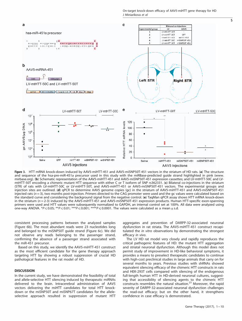
Optimized miHTT-451 and miSNP50T-451 expressed from AAV5vectors induced the mutant HTT mRNA knock-down in HD ratsAlthough the engineered mmu-miHTT-155 constructs generatesthe favorable miHTT effector sequence and is suitable for pilotexperiments addressing efficacy and distribution, it is not optimalfor clinical studies in humans due to a high risk of immuneresponse to GFP and possibility of off-target silencing caused bythe passenger strand.18 Previously, we showed that the silencingefficacy and processing of the miHTT construct are influenced bythe pre-miHTT precursor.18 We identified the miHTT-451 precursorthat showed no passenger strand in vivo opposite to themiHTT-101 and miHTT-135 precursors (Figure 3a). Moreover, themiHTT-451 construct showed efficacious HTT knock-down and nosigns of toxicity. Therefore, in this study we included themiHTT-451 precursor for the total HTT knock-down. For theallele-selective approach, we optimized the miSNP50T constructdesigned to preferentially bind to HTT messenger RNA (mRNA)carrying the U isoform of SNP rs362331, which is associated athigh frequencies with HD.25 This miSNP50T construct, whichexhibits stronger efficacy than miSNP67T in vitro, was not availablewhen the pilot study was initiated and therefore, it was introducedat this stage. Moreover, for both constructs the cytomegaloviruspromoter together with GFP was replaced by the strongcytomegalovirus immediate-early enhancer fused to chicken β-actin (CAG) promoter, which has been shown to be effective in thebrain.26 The resultant constructs are named miHTT-451 andmiSNP50T-451 (Figure 3b).To further establish mechanistic efficiency, safety, as well as
allele selectivity in vivo, we generated AAV5-miHTT-451 and AAV5-miSNP50T-451 vectors and co-injected them bilaterally in thestriatum of rats with a LV vector encoding a chimeric mutant HTTsequence with an 82Q chain and carrying target regions for themiHTT and miSNP50T constructs, named LV-mtHTT-50T(Figures 3b and c). In order to address the allele-selective potentialof the miSNP50T construct, we included a second LV vector,named LV-mtHTT-50C. The AAV5-miSNP50T-451 expression pro-duct was designed to have one nucleotide mismatch with the LV-mtHTT-50T transcripts and two nucleotide mismatches with theLV-mtHTT-50C transcripts. Therefore, this system enabled us totest the allele selectivity based on only a single SNP. As negativecontrols, we included a GFP expressed from the CAG promoterand saline control. Two months post-injection, rats wereeuthanized and the brains were processed to assess on-targetefficacy, HD-like neuronal dysfunction, and immune reaction.To measure AAV5 vector DNA in striata, we performed real-time
quantitative PCR (qPCR) with primers directed towards the CAGpromoter (Figure 3d). We observed high and comparable vectorDNA in the striatal homogenates from AAV5-GFP-, AAV5-miHTT--451- and AAV5-miSNP50T-451-injected rats. Similar to TaqManassays (data not shown), we detected a background level of~ 3000 genome copies in the saline control, which was consideredfor calculations of final vector DNA. Furthermore, the latterinversely correlated with the human-specific HTT mRNA knock-down from the same tissue homogenate demonstrated byTaqMan RT-qPCR (Figure 3e). We detected 62.3%± 21.6 HTTmRNA knock-down in AAV5-miHTT-451- and 81%± 4.3 HTT mRNAknock-down in AAV5-miSNP50T-451-injected rats as comparedwith the saline control. We observed similar HTT mRNA knock-
down by AAV5-miSNP50T-451 in LV-mtHTT-50T-injected ratscompared with LV-mtHTT-50C-injected rats where 65.5%± 9.2HTT mRNA reduction was detected.
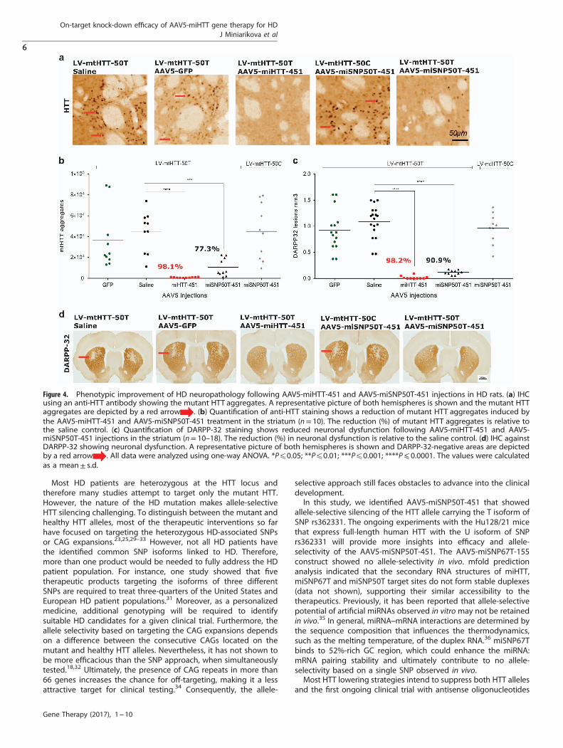
On-target silencing efficiency of AAV5-miHTT-451 and AAV5-miSNP50T-451 correlates with suppression of DARPP-32-associated neuronal dysfunction in HD ratsTo establish the ability of our optimized AAV5-miHTT-451 andAAV5-miSNP50T-451 vectors to suppress formation of mutant HTTaggregates, we analyzed striato-cortical sections by IHC using ananti-HTT antibody (Figure 4a). Here, in the AAV5-miHTT-451-injected rats almost no detectable mutant HTT aggregates wereobserved (843 ± 359, P⩽ 0.0001), whereas the aggregates wereabundant in the GFP (3.6x10⁴ ± 2.9x10⁴) and saline(4.4x10⁴ ± 2x10⁴) controls (Figure 4b). Based on the number ofmutant HTT aggregates, AAV5-miSNP50T-451 showed strongersilencing of the matched LV-mtHTT-50T expression product(1x10⁴ ± 0.8x10⁴, P⩽ 0.001) compared with the mismatchedLV-mtHTT-50C (4.5x10⁴ ± 2.6x10⁴), suggesting allele-selectivepotential.During development of symptomatic HD, the appearance of
mutant HTT aggregates precedes the death of medium spinyneurons located in the striatum.22 Therefore, we addressed thepotential of our AAV5-miHTT-451 and AAV5-miSNP50T-451vectors to suppress DARPP-32-associated neuronal dysfunctionby performing IHC against DARPP-32 (Figures 4c and d).Consistent with lowering of mutant HTT aggregates, AAV5-miHTT-451 (0.02 mm3± 0.03, P⩽ 0.0001) and AAV5-miSNP50T-451 (0.12 mm3± 0.04, P⩽ 0.0001) treatmentssignificantly suppressed the partial striatal lesion induced by theLV-mtHTT-SNP50T expression product compared with the GFP(0.92 mm3± 0.39) and saline controls (1.09 mm3± 0.31). In con-trast, AAV5-miSNP50T-451 treatment did not show strong reduc-tion in the size of DARPP-32 lesions in LV-mtHTT-SNP50C-injectedrats (0.96 mm3± 0.29), demonstrating allele selectivity.
AAV5 viruses delivering the miHTT-451 or miSNP50T-451precursors did not induce an overt immune response via GFAPand Iba1 activationAlthough AAV-based gene therapy is an attractive approach forthe delivery of gene products, such as therapeutic miRNAs, andoverall has demonstrated low toxicity to date, activation of theimmune response should be addressed for each therapeuticcandidate in a given target tissue. To evaluate the immuneresponse to our AAV5 vectors expressing therapeutic miRNAprecursors in the brain, we analyzed the activation of microgliaand infiltration of astrocytes in the injected areas by IHC using ananti-ionized calcium-binding adapter molecule 1 (Iba1) and anti-glial fibrillary acidic protein (GFAP) antibody, respectively(Figures 5a and b). We found similar levels of immunoreactivityin AAV5-miHTT-451- and AAV5-miSNP50T-451-treated rats com-pared to the saline control, indicating no apparent activation ofimmune reaction via microglia and astrocytes.
AAV5-miHTT-451 and miSNP50T-451 constructs do not generate apassenger strand in vivoPreviously, we showed that the miHTT-451 construct does notgenerate a passenger strand and is not toxic probably due tohigher specificity in vivo (Figure 6a).18 The absence of thepassenger strand from the miR-451 scaffold provides a bettersafety profile for therapeutic shRNAs or miRNAs since it reducesthe chance for off-target suppression of other genes by thepassenger strand.18,27,28 To establish the absence of a passengerstrand for the miSNP50T-451 construct in vivo, we analyzed theprocessing patterns in the striatum of AAV5-miSNP50T-451-injected rats by next-generation sequencing. We observed
On-target knock-down efficacy of AAV5-miHTT gene therapy for HDJ Miniarikova et al
4
Gene Therapy (2017), 1 – 10
consistent processing patterns between the analyzed samples(Figure 6b). The most abundant reads were 23 nucleotides longand belonged to the miSNP50T guide strand (Figure 6c). We didnot observe any reads belonging to the passenger strand,confirming the absence of a passenger strand associated withthe miR-451 precursor.Based on this study, we identify the AAV5-miHTT-451 construct
as the most efficient candidate for the gene therapy approachtargeting HTT by showing a robust suppression of crucial HDpathological features in the rat model of HD.
DISCUSSIONIn the current study, we have demonstrated the feasibility of totaland allele-selective HTT silencing induced by therapeutic miRNAsdelivered to the brain. Intracerebral administration of AAV5vectors delivering the miHTT candidates for total HTT knock-down or the miSNP50T and miSNP67T candidates for the allele-selective approach resulted in suppression of mutant HTT
aggregates and prevention of DARPP-32-associated neuronaldysfunction in rat striata. The AAV5-miHTT-451 construct recapi-tulated the in vitro observations by demonstrating the strongestefficacy in vivo.The LV HD rat model very closely and rapidly reproduces the
critical pathogenic features of HD: the mutant HTT aggregationand striatal neuronal dysfunction. Although this model does notpermit study of improvement in HD-like behavioral symptoms, itprovides a means to preselect therapeutic candidates to continuewith high-cost preclinical studies in large animals that carry on forseveral months to years. Previous studies with shRNAs showedequivalent silencing efficacy of the chimeric HTT constructs in ratsand HEK-293T cells compared with silencing of the endogenousfull-length human HTT in HD-derived neuronal cultures, suggest-ing that accessibility of silencing agents to the chimeric HTTconstructs resembles the natural situation.23 Moreover, the rapidseverity of DARPP-32-associated neuronal dysfunction challengesthe read-out efficacy; but on the other hand, it strengthensconfidence in case efficacy is demonstrated.
Figure 3. HTT mRNA knock-down induced by AAV5-miHTT-451 and AAV5-miSNP50T-451 vectors in the striatum of HD rats. (a) The structureand sequence of the hsa-pre-miR-451a precursor used in this study with the miRBase-predicted guide strand highlighted in pink (www.mirbase.org). (b) Schematic representation of the AAV5-miHTT-451 and AAV5-miSNP50T-451 expression cassettes; and LV-mtHTT-50C and LV-mtHTT-50T encoding a chimeric mutant HTT sequence with either C or T isoform of SNP rs362331. (c) Bilateral co-injections in the striatum(STR) of rats with LV-mtHTT-50C or LV-mtHTT-50T, and AAV5-miHTT-451 or AAV5-miSNP50T-451 vectors. The experimental groups andinjection sites are outlined. (d) qPCR to determine AAV5 genome copies (gc) in the striatum of AAV5-miHTT-451 and AAV5-miSNP50T-451injected rats (n= 3), two months post-injection. Primers directed to the CAG promoter were used and the gc values were calculated based onthe standard curve and considering the background signal from the negative control. (e) TaqMan qPCR assay shows HTT mRNA knock-downin the striatum (n= 2-3) induced by the AAV5-miHTT-451 and AAV5-miSNP50T-451 expression products. Human HTT-specific exon-spanningprimers were used and HTT values were subsequently normalized to GAPDH, an internal control set at 100%. All data were analyzed usingone-way ANOVA. *Pp0.05; **Pp0.01; ***Pp0.001; ****P⩽ 0.0001. The values were calculated as a mean± s.d.
On-target knock-down efficacy of AAV5-miHTT gene therapy for HDJ Miniarikova et al
5
Gene Therapy (2017), 1 – 10
Most HD patients are heterozygous at the HTT locus andtherefore many studies attempt to target only the mutant HTT.However, the nature of the HD mutation makes allele-selectiveHTT silencing challenging. To distinguish between the mutant andhealthy HTT alleles, most of the therapeutic interventions so farhave focused on targeting the heterozygous HD-associated SNPsor CAG expansions.23,25,29–33 However, not all HD patients havethe identified common SNP isoforms linked to HD. Therefore,more than one product would be needed to fully address the HDpatient population. For instance, one study showed that fivetherapeutic products targeting the isoforms of three differentSNPs are required to treat three-quarters of the United States andEuropean HD patient populations.31 Moreover, as a personalizedmedicine, additional genotyping will be required to identifysuitable HD candidates for a given clinical trial. Furthermore, theallele selectivity based on targeting the CAG expansions dependson a difference between the consecutive CAGs located on themutant and healthy HTT alleles. Nevertheless, it has not shown tobe more efficacious than the SNP approach, when simultaneouslytested.18,32 Ultimately, the presence of CAG repeats in more than66 genes increases the chance for off-targeting, making it a lessattractive target for clinical testing.34 Consequently, the allele-
selective approach still faces obstacles to advance into the clinicaldevelopment.In this study, we identified AAV5-miSNP50T-451 that showed
allele-selective silencing of the HTT allele carrying the T isoform ofSNP rs362331. The ongoing experiments with the Hu128/21 micethat express full-length human HTT with the U isoform of SNPrs362331 will provide more insights into efficacy and allele-selectivity of the AAV5-miSNP50T-451. The AAV5-miSNP67T-155construct showed no allele-selectivity in vivo. mfold predictionanalysis indicated that the secondary RNA structures of miHTT,miSNP67T and miSNP50T target sites do not form stable duplexes(data not shown), supporting their similar accessibility to thetherapeutics. Previously, it has been reported that allele-selectivepotential of artificial miRNAs observed in vitromay not be retainedin vivo.35 In general, miRNA–mRNA interactions are determined bythe sequence composition that influences the thermodynamics,such as the melting temperature, of the duplex RNA.36 miSNP67Tbinds to 52%-rich GC region, which could enhance the miRNA:mRNA pairing stability and ultimately contribute to no allele-selectivity based on a single SNP observed in vivo.Most HTT lowering strategies intend to suppress both HTT alleles
and the first ongoing clinical trial with antisense oligonucleotides
Figure 4. Phenotypic improvement of HD neuropathology following AAV5-miHTT-451 and AAV5-miSNP50T-451 injections in HD rats. (a) IHCusing an anti-HTT antibody showing the mutant HTT aggregates. A representative picture of both hemispheres is shown and the mutant HTTaggregates are depicted by a red arrow . (b) Quantification of anti-HTT staining shows a reduction of mutant HTT aggregates induced bythe AAV5-miHTT-451 and AAV5-miSNP50T-451 treatment in the striatum (n= 10). The reduction (%) of mutant HTT aggregates is relative tothe saline control. (c) Quantification of DARPP-32 staining shows reduced neuronal dysfunction following AAV5-miHTT-451 and AAV5-miSNP50T-451 injections in the striatum (n= 10–18). The reduction (%) in neuronal dysfunction is relative to the saline control. (d) IHC againstDARPP-32 showing neuronal dysfunction. A representative picture of both hemispheres is shown and DARPP-32-negative areas are depictedby a red arrow . All data were analyzed using one-way ANOVA. *Pp0.05; **Pp0.01; ***Pp0.001; ****P⩽ 0.0001. The values were calculatedas a mean± s.d.
On-target knock-down efficacy of AAV5-miHTT gene therapy for HDJ Miniarikova et al
6
Gene Therapy (2017), 1 – 10

employs this approach (https://clinicaltrials.gov/). Preclinical studiesusing HD animal models and non human primates indicate thatthere is a therapeutic window where total HTT knock-down is safeand yet beneficial for HD patients.37–39 Importantly, this strategywould provide a treatment for all HD patients independent fromtheir genotype. In this study, we showed that the miHTT constructprocessed from the miR-451 scaffold is able to strongly suppressmutant HTT aggregation and ultimately dramatically reduce thegeneration of striatal lesions at two months post-injection.Moreover, we did not observe immune reaction to the AAV5-miHTT-451 vector via astrocytes or microglia activation, suggestinga favorable safety profile.Altogether, these data provide strong evidence for the
therapeutic efficacy of the AAV5-miHTT-451 vector by inducingfunctional improvements downstream in the HD pathologicalprocess without raising safety concerns in a rodent model of HD.These promising results support our ongoing efforts for thedevelopment of an HD-modifying treatment by silencing thehuman HTT gene.
MATERIALS AND METHODSHTT target sequencesHomo sapiens HTT mRNA, complete CDS (gb|L12392.1|HUMHDA) obtainedfrom http://blast.ncbi.nlm.nih.gov/Blast.cgi, was used to identify targetsequences for the artificial miHTT targeting exon 1, the miSNP50Tconstruct binding to the U isoform of SNP rs362331, and the miSNP67Tconstruct targeting the U isoform of SNP rs362307.
DNA constructsTo generate the miHTT-155 and miSNP67T-155 constructs, their guidesequences targeting HTT transcripts were embedded into the engineeredmurine pre-miR-155 backbone of pcDNA6.2-GW/EmGFP-miR (Invitrogen,Carlsbad, CA, USA) by annealing complementary oligonucleotides (Qiagen,Valencia, CA, USA) followed by ligation into the linearized pcDNA6.2plasmid.18
To generate miHTT-451 and miSNP50T-451 constructs, the guide andpassenger strand sequences were incorporated in the hsa-pri-miR-451ascaffold. 200 nucleotides long 5′ and 3′ encompassing flanking regionswere included with EcoRV and BamHI restriction sites and the completesequences were ordered from GeneArt gene synthesis (Invitrogen). In
Figure 5. No apparent activation of microglia and astrocytes following AAV5-miHTT-451 and AAV5-miSNP50T-451 injections in HD rats. (a) IHCagainst Iba1 to show microglial activity. A representative picture of the right hemisphere is shown. (b) IHC against GFAP to show the astrocyteactivity. A representative picture of the right hemisphere is shown.
Figure 6. Next-generation sequencing analysis of the pre-miHTT-451 and pre-miSNP50T-451 processing patterns shows no passenger strandin vivo. (a) Sequence distribution (%) of reads mapping to pre-miHTT-451 in vivo.18 The predicted guide strand is indicated in red and amismatch with the reference sequence in light blue. (b) Sequence distribution (%) of reads mapping to pre-miSNP50T in vivo. The predictedguide strand is indicated in red and a mismatch with the reference sequence in light blue. (c) The length distribution of reads mapping to thepre-miSNP50T precursor (n= 2). For the read alignments, up to 3 mismatches with the reference sequence were allowed. Reads representedwith less than 3% were excluded from the figure.
On-target knock-down efficacy of AAV5-miHTT gene therapy for HDJ Miniarikova et al
7
Gene Therapy (2017), 1 – 10
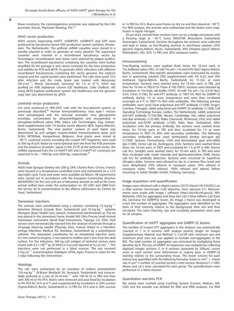
these constructs, the cytomegalovirus promoter was replaced by the CAGpromoter (Inovio, Plymouth Meeting, PA).18
AAV5 vector productionAAV5 vectors expressing miHTT, miSNP50T, miSNP67T and GFP wereproduced by baculovirus-based AAV production system (uniQure, Amster-dam, The Netherlands). The artificial miRNA cassettes were cloned in atransfer plasmid in order to generate an entry plasmid. The expressioncassettes were inserted in a recombinant baculovirus vector byhomologous recombination and clones were selected by plague purifica-tion. The recombinant baculovirus containing the cassettes were furtheramplified till the passage 6 and clones screened for the best productionand stability by PCR and qPCR. To generate AAV5, infections with differentrecombinant baculoviruses containing the vector genome, the repliconenzyme and the capsid protein were performed. The cells were lysed 72 hafter infection and the crude lysate was treated with Benzonase(50 U ml− 1; Merck, Darmstadt, Germany) for 1 h at 37 °C. AAV5 waspurified on AVB Sepharose column (GE Healthcare, Little Chalfont, UK)using AKTA Explorer purification system (GE Healthcare) and the genomecopy titer was determined by qPCR.
Lentiviral vector productionLVs were produced in HEK-293T cells with the four-plasmid system, aspreviously described.40 Human immunodeficiency virus type-1 vectorswere pseudotyped with the vesicular stomatitis virus glycoproteinenvelope, concentrated by ultracentrifugation and resuspended inphosphate-buffered saline (PBS, Gibco, Life Technologies, Zug, Switzer-land) supplemented with 1% bovine serum albumin (BSA, Sigma-Aldrcich,Buchs, Switzerland). The viral particle content of each batch wasdetermined by p24 antigen enzyme-linked immunosorbent assay (p24ELISA, RETROtek, Kampenhout, Belgium). Viral stocks were stored at− 80 °C. Lentiviral vectors encoding the mutant HTT fragments were usedat 300 ng of p24. Based on transcriptional start site from the PGK promoterand the presence of polyA+ signal in the 3′LTR of the lentiviral vector, themRNAs expressed from the LV-mtHTT-50T and LV-mtHTT-67 constructs areexpected to be ~ 1993 bp and 2059 bp, respectively.41
AnimalsAdult male Sprague Dawley rats (200 g; OFA, Charles River, Oncins, France)were housed in a temperature-controlled room and maintained on a 12 hday/night cycle. Food and water were available ad libitum. All experimentswere carried out in accordance with the European Community directive(86/609/EEC) for the care and use of laboratory animals as well as the Swissanimal welfare laws under the authorization no. VD 2487 and 2889 fromthe Service de la consommation et des affaires vétérinaires du Canton deVaud, Switzerland.
Stereotaxic injectionsThe animals were anesthetized using a solution containing 75 mg kg− 1
ketamine (Ketasol, Graeub, Bern, Switzerland) and 10 mg kg− 1 xylazine(Rompun, Bayer Health Care, Uznach, Switzerland) administered i.p. The ratwas placed in the stereotaxic frame (model 963 Ultra Precise Small AnimalStereotaxic Instrument, David Kopf Instruments, Tujunga, CA, USA). Viralvectors were stereotaxically injected into the striatum of animals through a34-gauge blunt-tip needle (Phymep, Paris, France) linked to a Hamiltonsyringe (Hamilton Medical AG, Bonaduz, Switzerland) by a polyethylenecatheter. The stereotaxic coordinates for an intrastriatal injection were:0.5 mm rostral to bregma, 3 mm lateral to midline and 5 mm from the skullsurface. For the infections, 300 ng p24 antigen of lentiviral vectors weremixed with 6.5 × 10E10 gc of AAV5 in 4 μl and injected at 0.2 μl min− 1. Theinjections were not performed in a blind manner. The rats received2 mg ml− 1 acetaminophen (Dafalgan, UPSA, Agen, France) in water for the3 days following the intervention.
HistologyThe rats were euthanized by an overdose of sodium pentobarbital150 mg kg− 1 (B-Braun Mediacal SA, Sempach, Switzerland) and transcar-dially perfused at a rate of 20 ml min− 1 with 100 ml of 1x PBS and thenwith 300 ml of 4% PFA. Brains were removed and post-fixed by incubationin 4% PFA for 24 h at 4 °C and cryoprotected by incubation in 20% sucrose(Sigma-Aldrich, Buchs, Switzerland) in 1x PBS for 24 h and in 30% sucrose
in 1x PBS for 24 h. Brains were frozen on dry ice and then stored at − 80 °C.For RNA analysis, the animals were euthanized and the brains were snap-frozen in liquid nitrogen.25 μm-thick coronal brain sections were cut on a sledge microtome with
a freezing stage at − 30 °C (Leica SM2010R, Biosystems Switzerland,Nunningen, Switzerland). Sections throughout the striatum were collectedand kept in tubes, as free-floating sections in anti-freeze solution (25%glycerol (Sigma-Aldrich, Buchs, Switzerland), 30% Ethylene glycol (Merck,Nottingham, UK), 25% 1x PBS and 20% nanopure water).
ImmunostainingFree-floating sections were washed three times, for 10 min each, inTris-buffered saline (TBS, 10 mM Tris pH=7.6 and 0.9% NaCl Sigma-Aldrich,Buchs, Switzerland). Non-specific peroxidases were inactivated by incuba-tion in quenching solution (TBS supplemented with 3% H2O2 and 10%methanol, Sigma-Aldrich, Buchs, Switzerland) for 15 min at roomtemperature. Sections were washed twice, for 10 min each, in TBS, andthen for 10 min in TBS-0.1% Triton X-100 (TBST). Sections were blocked byincubation in Tris-high salt-buffer (THST, 50 mM Tris pH=7.6, 0.5 M Nacl,0.5% Triton X-100,) for anti-HTT antibody or TBST-5% NGS for DARPP-32antibody during 1 h at room temperature and were then incubatedovernight at 4 °C in TBST-1% BSA with antibodies. The following primaryantibodies were used: Goat polyclonal anti-HTT antibody (1/1000, Sicgen,Coimbra, Portugal), rabbit polyclonal anti-DARPP-32 antibody (1/1000, CellSignaling Technology Europe, Leiden, The Netherlands), rabbit polyclonalanti-GFP antibody (1/100 000, Abcam, Cambridge, UK), rabbit polyclonalanti-IbaI antibody (1/25 000, Wako Chemicals, Richmond, USA) and rabbitpolyclonal anti-GFAP antibody (1/500, Dako, Baar, Switzeland). Afterincubation with the primary antibody, the sections were washed threetimes, for 10 min each, in TBS and then incubated for 1 h at roomtemperature in TBST-1% BSA with secondary antibodies. The followingsecondary antibodies were used: biotinylated donkey anti-goat IgG(1/1000, Jackson, Pennsylvania, USA) and biotinylated goat anti-rabbitIgG (1/400, Vector Lab Inc, Burlingame, USA). Sections were washed threetimes, for 10 min each, in TBST and incubated for 1 h at RT in ABC (VectorLab Inc). Samples were washed twice, for 10 min each, in TBS and werethen incubated with nickel chloride and diaminobenzidine (DAB, VectorLab Inc) for antibody detection. Sections were mounted on SuperfrostUltraplus slides. Sections were allowed to dry in a laminar flow hood andwere dehydrated (70% ethanol in nanopure water, 95% ethanol innanopure water, 100% ethanol, 100% ethanol and xylene) beforemounting in Eukitt (Kindler GmbH, Freiburg, Germany).
Image acquisition and quantificationImages were obtained with a digital camera (3CCD Hitachi HV-F202SCL) ona slide scanner microscope (×20 objective, Zeiss axioscan Z1). Measure-ments were made with Image J software (http://rsb.info.nih.gov/ij/, NIH,Bethesda, USA) for aggregates and Zeiss Zen Lite 2 (blue Edition; Carl ZeissAG, Germany) for DARPP32 lesion. An Image J macro was developed tocount the number of aggregates. The aggregates were identified on thebasis of their intensity relative to the background, their size and theircircularity. The same intensity, size and circularity parameters were usedfor all samples.
Quantification of mtHTT aggregates and DARPP-32 lesionsThe number of mutant HTT aggregates in the striatum was automaticallycounted in 1 in 8 sections with analyze particle plugin on ImageJ(Supplementary Material and Method 1). Cut-off with minimum size andmaximum pixel area size was applied to exclude non-aggregates in theROI. The total number of aggregates was estimated by multiplying thesedensities by 8. The loss of DARPP-32 expression was analyzed by collectingdigitized images sections (1 in 8 sections separated by 200μm). Lesionareas in each section were determined as regions poor in DARPP-32staining relative to the surrounding tissue. The lesion volume for eachanimal was quantified with the following formulae: lesion in mm3 = (meansize in μm2×number of counted section× inter-section distance)/1 × 10E9.Means and s.d.’s were calculated for each group. The quantifications wereperformed in a blind manner.
Quantitative real-time PCRRat striata were crushed using CryoPrep System (Covaris, Woburn, MA,USA) and the powder was divided for RNA and DNA analyses. For RNA
On-target knock-down efficacy of AAV5-miHTT gene therapy for HDJ Miniarikova et al
8
Gene Therapy (2017), 1 – 10
isolation, the striatal powder was homogenized in Trizol using gentleMACSDissociator (Miltenyi Biotec) and total RNA was isolated from Trizolaccording to the manufacturer’s protocol (Invitrogen). To remove genomicDNA, RNA was treated with dsDNase (Thermo Scientific, Waltham, MA,USA). Quantitative real-time PCR reaction was performed to detectHTT mRNA knock-down using TaqMan gene expression assays(Applied Biosystems, Foster City, CA, USA), human HTT(Hs00918134_m1), rat GAPDH (Rn01462662_g1); and miHTT expressionlevels using custom TaqMan microRNA expression assay or U6 snRNA(001973; Applied Biosystems). The expression level of each gene wasnormalized to either endogenous GAPDH or U6 snRNA levels. Fold change/percentages in HTT mRNA knock-down or miHTT expression werecalculated based on 2^DDCT method (Supplementary Material andMethod 2).For the genomic DNA isolation, the striatal powder was processed using
DNeasy96 Blood and Tissue kit (Qiagen). qPCR reaction was performed todetect AAV5 vector titers using a standard line and SYBR Green protocol(Applied Biosystems). Forward primer sequence: 5′-GAGCCGCAGCCATTGC-3′ and reverse primer sequence: 5′-CACAGATTTGGGACAAAGGAAGT-3′. Based on the standard line, genome copies per microgram DNA werecalculated.
Next-generation sequencing data analysisThe raw sequencing data were produced as previously described.18 SmallRNA raw data sets were analyzed using the CLC Genomics Workbench 6(Qiagen). All reads containing ambiguity N symbols, reads shorter than 10nucleotides, longer than 45 nucleotides and reads represented less than 10times were discarded. The obtained unique small RNA reads were alignedto the reference sequences of the pre-miHTT and pre-miSNP50T constructswith a max. of 3 nucleotide mismatches allowed. The percentages of readsbased on the total number of reads matching the reference sequenceswere calculated.
Sample sizes, calculations and statistical analysisSample sizes of saline n=18, GFP n= 16, AAV5-miHTT-155 n= 18, AAV5-miHTT-451 n= 16, AAV5-miSNP67T-155 n= 16 and AAV5-miSNP50T-451n= 16 represent the number of striata injected per experimental group.The sample size was chosen to consider statistical variability due tosurgical procedure based on previous studies. All experiments involvinganimals were performed once. The only exclusion criterion was if aproblem was encountered during the injection procedure. The knock-down percentages were calculated using the following formula: Knock-down (%) = [AAV5-miRNA treatment]/[saline treatment] × 100%. Data wereanalyzed using one-way ANOVA. *Pp0.05; **Pp0.01; ***Pp0.001;****P⩽ 0.0001. Differences were considered significant when Pp0.05.The values were calculated as a mean± s.d.
CONFLICT OF INTERESTJM, RM, CCB, JL, MME, HP and PK are employees and shareholders at uniQure. ND andCHUV have a collaborative agreement with uniQure. uniQure has sponsored aportion of the research performed in ND lab.
ACKNOWLEDGEMENTSWe thank Dr Eileen Sawyer and Dr Olivier ter Brake for critically reviewing themanuscript.
REFERENCES1 Rawlins MD, Wexler NS, Wexler AR, Tabrizi SJ, Douglas I, Evans SJW et al. The
prevalence of huntington’s disease. Neuroepidemiology 2016; 46: 144–153.2 Langbehn DR, Brinkman RR, Falush D, Paulsen JS, Hayden MR. A new model for
prediction of the age of onset and penetrance for Huntington’s disease based onCAG length. Clin Genet 2004; 65: 267–277.
3 The Huntington's Disease Collaborative Research Group. A novel gene containinga trinucleotide repeat that is expanded and unstable on Huntington's diseasechromosomes. Cell 1993; 72: 971–983.
4 Ross CA, Tabrizi SJ. Huntington’s disease: from molecular pathogenesis to clinicaltreatment. Lancet Neurol 2011; 10: 83–98.
5 Bates GP, Dorsey R, Gusella JF, Hayden MR, Kay C, Leavitt BR et al. Huntingtondisease. Nat Rev Dis Primers 2015; 1: 15005.
6 DiFiglia M, Sapp E, Chase KO, Davies SW, Bates GP, Vonsattel JP et al. Aggregationof huntingtin in neuronal intranuclear inclusions and dystrophic neurites in brain.Science 1997; 277: 1990–1993.
7 Ross CA, Aylward EH, Wild EJ, Langbehn DR, Long JD, Warner JH et al. Huntingtondisease: natural history, biomarkers and prospects for therapeutics. Nat Rev Neurol2014; 10: 204–216.
8 Yamamoto A, Lucas JJ, Hen R. Reversal of neuropathology and motor dysfunctionin a conditional model of Huntington’s disease. Cell 2000; 101: 57–66.
9 Fire A, Xu S, Montgomery MK, Kostas SA, Driver SE, Mello CC. Potent and specificgenetic interference by double-stranded RNA in Caenorhabditis elegans. Nature1998; 391: 806–811.
10 Boudreau RL, Rodríguez-Lebrón E, Davidson BL. RNAi medicine for the brain:progresses and challenges. Hum Mol Genet 2011; 20: 21–27.
11 Aronin N, DiFiglia M. Huntingtin-lowering strategies in Huntington’s disease:antisense oligonucleotides, small RNAs, and gene editing. Mov Disord 2014; 29:1455–1461.
12 Kantor B, Bailey RM, Wimberly K, Kalburgi SN, Gray SJ. Methods For Gene TransferTo The Central Nervous System. Adv Genet 2014; 87: 125–197.
13 Srivastava A. In vivo tissue-tropism of adeno-associated viral vectors. Curr OpinVirol 2016; 21: 75–80.
14 Markakis EA, Vives KP, Bober J, Leichtle S, Leranth C, Beecham J et al. Comparativetransduction efficiency of AAV vector serotypes 1–6 in the substantia nigra andstriatum of the primate brain. Mol Ther 2010; 18: 588–593.
15 Burger C, Gorbatyuk OS, Velardo MJ, Peden CS, Williams P, Zolotukhin S et al.Recombinant AAV viral vectors pseudotyped with viral capsids from serotypes 1,2, and 5 display differential efficiency and cell tropism after delivery to differentregions of the central nervous system. Mol Ther 2004; 10: 302–317.
16 Lin D, Fantz CR, Levy B, Rafi MA, Vogler C, Wenger DA et al. AAV2/5 vectorexpressing galactocerebrosidase ameliorates CNS disease in the murine modelof globoid-cell leukodystrophy more efficiently than AAV2. Mol Ther 2005; 12:422–430.
17 Samaranch L, Blits B, San Sebastian W, Hadaczek P, Bringas J, Sudhakar V et al.MR-guided parenchymal delivery of adeno-associated viral vector serotype 5 innon-human primate brain. Gene Ther 2017; 24: 253–261.
18 Miniarikova J, Zanella I, Huseinovic A, van der Zon T, Hanemaaijer E, Martier Ret al. Design, characterization, and lead selection of therapeutic miRNAs targetinghuntingtin for development of gene therapy for Huntington’s disease. Mol TherNucleic Acids 2016; 5: e297.
19 Menalled L, Brunner D. Animal models of Huntington’s disease for translation tothe clinic: best practices. Mov Disord 2014; 29: 1375–1390.
20 Ramaswamy S, Mcbride JL, Kordower JH. Animal models of Huntington’s disease.ILAR J 2007; 48: 356–373.
21 Pouladi MA, Morton AJ, Hayden MR. Choosing an animal model for the study ofHuntington’s disease. Nat Rev Neurosci 2013; 14: 708–721.
22 de Almeida LP, Ross CA, Zala D, Aebischer P, Déglon N. Lentiviral-mediateddelivery of mutant huntingtin in the striatum of rats induces a selective neuro-pathology modulated by polyglutamine repeat size, huntingtin expression levels,and protein length. J Neurosci 2002; 22: 3473–3483.
23 Drouet V, Ruiz M, Zala D, Feyeux M, Auregan G, Cambon K et al. Allele-specificsilencing of mutant huntingtin in rodent brain and human stem cells. PLoS ONE2014; 9: e99341.
24 Ouimet CC, Langley-Gullion KC, Greengard P. Quantitative immunocytochemistryof DARPP-32-expressing neurons in the rat caudatoputamen. Brain Res 1998; 808:8–12.
25 Lombardi MS, Jaspers L, Spronkmans C, Gellera C, Taroni F, Di Maria E et al.A majority of Huntington’s disease patients may be treatable by individualizedallele-specific RNA interference. Exp Neurol 2009; 217: 312–319.
26 Klein RL, Hamby ME, Gong Y, Hirko AC, Wang S, Hughes JA et al. Dose andpromoter effects of adeno-associated viral vector for green fluorescent proteinexpression in the rat brain. Exp Neurol 2002; 176: 66–74.
27 Ma H, Zhang J, Wu H. Designing Ago2-specific siRNA/shRNA to avoid competitionwith endogenous miRNAs. Mol Ther Nucleic Acids 2014; 3: e176.
28 Herrera-Carrillo E, Harwig A, Liu YP, Berkhout B. Probing the shRNA characteristicsthat hinder Dicer recognition and consequently allow Ago-mediated processingand AgoshRNA activity. RNA 2014; 20: 1410–1418.
29 Yu D, Pendergraff H, Liu J, Kordasiewicz HB, Cleveland DW, Swayze EE et al.Single-stranded RNAs use RNAi to potently and allele-selectively inhibit mutanthuntingtin expression. Cell 2012; 150: 895–908.
30 Carroll JB, Warby SC, Southwell AL, Doty CN, Greenlee S, Skotte N et al. Potent andselective antisense oligonucleotides targeting single-nucleotide polymorphismsin the Huntington disease gene/allele-specific silencing of mutant huntingtin. MolTher 2011; 19: 2178–2185.
31 Pfister EL, Kennington L, Straubhaar J, Wagh S, Liu W, DiFiglia M et al. Five siRNAstargeting three SNPs may provide therapy for three-quarters of Huntington'sdisease patients. Curr Biol 2009; 19: 774–778.
On-target knock-down efficacy of AAV5-miHTT gene therapy for HDJ Miniarikova et al
9
Gene Therapy (2017), 1 – 10
32 Kay C, Skotte NH, Southwell AL, Hayden MR. Personalized gene silencingtherapeutics for Huntington disease. Clin Genet 2014; 86: 29–36.
33 Warby SC, Montpetit A, Hayden AR, Carroll JB, Butland SL, Visscher H et al.CAG expansion in the Huntington disease gene is associated with a specificand targetable predisposing haplogroup. Am J Hum Genet 2009; 84:351–366.
34 Butland SL, Devon RS, Huang Y, Mead C-L, Meynert AM, Neal SJ et al. CAG-encoded polyglutamine length polymorphism in the human genome. BMCGenomics 2007; 8: 126.
35 Monteys AM, Wilson MJ, Boudreau RL, Spengler RM, Davidson BL. Artificial miR-NAs targeting mutant huntingtin show preferential silencing in vitro and in vivo.Mol Ther Nucleic Acids 2015; 4: e234.
36 Bartel DP. MicroRNAs: genomics, biogenesis, mechanism, and function. Cell 2004;136: 215–233.
37 Stiles DK, Zhang Z, Ge P, Nelson B, Grondin R, Ai Y et al. Widespread suppressionof huntingtin with convection-enhanced delivery of siRNA. Exp Neurol 2012; 233:463–471.
38 McBride JL, Pitzer MR, Boudreau RL, Dufour B, Hobbs T, Ojeda SR et al. Preclinicalsafety of RNAi-mediated HTT suppression in the rhesus macaque as a potentialtherapy for Huntington’s disease. Mol Ther 2011; 19: 2152–2162.
39 Grondin R, Kaytor MD, Ai Y, Nelson PT, Thakker DR, Heisel J et al. Six-month partialsuppression of Huntingtin is well tolerated in the adult rhesus striatum. Brain2012; 135: 1197–1209.
40 Hottinger AF, Azzouz M, Déglon N, Aebischer P, Zurn AD. Complete and long-term rescue of lesioned adult motoneurons by lentiviral-mediated expression ofglial cell line-derived neurotrophic factor in the facial nucleus. J Neurosci 2000; 20:5587–5593.
41 Adra CN, Boer PH, McBurney MW. Cloning and expression of the mouse pgk-1gene and the nucleotide sequence of its promoter. Gene 1987; 60: 65–74.
This work is licensed under a Creative Commons Attribution 4.0International License. The images or other third party material in this
article are included in the article’s Creative Commons license, unless indicatedotherwise in the credit line; if the material is not included under the Creative Commonslicense, users will need to obtain permission from the license holder to reproduce thematerial. To view a copy of this license, visit http://creativecommons.org/licenses/by/4.0/
© The Author(s) 2017
Supplementary Information accompanies this paper on Gene Therapy website (http://www.nature.com/gt).
On-target knock-down efficacy of AAV5-miHTT gene therapy for HDJ Miniarikova et al
10
Gene Therapy (2017), 1 – 10