Aasld guidelines for the treatment of hepatocellular carcinoma · Liver Diseases (AASLD) on the...
68
AASLD GUIDELINES FOR THE TREATMENT OF HEPATOCELLULAR CARCINOMA Julie Heimbach, MD, Laura M. Kulik, MD Richard Finn, MD, Claude B. Sirlin, MD, Michael Abecassis, MD, Lewis R. Roberts, MD, and Andrew Zhu, MD, PhD, M. Hassan Murad, Jorge Marrero, MD Corresponding Author: Julie Heimbach, MD Mayo Clinic Transplant Center Charlton 10 200 First Street SW Rochester, MN 55905-0001 United States (507) 266-6640 [email protected] FUNDING The funding for the development of this Practice Guideline was provided by the American Association for the Study of Liver Diseases. ACKNOWLEDGEMENTS This practice guideline was produced in tandem with three denovo systematic reviews that were written by the same writing group, including M. Hassan Murad, M.D., M.P.H., who participated in the selection of the clinical questions and provided expertise regarding the GRADE approach. The AASLD Practice Guidelines Committee approved the scope and directed the development of the practice guideline and provided the peer review. Members of the committee included Raphael B. Merriman, MD, FACP, FRCPI (Chair), Tram T. Tran, MD (Vice-Chair), Michael W. Fried, MD, FAASLD (Board Liaison), Jawad Ahmad, MD, FAASLD, Joseph Ahn, MD, Fredric Gordon, MD, FAASLD, Julie Heimbach, MD, Simon P. Horslen, MD, Christine Hsu, MD, Whitney E. Jackson, MD, Fasiha Kanwal, MD, MSHS, Michael D. Leise, MD, Jacqueline G. O'Leary, MD, Michael L. Schilsky, MD, FAASLD, Amit Singal, MD (Committee Liaison), James R. Spivey, MD, R. Todd Stravitz, MD, FAASLD, Jayant A. Talwalkar, MD, MPH, FAASLD, Helen S. Te, MD, FAASLD, and Michael Volk, MD. AASLD APPROVAL This practice guideline was approved by AASLD on December 8, 2016 This article has been accepted for publication and undergone full peer review but has not been through the copyediting, typesetting, pagination and proofreading process which may lead to differences between this version and the Version of Record. Please cite this article as doi: 10.1002/hep.29086 This article is protected by copyright. All rights reserved.
Transcript of Aasld guidelines for the treatment of hepatocellular carcinoma · Liver Diseases (AASLD) on the...

AASLD GUIDELINES FOR THE TREATMENT OF HEPATOCELLULAR
CARCINOMA
Julie Heimbach, MD, Laura M. Kulik, MD Richard Finn, MD, Claude B. Sirlin, MD, Michael Abecassis,
MD, Lewis R. Roberts, MD, and Andrew Zhu, MD, PhD, M. Hassan Murad, Jorge Marrero, MD
Corresponding Author:
Julie Heimbach, MD Mayo Clinic Transplant Center Charlton 10 200 First Street SW Rochester, MN 55905-0001 United States (507) 266-6640 [email protected]
FUNDING
The funding for the development of this Practice Guideline was provided by the American Association
for the Study of Liver Diseases.
ACKNOWLEDGEMENTS
This practice guideline was produced in tandem with three denovo systematic reviews that were written
by the same writing group, including M. Hassan Murad, M.D., M.P.H., who participated in the selection
of the clinical questions and provided expertise regarding the GRADE approach. The AASLD Practice
Guidelines Committee approved the scope and directed the development of the practice guideline and
provided the peer review. Members of the committee included Raphael B. Merriman, MD, FACP, FRCPI
(Chair), Tram T. Tran, MD (Vice-Chair), Michael W. Fried, MD, FAASLD (Board Liaison), Jawad Ahmad,
MD, FAASLD, Joseph Ahn, MD, Fredric Gordon, MD, FAASLD, Julie Heimbach, MD, Simon P. Horslen, MD,
Christine Hsu, MD, Whitney E. Jackson, MD, Fasiha Kanwal, MD, MSHS, Michael D. Leise, MD, Jacqueline
G. O'Leary, MD, Michael L. Schilsky, MD, FAASLD, Amit Singal, MD (Committee Liaison), James R. Spivey,
MD, R. Todd Stravitz, MD, FAASLD, Jayant A. Talwalkar, MD, MPH, FAASLD, Helen S. Te, MD, FAASLD,
and Michael Volk, MD.
AASLD APPROVAL
This practice guideline was approved by AASLD on December 8, 2016
This article has been accepted for publication and undergone full peer review but has not beenthrough the copyediting, typesetting, pagination and proofreading process which may lead todifferences between this version and the Version of Record. Please cite this article asdoi: 10.1002/hep.29086
This article is protected by copyright. All rights reserved.

Submitted to Hepatology for Publication January 10, 2017
2
GUIDING PRINCIPLES AND OBJECTIVES
Guiding Principles
This document presents official recommendations of the American Association for the Study of
Liver Diseases (AASLD) on the surveillance, diagnosis, and treatment of hepatocellular
carcinoma (HCC) occurring in the setting of adults with cirrhosis. Unlike previous AASLD
practice guidelines, the current guideline was developed in compliance with the Institute of
Medicine standards for trustworthy practice guidelines and uses the Grading of Recommendation
Assessment, Development and Evaluation (GRADE) approach1. Multiple systematic reviews of
the literature were conducted to support the recommendations in this practice guideline. An
enhanced understanding of the guideline can be obtained by reading the applicable portions of
the systematic reviews. In addition, more detailed information may be found in the associated
guidance document related to clinically important aspects of HCC that lacked sufficient evidence
to warrant a systematic review.
The guideline focuses on a broad spectrum of clinical practice, including surveillance of patients
with cirrhosis for HCC, establishing the diagnosis of HCC, and various therapeutic options for
the treatment of HCC. To address other issues on HCC such as epidemiology, staging, and
additional aspects of diagnosis and treatment, the authors have created a new guidance document
that will be published soon (reference when available), which is based upon the previous HCC
AASLD guidelines by Bruix and Sherman2.
Key Questions
This article is protected by copyright. All rights reserved.

Submitted to Hepatology for Publication January 10, 2017
3
The guideline developers from the AASLD identified key questions that health care providers
are faced with frequently in the evaluation and management of patients with HCC. These
questions were:
1. Should adults with cirrhosis undergo surveillance for HCC? If, so which
surveillance test is best?
2. Should adults with cirrhosis and suspected HCC undergo diagnostic evaluation with
multiphasic CT or multiphasic MRI?
3. Should adults with cirrhosis and an indeterminate hepatic nodule undergo a biopsy,
repeated imaging, or alternative imaging for the diagnostic evaluation?
4. Should adults with Child’s class A cirrhosis and early-stage HCC (T1 or T2) be
treated with resection or locoregional therapy?
5. Should adults with cirrhosis and HCC that has been resected or ablated successfully
undergo adjuvant therapy or not?
6. Should adults with cirrhosis awaiting liver transplantation and HCC (T1) be treated
or undergo observation?
7. Should adults with cirrhosis awaiting liver transplantation and HCC (Organ
Procurement and Transplantation Network [OPTN] T2) undergo transplant alone
or transplant with bridging therapy while waiting?
8. Should adults with cirrhosis awaiting liver transplantation and HCC beyond Milan
criteria (T3) be transplanted following downstaging to within Milan criteria?
9. Should adults with cirrhosis and HCC (T2 or T3, no vascular involvement) who are
not candidates for resection or transplantation be treated with transarterial
chemoembolization, transarterial radioembolization, or external radiation?
This article is protected by copyright. All rights reserved.

Submitted to Hepatology for Publication January 10, 2017
4
10. Should adults with Child’s A/B cirrhosis and advanced HCC with macrovascular
invasion and/or metastatic disease be treated with systemic or locoregional therapies
or no therapy?
Target Audience
This guideline is intended primarily for health care providers caring for patients with cirrhosis.
Additionally, the guideline may inform policy decisions regarding patients with HCC.
BACKGROUND
Burden of Disease
According to the World Health Organization, HCC is the fifth most common tumor worldwide
and the second most common cause of cancer-related death ( globacan.iarc.fr ). Male to female
predominance is greater than 2:1 with liver cancer, and approximately 83% of the estimated
782,000 new HCC cases in 2012 occurred in less developed regions of the world, with East and
South Asia plus sub-Saharan Africa being the high-incidence regions, while Southern Europe
and North America are the intermediate regions, and Northern Europe and South Central Asia
are the low-incidence regions3.
The incidence of HCC has been rapidly rising in the United States over the last 20 years4.
According to estimates from the Surveillance Epidemiology End Result (SEER) program of the
National Cancer Institute, the United States will witness an estimated 39,230 cases of HCC and
27,170 HCC deaths in 2016 ( seer.cancer.gov ). In addition, a recent study using the SEER
registry projects that the incidence of HCC will continue to rise until 20305, with the highest
This article is protected by copyright. All rights reserved.

Submitted to Hepatology for Publication January 10, 2017
5
increase in Hispanics, followed by African Americans, and then Caucasians, with a decrease
noted among Asian Americans. The increase in incidence of HCC in the United States is
attributed primarily to the hepatitis C (HCV) epidemic, prompting Petrick et al. to suggest that
preventive efforts should target the birth cohort with the highest prevalence of HCV infection
(1945-1965)4. Recent data have also shown that metabolic disorders—defined as nonalcoholic
fatty liver disease (NAFLD) and the metabolic syndrome—contribute numerically more to the
burden of HCC than any other risk factor including HCV infection6, which is due primarily to
the high prevalence of NAFLD in the population overall.
High-Risk Group
The presence of cirrhosis represents a key risk factor for the development of HCC. The
prevalence of cirrhosis among patients with HCC has been estimated to be 85%-95%7, 8
, and the
HCC incidence rate among patients with cirrhosis has been shown to be 2%-4% per year9.
Therefore, patients with cirrhosis constitute a high-risk group for efforts at prevention and early
detection. The fact that patients with HCC have underlying liver disease significantly impacts the
management and therapeutic options.
The key questions posed above reflect common scenarios in this patient population and provide
the framework for this practice guideline. We used the Child classification to define the
underlying degree of liver dysfunction instead of the Model for End Stage Liver Disease
(MELD) classification, mainly because it is more commonly used in this context.
METHODS OF GUIDELINE DEVELOPMENT
This article is protected by copyright. All rights reserved.

Submitted to Hepatology for Publication January 10, 2017
6
An experienced methodologist moderated and facilitated the process of selecting the “key
questions” (see above). A group of AASLD content experts worked collaboratively with an
independent research group specializing in conducting systematic reviews to synthesize the
available evidence. The research group provided curated evidence summaries following the
GRADE approach (Table 1)1. In this approach, the quality of evidence in each systematic review
is rated as high, moderate, low, or very low based on the domains of precision, directness,
consistency, and risk of bias. Following a comprehensive analysis of each systematic review, the
guideline-writing group based its recommendations on the quality of the evidence, balance of
benefits and harms, patients’ values and preferences, and other clinical considerations. Based on
this assessment, the guideline writing group generated AASLD recommendations that are graded
as either strong (apply to most patients with minimal variation) or conditional (apply to a
majority of patients). The strength of recommendation is not only determined by the quality of
evidence. Other factors, such as the balance of benefits and harms, patients’ values and
preferences, and feasibility of the recommended action, all play a role in determining the strength
of recommendations. Technical remarks are added to recommendations to help reconcile the
level of the recommendation with the quality of the evidence and in order to facilitate
implementation. Evidence profiles for the corresponding systematic review for each of the key
questions are presented as an appendix to this article. For the key questions with sparse, indirect
evidence, relevant studies are summarized after each recommendation.
1. SHOULD ADULTS WITH CIRRHOSIS UNDERGO SURVEILLANCE FOR
HCC, AND IF SO, WHICH SURVEILLANCE TEST IS BEST?
Recommendations
This article is protected by copyright. All rights reserved.

Submitted to Hepatology for Publication January 10, 2017
7
1A. The AASLD recommends surveillance of adults with cirrhosis because it improves
overall survival.
Quality/Certainty of Evidence: Moderate
Strength of Recommendation: Strong
1B. The AASLD suggests surveillance using ultrasound (US), with or without alpha-
fetoprotein (AFP), every 6 months.
Quality/Certainty of Evidence: Low
Strength of Recommendation: Conditional
1C. The AASLD suggests not performing surveillance of patients with cirrhosis with
Child’s class C unless they are on the transplant waiting list, given the low anticipated
survival for patients with Child’s C cirrhosis.
Quality/Certainty of the Evidence: Low
Strength of Recommendation: Conditional
Technical Remarks
1. It is not possible to determine which type of surveillance test, US alone or the
combination of US plus AFP, leads to a greater improvement in survival.
2. The optimal interval of surveillance ranges from 4-8 months.
3. Modification in surveillance strategy based on etiology of liver diseases or risk
stratification models cannot be recommended at this time.
This article is protected by copyright. All rights reserved.

Submitted to Hepatology for Publication January 10, 2017
8
Background
The goal of surveillance and screening is to reduce mortality10
. HCC meets the criteria for the
development of a surveillance program11
given that patients with cirrhosis are a high-risk group7
and they can be readily identified. The previous AASLD guidelines on HCC2 summarize the
populations at the highest risk to have chronic viral hepatitis B and cirrhosis due to hepatitis C. A
randomized surveillance study performed in another high-risk group, hepatitis B (HBV) carriers,
showed a 37% reduction in mortality for those who underwent surveillance12
. However, there are
no randomized trials in Western populations with cirrhosis secondary to chronic hepatitis C or
fatty liver disease, and thus there is some controversy surrounding whether surveillance truly
leads to a reduction in mortality in this population of patients with cirrhosis. Another source of
controversy is which surveillance test(s) should be utilized. While it is well established that US
should be part of surveillance, it is unknown whether the addition of biomarkers, such as AFP,
allows for improved survival. The previous AASLD guidelines recommends that US was the
primary modality to be used2. Because of these uncertainties, the aim of this question was to
determine whether current data are in support of HCC surveillance in adults with cirrhosis, and if
so, what type of surveillance is best.
Evidence and Rationale
The evidence profile of surveillance for HCC is included in Supporting Table 1, which utilizes
the data from a recent systematic review on surveillance13
. There were no randomized controlled
trials (RCTs) of surveillance in patients with cirrhosis. There were 38 observational cohort
studies that evaluated surveillance in patients with cirrhosis, making the overall quality of the
evidence moderate (Supporting Table 1). The majority of the data was reported with 3-year
survival. The pooled 3-year survival rate was 50.8% among the 4735 patients who underwent
This article is protected by copyright. All rights reserved.

Submitted to Hepatology for Publication January 10, 2017
9
HCC surveillance, compared to only 27.9% among the 6115 patients without prior surveillance,
with an odds ratio of 1.90 (95% CI: 1.67-2.17; P < 0.001). There were 6 studies that controlled
for lead-time bias, and the improvement in survival persisted (3-year survival rates of 39.7% for
surveillance vs 29.1% without surveillance, P < 0.001). Of the 23 studies evaluated, 10 were
considered high-quality studies in which the 3-year survival with surveillance was greater than
no surveillance (45.6% vs 28.8%, P < 0.001.)
In addition to improved survival, surveillance also led to an increase in the detection of early-
stage HCC, with an odds ratio of 2.11 (95% CI: 1.88 to 2.33) compared to no surveillance. In
terms of anticipated absolute effects, surveillance led to 163 per 1,000 more patients detected at
early stages compared to no surveillance. In addition, surveillance led to more curative
treatments compared to no surveillance (61.8% vs 38.2%, P < 0.001). Thus, improvement in
survival seen with surveillance appears to be due to higher early-stage detection and higher
curative treatment rates.
The surveillance tests most commonly used were US and AFP. Of the studies identified, only 4
studies used US alone, while the rest of the studies relied on US and AFP at 6-month intervals.
The use of US plus AFP improves detection of early-stage HCC compared with no surveillance,
with an odds ratio of 2.16 (95% CI: 1.80 to 2.60), while US alone had an odds ratio of 2.04 (95%
CI: 1.55 to 2.68). Both US alone and US plus AFP led to similar rates of curative treatment (odds
ratio 2.23 for US [95% CI: 1.83-2.71) and 2.19 for US plus AFP [95% CI: 1.89 to 2.53]). There
were no studies that directly compared US alone versus US plus AFP to determine which was
superior in terms of early-stage detection or curative therapy.
This article is protected by copyright. All rights reserved.

Submitted to Hepatology for Publication January 10, 2017
10
The studies were also evaluated to determine whether US alone or US plus AFP improved
survival. US plus AFP had a pooled risk ratio of 1.86 (95% CI: 1.76-1.97) for improving
survival, while US alone had a slightly lower pooled risk ratio of 1.75 (95% CI: 1.56-1.98) for
improving survival. There was no statistical difference between the 2 strategies. However, there
are serious issues when comparing these surveillance tests for their impact on survival, which
include: (1) no description of the trigger to perform a diagnostic test, (2) some studies appear to
evaluate AFP or US rather than the combination, (3) no mention of the performance
characteristics of these tests, and (4) most importantly, the studies were not powered to
determine an improvement in survival.
Future Research
Given the current burden of HCC and the projected continued increase in incidence of this
tumor, better studies including appropriate study design comparing US to US plus AFP as
surveillance strategies are needed. Such studies should evaluate the characteristics of US,
including its operator dependency and reliability as a surveillance test in specific patient
populations. In addition, it would be important to determine whether other serum biomarkers in
addition to AFP complement US, such as des-gamma carboxy prothrombin, AFP L3, and other
novel serum tests14
.
2. SHOULD ADULTS WITH CIRRHOSIS AND SUSPECTED HCC UNDERGO
DIAGNOSTIC EVALUATION WITH MULTIPHASIC CT OR MULTIPHASIC MRI?
This article is protected by copyright. All rights reserved.

Submitted to Hepatology for Publication January 10, 2017
11
Recommendations
2. The AASLD recommends diagnostic evaluation for HCC with either multiphasic CT or
multiphasic MRI because of similar diagnostic performance characteristics.
Quality/Certainty of Evidence: Low for CT versus MRI
Strength of Recommendation: Strong
Technical Remarks
1. The selection of the optimal modality and contrast agent for a particular patient depends
on multiple factors beyond diagnostic accuracy. These include modality availability, scan time,
throughput, scheduling backlog, institutional technical capability, exam costs and charges,
radiologist expertise, patient preference, and safety considerations.
2. All studies were performed at academic centers. Because of the greater technical complexity
of multiphasic MRI compared to multiphasic CT, generalizability to practices without liver MRI
expertise is not yet established.
Background
In patients with cirrhosis and suspected HCC, diagnostic imaging is used to noninvasively verify
the presence of HCC (diagnosis) and determine its extent (radiological staging). The goals are to
measure tumor burden, guide management, and help prioritize patients for possible liver
transplantation. Unlike most other malignancies, the diagnosis of HCC can be established
noninvasively, and treatment may be initiated based on imaging alone, without confirmatory
This article is protected by copyright. All rights reserved.

Submitted to Hepatology for Publication January 10, 2017
12
biopsy. The rationale is that in patients with cirrhosis, the pretest probability of HCC is
sufficiently high, and the pretest probability of lesions that may mimic HCC at imaging is
sufficiently low such that a lesion meeting HCC imaging criteria can be assumed reliably and
confidently to be HCC. Although there is strong consensus that the imaging diagnosis of HCC
requires multiphasic imaging, there is not agreement about which diagnostic imaging test to use.
Commonly used methods in clinical practice include multiphasic CT with extracellular agents,
multiphasic MRI with extracellular agents (gadolinium-based compounds that stay in the
extracellular space and permit characterization of blood flow), and multiphasic MRI with
gadoxetate disodium (a specific gadolinium-based compound that accumulates in hepatocytes
and permits characterization of hepatocellular “function” in addition to blood flow).
Evidence and Rationale
The evidence profile of diagnostic accuracy for HCC is included in Supporting Table 2, which
utilizes the data from a de novo systematic review on imaging in HCC performed to address this
question (reference when available). There were no randomized comparative studies of CT vs
MRI, no studies identified that compared multiphasic MRI with an extracellular agent vs
multiphasic MRI with gadoxetate disodium, and no data on patient preference. There were 19
observational studies in patients with cirrhosis and suspected HCC that compared the per-lesion
diagnostic accuracy of CT and MRI, reporting true positive, false positive, false negative, and
true negative values. An additional 14 studies reported only detection rate (sensitivity), but these
are not further discussed, as sensitivity cannot be interpreted in the absence of data on specificity
and/or positive predictive value. Quality of evidence was low and was downgraded because of
This article is protected by copyright. All rights reserved.

Submitted to Hepatology for Publication January 10, 2017
13
the methodological limitations of the included studies, inconsistency across studies, and possible
publication bias. The performance characteristics of these imaging modalities overall and for
lesions of different sizes are reviewed below.
With regard to overall accuracy, 8 studies compared multiphasic MRI using an extracellular
agent vs multiphasic CT. MRI with an extracellular agent provided higher pooled sensitivity than
CT (0.76 [0.72, 0.81] vs 0.63 [0.57, 0.69], P < 0.001) with similar specificity (0.78 [0.63, 0.88]
vs 0.82 [0.71, 0.89], P = 0.62). Eight studies compared multiphasic MRI with gadoxetate
disodium vs multiphasic CT. MRI with gadoxetate disodium provided higher pooled sensitivity
than CT (0.87 [0.79, 0.93] vs 0.73 [0.64, 0.81], P < 0.02) with similar specificity (0.94 [0.90,
0.97] vs 0.96 [0.90, 0.98], P = 0.47).
When looking specifically at lesions larger than 2 cm, 3 studies compared multiphasic MRI with
an extracellular agent vs multiphasic CT and showed a similar pooled sensitivity, with a higher
pooled specificity of 0.87 versus 0.7, P = 0.02. Examining accuracy in HCC between 1-2 cm,
there were 6 studies that compared multiphasic MRI vs CT, and this also showed similar
sensitivity and specificity. For HCC < 1 cm, 2 studies compared multiphasic CT vs multiphasic
MRI with an extracellular agent. The sensitivity of MRI for <1 cm was significantly higher
compared to CT (0.69 vs 0.49, P = 0.049), while the specificity was, at a trend level, lower (0.46
vs 0.69, P = 0.08).
While multiphasic MRI may be marginally more sensitive than CT in a pooled analysis of
comparative studies, the differences in pooled diagnostic performance are insufficient to
This article is protected by copyright. All rights reserved.

Submitted to Hepatology for Publication January 10, 2017
14
recommend MRI over CT. Mitigating factors include the low quality of the evidence, concerns
about generalizability to nonacademic settings, and recognition that multiple factors beyond
diagnostic accuracy inform the selection of optimal imaging modalities in individual patients.
Compared to multiphasic CT, multiphasic MRI has important advantages and disadvantages.
Advantages include greater soft tissue contrast, more comprehensive assessment of nodule and
background liver tissue properties, and absence of ionizing radiation. Disadvantages include
greater technical complexity, longer scan times, lower throughput, increased susceptibility to
artifact, less consistent image quality (largely because of patient factors such as breath holding,
difficulty holding still, or high-volume ascites), larger number of potential contraindications,
higher charges, and—especially outside the United States—lower availability and longer
scheduling backlogs. From a patient perspective, CT is faster, more spacious, and provokes less
claustrophobia, but it exposes patients to radiation. Both modalities require IV access and
contrast agents, the use of which may be problematic in patients with acute kidney injury or
chronic renal failure15,16
.
Future Research
Although not used widely in North America, multiphasic contrast-enhanced ultrasound (CEUS)
also can be used to diagnose HCC noninvasively, and further studies are needed17-24
. Prospective
studies should include multiphasic CT, multiphasic MRI with an extracellular agent, and
multiphasic MRI with gadoxetate disodium 8, and data on costs and patient preference should be
collected. Of note, a multicenter trial of US transplant patients with HCC underwent both MRI
and CT at multiple fixed time points while awaiting transplantation has recently completed
This article is protected by copyright. All rights reserved.

Submitted to Hepatology for Publication January 10, 2017
15
enrollment and may further elucidate which technique is optimal in this particular patient
population (NCT01082224.)
3. SHOULD ADULTS WITH CIRRHOSIS AND AN INDETERMINATE HEPATIC
NODULE UNDERGO A BIOPSY, REPEATED IMAGING, OR ALTERNATIVE
IMAGING FOR THE DIAGNOSTIC EVALUATION?
Recommendation
3A. AASLD suggests several options in patients with cirrhosis and an indeterminate
nodule, including follow-up imaging, imaging with an alternative modality or alternative
contrast agent, or biopsy, but cannot recommend one option over the other.
Quality/Certainty of Evidence: Very low
Strength of Recommendation: Conditional
3B. AASLD suggests against routine biopsy of every indeterminate nodule
Quality/Certainty of Evidence: very low
Strength of Recommendation: conditional
Technical Remarks
1. Biopsy may be required in selected cases, but its routine use is not suggested. Biopsy has the
potential to establish a timely diagnosis in cases in which a diagnosis is required to affect
therapeutic decision making; however, biopsy has a risk of bleeding, tumor seeding, and the
This article is protected by copyright. All rights reserved.

Submitted to Hepatology for Publication January 10, 2017
16
possibility that a negative biopsy is due to the failure to obtain tissue representative of the nodule
rather than a truly benign nodule.
2. Stringent imaging criteria with high specificity for ≥10 mm HCC have been developed by the
American College of Radiology (ACR) through its Liver Imaging Reporting And Data System
(LI-RADS)25
, by the OPTN26
, and by prior AASLD guidelines2, and include arterial phase
hyperenhancement in combination with washout appearance and/or capsule appearance. Lesions
which do not meet these guidelines or are smaller than 1 cm are considered indeterminate.
Background
In its prior HCC clinical practice guidelines2, the AASLD recommended biopsy for all
indeterminate lesions initially detected by surveillance ultrasound, with the presumed rationale
being that biopsy can establish a definitive diagnosis, thereby permitting earlier intervention.
Because of its many limitations, however, biopsy may not be an optimal strategy in all cases.
Biopsy is expensive, may cause anxiety or pain, and has a risk of complications, including tumor
track seeding and bleeding27
. Sampling error, especially for very small lesions, is an additional
drawback. A negative biopsy may not exclude malignancy, and repeated biopsies may be
necessary to establish a diagnosis. Follow-up imaging may be especially relevant in patients
awaiting liver transplantation with a single small, indeterminate nodule, given that biopsy
confirmation of <20 mm HCC would not change management or contribute to liver
transplantation priority. Because there is controversy regarding optimal workup for an
indeterminate nodule, the aim of this question was to determine whether current data are able to
elucidate an optimal strategy.
This article is protected by copyright. All rights reserved.

Submitted to Hepatology for Publication January 10, 2017
17
Evidence and Rationale
The evidence profile is included in Supporting Table 2, which utilizes the data from a de novo
systematic review on imaging in HCC performed to address this question (reference when
available). Based on an extensive search strategy detailed in the systematic review, there were
no comparative studies identified that directly address this question, although 2 single-center,
noncomparative studies were identified that examined the role of biopsy.
Forner et al. in 2008 reported outcomes for ≤2 cm hepatic nodules detected during surveillance
ultrasound in patients with cirrhosis17
. The authors performed percutaneous biopsy of ≤2 cm
nodules in addition to MRI and CEUS. They found a sensitivity and specificity of MRI to be
61.7% and 96.6%, while CEUS was 51.7% and 93.1% compared to the standard, which was
biopsy. When both tests were in concordance, the sensitivity was only 33%, with 100%
specificity. Biopsy had a false negative rate of 30%, as patients with suspicious imaging findings
or growth were rebiopsied up to 3 times. In 2011, Khalili et al. reported that in patients with
cirrhosis, only 14%-23% of 1-2 cm indeterminate nodules initially detected at surveillance
ultrasound are malignant28
. Given the low likelihood of malignancy, they argued that biopsy for
all indeterminate hepatic nodules may be impractical and suggested an alternative strategy of
close follow-up imaging with sequential contrast imaging using an alternate technique for most
indeterminate ≤2 cm nodules, with biopsy reserved for 1-2 cm nodules with arterial phase
hyperenhancement or in the presence of a synchronous HCC. Numerous other studies also
reported low likelihoods of malignancy among ≤2 cm indeterminate nodules, as characterized by
CT or MRI19, 23, 29-37
.
This article is protected by copyright. All rights reserved.
Submitted to Hepatology for Publication January 10, 2017
18
Since many if not most indeterminate small hepatic nodules are nonmalignant, strategies for risk
stratification are needed. Tanabe et al. evaluated the natural history of indeterminate lesions
detected at CT or MRI38
. The indeterminate lesions were categorized as probably benign,
intermediate probability of HCC, and probably HCC based only on imaging features25
. No
lesions initially categorized as probably benign progressed to definite HCC during follow-up,
while 7% of lesions initially categorized as intermediate probability progressed to HCC, and
38% of lesions initially categorized as probably HCC progressed to definite HCC. Similarly,
Darnell et al. in 2015 showed that the various LI-RADS categories are associated with different
likelihood of HCC in patients with cirrhosis, using contemporaneous biopsy as the reference
standard39
.
Taken together, these studies suggest that a substantial proportion of 1-2 cm indeterminate
nodules are nonmalignant histologically and unlikely to progress to HCC during imaging follow-
up. Thus, a strategy of obtaining a biopsy of all indeterminate nodules would result in a
considerable number of unnecessary biopsies. However, indeterminate nodules do require further
evaluation. Other diagnostic options include follow-up imaging, imaging with an alternative
modality or contrast agent, and referral to a specialty center. A study by Serste et al. performed
CT, MRI, and biopsy for a series of 74 patients with nodules identified by surveillance
ultrasound. The authors concluded that sensitivity and specificity of the combination of the two
diagnostic tests was 98% and 81% respectively, and that biopsy could be reserved for those
without definitive findings on either CT or MRI40
. An individualized diagnostic workup based on
clinical context and imaging findings such as nodule characteristics, feasibility of biopsy, and
This article is protected by copyright. All rights reserved.
Submitted to Hepatology for Publication January 10, 2017
19
institutional expertise may be the optimal approach. In selected circumstances, a
multidisciplinary group may elect to treat a probable HCC without biopsy confirmation, though
practitioners and patients need to be aware that such treatment may affect transplant priority.
Future Research
Future research is needed to standardize the definition of and independently verify the prognostic
value of different nodule characteristics and to identify additional nonimaging features to more
precisely predict lesion progression38, 39
, potentially including endpoints other than survival, such
as patient preference or drop-off from the transplant waiting list.
4. SHOULD ADULTS WITH CHILD’S CLASS A CIRRHOSIS AND EARLY-STAGE
HCC (T1 OR T2) BE TREATED WITH RESECTION OR LOCOREGIONAL
THERAPY?
Recommendation
1. The AASLD suggests that adults with Child’s A cirrhosis and resectable T1 or T2 HCC
undergo resection over radiofrequency ablation.
Quality/Certainty of Evidence: Moderate
Strength of Recommendation: Conditional
Technical Remarks
1. Direct comparative studies of resection versus other types of locoregional therapy
(LRT)—such as TARE and TACE or other forms of ablative therapy, such as radiation
and microwave—are not available, though indirect evidence favors resection.
This article is protected by copyright. All rights reserved.

Submitted to Hepatology for Publication January 10, 2017
20
2. The definition of resectability is not uniform across studies or in clinical practice, and
variability is seen not only in what is defined as resectable from a purely technical
standpoint but also in patient-related factors such as acceptable degree of portal
hypertension and performance status. This variability leads to challenges in comparing
study findings.
3. Stage T1 and T2 HCC include a wide range of tumor sizes from <1 cm to 5 cm, and the
effectiveness of available therapies depend in large part on the size, number, and location
of the tumors. Whereas smaller, single tumors (<2.5cm) that are favorably located may be
equally well treated by either resection or ablation, tumors larger than 2.5-3 cm,
multifocal, or near major vascular or biliary structures may have limited ablative options.
Multiple tumors which are bilobar or centrally located may not be resectable.
4. Randomized trials performed to date comparing radiofrequency ablation (RFA) to
resection have been performed primarily in East Asian patients, in whom there is a higher
etiologic prevalence of HBV (including noncirrhotic HBV–associated HCC) and a lower
prevalence of other liver diseases such as NAFLD or HCV compared to Western patients.
The impact of these demographic differences on oncologic outcomes of different
therapies is unknown.
Background
Because cirrhosis is one of the primary risk factors for HCC, the selection of treatment modality
depends as much on the underlying liver function and the degree of portal hypertension as on the
oncologic stage of the tumor. Therefore, while therapeutic options are limited for patients who
present with advanced liver disease and/or advanced tumor stages, multiple options exist for
those presenting with well-compensated cirrhosis and smaller, potentially resectable tumors.
This article is protected by copyright. All rights reserved.

Submitted to Hepatology for Publication January 10, 2017
21
These include ablative strategies such as radiofrequency, microwave, chemical or cryoablation,
as well as surgical resection. Most studies define patients with resectable HCC as those (i) with
1-3 unilobar lesions, with an upper size limit of 5 cm for single lesions and 3 cm for more than 1
lesion (some trials accept 2 lesions up to 4 cm); (ii) without radiographic evidence of
extrahepatic disease or macrovascular invasion; and (iii) occurring in the setting of minimal or
no portal hypertension and in the absence of synthetic dysfunction (BCLC 0 or A). However, a
number of clinical and laboratory variables and circumstances, including the availability of
alternative therapies, can influence the individual clinician’s decision to proceed with resection.
The absence of a standard definition of resectability constitutes a limitation of the interpretation
of data from analyses of studies comparing resection to ablation of “resectable” tumors and may
lead to biased analyses and conclusions.
In addressing this particular question, it should be noted that the existing evidence was reviewed
in order to compare resection to ablative therapy (also comparing different ablative options)
specifically to determine the optimal therapeutic option for patients with early-stage (T1-T2),
potentially resectable HCC occurring in the setting of compensated cirrhosis (minimal or no
portal hypertension and preserved synthetic function). Given that liver transplantation is reserved
for patients with unresectable HCC, we did not include a review of studies comparing
transplantation to either resection or ablative therapies.
Evidence and Rationale
The evidence profile is included in Supporting Table 3, which utilizes the data from a recent
systematic review performed by Weis et al.41
on treatment for early-stage HCC in patients with
This article is protected by copyright. All rights reserved.

Submitted to Hepatology for Publication January 10, 2017
22
Child’s A or B cirrhosis. This systematic review did not cover the use of TACE or TARE,
though it covered multiple other comparative groups—including RCTs comparing RFA to
percutaneous ethanol or acetic acid ablation—and found moderate quality evidence that RFA
prolonged survival. In both the RFA versus resection comparison and the RFA versus other
techniques comparison, the authors of the systematic review concluded that the total number of
included patients was too low to reach a firm conclusion.
Importantly, there were 3 RCTs which compared RFA to resection, including a total of 578
patients42-44
. Two of these 3 trials had a low risk of bias and moderate evidence quality42,43
,
while one had a high risk of bias44
. The results of the 2 low-risk-of-bias trials demonstrate that
hepatic resection is more effective than RFA regarding overall survival (HR 0.56; 95% CI 0.40
to 0.78) as well as 2-year survival (HR 0.38; 95% CI 0.17 to 0.84). When a third trial with a high
risk of bias is added to the analysis, the difference in survival between resection and RFA
became insignificant (overall survival: HR 0.71; 95% CI 0.44 to 1.15). The reason for an
increased risk of bias in the third study is related to an unusually high number of patients (N =
19) who switched from the RFA arm to the resection arm yet were still counted within the RFA
group because of intention to treat, thus potentially overstating the benefit of RFA. The
additional endpoints of 2-year event-free survival and local progression favored resection
regardless of inclusion of the potentially biased trial. Not unexpectedly, the complication rate
was higher for resection compared to RFA (O.R. = 8.3).
In addition to the trials included in the systematic review by Weis et al. comparing
resection to RFA, there are two additional RCTs published more recently that confirm the
This article is protected by copyright. All rights reserved.

Submitted to Hepatology for Publication January 10, 2017
23
findings of improved survival for patients following resection45, 46
. One single-center RCT (Lui
et al. 2016) compared resection to RFA combined with TACE (TACE was performed first,
followed by RFA within 4 weeks) and demonstrated improved survival at 1, 3, and 5 years for
the resection group (P = 0.007)45
. Another RCT trial by Yin et al. (2014) compared resection to
TACE alone for lesions up to and exceeding Milan criteria (up to 5 tumors, with the largest being
<5 cm) and found resection to be superior in 1 and 3 years of follow-up (HR 0.4, P < 0.001)46
.
Size of the lesion was a risk factor for worse outcome in both arms of the systematic review.
This is not surprising given that it is known that RFA is more effective in lesions <3 cm.
However, the specific question of survival for patients with single HCC lesions <3 cm treated
with resection versus RFA has not been addressed in a RCT. A recent multicenter retrospective
report from Italy did examine this question47
. This report included 544 Child’s A patients from
15 centers, and the authors observed similar complication rates (4.5% for resection, 2.0% for
RFA, P = 0.101), recurrence rates (56% for resection, 57.1% for RFA, P = 0.765), and 4-year
survival rates (74.4% for resection, 66.2% for RFA, P = 0.353). A subgroup analysis for
outcomes of smaller single lesions was not performed by Weis et al., but examining the 3
individual RCT trials included in the systematic review, Huang et al. (2010)42
demonstrated that
survival following resection remained favorable compared to RFA (P = 0.03) in patients with
smaller tumors. This subgroup analysis was not performed in the other two RCTs.
Future Research
The comparative effectiveness of ablative strategies other than RFA techniques, such as
stereotactic body radiation and microwave ablation, remain unclear. In addition, the effectiveness
This article is protected by copyright. All rights reserved.

Submitted to Hepatology for Publication January 10, 2017
24
of embolization strategies such as transarterial approaches (TACE and TARE) have not been
systematically compared to either resection or ablative strategies in Child’s A patients with T1 or
T2 HCC.
5. SHOULD ADULTS WITH CIRRHOSIS AND HCC THAT HAS BEEN RESECTED
OR ABLATED SUCCESSFULLY UNDERGO ADJUVANT THERAPY?
Recommendation
5. The AASLD suggest against the routine use of adjuvant therapy for patients with HCC
following successful resection or ablation.
Quality/Certainty of Evidence: Low
Strength of Recommendation: Conditional
Technical Remarks
1. The modified Response Evaluation Criteria in Solid Tumors (mRECIST) may be the most
common criteria used to evaluate radiological response in patients affected by HCC and treated
with LRT, though other classification systems are also used48
.
2. The risk of recurrence after surgical resection or ablation is related to characteristics of
the tumor at the time of surgery, such as size, degree of differentiation, and the presence or
absence of lymphovascular invasion.
Background
This article is protected by copyright. All rights reserved.

Submitted to Hepatology for Publication January 10, 2017
25
Given the unique biology of HCC in which risk includes both recurrence of the primary tumor
and the development of de novo tumors, the ideal adjuvant therapy would have an antineoplastic
component aimed at the original tumor and a chemopreventive effect aimed at the development
of a de novo tumor. The distinction of these two scenarios is difficult and often based on the time
of the recurrence (e.g., early vs late, with the latter believed to be related to the development of a
de novo tumor)49
. Early studies with the adjuvant use of acyclic retinoids were promising50
, with
a decrease in the development of secondary tumors, but larger studies did not confirm a benefit51
.
The lack of proven active agents in advanced disease has hampered the development of agents
targeting early-stage disease. To date, most of the adjuvant agents studied did not have clinical
evidence that they improve survival in any stage of HCC. Of the agents evaluated in the adjuvant
setting, only sorafenib has been shown to improve survival in advanced disease52
, yet it
ultimately did not show any improvement in outcomes for the adjuvant treatment of HCC in
randomized studies53
. Resection of HCC with curative intent or ablation is associated with rates
of recurrence at 5 years as high as 75%47
. Therefore, there is a clear need for adjuvant systemic
therapies.
Evidence and Rationale
The evidence profile is included in Supporting Table 5, which utilizes the data from a recent
systematic review performed by Wang et al.54
on adjuvant treatment for HCC after treatment.
The systematic review by Wang et al.54
identified that adjuvant IFN therapy can improve both
recurrence-free and overall survival in patients with virus-associated liver disease; however, the
side effects of interferon are significant, limiting its use in clinical practice55
. RCTs of adjuvant
chemotherapy, internal radiation, and heparanase inhibitor PI-88 therapy were included in the
systematic review and failed to improve recurrence-free or overall survival. The efficacy of
This article is protected by copyright. All rights reserved.

Submitted to Hepatology for Publication January 10, 2017
26
several cytotoxic chemotherapy regimens has also been tested in RCTs and has never been
shown to improve survival in advanced HCC56
, which limits their use in the adjuvant setting.
Future Research
There is a clear need for the development of new, effective chemotherapy agents for treatment of
HCC in both the advanced setting and in the adjuvant setting. In addition, the impact of HCV
eradication by direct-acting antiviral therapies on the future risk of HCC is uncertain and requires
further study57
. Finally, the role of statin therapy in the adjuvant setting is unknown, though it
may warrant investigation given the recent reports of an associated reduction in HCC risk for
patients with HBV who are on statin therapy58
.
6. SHOULD ADULTS WITH CIRRHOSIS AWAITING LIVER TRANSPLANTATION
AND T1 HCC BE TREATED OR UNDERGO OBSERVATION?
Recommendation
6. The AASLD suggests observation with follow-up imaging over treatment for patients
with cirrhosis awaiting liver transplantation who develop T1 HCC.
Quality/Certainty of Evidence: Very Low
Strength of Recommendation: Conditional
Technical Remarks
1. This recommendation is intended for patients who are already on the liver transplantation
waitlist—and thus presumably with an indication for transplantation in addition to HCC—and is
based on current organ allocation policies in the United States. Future allocation policy revisions
may impact this recommendation.
This article is protected by copyright. All rights reserved.

Submitted to Hepatology for Publication January 10, 2017
27
2. The choice of observation with follow-up imaging versus treatment depends on several
factors including patient preference, anticipated waiting time, rate of growth of the lesion, degree
of liver decompensation, and AFP.
Background
The decision to offer local-regional therapy (LRT) consisting of either local ablation or
transarterial treatment to patients with cirrhosis who have a single HCC nodule between 1 and 2
cm (T1) and are listed for liver transplantation is dependent in large part on an assessment of the
patient’s underlying liver function and ability to safely undergo LRT, anticipated wait time, and
organ allocation policy. In the US, current liver allocation policy prioritizes patients with OPTN
T2 stage HCC (either a single lesion between 2-5 cm, or 2 or 3 lesions each between 1-3 cm) but
not for those with OPTN stage T1 (https://optn.transplant.hrsa.gov/governance/policies/).
Therefore, if a T1 lesion is treated with LRT, it may not reach stage T2, denying the patient
increased priority for transplantation. LRT of a T1 HCC may be of significant benefit to patients
who are well compensated and have no other indication for transplantation, as they may be able
to avoid transplantation. Importantly, the patient will remain at risk for HCC recurrence and will
require continued monitoring. This is the outcome assessed by the study by Huo et al., discussed
below59
.
If the patient has other indications for transplant other than the presence of HCC, especially if
these complications are not captured by the current MELD-Na score, such as encephalopathy or
ascites, the decision to treat with LRT requires careful consideration. If observation is
contemplated, a key consideration is the possibility that the tumor, if untreated, may grow to
This article is protected by copyright. All rights reserved.

Submitted to Hepatology for Publication January 10, 2017
28
beyond T2 criteria and/or metastasize during the observation period. This is the question
addressed in the observational study by Mehta et al., discussed below60
.
Evidence and Rationale
The data are summarized in Supporting Table 6, including the findings of a de novo systematic
review of all studies that enrolled adults with cirrhosis awaiting liver transplantation and treated
with bridging or down-staging therapies before transplant. There were no RCTs. Eighty-seven
noncomparative trials were identified, and only 2 of these address the question of waitlist
outcomes for patients with T1 HCC who were or were not treated with LRT.
The study by Mehta et al.60
is a retrospective observational study of 114 patients with T1 HCC
listed for liver transplantation at a single US institution between 2004-2012 who were not treated
with LRT. The median age was 60, with equal proportions in Child’s A (48%) and Child’s B/C
(52%) Median follow-up was 2.4 years, and during the observation period, 100 patients (87%)
progressed from T1 to T2 at a median of 6.9 months. Six patients (5.3%) remained within T1, 6
other patients (5.3%) progressed from T1 to beyond T2 at a median of 5.1 months from listing,
and 2 additional patients died of non-HCC causes. The cumulative probability of waitlist dropout
was 4.5% within 6 months, 7.1% within 1 year, and 15.6% within 2 years, and the rate of tumor
growth was estimated to be 0.14 cm per month. Risks for wait list dropout included AFP >500
and rapid growth. The authors concluded that observation for patients with T1 HCC waiting for
liver transplantation is an acceptable strategy, though based on their observations of the patients
who dropped out, they recommended LRT rather than observation for patients with T1 HCC
with high AFP >500 or with rapid growth. It is important to note that this study was performed in
This article is protected by copyright. All rights reserved.

Submitted to Hepatology for Publication January 10, 2017
29
an area with prolonged waiting time, and the findings may not be generalizable to areas with
shorter wait times.
The study by Huo et al.59
reported on outcomes for 390 patients in Taiwan with T1 (n = 94) and
T2 (n = 296) HCC who were eligible for transplant but who were treated instead with LRT.
Patients were treated with a number of different methods including RFA, percutaneous ethanol
or acetic acid injection (PEI, PAI), and TACE. Patients treated with RFA had the lowest rate of
waitlist dropout. Overall, patients with T1 HCC had a 6-month waitlist dropout rate of 5.3% for
tumor progression beyond T2 criteria, though this represented only 2% of patients treated with
RFA. Notably, a majority of patients in the study had HBV and were of an age slightly older than
the typical transplant patient, which may limit the generalizability of the findings. In addition,
the primary aim of the study by Huo et al. was to validate a potential allocation score proposal
called the HCC-MELD score rather than to observe the impact of LRT on waitlisted patients
with T1 or T2 HCC.
Future Research
Additional longitudinal data from multicenter cohorts of patients with T1 HCC would be
beneficial in order to gain a better understanding of its natural history. In addition, predictive
markers of poor biologic behavior such as rapid progression would also better inform decisions
about nontreatment of T1 HCC in regards to a risk/benefit analysis.
7. SHOULD ADULTS WITH CIRRHOSIS AWAITING LIVER TRANSPLANTATION
AND HCC (OPTN T2) UNDERGO TRANSPLANT ALONE OR TRANSPLANT WITH
BRIDGING THERAPY WHILE WAITING?
This article is protected by copyright. All rights reserved.

Submitted to Hepatology for Publication January 10, 2017
30
Recommendation
7A. The AASLD suggests bridging to transplant in patients listed for liver transplantation
within OPTN T2 (Milan) criteria to decrease progression of disease and subsequent
dropout from the waiting list
Quality/Certainty of Evidence: Very low
Strength of Recommendation: Conditional
7B. The AASLD does not recommend one form of liver-directed therapy over another for
the purposes of bridging to liver transplantation for patients within OPTN T2 (Milan)
criteria.
Quality/Certainty of Evidence: Very low
Strength of Recommendation: Conditional
Technical Remarks
1. Bridging is defined as the use of locoregional therapy (LRT)—such as TACE, Y90,
ablative therapy, or a combination of different types of LRT such as TACE and
ablation—to induce tumor death and deter tumor progression beyond the Milan criteria.
2. The risk of hepatic decompensation because of LRT must be considered when selecting
patients for bridging therapy.
3. Patients in the United States with HCC within Milan criteria have been granted access to
liver transplant via MELD exception point allocation since February 2002. While patients
with T2 HCC have continued to have access to deceased donor liver transplantation,
multiple changes to the policy to reduce access combined with ever-increasing waiting
times have impacted the interpretation of studies before and in the early days following
adoption of MELD allocation compared to current practice.
4. Given that organ availability is variable, the practices for liver transplant for HCC may
differ based on geographic location and access to living and deceased donor organs.
This article is protected by copyright. All rights reserved.

Submitted to Hepatology for Publication January 10, 2017
31
5. The MELD allocation system with additional prioritization for HCC is not practiced
worldwide.
Background
The primary aim of bridging therapy is to minimize the risk of HCC progression while awaiting
liver transplantation. Patients with T2 tumors, synonymous with the Milan criteria, have been
granted additional HCC MELD exception points since 2002 because of an excellent overall
survival with a low risk for HCC recurrence posttransplant (10%-15%)61
. Progression beyond the
Milan criteria while awaiting transplant eliminates access to exception points, and thus,
maintaining tumor burden within or below T2 while waiting for transplant is the only way to
continue earning exception points. Studies have demonstrated that without liver-directed therapy,
the dropout rate is as high as 25% and 38% at 6 months and 12 months, respectively62-64
. This
question assesses the benefit of the addition of bridging therapy for patients with T2 HCC
awaiting LT.
Evidence and Rationale
The data are summarized in Supporting Table 6, including the findings of a de novo systematic
review all studies that enrolled adults with cirrhosis awaiting liver transplantation and treated
with bridging or down-staging therapies before transplant. There were 18 comparative studies
which reported the outcome of interest, though there were no RCTs. The reported outcomes
included dropout because of HCC progression and because of all causes, recurrence rate, and
overall recurrence-free survival post–liver transplant. Among the comparative studies, 1 study
enrolled only patients meeting Milan criteria, 6 enrolled patients both within and exceeding the
Milan criteria, and 2 did not specifically define criteria. The quality of the evidence overall was
very low because of studies with significant risk of bias and imprecision. The data were analyzed
This article is protected by copyright. All rights reserved.

Submitted to Hepatology for Publication January 10, 2017
32
using all included studies and among the subset of those performed in the United States to
control for the MELD era effect. This stratification did not reveal any significant difference
among the various outcomes. Importantly, there was a trend toward lower dropout because of
progression and lower dropout from all causes in patients who received bridging LRT (RR 0.32
and 0.38, respectively), but the difference did not reach statistical significance. Posttransplant
recurrence and survival rates were not significantly different between the two reported cohorts,
despite the lack of randomization and potential for selection bias regarding which patients were
selected to receive bridging. Outcomes were noted to be similar when examined by TACE,
transarterial embolization (TAE), RFA, TACE + RFA, or multitherapies. The RR of recurrence
was < 1 in those treated with TACE + RFA and RFA alone with noted wide CI and was limited
to single studies with relatively small numbers in each respective therapy. Despite this limited
evidence, bridging therapy is conditionally recommended because of selection bias for the
patients selected to receive LRT as well as shorter waiting time during the study period
compared to the present time and the relatively low risk of harm for the intervention compared to
the potential benefit. Noncomparative studies of LRT have been associated with lower rates of
waitlist dropout of 8.7% at 6 months and 22.9% at and 12 months, respectively65
. Furthermore,
3-year observed survival (OS) post–liver transplant has been reported to be significantly
improved in patients with HCC who received LRT compared to those who did not using the
SRTR data: 76% vs71%, (P = 0.03)66
. The decision to bridge patients with HCC to transplant is
largely dependent upon their anticipated waiting time, with those exceeding 6 months being
considered for LRT if deemed appropriate based on the degree of hepatic dysfunction 67
.
Future Research
This article is protected by copyright. All rights reserved.

Submitted to Hepatology for Publication January 10, 2017
33
A RCT comparing bridging LRT versus not receiving bridging LRT for waitlisted patients with
HCC is unlikely to be performed due primarily to logistical reasons, including geographically
variable wait time within the United States for deceased donor transplants in patients with HCC.
Greater attention to stratifying outcomes based on pretransplant radiographic response using
mRECIST may help to delineate the true potential benefit derived from LRT. The addition of
biomarker data may also help stratify HCC with regard to its biologic behavior and response to
LRT.
8. SHOULD ADULTS WITH CIRRHOSIS AND HCC BEYOND MILAN CRITERIA
(T3) BE TRANSPLANTED FOLLOWING DOWN-STAGING TO WITHIN MILAN
CRITERIA?
Recommendation
8. The AASLD suggests that patients beyond the Milan criteria (T3) should be considered
for LT after successful down-staging into the Milan criteria.
Quality/Certainty of Evidence: Very low
Strength of Recommendation: Conditional
Technical Remarks
1. The optimal form of liver-directed therapy for the purposes of down-staging cannot be
determined based on the available data.
2. Currently, in the US, MELD exception may be granted by appeal to the regional review board
system for patients initially presenting with T3 HCC after successful down-staging to within
T2/Milan criteria, or they may appeal with a T3 tumor, though this is not a practice which is
This article is protected by copyright. All rights reserved.

Submitted to Hepatology for Publication January 10, 2017
34
widely accepted. HCC organ allocation policy may be revised in the future to allow access to
standardized MELD exception for down-staged patients rather than requiring appeal.
3. There is no standard, agreed-upon waiting period following down-staging to determine
efficacy of down-staging and subsequent optimal timing for liver transplant.
4. Many studies define down-staging as a reduction in tumor burden to within Milan criteria
based on radiographic findings, though some studies define down-staging as a complete absence
of tumor by radiographic findings. Other studies use explant pathology to define successful
down-staging, which is not useful in patient selection and makes direct comparison of results
challenging.
Background
Down-staging is defined as a reduction in tumor burden to predefined criteria, most commonly
the Milan criteria, through the use of LRT. While some may consider the Milan criteria to be too
restrictive, the severe organ shortage and concerns about futility support limiting access to
organs to patients within these criteria. Within the US, patients who exceed these criteria who
can be successfully down-staged to within the Milan criteria may become eligible for HCC
MELD exception points after undergoing review by their respective regional review board.
Reported success with down-staging is highly variable (24 to 90%)68
. This variability is largely
because of differences in tumor burden prior to LRT, type of LRT used, definition of successful
down-staging, as well as differing methods to assess radiographic response (WHO, EASL,
RECIST, mRECIST) and lack of a standardized time period at which response to therapy is
gauged. Furthermore, some have proposed the incorporation of tumor markers in addition to
tumor size and number to meet criteria for successful down-staging. This key question attempts
This article is protected by copyright. All rights reserved.
Submitted to Hepatology for Publication January 10, 2017
35
to determine whether patients with HCC burden beyond Milan criteria should undergo LT after
successful down-staging to within Milan criteria.
Evidence and Rationale
The data are summarized in Supporting Table 6, including the findings of a de novo systematic
review of all studies that enrolled adults with cirrhosis awaiting liver transplantation and treated
them with bridging or down-staging therapies before transplant. There were a total of 24 studies
examined for outcomes associated with down-staging and transplantation. There were no RCTs.
Only 3 of these compared down-staging of T3 tumors versus T2 tumors with no down-staging
prior to liver transplant, while the remaining studies were noncomparative, as summarized in
Supporting Table 6. There were no comparative studies for transplantation of T3 with and
without down-staging. The outcomes reported in the 3 comparative studies were limited to post–
liver transplant overall (1, 3, and 5 years) and recurrence-free survival (1 and 5 years). Down-
staging of T3 patients compared to no therapy (in T2 patients) prior to liver transplant was
associated with similar overall and recurrence-free survival. The 5-year observed survival with
down-staging had a RR of 1.17 (CI 1.03-1.32), relative to no down-staging.
Heckman et al. provided the only comparative yet nonrandomized US study, which includes 123
patients transplanted from 2000-2006, spanning both pre- and post-MELD era patients69
. In this
series, patients had a very short wait list time: 28 days in the 50 patients receiving LRT (TACE,
Y90, RFA, or resection) prior to transplant and 24 days in those without LRT pretransplant.
There were 12 of 50 patients who were successfully down-staged from T3 to within T2 at the
time of transplantation. No significant difference in OS was noted between the 12 that were
This article is protected by copyright. All rights reserved.

Submitted to Hepatology for Publication January 10, 2017
36
down-staged compared to the remaining patients in the LRT group, of whom most were T2 at the
time of transplantation.
Holowko et al. present outcomes for patients reported to be beyond T2 treated with LRT,
compared to those within T2 who were not treated with LRT, noting no difference in 5-year
OS70
. The third comparative study was from Asia and consisted predominately of living donor
liver transplants (LDLT), with down-staging consisting mostly of TACE71
. A total of 51 T3
patients were successfully down-staged radiographically to the Milan criteria and were compared
to 110 patients who presented within Milan criteria and thus underwent LT without LRT. A
small number of T3 patients underwent resection for down-staging. There was a trend favoring
LRT for both OS and RFS, despite the down-staged patients being at a more advanced stage,
though these differences did not reach significance (OS 83.7% vs 78.9%; RFS 90% vs 86%).
The majority of the remaining studies that examined down-staging were noncomparative studies.
Among the 21 noncomparative studies, 14 reported recurrence rates posttransplantation that
averaged 20.4% (CI 0.15-27.7), with the lowest recurrence rate noted to be in studies that
employed multitherapies. Overall, the 5-year post LT OS was 77.6%. These outcomes are
comparable to what has been reported posttransplant among patients with HCC within Milan
criteria. The number of studies that examined various individual modalities (including Y90,
DEB-TACE, PEI, RFA, TACE, TACI, and TAE) were small, with a range 1-4 for each modality.
The highest 5-year OS was reported in those treated with multitherapies (84.4%), and the lowest
5-year OS was seen in those that were treated with TACI (54.1%). A lack of a comparative
group beyond historical controls severely limits interpretation. Noncomparative studies
This article is protected by copyright. All rights reserved.

Submitted to Hepatology for Publication January 10, 2017
37
examining the success of down-staging may include patients that are not deemed liver transplant
candidates for other reasons (such as advanced age or significant co-morbidities), and thus the
results of these studies may be affected by the inclusion of nontransplant candidates in whom
LRT is palliative in intent.
Future Research
Determining the variables which predict outcomes after down-staging as well as the optimal
waiting period between down-staging and transplantation are key targets for future studies.
Effectiveness of down-staging prior to transplant can only be determined if the many variables
that can confound these analyses are standardized, and Parikh et al. have proposed criteria that
should be included in all down-staging studies, including patient demographics, center
characteristics such as volume and waiting time, tumor characteristics such as BCLC stage,
treatment details, and posttransplant details such as recurrence and survival72
.
9. SHOULD ADULTS WITH CIRRHOSIS AND HCC (T2 OR T3, NO VASCULAR
INVOLVEMENT) WHO ARE NOT CANDIDATES FOR RESECTION OR
TRANSPLANTATION BE TREATED WITH TRANSARTERIAL
CHEMOEMBOLIZATION, TRANSARTERIAL RADIOEMBOLIZATION, OR
EXTERNAL RADIATION?
Recommendation
9A. The AASLD recommends LRT over no treatment in adults with cirrhosis and HCC
(T2 or T3, no vascular involvement) who are not candidates for resection or
transplantation.
Quality/Certainty of Evidence:
This article is protected by copyright. All rights reserved.

Submitted to Hepatology for Publication January 10, 2017
38
Transarterial chemoembolization (TACE): Moderate
Transarterial bland embolization (TABE): Very low
Transarterial radioembolization (TARE): Very low
External radiation (XRT): Very low
Strength of Recommendation: Strong
9B. The AASLD does not recommend one form of LRT over another.
Quality/Certainty of Evidence: Very low
Strength of Recommendation: Conditional
Technical Remarks
1. The available evidence is for Child’s A and highly selected Child’s B. There are no data to
support the use of LRT for patients with Child’s C or poor performance status, and use of LRT
should be weighed against the risk of harm.
2. The data for the use of TARE and external beam radiotherapy is emerging. As discussed
below, the results to date are encouraging but inadequate to make a recommendation.
3. Radiofrequency ablation (RFA) is another treatment strategy that may be utilized for selected
patients with unresectable T2 HCC, depending on the size, location, and number of lesions.
Background
TACE and bland TAE are widely used in patients with unresectable HCC, either as bridge to
transplantation or as a recommended treatment to extend survival in the setting of patients with
HCC not amenable to either resection or transplantation. More recently, with advances in
technology to improve precision, external beam radiotherapy and TARE have also been utilized
This article is protected by copyright. All rights reserved.
Submitted to Hepatology for Publication January 10, 2017
39
as a treatment strategy for HCC. The intent of this question was to review the existing evidence
in order to attempt to determine the optimal therapy for those patients with larger (>2.5 cm) or
multinodular T2 or T3 tumors with no evidence of distant metastasis or macrovascular invasion
who are not eligible for resection or liver transplantation.
Evidence and Rationale
The data used for this question are based on recent existing systematic reviews. A meta-
analysis performed by Llovet and Bruix in 2002 comparing TACE versus placebo identified 7
RCTs on TACE versus placebo with a total of 545 patients73
, establishing TACE as an effective
strategy for unresectable multinodular HCC occurring in patients with compensated cirrhosis.
The analysis demonstrated improvement in 2-year survival for patients treated with TACE versus
placebo (41% versus 27%, OR 0.53, P = 0.017.) However, Oliveri et al. performed a more
recent systematic review that questioned the beneficial effect of TACE74
. In this report, TACE
or TAE were compared to placebo for T2 or T3 HCC not amenable to resection or
transplantation. The primary outcome was all-cause mortality, with secondary outcomes of
tumor response, adverse events, and quality of life also included. In this analysis, there were 9
RCT identified on the use of TACE (6) or TAE (3), published from 1990-2005, reporting on a
total of 645 patients75-84
. Compared to the meta-analysis by Llovet and Bruix, there were 2
additional RCTs included77,75
. Of the 9 included trials, 2 were noted to have a high risk of bias.
Analysis of the HRs from seven trials with low risk of bias showed no significant effect of
TACE or TAE compared to placebo on survival (overall HR 0.88; 95% CI 0.71 to 1.10; P =
0.27), though the data from TACE were pooled with the data from TAE. The 2 trials with high
risk of bias showed a significant effect (HR 0.53; 95% CI 0.34 to 0.83; P = 0.005). When all 9
This article is protected by copyright. All rights reserved.
Submitted to Hepatology for Publication January 10, 2017
40
trial trials were analyzed together for overall mortality, no significant intervention effect (HR
0.81; 95% CI 0.64 to 1.02; P = 0.07) was noted. Notably, a subgroup analysis of TACE only was
performed, and this still failed to demonstrate a statistically significant benefit to TACE (HR
0.79; 95% CI 0.58 to 1.06; P = 0.11). The authors calculated the number of subjects that was
required to be included in a meta-analysis in order to accept or reject an intervention effect at N
= 1028, and therefore only about two-thirds of the required number of study subjects were
available to be included in the analysis. Looking at the overall outcomes for the more recent
trials of TACE only versus TACE plus an ablative strategy without a placebo control arm, the
overall survival for the TACE-only groups in these studies is superior to the TACE-only groups
from the earlier studies, suggesting that refinements in techniques may have had an impact on
outcomes following TACE alone.
A meta-analysis specifically comparing TACE using drug eluting beads (DEB-TACE) versus
conventional TACE (cTACE) treatment performed by Facciorusso et al. (2015) identified 4
RCTs and 8 observational trials85
. Nonsignificant trends were noted in 1-, 2-, and 3-year survival
in favor of DEB-TACE compared to cTACE. Pooled analysis of objective response and of
complications showed no difference between the two therapies.
In order to determine the potential benefit of TARE using yttrium-90 microsphere
radioembolization, a systematic review was performed by Abdel-Rahmen and Elseyed (2016)86
.
In this analysis, two RCTs were identified with a total of 64 patients. One of the trials compared
TARE to TACE for intermediate-stage HCC, while the other was a planned interim analysis of
TARE plus sorafenib versus sorafenib alone for advanced stage cancer, which is not the
This article is protected by copyright. All rights reserved.

Submitted to Hepatology for Publication January 10, 2017
41
population addressed in this question. Neither trial reported on mortality or disease progression.
Both trials were classified as having a high risk of bias and low quality. Both trials demonstrated
a similar adverse event frequency in each arm. Looking specifically at the Kolligs et al. (2015)
trial for TARE versus TACE, there were a total of 28 patients included, with 13 patients treated
with TARE and 15 treated with TACE87
. There were 2 patients in each arm successfully down-
staged to either undergo LT (n = 3) or RFA (n = 1). Though the current data is simply too sparse
to make an assessment of efficacy, the authors identified 5 ongoing trials, so additional data is
anticipated in the near future. Importantly, a single-center RCT comparing TARE versus TACE
performed at a US transplant center has just been reported and has demonstrated longer time to
progression for waitlisted patients with HCC receiving TARE compared to TACE88
. This trial
was not adequately powered to detect a survival advantage.
An additional meta-analysis of trials performed primarily in China published in 2015 assessed
the available data for the combination of TACE plus PEI compared to TACE alone for T2 and
T3 unresectable tumors and identified 19 RCTs that met this inclusion criteria89
. This analysis
included 1,948 patients and found a benefit to the combination of TACE and PEI in both survival
at 1 and 2 years as well as in local tumor response rates and decreased AFP values. The patients
who benefited the most from the combination therapy were those with preserved liver function.
There was heterogeneity in the included studies, and while the authors concluded that
combination therapy appears to be beneficial compared to TACE alone, further multicenter
RCTs are clearly needed. Another meta-analysis of trials of TACE alone versus TACE plus
external beam radiotherapy has also been performed by Huo and Eslick that included 11 RCT
and 25 trials overall90
. This analysis demonstrated significant benefit to the combination therapy
This article is protected by copyright. All rights reserved.

Submitted to Hepatology for Publication January 10, 2017
42
for OS at 1, 2, 3 and 5 years posttreatment as well as for local tumor control. The RCTs analyzed
in this trial were all from Asian centers, and the eligibility criteria were variable among the
studies. It is unknown if RFA or PEI would be equivalent to external beam radiotherapy in
combination to TACE.
Future Research
Additional data on the efficacy of TARE and external beam radiotherapy is anticipated in the
near future. Efforts will likely need to focus on defining which patient characteristics (tumor
number, location, size, underlying liver disease, and degree of liver dysfunction) are most
important in determining efficacy of therapy. Patient factors may also determine which patients
may benefit from combination therapy of TACE or TARE plus an ablative strategy and which
ablative strategy should be used.
10. SHOULD ADULTS WITH CHILD–PUGH A/B CIRRHOSIS AND ADVANCED HCC
WITH MACROVASCULAR INVASION AND/OR METASTATIC DISEASE BE
TREATED WITH SYSTEMIC OR LOCOREGIONAL THERAPY (LRT) OR NO
THERAPY?
Recommendation
10: The AASLD recommends the use of systemic therapy over no therapy for patients with
Child–Pugh A cirrhosis or well-selected patients with Child’s B cirrhosis plus advanced
HCC with macrovascular invasion and/or metastatic disease.
Quality/Certainty of Evidence: Moderate
Strength of Recommendation: Strong
This article is protected by copyright. All rights reserved.
Submitted to Hepatology for Publication January 10, 2017
43
Technical Remarks
1. It was not possible to make a recommendation for systemic therapy over LRT as there was
inadequate evidence to inform the balance of benefit versus harm.
2. Advanced HCC is a heterogeneous group. The selection of treatment type may vary depending
on the extent of macrovascular invasion and/or metastatic disease, the degree of underlying
cirrhosis, and patient’s performance status, and when patients have very poor performance status
and/or advanced cirrhosis, no therapy may be the best option.
3. It is not possible to identify a preferred type of LRT based on the available evidence.
4. Most patients involved in the studies had Child’s A cirrhosis, although studies were mixed and
included some patients with Child’s B.
Background
Patients with advanced HCC (macrovascular invasion and/or metastatic disease) represent a
unique clinical challenge. The prognosis and treatment decision is generally dependent on the
extent of the vascular invasion and/or metastatic disease, the severity of underlying cirrhosis, and
the performance status of the patient. Even for patients with metastatic disease, particularly those
with limited extrahepatic tumor burden, the presence of concurrent macrovascular invasion often
leads to rapid tumor progression with disease-related symptoms. Therefore, many patients with
limited extrahepatic metastatic disease burden and concurrent macrovascular vascular invasion
have been treated with LRT. While various LRTs are provided in this setting, the evidence
supporting the routine use of many of these approaches has not been established, and thus far,
regardless of the treatment strategy utilized, the prognosis remains poor.
This article is protected by copyright. All rights reserved.

Submitted to Hepatology for Publication January 10, 2017
44
The intent of this question was to review the existing evidence in order to determine the optimal
treatment recommendation for those patients with advanced HCC (macrovascular invasion
and/or metastatic disease) in the setting of underlying Child–Pugh A/B cirrhosis.
Evidence and Rationale
The evidence of a de novo systematic review including all studies that enrolled adults with
advanced HCC is summarized in Supporting Table 7. Of the 15 studies identified, 4 were RCTs,
while the additional 11 were observational studies. The 4 RCTs were not designed to compare
the outcome of sorafenib versus local-regional therapy in advanced HCC. There were no
comparative trials and only a few noncomparative studies that addressed the question of whether
patients should be treated with either sorafenib or LRT. The only level-one evidence that exists
in patients with advanced HCC (macrovascular invasion and/or metastatic disease) is the
randomized Phase III trial with sorafenib in comparison with placebo. In the pivotal SHARP
trial, of the total 602 patients enrolled, 231 patients had macrovascular invasion and 309 patients
had extrahepatic metastasis. In the sorafenib arm, there were 108 patients (35%) with
macrovascular invasion versus the placebo arm that had 123 patients (41%) with macrovascular
invasion. Additionally, in the sorafenib arm, 159 patients (53%) had extrahepatic disease versus
the placebo arm that had 150 patients (50%) with extrahepatic disease. Of note, the extent of
macrovascular invasion was not detailed, and the extent of metastatic disease was only provided
for lungs and lymph nodes. Sorafenib significantly improved the median overall survival in the
entire population included in the study (sorafenib 10.7 months versus placebo arm 7.9 months,
HR 0.69, 0.55-0.87) and demonstrated a trend for improvement both for patients with
macrovascular invasion (sorafenib 8.1 months versus placebo arm 4.9 months, HR 0.68, 0.49-
This article is protected by copyright. All rights reserved.

Submitted to Hepatology for Publication January 10, 2017
45
0.93) and for patients with metastatic disease (sorafenib 8.9 months versus placebo arm 8.3
months, HR 0.85, 0.64-1.15)52, 91
.
Similarly, in the Asia-Pacific phase III trial, of the 226 patients randomized, 80 (35%) patients
had macrovascular invasion and 155 (69%) patients had extrahepatic disease. Sorafenib
significantly improved the median OS in comparison with placebo in the whole study population
(sorafenib 6.5 months versus placebo arm 4.2 months, HR 0.68, 0.50-0.93) and demonstrated a
positive trend in both patients with macrovascular invasion (HR 0.63, 0.39-1.03) and with
metastatic disease to either lungs or lymph nodes (HR 0.82, 0.57-1.18) 92, 93
.
The definitive benefits of sorafenib in advanced HCC with underlying Child’s B cirrhosis has not
been clearly established, though an ongoing randomized phase III trial conducted in Italy is
evaluating sorafenib versus placebo in patients with advanced HCC and underlying Child–Pugh
B cirrhosis (NCT01405573). There have been 4 published phase III randomized trials
comparing sorafenib versus either other targeted agents (sunitinib, brivanib, linifanib) or the
combination of sorafenib with erlotinib92, 94, 95
. Collectively, there were an additional 2,001
patients enrolled in the sorafenib arm, with 688 patients with macrovascular invasion and 1,220
patients with metastatic disease, reinforcing the benefits of sorafenib in advanced HCC. No
RCTs have been published to critically assess the relative benefits of sorafenib versus LRT in
advanced HCC with either macrovascular invasion or metastatic disease.
Specific to patients with macrovascular disease, one single-center retrospective
observational study (N = 557) has attempted to compare the relative benefits of TACE alone (n =
This article is protected by copyright. All rights reserved.

Submitted to Hepatology for Publication January 10, 2017
46
295) or TACE with radiation (RT) (n = 196) with sorafenib (n = 66) in patients with advanced
HCC with portal vein thrombosis (PVTT)96
. The TACE/RT group had longer median time to
progression (TTP) and OS than the chemoembolization alone and sorafenib groups (P < 0.001).
In an observational retrospective study, Nakazawa and colleagues compared the survival benefits
of sorafenib versus RT in patients with advanced HCC with PVTT in the main trunk or its first
branch97
. Of the 97 patients included, 40 received sorafenib and 57 received RT. Median survival
did not differ significantly between the sorafenib group (4.3 mo) and the RT group (5.9 mo;
P = 0.115). In another retrospective observational study, Song and colleagues attempted to
compare the efficacy of hepatic arterial infusion chemotherapy (HAIC)—which involves an
actual infusion catheter directly in the hepatic artery as opposed to embolized particles mixed
with chemotherapy released in the artery—with sorafenib in advanced HCC with PVTT98
. The
median OS was significantly longer in the HAIC group than in the sorafenib group (7.1 vs 5.5
mo, P = 0.011).
Future Research
Given the recognized poor prognosis for patients with advanced HCC with macrovascular
invasion, clinical trials with combined strategies using sorafenib and LRT are ongoing. Two
phase III trials are comparing the survival benefits of sorafenib versus radioembolization in
advanced HCC with macrovascular invasion (NCT01135056, NCT01482442). In addition, the
added benefits of LRT (radiation, TACE, and HAIC) combined with sorafenib versus sorafenib
alone is being studied in ongoing phase III clinical trials (NCT01730937, NCT01829035,
NCT02774187, and NCT01214343). For patients with metastatic disease, there is an attempt to
assess the added benefits of stereotactic body radiation (SBRT) to sorafenib (RTOG 1112)
This article is protected by copyright. All rights reserved.

Submitted to Hepatology for Publication January 10, 2017
47
through a randomized phase III trial comparing sorafenib with or without SBRT in patients with
advanced HCC (NCT01730937). Phase III trials comparing lenvatinib or nivolumab with
sorafenib are ongoing in an attempt to improve the survival in patients with advanced HCC with
metastatic disease (NCT01761266 and NCT02576509).
References
1. Atkins D, Best D, Briss PA, Eccles M, Falck-Ytter Y, Flottorp S, et al. Grading quality of evidence and strength of recommendations. BMJ. 2004;328(7454):1490. 2. Bruix J, Sherman M. Management of hepatocellular carcinoma: An update. Hepatology. 2011;53(3):1020–2. 3. Choo SP, Tan WL, Goh BK, Tai WM, Zhu AX. Comparison of hepatocellular carcinoma in Eastern versus Western populations. Cancer. 2016. doi: 10.1002/cncr.30237. 4. Petrick JL, Braunlin M, Laversanne M, Valery PC, Bray F, McGlynn KA. International trends in liver cancer incidence, overall and by histologic subtype, 1978-2007. International Journal of Cancer. 2016;139(7):1534-45. doi: 10.1002/ijc.30211. 5. Petrick JL, Kelly SP, Altekruse SF, McGlynn KA, Rosenberg PS. Future of Hepatocellular Carcinoma Incidence in the United States Forecast Through 2030. Journal of Clinical Oncology. 2016;34(15):1787-94. doi: 10.1200/jco.2015.64.7412. 6. Makarova-Rusher OV, Altekruse SF, McNeel TS, Ulahannan S, Duffy AG, Graubard BI, et al. Population attributable fractions of risk factors for hepatocellular carcinoma in the United States. Cancer. 2016;122(11):1757-65. doi: 10.1002/cncr.29971. 7. Fattovich G, Stroffolini T, Zagni I, Donato F. Hepatocellular carcinoma in cirrhosis: Incidence and risk factors. Gastroenterology. 2004;127(5 Suppl 1):S35-S50. doi: 10.1053/j.gastro.2004.09.014.
This article is protected by copyright. All rights reserved.

Submitted to Hepatology for Publication January 10, 2017
48
8. Kanwal F, Hoang T, Kramer JR, Asch SM, Goetz MB, Zeringue A, et al. Increasing Prevalence of HCC and Cirrhosis in Patients With Chronic Hepatitis C Virus Infection. Gastroenterology. 2011;140(4):1182-8.e1. doi: 10.1053/j.gastro.2010.12.032. 9. El-Serag HB. Hepatocellular Carcinoma. N Engl J Med. 2011;365:1118-27. 10. Smith R. Screening Fundamentals. J Natl Cancer Inst Monogr. 2015;1997(22):15-9. 11. Cole P, Morrison AS. Basic issues in population screening for cancer. J Natl Cancer Inst. 1980;64(5):1263-72. 12. Zhang B-H, Yang B-H, Tang Z-Y. Randomized controlled trial of screening for hepatocellular carcinoma. Journal of Cancer Research and Clinical Oncology. 2004;130(7):417-22. doi: 10.1007/s00432-004-0552-0. 13. Singal AG, Pillai A, Tiro J. Early Detection, Curative Treatment, and Survival Rates for Hepatocellular Carcinoma Surveillance in Patients with Cirrhosis: A Meta-analysis. PLoS Med. 2014;11(4):e1001624. doi: 10.1371/journal.pmed.1001624. 14. Chaiteerakij R, Addissie BD, Roberts LR. Update on biomarkers of hepatocellular carcinoma. Clin Gastroenterol Hepatol. 2015;13(2):237-45. 15. Thomsen HS. Nephrogenic Systemic Fibrosis: History and Epidemiology. Radiologic Clinics of North America. 2009;47(5):827-31. doi: 10.1016/j.rcl.2009.05.003. 16. Tao SM, Wichmann JL, Schoepf UJ, Fuller SR, Lu GM, Zhang LJ. Contrast-induced nephropathy in CT: incidence, risk factors and strategies for prevention. Eur Radiol. 2016;26(9):3310-8. doi: 10.1007/s00330-015-4155-8. 17. Forner A, Vilana R, Ayuso C, Bianchi L, Solé M, Ayuso JR, et al. Diagnosis of hepatic nodules 20 mm or smaller in cirrhosis: Prospective validation of the noninvasive diagnostic criteria for hepatocellular carcinoma. Hepatology. 2008;47(1):97-104. doi: 10.1002/hep.21966. 18. Jang H-J, Kim TK, Wilson SR. Small nodules (1–2cm) in liver cirrhosis: Characterization with contrast-enhanced ultrasound. European Journal of Radiology. 2009;72(3):418-24. doi: 10.1016/j.ejrad.2008.08.011. 19. Leoni S, Piscaglia F, Golfieri R, Camaggi V, Vidili G, Pini P, et al. The Impact of Vascular and Nonvascular Findings on the Noninvasive Diagnosis of Small Hepatocellular Carcinoma Based on the EASL and AASLD Criteria. Am J Gastroenterol. 2010;105(3):599-609. doi: 10.1038/ajg.2009.654. 20. Leoni S, Piscaglia F, Granito A, Borghi A, Galassi M, Marinelli S, et al. Characterization of Primary and Recurrent Nodules in Liver Cirrhosis Using Contrast-Enhanced Ultrasound: Which Vascular Criteria Should Be Adopted? Ultraschall in Med. 2013;34(03):280-7. doi: 10.1055/s-0033-1335024. 21. Manini MA, Sangiovanni A, Fornari F, Piscaglia F, Biolato M, Fanigliulo L, et al. Clinical and economical impact of 2010 AASLD guidelines for the diagnosis of hepatocellular carcinoma. Journal of Hepatology. 2014;60(5):995-1001. doi: 10.1016/j.jhep.2014.01.006. 22. Pompili M, Riccardi L, Semeraro S, Orefice R, Elia F, Barbaro B, et al. Contrast-enhanced ultrasound assessment of arterial vascularization of small nodules arising in the cirrhotic liver. Digestive and Liver Disease. 2008;40(3):206-15. doi: 10.1016/j.dld.2007.10.006. 23. Sangiovanni A, Manini MA, Iavarone M, Romeo R, Forzenigo LV, Fraquelli M, et al. The diagnostic and economic impact of contrast imaging techniques in the diagnosis of small hepatocellular carcinoma in cirrhosis. Gut. 2010;59(5):638-44. doi: 10.1136/gut.2009.187286. 24. Shin SK, Kim YS, Choi SJ, Shim YS, Jung DH, Kwon OS, et al. Contrast-enhanced ultrasound for the differentiation of small atypical hepatocellular carcinomas from dysplastic nodules in cirrhosis. Digestive and Liver Disease. 2015;47(9):775-82. doi: 10.1016/j.dld.2015.05.001. 25. Radiology ACo. Liver Imaging Reporting and Data System. 2016. doi: http://www.acr.org/quality-safety/resources/LIRADS.
This article is protected by copyright. All rights reserved.

Submitted to Hepatology for Publication January 10, 2017
49
26. Organ Procurement and Transplantation Network 2016. 27. Stigliano R, Marelli L, Yu D, Davies N, Patch D, Burroughs AK. Seeding following percutaneous diagnostic and therapeutic approaches for hepatocellular carcinoma. What is the risk and the outcome? Seeding risk for percutaneous approach of HCC. Cancer Treatment Reviews. 2007;33(5):437-47. doi: 10.1016/j.ctrv.2007.04.001. 28. Khalili K, Kyoung Kim T, Jang H-J, Kochak Yazdi L, Guindi M, Sherman M. Indeterminate 1-2-cm nodules found on hepatocellular carcinoma surveillance: Biopsy for all, some, or none? Hepatology. 2011;54(6):2048-54. doi: 10.1002/hep.24638. 29. Holland AE, Hecht EM, Hahn WY, Kim DC, Babb JS, Lee VS, et al. Importance of Small (≤20-mm) Enhancing Lesions Seen Only during the Hepatic Arterial Phase at MR Imaging of the Cirrhotic Liver: Evaluation and Comparison with Whole Explanted Liver. Radiology. 2005;237(3):938-44. doi: 10.1148/radiol.2373041364. 30. O'Malley ME, Takayama Y, Sherman M. Outcome of Small (10-20 mm) Arterial Phase-Enhancing Nodules Seen on Triphasic Liver CT in Patients with Cirrhosis or Chronic Liver Disease. The American Journal of Gastroenterology. 2005;100(7):1523-8. doi: 10.1111/j.1572-0241.2005.41814.x. 31. Byrnes V, Shi H, Kiryu S, Rofsky NM, Afdhal NH. The Clinical Outcome of Small (<20 mm) Arterially Enhancing Nodules on MRI in the Cirrhotic Liver. The American Journal of Gastroenterology. 2007;102(8):1654-9. doi: 10.1111/j.1572-0241.2007.01338.x. 32. Hwang SH, Yu J-S, Kim KW, Kim JH, Chung J-J. Small Hypervascular Enhancing Lesions on Arterial Phase Images of Multiphase Dynamic Computed Tomography in Cirrhotic Liver. Journal of Computer Assisted Tomography. 2008;32(1):39-45. doi: 10.1097/rct.0b013e318064c76b. 33. Yu JS, Lee JH, Chung JJ, Kim JH, Kim KW. Small Hypervascular Hepatocellular Carcinoma: Limited Value of Portal and Delayed Phases on Dynamic Magnetic Resonance Imaging. Acta Radiologica. 2008;49(7):735-43. doi: 10.1080/02841850802120045. 34. Kim YK, Lee YH, Kwak HS, Kim CS, Han YM. Clinical implication of small (<20 mm) enhancing hepatic nodules observed only during three-dimensional gadobenate dimeglumine-enhanced hepatic arterial-phase MRI of the hepatitis B virus-induced mild cirrhosis. Clinical Imaging. 2008;32(6):453-9. doi: 10.1016/j.clinimag.2008.05.002. 35. Khan AS, Hussain HK, Johnson TD, Weadock WJ, Pelletier SJ, Marrero JA. Value of delayed hypointensity and delayed enhancing rim in magnetic resonance imaging diagnosis of small hepatocellular carcinoma in the cirrhotic liver. J Magn Reson Imaging. 2010;32(2):360-6. doi: 10.1002/jmri.22271. 36. Kim TK, Lee KH, Jang HJ, Haider MA, Jacks LM, Menezes RJ, et al. Analysis of Gadobenate Dimeglumine–enhanced MR Findings for Characterizing Small (1–2-cm) Hepatic Nodules in Patients at High Risk for Hepatocellular Carcinoma. Radiology. 2011;259(3):730-8. doi: 10.1148/radiol.11101549. 37. Rimola J, Forner A, Tremosini S, Reig M, Vilana R, Bianchi L, et al. Non-invasive diagnosis of hepatocellular carcinoma ≤2cm in cirrhosis. Diagnostic accuracy assessing fat, capsule and signal intensity at dynamic MRI. Journal of Hepatology. 2012;56(6):1317-23. doi: 10.1016/j.jhep.2012.01.004. 38. Tanabe M, Kanki A, Wolfson T, Costa EAC, Mamidipalli A, Ferreira MPFD, et al. Imaging Outcomes of Liver Imaging Reporting and Data System Version 2014 Category 2, 3, and 4 Observations Detected at CT and MR Imaging. Radiology. 2016;281(1):152173. doi: 10.1148/radiol.2016152173. 39. Darnell A, Forner A, Rimola J, Reig M, García-Criado Á, Ayuso C, et al. Liver Imaging Reporting and Data System with MR Imaging: Evaluation in Nodules 20 mm or Smaller Detected in Cirrhosis at Screening US. Radiology. 2015;275(3):698-707. doi: 10.1148/radiol.15141132. 40. Sersté T, Barrau V, Ozenne V, Vullierme M-P, Bedossa P, Farges O, et al. Accuracy and disagreement of computed tomography and magnetic resonance imaging for the diagnosis of small
This article is protected by copyright. All rights reserved.

Submitted to Hepatology for Publication January 10, 2017
50
hepatocellular carcinoma and dysplastic nodules: Role of biopsy. Hepatology. 2012;55(3):800-6. doi: 10.1002/hep.24746. 41. Weis S, Franke A, Mössner J, Jakobsen JC, Schoppmeyer K. Radiofrequency (thermal) ablation versus no intervention or other interventions for hepatocellular carcinoma. Cochrane Database of Systematic Reviews. 2013(12):1-73. doi: 10.1002/14651858.cd003046.pub3. 42. Huang J, Yan L, Cheng Z, Wu H, Du L, Wang J, et al. A randomized trial comparing radiofrequency ablation and surgical resection for HCC conforming to the Milan criteria. Ann Surg. 2010;252(6):903-12. doi: 10.1097/SLA.0b013e3181efc656. 43. Feng K, Yan J, Li X, Xia F, Ma K, Wang S, et al. A randomized controlled trial of radiofrequency ablation and surgical resection in the treatment of small hepatocellular carcinoma. Journal of Hepatology. 2012;57(4):794-802. doi: 10.1016/j.jhep.2012.05.007. 44. Chen M-S, Li J-Q, Zheng Y, Guo R-P, Liang H-H, Zhang Y-Q, et al. A Prospective Randomized Trial Comparing Percutaneous Local Ablative Therapy and Partial Hepatectomy for Small Hepatocellular Carcinoma. Annals of Surgery. 2006;243(3):321-8. doi: 10.1097/01.sla.0000201480.65519.b8. 45. Liu H, Wang ZG, Fu SY, Li AJ, Pan ZY, Zhou WP, et al. Randomized clinical trial of chemoembolization plus radiofrequency ablation versus partial hepatectomy for hepatocellular carcinoma within the Milan criteria. British Journal of Surgery. 2016;103(4):348-56. doi: 10.1002/bjs.10061. 46. Yin L, Li H, Li A-J, Lau WY, Pan Z-y, Lai ECH, et al. Partial hepatectomy vs transcatheter arterial chemoembolization for resectable multiple hepatocellular carcinoma beyond Milan criteria: A RCT. Journal of Hepatology. 2014;61(1):82-8. doi: 10.1016/j.jhep.2014.03.012. 47. Pompili M, Saviano A, de Matthaeis N, Cucchetti A, Ardito F, Federico B, et al. Long-term effectiveness of resection and radiofrequency ablation for single hepatocellular carcinoma ⩽3cm. Results of a multicenter Italian survey. Journal of Hepatology. 2013;59(1):89-97. doi: 10.1016/j.jhep.2013.03.009. 48. Vincenzi B, Di Maio M, Silletta M, D’Onofrio L, Spoto C, Piccirillo MC, et al. Prognostic Relevance of Objective Response According to EASL Criteria and mRECIST Criteria in Hepatocellular Carcinoma Patients Treated with Loco-Regional Therapies: A Literature-Based Meta-Analysis. PLOS ONE. 2015;10(7):e0133488. doi: 10.1371/journal.pone.0133488. 49. Poon RT-P, Fan S-T, Ng IO-L, Lo C-M, Liu C-L, Wong J. Different risk factors and prognosis for early and late intrahepatic recurrence after resection of hepatocellular carcinoma. Cancer. 2000;89(3):500-7. doi: 10.1002/1097-0142(20000801)89:3<500::aid-cncr4>3.0.co;2-o. 50. Muto Y, Moriwaki H, Ninomiya M, Adachi S, Saito A, Takasaki KT, et al. Prevention of Second Primary Tumors by an Acyclic Retinoid, Polyprenoic Acid, in Patients with Hepatocellular Carcinoma. Hepatoma Prevention Study Group. New England Journal of Medicine. 1996;334(24):1561-7. doi: 10.1056/nejm199606133342402. 51. Okita K, Izumi N, Matsui O, Tanaka K, Kaneko S, Moriwaki H, et al. Peretinoin after curative therapy of hepatitis C-related hepatocellular carcinoma: a randomized double-blind placebo-controlled study. Journal of Gastroenterology. 2015;50(2):191-202. doi: 10.1007/s00535-014-0956-9. 52. Llovet JM, Ricci S, Mazzaferro V, Hilgard P, Gane E, Blanc J-F, et al. Sorafenib in Advanced Hepatocellular Carcinoma. New England Journal of Medicine. 2008;359(4):378-90. doi: 10.1056/nejmoa0708857. 53. Bruix J, Takayama T, Mazzaferro V, Chau G-Y, Yang J, Kudo M, et al. Adjuvant sorafenib for hepatocellular carcinoma after resection or ablation (STORM): a phase 3, randomised, double-blind, placebo-controlled trial. The Lancet Oncology. 2015;16(13):1344-54. doi: 10.1016/s1470-2045(15)00198-9.
This article is protected by copyright. All rights reserved.

Submitted to Hepatology for Publication January 10, 2017
51
54. Wang J, He XD, Yao N, Liang WJ, Zhang YC. A Meta-Analysis of Adjuvant Therapy after Potentially Curative Treatment for Hepatocellular Carcinoma. Canadian Journal of Gastroenterology. 2013;27(6):351-63. doi: 10.1155/2013/417894. 55. Zhuang L, Zeng X, Yang Z, Meng Z. Effect and Safety of Interferon for Hepatocellular Carcinoma: A Systematic Review and Meta-Analysis. PLoS ONE. 2013;8(9):e61361. doi: 10.1371/journal.pone.0061361. 56. Chen K, Ou TM, Hsu CW, Horng CT, Lee CC, Tsai YY, et al. Current systemic treatment of hepatocellular carcinoma: A review of the literature. World J Hepatol. 2015;7(10):1412-20. doi: 10.4254/wjh.v7.i10.1412. 57. Conti F, Buonfiglioli F, Scuteri A, Crespi C, Bolondi L, Caraceni P, et al. Early occurrence and recurrence of hepatocellular carcinoma in HCV-related cirrhosis treated with direct-acting antivirals. Journal of Hepatology. 2016;65(4):727-33. doi: 10.1016/j.jhep.2016.06.015. 58. Hsiang JC, Wong GL-H, Tse Y-K, Wong VW-S, Yip TC-F, Chan HL-Y. Statin and the risk of hepatocellular carcinoma and death in a hospital-based hepatitis B-infected population: A propensity score landmark analysis. Journal of Hepatology. 2015;63(5):1190-7. doi: 10.1016/j.jhep.2015.07.009. 59. Huo T-I, Huang Y-H, Su C-W, Lin H-C, Chiang J-H, Chiou Y-Y, et al. Validation of the HCC-MELD for dropout probability in patients with small hepatocellular carcinoma undergoing locoregional therapy. Clinical Transplantation. 2008;22(4):469-75. doi: 10.1111/j.1399-0012.2008.00811.x. 60. Mehta N, Sarkar M, Dodge JL, Fidelman N, Roberts JP, Yao FY. Intention to treat outcome of T1 hepatocellular carcinoma with the “wait and not ablate” approach until meeting T2 criteria for liver transplant listing. Liver Transpl. 2016;22(2):178-87. doi: 10.1002/lt.24360. 61. Mazzaferro V, Regalia E, Doci R, Andreola S, Pulvirenti A, Bozzetti F, et al. Liver Transplantation for the Treatment of Small Hepatocellular Carcinomas in Patients with Cirrhosis. New England Journal of Medicine. 1996;334(11):693-700. doi: 10.1056/nejm199603143341104. 62. Llovet JM, Bruix J, Fuster J, Castells A, Garcia-Valdecasas JC, Grande L, et al. Liver transplantation for small hepatocellular carcinoma: The tumor-node-metastasis classification does not have prognostic power. Hepatology. 1998;27(6):1572-7. doi: 10.1002/hep.510270616. 63. Bismuth H, Majno P, Adam R. Liver Transplantation for Hepatocellular Carcinoma. Semin Liver Dis. 1999;19(03):311-22. doi: 10.1055/s-2007-1007120. 64. Yao F, Bass NM, Nikolai B, Merriman R, Davern TJ, Kerlan R, et al. A follow-up analysis of the pattern and predictors of dropout from the waiting list for liver transplantation in patients with hepatocellular carcinoma: Implications for the current organ allocation policy. Liver Transplantation. 2003;9(7):684-92. doi: 10.1053/jlts.2003.50147. 65. Park S-J, Freise CE, Hirose R, Kerlan RK, Yao FY, Roberts JP, et al. Risk factors for liver transplant waitlist dropout in patients with hepatocellular carcinoma. Clinical Transplantation. 2012;26(4):E359-E64. doi: 10.1111/j.1399-0012.2012.01668.x. 66. Freeman RB Jr, Steffick DE, Guidinger MK, Farmer DG, Berg CL, Merion RM. Liver and intestine transplantation in the United States, 1997-2006. Am J Transplant. 2008 8(4 Pt 2):958-76. doi: 10.1111/j.1600-6143.2008.02174.x. 67. Clavien P-A, Lesurtel M, Bossuyt PMM, Gores GJ, Langer B, Perrier A. Recommendations for liver transplantation for hepatocellular carcinoma: an international consensus conference report. The Lancet Oncology. 2012;13(1):e11-e22. doi: 10.1016/s1470-2045(11)70175-9. 68. Toso C, Mentha G, Kneteman NM, Majno P. The place of downstaging for hepatocellular carcinoma. J Hepatol.52(6):930-6. doi: 10.1016/j.jhep.2009.12.032.
This article is protected by copyright. All rights reserved.

Submitted to Hepatology for Publication January 10, 2017
52
69. Heckman JT, deVera MB, Marsh JW, Fontes P, Amesur NB, Holloway SE, et al. Bridging Locoregional Therapy for Hepatocellular Carcinoma Prior to Liver Transplantation. Annals of Surgical Oncology. 2008;15(11):3169-77. doi: 10.1245/s10434-008-0071-3. 70. Hołówko W, Wróblewski T, Wojtaszek M, Grąt M, Kobryń K, Ziarkiewicz-Wróblewska B, et al. Transarterial Chemoembolization Prior to Liver Transplantation in Patients with Hepatocellular Carcinoma. Annals of Transplantation. 2015;20:764-8. doi: 10.12659/aot.896778. 71. Kim PTW, Onaca N, Chinnakotla S, Davis GL, Jennings LW, McKenna GJ, et al. Tumor biology and pre-transplant locoregional treatments determine outcomes in patients with T3 hepatocellular carcinoma undergoing liver transplantation. Clinical Transplantation. 2013;27(2):311-8. doi: 10.1111/ctr.12089. 72. Parikh ND, Waljee AK, Singal AG. Downstaging hepatocellular carcinoma: A systematic review and pooled analysis. Liver Transplantation. 2015;21(9):1142-52. doi: 10.1002/lt.24169. 73. Llovet J, Bruix J. Systematic review of randomized trials for unresectable hepatocellular carcinoma: Chemoembolization improves survival. Hepatology. 2003;37(2):429-42. doi: 10.1053/jhep.2003.50047. 74. Oliveri RS, Wetterslev J, Gluud C. Transarterial (chemo)embolisation for unresectable hepatocellular carcinoma. Cochrane Database of Systematic Reviews. 2011(3):1-60. doi: 10.1002/14651858.cd004787.pub2. 75. Akamatsu M, Yoshida H, Obi S, Sato S, Koike Y, Fujishima T, et al. Evaluation of transcatheter arterial embolization prior to percutaneous tumor ablation in patients with hepatocellular carcinoma: a randomized controlled trial. Liver International. 2004;24(6):625-9. doi: 10.1111/j.1478-3231.2004.0963.x. 76. Bruix J, Llovet JM, Castells A, Montañá X, Brú C, Ayuso MDC, et al. Transarterial embolization versus symptomatic treatment in patients with advanced hepatocellular carcinoma: Results of a randomized, controlled trial in a single institution. Hepatology. 1998;27(6):1578-83. doi: 10.1002/hep.510270617. 77. Doffoël M, Bonnetain F, Bouché O, Vetter D, Abergel A, Fratté S, et al. Multicentre randomised phase III trial comparing Tamoxifen alone or with Transarterial Lipiodol Chemoembolisation for unresectable hepatocellular carcinoma in cirrhotic patients (Fédération Francophone de Cancérologie Digestive 9402). European Journal of Cancer. 2008;44(4):528-38. doi: 10.1016/j.ejca.2008.01.004. 78. Groupe Francophone d’Etude et de Traitement du Carcinome Hepatocellulaire. Treatment of unresectable hepatocellular carcinoma (HCC) by lipiodol-targeted transcatheter arterial chemoembolization (TACE). A multicenter randomized trial. Hepatology. 1993;18(4):A58. doi: 10.1016/0270-9139(93)91759-l. 79. Li Q, Wang J, Sun Y, Cui YL, Juzi JT, Qian BY, et al. Postoperative Transhepatic Arterial Chemoembolization and Portal Vein Chemotherapy for Patients with Hepatocellular Carcinoma: A Randomized Study with 131 Cases. Digestive Surgery. 2006;23(4):235-40. doi: 10.1159/000095396. 80. Lo C, Ngan H, Tso WK, Liu CL, Lam CM, Poon RT, et al. Randomized controlled trial of transarterial lipiodol chemoembolization for unresectable hepatocellular carcinoma. Hepatology. 2002;35(5):1164-71. doi: 10.1053/jhep.2002.33156. 81. Pelletier G, Roche A, Ink O, Anciaux ML, Derhy S, Rougier P, et al. A randomized trial of hepatic arterial chemoembolization in patients with unresectable hepatocellular carcinoma. Journal of Hepatology. 1990;11(2):181-4. doi: 10.1016/0168-8278(90)90110-d. 82. Pelletier G, Ducreux M, Gay F, Luboinski M, Hagège H, Thong D, et al. Treatment of unresectable hepatocellular carcinoma with lipiodol chemoembolization: a multicenter randomized trial. Groupe CHC. Journal of Hepatology. 1998;29(1):129-34. doi: 10.1016/s0168-8278(98)80187-6.
This article is protected by copyright. All rights reserved.

Submitted to Hepatology for Publication January 10, 2017
53
83. Llovet JM, Real MI, Montaña X, Planas R, Coll S, Aponte J, et al. Arterial embolisation or chemoembolisation versus symptomatic treatment in patients with unresectable hepatocellular carcinoma: a randomised controlled trial. The Lancet. 2002;359(9319):1734-9. doi: 10.1016/s0140-6736(02)08649-x. 84. Llovet J, Real MI, Vilana R, Planas R, Coll S, Aponte J, et al. Chemoembolization improves survival in patients with unresectable hepatocellular carcinoma (HCC). Journal of Hepatology. 2001;34(0):11. doi: 10.1016/s0168-8278(01)80036-2. 85. Facciorusso A, Licinio R, Muscatiello N, Di Leo A, Barone M. Transarterial chemoembolization: Evidences from the literature and applications in hepatocellular carcinoma patients. World J Hepatol. 2015;7(16):2009-19. doi: 10.4254/wjh.v7.i16.2009. 86. Abdel-Rahman OM, Elsayed Z. Yttrium-90 microsphere radioembolisation for unresectable hepatocellular carcinoma. Cochrane Database of Systematic Reviews. 2016(2):1-31. doi: 10.1002/14651858.cd011313.pub2. 87. Kolligs FT, Bilbao JI, Jakobs T, Iñarrairaegui M, Nagel JM, Rodriguez M, et al. Pilot randomized trial of selective internal radiation therapy vs chemoembolization in unresectable hepatocellular carcinoma. Liver International. 2015;35(6):1715-21. doi: 10.1111/liv.12750. 88. Salem R, Gordon AC, Mouli S, Hickey R, Kallini J, Gabr A, et al. Y90 Radioembolization Significantly Prolongs Time to Progression Compared With Chemoembolization in Patients With Hepatocellular Carcinoma. Gastroenterology. 2016. doi: 10.1053/j.gastro.2016.08.029. 89. Fu Y, Zhao X, Yun Q, Zhu X, Zhu Y, Li Q, et al. Transarterial chemoembolization (TACE) plus percutaneous ethanol injection (PEI) for the treatment of unresectable hepatocellular carcinoma: a meta-analysis of randomized controlled trials. Int J Clin Exp Med. 2015;8(7):10388-400. 90. Huo YR, Eslick GD. Transcatheter Arterial Chemoembolization Plus Radiotherapy Compared With Chemoembolization Alone for Hepatocellular Carcinoma. JAMA Oncology. 2015;1(6):756. doi: 10.1001/jamaoncol.2015.2189. 91. Bruix J, Raoul J-L, Sherman M, Mazzaferro V, Bolondi L, Craxi A, et al. Efficacy and safety of sorafenib in patients with advanced hepatocellular carcinoma: Subanalyses of a phase III trial. Journal of Hepatology. 2012;57(4):821-9. doi: 10.1016/j.jhep.2012.06.014. 92. Cheng A-L, Kang Y-K, Chen Z, Tsao C-J, Qin S, Kim JS, et al. Efficacy and safety of sorafenib in patients in the Asia-Pacific region with advanced hepatocellular carcinoma: a phase III randomised, double-blind, placebo-controlled trial. The Lancet Oncology. 2009;10(1):25-34. doi: 10.1016/s1470-2045(08)70285-7. 93. Cheng A-L, Guan Z, Chen Z, Tsao C-J, Qin S, Kim JS, et al. Efficacy and safety of sorafenib in patients with advanced hepatocellular carcinoma according to baseline status: Subset analyses of the phase III Sorafenib Asia–Pacific trial. European Journal of Cancer. 2012;48(10):1452-65. doi: 10.1016/j.ejca.2011.12.006. 94. Cainap C, Qin S, Huang WT, Chung IJ, Pan H, Cheng Y, et al. Linifanib Versus Sorafenib in Patients With Advanced Hepatocellular Carcinoma: Results of a Randomized Phase III Trial. Journal of Clinical Oncology. 2014;33(2):172-9. doi: 10.1200/jco.2013.54.3298. 95. Zhu AX, Rosmorduc O, Evans TRJ, Ross PJ, Santoro A, Carrilho FJ, et al. SEARCH: A Phase III, Randomized, Double-Blind, Placebo-Controlled Trial of Sorafenib Plus Erlotinib in Patients With Advanced Hepatocellular Carcinoma. Journal of Clinical Oncology. 2014;33(6):559-66. doi: 10.1200/jco.2013.53.7746. 96. Kim G-A, Shim JH, Yoon SM, Jung J, Kim JH, Ryu M-H, et al. Comparison of Chemoembolization with and without Radiation Therapy and Sorafenib for Advanced Hepatocellular Carcinoma with Portal
This article is protected by copyright. All rights reserved.

Submitted to Hepatology for Publication January 10, 2017
54
Vein Tumor Thrombosis: A Propensity Score Analysis. Journal of Vascular and Interventional Radiology. 2015;26(3):320-9.e6. doi: 10.1016/j.jvir.2014.10.019. 97. Nakazawa T, Hidaka H, Shibuya A, Okuwaki Y, Tanaka Y, Takada J, et al. Overall survival in response to sorafenib versus radiotherapy in unresectable hepatocellular carcinoma with major portal vein tumor thrombosis: propensity score analysis. BMC Gastroenterol. 2014;14(1):84. doi: 10.1186/1471-230x-14-84. 98. Song DS, Song MJ, Bae SH, Chung WJ, Jang JY, Kim YS, et al. A comparative study between sorafenib and hepatic arterial infusion chemotherapy for advanced hepatocellular carcinoma with portal vein tumor thrombosis. Journal of Gastroenterology. 2015;50(4):445-54. doi: 10.1007/s00535-014-0978-3.
This article is protected by copyright. All rights reserved.

Submitted to Hepatology for Publication January 10, 2017
55
Table 1. The GRADE Approach
1. Rating the Quality of Evidence
Study design Initial rating of quality of evidence Rate down when: Rate up when: RCT High Risk of bias Large effect (e.g. RR: 0.5) Moderate Inconsistency Very large effect (e.g. RR: 0.2) Imprecision Dose response gradient Observational Low Indirectness All plausible
confounding would increase the association Very low Publication bias
2. Determinants of the Strength of a Recommendation
• Quality of evidence
• Balance of benefit and harms
• Patient values and preferences
• Resources and costs
3. Implication of the Strength of a Recommendation
Strong
• Population: Most people in this situation would want the recommended course of action and only a small proportion would not.
• Health care workers: Most people should receive the recommended course of action.
• Policy makers: The recommendation can be adapted as policy in most situations. Conditional
• Population: The majority of people in this situation would want the recommended course of action, but many would not.
• Health care workers: Be prepared to help patients make a decision that is consistent with their values using decision aids and shared decision making.
• Policy makers: There is a need for substantial debate and involvement of stakeholders.
This article is protected by copyright. All rights reserved.

Submitted to Hepatology for Publication January 10, 2017
56
Table 2. Clinical Questions Evaluated
Question Population Intervention Comparison Outcome 1 Adults with cirrhosis Surveillance for HCC No surveillance Survival
2 Adults with cirrhosis and suspected HCC
Diagnostic evaluation with multiphasic CT
Diagnostic evaluation with multiphasic MRI
Sensitivity and specificity
3 Adults with cirrhosis and an indeterminate hepatic nodule
Biopsy Repeated or alternative imaging
Sensitivity and specificity
4 Adults with Child’s class A cirrhosis and stage T1 or T2 HCC
Resection Local-regional therapy Survival, recurrence, morbidity
5 Adults with cirrhosis and HCC successfully resected or ablated
Adjuvant therapy No adjuvant therapy Survival
6 Adults with cirrhosis awaiting liver transplantation and T1 HCC
Local-regional therapy Observation Survival, progression to T3/waitlist dropout
7 Adults with cirrhosis awaiting liver transplantation and T2 HCC
Bridging therapy Observation Survival, progression to T3/waitlist dropout
8 Adults with cirrhosis awaiting liver transplantation and T3 HCC
Downstaging and transplant
No transplant Posttransplant survival, recurrence
9 Adults with cirrhosis and HCC (T2 or T3, no vascular involvement) who are not candidates for resection or transplantation
Transarterial chemoembolization
Transarterial radioembolization or external radiation
Survival
10 Adults with Child’s A/B cirrhosis and advanced HCC with macrovascular invasion and/or metastatic disease
Systemic therapy Local-regional therapy or no therapy
Survival
This article is protected by copyright. All rights reserved.

Submitted to Hepatology for Publication January 10, 2017
57
Appendix. Evidence Profiles
Table 1. Evidence profile for Q 1: Should adults with cirrhosis undergo surveillance for HCC,
and if so, which surveillance test is best?
Outcomes No. of participants
(studies)
Overall quality of
evidence
Relative
effect
(95% CI)
Early tumor detection rate 10 904
(38 observational
studies)
⨁⨁◯◯
LOW
OR 2.11
(1.88 to
2.33)
Early tumor detection rate (defined
early stage using BCLC or Milan
criteria)
6348
(23 observational
studies)
⨁⨁◯◯
LOW
OR 2.08
(1.80 to
2.37)
Early tumor detection rate (using
BCLC to define early stage)
6573
(6 observational
studies)
⨁⨁◯◯
LOW
OR 1.96
(1.41 to
2.73)
Curative treatment rate 24.374
(34 observational
studies)
⨁⨁⨁◯*
MODERATE
OR 2.24
(1.99 to
2.52)
3-year survival rate* 10 850
(23 observational
studies)
⨁⨁⨁◯*
MODERATE
OR 1.90
(1.67 to
2.17)
Early detection (ultrasound only)
(5 observational
studies)
⨁⨁◯◯
LOW
OR 2.04
(1.55 to
2.68)
Early detection (ultrasound +/- AFP)
(14 observational
studies)
⨁⨁◯◯
LOW
OR 2.16
(1.80 to
2.60)
Receipt of curative treatment
(ultrasound only)
(8 observational
studies)
⨁⨁◯◯
LOW
OR 2.23
(1.83 to
2.71)
Receipt of curative treatment
(ultrasound +/- AFP)
(24 observational
studies) ⨁⨁◯◯
LOW
OR 2.19
(1.89 to
2.53)
*Upgraded because of large effect size
This article is protected by copyright. All rights reserved.

Submitted to Hepatology for Publication January 10, 2017
58
Table 2. Evidence profile for Q 2: Should adults with cirrhosis and suspected HCC undergo
diagnostic evaluation with multiphasic CT or multiphasic MRI?
Modality Studies
(n)
Sensitivity
(95% CI)
I2 (%)
P
value
Specificity
(95% CI)
I2 (%)
P
value
+
likelihood
Ratio
(95% CI)
P
value
-
likelihood
Ratio
(95% CI)
P
value
Diagnostic
Odds
Ratio
(95% CI)
P
value
Quality of
Evidence
Contrast-
enhanced
CT
19 0.66 (
0.60,
0.72)
I2 = 72.53
0.0003 0.92 (0.84,
0.96)
I2 = 86.74
0.83 8.1 ( 4.1,
16.2)
0.86 0.37 (
0.30-
0.44)
0.001 22 (10, 50) 0.24 ⨁⨁⨁◯*
MODERATE
MRI
with and
without
contrast
19 0.82 (
0.75,
0.87)
I2 = 72.90
0.91 (0.82,
0.95)
I2 = 89.81
8.8 (4.6,
16.9)
0.20 (
0.15-
0.28)
43 (20, 92) ⨁⨁⨁◯*
MODERATE
*serious risk of bias and possible publication bias
This article is protected by copyright. All rights reserved.

Submitted to Hepatology for Publication January 10, 2017
59
Table 3. Evidence profile for Q 4: Should adults with Child’s A cirrhosis and early-stage HCC (T1 or
T2) be treated with resection or locoregional therapy?
Outcomes
No. of
participants
(studies)
Overall quality of
evidence
Relative effect
(95% CI)
Overall survival (low-risk-of-bias
studies)
230
(2 RCTs) ⨁⨁⨁◯
3
MODERATE
RR 0.60
(0.44 to 0.82)
Event-free survival 578
(3 RCTs) ⨁⨁⨁◯
1
MODERATE
HR 0.70
(0.54 to 0.91)
2-year survival (low risk of bias
studies)
398
(2 RCTs) ⨁⨁⨁◯
3
MODERATE
HR 0.38
(0.17 to 0.84)
Local progression 168
(1 RCT) ⨁⨁⨁◯
3
MODERATE
HR 0.48
(0.28 to 0.82)
Rate of complications (low risk
of bias studies)
398
(2 RCTs) ⨁⨁◯◯
2
LOW
OR 4.6
(1.4 to 14.7)
Length of hospital stay (days) 559
(3 RCTs) ⨁⨁⨁◯
1
MODERATE
Standardized
Mean
Difference 2.18
(1.97 to 2.39) 1High risk of bias 2High heterogeneity 3Small number of events
This article is protected by copyright. All rights reserved.

Submitted to Hepatology for Publication January 10, 2017
60
Table 4. Evidence profile for Q5: Should adults with cirrhosis and HCC that has been resected or
ablated successfully undergo adjuvant therapy?
Outcomes
No. of
participants
(studies)
Overall quality of
evidence
Relative effect
(95% CI)
Recurrence-free survival
(Chemotherapy) (8 RCTs) ⨁◯◯◯
1,2,3
VERY LOW
HR 0.94 (0.80 to 1.10)
Recurrence-free survival (internal
radiation therapy) (1 RCT) ⨁⨁◯◯
1,3
LOW
HR 0.6 (0.3 to 1.3)
Recurrence-free survival
(radioimmunotherapy) (1 RCT) ⨁⨁◯◯
1,3
LOW
HR 0.3 (0.1 to 0.7)
Recurrence-free survival
(heparanase inhibitor PI-88
therapy
(1 RCT) ⨁⨁◯◯
1,3
LOW
HR 0.6 (0.3 to 1.2)
Recurrence-free survival
(transhepatic arterial
chemotherapy)
(3 RCTs) ⨁⨁◯◯
1,3
LOW
HR 0.7 (0.4 to 1.2)
Recurrence-free survival
(systematic and transhepatic
chemotherapy)
(2 RCTs) ⨁⨁◯◯
1,3
LOW
HR 1.5 (0.9 to 2.4)
Overall survival (chemotherapy) (7 RCTs)
⨁⨁◯◯1,3
LOW
HR 1.01 (0.80 to 1.40)
Overall survival (internal
radiation therapy) (1 RCT) ⨁⨁◯◯
1,3
LOW
HR 0.7 (0.3 to 1.5)
Overall survival
(radioimmunotherapy) (1 RCT) ⨁⨁◯◯
1,3
LOW
HR 0.3 (0.1 to 0.8)
Overall survival (oral
chemotherapy) (3 RCTs) ⨁◯◯◯
1,2,3
VERY LOW
HR 1.01 (0.70 to 1.50)
Overall survival (transhepatic
arterial chemotherapy) (2 RCTs) ⨁◯◯◯
1,2,3
VERY LOW
HR 0.6 (0.3 to 1.1)
Overall survival (systematic and
transhepatic chemotherapy)
(2 RCTs) ⨁⨁◯◯
1,3
LOW
HR 1.7 (0.9 to 3.2)
1High risk of bias 2High heterogeneity (>50%) 3Small number of events
This article is protected by copyright. All rights reserved.

Submitted to Hepatology for Publication January 10, 2017
61
Table 5. Evidence profile for Q 6: Should adults with cirrhosis awaiting liver transplantation and
T1 HCC be treated or undergo observation?
Outcomes
№ of
participants
(studies)
Overall quality of
evidence
Relative effect
(95% CI)
Dropout from all causes (at 6
months)
1 noncomparative
study ⨁◯◯◯
1,2
VERY LOW
0.05 (0.01-0.1)
Dropout from all causes 1 noncomparative
study ⨁◯◯◯
1,2
VERY LOW
0.30 (0.20-0.32)
Progression to T2 1 noncomparative
study ⨁◯◯◯
1,2
VERY LOW
0.88(0.82-0.94)
1High risk of bias 2Small number of events
This article is protected by copyright. All rights reserved.

Submitted to Hepatology for Publication January 10, 2017
62
Table 6. Evidence profile for Q 7: Should adults with cirrhosis awaiting liver transplantation and HCC
(OPTN T2) undergo transplant alone or transplant with bridging therapy while waiting?
Intervention Outcomes
No. of
participants
(studies)
Follow-up
Overall quality of
evidence
OR
(95% CI)
Any bridging therapy Dropout due to
progression 2
⨁◯◯◯
VERY LOW*†
0.321 (0.056-
1.851)
Dropout from all causes 3
⨁◯◯◯
VERY LOW*†‡
0.378 (0.060-
2.370)
All-cause mortality (post-
LT) 5
⨁◯◯◯
VERY LOW*†
1.028 (0.752-
1.404)
Recurrence (post-LT) 10
⨁◯◯◯
VERY LOW *
1.445
(0.0911-2.29)
3-year survival (post-LT) 5
⨁◯◯◯
VERY LOW*†
1.010 (0.890-
1.147)
5-year survival (post-LT) 5
⨁◯◯◯
VERY LOW*†
0.879 (0.762-
1.014)
1-year survival (post-LT) 3
⨁◯◯◯
VERY LOW *†
1.008 (0.945-
1.076)
5-year recurrence-free
survival (post-LT) 3
⨁◯◯◯
VERY LOW*† ‡
0.920 (0.75-
1.127)
1-year recurrence-free
survival (post-LT) 2
⨁◯◯◯
VERY LOW *†
1.007 (0.944-
1.075)
3-year recurrence-free
survival (post-LT) 2
⨁◯◯◯
VERY LOW *†
1.072 (0.965-
1.190)
TACE Dropout because of
progression 1
⨁◯◯◯
VERY LOW *†
0.371 (0.043-
3.185)
Dropout from all causes 1
⨁◯◯◯
VERY LOW *†
0.212 (0.027-
1.650)
All-cause mortality (post-
LT) 1
⨁◯◯◯
VERY LOW *†
1.000
(0.270-3.705)
Recurrence (post-LT) 3
⨁◯◯◯
VERY LOW *†
1.74 (0.49-
6.15)
3-year survival (post-LT) 2
⨁◯◯◯
VERY LOW *†
0.929
(0.717-1.203)
5-year survival (post-LT) 3
⨁◯◯◯
VERY LOW *†‡
0.888
(0.534-1.475)
1-year survival (post LT) 2
⨁◯◯◯
VERY LOW *†
1.036
(0.871-1.231)
5-year recurrence-free
survival (post-LT) 1
⨁◯◯◯
VERY LOW *†
0.799
(0.667-0.956)
TACE and RFA Recurrence (post-LT) 1
⨁◯◯◯
VERY LOW *†
0.72 (0.0.18-
2.91)
TAE All-cause mortality (post-
LT) 1
⨁◯◯◯
VERY LOW *†
1.124
(0.675-1.873)
Recurrence (post-LT) 1
⨁◯◯◯
VERY LOW *†
2.374
(0.609-9.252)
Multitherapies Dropout from all causes 1
⨁◯◯◯
VERY LOW *†
0.131
(0.038-0.449)
This article is protected by copyright. All rights reserved.

Submitted to Hepatology for Publication January 10, 2017
63
All-cause mortality (post-
LT) 2
⨁◯◯◯
VERY LOW *†
1.145
(0.688-1.907)
Recurrence (post-LT) 4
⨁◯◯◯
VERY LOW *†
1.49 (0.826-
2.7)
3-year survival (post-LT) 3
⨁◯◯◯
VERY LOW *†
1.049
(0.868-1.268)
5-year survival (post-LT) 2
⨁◯◯◯
VERY LOW *†
0.880
(0.784-0.988)
1-year survival (post-LT) 1
⨁◯◯◯
VERY LOW *†
1.004
(0.936-1.077)
1-year recurrence-free
survival (post-LT) 2
⨁◯◯◯
VERY LOW *†
1.007
(0.944-1.075)
3-year recurrence-free
survival (post-LT) 2
⨁◯◯◯
VERY LOW *†
1.072 (0.965
- 1.190)
5-year recurrence-free
survival (post-LT) 2
⨁◯◯◯
VERY LOW *†
0.986
(0.758-1.282)
RFA Dropout because of
progression 1
⨁◯◯◯
VERY LOW *†
0.241
(0.012-4.946)
Dropout from all causes 1
⨁◯◯◯
VERY LOW *†
1.434
(0.793-2.594)
All-cause mortality (post-
LT) 1
⨁◯◯◯
VERY LOW *†
0.706
(0.347-1.435)
Recurrence (post-LT) 1
⨁◯◯◯
VERY LOW *†
0.745
(0.069-8.003)
*Serious risk of bias. †Imprecision ‡ Inconsistency
This article is protected by copyright. All rights reserved.

Submitted to Hepatology for Publication January 10, 2017
64
Table 7. Evidence profile for Q 8: Should adults with cirrhosis and HCC beyond Milan criteria
(T3) be transplanted following down-staging to within Milan criteria?
Intervention Outcomes
No. of
participants
(studies)
Follow-up
Overall quality of
evidence
Relative
effect
(95% CI)
All bridging therapies 1-year survival (post-LT) 2 ⨁◯◯◯
VERY LOW *†
1.11 (1.01-
1.23)
5-year survival (post-LT) 1 ⨁◯◯◯
VERY LOW *†
1.17 (1.03-
1.32)
3-year survival (post-LT) 1 ⨁◯◯◯
VERY LOW *†
1.02 (0.77-
1.34)
1-year recurrence-free
survival (post-LT)
1 ⨁◯◯◯
VERY LOW *†
0.99 (0.91-
1.07)
5-year recurrence-free
survival (post-LT)
2 ⨁◯◯◯
VERY LOW *†
1.04 (0.93-
1.16)
*Serious risk of bias. †Imprecision
This article is protected by copyright. All rights reserved.

Submitted to Hepatology for Publication January 10, 2017
65
Table 8. Evidence profile for Q 9: Should adults with cirrhosis and HCC (T2 or T3, no vascular
involvement) who are not candidates for resection or transplantation be treated with transarterial
chemoembolization, transarterial radioembolization, or external radiation?
Intervention Outcomes
No. of
participants
(studies)
Follow-up
Overall quality of
evidence
Relative effect
(95% CI)
TACE or TAE
Overall survival 7/RCTs ⨁⨁⨁◯
MODERATE*
HR
0.88 (0.71-
1.10)
TACE
Overall survival 5/RCTs ⨁⨁⨁◯
MODERATE*
HR
0.87 (0.67-
1.14)
Drug-eluting beads
TACE vs conventional
TACE
Overall survival 2/ RCTs ⨁⨁◯◯
LOW *†
HR
1.07 (0.7-1.67)
TACE and PEI vs
monotherapy (TACE or
PEA)
1-year survival rate 17/ RCTs
⨁⨁⨁◯
MODERATE†
RR
1.24 (1.17-
1.31)
3-year survival rate 10/ RCTs
⨁⨁⨁◯
MODERATE†
RR
2.27(1.93-2.67)
TACE + RT vs TACE
Huo, 20154 1-year survival 12/RCTs
⨁⨁⨁◯
MODERATE†
OR
1.36 (1.12-
1.66)
3-year survival 9/ RCTs
⨁⨁⨁◯
MODERATE†
OR
2.32 (1.64-
3.28)
5-year survival rate 2/RCTs ⨁⨁⨁◯
MODERATE†
OR
6.32 (1.58-
25.30)
RFA vs PEI Overall survival 5/RCTs
⨁⨁⨁◯
MODERATE†
HR
0.67 (0.51-
0.87)
*Serious risk of bias. †Imprecision
This article is protected by copyright. All rights reserved.

Submitted to Hepatology for Publication January 10, 2017
66
Table 9. Evidence profile for Q10: Should adults with Child–Pugh A/B cirrhosis and advanced
HCC with macrovascular invasion and/or metastatic disease be treated with systemic or
locoregional therapy (LRT) or no therapy?
Intervention vs
comparison
Design Studie
s (n)
Child–
Pugh
Outcom
e
Patient
s (n)
ES (95%
CI)
GRADE
Macrovascular invasion:
Sorafenib vs placebo RCTs 2 Class A
(96.6%)
Class B
(0.4%)
Overall
Survival
311 HR 0.66
(0.51-
0.87), I2 =
0%
⨁⨁⨁◯
MODERATE
†
**Sorafenib-cryoRx
vs sorafenib
RCT 1 Class A
(80.9%)
Class B
(0.19%)
1-year
survival
rate
104 RR 1.7
(0.99-
2.78)
⨁⨁⨁◯
MODERATE
†
**Percutaneous RFA
vs control
Observationa
l study
1 Class A
(78.9%)
Class B
(21.1%)
Mortalit
y
57 RR 0.81
(0.67-
0.97)
⨁◯◯◯
VERY LOW
*†
**TACE vs Y 90 Observationa
l study
1 NR Median
Survival
323 OR 2.1
(1.04-4.2) ⨁◯◯◯
VERY LOW
*†
**131 I-lipiodol vs
TACE/TAE
Observationa
l study
1 Class A
(59.7%)
Class B
(33.9%)
Class C
(6.4%)
1-year
survival
rate
20 RR 2.6
(0.39-
16.9)
⨁◯◯◯
VERY LOW
*†
Cytotoxic
chemotherapy vs
sorafenib
Observationa
l study
1 Class A
(76.1%)
Class B
(23.9%)
Overall
Survival
49 HR 0.5
(0.1-1.7) ⨁◯◯◯
VERY LOW
*†
**Transhepatic
arterial chemotherapy
vs control
Observationa
l study
1 Interventio
n
(7.0 ± 2.10)
Control
(8.5 ± 2.20
)
6-month
survival
rate
23 RR 11.5
(0.69 –
190.8)
⨁◯◯◯
VERY LOW
*†
**Chemoembolizatio
n with or without RT
vs sorafenib
Observationa
l study
1 Class A
(64.4%)
Class B
(35.6%)
Overall
survival
262 HR 0.28
(0.20-
0.40)
⨁◯◯◯
VERY LOW
*†
**Chemoembolizatio
n with or without RT
vs sorafenib
Observationa
l study
1 Class A
(100%)
Overall
survival
413 HR 0.34
(0.24-
0.48)
⨁◯◯◯
VERY LOW
*†
**Chemoembolizatio
n with or without RT
vs sorafenib
Observationa
l study
1 Class B
(100%)
Overall
survival
144 HR 0.26
(0.16-
0.43)
⨁◯◯◯
VERY LOW
*†
**Chemoembolizatio
n vs sorafenib
Observationa
l study
1 Class A
(79.8%)
Class B
Overall
survival
361 HR
0.67(0.47
–0.95)
⨁◯◯◯
VERY LOW
*†
This article is protected by copyright. All rights reserved.
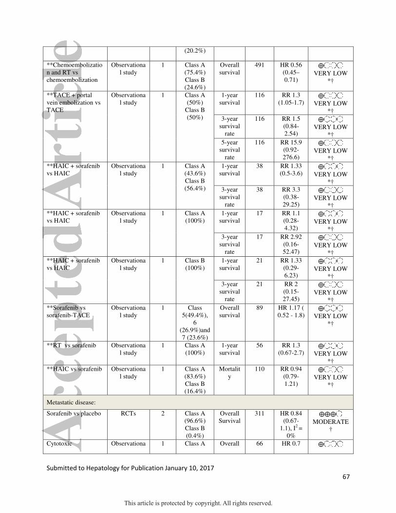
Submitted to Hepatology for Publication January 10, 2017
67
(20.2%)
**Chemoembolizatio
n and RT vs
chemoembolization
Observationa
l study
1 Class A
(75.4%)
Class B
(24.6%)
Overall
survival
491 HR 0.56
(0.45–
0.71)
⨁◯◯◯
VERY LOW
*†
**TACE + portal
vein embolization vs
TACE
Observationa
l study
1 Class A
(50%)
Class B
(50%)
1-year
survival
116 RR 1.3
(1.05-1.7) ⨁◯◯◯
VERY LOW
*†
3-year
survival
rate
116 RR 1.5
(0.84-
2.54)
⨁◯◯◯
VERY LOW
*†
5-year
survival
rate
116 RR 15.9
(0.92-
276.6)
⨁◯◯◯
VERY LOW
*†
**HAIC + sorafenib
vs HAIC
Observationa
l study
1 Class A
(43.6%)
Class B
(56.4%)
1-year
survival
38 RR 1.33
(0.5-3.6) ⨁◯◯◯
VERY LOW
*†
3-year
survival
rate
38 RR 3.3
(0.38-
29.25)
⨁◯◯◯
VERY LOW
*†
**HAIC + sorafenib
vs HAIC
Observationa
l study
1 Class A
(100%)
1-year
survival
17 RR 1.1
(0.28-
4.32)
⨁◯◯◯
VERY LOW
*†
3-year
survival
rate
17 RR 2.92
(0.16-
52.47)
⨁◯◯◯
VERY LOW
*†
**HAIC + sorafenib
vs HAIC
Observationa
l study
1 Class B
(100%)
1-year
survival
21 RR 1.33
(0.29-
6.23)
⨁◯◯◯
VERY LOW
*†
3-year
survival
rate
21 RR 2
(0.15-
27.45)
⨁◯◯◯
VERY LOW
*†
**Sorafenib vs
sorafenib-TACE
Observationa
l study
1 Class
5(49.4%),
6
(26.9%)and
7 (23.6%)
Overall
survival
89 HR 1.17 (
0.52 - 1.8) ⨁◯◯◯
VERY LOW
*†
**RT vs sorafenib Observationa
l study
1 Class A
(100%)
1-year
survival
56 RR 1.3
(0.67-2.7) ⨁◯◯◯
VERY LOW
*†
**HAIC vs sorafenib Observationa
l study
1 Class A
(83.6%)
Class B
(16.4%)
Mortalit
y
110 RR 0.94
(0.79-
1.21)
⨁◯◯◯
VERY LOW
*†
Metastatic disease:
Sorafenib vs placebo RCTs 2 Class A
(96.6%)
Class B
(0.4%)
Overall
Survival
311 HR 0.84
(0.67-
1.1), I2 =
0%
⨁⨁⨁◯
MODERATE
†
Cytotoxic Observationa 1 Class A Overall 66 HR 0.7 ⨁◯◯◯
This article is protected by copyright. All rights reserved.

Submitted to Hepatology for Publication January 10, 2017
68
chemotherapy vs
sorafenib
l study (76.1%)
Class B
(23.9%)
Survival (0.2-1.9) VERY LOW
*†
Chemoembolization
with or without RT vs
sorafenib
Observationa
l study
1 Class A
(64.4%)
Class B
(35.6%)
Overall
Survival
101 HR 0.66
(0.43-
1.02)
⨁◯◯◯
VERY LOW
*†
*Serious risk of bias. †Imprecision
** Studies included only portal vein tumor thrombosis
This article is protected by copyright. All rights reserved.