THE SALIVARY GLANDS DISEASES. Clinical Anatomy of the Salivary Glands.
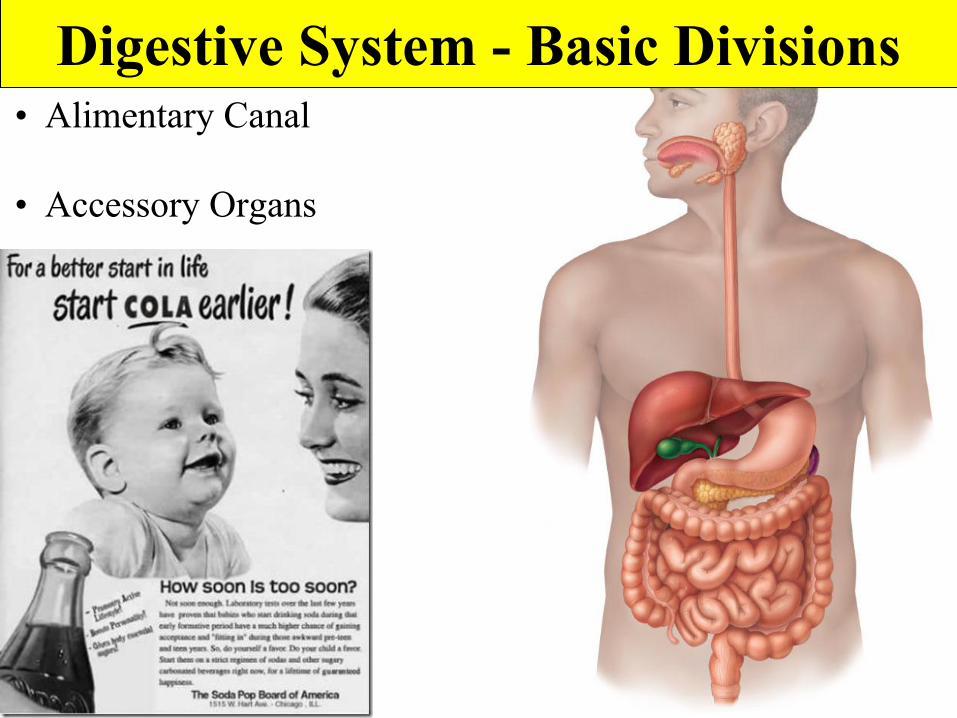
Digestive System - Basic Divisions• Alimentary Canal
• Accessory Organs
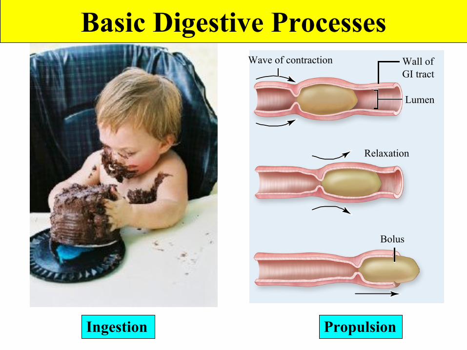
Wave of contraction
Bolus
Relaxation
Wall ofGI tract
Lumen
Basic Digestive Processes
Ingestion Propulsion
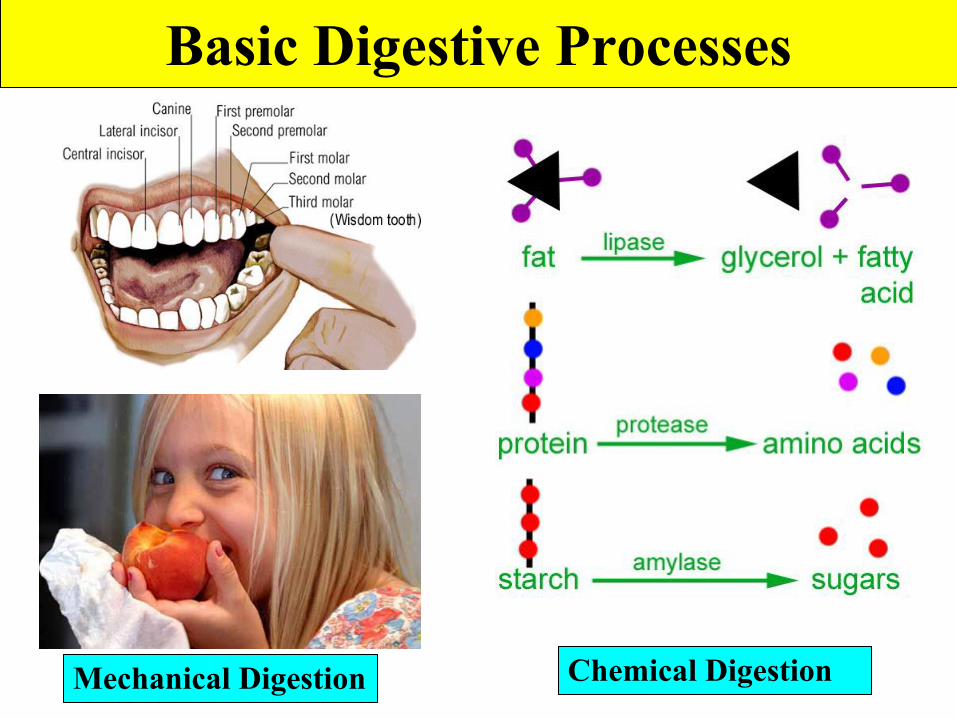
Basic Digestive Processes
Mechanical Digestion Chemical Digestion
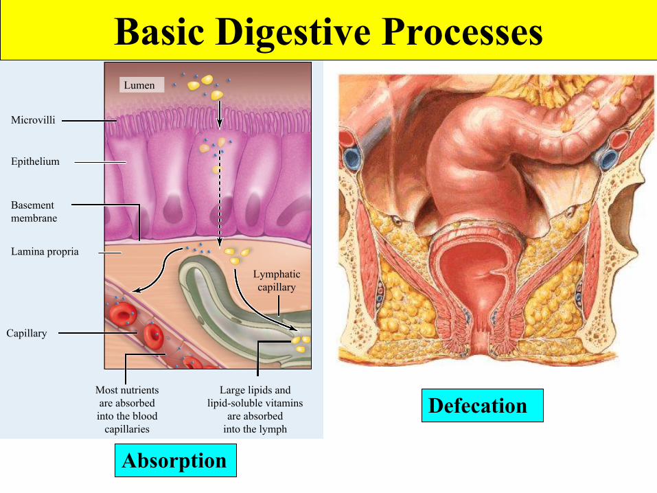
Microvilli
Epithelium
Basementmembrane
Lymphaticcapillary
Large lipids andlipid-soluble vitamins
are absorbedinto the lymph
Most nutrientsare absorbedinto the blood
capillaries
Capillary
Lamina propria
Lumen
Basic Digestive Processes
Absorption
Defecation

Oral Cavity• Extends from oral orifice to oropharynx• Site of ingestion, mechanical digestion, chemical
digestion, and propulsion (deglutition).

Intrinsic Salivary Glands
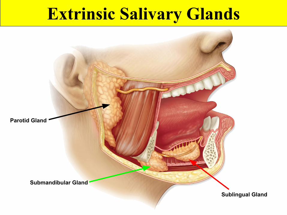
Extrinsic Salivary Glands
Parotid Gland
Submandibular Gland
Sublingual Gland
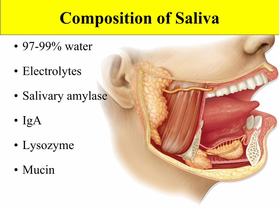
Composition of Saliva• 97-99% water
• Electrolytes
• Salivary amylase
• IgA
• Lysozyme
• Mucin
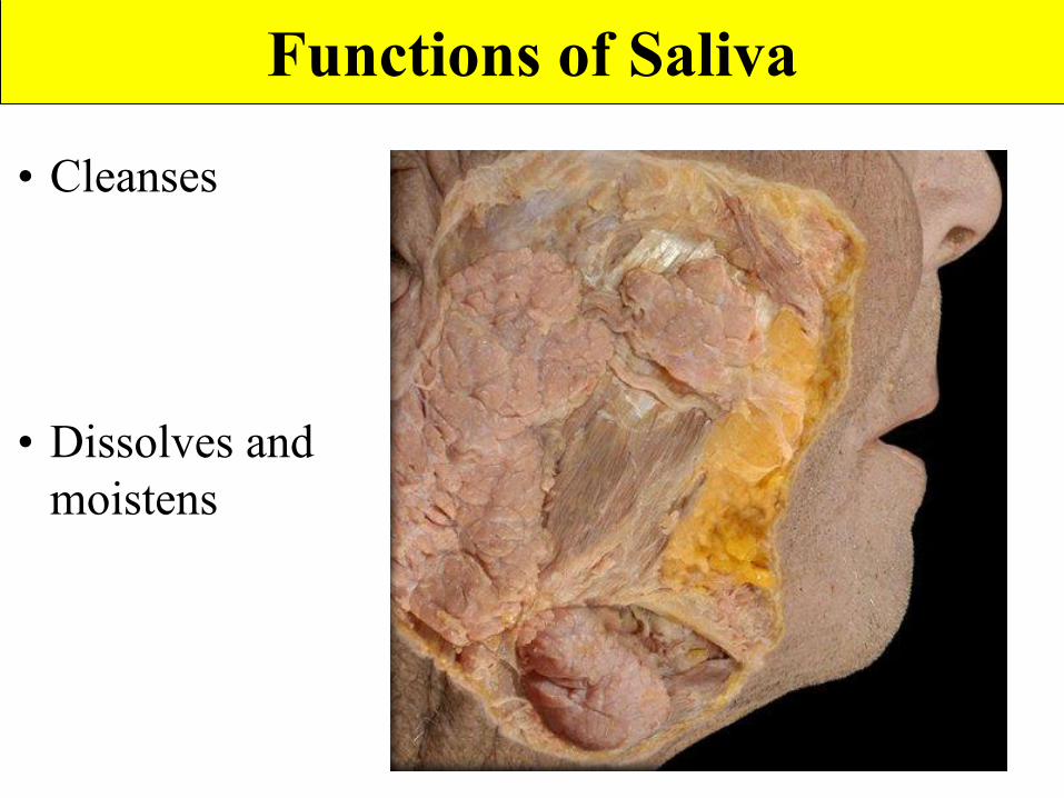
Functions of Saliva
• Cleanses
• Dissolves and moistens
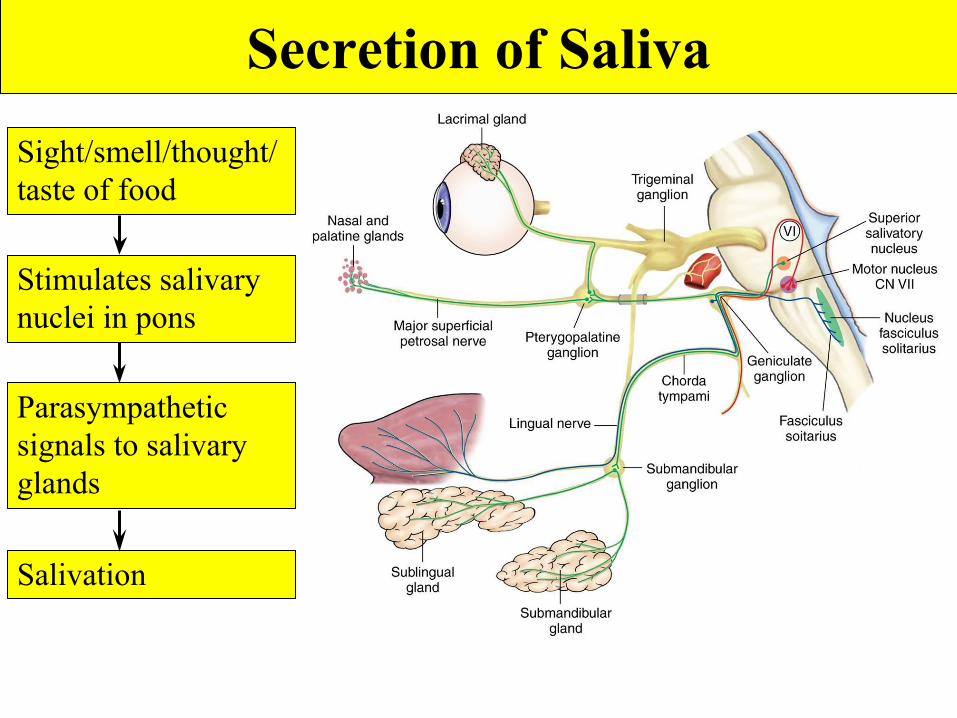
Sight/smell/thought/taste of food
Stimulates salivary nuclei in pons
Parasympathetic signals to salivary glands
Salivation
Secretion of Saliva
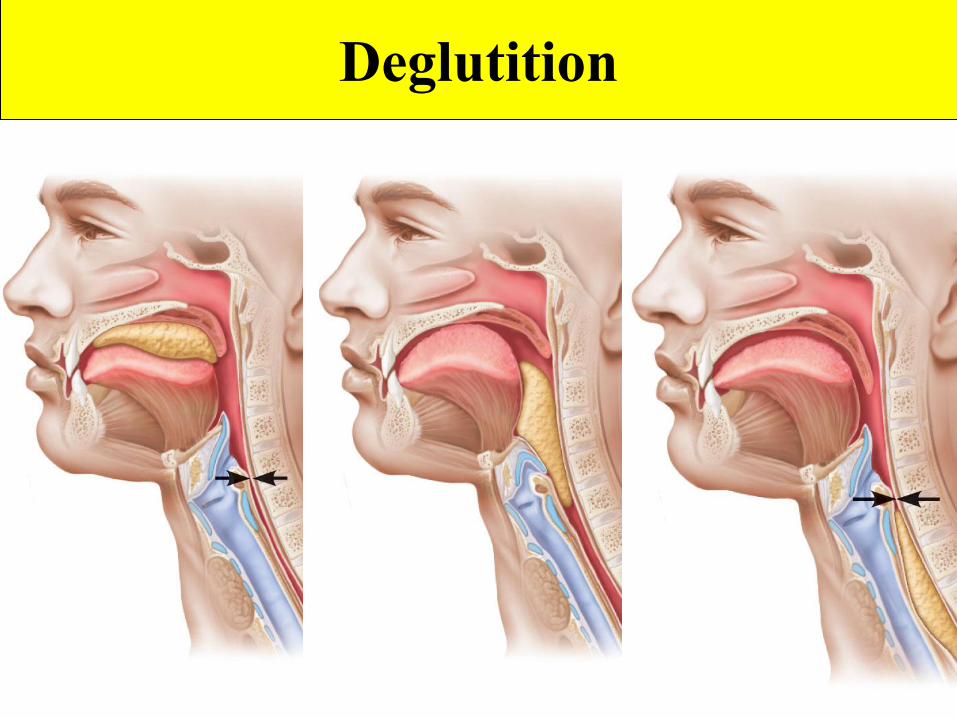
Deglutition
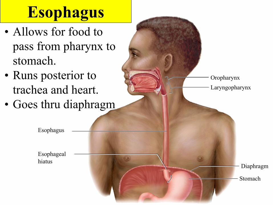
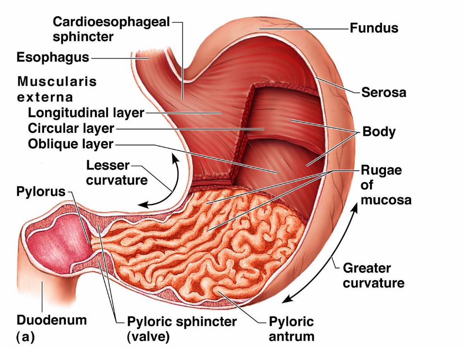
Esophagus
Esophagealhiatus
Stomach
Diaphragm
OropharynxLaryngopharynx
Esophagus• Allows for food to
pass from pharynx to stomach.
• Runs posterior to trachea and heart.
• Goes thru diaphragm
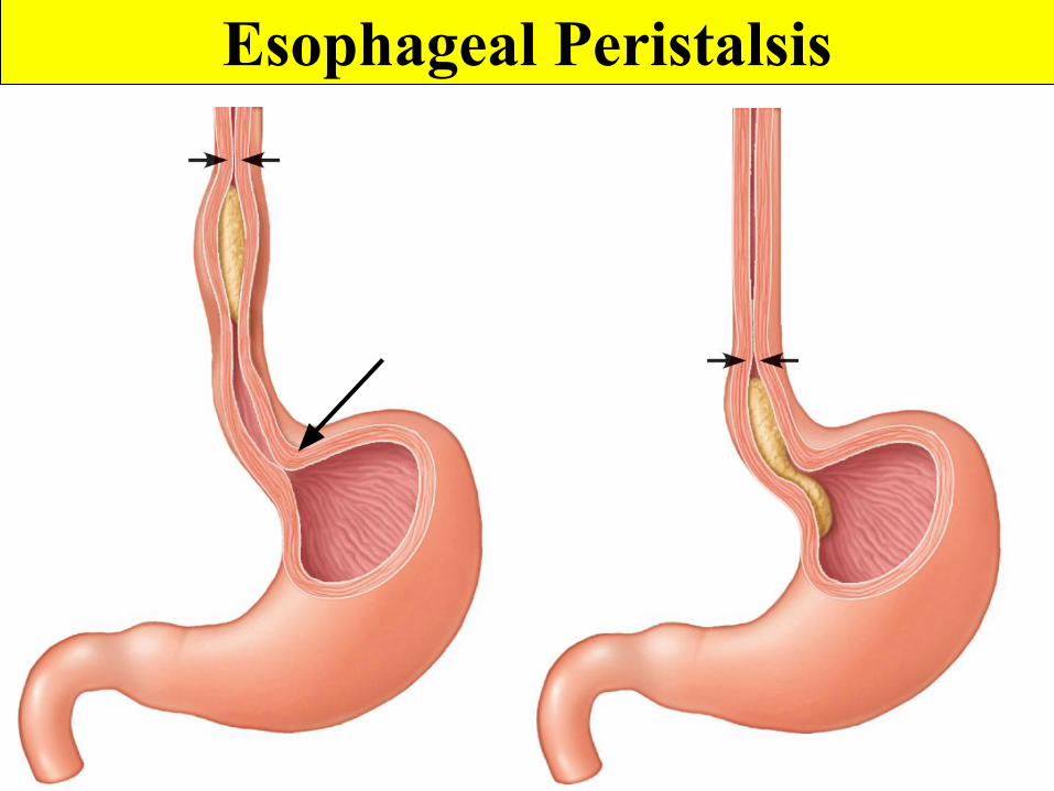
Esophageal Peristalsis
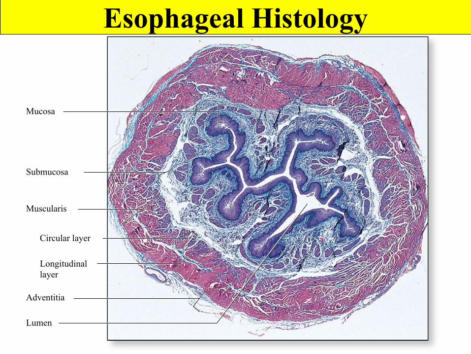
Lumen
Mucosa
Submucosa
Muscularis
Circular layer
Longitudinallayer
Adventitia
Esophageal Histology
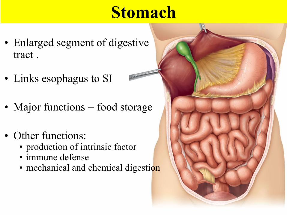
Stomach• Enlarged segment of digestive
tract .
• Links esophagus to SI
• Major functions = food storage
• Other functions:• production of intrinsic factor• immune defense• mechanical and chemical digestion
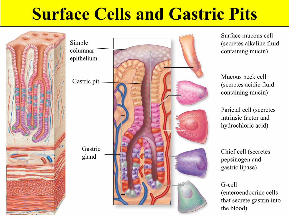
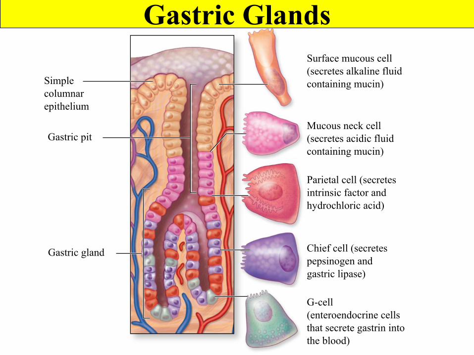
Surface Cells and Gastric Pits
Gastric gland
Surface mucous cell(secretes alkaline fluidcontaining mucin)
Mucous neck cell(secretes acidic fluidcontaining mucin)
Parietal cell (secretesintrinsic factor andhydrochloric acid)
Chief cell (secretespepsinogen andgastric lipase)
G-cell(enteroendocrine cellsthat secrete gastrin intothe blood)
Simplecolumnarepithelium
Gastric pit
Gastric Glands
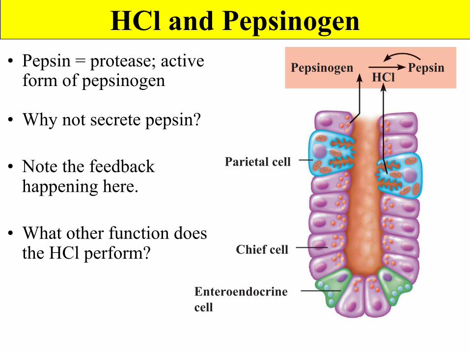
Pepsinogen PepsinHCl
Chief cell
Enteroendocrinecell
Parietal cell
HCl and Pepsinogen• Pepsin = protease; active
form of pepsinogen
• Why not secrete pepsin?
• Note the feedback happening here.
• What other function does the HCl perform?
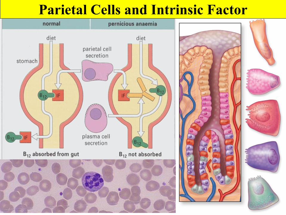
Parietal Cells and Intrinsic Factor
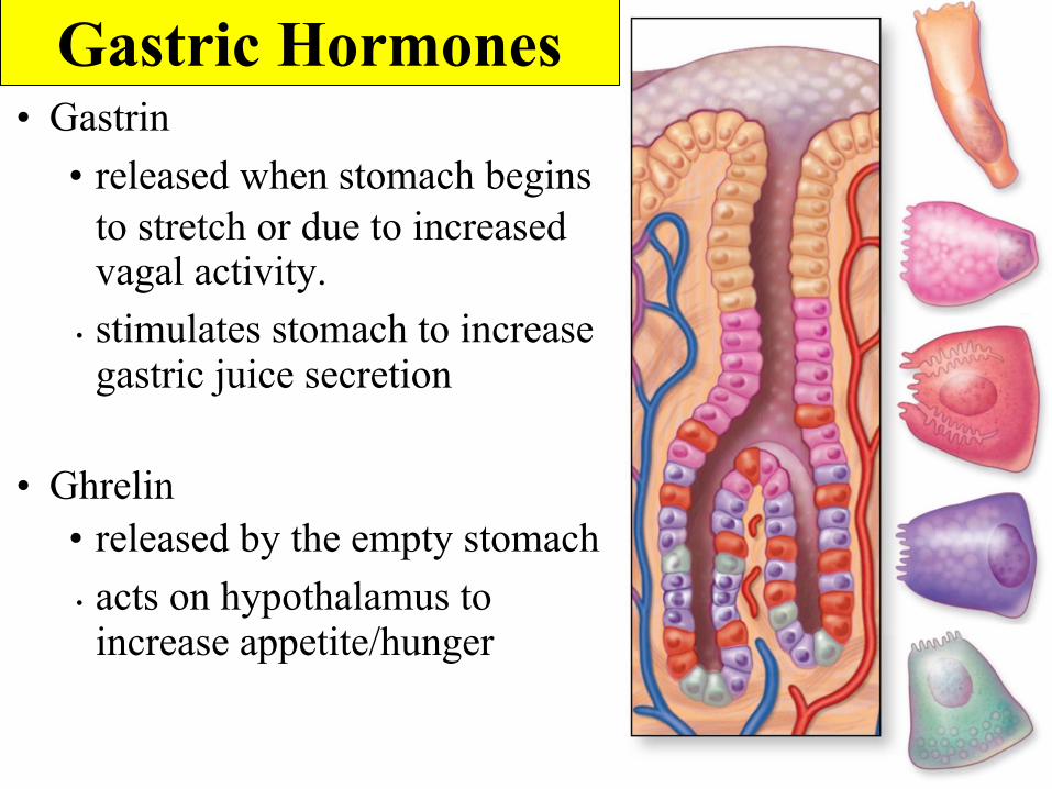
Gastric Hormones• Gastrin
• released when stomach begins to stretch or due to increased vagal activity.
• stimulates stomach to increase gastric juice secretion
• Ghrelin• released by the empty stomach• acts on hypothalamus to increase appetite/hunger
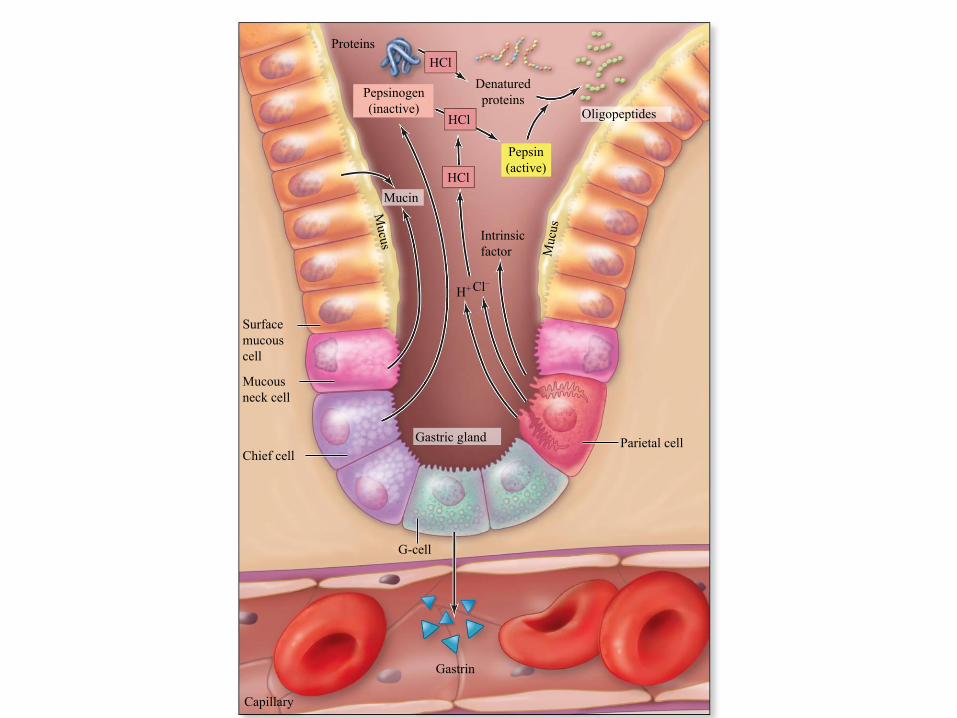
H+
Muc
us
Mucus
ProteinsHCl
Denaturedproteins
Pepsin(active)
HCl
HCl
Surfacemucouscell
Intrinsicfactor
Mucousneck cell
Chief cellParietal cell
G-cell
Gastrin
Capillary
Pepsinogen(inactive)
Cl–
Mucin
Oligopeptides
Gastric gland
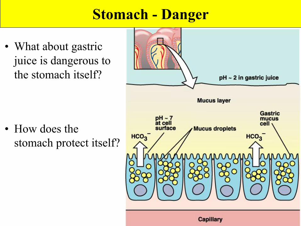
• What about gastric juice is dangerous to the stomach itself?
• How does the stomach protect itself?
Stomach - Danger
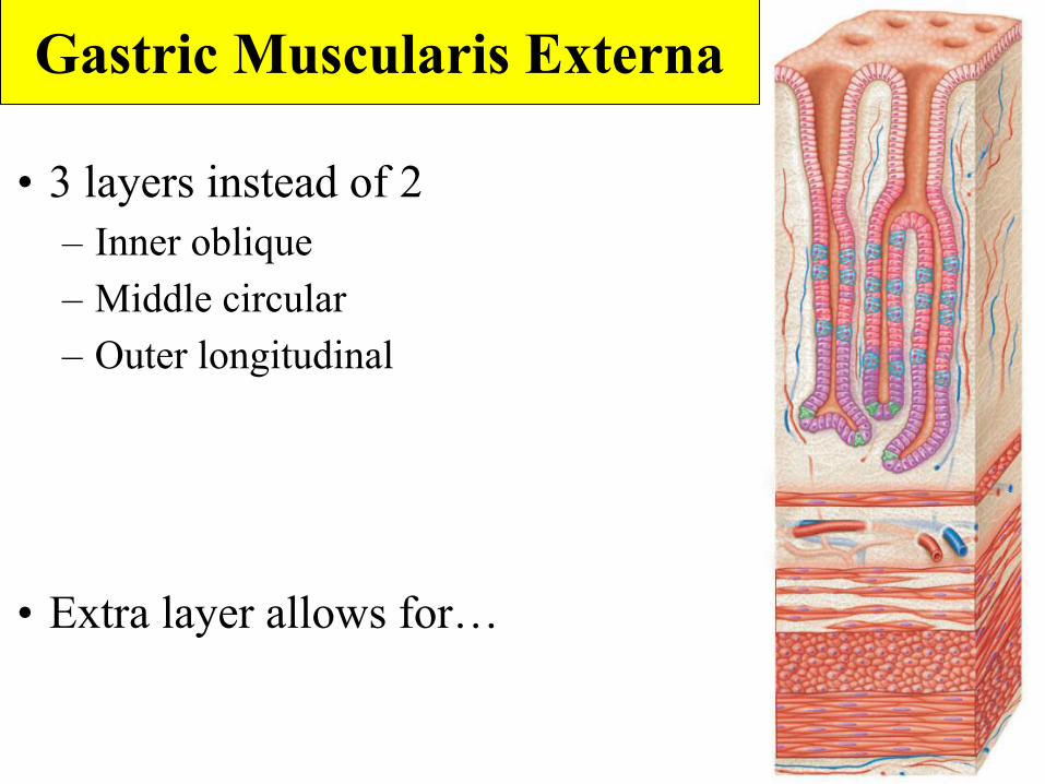
• 3 layers instead of 2– Inner oblique – Middle circular – Outer longitudinal
• Extra layer allows for…
Gastric Muscularis Externa
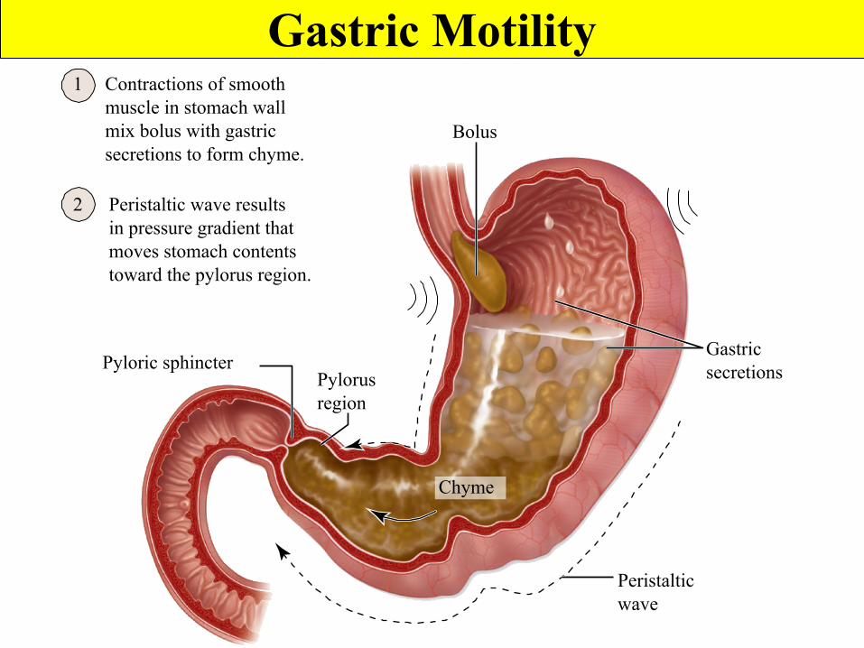
1
2
Contractions of smoothmuscle in stomach wallmix bolus with gastricsecretions to form chyme.
Peristaltic wave resultsin pressure gradient thatmoves stomach contentstoward the pylorus region.
Pyloric sphincterPylorusregion
Peristalticwave
Gastricsecretions
Bolus
Chyme
Gastric Motility
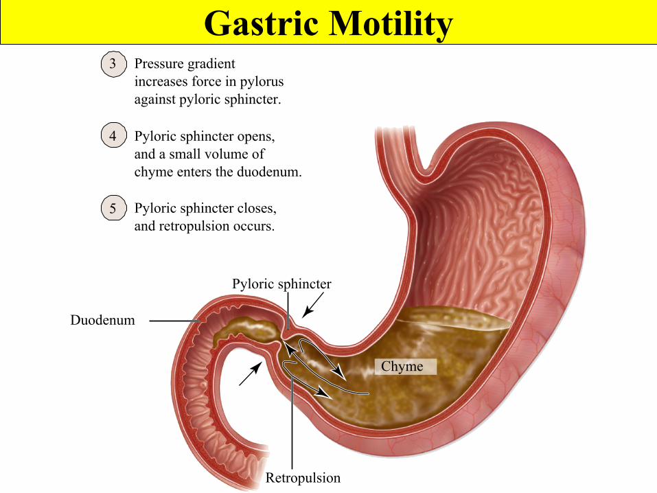
3
4
5
Retropulsion
Duodenum
Pyloric sphincter
Pyloric sphincter closes, and retropulsion occurs.
Pyloric sphincter opens,and a small volume ofchyme enters the duodenum.
Pressure gradientincreases force in pylorusagainst pyloric sphincter.
Chyme
Gastric Motility
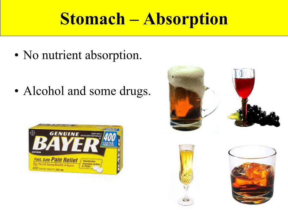
Stomach – Absorption
• No nutrient absorption.
• Alcohol and some drugs.
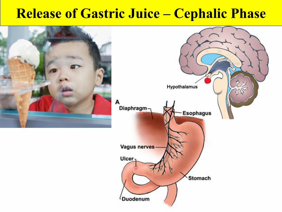
Release of Gastric Juice – Cephalic Phase
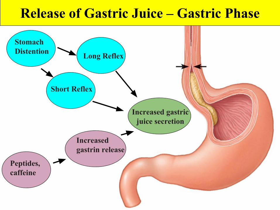
Release of Gastric Juice – Gastric Phase
Stomach Distention Long Reflex
Increased gastric juice secretion
Short Reflex
Peptides, caffeine
Increased gastrin release
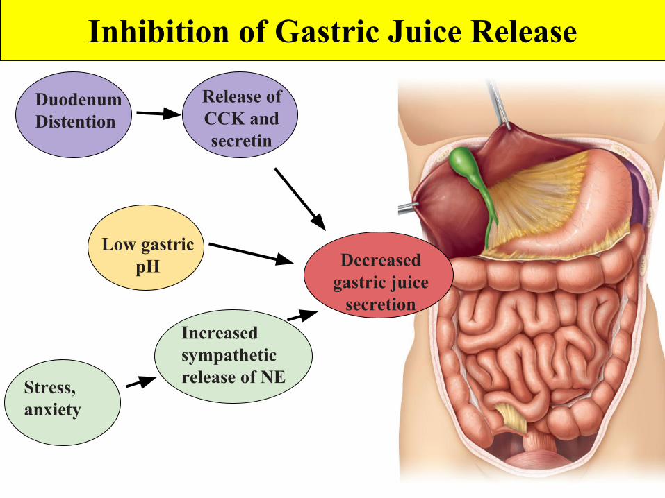
Inhibition of Gastric Juice Release
Duodenum Distention
Release of CCK and secretin
Decreased gastric juice
secretion
Stress, anxiety
Increased sympathetic release of NE
Low gastric pH
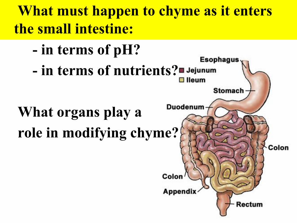
What must happen to chyme as it enters the small intestine:
- in terms of pH?- in terms of nutrients?
What organs play a role in modifying chyme?
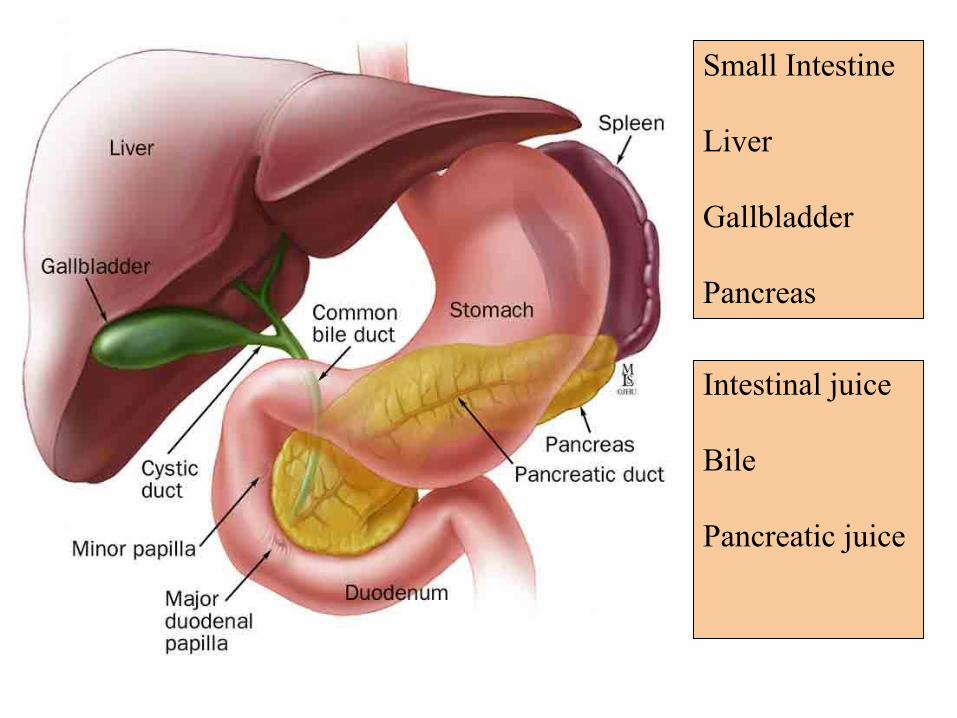
Small Intestine
Liver
Gallbladder
Pancreas
Intestinal juice
Bile
Pancreatic juice
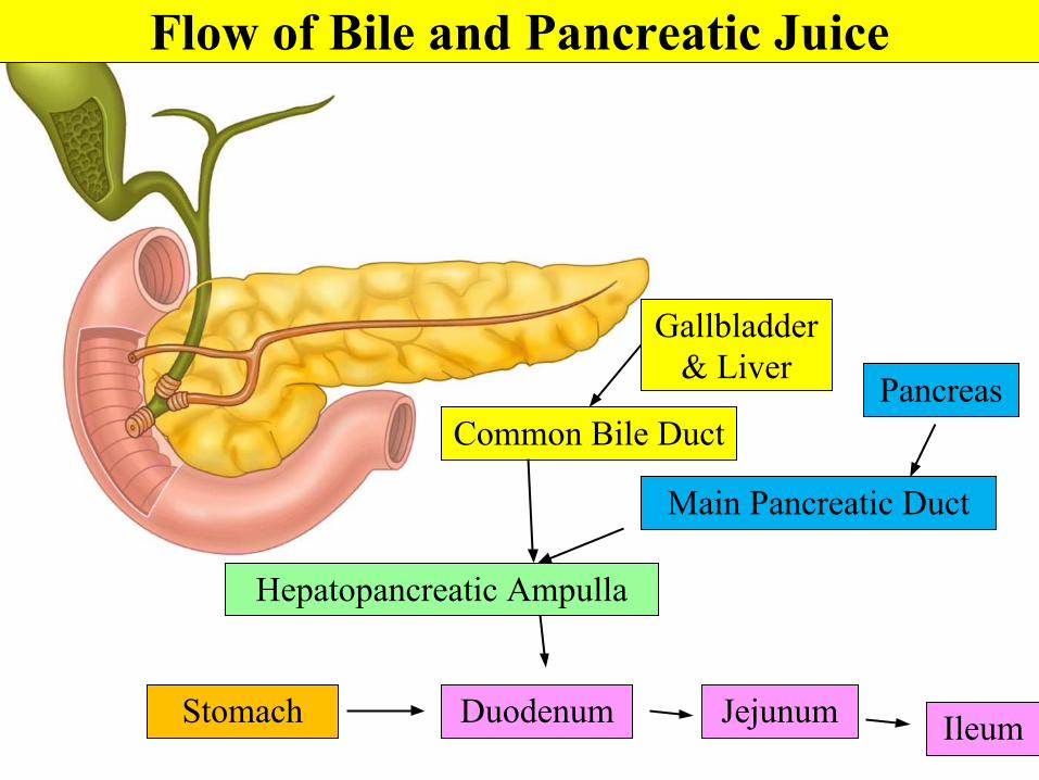
Common Bile Duct
Gallbladder & Liver
Pancreas
Hepatopancreatic Ampulla
Duodenum
Main Pancreatic Duct
Stomach Jejunum Ileum
Flow of Bile and Pancreatic Juice
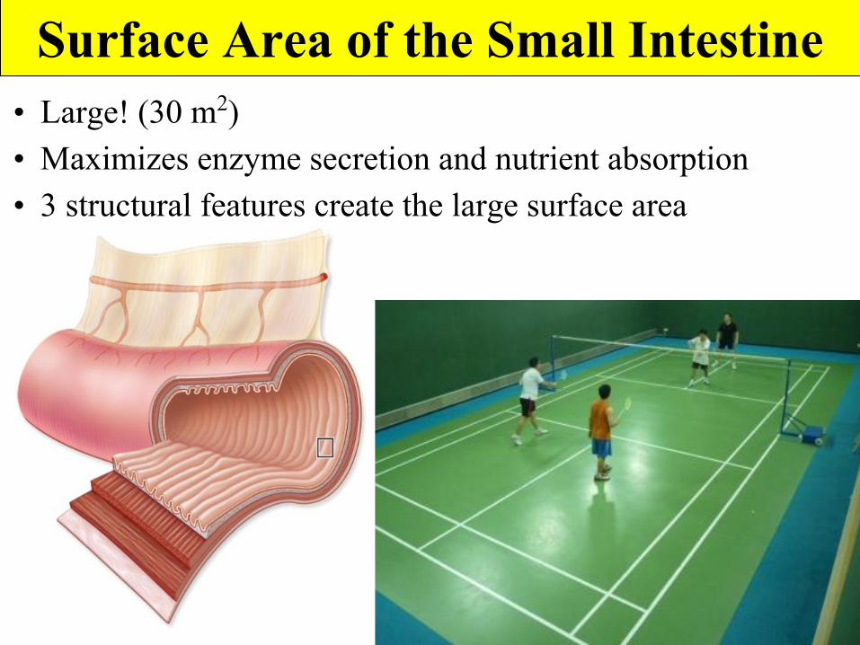
Surface Area of the Small Intestine• Large! (30 m2)• Maximizes enzyme secretion and nutrient absorption• 3 structural features create the large surface area
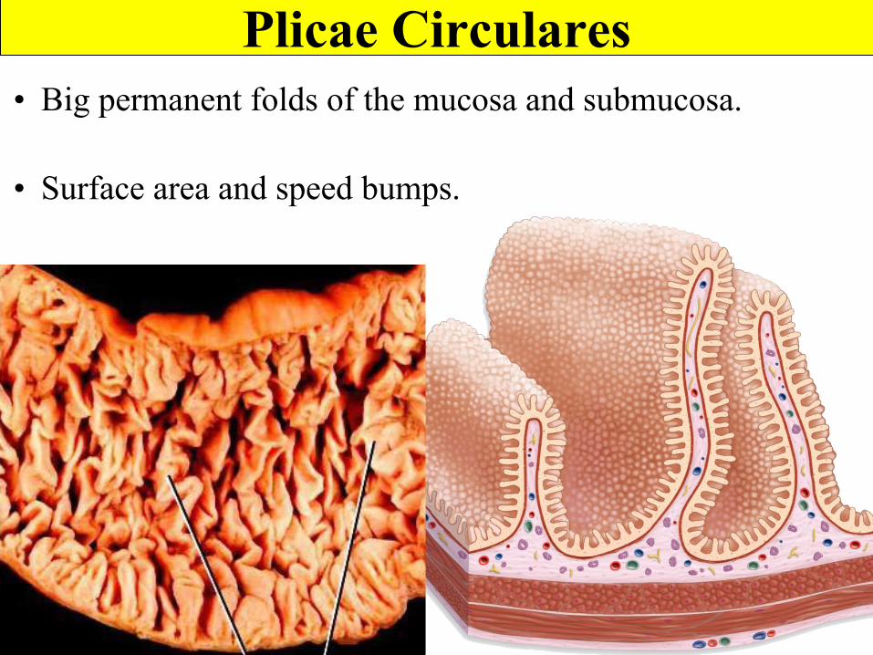
Plicae Circulares• Big permanent folds of the mucosa and submucosa.
• Surface area and speed bumps.
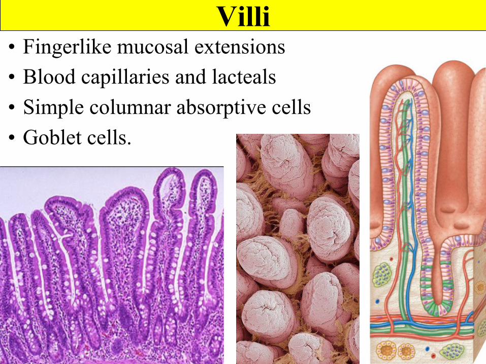
Villi• Fingerlike mucosal extensions• Blood capillaries and lacteals• Simple columnar absorptive cells • Goblet cells.
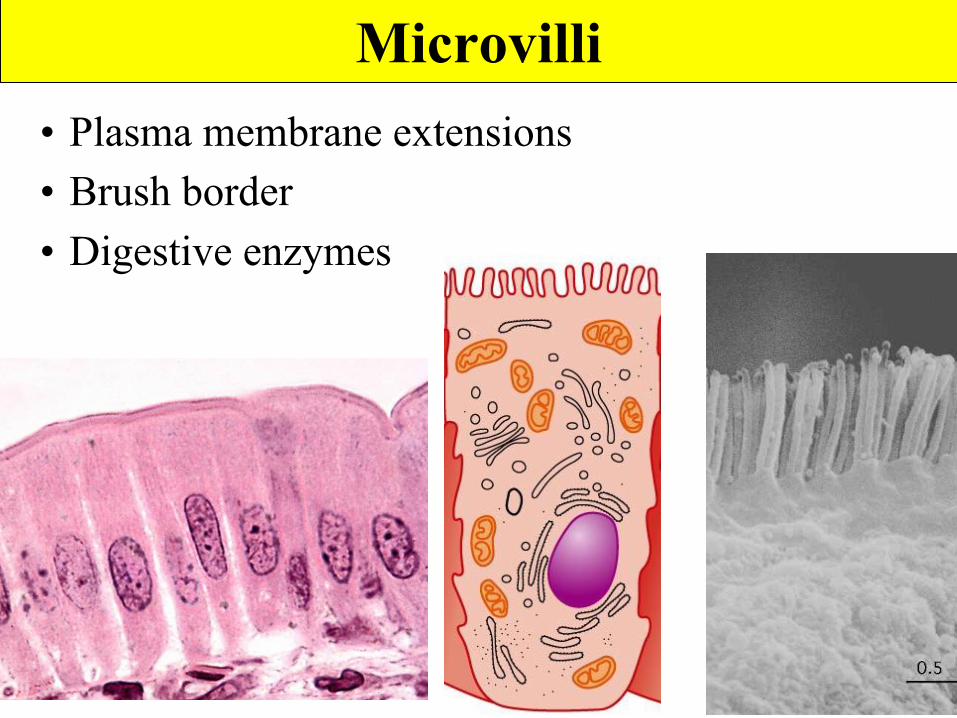
Microvilli• Plasma membrane extensions• Brush border• Digestive enzymes
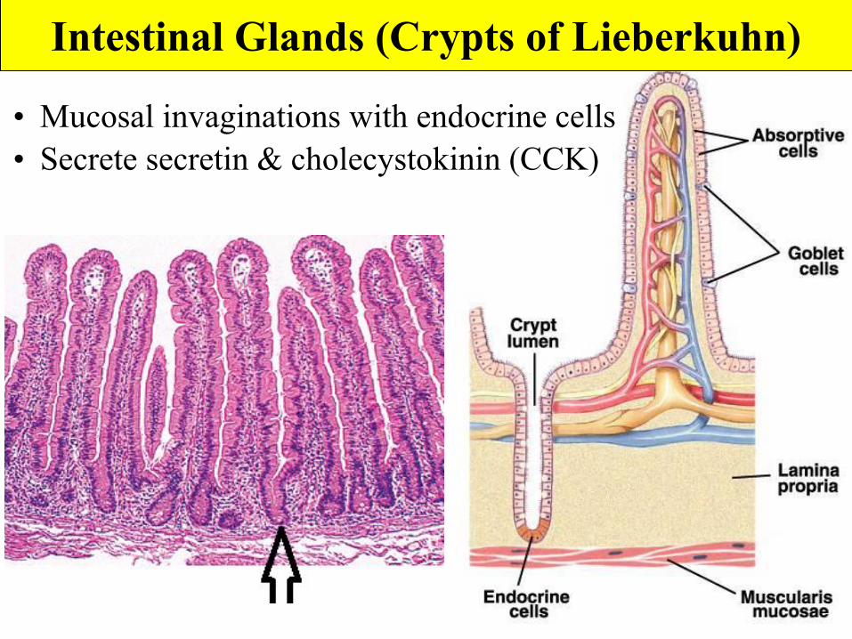
• Mucosal invaginations with endocrine cells• Secrete secretin & cholecystokinin (CCK)
Intestinal Glands (Crypts of Lieberkuhn)
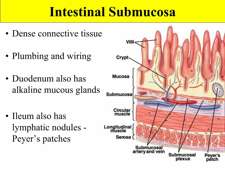
Intestinal Submucosa• Dense connective tissue
• Plumbing and wiring
• Duodenum also has alkaline mucous glands
• Ileum also has lymphatic nodules - Peyer’s patches
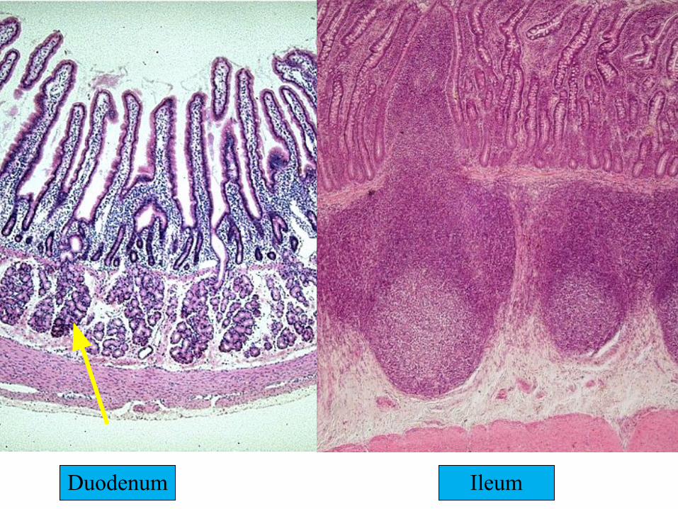
Duodenum Ileum
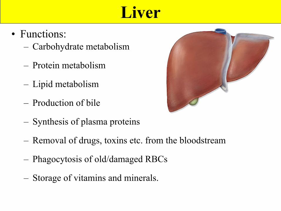
Liver• Functions:
– Carbohydrate metabolism
– Protein metabolism
– Lipid metabolism
– Production of bile
– Synthesis of plasma proteins
– Removal of drugs, toxins etc. from the bloodstream
– Phagocytosis of old/damaged RBCs
– Storage of vitamins and minerals.
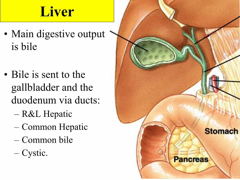
Liver• Main digestive output
is bile
• Bile is sent to the gallbladder and the duodenum via ducts:– R&L Hepatic– Common Hepatic– Common bile– Cystic.
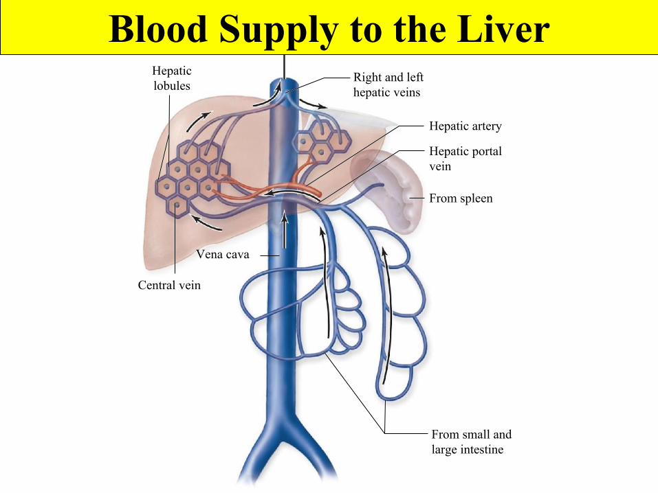
From small andlarge intestine
From spleen
Hepatic portalvein
Right and lefthepatic veins
Hepatic artery
Hepaticlobules
Central vein
Vena cava
Blood Supply to the Liver
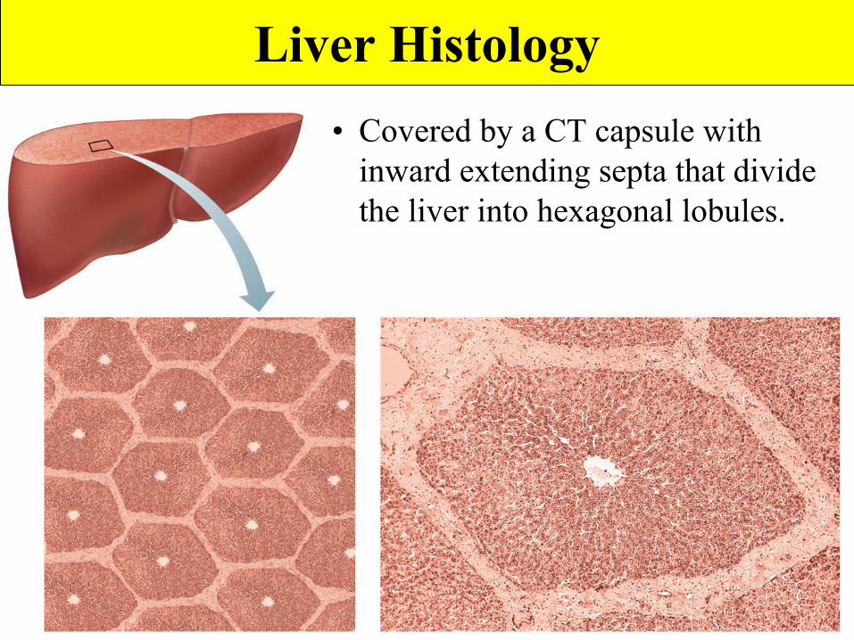
Liver Histology• Covered by a CT capsule with
inward extending septa that divide the liver into hexagonal lobules.
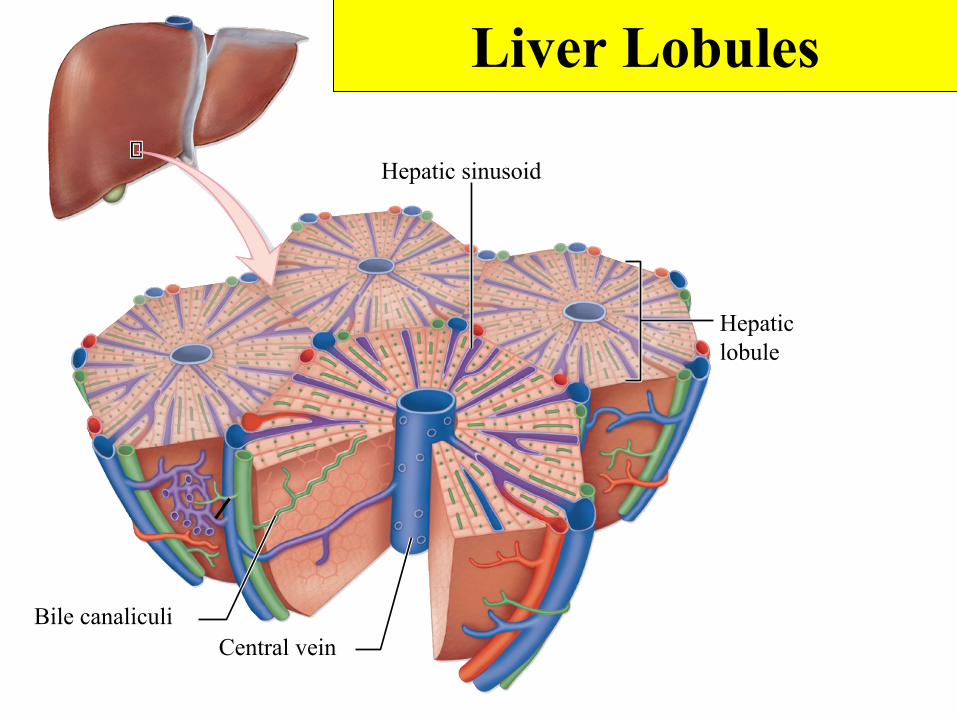
Hepatic sinusoid
Hepaticlobule
Bile canaliculiCentral vein
Liver Lobules
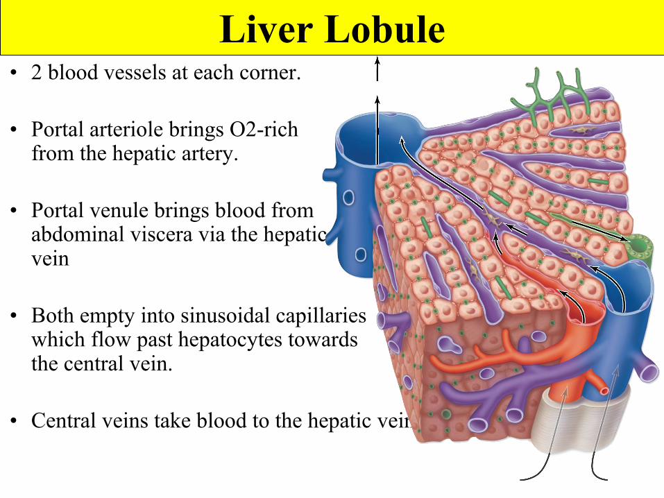
• 2 blood vessels at each corner.
• Portal arteriole brings O2-rich blood from the hepatic artery.
• Portal venule brings blood from abdominal viscera via the hepatic portal vein
• Both empty into sinusoidal capillaries which flow past hepatocytes towards the central vein.
• Central veins take blood to the hepatic vein.
Liver Lobule
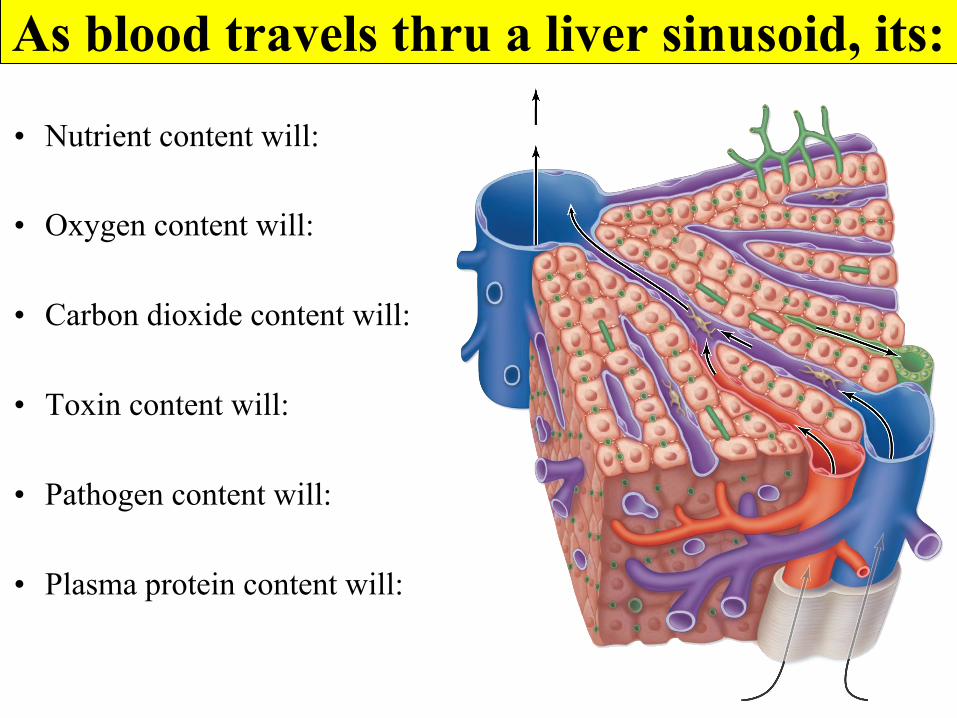
As blood travels thru a liver sinusoid, its:
• Nutrient content will:
• Oxygen content will:
• Carbon dioxide content will:
• Toxin content will:
• Pathogen content will:
• Plasma protein content will:
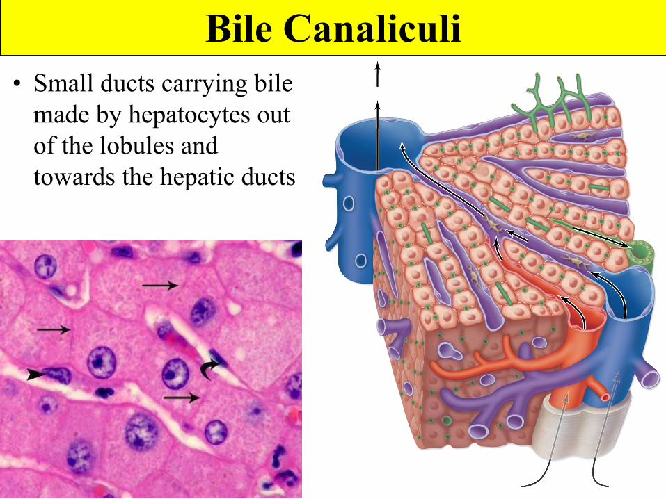
Bile Canaliculi• Small ducts carrying bile
made by hepatocytes out of the lobules and towards the hepatic ducts
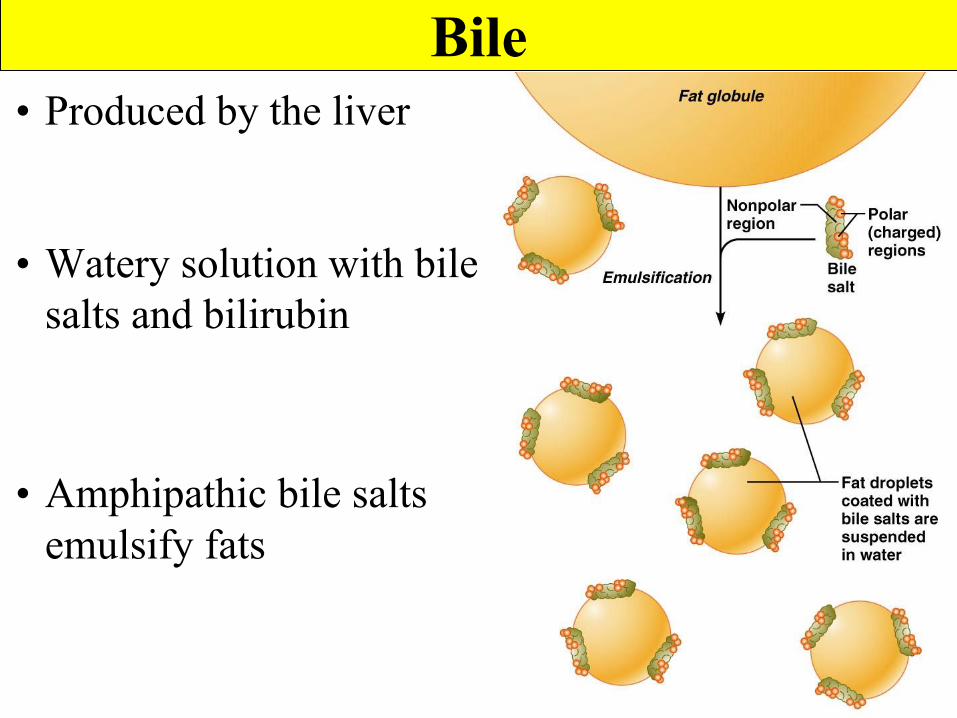
Bile• Produced by the liver
• Watery solution with bile salts and bilirubin
• Amphipathic bile salts emulsify fats
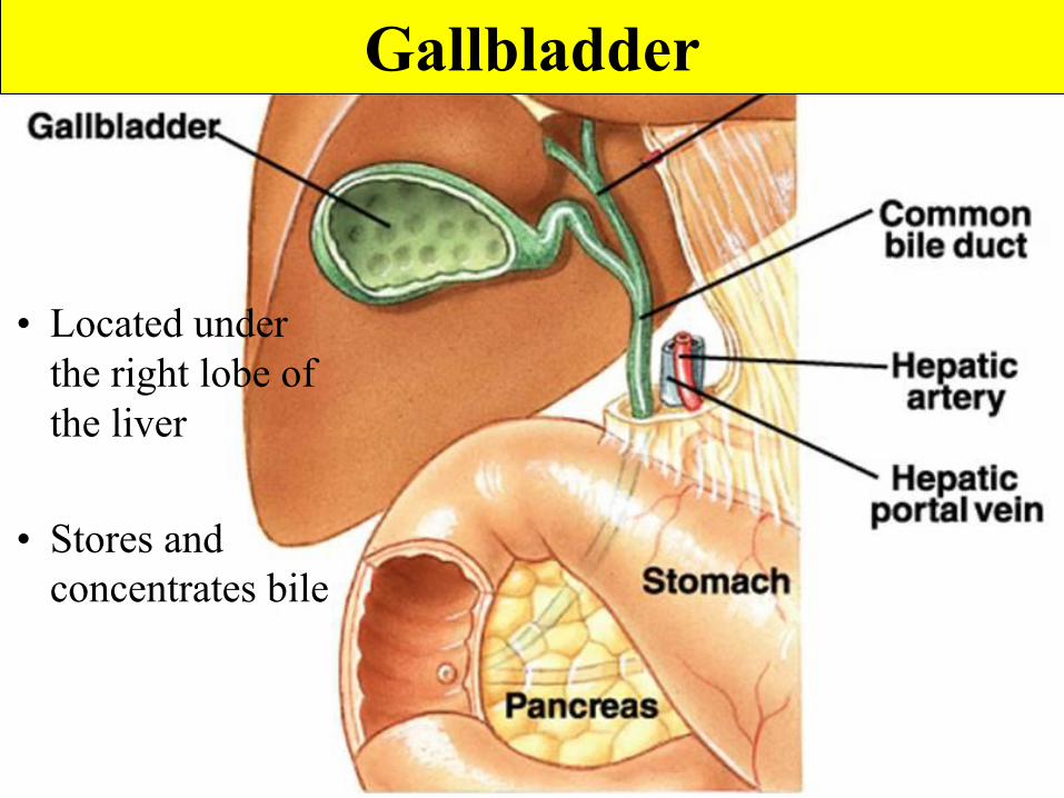
Gallbladder
• Located under the right lobe of the liver
• Stores and concentrates bile

Gallbladder - Histology
• Rugae
• Simple columnar epithelium with microvilli.
• Thick smooth muscle muscularis.
• Serosa
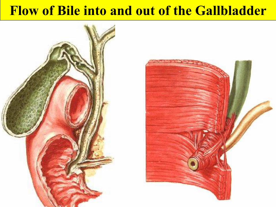
Flow of Bile into and out of the Gallbladder
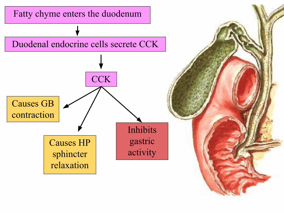
Fatty chyme enters the duodenum
Duodenal endocrine cells secrete CCK
CCK
Causes GB contraction
Causes HP sphincter relaxation
Inhibits gastric activity
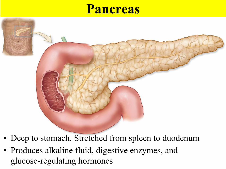
Pancreas
• Deep to stomach. Stretched from spleen to duodenum• Produces alkaline fluid, digestive enzymes, and
glucose-regulating hormones
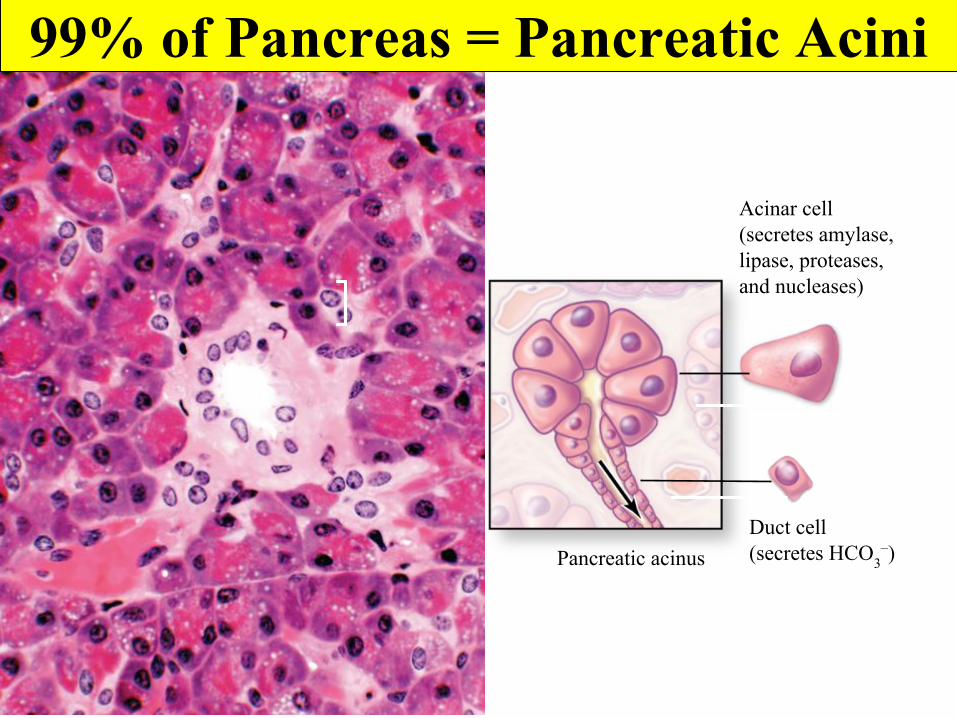
Pancreatic acinusDuct cell(secretes HCO3
–)
Acinar cell(secretes amylase,lipase, proteases,and nucleases)
99% of Pancreas = Pancreatic Acini
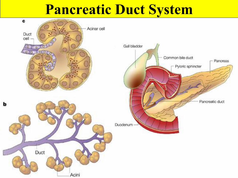
Pancreatic Duct System
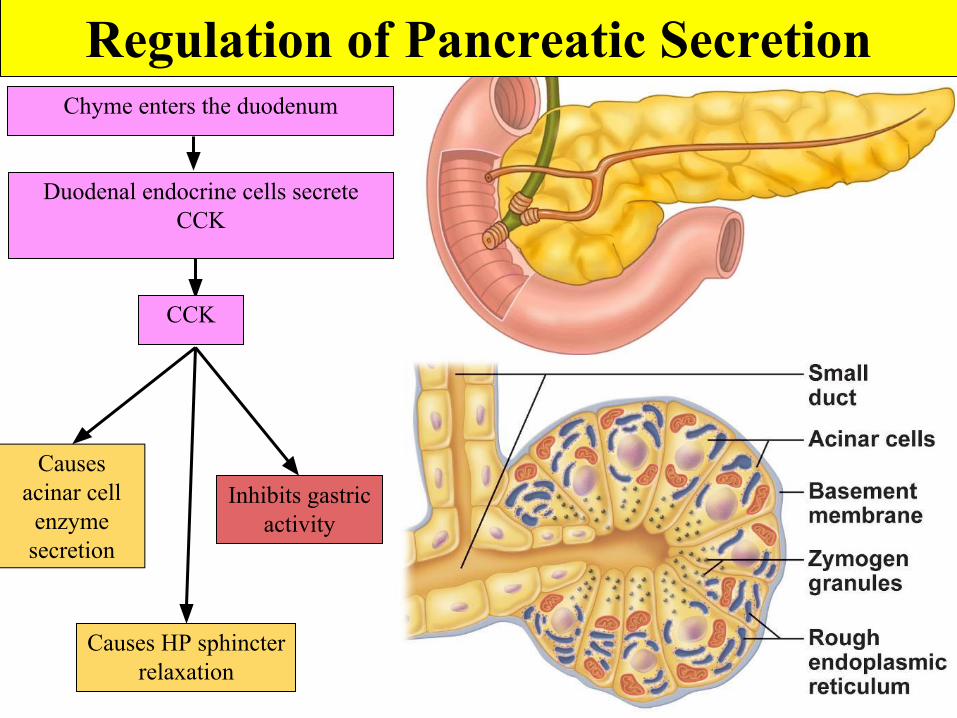
Chyme enters the duodenum
Duodenal endocrine cells secrete CCK
CCK
Causes acinar cell
enzyme secretion
Causes HP sphincter relaxation
Inhibits gastric activity
Regulation of Pancreatic Secretion
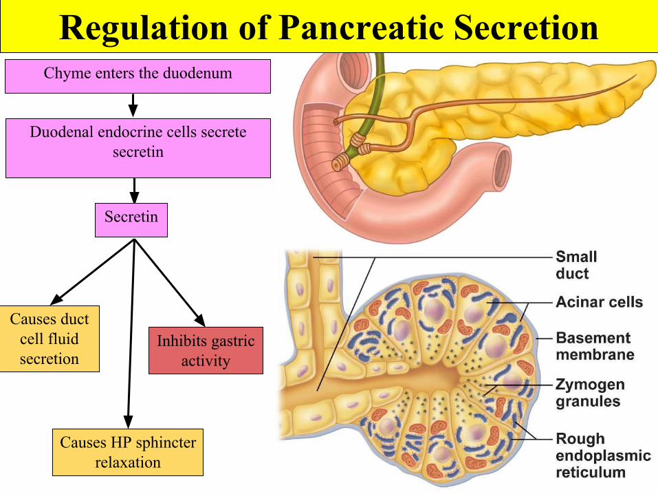
Chyme enters the duodenum
Duodenal endocrine cells secrete secretin
Secretin
Causes duct cell fluid secretion
Causes HP sphincter relaxation
Inhibits gastric activity
Regulation of Pancreatic Secretion
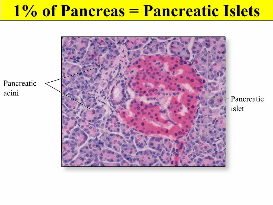
Pancreaticacini
Pancreaticislet
1% of Pancreas = Pancreatic Islets
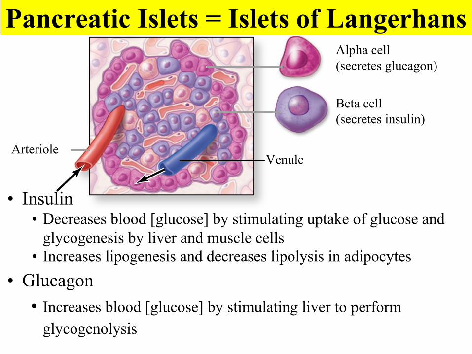
Alpha cell(secretes glucagon)
Beta cell(secretes insulin)
VenuleArteriole
Pancreatic Islets = Islets of Langerhans
• Insulin• Decreases blood [glucose] by stimulating uptake of glucose and
glycogenesis by liver and muscle cells• Increases lipogenesis and decreases lipolysis in adipocytes
• Glucagon• Increases blood [glucose] by stimulating liver to perform
glycogenolysis

Horrible Breakfasts
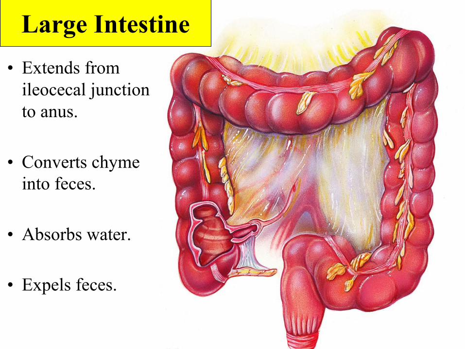
• Extends from ileocecal junction to anus.
• Converts chyme into feces.
• Absorbs water.
• Expels feces.
Large Intestine
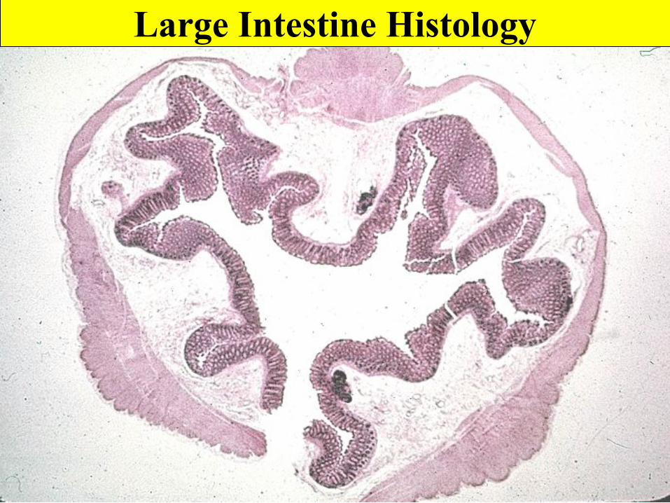
Large Intestine Histology
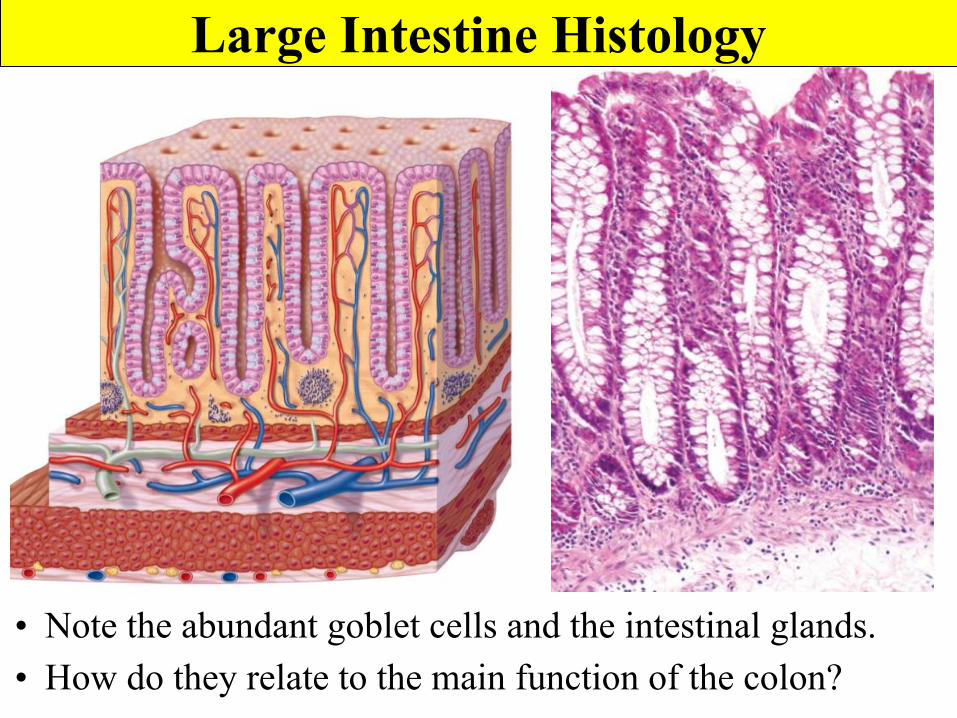
Large Intestine Histology
• Note the abundant goblet cells and the intestinal glands.• How do they relate to the main function of the colon?
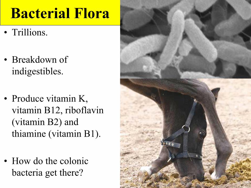
Bacterial Flora• Trillions.
• Breakdown of indigestibles.
• Produce vitamin K, vitamin B12, riboflavin (vitamin B2) and thiamine (vitamin B1).
• How do the colonic bacteria get there?
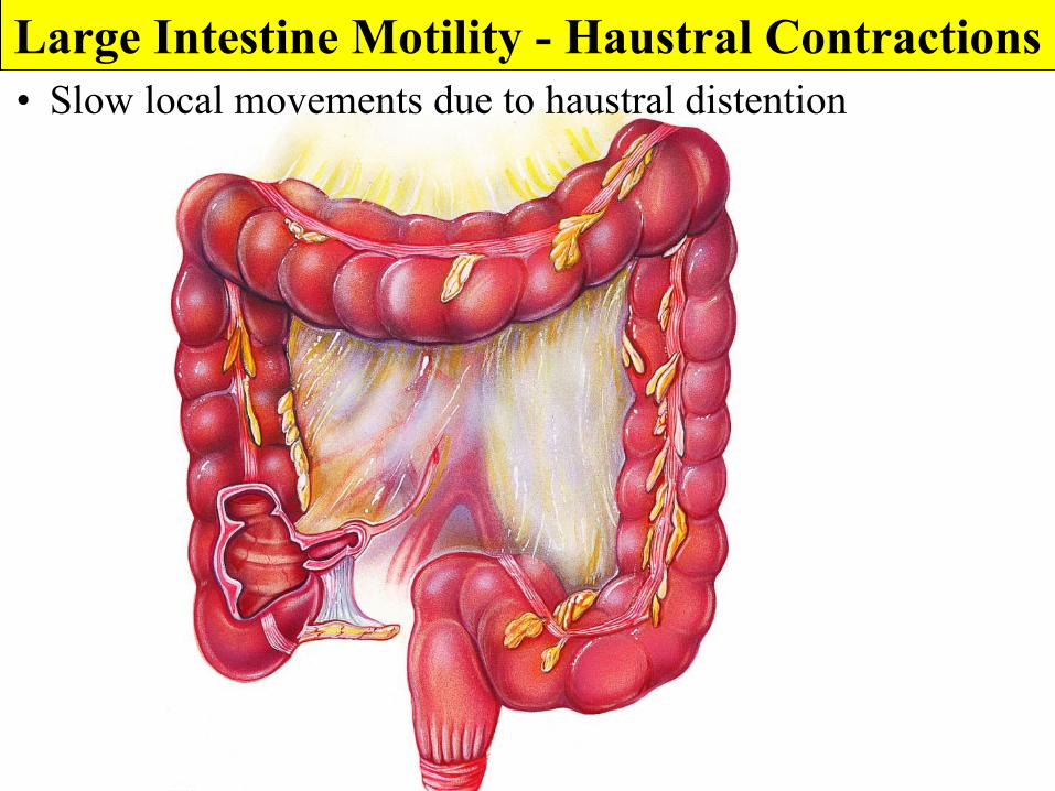
• Slow local movements due to haustral distentionLarge Intestine Motility - Haustral Contractions
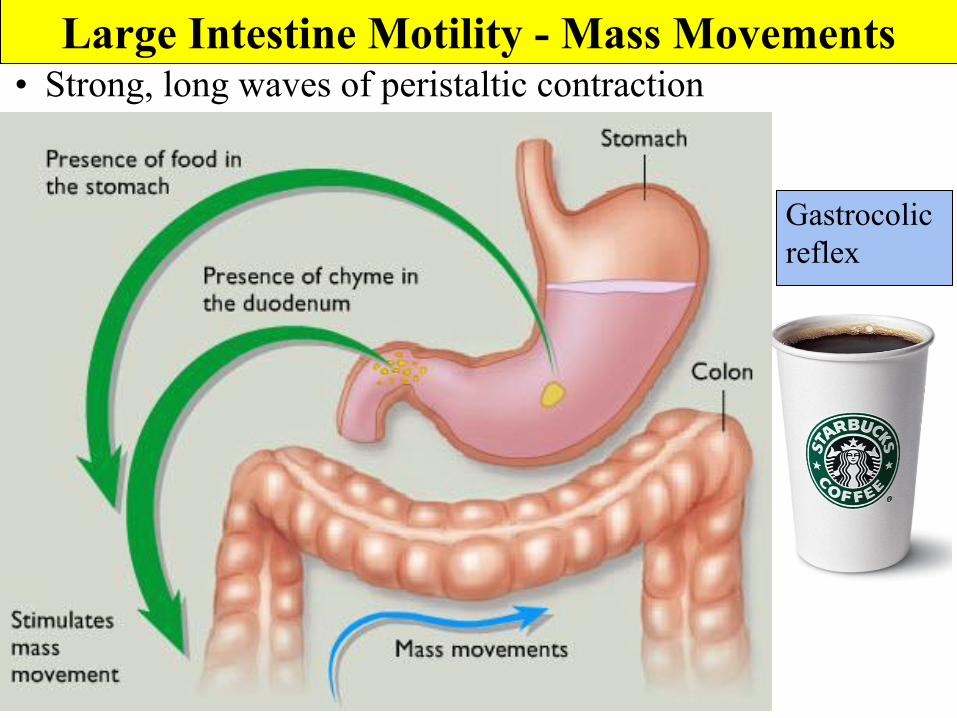
• Strong, long waves of peristaltic contractionLarge Intestine Motility - Mass Movements
Gastrocolic reflex
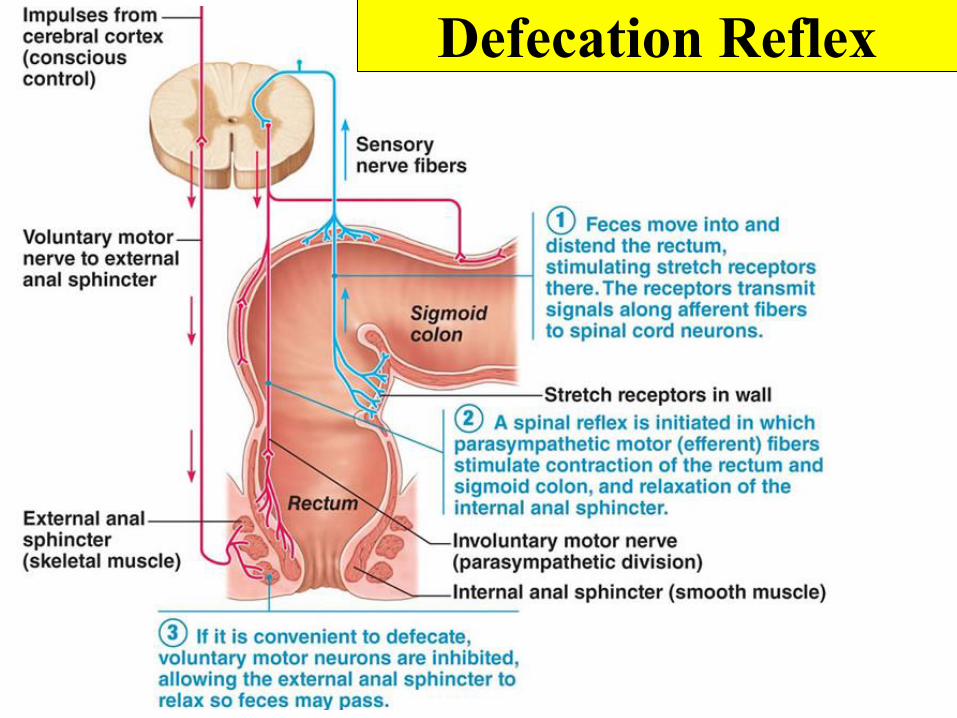
Defecation Reflex
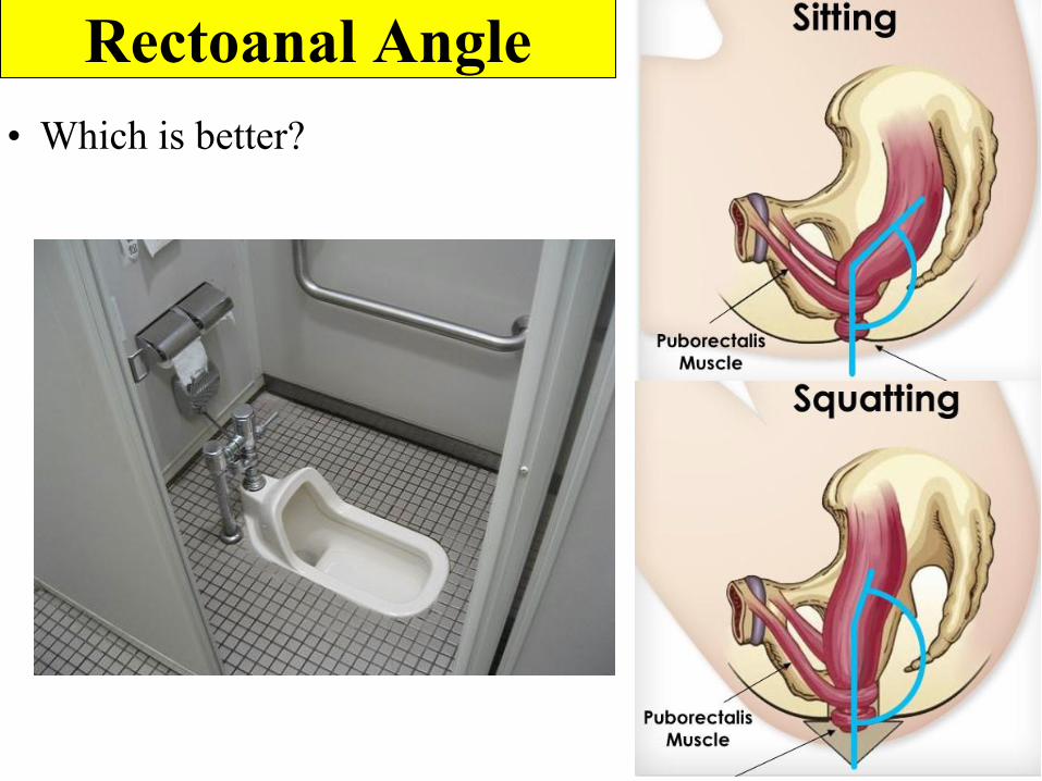
Rectoanal Angle• Which is better?
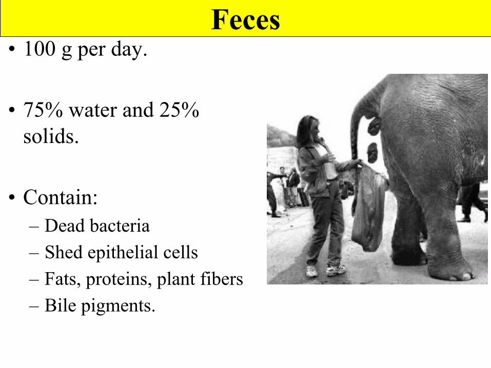
Feces• 100 g per day.
• 75% water and 25% solids.
• Contain:– Dead bacteria– Shed epithelial cells– Fats, proteins, plant fibers– Bile pigments.