Aalborg Universitet Development of methods for studying the physiology...
64
Aalborg Universitet Development of methods for studying the physiology behind the recovery of individuals after stroke Iftime, Simona Denisia Publication date: 2011 Document Version Publisher's PDF, also known as Version of record Link to publication from Aalborg University Citation for published version (APA): Iftime Nielsen, S. D. (2011). Development of methods for studying the physiology behind the recovery of individuals after stroke. Center for Sensory-Motor Interaction (SMI), Department of Health Science and Technology, Aalborg University. General rights Copyright and moral rights for the publications made accessible in the public portal are retained by the authors and/or other copyright owners and it is a condition of accessing publications that users recognise and abide by the legal requirements associated with these rights. ? Users may download and print one copy of any publication from the public portal for the purpose of private study or research. ? You may not further distribute the material or use it for any profit-making activity or commercial gain ? You may freely distribute the URL identifying the publication in the public portal ? Take down policy If you believe that this document breaches copyright please contact us at [email protected] providing details, and we will remove access to the work immediately and investigate your claim. Downloaded from vbn.aau.dk on: juni 11, 2018
Transcript of Aalborg Universitet Development of methods for studying the physiology...
Aalborg Universitet
Development of methods for studying the physiology behind the recovery ofindividuals after strokeIftime, Simona Denisia
Publication date:2011
Document VersionPublisher's PDF, also known as Version of record
Link to publication from Aalborg University
Citation for published version (APA):Iftime Nielsen, S. D. (2011). Development of methods for studying the physiology behind the recovery ofindividuals after stroke. Center for Sensory-Motor Interaction (SMI), Department of Health Science andTechnology, Aalborg University.
General rightsCopyright and moral rights for the publications made accessible in the public portal are retained by the authors and/or other copyright ownersand it is a condition of accessing publications that users recognise and abide by the legal requirements associated with these rights.
? Users may download and print one copy of any publication from the public portal for the purpose of private study or research. ? You may not further distribute the material or use it for any profit-making activity or commercial gain ? You may freely distribute the URL identifying the publication in the public portal ?
Take down policyIf you believe that this document breaches copyright please contact us at [email protected] providing details, and we will remove access tothe work immediately and investigate your claim.
Downloaded from vbn.aau.dk on: juni 11, 2018

1
Development of methods for studying the physiology
behind the recovery of individuals after stroke
– PhD THESIS –
SIMONA DENISIA IFTIME NIELSEN
Center for Sensory-Motor Interaction, Aalborg University
Aalborg, March 2011

2
ISBN (print edition): 978-87-7094-109-9
ISBN (electronic edition): 978-87-7094-110-5
3
The PhD thesis is based on the following articles:
ARTICLE 1
Simona Denisia Iftime Nielsen, Rune Jersin Vingborg, Thomas Sinkjær, Mark Schram Christense,
Andreas Roepstorff & Michael James Grey.‟ Interaction of electrical stimulation and voluntary hand
movement in SII and the cerebellum during simulated therapeutic Functional Electrical Stimulation‟.
Human Brain Mapping (published) (DOI 10.1002/hbm.21191)
ARTICLE 2
Simona Denisia Iftime Nielsen, Thomas Sinkjær, Michael James Grey, Omar Feix do Nascimento.„ The
dynamics of cortical modulation associated with voluntary movement task and peripheral electrical
stimulation task. Journal of Neuroscience Methods (submitted)
ARTICLE 3
Simona Denisia Iftime Nielsen; Strahinja Došen, Mirjana Popović, Dejan Popović. „Learning of arm/hand
coordination with an altered visual input‟. Computational Intelligence and Neuroscience (published)
(DOI pii: 520781)
The research work has been carried out at the Center for Sensory-Motor Interaction, Aalborg University,
Denmark and Center for Functionally Integrative Neuroscience, Aarhus Hospital.

4
Acknowledgements
I am grateful to my initial supervisor Thomas Sinkjær and co-supervisor Mike James Grey for their
involvement and active support throughout these years. I greatly appreciate their availability and
dedication.
I wish to express my sincere thanks to my final supervisor Dejan Popovic for all his effort, positive
attitude and constructive criticism that guided me all these years. His advice and support gave me all the
time the right direction. My special tanks go also to Mirjana Popovic for excellent input and meaningful
discussions. Everything that I learned working with them was invaluable to me.
I am grateful to my co-authors, Andreas Ropestorff, Omar Feix do Nasimento, Mark Schram Christensen
and Rune Vingborg for their important and insightful inputs. Thank you for an excellent collaboration and
for sharing with me knowledge and professional experience.
Thanks to Line and Gery, my officemates, and to all my PhD colleagues and friends from SMI for
interesting discussions and for sharing with me time and ideas.
Thanks to SMI administrative and technical staff for being a true support all these years and for providing
the assistance that I needed.
This PhD thesis is dedicated to my parents, Elena and Mihai, and my sister Diana that actively
accompanied me throughout this PhD journey. The thesis is also dedicated to my son Nicolai that is
filling my life with smiles.
A journey is ending, a new one is starting.
March 2011 Simona D.I. Nielsen
5
MOTIVATION FOR THIS RESEARCH .................................................................................................................................. 7
REFERENCES ........................................................................................................................................................................ 10
CHAPTER 1 ............................................................................................................................................................................... 12
INTRODUCTION ...................................................................................................................................................................... 12
1.1 CORTICAL PLASTICITY ................................................................................................................................................ 13 1.2 MOTOR LEARNING: IMPLICATIONS FOR REHABILITATION ............................................................................... 14 1.3 THERAPIES ...................................................................................................................................................................... 17 1.4 NEURONAL CHANGES THAT FOLLOW THE THERAPIES ...................................................................................... 23 1.5 PHD PROJECT GOALS .................................................................................................................................................... 26 1.6. ORGANIZATION OF PHD THESIS ................................................................................................................................ 30 REFERENCES ........................................................................................................................................................................ 32
CHAPTER 2 ............................................................................................................................................................................... 38
METHODOLOGY ..................................................................................................................................................................... 38
2.1TECHNIQUES TO STUDY CORTICAL REORGANIZATION ...................................................................................... 38 2.2 ACTIVITIES STUDIED WITH THE COMPLEXITY OF DISTINGUISHING ............................................................... 39 2.3 STUDYING LEARNING .................................................................................................................................................. 44 REFERENCES ........................................................................................................................................................................ 46
CHAPTER 6 ............................................................................................................................................................................... 47
DISCUSSIONS ........................................................................................................................................................................... 47
6. 1 STUDY 1 .......................................................................................................................................................................... 47 6. 2 STUDY 2 .......................................................................................................................................................................... 49 6.3 STUDY 3 ........................................................................................................................................................................... 50 REFERENCES ........................................................................................................................................................................ 54
CHAPTER 7 ............................................................................................................................................................................... 55
CONSIDERATIONS AND FUTURE PROSPECTRIVES .................................................................................................... 55
REFERENCES ........................................................................................................................................................................ 57
SUMMARY ................................................................................................................................................................................ 58
SAMMENFATNING ................................................................................................................................................................. 61

6
List of abbreviations ARAT Action Research Arm Test
BOLD blood oxygen-level dependent
CIMT Constraint Induced Movement Therapy
CNS Central Nervous System
EEG Electroencephalography
ERP Evoked responses potentials
ES Electrical stimulation
fMRI functional Magnetic Resonance Imaging
FES Functional Electrical Stimulation
FET Functional Electrical Therapy
MEG Magnetoencephalography
MRCPs Movement related cortical potentials
PET Positron Emission Tomography
SII Secondary somatosensory areas
tFES therapeutic FES
TENS Transcutaneous electrical nerve stimulation
TMS Transcranial magnetic stimulation
VR Virtual Reality
7
MOTIVATION FOR THIS RESEARCH
Rationale for neuroimaging research: understanding the mechanism
Motor impairment after stroke is common following damage to areas of the brain normally involved in
planning and executing motor commands. Regeneration of damaged tissue in adults is limited, which
suggests that real improvement in motor function observed over weeks or months following stroke is a
consequence of reorganization of the surviving elements of the motor network. The mainstay of treatment
is neurorehabilitation (Popović et al. 2003; (Rossini et al. 2003; Popović et al. 2004; Young and Forster
2007). The overall approach is effective and the benefit of strategies aimed at helping patients adapt to
impairment well proven (Ward et al. 2003). A better understanding of the mechanisms underlying
recovery (or deterioration) of function after a CNS lesion, as well as those leading to maladaptive or
unfavourable outcomes, would be essential for directing specific and effective rehabilitative strategies as
well as avoiding potentially harmful interventions (Rossini et al. 2003).
The biological basis of post-stroke recovery of function, particularly that occurring after following a
rehabilitation therapy, has long remained elusive. In spite of clinical research (Ward et al. 2003; Weiller
et al. 1992; Cramer et al. 1997; Marshall et al. 2000), there is a lack of objective methodologies
applicable to humans. The decision regarding the choice of the appropriate therapy to patient subgroups is
based on clinical examination and specialists‟ experience. It should take in account the best fit between
the neural status of the patient and the expected neural consequences of the therapy.
The success of a treatment depends on the ability to drive functionally relevant reorganization in
surviving brain regions and networks. This will vary dramatically across patients. Neuroimaging
techniques can allow the assessment of how treatments interact with residual functional anatomy, which
will inform mechanisms of action and allow targeted application of therapies based on neuroscientific
principles (Ward 2005).
Functional Electrical Stimulation (FES) is used in the rehabilitation therapy of patients after stroke to
improve their motor abilities. Its principle lies in applying repeated electrical stimulation to the relevant
nerves or muscles for eliciting either isometric or concentric contractions of the treated muscles
(Blickenstorfer et al. 2009).
Electrical stimulation (ES), when paired with voluntary component (therapeutic FES), appears to
facilitate recovery in an additive or interactive way, in clinical studies (Popović et al. 2003; Popović et al.
2004; Popović et al. 2002). Enhancing plasticity is creating a permissive state for learning. Results from
studies where therapeutic FES was applied in acute and in chronic hemiplegia, suggest better recovery of
function compared with conventional treatment (Popović et al. 2004).
8
An open issue is represented by the mechanisms for these observed effects. Electrical stimulation is
proposed to work through a sensorimotor coupling mechanism (Cauraugh et al. 2000; Woldag and
Hummelsheim 2002). Increased proprioceptive signals from evoked movements are thought to bombard
the somatosensory cortex (Cauraugh et al. 2000; Rosenkranz and Rothwell 2006), thereby increasing
motor corticoneuronal excitability (Ridding and Taylor 2001; Luft et al. 2002; Sawaki et al. 2006). The
increased motor cortical excitability may, then, facilitate greater voluntary activation of the relevant
neuronal network that, again, lead to improved function (Wu et al. 2005).
Applying electrical stimulation with voluntary movement (tFES) provides an intensive traffic of neural
information towards the brain, which occurs in a predictable manner, and that this may promote neural
plasticity (Popović et al. 2003). Kinematic studies (Popović et al. 2003; Popović et al. 2004) demonstrated
spatial and temporal reorganization of the movement and a novel coordination between neighboring joints
as measured by the Drawing test (Eder et al. 2005). This suggests that its effect is not only peripheral, but
that it, somehow, facilitates cortical changes.
While the positive benefits of therapeutic FES intervention following stroke are readily apparent, no
investigation to date has examined the specific mechanism that may contribute to optimal motor output in
the hemiplegic hand. Several reports have demonstrated that motor training causes cortical reorganization
and somatosensory inputs lead to changes in the cortical excitability (Ridding and Taylor 2001; Kaelin-
Lang et al. 2005). Therapeutic FES uses both somatosensory inputs and passive movements as means to
improve motor performances (Lotze et al. 2003).
Neuroimaging studies demonstrated that passive movements result in cortical reorganization, meaning
mere external treatment caused changes in functional brain activations to resemble the ones elicited by
active movements (Lotze et al. 2003; Weiller et al. 1996; Carel et al. 2000). However, Lotze et al 2003
and a recent study from Kaelin-Lang et al. 2005 found that active training leads to better motor
performance and more prominent increases in fMRI activation than passive training. Their findings
consolidate the pivotal role of voluntary drive in motor learning and neurorehabilitation.
However, the functional brain correlates of therapeutic FES have yet to be determined. Having a good
understanding of how therapeutical FES may interact with the central nervous system may therefore be
crucial to improve and optimize the treatment. Noninvasive techniques to study brain function, including
functional magnetic resonance imaging (fMRI), electroencephalography (EEG), could help to document
the relationship between cortical reorganization and the recovery of motor function (Rossini et al. 2003).
It is possible that the clinical results reported with therapeutic FES are due to the enhanced cortical
plasticity from the cumulative effects of increased cortical excitability due to FES with those due to
voluntary activation. The lack of this knowledge was the motivation for this project. The purpose of this
9
research was to contribute to the understanding of the neural consequences of the applied therapeutic FES
to promote reaching and grasping and this was materialized in Study 1 and Study 2.
In addition, plasticity of the cortex is considered as learning of novel tasks. The necessity for learning
comes after stroke due to the fact that proprioception/exteroception is modified and motor pathways do
not operate; however the vision works fine. Scientists now are trying to determine how exactly practice
makes perfect, how the brain learns the physical properties of its own body, and how the visual system
coordinates with the motor system (Krakauer 2006). As Krakauer describes, simply reaching out one's
hand involves translating a vast series of physical and visual properties into action: Extrinsic, vectorial
coordinates seen in the visuomotor system must be transformed first into proprioceptive coordinates and
then into intrinsic muscle coordinates.
In parallel with the neural consequences of the therapeutic FES, we were interested in other aspects such
learning of a novel task. This led us to Study 3 where we are presenting how the learning takes place
when vision is modified, while the propriopection/exteroception and motor pathways are intact.
10
REFERENCES Blickenstorfer A, Kleiser R, Keller T, Keisker B, Meyer M, Riener R, Kollias S (2009) Cortical and subcortical
correlates of functional electrical stimulation of wrist extensor and flexor muscles revealed by fMRI. Hum Brain
Mapp. 30:963-975
Carel C, Loubinoux I, Boulanouar K, Manelfe C, Rascol O, Celsis P, Chollet F (2000)
Neural substrate for the effects of passive training on sensorimotor cortical representation: A study with functional
magnetic resonance imaging in healthy subjects. J Cereb Blood Flow Metab 20:478-484
Cauraugh J, Light K, Kim S, Thigpen M, Behrman A (2000) Chronic motor dysfunction after stroke: recovering
wrist and finger extension by electromyography-triggered neuromuscular stimulation. Stroke 31:1360-1364
31:1360-1364
Cramer SC, Nelles G, Benson RR, Kaplan JD, Parker RA, Kwong KK (1997) A functional MRI study of subjects
recovered from hemiparetic stroke. Stroke 28:2518-2527
Eder C, Popović MB, Popović DB, Stefanović A, Schwirtlich L, Jović J (2005) The Drawing Test: Assessment of
coordination abilities and correlation with the clinical measure of spasticity. Arch Phys Med. Rehabil 86:289-295
Kaelin-Lang A, Sawaki L, Cohen LG (2005) Role of voluntary drive in encoding an elementary motor memory. J
Neurophysiol 93:1099-1103
Krakauer JW (2006) Motor learning: its relevance to stroke recovery
and neurorehabilitation. Current Opinion in Neurology 19:84-90
Lotze M, Braun C, Birbaumer N, Anders S, Cohen LG (2003)
Motor learning elicited by voluntary drive. Brain 126:866-872
Luft AR, Smith GV, Forrester L, Whital lJ, Macko RF, Hauser TK, Goldberg AP, Hanley DF (2002) Comparing
brain activation associated with isolated upper and lower limb movement across corresponding joints. Hum Brain
Mapp 17:131-140
Marshall RS, Perera GM, Lazar RM, Krakauer JW, Constantine RC, DeLaPaz RL (2000)
Evolution of cortical activation during recovery from corticospinal tract infarction. Stroke 31:656-661
Popović MB, Popović DB, Schwirtlich L, Sinkjær T (2004) Clinical Evaluation of Functional Electrical Therapy
(FET) in chronic hemiplegic subjects. Neuromod 7:133-140
Popović MB, Popović DB, Sinkjær T, Stefanović A, Schwirtlich L (2003) Clinical evaluation of functional
electrical therapy in acute hemiplegic subjects. Journal of Rehabilitation Research and Development 40
Popović MB, Popović DB, Sinkjær T, Stefanović A, Schwirtlich L (2002) Restitution of reaching and grasping
promoted by functional electrical therapy. Artif Organs 26:271-275
Ridding MC, Taylor JL (2001) Mechanisms of motor-evoked potential facilitation following prolonged dual
peripheral and central stimulation in humans. J Physiol 537:623-631
Rosenkranz K, Rothwell JC (2006) Differences between the effects of three plasticity inducing protocols on the
organization of the human motor cortex. Eur J Neurosci 23:822-829
11
Rossini PM, Calautti C, Pauri F, Baron JC (2003) Post-stroke plastic reorganisation in the adult brain. Lancet
Neurology 2:493-502
Sawaki L, Wu CW, Kaelin-Lang A, Cohen LG (2006) Effects of somatosensory stimulation on use-dependent
plasticity in chronic stroke. Stroke 37:246-247
Ward NS (2005) Mechanisms underlying recovery of motor function afterstroke. Postgrad Med J 81:510-514
Ward NS, Brown MM, Thompson AJ, Frackowiak RSJ (2003)
Neural correlates of outcome after stroke: a cross-sectional fMRI study. Brain 126:1430-1448
Weiller C, Chollet F, Friston KJ, Wise RJ, Frackowiak RS (1992) Functional reorganization of the brain in
recovery from striatocapsular infarction in man. Ann Neurol 31:463-472
Weiller C, Juptner M, Fellows S, Rijntjes M, Leonhardt G, Kiebel S, Muller S, Diener HC, Thilmann AF (1996)
Brain representation of active and passive movements. Neuroimage 4:105-110
Woldag H, Hummelsheim H (2002) Evidence-based physiotherapeutic concepts for improving arm and hand
function in stroke patients: a review. J Neurol 249:518-528
Wu CW, van GP, Hanakawa T, Yaseen Z, Cohen LG (2005) Enduring representational plasticity after
somatosensory stimulation. Neuroimage 27:872-884
Young J, Forster A (2007) Rehabilitation after stroke. BMJ 334:86-90
12
CHAPTER 1
INTRODUCTION In this chapter, we provide background information, introduce terms used throughout the thesis, set the
aim of the thesis, define open problems, and formulate research questions that are to be answered by the
thesis. Overall goal of this research was to develop a set of objective methods and tools that can study
plasticity induced by different experimental conditions. The methods that we propose here should be
understood as contribution to the actual effort of the researchers in the rehabilitation field. This tool
should distinguish between the experimental conditions and should find application later in the
rehabilitation field. Specifically, the aim was to develop a tool for study plasticity induced by therapeutic
FES. In parallel with the neural consequences of the therapeutic FES, the learning process that takes place
when vision is modified while the propriopection/exteroception and motor pathways are intact was
investigated.
Stroke is a major cause of impairment and functional disability in millions of people worldwide (Rossini
et al. 2003; Young and Forster 2007). A stroke can be seen as a massive distortion of the capacity of the
brain to process neural information, with heterogeneous consequences. The residual impairment in a
number of functions fundamental for everyday activities, such as movement programming and execution,
sensorimotor integration, language, and other cognitive functions have a chronic impact on overall level
of functioning and quality of life. Not only the motor system is affected after a stroke, but also the
cognitive and emotional systems may be seriously impaired (de Vries and Mulder 2007).
It is estimated that after acute stroke approximately 80% of the patients have some form of motor
impairment (Barker and Mullooly 1997). About 20% of these patients regain at least part of their lost
motor functions in the subsequent months; thus, of the patients surviving stroke, 50–60% are left with a
chronic motor disorder (Hendricks et al. 2002). These disorders are often related to balance, timing and
co-ordination, and to loss of strength and/or spasticity in the affected limbs.
Although stroke damage can be devastating, many patients survive the initial event and undergo some
spontaneous recovery, which can be further augmented by rehabilitative therapy (Murphy and Corbett
2009). Hesse suggests that the goal of rehabilitation is to enable an individual who has experienced a
stroke to reach the highest possible level of independence and be as productive as possible (Hesse et al.
2005).
Rehabilitation aims to enable stroke patients to regain hand/arm/leg function, as well as other vital
functions, and to return to independent life-style in the easiest, simplest, and fastest way. Efficacy of
rehabilitation depends on the degree of initial severity of stroke and the initial treatment, as well as on the
13
time interval from stroke to initiation of voluntary movement. Rehabilitation encompasses various
techniques which are used to manipulate elements of the central and peripheral nervous system and
includes traditional conventional motor therapy, constraint intensive movement therapy, amphetamine,
mirror therapy, and electrical stimulation (Ward and Cohen 2004a; Homberg 2005; Ward 2005b;
Krakauer 2006).
It is clear from clinical studies that post-injury training is an important element in promoting recovery.
The quality of the post-injury experience is crucial to the rate and extent of recovery. Much therapeutic
effort is invested in functional recovery of motor skills after stroke (de Vries and Mulder 2007).
1.1 CORTICAL PLASTICITY
For many years, the central nervous system (CNS) has been viewed as a rigid structure with little capacity
for modification and adaptation. In the last two decades, however, there has been a paradigm shift
characterized by the understanding of the CNS as a plastic organ, capable of adaptation or modification
when confronted with environmental challenges or lesions (Celnik and Cohen 2004a).
In this chapter, we are discussing strategies geared to influence motor function and cortical plasticity in
the human CNS.
Santiago Ramon Y Cahal stated that that the production of new neurons occurs only in the developmental
stages of life and never in the adult organisms.
More than 50 years ago, Donald Hebb postulated that increments in synaptic efficacy occur during
learning when firing of one neuron repeatedly produces firing in another neuron to which it is connected,
leading to the notion of plasticity as a behavioral adaptation (ie, learning) that is associated with a change
of function at the level of the synapse.
Details of cortical map structure are largely created and altered by experience. If a body part becomes less
or more active, such as by deafferentation or by repeated use in learning paradigms, its topographical
representation in the somatosensory cortex shrinks or enlarges, respectively (Merzenich et al. 1983;
Recanzone et al. 1992; Buonomano and Merzenich 1998). Often, these changes cause proportional
14
enlargement or shrinkage of adjacent cortical representational areas, apparently in order to utilize cortical
space and neurons more efficiently.
Experiments in both animals and humans show that some regions in the normal adult brain, particularly
the cortex, have the capacity to change structure and consequently function during learning or in response
to exposure to enriched environments. This process is often referred to as plasticity (Ward 2005a).
Neuroplasticity occurs in the brain 1) at the beginning of life: when the immature brain organizes itself, 2)
in case of brain injury: to compensate for lost functions or maximize remaining functions, 3) through
adulthood: whenever something new is learned and memorized.
The brain has a remarkable ability to reorganize its neural connections in response to sensory stimulation
and after injury. It is believed that the adaptations in the brain in response to injury (i.e. plasticity) are
correlated with recovery of function and are effected by rehabilitation therapies (Ward 2005b). Brain
plasticity is why intensive therapy is such a critical element of stroke recovery (Rossini et al. 2003; Ward
and Cohen 2004b). The best rehabilitation strategies, therefore, will enhance cortical plasticity.
Two related factors enable plasticity in the adult brain after stroke. First, a surprising amount of diffuse
and redundant connectivity exists in the CNS and, second, new structural and functional circuits can form
through remapping between related cortical regions (Murphy and Corbett 2009).
Functional recovery is attributed to reorganization processes in the damaged brain. Within-system
reorganization (selforganization) may be possible when damage to a functional system is partial.
However, when a functional system is completely damaged, recovery is achieved largely by a process of
substitution, i.e. other brain areas are recruited to take over the functions of the areas damaged by stroke
(de Vries and Mulder 2007; Seitz and Freund 1997).
The efficiency and speed of the (motor) recovery process depends partly on the availability of (sensory)
information provided by motor activity (Kwakkel et al. 2004). Traditionally, 5 sources of information can
be distinguished in relation to motor relearning: (1) proprioceptive information; (2) tactile information;
(3) vestibular information; (4) visual information; and (5) (to a lesser extent) auditory information (de
Vries and Mulder 2007).
1.2 MOTOR LEARNING: IMPLICATIONS FOR REHABILITATION
Rehabilitation, for patients, is fundamentally a process of relearning how to move to carry out their needs
successfully (Gilmore and Spaulding 2001). Motor learning theories have contributed to the way
rehabilitation therapists work with people who have experienced a CVA and motor learning principles
have been applied to the functional retraining of clients with neurological impairments (Krakauer 2006).
There are four factors that contribute to motor learning: stages of learning, types of task, practice, and
15
feedback. All four factors must be considered by clinicians when designing treatment programs for
patients who have experienced a CVA. Practice and feedback are considered to be the two most important
factors in skill acquisition (Krakauer 2006).
(Xu et al. 2010) did an experiment with one month old mice. They taught the mice a task and then
imaged their brains, at the level of individual dendrites. They showed that, within one hour of the training
session, the mice that did well at the task (that is, had learnt it to some extent), had an increase in
dendritic spines of about 10%. That is, the brain had undergone structural as well as functional changes.
What‟s more, about 50% of the new spines were still there two weeks later and, for mice training for 16
days, 40% of the new spines were still there three months later. The authors conclude: „these data
indicate that motor learning selectively stabilizes learning-induced new spines and destabilizes pre-
existing spines. The prolonged persistence of learning-induced synapses provides a potential cellular
mechanism for the consolidation of lasting, presumably permanent, motor memories.‟ „Practice of novel,
but not previously learned, tasks further promotes dendritic spine formation in adulthood‟ (Xu et al.
2010). Furthermore, they showed that different motor skills are encoded by different sets of synapses.
Practice of novel, but not previously learned, tasks further promotes dendritic spine formation in
adulthood. Their findings reveal that rapid, but long-lasting, synaptic reorganization is closely associated
with motor learning. The study of how the brain rewires and regrows neurons after injury or to facilitate
learning is one of the most exciting frontiers of neuroscience (Krakauer 2006).
Experiments in monkeys clearly demonstrate the importance of learning for recovery of function (Nudo
and Friel 1999). A subtotal lesion confined to a small portion of the representation of one hand resulted in
further loss of hand territory in the adjacent, undamaged cortex of adult squirrel monkeys if the hand was
not used. Subsequent reaching relied on compensatory proximal movements of the elbow and shoulder.
Forced retraining of skilled hand use, however, prevented loss of hand territory adjacent to the infarct. In
some instances, the hand representations expanded into regions formerly occupied by representations of
the elbow and shoulder. This functional reorganization in the undamaged motor cortex was accompanied
by behavioral recovery of skilled hand function. These results suggest that, after local damage to the
motor cortex, rehabilitative training can shape subsequent recovery-related reorganization in the adjacent
intact cortex.
After focal brain damage work in animal models has clearly shown that the molecular and cellular
substrates of plasticity are changed in both perilesional and distant brain regions (Schallert et al. 2000).
There is also evidence of reduced GABAergic inhibition and increased hyperexcitability in both
perilesional and distant cortex after focal injury. This finding is of particular interest as it is easier to
induce long term potentiation, long considered a key substrate of learning, under such conditions
16
(Hagemann et al. 1998). Taken together, these changes suggest that the damaged brain is more amenable
to activity driven changes in structure and consequently function. In other words it is more plastic.
Similar injury induced changes are likely to occur in the human brain, and manipulation of these
processes might provide a means of maximizing the recovery potential in patients with focal brain
damage (Ward and Cohen 2004a).
Prediction: forward model. The concept of forward internal model is a widely accepted tenant in motor
control. The internal model hypothesis posits that there exist neural mechanisms that mimic the
input/output characteristics of motor commands and compare this idealized output to actual performance
signalled in the form of sensory feedback (Ito 1970; Ito 1970; Wolpert et al. 1995; Wolpert and
Ghahramani 2000) (Ito 2005) . Internal models predict the sensory consequences of self-generated
movements using efference copies of motor commands and they calculate feedforward motor commands
from desired trajectory information. It believed that the construction of the internal model and its
comparison with actual performance is carried out in the cerebellum (Ito 1970; Ito 2005; Blakemore et al.
1998; Blakemore et al. 2001). An accurate movement prediction then attenuates the sensation in the
somatosensory cortex (Blakemore et al. 1998). These mechanisms are proposed to underlie motor
adaptation/learning by using discrepancies between ideal and actual trajectories as sensory error signals to
update internal model parameters in order to perform better the next time around.
The cerebellum is a likely site for a forward model of the motor apparatus that provides predictions of the
sensory consequences of motor commands, which are then compared with the actual sensory feedback
from the movement, according to computational and neurophysiological data. The error signals from this
comparison may be used to modify motor commands during performance, to modulate neural responses
to the sensory consequences of the movement, and to update the forward model (Blakemore et al. 2001).
The ability to predict the consequences of our own actions using an internal model of both the motor
system and the external world has emerged as an important theoretical concept in motor control (Wolpert
et al. 1995; Kawato et al. 1987; Jordan and Rumelhart 1992; Miall and Wolpert 1996). Such models are
known as forward models because they capture the forward or causal relationship between actions, as
signaled by efference copy and outcomes. Such forward models may play a fundamental role in
coordinative behavior.
An experimental paradigm that is widely used to study motor learning involves having subjects hold the
handle of a robotic arm and make planar reaching movements in a horizontal plane to visual targets
displayed on a screen (Shadmehr and Mussa-Ivaldi 1994). When first exposed to the viscous curl field,
subjects make skewed trajectories, but with practice are able to adapt to the force-field and again make
17
smooth and nearly straight movements. When subjects are in this adapted state and the force-field is
turned off, „after-effects‟ occur, with trajectories now skewed in the direction opposite to that seen during
initial adaptation. The presence of after-effects is strong evidence that the central nervous system can alter
motor commands to the arm to predict the effects of the force field and form a new mapping between
limb state and muscle forces (internal model). Experiments indicate that internal models learned for one
type of movement can generalize to other movements (Conditt et al. 1997). The importance of the
concept of internal model to rehabilitation is that the model can be updated as the state of the limb
changes. Thus rehabilitation needs to emphasize techniques that promote formation of appropriate
internal models and not just repetition of movements (Krakauer 2006). They examined the effects of the
removal of visual feedback during movement on the learning of both stable and unstable dynamics in
comparison with the case when both vision and proprioception are available. Subjects were able to learn
to make smooth movements in both types of novel dynamics after learning with or without visual
feedback. By examining the endpoint stiffness and force after learning it could be shown that subjects
adapted to both types of dynamics in the same way whether they were provided with visual feedback of
their trajectory or not. The main effects of visual feedback were to increase the success rate of
movements, slightly straighten the path, and significantly reduce variability near the end of the
movement. These findings suggest that visual feedback of the hand during movement is not necessary for
the adaptation to either stable or unstable novel dynamics. Instead vision appears to be used to fine-tune
corrections of hand trajectory at the end of reaching movements.
1.3 THERAPIES
Concepts from research in motor control can generate fresh thinking with regard to rehabilitation. There
is a growing awareness that motor learning and motor recovery share overlapping neural substrates.
Understanding the motor learning capability in stroke survivors has important practical implications for
rehabilitation since the reacquisition of motor skills is an important part of functional motor recovery
(Winstein et al. 1999). Ward and Cohen (Ward and Cohen 2004b) have suggested that it is important to
maximize the neuronal input to the affected area. As an example, paretic hand recovery is improved by
reducing the somatosensory input from the intact hand by cutaneous anesthesia or by the immobilization
of the intact hand in patients undergoing constraint induced movement therapy (Taub and Uswatte 2003).
Another strategy is to maximize the somatosensory input from the paretic hand with intensive exercise
(Sunderland et al. 1992) or robot-induced therapy (Volpe et al. 2000) or to use repetitive low frequency
transcranial magnetic stimulation directly on the somatosensory cortex (Peinemann et al. 2000; Schambra

18
et al. 2003). In this section we will review the rehabilitation techniques that to some extent rely on motor
learning and relearning.
Physical Therapy (PT). For most stroke patients, physical therapy is the cornerstone of the rehabilitation
process. A physical therapist uses training, exercises, and physical manipulation of the stroke patient's
body with the intent of restoring movement, balance, and coordination. The aim of PT is to have the
stroke patient relearn simple motor activities such as walking, sitting, standing, lying down, and the
process of switching from one type of movement to another. Although exercise programs constitute an
essential component of poststroke rehabilitation, stroke survivors may not regain enough voluntary motor
control in the upper extremity with traditional rehabilitation methods to fully and effectively grasp and
manipulate objects. To address this shortcoming, newer and more technologically advanced rehabilitation
methods have been investigated (Young and Forster 2007).
Constraint-Induced Movement Therapy (CIMT). Traditional physical therapy has been criticized for its
lack of intensity (Page 2003). Taub argued that one very important aspect in the rehabilitation process is
the intensity with which the selected rehabilitation technique is applied to patients rather than its nature
(Taub et al. 1993; Taub et al. 1999). Animal and human studies have shown that important variables in
learning and relearning motor skills and in changing neural architecture are the quantity, duration and
intensity of training sessions. There is evidence to demonstrate that plasticity is “use-dependent” and
intensive massed and repeated practice may be necessary to modify neural organization (Taub et al.
1999). The decreased ability to use the affected limb is a common deficit in individuals who have had a
stroke. The amount of movement performed with the affected limb is decreasing (learned non-use) and
over time such a decrease in movement leads the brain to extinguish movements that are no longer being
used. CIMT refers to a family of treatments for motor disability that combines constraint of movement,
massed practice, and shaping of behaviour to improve the amount of use of the targeted limb (Taub et al.
1999). CIMT has two components and is usually given over 2 weeks: (i) restraint of the less-affected
extremity for 90% of waking hours; (ii) massed practice with the affected limb for 6 hours a day using
shaping (Krakauer 2006). CIMT has its roots in animal experiments. When the monkey‟s forelimbs were
deafferented, the monkey ceased to use the affected limbs. This nonuse was „unlearned‟ by restricting the
intact limb with a sling. Restriction for 1 to 2 weeks resulted in restoration of use of the previously
ignored limb (Taub and Uswatte 2003). This therapy has garnered a large amount of attention because it
has shown that even patients with chronic stroke (> 6 months out) can show meaningful gains (Taub et al.
2003). Thus, there are many studies demonstrating that in human patients with an affected upper limb, a

19
positive therapeutic effect can be achieved following CIMT principles (Taub et al. 2003; Tarkka et al.
2005; Tarkka et al. 2008). One multicenter trial was also performed addressing the effects of CIMT in
appropriate subjects within 3 to 9 months after stroke (Taub et al. 2006). Not surprisingly, the multicenter
trial demonstrated that practice improved hand motor skills more than no practice.
Electrical Stimulation.
Low TENS. Low-TENS was first evaluated in 44 stroke patients. Three months of treatment resulted in a
significant increase of motor function in the treatment group compared to controls; no decrease in pain or
spasticity occurred (Sonde et al. 1998). The follow up of the use of Low-TENS after three years (Sonde et
al. 2000) included 28 stroke patients. Fugl-Meyer Motor Assessment, Ashworth Scale to assess spasticity,
and the Barthel Index scores showed that motor function of the paretic arm had deteriorated, and
spasticity was increased in both groups.
MESH Glove. The glove is used with no intention to generate movement in three weeks, 20-minute
sessions once or twice daily. Functional abilities were evaluated before and after MESH glove treatment
(Modified Motor Assessment Scale, sensory and motor testing, somatosensory evoked potentials) in 51
chronic post-stroke patients (mean time 3.7 years post-onset of stroke). The results suggest improved arm
and hand sensation, normalized hand temperature, decreased swelling, decreased spasticity, and improved
voluntary motor control (Peurala et al. 2002).
Cyclic Electrical Stimulation (ES). Chae (Chae et al. 1998a; Chae and Yu 1999) suggested that active
repetitive exercise induced by cyclic ES enhances motor recovery in sub-acute stroke. Stroke patients
from the ES-treated group exhibited significantly greater upper extremity motor recovery than control
subjects. However, the gains in motor function did not translate into significant improvement in the
performance of basic self-care activities. Measures used to document the recovery were the Fugl-Meyer
Motor Assessment and the Functional Independence Measure (FIM), conducted at the start and after 3
months of treatment.
Powell (Powell et al. 1999) studied 60 acute hemiparetic patients, 2 to 4 weeks after stroke. At both 8 and
32 weeks, the change in the isometric strength of wrist extensors was significantly greater in the ES group
than in the control group. At week 8, grasp and grip subscores of the Action Research Arm Test (ARAT)
increased significantly in the ES group compared with those in the control group.
EMG-triggered electrical stimulation. Francisco et al. (1998) assessed the efficacy of EMG triggered
neuromuscular stimulation in enhancing upper extremity motor recovery and functional recovery of acute

20
stroke survivors. The subjects treated with EMG-triggered stimulation exhibited significantly greater
gains in Fugl- Meyer and Functional Independence Measure (FIM) scores compared with controls.
Cauraugh et al. (2000b) used electrical stimulation triggered by electromyographical (EMG) activity to
improve hand function in poststroke subjects. The results indicated that participants in the treatment
group achieved significantly higher gains on hand function tests and force generation measures following
treatment when compared to a control group. EMG-triggered FES was also used by Chae et al. (2001) in
an active repetitive movement training program for the finger extensors of stroke survivors in which
notable improvements in function were obtained. Improvements in self-care tasks have also been
observed following treatment with EMG-triggered FES as well (Francisco et al. 1998).
A recent meta-analysis of EMG-triggered neuromuscular stimulation reveals that it is an effective post-
stroke treatment in the acute, subacute and chronic phases of recovery (Krakauer 2006). Importantly, it
has been shown that simple suprathreshold sensory stimulation, unrelated to movement, is of limited
functional value (Krakauer 2006).
Functional Electrical Stimulation (FES). In one of the earliest studies, Merletti et al. (1975) applied a 2
channel functional electrical stimulation in order to augment elbow and fingers/wrist extensions. The
conclusions were that FES contributed greatly to recovery of hand and elbow movements in 5 stroke
subjects, yet in the remaining 3, the improvement was significant only at the elbow joint.
Electrical therapy has been applied as a therapy in humans with central nervous system injuries although
there are no definite conclusions on which technique works the best for a given indication. Kraft et al.
(1992) reported that subjects assigned to electrical therapy improved their aggregated Fugl-Meyer score
significantly from pretreatment to posttreatment, and the improvement was maintained at 3 and 9 month
follow-ups. Feys et al. (1998) used FES for 6 weeks in stroke subjects. Subjects performed better on the
Fugl-Meyer test compared to the control group throughout the study period, but differences were
significant only at follow-up. Twenty-six subjects were randomly assigned to receive either
neuromuscular stimulation or placebo (Chae et al. 1998b). The treatment group received surface
neuromuscular stimulation to produce wrist and finger extension exercises and the controls received
placebo stimulation over the paretic forearm 1 h per day for a total of 15 sessions. Parametric analyses
revealed gains in Fugl-Meyer scores for the treatment group immediately and after 4 and 12 weeks of
treatment. The Handmaster NMS-1 system is becoming widely used for therapy in stroke subjects
(Nathan 1997). Evaluation of the Bionic Glove (Popovic et al. 1999), and the Belgrade Grasping System
(Popovic et al. 1998) in chronic tetraplegic subjects showed that FES improves the reach and grasp. The
common conclusions from all studies are that combined electrical stimulation and extensive physical
21
exercise with enhanced feedback contribute to the recovery and that the contribution is greater if the
treatment is applied in a timely fashion, i.e., shortly after the stroke.
FES is used in the rehabilitation therapy of patients after stroke or spinal cord injury to improve their
motor abilities. Its principle lies in applying repeated electrical stimulation to the relevant nerves or
muscles for eliciting either isometric or concentric contractions of the treated muscles (Blickenstorfer et
al. 2009). Electrical stimulation can be especially beneficial when traditional active-motor approaches
may be difficult to implement (Gritsenko and Prochazka 2004).
Functional Electrical Therapy (FET). Most of clinical studies agree that active rehabilitation is better
than passive and that early treatment leads to better recovery, as well as that task related exercise is
important (Popović et al. 2002). Popović et al. (2002) found that electrical stimulation combined with a
voluntary exercise program was more effective in improving hand function in stroke survivors when
compared to a group not receiving electrical stimulation.
FET combines intensive voluntary activation of proximal muscles and patterned multichannel electrical
stimulation of distal muscles providing grasp and release functions in the paretic hand (Popović et al.
2003; Popović et al. 2004). The essential difference between FET and other electrical stimulation
methods is that while electrical stimulation assists the opening, closing, and releasing functions, in
parallel, a hemiplegic subject can concentrate on manipulation, that is, on shoulder and elbow
movements. This added ability to grasp and release objects motivates a hemiplegic subject to exercise in a
functional manner, i.e., to practice typical movements that were part of his or her normal daily activities
before the cerebrovascular accident (Popović et al. 2002; Popović et al. 2003).
Direct brain stimulation. Cortical stimulation can modify activity in the motor cortex in animals
and modulates cortical plasticity in humans (Celnik and Cohen 2004b). For example, TMS synchronously
applied to a human motor cortex engaged in a motor training task enhances use-dependent plasticity in
the contralateral hand. These findings suggest that noninvasive cortical stimulation could represent an
adjuvant to motor training in efforts to recover lost function after cortical lesions like stroke (Plautz et al.
2003).
Activity within the intact motor cortex may be down-regulated. In addition to local effects under the
stimulated location, cortical stimulation applied to one site can induce distant effects on cortical function
and behavior (Siebner et al. 2000). For example, TMS applied to one motor cortex elicits activation
changes in positron emission tomographic scans in the opposite motor cortex (Ward and Cohen 2004b).
Low-frequency repetitive TMS applied to one motor cortex down-regulates motor cortical excitability in
22
the homonymous motor representation in the opposite hemisphere (Schambra et al. 2003) consistent with
the concept of a physiologic balance of reciprocal inhibitory projections between both hemispheres.
Recent studies showed that this balance is disturbed in patients with cortical lesions such as stroke in the
process of generation of a voluntary movement by the paretic hand. Specifically, some of these patients
show an abnormally high interhemispheric inhibitory drive from M1 in the intact hemisphere to M1 in the
affected hemisphere (Murase et al. 2004) a finding that is more prominent in more impaired individuals.
Therefore, it is possible that one way to enhance motor function in the paretic hand is the down-regulation
of activity in the ipsilateral, intact motor cortex (with the purpose of reducing abnormal inhibition from
the intact to the affected hemisphere), a hypothesis under investigation. A previous study indeed showed
that 1-Hz TMS applied to one motor cortex in healthy individuals results in improvements in motor
performance in the ipsilateral hand (Kobayashi and Pascual-Leone 2003).
Motor imagery. Neural reorganization depends on the information provided by sensorimotor efferent-
afferent feedback loops. It has, however, been shown that the motor system can also be activated”offline”
by imagining (motor imagery) (de Vries and Mulder 2007). Motor imagery intervention also leads to an
improvement in arm function compared to a control group in acute (Page 2001) and chronic (Liu et al.
2004) stroke patients. This suggests that motor imagery could potentially lead to recovery of basic motor
skills.
Virtual reality-based rehabilitation (VR). The main idea behind VR is attractive and plausible, namely
that it can provide a varied and enjoyable environment in which patients can sustain the motivation to
practice for extended periods of time and attend to specific components of error feedback (Krakauer
2006). The critical questions that need to be answered before investing in expensive equipment concern
whether motor learning in a virtual environment generalizes to the real world and whether there are
advantages of practice in a virtual versus a real environment (Adamovich et al. 2009). There is
affirmative evidence for both questions in patients with chronic stroke, trained on VR tasks for the hand
and arm (Adamovich et al. 2009). Although these studies are small and have not included controls, they
highlight the potential of an approach that emphasizes principles of motor learning and then amplifies
them in the VR environment (Krakauer 2006).
Interactive robotic therapy. The use of robot-induced force-fields to study adaptation to dynamic
perturbations and growing awareness that motor learning and motor recovery share overlapping neural
substrates, led to the idea that robotic devices could be developed to provide rehabilitation. The first robot
23
rehabilitation trial used the robot to assist patients with an impedance controller when they made self-
initiated planar reaching and drawing movements (Krebs et al. 1998). A study that compared robot
assisted therapy with intensive conventional therapy showed a significantly greater benefit of the robot on
both measures of impairment and activities of daily living. Robots can also be used to have patients adapt
to novel force-fields, as has been done in healthy subjects. Studies (Volpe et al. 2000) are suggesting that
patients with hemiparesis do not learn or implement new internal models as well as controls.
Nevertheless, a force-field environment generated by the robot could challenge patients to learn an
internal model in a varying environment. An advantage of the robot is that it provides a way to control
and measure therapeutic efficacy of both robotic therapy and other rehabilitation techniques. Precise
kinematic measurements can be obtained and, if patients are adequately constrained so that they cannot
make compensatory trunk movements, it can be ascertained if true recovery, defined by the ability to
make straight and smooth movements, can actually result from rehabilitation (Krakauer 2006).
1.4 NEURONAL CHANGES THAT FOLLOW THE THERAPIES
Plastic changes in the intact and lesioned CNS can be induced by a variety of experimental manipulations
and daily life events (Ward and Cohen 2004b). Therapeutic strategies to promote recovery from stroke
are now beginning to utilize current knowledge of neural plasticity and the neuromodulatory role of
physical rehabilitation. Current interests are also focused on therapies that may enhance plasticity
associated with recovery and rehabilitation
The key lesson from animal models of focal damage is that manipulation of environmental, behavioural,
or pharmacological context does not have an effect on recovery on its own; rather it can influence the
effect of a specific therapy. In other words some techniques seem to „„condition‟‟ the brain, so that it is
temporarily more responsive to afferent input, and the best chance of driving cerebral reorganisation and
functional recovery occurs when the brain is most receptive to afferent signals (Ward 2005b). Reducing
somatosensory input from the unaffected hand can lead to improvements in motor performance in the
non-anaesthetised affected hand that briefly outlast the duration of the anaesthesia. Immobilising the
unaffected hand to encourage use of the affected hand (constraint induced movement therapy) may also
reduce somatosensory input from the unaffected hand. Increasing somatosensory input from the affected
hand using median nerve stimulation has been shown to improve motor function in stroke patients.
Increasing the excitability of affected hemisphere M1 by means of repetitive TMS as a means of
„„conditioning‟‟ the brain to be more responsive during therapy is an approach to the treatment of motor
impairment (Ward 2005b).
This chapter discusses the influence of somatosensory input on motor function and cortical plasticity.
24
CIMT. fMRI and TMS methods have been used to analyze changes in brain activation after participation
in CIMT (Tarkka et al. 2005; Tarkka et al. 2008; Kobayashi and Pascual-Leone 2003). This therapy leads
to activation in the hemisphere ipsilateral to the affected limb and also to activation contralateral to the
affected side in the undamaged hemisphere. Results of fMRI-studies suggest that gains in motor function
produced by CIMT may be associated with a shift in laterality of motor cortical activation toward the
undamaged hemisphere (Kobayashi and Pascual-Leone 2003). Another fMRI study observed new
activation in the contralateral to the affected hand motor/premotor cortices in three subjects and increased
activation of the ipsilateral to the affected hand motor cortex and SMA in two patients after CIMT
(Hamzei et al. 2006). TMS data (Woldag et al. 2004; Ziemann 2004) point to a modulation in the
excitability of the cortical hand motor area in the affected side after CIMT. These modulations, which can
be excitatory and/or inhibitory, are reflections of cortical reorganization and the plastic changes occurring
in response to the task-specific exercise. It seems that in order to obtain biological indications of
reorganization, a definite learning component has to be present in the exercise regimen. The CIMT
provides an increasingly difficult motor challenge with a motor learning component, and thus provides
activation in the brain that may enhance reorganization related to motor control.
Plasticity after FES. Suggestions have been made that the benefits of FES may go beyond the
peripheral muscular level and that cortical activity may be stimulated during this type of intervention as
well. Short-term changes in motor-evoked potential and in cortical blood flow as an effect of peripheral
ES were demonstrated with TMS (Barsi et al. 2008), positron emission tomography (Ledberg et al. 1995),
magnetoencefalography (Lin and Forss 2002), and fMRI (Backes et al. 2000; Smith et al. 2003).
Additionally, event-related synchronization was seen in electroencephalogram (EEG) measures during
wrist movements induced by FES in healthy subjects, suggesting that the cortical processes that regulate
active voluntary movement are similar to the cortical activity seen during FES (Houdayer et al. 2006).
It is possible that the clinical results reported with therapeutic FES are due to the enhanced cortical
plasticity from the cumulative effects of increased cortical excitability due to FES with those due to
voluntary activation. Both ES and FES have been shown to lead to cortical reorganization (Blickenstorfer
et al. 2009; Barsi et al. 2008). In addition, the repetition of even a simple movement can produce changes
in cortical excitability that lead to a transient reorganization in motor connectivity (Classen et al. 1998).
Moreover, active involvement in task performance leads to a substantial increase in cortical excitability
compared to non-skilful or passive training and when combined with FES (Barsi et al. 2008).
25
Electrical stimulation is proposed to work through a sensorimotor coupling mechanism whereby
increased proprioceptive signals from evoked movements activate the somatosensory cortex thereby
increasing motor corticoneuronal excitability (Cauraugh et al. 2000a; Rosenkranz and Rothwell 2006).
The increased motor cortical excitability may then facilitate greater voluntary activation of the relevant
neuronal network thereby leading to improved function (Wu et al. 2005). It is possible that the additional
proprioceptive and/or somatosensory information provided by the electrical stimulation is used in a
forward internal model.
A good understanding of how therapeutic FES interacts with the central nervous system may allow us to
improve the therapy. Noninvasive techniques to study brain function, including functional magnetic
resonance imaging, could help to document the relationship between cortical reorganization and the
recovery of motor function (Ward et al. 2003). (Blickenstorfer et al. 2009) demonstrated that fMRI
experiments during FES are feasible and suggested that fMRI could be used to monitor cortical
organization.
Key
SUMMARY:
Key point 1: Functionally relevant reorganisation occurs in the human brain after stroke.
Key point 2: Rehabilitation treatments aimed at minimizing impairment are a key part of the
rehabilitation process.
Key point 3: Plastic changes in the CNS can be induced by a variety of experimental manipulations and
daily life events. Each therapy will contribute in a way to recovery; a therapy is better than no therapy.
Key point 4: - Two combined therapies works better than one.

26
1.5 PhD PROJECT GOALS
As presented in the previous sections there are some limitations regarding the choice of the appropriate
therapy to post stroke patient. The decision should take in account the best fit between the neural status of
the patient and the expected neural consequences of the therapy. In spite of clinical research, there is a
lack of objective methodologies applicable to humans. Most researches would agree that any therapy the
patient will follow, it will contribute to an extent to the functional recovery. However, the details of how
the therapies operate to generate the recovery are still largely unknown.
The goal of this thesis was to develop a set of objective methods and tools that can study plasticity
induced by different experimental conditions. The methods that we propose here should be understood as
contribution to the actual effort of the researchers in the rehabilitation field. By analysing the stare of the
art in the rehabilitation field, we identified following open problems.
PROBLEM 1: Functional Electrical Stimulation is used in the rehabilitation therapy of patients after
stroke to improve their motor abilities. Electrical stimulation when paired with voluntary component
(therapeutic FES), appears to facilitate recovery in an additive or interactive way, in clinical studies. An
open issue is represented by the mechanisms for these observed effects. Having a good understanding of
how therapeutic FES may interact with the central nervous system may therefore be crucial to improve
and optimize the treatment.
Electrical stimulation has been proposed to work via a sensorimotor coupling mechanism whereby
increased proprioceptive signals from evoked movements activate the somatosensory cortex thereby
increasing motor corticoneuronal excitability (Cauraugh et al. 2000; Luft et al. 2002; Ridding and Taylor
2001; Rosenkranz and Rothwell 2006; Sawaki et al. 2006; Woldag and Hummelsheim 2002). The
increased motor cortical excitability may then facilitate greater voluntary activation of the relevant
neuronal network, thereby leading to improved function (Wu et al. 2005). It is possible that the additional
proprioceptive and/or somatosensory information provided by the electrical stimulation is used in a
forward internal model.
Neuroimaging techniques could allow the assessment of how treatments interact with residual functional
anatomy and allow targeted application of therapies. Noninvasive techniques to study brain function,
including functional magnetic resonance imaging, electroencephalography, could help to document
objectively the relationship between cortical reorganization and the recovery of motor function.
By analysing the problem, we formulated the following research questions:
27
RESEARCH QUESTION 1: Is fMRI an appropriate objective technique to evaluate the neural
consequences of the therapeutic FES?
We started by applying fMRI technique to obtain objective information about the neural consequences of
the therapeutic FES (Study 1). The study analysed the combining stimulation approaches with
behavioural training (eg., motor, cognitive training) and its related neural network. The aim was to
develop a tool for study plasticity induced by therapeutic FES. This tool should distinguish between the
experimental conditions and should find application later in the rehabilitation field. The importance of
studying the neural correlates associated with FES for clinical populations (ie., stroke) and therapeutic
application constituted the motivation for conducting the research. The study subjects included were
healthy individuals.
Additionally, the internal model concept is important in rehabilitation because the model can be updated
as the state of the limb changes. Therefore rehabilitation may need to emphasize techniques that promote
the formation of appropriate internal models rather than simple repetition of movements (Krakauer 2006).
To determine if internal models are appropriately incorporated during FES-assisted voluntarily
movement, one must test if internal model networks are appropriately engaged during the task.
PROBLEM 2: Functional MRI relies on the paramagnetic characteristics of deoxyhemoglobin,
measuring its concentration changes in brain tissue, in response to task dependent neuronal activation.
The fMRI technique gives a comprehensive view of the distributed network that governs a particular
brain function and it allows a detailed analysis of the relationship between function and anatomy. EEG
records electrical brain activity on a millisecond time scale and thus permits temporal dynamics of brain
function to be analyzed. A more complete picture of the cortical reorganization and when and where
changes take place could be obtained with multimodal approaches.
RESEARCH QUESTION 2: Is EEG an appropriate objective technique to evaluate the neural
consequences of the therapeutic FES? Could EEG analysis complement our previous fMRI results
helping through a better understanding of the mechanism by which therapeutic FES interacts with the
central nervous system which may allow us to improve the therapy? More specifically: will different task-
related afferent input be reflected in the modulation of alpha and beta rhythms and movement related
cortical potentials?
PROBLEM 3: Plasticity of the cortex is considered as learning of novel tasks. The necessity for
learning comes after stroke due to the fact that proprioception/exteroception is modified and motor
28
pathways do not operate; however the vision works fine. Scientists now are trying to determine how
exactly practice makes perfect, how the brain learns the physical properties of its own body, and how the
visual system coordinates with the motor system. By analyzing the problem, we formulated the
corresponding research question:
RESEARCH QUESTION 3: how the learning takes place when vision is modified, while the
propriopection/exteroception and motor pathways are intact?
Study 3 presents a novel tool to manipulate visual stimuli by virtually altering point of view and so visual
feedback necessary to perform a visuoguided movement and to correct it online, in order to see the effect
of that above the reaching/grasping performance and its kinematics parameters. This study is directly
related to the results presented in the literature related to the so-called perceptual recalibration that takes
place when the subject is exposed to the altered visual input. Namely, when there is a discrepancy
between the seen and felt location of the object, the performance suffers. However, the sensory systems
rapidly adapt to this discrepancy, returning perception and performance to near normal. One of the
suggestions is that this adaptation consists of "recalibrating" the transformation between the visual and
proprioceptive perception of spatial location because visuomotor adaptation is a perceptual recalibration
that depends on the subject‟s familiarity with the trajectory.
The three performed studies are listed below together with their working hypotheses.
STUDY 1: Interaction of Voluntary Hand Movement and Electrical Stimulation in
secondary somatosensory areas and the cerebellum during simulated therapeutic
Functional Electrical Stimulation
We designed an fMRI experiment to study brain activity evoked with three tasks hand grasp tasks
involving voluntary motor activation (VOL), patterned electrical stimulation of finger flexor/extensor
muscles to effect movement without voluntary activation (FES), and voluntary activation performed
together with FES (FESVOL).
The internal model concept is important in rehabilitation because the model can be updated as the state of
the limb changes. Therefore rehabilitation may need to emphasize techniques that promote the formation
of appropriate internal models rather than simple repetition of movements (Krakauer 2006). To determine
if internal models are appropriately incorporated during FES-assisted voluntarily movement, one must
test if internal model networks are appropriately engaged during the task.
29
Based on the studies of (Blakemore et al. 1998b; Blakemore et al. 1999; Blakemore et al. 2001), which
specifically test mechanisms of the internal model networks, we hypothesized that FESVOL, as opposed
to FES would activate the motor part of the cerebellum (Grodd et al. 2001), in particular to make
predictions of the sensory consequences of motor commands. In turn, this should reduce the SII activity
in FESVOL compared with FES due to a better prediction of the sensory input.
Each subject was instrumented with surface stimulation electrodes positioned over the motor points of the
finger flexors/extensors muscles on the right arm. A button press with the left index finger initiated
patterned electrical stimuli (50 Hz, 200 μs pulse duration, 8-15 mA pulse amplitude) to produce right
hand opening and closing. Right index finger flexion/extension was recorded with a goniometer.
FESVOL revealed greater cerebellar activity compared with FES alone and reduced activity bilaterally in
secondary somatosensory areas (SII) compared with VOL alone. Reduced activity was also observed for
FESVOL compared with FES alone in the angular gyrus, middle frontal gyrus and inferior frontal gyrus.
These findings indicate that during the VOL condition the cerebellum predicts the sensory consequences
of the movement and diminish the subsequent activation in SII. The decreased SII activity may reflect a
better match between the internal model and the actual sensory feedback. The greater cerebellar activity
coupled with reduced angular gyrus activity in FESVOL compared with FES may indicate that the cortex
may interpret sensory information during the FES condition as an error-like signal due to the lack of a
voluntary component in the movement.
STUDY 2: The dynamics of cortical modulation associated with voluntary movement task
and peripheral electrical stimulation task
The aim of this study was to investigate differences in the cortical activity evoked with three grasp tasks
involving voluntary motor activation, patterned electrical stimulation of finger flexor/extensor muscles to
effect movement without voluntary activation, and their combination. We assessed cortical function,
using multichannel surface EEG, with analysis of alpha and beta oscillatory activity and movement-
related cortical potentials (MRCPs). We hypothesized that the central processing of peripheral input
would be reflected differently in the modulation of cortical alpha and beta oscillatory activity and
MRCPs. The power spectra of EEG signal decreased in the alpha band bilaterally with movement
initiation over the motor and parietal areas under FESVOL condition. Power also decreased in the beta
band with an ipsilateral distribution over motor areas and a bilateral distribution over the parietal areas
under the VOL condition, a contralateral distribution over motor areas under the FES condition and an
ipsilateral distribution over the centro-parietal areas. In addition, the three tasks VOL, FES, and FESVOL
30
utilized a different distribution of brain areas for the preparation, execution and control phases of the
movement as quantified by movement-related cortical potentials.
STUDY 3: Learning of arm/hand coordination with an altered visual input
The focus of this study was to test a novel tool for studying motor coordination with an altered visual
input. The altered visual input was created using special glasses that presented the view as recorded by a
video camera placed at various positions around the subject. The camera was positioned at a frontal (F),
lateral (L), or top (T) position with respect to the subject. We studied the differences between the hand
trajectories while grasping between altered vision and normal vision (N) in ten subjects. The outcome
measures from the analysis were the trajectory errors and the time of execution. We found substantial
trajectory errors and an increased execution time at the beginning of the task. We also found that the
trajectory errors decreased after three days of practice with the altered vision for 20 minutes per day,
suggesting that recalibration of the visual systems occurred relatively quickly. The results indicate that
this recalibration has occurred by movement training. The results also suggest that the recalibration is
more difficult to achieve for altered vision in F and L conditions than in T condition.
1.6. ORGANIZATION OF PhD THESIS
The PhD thesis contains 7 chapters. The thesis starts with the motivation for this research. The main
framework of the section was to consider the rationale and methods for conducting neuroimaging studies
in the service of understanding mechanisms of recovery after therapeutic interventions.
Chapter 1 provides background information, introduce terms used throughout the thesis, set the aim of
the thesis, define open problems, and formulate research questions that are to be answered by the thesis.
Chapter 2 presents the methods that were applied to answer the research questions. Chapters 3, 4 and 5
are based on the following articles:
Chapter 3: Simona Denisia Iftime Nielsen, Rune Jersin Vingborg, Thomas Sinkjær, Mark, Andreas
Roepstorff & Michael James Grey.‟ Interaction of electrical stimulation and voluntary hand movement in
SII and the cerebellum during simulated therapeutic Functional Electrical Stimulation‟. Human Brain
mapping journal (DOI 10.1002/hbm.21191)

31
Chapter 4: Simona Denisia Iftime Nielsen, Thomas Sinkjær, Michael James Grey, Omar Feix do
Nascimento.„ The dynamics of cortical reorganization associated with voluntary and peripheral electrical
stimulus‟ (submitted)
Chapter 5: Simona Denisia Iftime Nielsen; Strahinja Došen, Mirjana Popović, Dejan Popović. „Learning
of arm/hand coordination with an altered visual input‟ (DOI pii: 520781)
Chapter 6 summarizes how the applied methods answered the research questions.
Chapter 7 includes considerations and future prospective.

32
REFERENCES Adamovich SV, Fluet GG, Tunik E, Merians AS (2009) Sensorimotor training in virtual reality: A review.
NeuroRehabilitation 25:29-44
Backes WH, Mess WH, van Kranen-Mastenbroek V, Reulen JP (2000) Somatosensory cortex responses to median
nerve stimulation: fMRI effects of current amplitude and selective attention. Clin Neurophysiol 111:1738-1744
Barker WH, Mullooly JP (1997) Stroke in a defined elderly population, 1967–1985. A less lethal and disabling but
no less common disease. Stroke 28:284-290
Barsi GI, Popovic DB, Tarkka IM, Sinkjaer T, Grey MJ (2008) Cortical excitability changes following grasping
exercise augmented with electrical stimulation. Exp Brain Res 191:57-66
Blakemore SJ, Frith CD, Wolpert DM (2001) The cerebellum is involved in predicting the sensory consequences of
action. Neuroreport 12:1879-1884
Blakemore SJ, Wolpert DM, Frith CD (1998) Central cancellation of self-produced tickle sensation. Nat Neurosci
1:635-640
Blickenstorfer A, Kleiser R, Keller T, Keisker B, Meyer M, Riener R, Kollias S (2009) Cortical and subcortical
correlates of functional electrical stimulation of wrist extensor and flexor muscles revealed by fMRI. Hum Brain
Mapp. 30:963-975
Buonomano DV, Merzenich MM (1998) Cortical plasticity: from synapses to maps. Annual Review of
Neuroscience 21:149-186
Cauraugh J, Light K, Kim S, Thigpen M, Behrman A (2000a) Chronic motor dysfunction after stroke: recovering
wrist and finger extension by electromyography-triggered neuromuscular stimulation. Stroke 31:1360-1364
31:1360-1364
Cauraugh J, Light K, Kim S, Thigpen M, Behrman A (2000b) Chronic motor dysfunction after stroke: recovering
wrist and finger extension by electromyography-triggered neuromuscular stimulation. Stroke 31:1360-1364
Celnik PA, Cohen LG (2004a) Modulation of motor function and cortical plasticity in health and disease.
Restorative Neurology and Neuroscience 22:261-268
Celnik PA, Cohen LG (2004b) Modulation of motor function and cortical
plasticity in health and disease. Restorative Neurology and Neuroscience 22:261-268
Chae J, Bethoux F, Bohine T, Dobos L, Davis T, Friedl A (1998a) Neuromuscular stimulation for upper extremity
motor and functional recovery in acute hemiplegia. Stroke 29:975-979
Chae J, Bethoux F, Bohine T, Dobos L, Davis T, Friedl A (1998b) Neuromuscular stimulation for upper extremity
motor and functional recovery in acute hemiplegia. Stroke 29:975-979
Chae J, Fang ZP, Walker M, Purmehdi S (2001) Intramuscular electromyographically controlled neuromuscular
electrical stimulation for upper limb recovery in chronic hemiplegia. Am J Phys Med Rehabil 80:935-941
Chae J, Yu D (1999) Neuromuscular stimulation for motor relearning in hemiplegia. Crit Rev Phys Med Rehabil
11:279-297

33
Classen J, Liepert J, Wise SP, Hallett M, Cohen LG (1998) Rapid plasticity of human cortical movement
representation induced by practice. J Neurophysiol 79:1117-1123
Conditt MA, Gandolfo F, Mussa-Ivaldi FA (1997) The motor system does not learn the dynamics of the arm by
rote memorization of past experience. J Neurophysiol. J Neurophysiol 78:554-560
de Vries SJ, Mulder T (2007) Motor imagery and stroke rehabilitation: a critical discussion. J Rehabil Med 2007
39:5-13
Feys HM, De Weerdt WJ, Selz BE, Cox Steck GA, Spichiger R, Vereeck LE, Putman KD, Van Hoydonck GA
(1998) Effect of a therapeutic intervention for the hemiplegic upper limb in the acute phase after stroke: a single-
blind, randomized, controlled multicenter trial. Stroke 29:785-792
Francisco G, Chae J, Chawla H, Kirshblum S, Zorowitz R, Lewis G, Pang S (1998)
Electromyogram-triggered neuromuscular stimulation for improving the arm function of acute stroke survivors: a
randomised pilot study. Arch Phys Med Rehabil 79:570-575
Gilmore PA, Spaulding SJ (2001) Motor Control and Motor Learning: Implications for Treatment of Individuals
Post Stroke. Physical & Occupational Therapy In Geriatrics 20:1-15
Gritsenko V, Prochazka A (2004) A functional electric stimulation-assisted exercise therapy system for hemiplegic
hand function. Archives of Physical Medicine and Rehabilitation 85:881-885
Hagemann G, Redecker C, Neumann-Haefelin T (1998) Increased long-term potentiation in the surround of
experimentally induced focal cortical infarction. Ann Neurol 44:255-258
Hamzei F, Liepert J, Dettmers C, Weiller C, Rintjes M (2006) Two different reorganization patterns after
rehabilitative therapy: An exploratory study with fMRI and TMS, NeuroImage 31:710-720
Hendricks HT, van Limbeek J, Geurts AC, Zwarts MJ (2002) Motor recovery after stroke: a systematic review of
the literature. ArchPhys Med Rehabil 83:1629-1637
Hesse S, Werner C, Pohl M, Rueckriem S, Mehrholz J, Lingnau ML (2005)
Computerized arm training improves the motor control of the severely affected arm after stroke: a single-blinded
randomized trial in two centers. Stroke 36:1960-1966
Homberg V (2005) Evidence based medicine in neurological rehabilitation - a critical review. Acta Neurochir 93:3-
14
Houdayer EL, Cassim F, Bourriez JL, Derambure P (2006) Relationship between event-related beta
synchronization and afferent inputs: analysis of finger movement and peripheral nerve stimulations, Clin.
Neurophysiol.:628-636
Ito M (2005) Bases and implications of learning in the cerebellum--adaptive control and internal model mechanism.
Prog Brain Res 148:95-109
Ito M (1970) Neurophysiological aspects of the cerebellar motor control system. Int J Neurol 7:162-176
Jordan MI, Rumelhart DE (1992) Forward models: supervised learning with a distal teacher. Cognit Sci 16:307-354
Kawato M, Furawaka K, Suzuki R (1987) A hierarchical neural network model for the control and learning of
voluntary movements. Biol Cybern 56:1-17
34
Kobayashi M, Pascual-Leone A (2003) Transcranial magnetic stimulation in neurology. The Lancet Neurology
2:145-156
Kraft GH, Fitts SS, Hammond MC (1992) Techniques to improve function of the arm and hand in chronic
hemiplegia. Arch Phys Med Rehabil 73:220-227
Krakauer JW (2006) Motor learning: its relevance to stroke recovery and neurorehabilitation. Current Opinion in
Neurology 19:84-90
Krebs HI, Hogan N, Aisen ML, Volpe BT (1998) Robot-aided neurorehabilitation. IEEE Trans Rehabil Eng 6:75-
87
Kwakkel G, van Peppen R, Wagenaar RC, Wood DS, Richards C, Ashburn A (2004)
Effects of augmented exercise therapy time after stroke: a meta-analysis. Stroke 35:2529-2539
Ledberg A, O'Sullivan BT, Kinomura S, Roland PE (1995) Somatosensory activations of the parietal operculum of
man. A PET study. Eur J Neurosci 7:1934-1941
Lin YY, Forss N (2002) Functional characterization of human second somatosensory cortex by
magnetoencephalography. Behav Brain Res 135:141-145
Liu KP, Chan CC, Lee TM, Hui-Chan CW (2004) Mental imagery for promoting relearning for people after stroke:
a randomized controlled trial. Arch Phys Med Rehabil 85:1403-1408
Merletti R, Acimovic R, Grobelnik S, Cvilak G (1975) Electrophysiological orthosis for the upper extremity in
hemiplegia: feasibility study. Arch Phys Med Rehabil 56:507-513
Merzenich MH, Kaas JH, Wall JT, Sur M, Nelson RJ, Felleman DJ (1983) Progression of change following median
nerve section in the cortical representation of the hand areas 3b and 1 in adult owl and squirrel monkeys.
Neuroscience 10:639-665
Miall RC, Wolpert DM (1996) Forward models for physiological motor control. Neural Networks 9:1265-1279
Murase N, Duque J, Mazzocchio R, Cohen LG (2004) Influence of interhemispheric interactions on motor function
in chronic stroke. Ann Neurol. 55:400-409
Murphy TH, Corbett D (2009) Plasticity during stroke recovery: from synapse to behaviour. Nature Reviews
Neuroscience 10:861-872
Nudo RJ, Friel KM (1999) Cortical plasticity after stroke: implications for rehabilitation, Rev Neurol 155:713-717
Page S (2003) Forced use after TBI: promoting plasticity and function through practice. Brain Injury 17:675-684
Page SJ (2001) Mental practice: a promising restorative technique in stroke rehabilitation. Top Stroke Rehabil
8:54-63
Peinemann A, Lehner C, Mentschel C, Munchau A, Conrad B, Siebner HR (2000) Subthreshold 5-Hz repetitive
transcranial magnetic stimulation of the human primary motor cortex reduces intracortical paired-pulse inhibition.
Neurosci Lett 296:21-24
Peurala SH, Pitkanen K, Sivenius J, Tarkka IM (2002) Cutaneous electrical stimulation may enhance sensorimotor
recovery in chronic stroke. Clin Rehabil 16:709-716
35
Plautz EJ, Barbay S, Frost SB, Friel KM, Dancause N, Zoubina EV, Stowe AM, Quaney BM, Nudo RJ (2003)
Post-infarct cortical plasticity and behavioral recovery using concurrent cortical stimulation and rehabilitative
training: a feasibility study in primates. Neurol Res 25:801-810
Popovic DB, Popovic MB, Stojanovic A, Pjanovic A, Radosavljevic S, Vulovic D (1998)
Clinical Evaluation of the Belgrade grasping system. Proceedings of the VI Vienna Functional Electrical
Stimulation Workshop:247-250
Popovic DB, Stojanovic A, Pjanovic A, Radosavljevic S, Popovic MB, Jovic S, Vulovic D (1999) Clinical
evaluation of the bionic glove. Arch Phys Med Rehabil 80:299-304
Popović MB, Popović DB, Schwirtlich L, Sinkjær T (2004) Clinical Evaluation of Functional Electrical Therapy
(FET) in chronic hemiplegic subjects. Neuromod 7:133-140
Popović MB, Popović DB, Sinkjær T, Stefanović A, Schwirtlich L (2003)
Clinical evaluation of functional electrical therapy in acute hemiplegic subjects. Journal of Rehabilitation Research
and Development 40
Popović MB, Popović DB, Sinkjær T, Stefanović A, Schwirtlich L (2002) Restitution of reaching and grasping
promoted by functional electrical therapy. Artif Organs 26:271-275
Powell J, Pandyan AD, Granat M, Cameron M, Stott DJ (1999)
Electrical Stimulation of Wrist Extensors in Poststroke Hemiplegia. Stroke 30:1384-1389
Recanzone GH, Merzenich MM, Jenkins WM, Grajski KA, Dinse HR (1992) Topographic reorganization of the
hand representation in cortical area 3b of owl monkeys trained in a frequency-discrimination
task. Journal of Neurophysiology 67:1031-1056
Rosenkranz K, Rothwell JC (2006) Differences between the effects of three plasticity inducing protocols on the
organization of the human motor cortex. Eur J Neurosci 23:822-829
Rossini PM, Calautti C, Pauri F, Baron JC (2003) Post-stroke plastic reorganisation in the adult brain. Lancet
Neurology 2:493-502
Schallert T, Leasure JL, Kolb B (2000) Experience-associated structural events, subependymal cellular
proliferative activity, and functional recovery after injury to the central nervous system. J Cereb Blood Flow Metab
20:1513-1528
Schambra HM, Sawaki L, Cohen LG (2003) Modulation of excitability of human motor cortex (M1) by 1 Hz
transcranial magnetic stimulation of the contralateral M1. Clin Neurophysiol. 114:130-133
Seitz RJ, Freund HJ (1997) Plasticity of the human motor cortex. Adv Neurol 73:321-333
Shadmehr R, Mussa-Ivaldi FA (1994) Adaptive representation of dynamics during learning of a motor task. J
Neurosci 14:3208-3224
Siebner HR, Peller M, Willoch F, Minoshima S, Boecker H, Auer C, Drzezga A, Conrad B, Bartenstein P (2000)
Lasting cortical activation after repetitive TMS of the motor cortex: a glucose metabolic study. Neurology 54:956-
963
36
Smith GV, Alon G, Roys SR, Gullapalli RP (2003) Functional MRI determination of a dose-response relationship
to lower extremity neuromuscular electrical stimulation in healthy subjects. Exp Brain Res 150:33-39
Sonde L, Gip C, Fernaeus SE, Nilsson CG, Viitanen M (1998) Stimulation with low frequency (1.7 Hz)
transcutaneous electric nerve stimulation (low-tens) increases motor function of the post-stroke paretic arm, Scand
J Rehabil Med 30:95-99
Sonde L, Kalimo H, Fernaeus SE, Viitanen M (2000) Low TENS treatment on post-stroke paretic arm: a three-year
follow-up. Clin Rehabil 14:14-19
Sunderland A, Tinson DJ, Bradley EL, Fletcher D, Langton HR, Wade DT (1992) Enhanced physical therapy
improves recovery of arm function after stroke. A randomised controlled trial. J Neurol Neurosurg Psychiatry
55:530-535
Tarkka IM, Kononen M, Pitkanen K, Sivenius J, Mervaala E (2008) Alterations in cortical excitability in chronic
stroke after constraint-induced movement therapy. Neurological Research 30:504-510
Tarkka IM, Pitkanen K, Sivenius J (2005) Paretic hand rehabilitation with constraint-induced movement therapy
after stroke. American Journal of Physical Medicine and Rehabilitation 84:501-505
Taub E, Uswatte G (2003) Constraint induced movement therapy: bridging from the primate laboratory to the
stroke rehabilitation laboratory. Journal of Rehabilitation Medicine 41:34-40
Taub E, Uswatte G, Morris DM (2003) Improved motor recovery after stroke and massive cortical reorganization
following Constraint-Induced Movement therapy. Phys Med Rehabil Clin N Am 14:77-91
Taub E, Uswatte G, Pidikiti R (1999) Constraint-induced movement therapy: a new family of techniques with
broad application to physical rehabilitation – A clinical review. Journal of Rehabilitation Research and
Development 36:1-17
Taub E, Uswatte G, King DK, Morris D, Crago JE, Chatterjee A (2006) A placebo-controlled trial of constraint-
induced movement therapy for upper extremity after stroke. Stroke 37:1045-1049
Taub E, Miller NE, Novack TA, Cook EW, Fleming WC, Nepomuceno CS, Connell JS, Crago JE (1993)
Technique to improve chronic motor deficit after stroke. Archives of Physical Medicine and Rehabilitation 74:347-
354
Volpe BT, Krebs HI, Hogan N (2000) A novel approach to stroke rehabilitation: robot-aided sensorimotor
stimulation. Neurology 54:1938-1944
Ward NS (2005a) Mechanisms underlying recovery of motor function afterstroke. Postgrad Med J 81:510-514
Ward NS (2005b) Plasticity and the functional reorganization of the human brain. Int J Psychophysiol 58:158-161
Ward NS, Cohen LG (2004a) Mechanisms Underlying Recovery of Motor Function After Stroke. Arch Neurol.
61:1844-1848
Ward NS, Cohen LG (2004b) Mechanisms underlying recovery of motor function after stroke. Arch Neurol
61:1844-1848
Ward NS, Brown MM, Thompson AJ, Frackowiak RSJ (2003) Neural correlates of outcome after stroke:a cross-
sectional fMRI study. Brain 126:1430-1448
37
Winstein CJ, Merians AS, Sullivan KJ (1999) Motor learning after unilateral brain damage Neuropsychologia
37:975-987
Woldag H, Lukhaup S, Renner C, Hummelsheim H (2004) Enhanced motor cortex excitability during ipsilateral
voluntary hand activation in healthy subjects and stroke patients. Stroke 35:2556-2559
Wolpert DM, Ghahramani Z (2000) Computational principles of movement neuroscience. Nat Neurosci 3:1212-
1217
Wolpert DM, Ghahramani Z, Jordan MI (1995) An internal model for sensorimotor integration. Science 269:1880-
1882
Wu CW, van GP, Hanakawa T, Yaseen Z, Cohen LG (2005) Enduring representational plasticity after
somatosensory stimulation. Neuroimage 27:872-884
Xu T, Yu X, Perlik AJ, Tobin WF, Zweig JA, Tennant K, Jones T, Zuo Y (2010) Rapid formation and selective
stabilization of synapses for enduring motor memories. Nature 462:915-919
Young J, Forster A (2007) Rehabilitation after stroke. BMJ 334:86-90
Ziemann U (2004) TMS induced plasticity in human cortex, Reviews in Neuroscience 15:253-266
38
CHAPTER 2
METHODOLOGY
2.1TECHNIQUES TO STUDY CORTICAL REORGANIZATION
Research in humans is performed largely at the systems level using techniques such as functional
magnetic resonance imaging, positron emission tomography, transcranial magnetic stimulation,
electroencephalography, Magnetoencephalography. They all present both significant advantages and
limitations (Rossini et al. 2003).
fMRI and PET permit measurement of measure regional blood flow and metabolic changes linked with
function-related changes in neuronal firing level. These methods provide good anatomical resolution but
relatively poor definition in the temporal domain. On the other hand, physiological techniques, such as
EEG and MEG, have excellent temporal resolution (Williamson and Kaufman 1990; Ward and Cohen
2004; Houdayer et al. 2006). When combined, these tools provide the investigator with useful strategies
to evaluate changes in brain organization associated with learning or lesions (Ward and Cohen 2004).
TMS is a safe, non-invasive way to excite or inhibit the human cortex with high temporal resolution
(Ward and Cohen 2004). TMS triggers transient electromyographic responses (motor evoked potentials)
in the target muscles. Because TMS excites discrete brain regions, it can be used to map the motor cortex
(Ward and Cohen 2004; Siebner et al. 2000; Schambra et al. 2003). EEG analyses the electromagnetic
properties of neuronal activation (Rossini et al. 2003). MEG is another non-invasive technique that
detects the electromagnetic fields produced by active neurons. It can identify and provide a precise three-
dimensional location of neurons that are synchronously firing, either spontaneously or in response to an
external stimulus, in restricted cortical areas (Williamson and Kaufman 1990).
Functional MRI relies on the paramagnetic characteristics of deoxyhemoglobin, measuring its
concentration changes in brain tissue, in response to task dependent neuronal activation. The MRI scanner
can detect this concentration difference and the computed signal is called blood oxygen-level dependent
(BOLD) signal (Rossini et al. 2003). The summation of all the BOLD signals acquired repeatedly over
time, permits to detect the task dependent activation with good anatomical resolution within a relatively
short period. fMRI measures the blood oxygen level dependent signal. Changes in deoxyhaemoglobin
concentration are dependent on blood flow, blood volume, and blood oxygen saturation. When neurons
are active, they need more oxygen and glucose so blood flow increases. However, the increase in
perfusion overcompensates for the increase in oxygen consumption, thus causing a decrease in local
deoxyhaemoglobin concentration (Logothetis et al. 2001). The temporal resolution of fMRI is determined

39
by the haemodynamic response and can detect differences in peak activation time between brain regions
in the order of 1–2 s (Menon and Kim 1999). Together PET and fMRI give a comprehensive view of the
distributed network that governs a particular brain function and provide a detailed analysis of the relation
between function and anatomy. However, both techniques have several limitations: the sequence of
activation events can be too quick to be accurately recorded; the inability to differentiate excitation from
inhibition; and lack of discrimination between motor output and sensory feedback (Frostig et al. 1990).
Temporal resolution in fMRI, on the other hand, if compared to the rapidity of neuronal activity is limited
by the timing of the hemodynamic response (neurovascular coupling). Both PET and fMRI pose problems
in discriminating the temporal sequence of a phenomenon and in differentiating neuronal firing decrease
from increase (exciting vs inhibiting net effects).
TMS is increasingly utilized to study brain plasticity. TMS, through a brief and intense magnetic field, is
applied directly to the scalp, and it permits, by recording the evoked responses, to map the cortical
representation areas located under the coil (Siebner et al. 2000)
TMS and MEG allow the detection of sensorimotor area reshaping, either due to neuronal reorganization
or to recovery of the previously damaged neural network. They have a high temporal resolution but suffer
from limitations. TMS, in fact, only provides bi-dimensional scalp maps while MEG allows for three-
dimensional identification of sources obtained by means of inverse procedures that rely on the choice of a
mathematical model of the head and sources. Nonetheless, a multi-technological combined approach in
which functional MRI and PET, despite their poor temporal resolution, are integrated with TMS and
MEG, constitute at present, the best way to evaluate plasticity phenomena underlying partial or total
recovery of hand function.
Another promising computational EEG approach is the analysis of the EEG rhythms (theta, alpha, and
beta) generated in different cortical areas as task activation related changes (Houdayer et al. 2006).
It is probably only through the integration of different neuroimaging techniques that it will be possible to
overcome the pitfalls of each methodology, in the study of both normal brain function as well as post-
stroke recovery (Rossini et al. 2003).
2.2 ACTIVITIES STUDIED WITH THE COMPLEXITY OF DISTINGUISHING
STUDY 1 and STUDY 2
The purpose of this research was to contribute to the understanding of the neural consequences of the
applied therapeutic FES to promote reaching and grasping. The idea was to understand the mechanism

40
behind therapeutic FES used in clinical rehabilitation. Patterned electrical stimulation was applied on the
right arm and the cortical activity elicited by neuromuscular stimulation combined with voluntary effort
was assessed, for details see (DOI 10.1002/hbm.21191). Studies 1 and Study 2 were comparing directly
and in a controlled setting, the interactions between brain activities generated by voluntary movement,
electrically evoked movement and imagined movement. Study 1 used as technique fMRI (see Figure 1)
while Study 2 used EEG (see Figure 2).
Temporal resolution of fMRI is bound by the time constants of neurovascular coupling and it cannot
describe the dynamics of mental activity at the millisecond level. The EEG is capable of detecting
changes in electrical activity in the brain on a millisecond-level making it as one of the few techniques
available that has such high temporal resolution.
fMRI allows an anatomically detailed measurement of neuronal activity including that of deeper cerebral
structures. However, for EEG, attempts to localize the neural sources of the surface electric field are
compromised by the “inverse problem”: a given electromagnetic field registered by scalp EEG can result
from an infinite number of different intracranial sources. Therefore, the topographical analysis of surface
EEG is limited in terms of its localizing capabilities. EEG is relatively tolerant of subject movement
versus an fMRI (where the subject must remain completely still). EEG is silent, which allows for better
study of the responses to auditory stimuli and does not cause claustrophobia. Hardware costs are
significantly lower for EEG sensors versus an fMRI machine. EEG can be used in subjects who are
incapable of making a motor response. Evoked response potentials (ERPs) can elucidate stages of
processing (rather than just the final end result). More details are presented in Table 1.
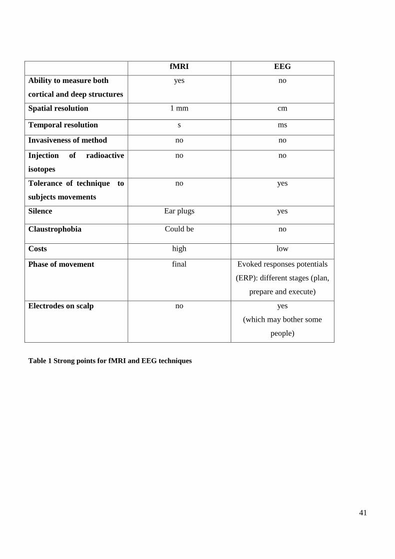
41
fMRI EEG
Ability to measure both
cortical and deep structures
yes no
Spatial resolution 1 mm cm
Temporal resolution s ms
Invasiveness of method no no
Injection of radioactive
isotopes
no no
Tolerance of technique to
subjects movements
no yes
Silence Ear plugs yes
Claustrophobia Could be no
Costs high low
Phase of movement final Evoked responses potentials
(ERP): different stages (plan,
prepare and execute)
Electrodes on scalp no yes
(which may bother some
people)
Table 1 Strong points for fMRI and EEG techniques

42
Figure 1 Experimental setup for Study 1. The subjects lay supine on the scanner bed with their arms resting
comfortably by their side for the duration of the experiment. An optic fibre goniometer monitored the extent of
finger flexion/extension. Electrical stimuli were delivered through round surface stimulation electrodes that were
fixed to the skin. The stimulation pattern was triggered by a push-button switch held in the left hand.
Optical fibre goniometer
Finger Flexors/Extensors
Button to press

43
Figure 2 Experimental setup for Study 2. The subjects lay on an armchair. A goniometer monitored the extent of
finger flexion/extension. Stimulation was delivered through round surface stimulation electrodes that were fixed to
the skin. The electrodes were positioned with cathodes over the motor points of the extensors (extensor digitorum
communis) and a common anode located on the forearm near the wrist. The stimulation pattern was triggered by a
push-button switch held in the left hand. The EEG recordings were performed with 32 channel digital DC EEG
amplifier.
Finger Extensors stimulation
Button to press
Goniometer
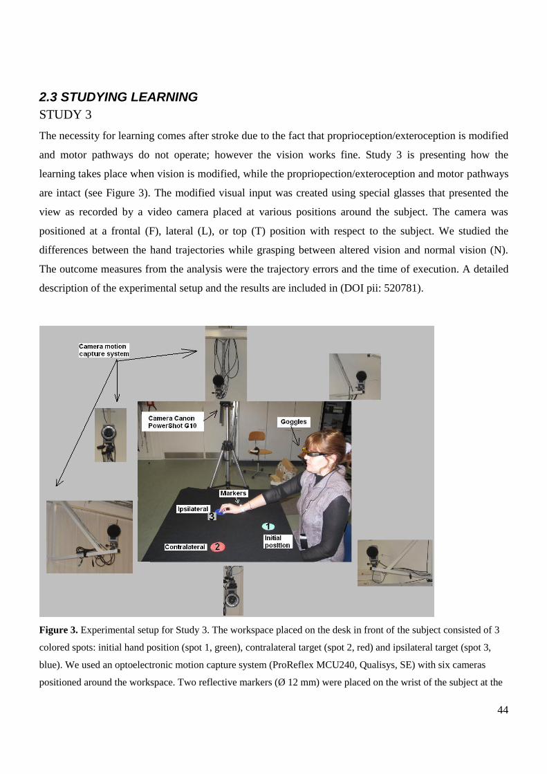
44
2.3 STUDYING LEARNING
STUDY 3
The necessity for learning comes after stroke due to the fact that proprioception/exteroception is modified
and motor pathways do not operate; however the vision works fine. Study 3 is presenting how the
learning takes place when vision is modified, while the propriopection/exteroception and motor pathways
are intact (see Figure 3). The modified visual input was created using special glasses that presented the
view as recorded by a video camera placed at various positions around the subject. The camera was
positioned at a frontal (F), lateral (L), or top (T) position with respect to the subject. We studied the
differences between the hand trajectories while grasping between altered vision and normal vision (N).
The outcome measures from the analysis were the trajectory errors and the time of execution. A detailed
description of the experimental setup and the results are included in (DOI pii: 520781).
Figure 3. Experimental setup for Study 3. The workspace placed on the desk in front of the subject consisted of 3
colored spots: initial hand position (spot 1, green), contralateral target (spot 2, red) and ipsilateral target (spot 3,
blue). We used an optoelectronic motion capture system (ProReflex MCU240, Qualisys, SE) with six cameras
positioned around the workspace. Two reflective markers (Ø 12 mm) were placed on the wrist of the subject at the
45
ulnar and radial styloid processes, respectively. The subject viewed the workspace through goggles (Myvu Crystal
Standard/Universal) connected to a camera (Canon PowerShot G10). The Canon camera was placed in 3 different
positions with respect to the workspace, resulting in 3 different experimental conditions: frontal (F), lateral (L,
shown in this figure), and top (T).
46
REFERENCES Frostig RD, Lieke EE, Ts‟o D, Grinvald A (1990) Cortical functional architecture and local coupling between
neuronal activity and the microcirculation revealed by in vivo high-resolution optical imaging of intrinsic signals.
Proc Natl Acad Sci USA 87:6082-6086
Houdayer EL, Cassim F, Bourriez JL, Derambure P (2006) Relationship between event-related beta
synchronization and afferent inputs: analysis of finger movement and peripheral nerve stimulations, Clin.
Neurophysiol.:628-636
Logothetis NK, Pauls J, Augath M, Trinath T, Oeltermann A (2001) Neurophysiological investigation of the basis
of the fMRI signal. Nature 412:150-157
Menon RS, Kim KG (1999) Spatial and temporal limits in cognitive neuroimaging with fMRI. Trends Cogn Sci
3:207-216
Rossini PM, Calautti C, Pauri F, Baron JC (2003) Post-stroke plastic reorganisation in the adult brain. Lancet
Neurology 2:493-502
Schambra HM, Sawaki L, Cohen LG (2003) Modulation of excitability of human motor cortex (M1) by 1 Hz
transcranial magnetic stimulation of the contralateral M1. Clin Neurophysiol. 114:130-133
Siebner HR, Peller M, Willoch F, Minoshima S, Boecker H, Auer C, Drzezga A, Conrad B, Bartenstein P (2000)
Lasting cortical activation after repetitive TMS of the motor cortex: a glucose metabolic study. Neurology 54:956-
963
Ward NS, Cohen LG (2004) Mechanisms Underlying Recovery of Motor Function After Stroke. Arch Neurol.
61:1844-1848
Williamson SJ, Kaufman L (1990) Evolution of neuromagnetic topographic mapping. Brain Topogr 3:113-127

47
CHAPTER 6
DISCUSSIONS
This research seeks to determine the neural consequences of the applied therapeutic FES to promote
reaching and grasping. In parallel with the neural consequences of the therapeutic FES, the role of the
modified vision in reaching and grasping was determined.
The potential benefits to be gained by this research will be the objective understanding of the effective
therapeutic FES treatment that assist post stroke individuals in achieving the hand/arm/leg function, as
well as other vital functions, and to return to independent life-style in the easiest, simplest, and fastest
way. This information will be extremely beneficial for the researchers investigating optimal methods of
intervention for stroke survivors. In addition, in Study 3 we presented an effective, yet simple new tool
for altering visual input when studying motor coordination. The results show that this alteration of the
visual input can be graded; hence, allow studying of different concepts of learning of the movement. The
methodology introduced here may be further explored for various experimental paradigms.
The results of this PhD thesis should be understood as contribution to the rehabilitation field. The studies
tried to use the available technology to answer the questions raised by the open identified problems.
6. 1 STUDY 1
The main research question was: what is the most appropriate technique to quantify the neural
consequences of the therapy? This main question leads us to the following specific research questions: is
fMRI an appropriate objective technique to evaluate the neural consequences of the therapeutic FES? A
summary is included below.
The internal model concept is important in rehabilitation because the model can be updated as the state of
the limb changes. To determine if internal models are appropriately incorporated during FES-assisted
voluntarily movement, one must test if internal model networks are appropriately engaged during the
task. We designed an fMRI experiment to study brain activity evoked with three tasks hand grasp tasks
involving voluntary motor activation (VOL), patterned electrical stimulation of finger flexor/extensor
muscles to effect movement without voluntary activation (FES), and voluntary activation performed
together with FES (FESVOL). Based on the studies of (Blakemore et al. 1998b; Blakemore et al. 1999;
Blakemore et al. 2001), which specifically test mechanisms of the internal model networks, we
hypothesized that FESVOL, as opposed to FES would activate the motor part of the cerebellum (Grodd et
al. 2001), in particular to make predictions of the sensory consequences of motor commands. In turn, this
48
should reduce the SII activity in FESVOL compared with FES due to a better prediction of the sensory
input.
FESVOL revealed greater cerebellar activity compared with FES alone and reduced activity bilaterally in
secondary somatosensory areas (SII) compared with VOL alone. Reduced activity was also observed for
FESVOL compared with FES alone in the angular gyrus, middle frontal gyrus and inferior frontal gyrus.
These findings indicate that during the VOL condition the cerebellum predicts the sensory consequences
of the movement and diminish the subsequent activation in SII. The decreased SII activity may reflect a
better match between the internal model and the actual sensory feedback. The greater cerebellar activity
coupled with reduced angular gyrus activity in FESVOL compared with FES may indicate that the cortex
may interpret sensory information during the FES condition as an error-like signal due to the lack of a
voluntary component in the movement.
6.1.1 MAIN RESULTS STUDY 1:
The greater cerebellar activity coupled with reduced angular gyrus activity in FESVOL compared
with FES may indicate that the cortex may interpret sensory information during the FES
condition as an error-like signal due to the lack of a voluntary component in the movement.
The combination of voluntary activity with FES- driven motor activity may help to make the
movement more predictable and, ultimately, more one‟s own.
An alternative explanation for the FESVOL finding is that the voluntary effort reduces the SII
activation and increases the cerebellar activation, and on top of this activation the electrical
stimulation activates the SII but not the cerebellum. This condition would then look more like the
VOL condition, which is likely better integrated than the FES alone condition. In part, this might
explain the common notion among FES practitioners why therapeutic FES appears to produce
better clinical results than does FES training alone.
6.1.2 IMPLICATIONS OF THE RESULTS FOR REHABILITATION
Our findings have implications for the physical rehabilitation of stroke related deficits. By combining
patterned electrical stimulation with attempts at voluntary movement, the movement might become ones
own and like ordinary voluntary movement will co-activates the motor control regions and the sensory-
feedback areas, and this co-activation may be crucial in motor recovery at the brain level. This may partly
explain why therapeutic FES (i.e. FES augmented voluntary activity) produces better clinical results than
does FES training alone.
49
One limitation of our results is that the findings in healthy brains cannot be not directly translated to
patient populations. These findings might be completely different from what ones would observe in
damaged brains. Patients with brain damage have obviously a neural re-organization and plasticity that is
not observed in healthy subjects.
6. 2 STUDY 2
The main research question was: what is the most appropriate technique to quantify the neural
consequences of the therapy? This main question leads us to the following specific research questions: is
EEG an appropriate objective technique to evaluate the neural consequences of the therapeutic FES?
Could EEG analysis complement our previous fMRI results helping through a better understanding of the
mechanism by which therapeutic FES interacts with the central nervous system which may allow us to
improve the therapy? More specifically: will different task-related afferent input be reflected in the
modulation of alpha and beta rhythms and movement related cortical potentials?. A summary is included
below. Our study demonstrates that different task-related afferent inputs were reflected in the modulation
of alpha and beta rhythms. In addition, the three tasks VOL, FES, and FESVOL utilized a different
distribution of brain areas for the preparation, execution and control phases of the movement as quantified
by MRCPs. The EEG analysis is complementing our previous fMRI results helping through a better
understanding of the mechanism by which therapeutic FES interacts with the central nervous system
which may allow us to improve the therapy. Future studies should investigate whether patients with
altered strengths of afferent input show similar patterns.
6.2.1 MAIN RESULTS STUDY 2:
VOL, FES, FESVOL determine different distribution in brain areas in preparation and execution
phases of the movement as quantified by movement-related cortical potentials (MRCPs).
The three different task-related afferent inputs were reflected in the modulation of alpha and beta
rhythms.
6.2.2 IMPLICATIONS OF THE RESULTS FOR REHABILITATION
MRCPs were studied in connection with stroke rehabilitation. Studies demonstrated topographical
alterations of some MRP components during the recovery period after a stroke (Honda et al. 1997; Green
et al. 1999; Platz et al. 2000). The strength of the EEG technique is its high temporal resolution, which
allows the separate assessment of movement preparation and movement execution and makes it possible
50
to distinguish slowly evolving movement-related potentials and sensorimotor rhythms, and thereby
different types of electric brain activity that are closely related to neural function.
Our results indicate that alpha band energy showed a bilateral decreased distribution over the motor areas
and ipsilateral parietal (in relation to the right hand involved in the task). Beta showed an ipsilateral
centroparietal decrease. EEG combined with fMRI studies showed that the strength of rolandic alpha, and
rolandic beta rhythms inversely correlates with the fMRI signal in „„its cortical area‟‟. A decrease in the
power of sensorimotor rhythms could be correlated to an activated cortical network, servicing planning
and execution, while power increase, might reflect deactivation. (Laufs et al. 2003) found robust pattern
of negative correlation between BOLD signal change in association with alpha power in frontal and
parietal cortices. (Goldman et al. 2002) also observed BOLD signal changes in parts of the frontal and of
the occipital lobe to correlate negatively with alpha power. In summary, the bilateral activation might
reflect an excitatory activity.
(Platz et al. 2000; Muller et al. 2003) studied movement-related slow cortical potentials and event-related
desynchronization of alpha (alpha-ERD) and beta (beta-ERD) activity
after self-paced voluntary
triangular finger movements in 13 ischaemic supratentorial stroke patients. The multimodal EEG analysis
suggested impairment-specific changes in the movement-related electrical activity of the brain.
The
readiness potential of paretic subjects was centered more anteriorly and laterally; during movement, they
showed increased beta-ERD at left lateral frontal recording sites. Patients showed reduced alpha-ERD and
beta-ERD during both movement preparation and actual performance. They concluded that (i) disturbed
motor efference is associated with an increased need for excitatory drive of
pyramidal cells in motor and
premotor areas or an attempt to drive movements through projections from these areas to brainstem
motor
systems during movement preparation; (ii) an undisturbed somatosensory afference might contribute to
the release of relevant cortical areas from their `idling' state when movements are
prepared and
performed; and (iii) patients have a relative lack of activity of the frontal motor system and the
left
parietal cortex.
6.3 STUDY 3
The main research question was: how the learning takes place when vision is modified, while the
proprioception/exteroception and motor pathways are intact? A summary is included below.
6.3.1 MAIN RESULTS STUDY 3:
The results indicate that fast learning (recalibration) of the visual systems occurred relatively fast
and has been created through the training of movement.
51
The recalibration is more difficult to achieve for altered vision in frontal and lateral conditions
than in top condition
The paper presents an effective, yet simple new tool for altering visual input when studying motor
coordination.
The results show that this alteration of the visual input can be graded; hence, allow studying of
different concepts of learning of the movement.
The methodology introduced here may be further explored for various experimental paradigms,
especially if the camera connected to the input of the Myvu Crystal EV is replaced with the
computer output providing virtual reality.
Study 3 tested a new tool for studying motor learning/coordination under altered visual input. In the
experiment, the visual input was presented to subjects in four different conditions: normal (N), frontal (F),
lateral (L), and top (T). The experiment consisted of five sessions that took place on five consecutive
days as follows. Day 1 was used for the baseline assessment. Day 2, 3, and 4 were allocated for training.
Day 5 was used for evaluation. This paper was indented as a possible application for people after a CVA.
The necessity for learning comes after stroke due to the fact that proprioception/exteroception is modified
and motor pathways do not operate; however the vision works fine. The goal was to determine how
exactly practice makes perfect and how the visual system coordinates with the motor system.
Therefore we looked at the dissociation between proprioception and vision during manipulation of the
arm in reach to grasp task. This dissociation translated in additional cognitive operations (translation,
rotation, scaling of the visual input on the screen of the goggles into the plane of motion of the arm on the
table) that are presumably required to translate visual information about target location on the monitor
screen into an estimate of the metrics o the desired arm movement.
Proprioception contrasted with vision, so the brain computing tried a way to recalibrate reference frame
for that computation. Errors scaled proportionally with the difficulty of the task. Our subjects ranked the
level of difficulty of the F condition as the highest, followed by the L and T conditions.
On Day 1, the errors were generally larger for L condition than for F condition. The largest values for the
contralateral 1 EE and ipsilateral EE were observed in the L condition, while the contralateral 2 EE
reached a maximum value in the F condition.
This suggests that altered vision in the L condition had the greatest effect on segments 1 and 2, while
altered vision in the F condition mostly affected segment 3 of the movement.
Some adaptation occurred even on Day 1 for F and L conditions from the first trials performed. The error
decreased from Trial 1 to Trial 10 for F and L conditions through this adaptation was not complete at the
end of the 10 trials.
52
On Day 5, the magnitude of the errors decreased for F and L conditions compared with Day 1. F
condition showed the largest decrease of contralateral 2 EE, while L condition for ipsilateral EE (> 50%
Day 1). This suggests that the training had the greatest effect on segment 3 for F condition and segment 1
and 2 for L condition.
The magnitude of the errors was substantially smaller in T condition compared with F and L condition
both on Day 1 and Day 5.
This trend is consistent with the scatter plot of the trajectory end points (see DOI pii: 52078) and the
analysis of velocity profiles (see DOI pii: 520781).
On Day 1, F and L condition, i.e. with the camera positioned in front and lateral (right) to the subject
respectively, showed trajectories end points scattered covering almost the entire workspace. T condition,
with the camera positioned on top of the subject showed end points clustered together within the
reference spots.
The analysis of velocity profiles showed that the adaptation occurred for F, L, T conditions in the way
that the velocities had near symmetrical bell-shaped profiles. The peak velocity reached the value typical
of normal reaching movements for L condition (segment 1, 3, and 4), for F (segment 2 and 4), for T
condition (segment 3), see (DOI pii: 520781).
6.3.2 IMPLICATIONS OF THE RESULTS FOR REHABILITATION
Generalization to other type that was not trained
The subjects were trained only in F condition. However, the results indicate an improved performance on
Day 5 for the L and T conditions, although these conditions were not used for training. It could indicate
that training only with F condition affected the motor learning ability of the subjects in general. Learning
reach to grasp new trajectories and the new use of the hand in injured persons could be a possible
application of the results of this paper.
Cognitive component
Our results suggest that the difference between altered vision tasks and normal visually guided reaching
leads to an adaptation in the form of perceptual recalibration where proprioception was calibrated in terms
of the visual system. If the adaptation is expected to take the guise of a more cognitive, problem-solving
process, we can refer to this as the visual-motor skill acquisition. The ability to predict with some
confidence which of these two types of adaptation a peripheral manipulation will produce would allow for
a prediction of whether significant improvement is likely to occur on training, how persistent the
adaptation will be, and whether it will result in after-effects. The methodology introduced here may be

53
further explored for various experimental paradigms; the manipulation of viewing angle would be useful
is not reachable working space for disable patients (Ward and Frackowiak 2006).
54
REFERENCES Goldman RI, Stern JM, Engel JJ, Cohen M (2002) Simultaneous EEG and fMRI of the alpha rhythm.
NeuroReport:2487-2492
Green JB, Bialy Y, Sora E, Ricamato A (1999) High-resolution EEG in poststroke hemiparesis can identify
ipsilateral generators during motor tasks. Stroke 30:2659-2665
Honda M, Nagamine T, Fukuyama H, Yonekura Y, Kimura J, Shibasaki H (1997)
Movement-related cortical potentials and regional cerebral blood flow change in patients with stroke after motor
recovery. Journal of Neurological Science 146:117-126
Laufs H, Kleinschmidt A, Beyerle A, Eger E, Salek-Haddadi A, Preibisch C, Krakow K (2003) EEG-correlated
fMRI of human alpha activity, NeuroImage:1463-1476
Muller GR, Neuper C, Rupp R, Keinrath C, Gerner HJ, Pfurtscheller G (2003) Event-related beta EEG changes
during wrist movements induced by functional electrical stimulation of forearm muscles in man. Neuroscience
Letters:143-147
Platz T, Kim IH, Pintschovius H (2000) Multimodal EEG analysis in man suggests impairment-specific changes in
movement-related electric brain activity after stroke. Brain 123:2475-2490
Ward NS, Frackowiak RSJ (2006) The functional anatomy of cerebral reorganisation
after focal brain injury. Journal of Physiology 99:425-436
55
CHAPTER 7
CONSIDERATIONS AND FUTURE PROSPECTRIVES
The present PhD thesis offered an overview of the strong and weak points and contribution of different
techniques and methods in defining objectively the neural consequences of the therapeutic FES (STUDY
1 and STUDY 2) and how the learning takes place when vision is modified, while the
propriopection/exteroception and motor pathways are intact (STUDY 3).
The participants involved in all three studies were healthy volunteers. The protocol in STUDY 1 and
STUDY 2 tried to mimic the clinical therapeutic FES applied to post-stroke patients. It would be of
interest to apply the protocol in STUDY 1 and STUDY 2 in patients who had major disability in their
paretic hand and arm. However, the extension of the results to the stroke population should be done
cautiously. In post stroke patients there is invariably some degree of functional recovery, ranging from
minimal to complete.
Functional imaging studies of the motor system in previously hemiparetic patients have described task-
related brain activation in recovered patients over and above control subjects in contralesional
sensorimotor and premotor cortex, ipsilesional cerebellum, bilateral supplementary motor area (SMA)
and parietal cortex (Weiller et al. 1992). (Ward et al. 2003) asked control subjects to perform passive
movement of the wrist and obtained increases in brain activation in
the contralateral sensorimotor cortex
and supplementary motor area, the bilateral inferior parietal cortex and secondary somatosensory
areas,
and the ipsilateral cerebellum. Increases in brain activation correlating with motor recovery were observed
in both the ipsilesional primary sensory and primary motor cortex in 1 patient with good
motor recovery
but not in another patient with poor recovery. They concluded that functionally relevant changes in
cerebral organization can be identified in individual patients.
Healthy volunteer group results are very important for having a „proof of principle‟. Functional imaging
techniques can be used as estimators of the outcome of an applied therapy and can contribute toward an
understanding of the general principles of cerebral reorganization after stroke. But we should consider of
course the intersubject variability in post stroke population.
In addition, STUDY 1 investigates the short term effect of applied peripheral electrical stimulus
combined with voluntary effort. It will be of great interest to investigate the long term effects and to
describe the learning process that will occur. Current models of motor learning suggest that during early
learning, there is a dynamic interaction between a frontoparietal network encoding movement in terms of
spatial coordinates, that requires high levels of attention, and motor cortex which encodes movement in
terms of a kinematic system of joints, muscles, limb trajectories, etc. Motor cortex is dominant once
learning has occurred and a movement has become automatic (Hikosaka et al. 2002). A number of
empirical findings support such a model: decreases in brain activation as a function of motor learning
56
have been reported in lateral premotor cortex, prefrontal cortex, pre-SMA, superior parietal cortex,
anterior cingulate, cerebellum, cerebellar vermis and caudate (Hikosaka et al. 2002); the cerebellum is
involved in detecting error between internal models of movement and the sensory consequences of actual
movement (Blakemore et al. 1998; Blakemore et al. 2001). (Ward et al. 2003; Ward 2005) describe
decreases in activation with recovery consistent with the notion of a transfer of reliance from the
frontoparietal to the primary motor system.
Technical Challenges and Issues in Experimental Design and Analysis
The results of functional imaging experiments are only as reliable as the care with which the experiment
is constructed and executed. The choice of experimental task, patient monitoring, patient selection, and
approaches to analysis require careful consideration if a study is to successfully address its stated
hypothesis. Thereafter any functional recovery is associated with a focusing of brain activation patterns
towards that seen in controls (Ward 2005). This focusing is similar to that seen in the normal brain during
motor skill learning. However, although it is unsurprising that the damaged brain will attempt to use
highly preserved neural systems such as those subserving motor skill learning to maximize functional
motor recovery, the degree to which this is successful will depend on the integrity of such networks
(Ward 2005).
57
REFERENCES Blakemore SJ, Frith CD, Wolpert DM (2001) The cerebellum is involved in predicting the sensory consequences of
action. Neuroreport 12:1879-1884
Blakemore SJ, Wolpert DM, Frith CD (1998) Central cancellation of self-produced tickle sensation. Nat Neurosci
1:635-640
Hikosaka O, Nakamura K, Sakai K, Nakahara H (2002) Central mechanisms of motor skill learning. Current
Opinion in Neurobiology 12:217-222
Ward NS (2005) Mechanisms underlying recovery of motor function afterstroke. Postgrad Med J 81:510-514
Ward NS, Brown MM, Thompson AJ, Frackowiak RSJ (2003) Neural correlates of outcome after stroke: a cross-
sectional fMRI study. Brain 126:1430-1448
Weiller C, Chollet F, Friston KJ, Wise RJ, Frackowiak RS (1992) Functional reorganization of the brain in
recovery from striatocapsular infarction in man. Ann Neurol 31:463-472
58
SUMMARY
Functional Electrical Stimulation is used in the rehabilitation therapy of patients after stroke to improve
their motor abilities. Electrical stimulation when paired with voluntary component (therapeutic FES),
appears to facilitate recovery in an additive or interactive way, in clinical studies. An open issue is
represented by the mechanisms for these observed effects. Having a good understanding of how
therapeutic FES may interact with the central nervous system may therefore be crucial to improve and
optimize the treatment. Noninvasive techniques to study brain function, including functional magnetic
resonance imaging, electroencephalography, could help to document objectively the relationship between
cortical reorganization and the recovery of motor function.
The goal of this thesis was therefore to develop a set of methods and tools that can study plasticity
induced by different experimental conditions. The methods that we propose here should be understood as
contribution to the actual effort of the researchers in the rehabilitation field.
We started by applying fMRI technique to obtain objective information about the neural consequences of
neuromuscular stimulation combined with voluntary effort (FESVOL) peripheral electrical stimulation
alone (FES) or voluntary activation alone (VOL) (STUDY 1). The aim was to develop a tool for study
plasticity induced by therapeutic FES. The importance of studying the neural correlates associated with
FES for clinical populations (ie., stroke) and therapeutic application constituted the motivation for
conducting the research. FESVOL revealed greater cerebellar activity compared with FES alone and
reduced activity bilaterally in secondary somatosensory areas (SII) compared with VOL alone. Reduced
activity was also observed for FESVOL compared with FES alone in the angular gyrus, middle frontal
gyrus and inferior frontal gyrus. These findings indicate that during the VOL condition the cerebellum
predicts the sensory consequences of the movement and diminish the subsequent activation in SII. The
decreased SII activity may reflect a better match between the internal model and the actual sensory
feedback. The greater cerebellar activity coupled with reduced angular gyrus activity in FESVOL
compared with FES may indicate that the cortex may interpret sensory information during the FES
condition as an error-like signal due to the lack of a voluntary component in the movement.
Functional MRI relies on the paramagnetic characteristics of deoxyhemoglobin, measuring its
concentration changes in brain tissue, in response to task dependent neuronal activation. fMRI pose
problems in differentiating neuronal firing decrease from increase (exciting vs inhibiting net effects). The
polarity and amplitude of the potentials observed in the electroencephalogram are directly related to
modality (excitatory or inhibitory) and intensity of the neural population at the cortical level. A more
complete picture of the cortical reorganization and when and where changes take place could be obtained
59
with multimodal approaches .The presented limitations lead us to the Study 2 where we applied EEG
technique to obtain objective information about the neural consequences of the therapeutic FES.
STUDY 2 evaluated the relationship between different task-related afferent inputs and cortical function.
Cortical function was assessed with analysis of the strength of alpha and beta oscillatory activity and
movement related cortical potentials in multichannel surface EEG. The working hypothesis was that
processing of peripheral electrical stimulus input is reflected differently in the modulation of cortical
alpha and beta oscillatory activity and movement related cortical potentials as the procession of the
voluntary input.
The first main finding was that different task-related afferent inputs determine different distribution in
brain areas in preparation and execution phases of the movement as quantified by movement related
cortical potentials. The second main finding was that different task-related afferent inputs are producing a
state of activation/deactivation (inhibition) of neural networks in different cortical areas reflected in the
modulation of alpha and beta rhythms.
Plasticity of the cortex is considered as learning of novel tasks. The necessity for learning comes after
stroke due to the fact that proprioception/exteroception is modified and motor pathways do not operate;
however the vision works fine. STUDY 3 tested a novel tool for studying motor coordination with an
altered visual input. The altered visual input was created using special glasses that presented the view as
recorded by a video camera placed at various positions around the subject. The camera was positioned at
a frontal (F), lateral (L), or top (T) position with respect to the subject. We studied the differences
between the hand trajectories while grasping between altered vision and normal vision (N) in ten subjects.
The outcome measures from the analysis were the trajectory errors and the time of execution. We found
substantial trajectory errors and an increased execution time at the beginning of the task. We also found
that the trajectory errors decreased after three days of practice with the altered vision for 20 minutes per
day, suggesting that recalibration of the visual systems occurred relatively quickly. The results indicate
that this recalibration has occurred by movement training. The results also suggest that the recalibration is
more difficult to achieve for altered vision in F and L conditions than in T condition.
The potential benefits to be gained by this research will be the objective understanding of the effective
therapeutic FES treatment that assist post stroke individuals in achieving the hand/arm/leg function, as
well as other vital functions, and to return to independent life-style in the easiest, simplest, and fastest
way. This information will be extremely beneficial for the researchers investigating optimal methods of
intervention for stroke survivors. In addition, in STUDY 3 we presented an effective, yet simple new tool
for altering visual input when studying motor coordination. The results show that this alteration of the
visual input can be graded; hence, allow studying of different concepts of learning of the movement. The
methodology introduced here may be further explored for various experimental paradigms.

60
The results of this PhD thesis should be understood as contribution of the available technology to the
rehabilitation field. The present PhD thesis offered an overview of the strong and weak points and
contribution of different techniques and methods in defining objectively the neural consequences of the
therapeutic FES (STUDY 1 and STUDY 2) and the role of normal/modified vision for the control of
reaching, grasping, manipulating movement of the arm that can be implemented in neural prosthesis for
grasping or artificial hands (STUDY 3).
61
SAMMENFATNING
Funktionel Elektrisk Stimulation anvendes ved genoptræning af patienter efter et slagtilfælde, for at
forbedre deres motoriske færdigheder. Elektrisk stimulation parret med en voluntær bevægelse
(terapeutisk FES) synes, i kliniske studier, at lette genoptræning på en additiv eller interaktiv facon.
Hvilke mekanismer der ligger til grund for de observerede effekter, er et åbent spørgsmål. En god
forståelse af hvordan terapeutisk FES kan interagere med det centrale nervesystem, kan derfor være
afgørende for at forbedre og optimere behandlingen. Ikke-invasive metoder til at undersøge
hjerneaktivitet, som Funktionel magnetisk resonans-billeddannelse og Elektroencefalografi, kan på en
objektiv måde dokumentere sammenhængen mellem kortikal reorganisation og genoptræning af
motoriske færdigheder.
Målet med afhandlingen var derfor at udvikle metoder og værktøjer, der kunne undersøge plasticitet
under forskellige eksperimentielle forhold. De metoder vi foreslår skal forstås som et bidrag til den
aktuelle forskning indenfor genoptræning.
Vi startede med at anvende fMRI teknik for, på en objekttiv måde, at få information om de neurale
konsekvenser af dels neuromuskulær stimulation kombineret med en voluntær bevægelse (FESVOL),
periferisk elektronisk stimulation (FES) og voluntær bevægelse (VOL) (STUDIE 1). Målet var at udvikle
et værktøj til at undersøge plasticitet som følge af terapeutisk FES. Vigtigheden af at undersøge de
neurale korrelater associeret med FES ved kliniske populationer (dvs. slagtilfælde) og terapeutiske
anvendelser, udgjorde motivationen for at udføre forskningen. FESVOL viste større cerebellar aktivitet
sammenholdt med FES og mindre aktivitet bilateralt i sekundære somatosensoriske områder (SII)
sammenholdt med VOL. Reduceret aktivitet blev også observeret for FESVOL sammenholdt med FES i
den angular gyrus, den middle frontal gyrus og den inferior frontal gyrus. Disse resultater indikerer, under
VOL, at cerebellum prædikterer de sensoriske konsekvenser af bevægelsen og reducerer den
efterfølgende aktivering i SII. Den reducerede SII aktivitet kan afspejle et bedre match mellem den
interne model og det aktuelle sensoriske feedback. Den større cerebellar aktivitet og reduceret angular
gyrus aktivitet i FESVOL sammenholdt med FES kan indikere, at cortex fortolker sensorisk information i
løbet af FES som et fejllignende signal, grundet mangel på en voluntær komponent i bevægelsen.
Funktionel MRI er afhængig af de paramagnetiske karakteristika af deoxyhemoglobin ved måling af
ændringer i dets koncentration i hjernevæv, som reaktion på opgaveafhængig neuronal aktivering. Det er
problematisk for fMRI at skelne en forøgelse i neuronal affyring fra en formindskelse (dvs. ophidsende
kontra hæmmende effekt). Polariteten og amplituden af de observerede potentialer i det
elektroencefalografi er direkte relateret til modalitet (excitatoriske eller hæmmende) og intensitet af den
neurale population, på det kortikale plan. Et mere fuldstændigt billede af den kortikale reorganisation, og
62
hvornår og hvor ændringer finder sted, kunne opnås med multimodale metoder. De præsenterede
begrænsninger førte os til STUDIE 2 hvor vi anvendte EEG teknikker for objektivt at opnå information
om de neurale konsekvenser af terapeutisk FES.
STUDIE 2 evaluerede sammenhængen mellem forskellige opgave relaterede afferente input og kortikal
funktion. Kortikal funktion blev vurderet ud fra en analyse af styrken af alpha og beta oscillerende
aktivitet og bevægelses relateret kortikal potentialer i multikanal overflade EEG. Hypotesen var, at
behandling af perifere elektriske stimulus input, reflekteres forskelligt i modulationen af kortikal alpha og
beta oscillerende aktivitet, og bevægelses relateret kortikal potentialer.
Det første hovedresultat var, at forskellige opgave relaterede afferente input bestemmer forskellige
fordelinger i områder i hjernen, i forbindelse med forberedelses og udførelses faser af bevægelsen,
kvantificeret ved bevægelses relateret kortikal potentialer. Det andet hovedresultat var, at forskellige
opgave relaterede afferente input, producerer en tilstand af aktivering/deaktivering af neurale netværk i
forskellige kortikal områder, der er afspejlet i modulationen af alpha og beta rytmerne.
Plasticitet af cortex betragtes som læring af nye opgaver. Behovet for at lære efter et slagtilfælde skyldes,
at proprioception/exteroception er ændret og motor veje ikke virker; dog fungerer synet fint. Studie 3
testede et nyt værktøj til at undersøge motor koordination med et ændret visuelt input. Det ændrede
visuelle input blev skabt ved at benytte specielle briller, der viste et billede optaget af et kamera, som blev
placeret forskellige steder rundt om forsøgspersonen. Kameraet blev opstillet i en frontal (F), lateral (L)
og top (T) positioner i forhold til forsøgspersonen. For ti forsøgspersoner, med normalt og ændret syn,
undersøgte vi forskelle i deres hånds bane ved opsampling af et emne. De opsamlede målinger fra
analysen var afvigelser fra banen og udførelsestid. Vi fandt betydelige afvigelser fra banen og en forøget
udførelsestid i begyndelsen af opgaven. Vi observerede også, at afvigelser fra banen aftog efter tre dages
træning, 20 minutter pr. dag., med ændret syn, hvilket antyder, at rekalibrering af det visuelle system sker
sker relativt hurtigt. Resultatet indekerer, at denne rekalibrering skyldes bevægelses træning. Resultaterne
indikerer også, at rekalibrering er sværere at opnå for ændret syn (F) og (L) end (T).
De potentielle fordele der kan opnås med denne forskning, vil være den objektive forståelse af
effektiviteten af terapeutisk FES behandling, der hjælper post slagtilfælde personer i at opnå brug af
hånd/arm/ben og andre vitale funktioner, og til at vende tilbage til en uafhængig livsstil på den letteste,
simpleste og hurtigste måde. Denne information vil være yderst gavnlig for forskere, der undersøger
optimale metoder til intervention for overlevende der er blevet ramt af et slagtilfælde. Derudover
præsenterede vi i STUDIE 3 et effektivt og simpelt nyt værktøj til ændring af visuelt input ved
undersøgelse af motor koordinering. Resultaterne viser, at ændring af visuelt input kan gradueres, og det
er dermed muligt at undersøge forskellige aspekter ved indlæring af bevægelse. Den introducerede
metode kan undersøges yderligere gennem forskellige eksperimentelle paradigmer.

63
Resultaterne af denne ph.d. afhandling skal ses som et bidrag til den tilgængelige teknologi inden for
genoptræning. I denne ph.d afhandling gives et overblik over de stærke og svage sider af forskellige
teknikker og metoder, der objektivt definerer de neurale konsekvenser af terapeutisk FES (STUDIE 1 og
STUDIE 2), og betydningen af normalt/ændret syn i forbindelse med styring af en arms række og gribe
bevægelser, generelt manipulation af en arms bevægelser, der kan implementeres i en neural protese til at
gribe med, eller i kunstige hænder (STUDIE 3).