A6 brochure - v3.8 - SINGLES w bleed
92
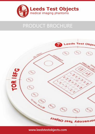
www.leedstestobjects.com PRODUCT BROCHURE
Transcript of A6 brochure - v3.8 - SINGLES w bleed
www.leedstestobjects.com
PRODUCT BROCHURE
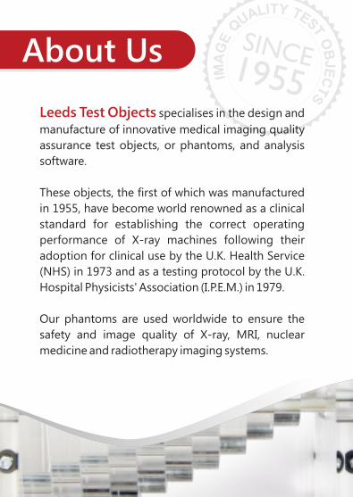
About Us
Leeds Test Objects specialises in the design and
manufacture of innovative medical imaging quality
assurance test objects, or phantoms, and analysis
software.
These objects, the first of which was manufactured
in 1955, have become world renowned as a clinical
standard for establishing the correct operating
performance of X-ray machines following their
adoption for clinical use by the U.K. Health Service
(NHS) in 1973 and as a testing protocol by the U.K.
Hospital Physicists' Association (I.P.E.M.) in 1979.
Our phantoms are used worldwide to ensure the
safety and image quality of X-ray, MRI, nuclear
medicine and radiotherapy imaging systems.
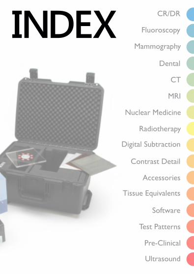
Radiotherapy
CR/DR
Ultrasound
Fluoroscopy
Mammography
Dental
CT
MRI
Nuclear Medicine
Digital Subtraction
Contrast Detail
Accessories
Tissue Equivalents
Software
Test Patterns
Pre-Clinical

EU
RO
PE
DistributorsN
OR
TH
AM
ER
ICA
SO
UT
H
AM
ER
ICA
OC
EA
NIA
RE
ST
OF
W
OR
LD

MID
DL
E-E
AS
T &
AF
RIC
A
AS
IA
CR/DRCR DDR Set
Conforms to standardKcare CR/DR Protocol
A set of test objects designed to be used quickly and easily on an annual / commissioning basis to provide an ongoing check of imaging performance, particularly those aspects which are most liable to deterioration.
Set includes:
• TO 20 threshold contrast test object • Resolution test object 3.4 - 10.0 LP/mm• TO M1 geometry test object• TO MS4 mesh test object• Small lead block, Steel ruler, Tape measure and 1.5 mm
Copper filter
See also: AutoPIA Software

CR/DRTOR CDR
A routine QA test phantom for radiography systems. TOR
CDR is used to provide an ongoing check of imaging
performance, particularly those aspects which are most
liable to deterioration. Used for conventional, computed
and digital radiography.
• Sensitometric measurements
• Resolution limit (0.5 to 14.3 LP/mm)
• Low-contrast large-detail detectability
• High-contrast small-detail detectability
Measure:
See also: AutoPIA Software

CR/DRPIX-13
conforms to standardsKcare CR/DR Protocol
A routine QA test phantom for radiography systems. PIX-13 is used to provide an ongoing check of imaging performance, particularly those aspects which are most liable to deterioration. PIX-13 should be used with either PMMA/Cu or Al attenuator plates (available separately).
Conforms to standardDIN 6868-13
• Dynamic range (7 different thickness steps Cu)
• Resolution limit (0.6 to 5.0 LP/mm)
• Low-contrast large-detail detectability (6 details, 15mm diameter)
• X-ray to light field alignment markers
Measure:
See also: AutoPIA Software

CR/DRTO ANSI
Leeds Test Objects' Patient-Equivalent phatom is designed according to AAPM report 31, and is to be used when checking the image quality performance of general radiography systems to these standards.
Conforms to standardAAPM 31
Fluoroscopy
See also: AutoPIA Software
SFS Set
Set includes:
A set of test objects designed to be used quickly and easily on an annual / commissioning basis to provide an ongoing check of imaging performance, particularly those aspects which are most liable to deterioration. An ongoing record of these numbers will reveal any trend towards deterioration in imaging performance.
• TO 10 threshold contrast test object• TO N3 contrast test object • TO GS2 grey scale test object• TO E1 edge test object• TO M1 geometry test object• TO MS1,TO MS3, TO MS4 mesh test objects, • 1.0 mm Copper filter x 1, 0.5 mm Copper filter x 2 • Resolution test pattern (0.5 to 5.0 LP/mm) • Video cable and adaptors
Fluoroscopy
See also: AutoPIA & Radia Software
A routine QA test phantom for fluoroscopy systems. TOR18FG is used to provide an ongoing check of imaging performance, particularly those aspects which are most liable to deterioration.
• Limiting spatial resolution (0.5 to 5.0 LP/mm)
• Grey Scale
• Low-contrast detectability
• Circular Geometry (Pb Circle)
A small F.O.V. phantom is available upon request, containing the same features.
Measure:
TOR 18FG

FluoroscopyTO XR21
Measure:
A phantom designed to allow the user to check image quality in accordance with the NEMA XR21 standard. This phantom is manufactured from PMMA and enables various QA checks.
The modular stacking design of the plates allows variation of phantom thickness in steps of 25mm, up to a total of 300mm, simulating a range of patient sizes.
• Spatial resolution• Contrast (iodine) resolution• Radiation field size• Beam alignment• Working thickness range• X-ray to light field alignment• Dose (dose meter not supplied)
conforms to standardsABCDEF & GHIJKL
Conforms to standards:NEMA XR21
Fluoroscopy
The TO CDRH Phantom enables quality assurance testing of Radiography / Fluoroscopy systems.
This phantom is suited to both commissioning and routine QA checks. The set includes Copper, Aluminium and Lead filters.
• Low-contrast resolution (Aluminium disc with 8 contrast discs)
• High-contrast resolution (Copper mesh targets from 12 – 80 lines per inch)
• Attachment point for dose measurements (dose meter not supplied)
Measure:
TO CDRH
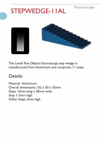
FluoroscopySTEPWEDGE-11AL
The Leeds Test Objects fluoroscopy step wedge is manufactured from Aluminium and comprises 11 steps.
Details:
Material: AluminiumOverall dimensions: 132 x 50 x 35mmSteps 12mm long x 50mm wideStep 1: 5mm highOther Steps: 3mm high
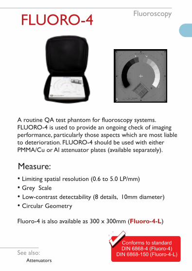
FluoroscopyFLUORO-4
A routine QA test phantom for fluoroscopy systems. FLUORO-4 is used to provide an ongoing check of imaging performance, particularly those aspects which are most liable to deterioration. FLUORO-4 should be used with either PMMA/Cu or Al attenuator plates (available separately).
Conforms to standardDIN 6868-4 (Fluoro-4)
DIN 6868-150 (Fluoro-4-L)
• Limiting spatial resolution (0.6 to 5.0 LP/mm)
• Grey Scale
• Low-contrast detectability (8 details, 10mm diameter)
• Circular Geometry
Fluoro-4 is also available as 300 x 300mm ( )Fluoro-4-L
Measure:
See also: Attenuators
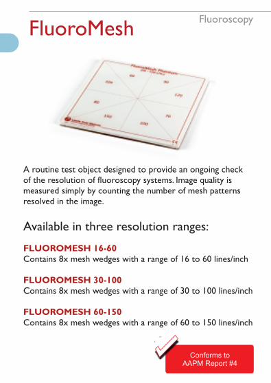
FluoroscopyFluoroMesh
A routine test object designed to provide an ongoing check of the resolution of fluoroscopy systems. Image quality is measured simply by counting the number of mesh patterns resolved in the image.
Available in three resolution ranges:
FLUOROMESH 16-60 Contains 8x mesh wedges with a range of 16 to 60 lines/inch
FLUOROMESH 30-100 Contains 8x mesh wedges with a range of 30 to 100 lines/inch
FLUOROMESH 60-150 Contains 8x mesh wedges with a range of 60 to 150 lines/inch
conforms to standardsABCDEF & GHIJKL
Conforms toAAPM Report #4

MammographyTORMAM
This test object is supplementary to TOR MAS or MAX and provides a more “natural” image which may be preferred by radiographers and radiologists.
One half contains a range of filaments, micro-particles and low-contrast details, representing pathological features in the breast. These are sensitive to the mammographic grey-scale, noise and unsharpness, and can be used to obtain an image-quality “score”. The other half simulates the appearance of breast tissue and contains micro-calcification in addition to fibrous and nodular details.
Contains features:
• 6 groups of multi-directional filaments
• 6 groups of micro-calcifications
• 6 groups of 3, low contrast details groups
See also: AutoPIA Software
Conforms toNHSBSP 0604

Mammography
Test objects designed to be used quickly and easily on a routine basis to provide an ongoing check of imaging performance, particularly those aspects which are most liable to deterioration.
Measure:
• Ten-step grey-scale plus two points for Sensitometric measurements
• Limiting Spatial Resolution (x2 in TOR MAX)
• Low-contrast large-detail detectability
• High-contrast small-detail detectability
• Irregular-shaped particles on a step-wedge background
• Low Contrast Resolution Pattern
See also: AutoPIA Software
TORMAS/MAX

MammographyPIXMAM 300x240
PIXMAM is a digital mammography image quality phantom comprising a stack of finely toleranced PMMA plates.
See also:TORMAM
Measure:
• AEC checks
• Detector homogeneity
• SNR
• CNR
Meets the requirements of standards:NHSBSP pub 604-3 :2009NHSBSP pub 702-1 :2007EUREF 4th edition of the European Guidelines for Quality Assurance in Breast Cancer Screening and Diagnosis
PIXMAM is also available as a series of semi-circular plates with a diameter of 250mm ( ).PIXMAM 250d

MammographyPIXMAM-3D
This phantom has been designed according to the EUREF Protocol for the Quality Control of the Physical and
Technical Aspects of Digital Breast Tomosynthesis Systems
PIXMAM-3D is a digital mammography image quality phantom comprising a stack of finely toleranced PMMA plates, some of which contain embedded test details.
Measure:
• AEC checks
• Detector homogeneity
• SNR
• CNR
• Image resolution
• Z-resolution test
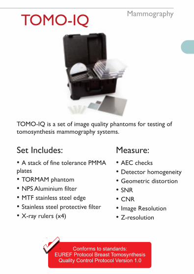
MammographyTOMO-IQ
TOMO-IQ is a set of image quality phantoms for testing of tomosynthesis mammography systems.
Set Includes:
• A stack of fine tolerance PMMA plates
• TORMAM phantom
• NPS Aluminium filter
• MTF stainless steel edge
• Stainless steel protective filter
• X-ray rulers (x4)
Measure:
• AEC checks
• Detector homogeneity
• Geometric distortion
• SNR
• CNR
• Image Resolution
• Z-resolution
Conforms to standards:EUREF Protocol Breast Tomosynthesis
Quality Control Protocol Version 1.0
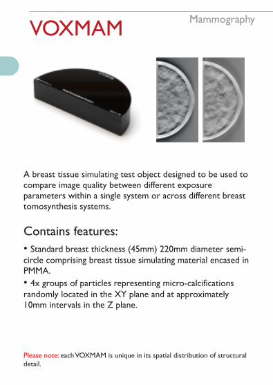
MammographyVOXMAM
A breast tissue simulating test object designed to be used tocompare image quality between different exposure parameters within a single system or across different breast tomosynthesis systems.
Contains features:
• Standard breast thickness (45mm) 220mm diameter semi-circle comprising breast tissue simulating material encased in PMMA.
• 4x groups of particles representing micro-calcifications randomly located in the XY plane and at approximately 10mm intervals in the Z plane.
Please note: each VOXMAM is unique in its spatial distribution of structural detail.

MammographyAgatha
Agatha is an image quality phantom used for constancy testing of digital breast tomosynthesis systems.
Measure:
• Missing tissue tests
• Line Object Spread Function (LOSF)
• Signal Difference to Noise Ratio (SDNR)
• Z-direction sensitivity profiles (ZSP)
• 3D MTF
• System geometry
• Tomographic motion
• Reconstruction effects on SDNR and ASF
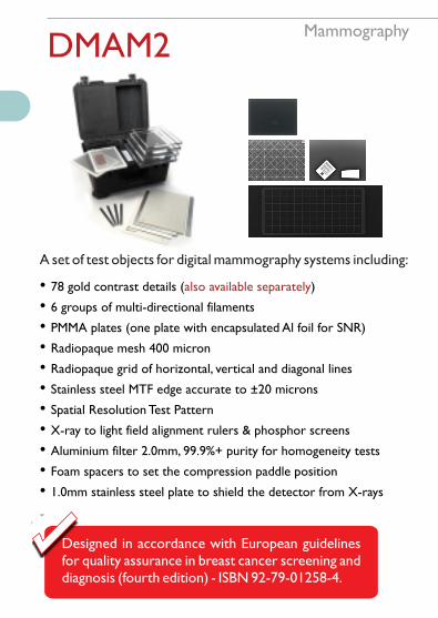
MammographyDMAM2
A set of test objects for digital mammography systems including:
• 78 gold contrast details ( )also available separately
• 6 groups of multi-directional filaments
• PMMA plates (one plate with encapsulated Al foil for SNR)
• Radiopaque mesh 400 micron
• Radiopaque grid of horizontal, vertical and diagonal lines
• Stainless steel MTF edge accurate to ±20 microns
• Spatial Resolution Test Pattern
• X-ray to light field alignment rulers & phosphor screens
• Aluminium filter 2.0mm, 99.9%+ purity for homogeneity tests
• Foam spacers to set the compression paddle position
• 1.0mm stainless steel plate to shield the detector from X-rays

MammographyCEDM
Leeds Test Objects' CEDM phantom is designed for quality control of contrast enhanced digital mammography.
The CEDM phantom is made of PMMA and contains independently fillable spherical voids with diameter 1.0, 2.0, 5.0, 10.0 and 15.0 mm, and five independently fillable fibrils.The voids can be filled with blood mimicking fluid (Leeds BMF) or saline solution mixed with contrast agent.
CEDM includes a syringe and needle.
Measure:
• CNR
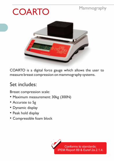
MammographyCOARTO
COARTO is a digital force gauge which allows the user to measure breast compression on mammography systems.
Set includes:
Breast compression scale:
• Maximum measurement: 30kg (300N)
• Accurate to 5g
• Dynamic display
• Peak hold display
• Compressible foam block
Conforms to standards:IPEM Report 89 & Euref 2a.2.1.4.

MammographyTO BIOPSY
This test object allows the user to check the accuracy and repeatability of the throw length for core needle biopsies using a stereotactic biopsy unit.
TO BIOPSY consists of a PMMA block containing two sets of holes of different depths and diameters.
Measure:
• Fine needle aspiriation stereotactic localisation testing
• Core biopsy stereotactic localisation testing

MammographyCoreView
A kit of test objects designed to be used for an ongoing check of imaging performance on dedicated core biopsy imaging systems, and specifically sized to fit the market-leading COREVISION system.
Set includes:
• Threshold contrast phantom
• Spatial resolution test pattern
• Geometric distortion phantom
• Uniformity phantom
• AEC check phantom

MammographyTO PASMAM
TO PASMAM is designed to be used to check the image quality performance of digital mammography systems, both on commissioning and on a routine basis.
TO PASMAM Comprises:
• Aluminium step wedge
• Structure plate
• 4 sets of steel balls (missing tissue test)
• PMMA insert with engraved square ROI
• High contrast insert
• CNR insert (Al square)
• 4 attenuator plates (2x 10mm and 2x 20mm)
Conforms to standardsPAS 1054 & DIN 6868-162

DentalTOR DEN Digital
TOR DEN Digital is designed to be used to check the image quality performance of digital intra-oral x-ray systems.
Conforms to standards:IEC 61223-3-4 & IEC 61223-2-7
TOR DEN Digital OPG is supplied with a tripod fitted with an alignment jig, to be used as a positioning device for OPG systems.
Measure:
• Limiting Spatial Resolution
• Low-contrast Resolution
• Image Receptor Dose
• Radiation Field Alignment
• Image Quality Homogeneity
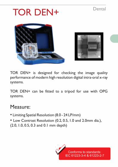
DentalTOR DEN+
TOR DEN+ is designed for checking the image quality performance of modern high resolution digital intra-oral x-ray systems.
TOR DEN+ can be fitted to a tripod for use with OPG systems.
Measure:
• Limiting Spatial Resolution (8.0 - 24 LP/mm)
• Low Contrast Resolution (0.2, 0.5, 1.0 and 2.0mm dia.), (2.0, 1.0, 0.5, 0.3 and 0.1 mm depth)
conforms to standardsABCDEF & GHIJKL
Conforms to standards:IEC 61223-3-4 & 61223-2-7
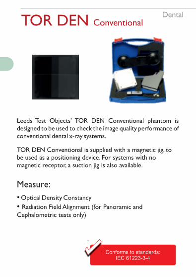
DentalTOR DEN Conventional
Leeds Test Objects’ TOR DEN Conventional phantom is designed to be used to check the image quality performance of conventional dental x-ray systems.
Measure:
• Optical Density Constancy
• Radiation Field Alignment (for Panoramic and Cephalometric tests only)
TOR DEN Conventional is supplied with a magnetic jig, to be used as a positioning device. For systems with no magnetic receptor, a suction jig is also available.
conforms to standardsABCDEF & GHIJKL
Conforms to standards:IEC 61223-3-4

DentalTO PAN
A test object that allows the complex focal trough of the panoramic x-ray unit to be quantified, as well as the capacity for spatial resolution testing using line-pair test patterns (available separately) and contrast resolution insert (available separately).
This allows full assessment of parameters affecting image quality. Can be used in film and digital systems.
Measure:
• Limiting spatial resolution
• Contrast resolution
• Quantification of the focal trough and image layer
See also: Test Patterns

DentalSedentexCT IQ
A PMMA cylinder (160 mm diameter) with recesses to house test inserts.
Conforms to standardEC No 172 : 2012
• Noise
• Uniformity
• Geometric Distortion
• Spatial Resolution
• Contrast Resolution
• Pixel Intensity Value / HU / CT Number
• Beam Hardening Artefacts
Measure:
See also: CT & Radia Software
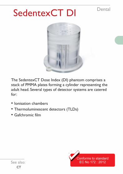
SedentexCT DI
Conforms to standardEC No 172 : 2012
Dental
The SedentexCT Dose Index (DI) phantom comprises a stack of PMMA plates forming a cylinder representing the adult head. Several types of detector systems are catered for:
• Ionisation chambers
• Thermoluminescent detectors (TLDs)
• Gafchromic film
See also: CT

DentalCBCT-161
CBCT-161 is used for constancy testing in Cone Beam CT.
• Pixel Intensity Value Linearity
• Noise
• CNR
Measure:
Conforms to standardDIN 6868-161See also:
AutoPIA Software
CBCT-161+As above, includes rods of Delrin 1, 2, 3, 4 and 5mm dia. - approx. 2% contrast.
• Homogeneity
• Artefacts
• MTF
CBCT-161-AAcceptance testing accessory to CBCT-161 (160 x 60, 160 x 50 mm PMMA cylinders).
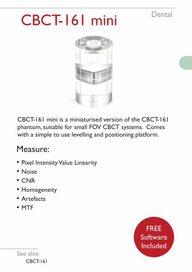
DentalCBCT-161 mini
See also: CBCT-161
CBCT-161 mini is a miniaturised version of the CBCT-161 phantom, suitable for small FOV CBCT systems. Comes with a simple to use levelling and positioning platform.
• Pixel Intensity Value Linearity
• Noise
• CNR
• Homogeneity
• Artefacts
• MTF
Measure:
FREESoftwareIncluded

DENTEST
Developed withPoole Hospital NHS
Dental
Leeds Test Objects’ DENTEST phantom is designed to be used to check the image quality performance of digital intra-oral dental x-ray systems. These test should be carried out on commissioning, and on a routine basis to check for any deterioration in performance.
• Dynamic range
• Spatial resolution
• Low contrast resolution
• Detector homogeneity
Measure:

DentalTO UNIDENT
Designed for use either with conventional intra-oral film or with digital detector systems, TO UniDENT is a step wedge test object which combines ease-of-use with a clinically-relevant indication of the performance of the entire imaging chain.
• For use with film or digital
• Quick, easy set-up
• Optimise exposure levels and routine QA checks
• Checks entire imaging process
• Highlights only clinically-important changes
Features:

CTCT AEC
CT AEC is used for constancy testing of CT AEC systems.
Set includes:
See also: SedentexCT IQ
11x 25mm thick PMMA ellipses which can be re-configured to any orderBlank inserts for AEC checksTubes to accommodate pencil ion chambers for dosimetry2x Sturdy end plates with carrying handlesSpirit level and centre markings for easy positioningWheeled protective storm case with telescopic handleSedentexCT-IQ test inserts (available separately)
•
• • • • • •
FREESoftwareIncluded

CTCT AEC-50
CT AEC-50 is used for constancy testing of CT AEC systems.
Set includes:
• 6x 50mm thick PMMA ellipses which can be re-configured to any order
• 2x Sturdy end plates with carrying handles
• Spirit level and centre markings for easy positioning
• Wheeled protective storm case with telescopic handle
FREESoftwareIncluded
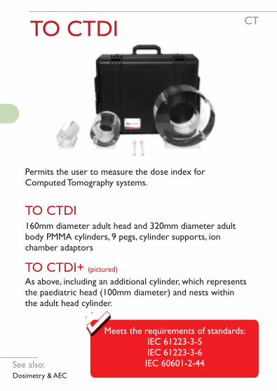
CTTO CTDI
Permits the user to measure the dose index for Computed Tomography systems.
160mm diameter adult head and 320mm diameter adult body PMMA cylinders, 9 pegs, cylinder supports, ion chamber adaptors
TO CTDI+ (pictured)
As above, including an additional cylinder, which represents the paediatric head (100mm diameter) and nests within the adult head cylinder.
conforms to standardsABCDEF & GHIJKL
Meets the requirements of standards:IEC 61223-3-5IEC 61223-3-6IEC 60601-2-44See also:
Dosimetry & AEC
TO CTDI
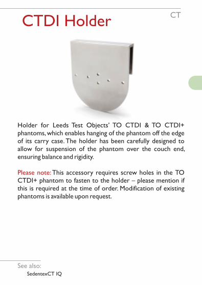
CTCTDI Holder
Holder for Leeds Test Objects’ TO CTDI & TO CTDI+ phantoms, which enables hanging of the phantom off the edge of its carry case. The holder has been carefully designed to allow for suspension of the phantom over the couch end, ensuring balance and rigidity.
Please note: This accessory requires screw holes in the TO CTDI+ phantom to fasten to the holder – please mention if this is required at the time of order. Modification of existing phantoms is available upon request.
See also: SedentexCT IQ
CTSedentexCT IQ
A PMMA cylinder (160 mm diameter) with recesses to house test inserts.
• Noise
• Uniformity
• Geometric Distortion
• Spatial Resolution
• Contrast Resolution
• Pixel Intensity Value / HU / CT Number
• Beam Hardening Artefacts
Measure:
See also: Dental & Radia Software
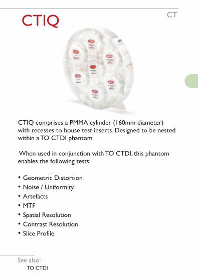
CTCTIQ
CTIQ comprises a PMMA cylinder (160mm diameter) with recesses to house test inserts. Designed to be nested within a TO CTDI phantom.
When used in conjunction with TO CTDI, this phantom enables the following tests:
• Geometric Distortion
• Noise / Uniformity
• Artefacts
• MTF
• Spatial Resolution
• Contrast Resolution
• Slice Profile
See also: TO CTDI

CTCBCT-150
CBCT-150 phantom is used for constancy testing of CBCT and 3D-Fluoroscopy systems.
Details:
• PMMA cuboid 120 x 120 x 60 mm
• Rows of 4x air holes of 0.5 - 1.3 mm diameter with Spatial Frequencies 0.38 - 1.0 mm
• Tripod for universal positioning
Measure:
• Spatial Resolution
Conforms to standardDIN 6868-150

MRIMagIQ
A QA test phantom for magnetic resonance imaging comprising a single chamber (MagIQ) or two identical chambers (MagIQ Duo), which may be filled with different solutions to create two test objects (e.g. high field and low field). Automated scoring software is available separately.
Conforms to standardIEC 62464-1:2007
Measure:
• Geometric Distortion
• Spatial Resolution
• Contrast Resolution - Fat Suppression
• SNR
• Ghosting
• Uniformity
• Slice Profile
• T1 and T2 gel tubes(available separately)
Supplied with 1.5T and 3T Solutions.
See also: Radia software
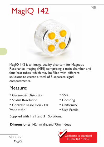
MRIMagIQ 142
MagIQ 142 is an image quality phantom for Magnetic Resonance Imaging (MRI) comprising a main chamber and four ‘test tubes’ which may be filled with different solutions to create a total of 5 separate signal compartments.
See also: MagIQ
Conforms to standardIEC 62464-1:2007
Measure:
• Geometric Distortion
• Spatial Resolution
• Contrast Resolution - Fat Suppression
• SNR
• Ghosting
• Uniformity
• Slice Profile
Supplied with 1.5T and 3T Solutions.
Dimensions: 142mm dia. and 75mm deep
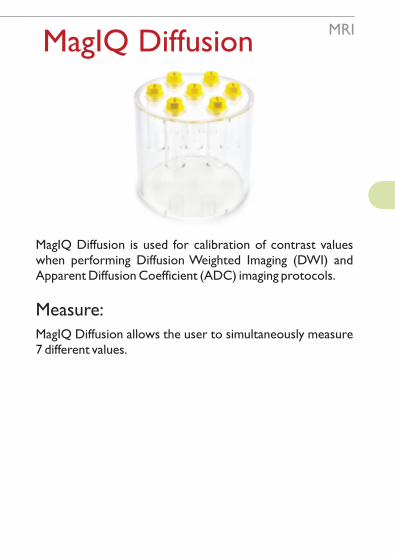
MRIMagIQ Diffusion
MagIQ Diffusion is used for calibration of contrast values when performing Diffusion Weighted Imaging (DWI) and Apparent Diffusion Coefficient (ADC) imaging protocols.
Measure:
MagIQ Diffusion allows the user to simultaneously measure 7 different values.

MRI
MagIQ Oncology is designed for image quality assessment in the diagnosis, treatment planning and therapy of cancer (especially breast and prostate tumours).
Measure:
• Homogeneity
• Contrast resolution
• Slice thickness
• MTF / line pairs
• Spherical gland volume
• Geometric distortion
• Silicon suppression techniques
• Fat saturation techniques
• Detachable section for T1 and T2 values and DCE
• Detachable section for Gadolinium doped water
MagIQ Oncology

MRI
Features:
• A set of two phantoms (one for each breast well)
• Each phantom has two separately filled chambers
• Sized to fit breast coils
Benefits:
• Quantitative measurement of SNR of the water signal from each phantom• Fat fraction using a combination of the water and fat only images in the fat chambers when using ‘Dixon’ fat separation techniques, or a ratio of T1 and T1 fat-sat to assess fat suppression
• Simple to use
• Easy set up, requiring only locally available pads to stabilise in breast well
MagIQ Breast
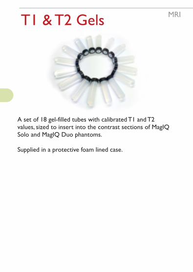
MRIT1 & T2 Gels
A set of 18 gel-filled tubes with calibrated T1 and T2 values, sized to insert into the contrast sections of MagIQ Solo and MagIQ Duo phantoms.
Supplied in a protective foam lined case.

Nuclear MedicinePET IQ
Leeds Test Objects' PET IQ Phantom is a realistic anthropomorphic PMMA vessel, which simulates the trunk of the human body.
On one face of the phantom is a circular plate into which six hollow PMMA spheres can be screwed. The wall thickness of the spheres is 1.25mm. The spheres are filled with water for cold lesion imaging and with 18FDG for hot lesion imaging.
PET IQ is supplied in a wheeled hard case with a telescopic handle.
Conforms to standardsNEMA NU2(2007) & IEC 61675-1(2008)

Gamma Resolution
Rigid PMMA and Pb bar phantom for the quality control of spatial resolution of gamma cameras. The Gamma resolution phantom consists of four sections, each with different bar dimensions, and features engraved, pigment filled centre lines for accurate and repeatable positioning.
Nuclear Medicine
• Quadrant 1Number of bars: 41Bar width and spacing: 3.2mmBar length: 190mm
• Quadrant 2Number of bars: 24Bar width and spacing: 4.0mmBar length: 260mm
• Quadrant 3Number of bars: 28Bar width and spacing: 4.8mmBar length: 190mm
• Quadrant 4Number of bars: 15Bar width and spacing: 6.4mmBar length: 260mm

Nuclear MedicineGamma Flood
Rigid PMMA flood phantom for the quality control of detector homogeneity and subsequent correction in gamma cameras.
The two filling and venting plugs of the Leeds Test Objects Gamma Flood phantom are situated on the flat surface, not on one of the edges as with many other manufacturers.
Thickness tolerances on materials and the fillable voids are ±1% of nominal ensuring high precision.

Nuclear MedicineGamma Distortion
Rigid PMMA phantom for the quality control of geometric distortion and spatial resolution of gamma cameras.
The Gamma distortion phantom consists of a series of 1mm dia. 3mm deep holes at 80mm pitch which can be filled with activity.
Measure (when filled):
• Point-to-point measurement
• Point spread function (PSF)
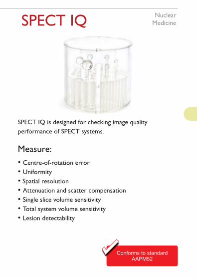
Nuclear MedicineSPECT IQ
SPECT IQ is designed for checking image quality
performance of SPECT systems.
Measure:
• Centre-of-rotation error
• Uniformity
• Spatial resolution
• Attenuation and scatter compensation
• Single slice volume sensitivity
• Total system volume sensitivity
• Lesion detectability
Conforms to standardAAPM52
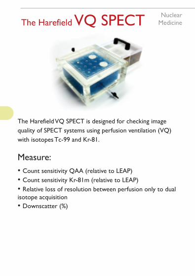
Nuclear MedicineThe Harefield VQ SPECT
The Harefield VQ SPECT is designed for checking image
quality of SPECT systems using perfusion ventilation (VQ)
with isotopes Tc-99 and Kr-81.
Measure:
• Count sensitivity QAA (relative to LEAP)
• Count sensitivity Kr-81m (relative to LEAP)
• Relative loss of resolution between perfusion only to dual isotope acquisition
• Downscatter (%)

Nuclear MedicinePET\CT\MR Cubes
A set of 7 clear PMMA cubes, 17x17x17mm, with a 5mm dia. spherical fillable void, which can be filled with any mixture of radionuclide, CT contrast agent or MR contrast agent and sealed with a nylon screw (provided).
These cubes can be used as:
• Point source markers
• Imaging aids
• Skin markers for clinical use

Radiotherapy
ISO Cube
ISO Cube is a routine phantom made from water equivalent material for testing of positioning, alignment and isocentre checks of CBCT imaging systems, in both kV (imaging) and MV (treatment) energy ranges. This phantom is supplied with an alignment plate for fast & easy set up.
Measure:
• Positioning and re-positioning
• Laser alignment
• Light-field size
• Light-field alignment
• kV and MV coordinate coincidence
• CBCT coordinate isocentre coincidence
See also: Radia Software
Conforms toTG-142 recommendations
Radiotherapy
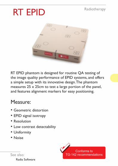
RT EPID
RT EPID phantom is designed for routine QA testing of the image quality performance of EPID systems, and offers a simple setup with its innovative design. The phantom measures 25 x 25cm to test a large portion of the panel, and features alignment markers for easy positioning.
Measure:
• Geometric distortion
• EPID signal isotropy
• Resolution
• Low contrast detectability
• Uniformity
• Noise
See also: Radia Software
Conforms toTG-142 recommendations

Radiotherapy
RT Align
RT Align is designed for routine QA testing of Radiotherapy systems. The phantom features laser alignment markers and field size engravings, as well as embedded high-contrast markers, for accurate, repeatable testing.
See also: Radia Software
Measure:
• Isocentre alignment
• Light field alignment
• Field size
• Couch travel (3D)
• EPID scaling and travel
• Couch rotation
Conforms toTG-142 recommendations

Radiotherapy
RT Align Rotate
Designed for routine QA testing of Radiotherapy systems, the RT Align Rotate contains a larger alignment plate and a jig to enable the plate to be positioned in any orientation.
Measure:
• Light field alignment
• Field size
• Couch travel (3D)
• EPID scaling and movement (embedded BB’s)
• Couch rotation

Digital SubtractionDSF Set
A set of four test objects, the DSF set is supplementary to the SFS set of test objects and checks those parameters which are particularly relevant to DSF imaging. After mask images have been obtained the image content is changed by the manipulation of various plates.
Set includes:
• TO J3
• TO Q3
• TO 20
• TO D3
See also: SFS Set, TO20

Digital SubtractionTO DSA
The TO DSA phantom is used for quality control testing of the image quality performance of digital subtraction angiography systems.
Measure:
• Video system stability
• Dynamic range
• Spatial resolution
• Contrast (Iodine) resolution
Conforms to AAPM Report 15Meets IEC 61223-3-3
• Mis-registration artefacts
• Contrast uniformity
• Contrast linearity
• Spatial uniformity
TO DSA is supplied in a wheeled hard case with a telescopic handle.

Digital SubtractionDSA 8/54
Leeds Test Objects DSA 8/54 phantom is designed for checking the image quality performance of digital subtraction fluoroscopy x-ray systems, on both a commissioning and routine basis.
DSA 8/54 comprises a PMMA phantom with a Cu step wedge and a dynamic PMMA insert with 4 Al strips, powered by a manual pneumatic bulb with a 6 metre hose.
Measure:
• Artefacts
• Dynamic range
• Logarithmic errors
• Contrast media sensitivity
Conforms to standardsDIN 6868-8 & 6868-54

Digital SubtractionTO DR
These test objects are designed for systems which operate by sensing mean signal levels. The TO DR tests dynamic range by measuring detail visibility over a wide range of input X-ray intensities. Different designs cater for subtraction and non-subtraction systems.
TO DR (4 Piece)
Supplied in a set that allows it to be used for both subtraction and non-subtraction systems.
TO DR (2 Piece)
For use with non-subtraction systems only.
See also: DSF Set

TO
10
These test objects are designed for quick quantitative assessments of image quality. The results can be plotted on a Threshold Detection Index Curve.
TO.12 Fluorography systems. A range of 9 contrasts per detail size.
TO.10 Fluoroscopic X-Ray systems. A range of 9 contrasts per detail size.
TO
12
TO 10 & TO 12Contrast
Detail
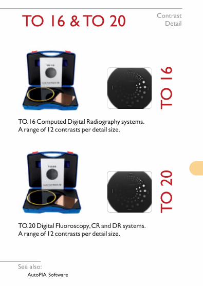
TO 16 & TO 20
conforms to standardsABCDEF & GHIJKL
TO
16
TO.20 Digital Fluoroscopy, CR and DR systems. A range of 12 contrasts per detail size.
TO
20
TO.16 Computed Digital Radiography systems. A range of 12 contrasts per detail size.
See also: AutoPIA Software
ContrastDetail
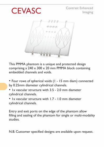
CEVASCContrast Enhanced
Imaging
This PMMA phantom is a unique and protected design comprising a 240 x 300 x 20 mm PMMA block containing embedded channels and voids.
• Four rows of spherical voids (1 - 15 mm diam) connected by 0.25mm diameter cylindrical channels.
• 1x vascular structure with 3.5 - 2.0 mm diameter cylindrical channels.
• 1x vascular structure with 1.7 - 1.0 mm diameter cylindrical channels.
Entry and exit ports on the edge of the phantom allow filling and sealing of the phantom for single or multi-modality studies.
N.B. Customer specified designs are available upon request.

TO FSJ General
The TO FSJ (gen) test object is designed for use with general radiography X-ray systems. This test object (when used with an appropriate test pattern) allows the medical physicist or radiographer to measure the focal spot of their x-ray system.
Features
• 30cm tower
• Quick central alignment check
• Can be used with line pair test pattern, star test pattern or slit camera
See also: Test Patterns
Accessories:Focal Spot
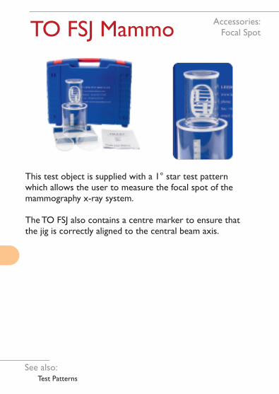
TO FSJ Mammo
This test object is supplied with a 1° star test pattern which allows the user to measure the focal spot of the mammography x-ray system.
The TO FSJ also contains a centre marker to ensure that the jig is correctly aligned to the central beam axis.
See also: Test Patterns
Accessories:Focal Spot
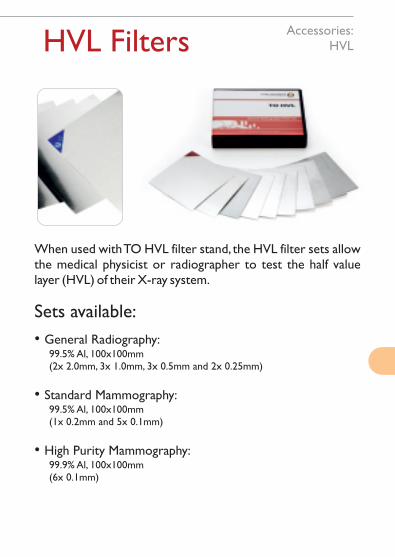
HVL Filters
When used with TO HVL filter stand, the HVL filter sets allow the medical physicist or radiographer to test the half value layer (HVL) of their X-ray system.
Sets available:
• General Radiography: 99.5% Al, 100x100mm (2x 2.0mm, 3x 1.0mm, 3x 0.5mm and 2x 0.25mm)
• Standard Mammography: 99.5% Al, 100x100mm (1x 0.2mm and 5x 0.1mm)
• High Purity Mammography: 99.9% Al, 100x100mm (6x 0.1mm)
Accessories:HVL
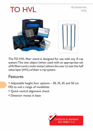
TO HVL
Conforms to standardIEC 60601-1-3
The TO HVL filter stand is designed for use with any X-ray system. This test object (when used with an appropriate set of Al filters and a multi-meter) allows the user to test the half value layer (HVL) of their x-ray system.
Features
• Adjustable height; four options – 30, 35, 45 and 50 cm FID, to suit a range of modalities
• Quick central alignment check
• Detector recess in base
Accessories:HVL

X-Ray RulersAccessories
Radiopaque rulers manufactured from PMMA and tungsten.
Available in various sizes and colours up to 2m in length.
Ruler division spacings can be specified on request.
Please ask for specific sizes.

TOR ABC
A routine test object used for beam alignment and centring in radiography and fluoroscopy.
TOR ABC is available on its own or with a vertical bucky jig as an optional extra (hanger or suction design).
A special variant of the vertical bucky jig is available for Siemens’ Vertix system.
Measure:
• The correspondence of the light field with the effective radiation field
• The determination of the position of the central beam
• The central beam angle deviation from 90°, relative to the plane of the film/detector, is below a limit beyond which the image quality would be adversely affected
Accessories:Beam Alignment
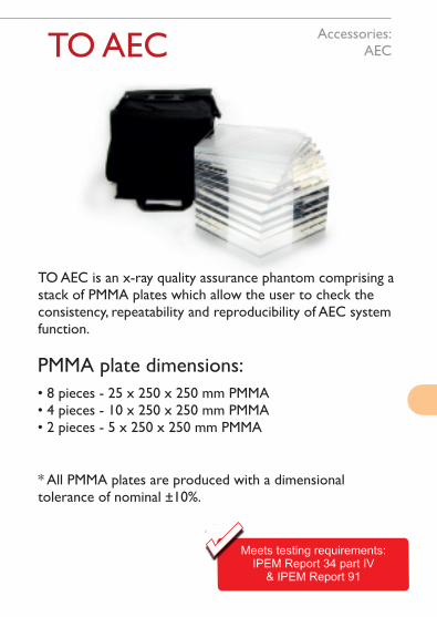
TO AEC
TO AEC is an x-ray quality assurance phantom comprising a stack of PMMA plates which allow the user to check the consistency, repeatability and reproducibility of AEC system function.
PMMA plate dimensions:
• 8 pieces - 25 x 250 x 250 mm PMMA • 4 pieces - 10 x 250 x 250 mm PMMA• 2 pieces - 5 x 250 x 250 mm PMMA
* All PMMA plates are produced with a dimensional tolerance of nominal ±10%.
conforms to standardsABCDEF & GHIJKL
Meets testing requirements:IPEM Report 34 part IV
& IPEM Report 91
Accessories:AEC

TO MTF
An MTF Edge test device comprising a Tungsten edge.
1mm thick tungsten plate (50mm diameter)
Tungsten purity of 90%+
Edge accuracy of 5µm
Repeatable angular adjustment
Conforms to standards:IEC 62220-3-2008, 62220-1:2004
& 62220-2-2007
•
•
•
•
Accessories
FREESoftwareIncluded
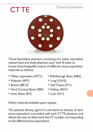
Tissue EquivalentsCT TE
Tissue Equivalent phantom consisting of a water equivalent nested head and body phantom each with 8 holes to house interchangeable inserts of different tissue equivalent materials as follows:
• Water equivalent (WT1)
• Adipose (AP7)
• Breast (BR12)
• Hard Cortical Bone (SB5)
• Inner Bone (Ib7)
• Rib/Average Bone (RB2)
• Lung (LN10)
• Soft Tissue (ST1)
• Kidney (KD1)
• Liver (LV1)
Other materials available upon request.
3The physical density (gm/cm ) and electron density of each tissue equivalent is provided with each CT TE phantom, and allows the user to determine the CT number corresponding to the different tissue equivalents.
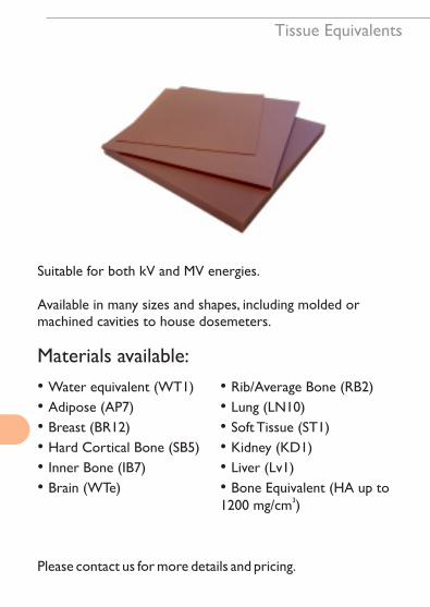
Tissue Equivalents
Suitable for both kV and MV energies.
Available in many sizes and shapes, including molded or machined cavities to house dosemeters.
Materials available:
• Water equivalent (WT1)
• Adipose (AP7)
• Breast (BR12)
• Hard Cortical Bone (SB5)
• Inner Bone (IB7)
• Brain (WTe)
• Rib/Average Bone (RB2)
• Lung (LN10)
• Soft Tissue (ST1)
• Kidney (KD1)
• Liver (Lv1)
• Bone Equivalent (HA up to 31200 mg/cm )
Please contact us for more details and pricing.

Tissue EquivalentsTO H2O
Constructed from PMMA and featuring two plugs, our water phantoms are available in four sizes:
• 300 x 300 x 50 mm
• 300 x 300 x 100 mm
• 300 x 300 x 150 mm
• 300 x 300 x 200 mm
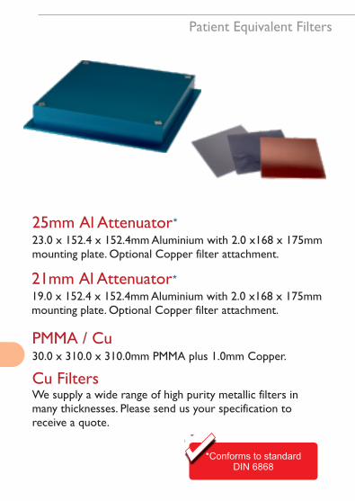
PMMA / Cu30.0 x 310.0 x 310.0mm PMMA plus 1.0mm Copper.
Cu FiltersWe supply a wide range of high purity metallic filters in many thicknesses. Please send us your specification to receive a quote.
*Conforms to standardDIN 6868
25mm Al Attenuator23.0 x 152.4 x 152.4mm Aluminium with 2.0 x168 x 175mm mounting plate. Optional Copper filter attachment.
*
Patient Equivalent Filters
21mm Al Attenuator19.0 x 152.4 x 152.4mm Aluminium with 2.0 x168 x 175mm mounting plate. Optional Copper filter attachment.
*
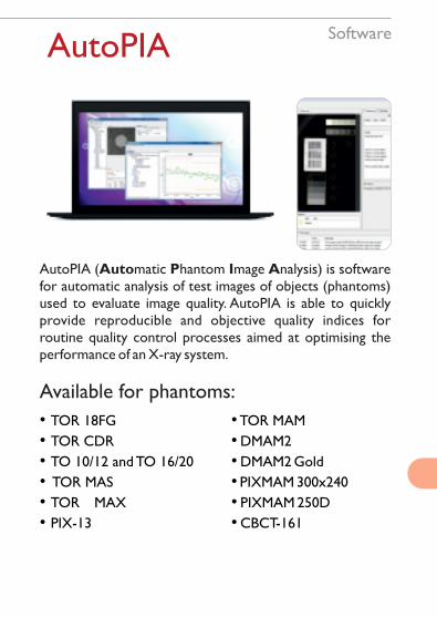
SoftwareAutoPIA
AutoPIA (Automatic Phantom Image Analysis) is software for automatic analysis of test images of objects (phantoms) used to evaluate image quality. AutoPIA is able to quickly provide reproducible and objective quality indices for routine quality control processes aimed at optimising the performance of an X-ray system.
Available for phantoms:
• TOR 18FG
• TOR CDR
• TO 10/12 and TO 16/20
• TOR MAS
• TOR MAX
• PIX-13
• TOR MAM
• DMAM2
• DMAM2 Gold
• PIXMAM 300x240
• PIXMAM 250D
• CBCT-161

SoftwareLTO Software
Analysis software for automated scoring and trending of diagnostic image quality phantoms. Radia allows the user to meet TG142 and insurance requirements and provides objective results for trending and tracking image quality.
Available for phantoms:
• TOR 18FG• SedentexCT IQ• MagIQ
• ISO Cube• RT EPID• RT Align
TEST PATTERNS
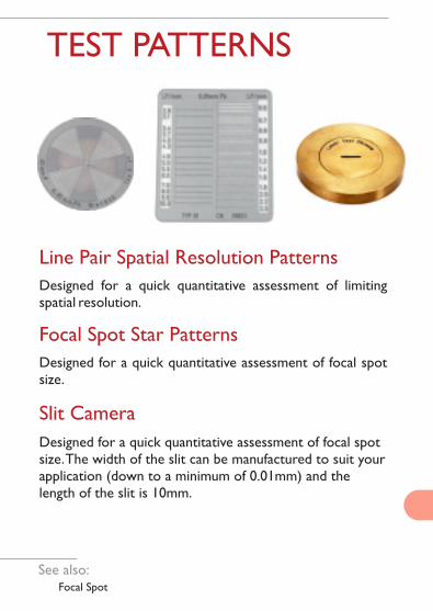
Line Pair Spatial Resolution Patterns
Designed for a quick quantitative assessment of limiting spatial resolution.
Focal Spot Star Patterns
Designed for a quick quantitative assessment of focal spot size.
Slit Camera
Designed for a quick quantitative assessment of focal spot size. The width of the slit can be manufactured to suit your application (down to a minimum of 0.01mm) and the length of the slit is 10mm.
See also: Focal Spot
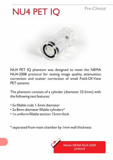
Pre-ClinicalNU4 PET IQ
NU4 PET IQ phantom was designed to meet the NEMA NU4-2008 protocol for testing image quality, attenuation correction and scatter correction of small Field-Of-View PET systems.
The phantom consists of a cylinder (diameter 33.5mm) with the following test features:
• 5x fillable rods 1-5mm diameter• 2x 8mm diameter fillable cylinders*• 1x uniform fillable section 15mm thick
* separated from main chamber by 1mm wall thickness
Meets NEMA NU4-2008protocol
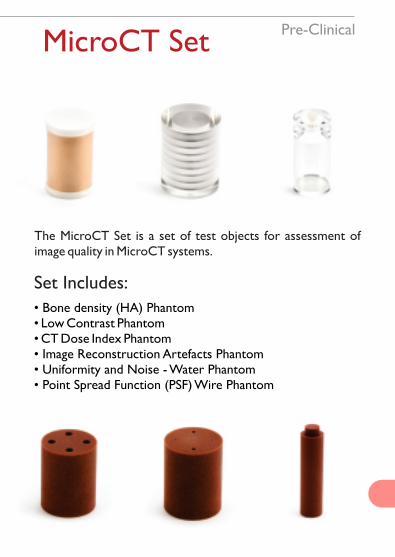
MicroCT Set
The MicroCT Set is a set of test objects for assessment of image quality in MicroCT systems.
Pre-Clinical
Set Includes:
• Bone density (HA) Phantom• Low Contrast Phantom• CT Dose Index Phantom• Image Reconstruction Artefacts Phantom• Uniformity and Noise - Water Phantom• Point Spread Function (PSF) Wire Phantom
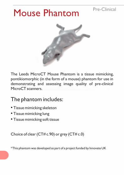
Mouse Phantom
The Leeds MicroCT Mouse Phantom is a tissue mimicking, pontikiomorphic (in the form of a mouse) phantom for use in demonstrating and assessing image quality of pre-clinical MicroCT scanners.
The phantom includes:
• Tissue mimicking skeleton
• Tissue mimicking lung
• Tissue mimicking soft tissue
Choice of clear (CT# c.90) or grey (CT# c.0)
* This phantom was developed as part of a project funded by Innovate UK
Pre-Clinical

Ultrasound
Coming Soon

Notes

v3.8
Leeds Test Objects Ltd.
MiRo House
Becklands Close
Boroughbridge
North Yorkshire
YO51 9NR
United Kingdom
Phone +44 (0)1423 320007
Web www.leedstestobjects.com
ISO 9001
www.leedstestobjects.com