(A4)Osteoarthritis of Knee - healthshare.org.uk · Femur (thigh bone) Fibula Tibia (shin bone)...
11
Osteoarthritis of the Knee Healthshare Information for Guided Patient Management
-
Upload
nguyenkien -
Category
Documents
-
view
233 -
download
4
Transcript of (A4)Osteoarthritis of Knee - healthshare.org.uk · Femur (thigh bone) Fibula Tibia (shin bone)...

Osteoarthritis of the KneeHealthshare Information for Guided Patient Management
Swanton
Stamp
Sharon
Stamp

Index
Introduction 2
Knee joint anatomy 2
What is Osteoarthritis? 2
What treatments are available? 3
• Conservative treatment 3
• Surgical management 4
• Knee replacement surgery 4
• When do I need a knee replacement? 5
• Am I too young or too old? 5
• What should I do before knee replacement? 5
• In hospital - after operation 5
• At home 5
• What are the possible complications? 6
• Exercises for Osteoarthritis 7
• Contact us 10
[ 1 ]
Ost
eo
art
hri
tis
of
the
Kn
ee
Swanton
Stamp
Sharon
Stamp
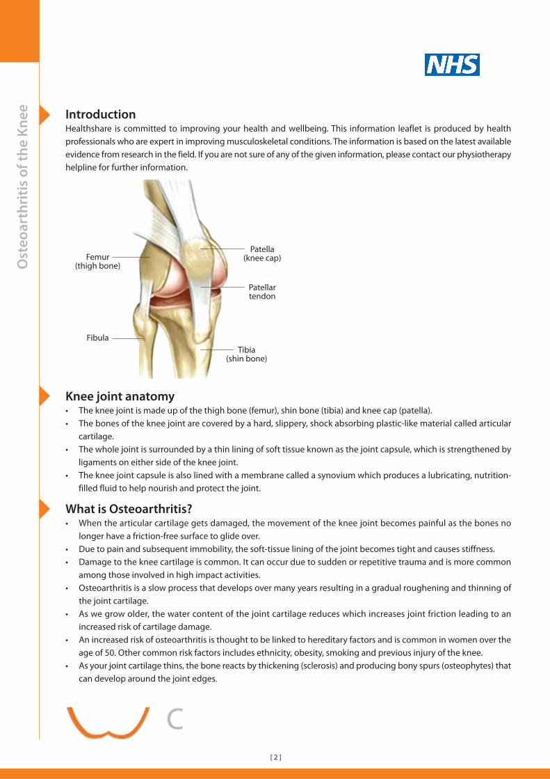
Femur(thigh bone)
FibulaTibia
(shin bone)
Patellartendon
Patella(knee cap)
Knee joint anatomy• The knee joint is made up of the thigh bone (femur), shin bone (tibia) and knee cap (patella).• The bones of the knee joint are covered by a hard, slippery, shock absorbing plastic-like material called articular
cartilage.• The whole joint is surrounded by a thin lining of soft tissue known as the joint capsule, which is strengthened by
ligaments on either side of the knee joint.• The knee joint capsule is also lined with a membrane called a synovium which produces a lubricating, nutrition-
filled fluid to help nourish and protect the joint.
What is Osteoarthritis?• When the articular cartilage gets damaged, the movement of the knee joint becomes painful as the bones no
longer have a friction-free surface to glide over.• Due to pain and subsequent immobility, the soft-tissue lining of the joint becomes tight and causes stiffness.• Damage to the knee cartilage is common. It can occur due to sudden or repetitive trauma and is more common
among those involved in high impact activities.• Osteoarthritis is a slow process that develops over many years resulting in a gradual roughening and thinning of
the joint cartilage.• As we grow older, the water content of the joint cartilage reduces which increases joint friction leading to an
increased risk of cartilage damage.• An increased risk of osteoarthritis is thought to be linked to hereditary factors and is common in women over the
age of 50. Other common risk factors includes ethnicity, obesity, smoking and previous injury of the knee.• As your joint cartilage thins, the bone reacts by thickening (sclerosis) and producing bony spurs (osteophytes) that
can develop around the joint edges.
Ost
eo
art
hri
tis
of
the
Kn
ee
[ 2 ]
IntroductionHealthshare is committed to improving your health and wellbeing. This information leaflet is produced by healthprofessionals who are expert in improving musculoskeletal conditions. The information is based on the latest availableevidence from research in the field. If you are not sure of any of the given information, please contact our physiotherapyhelpline for further information.
Swanton
Stamp
Sharon
Stamp

• Osteoarthritis of the knee joint also irritates thesynovium which leads to an increased productionof fluid, causing joint swelling (effusion).
• Osteoarthritis of the knee may cause a weaknessin the surrounding musculature and a thickeningof the ligaments, with a general instability of thejoint due to bony and soft tissue changes.
• Osteoarthritis of the knee commonly affectsthe inside of the joint and the knee cap. In mostcases osteoarthritis of the knee causes little orno pain but can become troublesome whentriggered by an incident such as a trip or fall.
• However, osteoarthritis of the knee joint canbecome severe, affecting the whole joint and can result in pain affecting your day-to-day activities, or interruptingsleep.
• In particular, osteoarthritis of the patella may affect your ability to kneel, climb stairs and sit for long periods oftime.
What treatments are available?
Conservative treatment
There are a number of treatment methods that you can try to help manage your symptoms before considering anysurgical intervention, such as a knee replacement. Remember these treatments will not cure your osteoarthritis, but
may help to reduce the pain and improve your functional independence and quality of life.
Physical Aids There are a number of devices you can try to help to improve your walking. These includewalking aids, shock-absorbing shoes, knee braces and splints. Your GP or physiotherapistshould be able to advise you on the appropriate use of these aids based on the severity ofyour condition and your activity requirements.
Physiotherapy Your physiotherapist may teach you an exercise programme which will aim to strengthenthe muscles that support the knee joint and improve its flexibility. They may also considerjoint and soft tissue manipulative therapy techniques. Treatment techniques such as TENSor acupuncture have also shown to be beneficial for improving pain and function in somecases.
Staying Active Performing regular exercise is key in maintaining the range of movement in your knee andhelping to slow down further deterioration. Regular exercise also helps to reduce pain.Swimming, walking and cycling have all been shown to have a beneficial effect on maintainingfunctional independence.
Medications Paracetamol and/or non-steroidal anti-inflammatory medications may be suggested byyour GP and can be beneficial in reducing pain and inflammation. Some patients reportglucosamine and/or chondroitin have proved beneficial, although evidence for thesesupplements is currently lacking. These are widely available from health food shops orpharmacies.
Joint Injections Corticosteroids and hyaluronic acid are the two most common drugs used for knee jointinjections to reduce knee pain. Corticosteroids are very strong anti-inflammatories, whilehyaluronic acid is the fluid naturally found in synovial joints for lubrication.
Ost
eo
art
hri
tis
of
the
Kn
ee
OA KneeNormal Knee
[ 3 ]
Swanton
Stamp
Sharon
Stamp

Surgical management
Arthroscopy (keyhole surgery): This is not routinely done for osteoarthritis as the current evidence does not showany significant benefit in reducing pain. However, if you have a cartilage tear that causes locking and loss of movementof the knee, then joint arthroscopy may help to improve your mobility and reduce pain levels.
Knee replacement surgery
• A knee replacement is an operation which involves the removal and replacement of the damaged or worn partsof the knee joint with a prosthesis that is made up of metal and plastic parts.
• A knee replacement is usually performed on patients who have severe osteoarthritis that adversely affects theirday-to-day activities.
• A total knee replacement involves the resurfacing of your whole knee joint, which can include the femur and tibiaonly, or the femur, tibia and patella. A partial knee replacement involves the resurfacing of one half of your kneei.e. either the inner or outer side of the joint.
• Partial knee replacements will only be performed on people who have arthritis on one side of the knee joint. It isalso typically performed for people under 55 who have an active lifestyle. Typically, movement will be regainedearlier with this procedure than with a total knee replacement.
• Not all people who suffer from arthritis in their knees need a knee replacement and many find conservativetreatment is sufficient in managing their symptoms.
• Only a small percentage of people who have knee osteoarthritis will need a knee replacement, and this will beidentified by a specialist doctor or a specialist physiotherapist.
Ost
eo
art
hri
tis
of
the
Kn
ee
[ 4 ]
Implant (side view)
Femur
Knee cap
Tibia
Femoralcomponent(knee)
Plasticsurface
Tibial plate
Fibula
Implant (front view)
Femoralcomponent(knee)
Plastic surface
Tibial plate
Half knee replacement
Total knee replacement
Swanton
Stamp
Sharon
Stamp
When do I need knee replacement?
• Conservative treatments have not improved your symptoms and your pain is getting worse with your knee hurtingmost of the time.
• Your knee pain is affecting the quality of your sleep.• The knee pain is limiting your enjoyment of life and activities of daily living such as work, gardening, walking etc.• Your knee keeps giving way or locking.• Knee X-Rays show severe arthritic changes and you are experiencing severe symptoms.
Am I too young or too old?
• There is no specific limit to the age of a patient who can have a knee replacement, but surgical intervention is lessfavoured in those under the age of 60.
• Typically materials used for a knee replacement only last for about 10-15 years and the probability of you requiringa second knee replacement is a lot higher if you are younger during your first replacement.
• When you have a second knee replacement, the rehabilitation after the operation is usually a lot more difficult.• Therefore, if you are younger and considering having a knee replacement, it is important that you are aware of
these potential problems and you should exhaust all other conservative options first.
What should I do before knee replacement?If you are waiting to have a knee replacement, there are a number of things you can do to help improve your post-operative recovery.
Lose weight: Excess weight will put extra pressure on your new knee reducing its longevity. Evidence suggests thatoverweight people are more likely to have problems after surgery. Losing weight will reduce the risk of post-operativecomplications and premature wear and tear of the new joint.
Exercise: Exercises prescribed by your physiotherapist will help improve your muscle strength before the operationand this will have a significant positive effect on your post-operative rehabilitation.
In hospital – after the operation• You may have a few tubes coming out of your knee which are drains that help prevent fluid collecting under the
incision site. These will probably be removed 1 to 2 days after the operation.• If you have had an epidural anaesthetic, you may not be able to feel or move your legs for several hours after the
operation. Once this has worn off, your knee may feel stiff and sore but should not be too painful.• If you experience severe pain, it is important to tell the nurse so that your medication can be adjusted appropriately.• Your physiotherapy sessions will start on the day after your operation, and will continue throughout your hospital
stay.• They will help to get your knee moving, and to get you mobile and walking within about 3-4 days with the use of
a walking aid. They will ensure that you are fully functional and independent before discharging you home.• You may be required to wear a compression stocking on your leg to help prevent a DVT (blood clot).
At home• You should continue to use the walking aids that were provided by the hospital for up to 6 weeks and gradually
reduce the amount of aid required, depending on progress.• On returning home, it is important that you continue with the exercises that you were shown in the hospital.• You should be able to start driving again after about 6-12 weeks, but before you do, it is important that you consult
with your GP or physiotherapist.• Post-operative physiotherapy will help you regain as much range of movement in your knee as possible and enable
a quicker return to independent and active living.
Ost
eo
art
hri
tis
of
the
Kn
ee
[ 5 ]
Swanton
Stamp
Sharon
Stamp

Ost
eo
art
hri
tis
of
the
Kn
ee What are the possible complications?
Complications of total knee replacement are minimal. Bleeding during or soon after the procedure is a commonlyreported complication. Infection and an abnormal reaction to the anaesthetic are possible complications of any surgery.
Complications specific to knee replacement surgery are:
• Wound and/or joint infection. Antibiotics are routinely given straight after surgery as a preventative measureagainst this.
• Instability of the new knee (this may require a further operation to rectify).• Patella dislocation due to new joint profile changes.• A leg length difference due to excessive bone loss. This can be rectified with a small raise in the shoe of the shorter
leg if necessary.• Excessive build-up of scar tissue in the knee which may restrict movement. However, this can often be addressed
by your physiotherapist who will try and break down this scar tissue. In rare cases a further operation may berequired to remove this tissue.
• After any big operation it is possible to develop a blood clot in the veins of the legs (DVT) which may break off andform a blockage in the lungs. It is usually treatable, and many measures will be taken before, during and after youroperation to help prevent this from happening (i.e. medication or wearing a compression stocking).
Please remember that risks for any operation will differ for every individual and it is important to discuss these
with your surgeon to determine how these risks may relate to you.
[ 6 ]
Swanton
Stamp
Sharon
Stamp

Ost
eo
art
hri
tis
of
the
Kn
ee Exercises for Osteoarthritis
Stretching exercises
Stage 1: Strengthening exercises
Heel slide
Lie on your back with one knee bent and the other straight.
Now slowly bend your knee while keeping your heel on thefloor. Move as far as your pain will allow you to and graduallyincrease the range of your knee.
Repeat 10 times, 3 times a day.
Quadriceps exercises
Sit on the floor with one leg outstretched in front of youwith a pillow/rolled up towel under the knee.
Push your knee into the pillow, straightening it as much asyou can. Your foot should lift off the floor as you do this. Itis important that you tighten your thigh muscles as youstraighten your knee.
Repeat 15 times x 3, 3 times a day.
Hamstring stretch - Standing
Stand on one leg with the affected leg on top of a chair orstool.
Keep your back straight and gentlylean forward while pulling your toestowards you.
Hold this position for 20 secondsx 5.
Repeat 3 times a day.
Hamstring stretch
Lie on your back with one leg straight, raise the bent kneetowards you and hold behind your knee.
Now slowly straighten your knee until a stretch is felt in theback of the thigh.
You can also use a towel to helpwith this stretch if needed.
Hold for 20 seconds x 5 reps.
Repeat 3 times a day.
Quadriceps stretch - Lying
Lie on the opposit side of the leg being stretched.
Slowly bend the leg to be stretched towards your buttockand pull the leg further by holding your hand or wrappinga towel around the ankle.
You should feel the stretch at the front of your knee.
Remember to keep your hip joint straight.
Hold this position for 20 seconds x 5 reps.
Repeat 3 times a day.
Calf stretch
Keeping your back leg straight,and your heel on the floorturned slightly outward, leaninto the wall until you feel astretch in the calf.
Hold this position for 20seconds x 5.
Repeat this at least 3 times aday.
[ 7 ]
Swanton
Stamp
Sharon
Stamp

Ost
eo
art
hri
tis
of
the
Kn
ee Exercises for Osteoarthritis/Continued
Stage 1: Strengthening exercises/continued
SLR (Straight leg raises)
Lie on your back with your arms next to by side. Keep oneleg straight and the other bent. Now raise the straightenedleg to the height of the bent knee.
Repeat 10 times x 3. Repeat this at least 3 times a day.Repeat on both sides.
You can vary this exercises by pointing your toes outwardsor inwards. You can also add some ankle weights tomake this exercise more difficult.
Bridging
Lie on your back with your back flat on the floor or a bed.
Now slowly pull your belly button towards your spine andslowly raise your buttocks from the floor. Try to bring yourchest, hips and legs into a straight line.
Hold this position for 10 seconds x 10 times.
Repeat 3 times a day.
Single leg bridging
Lie on your back, bend both knees to 90° with your feet flaton the bed/floor.
Slowly lift your hips off the floor and hold for 10 secondsx 10.
Repeat the same on the other side x 10.
Ball squeeze and push
Lying on your back with your knees bent, place a ball orpillow between your knees and squeeze.
Hold for 10 seconds x 10.Repeat 3 times a day.
Then place the ball betweenyour knee and the wall, pushout for 10 seconds x 10.Repeat 3 times day.
Wall slides
Stand leaning up against a wall, yourfeet a little away from the wall withyour toes pointing forwards.
Push your back against the wall andslowly lower your body into a seatedposition.
Now hold this position for 10 secondsx 10. Repeat 3 times a day.
This exercise must be pain free anddon’t bend more than the seatedposition.
Calf raises
Stand close to a support surfacesuch as a kitchen worktop or adoorway frame.
Now slowly raise up onto yourtoes first, then onto your big toe,then the middle of your foot andthen onto your little toe.
Repeat this sequence 10 x 3times a day.
[ 8 ]
Swanton
Stamp
Sharon
Stamp

Ost
eo
art
hri
tis
of
the
Kn
ee Exercises for Osteoarthritis/Continued
Stage 2: Strengthening exercises/continued
Stork standing
Balance on one leg for 30seconds and repeat withthe other leg x 5.
Repeat the above with youreyes closed x 5.
You can progress the aboveto standing on an unsteadysurface, e.g. a cushion or anarrow piece of wood.
Prone hamstring curls
Wrap a resistance band (theraband) around a stable objecti.e. table leg. Attach the free ends to ankle straps and secureto each ankle.
Lie on your stomach far enough from the attachment siteof the band so that there is no slack and then attempt tobring your foot towards your buttocks without allowingyour knee or hips to lift off the floor.
You can get the theraband from alocal sport store. Repeat 10 x 3 sets.
Quadriceps: Step-down backwards Quadriceps: Forward step-down
Stand on one leg on a step.
Slowly lower yourself by bending yourknee and touch the floor with your heel.
Return to the starting position withoutpushing off from the ground.
Repeat 10 x 2 sets.
Stand on one leg on a step.
Slowly lower yourself by bending your knee.
Return to the starting position without pushing off with theopposite leg.
Have your weight on your heelmore than your toe, but keep yourfoot flat. Your knee should be inline with your second toe whenbending it i.e. be aware thatyour knee and foot do not rollinwards.
Repeat 10 x 2 sets.
[ 9 ]
Semi squats
Stand with your feet hip width apart and hold onto a chairif you need to.
Bend the knees keeping your feet flat on the floor, andensure that your knees do not exceed a 90° angle.
Return to the starting position.
Repeat 10 times x 3 sets
Hip abduction and adduction
Attach a theraband around the table leg. Place the furthestleg in the loop of the band and raise it out to the side awayfrom your body.
Keep your knee straight andpelvis stable.
Now turn around and bringyour leg away from thetable towards your midline(the other leg).
Make sure you keep yourbody stable and just use your leg.
Repeat 10 x 3 sets.
Swanton
Stamp
Sharon
Stamp

Ost
eo
art
hri
tis
of
the
Kn
ee
[email protected] | http://healthshare.org.uk
Swanton
Stamp
Sharon
Stamp