
A Wearable Ultrasonic Assembly for Point-Of-Care ...swarup.ece.ufl.edu/papers/C/C101.pdf · A...
4
A Wearable Ultrasonic Assembly for Point-of-Care Autonomous Diagnostics of Malignant Growth Abhishek Basak 1 , Vaishnavi Ranganathan 1 and Swarup Bhunia 1 Abstract— Clinicians generally agree that the most effective way to treat a malignant tumor within body organs is to detect it early. Currently, detection is mostly based on clinical diagnostics triggered by specific symptoms, which is often too late allowing the malignancy to reach an advanced stage. Automated high-resolution monitoring of body parts at regular intervals can be highly effective for detection of an anomalous growth - either primary or recurrence - at an early stage. In this paper, we propose a wearable, point-of-care (POC) ultrasonic imaging assembly to perform unsupervised, periodic monitoring of body parts for early detection of cancerous growths. We use ultrasound technology, which is safe, low-cost, can be easily miniaturized, and amenable for long-term, convenient, online monitoring. The proposed system is capable of providing automated high-resolution images of susceptible volumes of interest in superficial cancer-prone organs (e.g. prostate, uterus, kidney, ovary and bladder) at periodic intervals. We explore the design space for such a system encompassing transducer design parameters, power and memory requirements. Next, we study the effectiveness of such a system using both ultrasonic software simulation (with Field II) as well as experimental evaluation (with medical ultrasound transducers and phantoms). We show that the proposed POC diagnostic system can reliably detect an anomaly at a much smaller size (mostly stage 1) than typically achieved through conventional symptomatic detection. I. I NTRODUCTION It is widely accepted among clinicians that the most effective way to fight against cancer is to detect it early for both primary and recurrence cases. The improvement in cancer survival rates over the past 30 years are often attributed to early detection [1]. According to a recent report, if diagnosed at a localized stage through regular screenings, the 5-year relative survival rate for cancer patients would be 60 - 85% depending on cancer type [1], as shown in Fig. 1(a). Unfortunately, at present, most cases of cancer are detected only when symptoms occur, which corresponds to a tumor of large size and sometimes metastasizing to different organs. Fig. 1(b) shows that the cumulative detection of cancers in stage 1 are usually less than 20%. Over 60% of patients with breast, lung, colon and ovarian cancer have hidden or overt metastatic colonies at the time of diagnosis and hence, even most advanced therapies produce poor results [2]. Detecting cancers as early as possible, preferably in the premalignant stage (i.e. stage 1), would lead to very high probability of successful treatment. Early detection for most forms of cancer would require periodic, high-resolution screening of susceptible regions. 1 Authors are with Department of Electrical Engineering and Computer Science, Case Western Reserve University, Cleveland, OH 44106, USA. Email: {axb594,vxn55,skb21}@case.edu. Fig. 1. a) Variation of 5-year survival rates with cancer staging; (b) variation of cumulative percentage of cancer detection with staging; (c) organs of human body considered in our point-of-care ultrasonic monitoring system. Existing screening modalities require a patient to access com- plex imaging instruments e.g. Magnetic Resonance Imaging (MRI) or Computed Tomography (CT) in a medical facility. They are generally difficult to access, expensive and not convenient for routine monitoring of an otherwise healthy person. The situation is much worse in developing coun- tries (compared to developed ones), where state-of-the-art diagnostic facilities are not easily available or affordable. Hence, an automated, high-resolution, affordable point-of- care (POC) screening technology can potentially change the face of cancer diagnostics. In this paper, we propose a wearable ultrasonic assem- bly for POC monitoring of anomalous growth. It includes ultrasonic transducer arrays placed at strategic locations, integrated transmit and receive circuitry along with the power management module, transceiver and associated backend computing unit for image rendering. The images of the super- ficial cancer-prone organs are taken periodically, compressed and transmitted out to an external computing unit e.g. a mobile phone, which performs pattern recognition to identify anomalous growth. If a diagnostic threshold is exceeded, the external computer can alert the patient or care-givers regard- ing a possible anomalous growth. We consider ultrasound based imaging since it can be easily miniaturized in a wear- able enclosure (e.g. a vest or bodysuit); can operate at low power budget (for untethered measurements); and is safe as well as inexpensive. We focus on monitoring the superficial cancer-prone organs such as prostrate, breast, bladder, uterus, ovaries, and cervix (as shown in Fig. 1(c), which provides acceptable imaging resolution using ultrasound technology. This corresponds over 40% of all cancer types in males and over 50% in females in 2012 [3] (including breast cancer). The proposed system can provide the following key 128 2013 IEEE Point-of-Care Healthcare Technologies (PHT) Bangalore, India, 16 - 18 January, 2013 978-1-4673-2767-1/13/$31.00 ©2013 IEEE
Transcript of A Wearable Ultrasonic Assembly for Point-Of-Care ...swarup.ece.ufl.edu/papers/C/C101.pdf · A...

A Wearable Ultrasonic Assembly for Point-of-Care AutonomousDiagnostics of Malignant Growth
Abhishek Basak1, Vaishnavi Ranganathan1 and Swarup Bhunia1
Abstract— Clinicians generally agree that the most effectiveway to treat a malignant tumor within body organs is todetect it early. Currently, detection is mostly based on clinicaldiagnostics triggered by specific symptoms, which is often toolate allowing the malignancy to reach an advanced stage.Automated high-resolution monitoring of body parts at regularintervals can be highly effective for detection of an anomalousgrowth - either primary or recurrence - at an early stage. In thispaper, we propose a wearable, point-of-care (POC) ultrasonicimaging assembly to perform unsupervised, periodic monitoringof body parts for early detection of cancerous growths. Weuse ultrasound technology, which is safe, low-cost, can beeasily miniaturized, and amenable for long-term, convenient,online monitoring. The proposed system is capable of providingautomated high-resolution images of susceptible volumes ofinterest in superficial cancer-prone organs (e.g. prostate, uterus,kidney, ovary and bladder) at periodic intervals. We explore thedesign space for such a system encompassing transducer designparameters, power and memory requirements. Next, we studythe effectiveness of such a system using both ultrasonic softwaresimulation (with Field II) as well as experimental evaluation(with medical ultrasound transducers and phantoms). We showthat the proposed POC diagnostic system can reliably detect ananomaly at a much smaller size (mostly stage 1) than typicallyachieved through conventional symptomatic detection.
I. INTRODUCTION
It is widely accepted among clinicians that the mosteffective way to fight against cancer is to detect it earlyfor both primary and recurrence cases. The improvementin cancer survival rates over the past 30 years are oftenattributed to early detection [1]. According to a recent report,if diagnosed at a localized stage through regular screenings,the 5-year relative survival rate for cancer patients wouldbe 60 − 85% depending on cancer type [1], as shown inFig. 1(a). Unfortunately, at present, most cases of cancer aredetected only when symptoms occur, which corresponds to atumor of large size and sometimes metastasizing to differentorgans. Fig. 1(b) shows that the cumulative detection ofcancers in stage 1 are usually less than 20%. Over 60% ofpatients with breast, lung, colon and ovarian cancer havehidden or overt metastatic colonies at the time of diagnosisand hence, even most advanced therapies produce poorresults [2]. Detecting cancers as early as possible, preferablyin the premalignant stage (i.e. stage 1), would lead to veryhigh probability of successful treatment.
Early detection for most forms of cancer would requireperiodic, high-resolution screening of susceptible regions.
1Authors are with Department of Electrical Engineering and ComputerScience, Case Western Reserve University, Cleveland, OH 44106, USA.Email: {axb594,vxn55,skb21}@case.edu.
Fig. 1. a) Variation of 5-year survival rates with cancer staging; (b) variationof cumulative percentage of cancer detection with staging; (c) organs ofhuman body considered in our point-of-care ultrasonic monitoring system.
Existing screening modalities require a patient to access com-plex imaging instruments e.g. Magnetic Resonance Imaging(MRI) or Computed Tomography (CT) in a medical facility.They are generally difficult to access, expensive and notconvenient for routine monitoring of an otherwise healthyperson. The situation is much worse in developing coun-tries (compared to developed ones), where state-of-the-artdiagnostic facilities are not easily available or affordable.Hence, an automated, high-resolution, affordable point-of-care (POC) screening technology can potentially change theface of cancer diagnostics.
In this paper, we propose a wearable ultrasonic assem-bly for POC monitoring of anomalous growth. It includesultrasonic transducer arrays placed at strategic locations,integrated transmit and receive circuitry along with the powermanagement module, transceiver and associated backendcomputing unit for image rendering. The images of the super-ficial cancer-prone organs are taken periodically, compressedand transmitted out to an external computing unit e.g. amobile phone, which performs pattern recognition to identifyanomalous growth. If a diagnostic threshold is exceeded, theexternal computer can alert the patient or care-givers regard-ing a possible anomalous growth. We consider ultrasoundbased imaging since it can be easily miniaturized in a wear-able enclosure (e.g. a vest or bodysuit); can operate at lowpower budget (for untethered measurements); and is safe aswell as inexpensive. We focus on monitoring the superficialcancer-prone organs such as prostrate, breast, bladder, uterus,ovaries, and cervix (as shown in Fig. 1(c), which providesacceptable imaging resolution using ultrasound technology.This corresponds over 40% of all cancer types in malesand over 50% in females in 2012 [3] (including breastcancer). The proposed system can provide the following key
128
2013 IEEE Point-of-Care Healthcare Technologies (PHT)Bangalore, India, 16 - 18 January, 2013
978-1-4673-2767-1/13/$31.00 ©2013 IEEE

TABLE ISYSTEM DESIGN CONSTRAINTS AND OBJECTIVES
Constraints/Objectives ValueSpatial Resolution [5], [6] <= 2cm
Scan Depth from Skin ∼ 10-12 cm
Scan Dimesion for 1 Kidney ∼ (10 cm x 4 cm)
Scan Dimesion for Lower Uro-Genital Organs ∼ (12 cm x 8 cm)
External Battery Capacity, Nominal Voltage 15 Ah, 3.6 V
Max. memory space in allowable form factor 500 GB
benefits: 1) early detection of both primary and recurrentcancer growth; 2) autonomous diagnosis convenient for homemonitoring of patients; 3) easy accessibility; and 4) reducedscreening cost. To our knowledge, this is the first studyshowing the effectiveness of a POC screening modality forvarious forms of cancer. In particular, the paper makes thefollowing contributions:
1) It proposes a wearable imaging system with integratedelectronics aimed at early detection of malignant growths(primary/recurrent).
2) It identifies the design requirements and exploresthe design parameter space (including power and memoryrequirements) for such a system for long-term convenientmonitoring of external organs e.g. in the uro-genital tracts.
3) It quantitatively verifies the advantages of proposedmonitoring in terms of detecting tumors of small size (mostlystage 1). System performance is evaluated using both awidely-used ultrasound simulation platform (Field II [4]) andexperimental validation using human tissue phantoms and amedical ultrasound machine.
II. SYSTEM DESIGN
The superficial organs of the human body, we have con-sidered as a case study for the system design, consists ofuro-genital tracts namely, kidney, prostrate, and bladder inmales and kidney, bladder, ovaries, uterus and cervix in thefemale anatomy. The constraints and optimization objectivesin the multidimensional design space are enlisted in Table I.
Fig. 2. System level block diagram with all the major components of thewearable POC imaging assembly.
Fig. 3. The proposed wearable ultrasonic assembly in the form of a vestfor autonomous monitoring of anomalous growth.
TABLE IIFINAL SYSTEM PARAMETERS
System Parameters ValueTransducer Type 2-D Array
Transducer Freq. 5 MHz
Scan Depth ∼ 13.2 cm
Element Pitch ∼ 150µm
No. of Elements 256 X 256
Sampling Frequency 40 MHz
The major system components are shown in Fig. 2 -1) Transducer - For 3-D scan and absence of mechanicalrotation (leads to reliability issues), 2-D phased arrays areused. Dynamic focusing is performed for better lateral reso-lution and beam steering for extending the volume coverage.
a) Frequency - For the desired resolution, the transducerfrequency is 5 MHz with ∼ 100% fractional bandwidth. Witha dynamic range of 80 dB and a soft tissue attenuation of0.6 dB/MHz/cm, the theoretical imaging depth is > 13 cm.
b) Element Pattern - For achieving desired lateral resolu-tion and SNR, the 2-D array would consist of 256X256 ele-ments (256 Tx and Rx channels). Manufacture of a 256X2562-D array at 5 MHz has been shown feasible with minimalcross talk and capacitance mismatch issues. For better minia-turization, enhanced bandwidth and efficient integration withfront-end electronics, Capacitive Micro-machined UltrasonicTransducers (CMUT’s) would be used. As in [7], we wouldbe using a sparse row-column addressing scheme based onchannel multiplexing, where columns of 256 elements wouldbe used for dynamic transmit and the row (correspondingto the focal point) would be used for reception, leading toenhanced focusing in both azimuthal and elevation planes.
As shown in Fig. 3, we use two array units, one for eachkidney, attached to the dorsal part of the upper abdomen,below the 12th rib. 2 array units would be placed laterallyin the the lower abdomen, to image the remaining organs.
2) Transmit (Tx) Circuitry - It is housed in the vest(Fig. 3), consisting of Tx beamformer, a digital-to-analogconverter (DAC) and a high voltage (HV) amplifier.
3) Receive (Rx) Circuitry - It is housed in the back sideof the wearable vest (Fig. 3) and includes the analog front
129
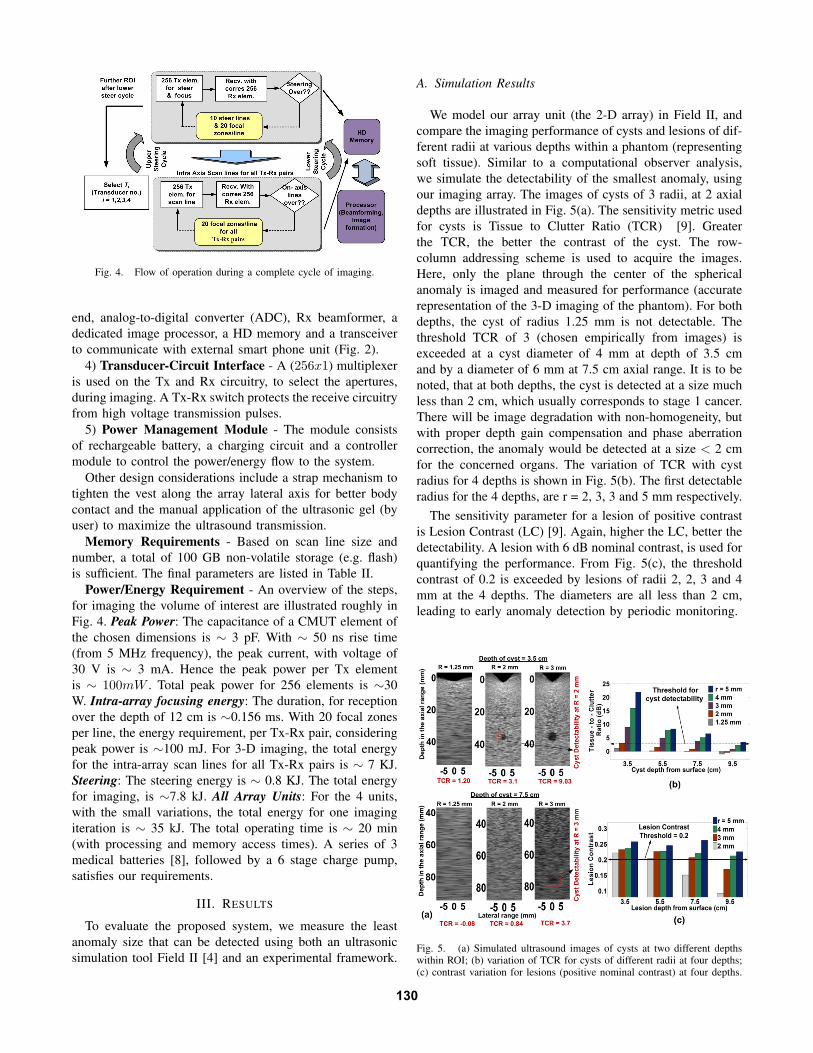
Fig. 4. Flow of operation during a complete cycle of imaging.
end, analog-to-digital converter (ADC), Rx beamformer, adedicated image processor, a HD memory and a transceiverto communicate with external smart phone unit (Fig. 2).
4) Transducer-Circuit Interface - A (256x1) multiplexeris used on the Tx and Rx circuitry, to select the apertures,during imaging. A Tx-Rx switch protects the receive circuitryfrom high voltage transmission pulses.
5) Power Management Module - The module consistsof rechargeable battery, a charging circuit and a controllermodule to control the power/energy flow to the system.
Other design considerations include a strap mechanism totighten the vest along the array lateral axis for better bodycontact and the manual application of the ultrasonic gel (byuser) to maximize the ultrasound transmission.
Memory Requirements - Based on scan line size andnumber, a total of 100 GB non-volatile storage (e.g. flash)is sufficient. The final parameters are listed in Table II.
Power/Energy Requirement - An overview of the steps,for imaging the volume of interest are illustrated roughly inFig. 4. Peak Power: The capacitance of a CMUT element ofthe chosen dimensions is ∼ 3 pF. With ∼ 50 ns rise time(from 5 MHz frequency), the peak current, with voltage of30 V is ∼ 3 mA. Hence the peak power per Tx elementis ∼ 100mW . Total peak power for 256 elements is ∼30W. Intra-array focusing energy: The duration, for receptionover the depth of 12 cm is ∼0.156 ms. With 20 focal zonesper line, the energy requirement, per Tx-Rx pair, consideringpeak power is ∼100 mJ. For 3-D imaging, the total energyfor the intra-array scan lines for all Tx-Rx pairs is ∼ 7 KJ.Steering: The steering energy is ∼ 0.8 KJ. The total energyfor imaging, is ∼7.8 kJ. All Array Units: For the 4 units,with the small variations, the total energy for one imagingiteration is ∼ 35 kJ. The total operating time is ∼ 20 min(with processing and memory access times). A series of 3medical batteries [8], followed by a 6 stage charge pump,satisfies our requirements.
III. RESULTS
To evaluate the proposed system, we measure the leastanomaly size that can be detected using both an ultrasonicsimulation tool Field II [4] and an experimental framework.
A. Simulation Results
We model our array unit (the 2-D array) in Field II, andcompare the imaging performance of cysts and lesions of dif-ferent radii at various depths within a phantom (representingsoft tissue). Similar to a computational observer analysis,we simulate the detectability of the smallest anomaly, usingour imaging array. The images of cysts of 3 radii, at 2 axialdepths are illustrated in Fig. 5(a). The sensitivity metric usedfor cysts is Tissue to Clutter Ratio (TCR) [9]. Greaterthe TCR, the better the contrast of the cyst. The row-column addressing scheme is used to acquire the images.Here, only the plane through the center of the sphericalanomaly is imaged and measured for performance (accuraterepresentation of the 3-D imaging of the phantom). For bothdepths, the cyst of radius 1.25 mm is not detectable. Thethreshold TCR of 3 (chosen empirically from images) isexceeded at a cyst diameter of 4 mm at depth of 3.5 cmand by a diameter of 6 mm at 7.5 cm axial range. It is to benoted, that at both depths, the cyst is detected at a size muchless than 2 cm, which usually corresponds to stage 1 cancer.There will be image degradation with non-homogeneity, butwith proper depth gain compensation and phase aberrationcorrection, the anomaly would be detected at a size < 2 cmfor the concerned organs. The variation of TCR with cystradius for 4 depths is shown in Fig. 5(b). The first detectableradius for the 4 depths, are r = 2, 3, 3 and 5 mm respectively.
The sensitivity parameter for a lesion of positive contrastis Lesion Contrast (LC) [9]. Again, higher the LC, better thedetectability. A lesion with 6 dB nominal contrast, is used forquantifying the performance. From Fig. 5(c), the thresholdcontrast of 0.2 is exceeded by lesions of radii 2, 2, 3 and 4mm at the 4 depths. The diameters are all less than 2 cm,leading to early anomaly detection by periodic monitoring.
Fig. 5. (a) Simulated ultrasound images of cysts at two different depthswithin ROI; (b) variation of TCR for cysts of different radii at four depths;(c) contrast variation for lesions (positive nominal contrast) at four depths.
130
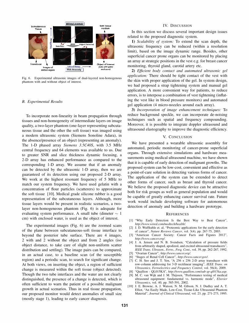
Fig. 6. Experimental ultrasonic images of dual-layered non-homogenousphantom with and without object of interest.
B. Experimental Results
To incorporate non-linearity in beam propagation throughtissues and non-homogeneity of intermediate layers on imagequality, a two-layer phantom (one-layer representing subcuta-neous tissue and the other the soft tissue) was imaged usinga modern ultrasonic system (Siemens Sonoline Adara), inthe absence/presence of an object (representing an anomaly).The 1-D phased array Siemens 3.5C40S, with 3.5 MHzcentral frequency and 64 elements was available to us. Dueto greater SNR and slice thickness dynamic focusing, a2-D array has enhanced performance as compared to thecorresponding 1-D array. We assume that if an anomalycan be detected by the ultrasonic 1-D array, then we areguaranteed of its detection using our proposed 2-D array.We work at the highest resonant frequency of 5 MHz tomatch our system frequency. We have used gelatin with aconcentration of flour particles (scatterers) to approximatethe soft tissue [10]. Medical grade silicone rubber is a goodrepresentation of the subcutaneous layers. Although, moretissue layers would be present in realistic scenarios, a two-layer non-homogeneous phantom (Fig. 6) is adequate forevaluating system performance. A small tube (dimeter ∼ 1cm) with enclosed water, is used as the object of interest.
The experimental images (Fig. 6) are the zoomed scansof the plane between subcutaneous-soft tissue interface tobeyond the posterior tube surface. There are 4 images,2 with and 2 without the object and from 2 angles (isoobject distance, to take care of slight non-uniform scatterdistribution and settling). The image pairs can be compared,in an actual case, to a baseline scan (of the susceptibleregion) and a periodic scan, to search for significant change.At both views, on inserting the object, a region of sufficientchange is measured within the soft tissue (object detected).Though the two tube interfaces and the water are not clearlydistinguished, the presence of a change is detected, which isoften sufficient to warn the patient of a possible malignantgrowth in actual scenarios. Thus in real tissue propagation,our proposed monitor would detect anomalies of small size(mostly stage 1), leading to early cancer diagnosis.
IV. DISCUSSION
In this section we discuss several important design issuesrelated to the proposed diagnostic system.
1) Scalability of system: To extend the scan depth, theultrasonic frequency can be reduced (within a resolutionlimit), based on the image dynamic range. Besides, othersuperficial cancer prone organs can be monitored by placingan array at strategic positions in the vest e.g. for breast cancermonitoring, thyroid gland, carotid artery etc.
2) Efficient body contact and automated ultrasonic gelapplication: There should be tight contact of the vest withthe skin with proper application of the gel. In system design,we had proposed a strap tightening system and manual gelapplication. A more convenient way for patients, to reduceerrors, is to interpose a combination of vest tightening (inflat-ing the vest like in blood pressure monitors) and automatedgel application (4 micro-nozzles around each array).
3) Incorporation of image enhancement techniques: Toreduce background speckle, we can incorporate de-noisingtechniques such as spatial and frequency compounding.Moreover, it is possible to integrate doppler ultrasound andultrasound elastography to improve the diagnostic efficiency.
V. CONCLUSION
We have presented a wearable ultrasonic assembly forautomated, periodic monitoring of cancer-prone superficialorgans. Through extensive simulations and hardware mea-surements using medical ultrasound machine, we have shownthat it is capable of early detection of malignant growths. Theproposed system can be low-cost, convenient and effective asa point-of-care solution in detecting various forms of cancer.The application of the system can be extended to detectother forms of cancer, such as breast and thyroid cancer.We believe the proposed diagnostic device can be attractiveboth for risk groups as well as general population and wouldbe capable of greatly enhancing cancer survival rate. Futurework would include developing software for autonomousdetection of anomaly and building a hardware prototype.
REFERENCES
[1] “Why Early Detection Is the Best Way to Beat Cancer”,http://www.wired.com/medtech/health/.
[2] J. D. Wulfkuhle et. al, “Proteomic applications for the early detectionof cancer”, Nature Reviews Cancer, vol. 3(4), pp. 267-75, 2003.
[3] “American Cancer Society: Cancer Facts and Figures 2012”,http://www.cancer.org/.
[4] J. A. Jensen and N. B. Svendsen, “Calculation of pressure fieldsfrom arbitrarily shaped, apodized, and excited ultrasound transducers”,IEEE Trans. Ultrason., Ferro., Freq. Cont., vol. 39, pp. 262-267, 1992.
[5] “Ovarian Cancer”, http://www.cancer.org/.[6] “Stages of Renal Cell Cancer”, http://www.cancer.gov/.[7] C. H. Seo and J. T. Yen, “A 256 x 256 2-D array transducer with
row-column addressing for 3-D rectilinear imaging”, IEEE Trans. onUltrasonics, Ferroelectrics and Frequency Control, vol. 56(4), 2009.
[8] “Quallion - QL015KA”, http://www.quallion.com/sub-sp-ql015ka.asp.[9] M. C. van Wijk and J. M. Thijssen, “Performance testing of medical
ultrasound equipment: fundamental vs. harmonic mode”, ElsevierUltrasonics, vol. 40, pp. 585-591, 2002.
[10] J. E. Browne, A. J. Watson, N. M. Gibson, N. J. Dudley and A. T.Elliot, “An Easily Made, Low-Cost, Tissue-Like Ultrasound PhantomMaterial”, Journal of Clinical Ultrasound, vol. 23, pp. 271-273, 1995.
131