
SPM Course May 2012 Segmentation and Voxel-Based Morphometry
Upload
nguyenthienCategory
view
223download
0
Volume 18 • Issue 2 • 1000237J Psychiatry Psychiatry, an open access journal
Research Article Open Access
Kim, et al., J Psychiatry 2015, 18:2 http://dx.doi.org/10.4172/Psychiatry.1000237
Research Article Open Access
Journal of Psychiatry
*Corresponding author: Kang Joon Lee, Department of Psychiatry, IlsanpaikHospital, Inje University College of Medicine, 170, Juhwa-ro, Ilsanseo-gu, Goyang-si, Gyeonggi-do, 411-706, South Korea, Tel: +82 31 910 7260, Fax: +82 31 9107268; E-mail: [email protected]
Received December 23, 2014; Accepted January 25, 2015; Published February 02, 2015
Citation: Kim H, Kim YS, Lee KJ (2015) A Voxel-Based Morphometry Study inAlzheimer’s Disease and Mild Cognitive Impairment. J Psychiatry 18: 237 doi: 10.4172/1994-8220.1000237
Copyright: © 2015 Kim H, et al. This is an open-access article distributed underthe terms of the Creative Commons Attribution License, which permits unrestricteduse, distribution, and reproduction in any medium, provided the original author andsource are credited
A Voxel-Based Morphometry Study in Alzheimer’s Disease and Mild Cognitive ImpairmentHyun Kim, Yo Sup Kim and Kang Joon Lee*1Department of Psychiatry, Ilsanpaik Hospital, Inje University College of Medicine, Goyang, South Korea2Ilsanpaik Hospital College of Medicine, South Korea
Keywords: Alzheimer’s disease; Mild cognitive impairment; Voxel-based morphometry
IntroductionAlzheimer’s disease (AD) is the most common cause of dementia
in the general population older than 60 years of age [1,2]. The clinical manifestations of AD are seen in the domains of memory and spatial navigation in the early phases of the disease, with almost all cognitive domains affected as the disease progresses, including attention, language, and visuospatial function, as well as executive function failure and emotional instability [3].
Mild cognitive impairment (MCI) is often viewed as a prodromal stage of AD and also as a good target for both early AD diagnosis and early therapeutic interventions [4,5]. Considerable progress has been made in identifying more mildly impaired subjects who may be in a transitional stage between normal aging and dementia [6]. Clinical AD is often preceded by MCI, a phase in which there is complaints and objective impairments in one or more cognitive domains, but activities of daily living are preserved [6]. MCI refers to the clinical state of individuals who have memory impairment but are otherwise functioning well and do not meet the clinical criteria for a diagnosis of dementia [6].
As AD progresses, or even during its pre-clinical diagnostic phase, histopathological microscopic lesions may lead to macroscopic changes of regional brain areas that can be detected in vivo by noninvasive morphometric magnetic resonance imaging (MRI) techniques, such as semi-automated voxel-based morphometry (VBM) [7,8]. In the current study, several authors applied an MRI VBM technique that maps the entire brain instead of being restricted to single regions. Voxels are represented mathematically in the three dimensions of height, width, and depth, and correspond to units of tissue volume. This method is very useful in the study of neurodegenerative diseases such as AD and allows changes to be detected in the prodromal states of the disease, such as MCI. Numerous structural MRI studies have demonstrated that atrophy of the medial temporal lobe, including the hippocampus
AbstractObjectives: A number of structural magnetic resonance imaging (MRI) studies have demonstrated grey matter
(GM) atrophy in subjects with mild cognitive impairment (MCI) and Alzheimer’s disease (AD). We used a voxel-based morphometric (VBM) approach to evaluate the patterns of atrophy in GM in subjects with AD and MCI in comparison to control subjects.
Methods: We performed brain MRI with VBM analysis in 53 subjects with AD, 32 subjects with MCI, and 32 normal elderly controls.
Results: In the AD group, we found GM atrophy in the left cingulate gyrus, left dorsal posterior cingulate cortex, left inferior temporal gyrus, and right supramarginal gyrus compared to control subjects, and in the right inferior frontal gyrus, right orbitofrontal area, left uncus, left ventral entorhinal cortex, and left inferior temporal gyrus compared to subjects in the MCI group. There were no significant differences in GM loss between MCI and control subjects.
Conclusion: We demonstrated the involvement of GM in AD, but not in MCI. The pattern of GM volume reductions can help us understand the underlying pathologic mechanisms in AD and MCI.
and entorhinal cortex, is a sensitive marker of early AD [9,10]. Moreover, it has been suggested that atrophy of medial temporal lobe structures might predict progression from MCI to AD [11,12]. Thus it is anticipated that comparison analysis of neuroanatomical images in MCI and AD would facilitate finding the progression from MCI to AD.
A recent meta-analysis of neuroimaging studies using voxel-based image analytic methods found that in patients with AD relative to healthy controls (HC), atrophy of the grey matter (GM) was present mainly in the bilateral medial temporal lobes, left medial thalamus, posterior insula, and left middle temporal gyrus/superior temporal sulcus, and that in patients with MCI relative to HC, GM atrophy was found mainly in the right gyrus rectus, left temporal pole/anterior superior temporal sulcus, and right amygdale [13].
The aim of the current study was to evaluate the patterns of GM atrophy of patients with AD and MCI in comparison to control subjects, using the technique of VBM. H0 is that there is no difference in imaging findings among the three groups. H1 is that there is significant VBM difference among the three groups.
MethodsSubjects
We enrolled 117 subjects older than 60 years, comprising 32
Journ
al of Psychiatry

Citation: Kim H, Kim YS, Lee KJ (2015) A Voxel-Based Morphometry Study in Alzheimer’s Disease and Mild Cognitive Impairment. J Psychiatry 18: 237 doi: 10.4172/Psychiatry.1000237
Volume 18 • Issue 2 • 1000237J Psychiatry Psychiatry, an open access journal
Page 2 of 6
subjects with MCI, 53 patients with AD, and 32 healthy controls from the psychiatric department of the Ilsanpaik Hospital between June 2011 and March 2014. Physical, neurological, psychiatric and routine laboratory examinations for dementia assessment were carried out in all subjects, along with MR imaging. The study protocol was approved by the Institutional Review Board (IRB) of Ilsanpaik Hospital.
A dementia screening test was performed using the Korean version of the Mini-Mental State Examination (MMSE-KC), the Global Deterioration Scale (GDS), and the Clinical Dementia Rating scale (CDR), and for probable AD diagnosis, we used the criteria of the National Institute of Neurological and Communicative Disorders and Stroke and Alzheimer’s Disease and Related Disorders Association (NINCDS-ADRDA) [14]. It is known that the internal consistency of MMSE-KC obtained by Cronbach's coefficient alpha was 0.826. The inter-rater reliability and test-retest reliability were 0.968 (p<0.001) and 0.825 (p<0.001), respectively [15]. Cronbach's alpha coefficients of the GDS were 0.9065 respectively, suggesting a high degree of internal consistency [16]. It is well known that the diagnostic value of CDR and NINCDS-ADRDA is high in Alzheimer’s disease [14,17]. MCI diagnosis followed the criteria of the International Working Group on Mild Cognitive Impairment [18]: (i) the person is neither normal nor demented; (ii) there is evidence of cognitive deterioration shown by either objectively measured decline over time and/or subjective report of decline by self and/or in formant in conjunction with objective cognitive deficits; and (iii) activities of daily living are preserved and complex instrumental functions are either intact or minimally impaired.
Patients with either a history of or the current presence of a major physical illness such as uncontrolled diabetes, obstructive pulmonary disease, cardiovascular disease, or those with comorbidities such as primary psychiatric or neurologic disorders, head injury with loss of consciousness, or alcohol or substance abuse were excluded from the study. The control group consisted of subjects with CDR scores of 0 and without a previous history of neurological or psychiatric disease, or memory complaints.
Brain MRI
All brain MRI studies were performed using the Siemens Magnetom Avanto 1.5-T MRI system. A three dimensional volumetric acquisition of a T1-weighted gradient echo sequence at 11.4/4.4/1 (TR/TE/excitation) produced a gapless series of contiguous, thin sagittal sections with the following parameters: flip angle, 15 degrees; acquisition matrix, 256 × 256; fields of view, 31.5cm; section thickness, 1.23mm.
Voxel-based MRI analysisData were analyzed using the VBM8 toolbox (http://dbm.neuro.
unijena.de/vbm8) with the default parameters of the statistical parametric mapping software package (SPM8, http://www.fil.ion.ucl.ac.uk/spm) executed using MATLAB (Math Works, Natick, MA, USA). The first anatomical standardization used affine transformation and fitted each individual brain to a standard template brain in 3-D in order to correct for differences in brain size and shape and to facilitate inter subject averaging. The normalized MR images were then segmented into grey matter, white matter, and cerebrospinal fluid. The segmented GM images were subjected to an affine and nonlinear anatomical standardization using a template of a priori GM. The anatomically standardized GM images were smoothed with an isotropic Gaussian kernel that was 8 mm in full width at half maximum to exploit the partial volume effects and to create a spectrum of GM intensities. The GM intensities are equivalent to the weighted average of GM voxels
that are located in the volume fixed by the smoothing kernel. Regional intensities can therefore be considered to be equivalent to the GM concentration.
Statistical analysis
Demographic and neuropsychological variables were compared across groups using analysis of variance (ANOVA) and chi-square tests for dichotomous variables. Differences between each of the groups were analyzed with the two-sample t-test. The smoothed GM images were analyzed with an analysis of covariance model using SPM8. Age, gender, education, and total intracranial volume (TIV) were included as covariates. SPM8,run with MATLAB, was utilized for preprocessing and analyzing the MRI data.
A family-wise error (FWE)-corrected statistical threshold of p<0.05 was applied. The anatomical labels of the Montreal Neurological Institute (MNI) coordinates were defined using automated anatomical labeling, which was implemented with the Wake Forest University (WFU) Pick Atlas software (v3.03) (http://www.fmri.wfubmc.edu). If predetermined labels did not exist for the MNI coordinates, then Talairach-converted coordinates from a web-based coordinate converter (bioimagesuite.yale.dcu/nmi2tal/index.aspx) and Talairach Client Version 2.4.3 (http://www.talairach.org/client.html) were used.
ResultsComparison among AD, MCI and control groups
Demographic and neuropsychological data for subjects in the AD, MCI, and control groups are shown in Table 1. A chi-square analysis revealed that there was no significant difference in gender distribution among the groups (χ2=0.068, df=2, p=0.967).As compared to the control group, both AD group (p<0.001) and MCI group (p<0.05) were significantly older, but there was no significant age difference between the AD group and the MCI group. Educational level was significantly higher in the control group than in the AD (p<0.001) and MCI (p<0.05) groups.
The mean MMSE-KC score of the AD group was significantly lower than that for any other group (p<0.001), and there was also a significant MMSE-KC score difference between the MCI group and the control group (p<0.001). In addition, the mean CDR and GDS scores of the AD group were significantly higher than those for any other group (p<0.001), and there were also significant differences in the mean CDR and GDS scores between the MCI group and the control group (p<0.001).
AD and control groups
Compared to normal controls, patients with AD had significantly lower GM volume in the left cingulate gyrus, left dorsal posterior cingulate cortex, left inferior temporal gyrus, and right supramarginal gyrus (Figure 1).
AD and MCI groups
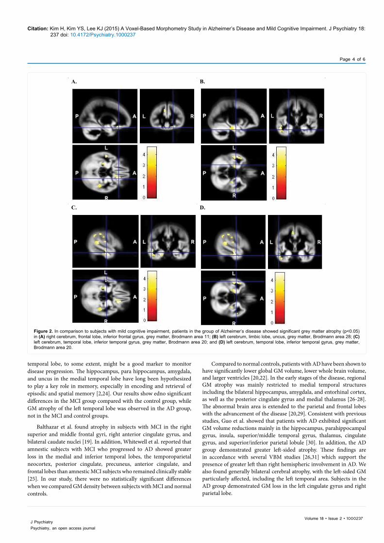
In comparison to subjects with MCI, patients in AD showed significant GM atrophy in the right inferior frontal gyrus, right orbitofrontal area, left uncus, left ventral entorhinal cortex, and left inferior temporal gyrus (Figure 2).
MCI and control groups
Compared with healthy controls, subjects with MCI showed no differences in the grey matter volume using a whole brain analysis.

Citation: Kim H, Kim YS, Lee KJ (2015) A Voxel-Based Morphometry Study in Alzheimer’s Disease and Mild Cognitive Impairment. J Psychiatry 18: 237 doi: 10.4172/Psychiatry.1000237
Volume 18 • Issue 2 • 1000237J Psychiatry Psychiatry, an open access journal
Page 3 of 6
DiscussionA number of brain MRI studies have indicated GM abnormalities
in patients with AD.Compared to normal controls, subjects with MCI and AD have been found to have significant lower global GM volume [19-22]. In the current study, we found atrophy of a part of the temporal lobe and a few other brain areas in AD patients in comparison with
MCI and control subjects.
Several authors found a similar pattern of GM atrophy among MCI and early AD patients, mainly in medial temporal structures. Derflinger et al. reported that the voxel-wise comparison of subjects with MCI and AD revealed GM loss in the entorhinal, temporoparietal, dorsofrontal, and occipital cortices as well as in the precuneus [23]. The left medial
Table 1: Demographic and neuropsychological data
NC (n=32) MCI (n=32) AD (n=53) p df StatisticsFemale 21 (0.66) 20 (0.63) 34 (0.64) 0.967 2 χ2=0.068Age (y) 70.59 (7.31) 75.56 (5.48) 77.19 (8.06) <0.01 2 F(2,114)=8.452
Education (y) 11.09 (2.88) 8.47 (4.52) 8.36 (4.03) <0.01 2 F(2,114)=5.550MMSE-K 27.41 (1.85) 24.00 (1.59) 17.74 (5.02) <0.01 2 F(2,114)=77.191
GDS 2.16 (0.37) 3.00 (0.25) 4.13 (0.81) <0.01 2 F(2,114)=115.145CDR 0.23 (0.25) 0.69 (0.25) 1.03 (0.40) <0.01 2 F(2,114)=59.563
NC: Normal Controls, MCI: Mild Cognitive Impairment, AD: Alzheimer’s Disease. Data are presented as mean (SD) or numbers (%).There was no significant difference in gender distribution among the groups. As compared to the control group, both AD group and MCI group were significantly older. Educational level was significantly higher in the control group than in the AD and MCI groups. The mean MMSE-KC score was significantly lower in AD group than that for any other group and the mean CDR and GDS scores were significantly higher.
A. B.
C.
Figure 1: When compared with normal controls, Alzheimer’s disease subjects displayed grey matter atrophy (p<0.05) in (A) left cerebrum, limbic lobe, cingulate gyrus, grey matter, Brodmann area 31; (B) left cerebrum, temporal lobe, inferior temporal gyrus, grey matter, Brodmann area 20; and (C) right cerebrum, parietal lobe, supramarginal gyrus, grey matter, Brodmann area 40.
Citation: Kim H, Kim YS, Lee KJ (2015) A Voxel-Based Morphometry Study in Alzheimer’s Disease and Mild Cognitive Impairment. J Psychiatry 18: 237 doi: 10.4172/Psychiatry.1000237
Volume 18 • Issue 2 • 1000237J Psychiatry Psychiatry, an open access journal
Page 4 of 6
temporal lobe, to some extent, might be a good marker to monitor disease progression. The hippocampus, para hippocampus, amygdala, and uncus in the medial temporal lobe have long been hypothesized to play a key role in memory, especially in encoding and retrieval of episodic and spatial memory [2,24]. Our results show edno significant differences in the MCI group compared with the control group, while GM atrophy of the left temporal lobe was observed in the AD group, not in the MCI and control groups.
Balthazar et al. found atrophy in subjects with MCI in the right superior and middle frontal gyri, right anterior cingulate gyrus, and bilateral caudate nuclei [19]. In addition, Whitewell et al. reported that amnestic subjects with MCI who progressed to AD showed greater loss in the medial and inferior temporal lobes, the temporoparietal neocortex, posterior cingulate, precuneus, anterior cingulate, and frontal lobes than amnestic MCI subjects who remained clinically stable [25]. In our study, there were no statistically significant differences when we compared GM density between subjects with MCI and normal controls.
Compared to normal controls, patients with AD have been shown to have significantly lower global GM volume, lower whole brain volume, and larger ventricles [20,22]. In the early stages of the disease, regional GM atrophy was mainly restricted to medial temporal structures including the bilateral hippocampus, amygdala, and entorhinal cortex, as well as the posterior cingulate gyrus and medial thalamus [26-28]. The abnormal brain area is extended to the parietal and frontal lobes with the advancement of the disease [20,29]. Consistent with previous studies, Guo et al. showed that patients with AD exhibited significant GM volume reductions mainly in the hippocampus, parahippocampal gyrus, insula, superior/middle temporal gyrus, thalamus, cingulate gyrus, and superior/inferior parietal lobule [30]. In addition, the AD group demonstrated greater left-sided atrophy. These findings are in accordance with several VBM studies [26,31] which support the presence of greater left than right hemispheric involvement in AD. We also found generally bilateral cerebral atrophy, with the left-sided GM particularly affected, including the left temporal area. Subjects in the AD group demonstrated GM loss in the left cingulate gyrus and right parietal lobe.
A. B.
C. D.
Figure 2. In comparison to subjects with mild cognitive impairment, patients in the group of Alzheimer’s disease showed significant grey matter atrophy (p<0.05) in (A) right cerebrum, frontal lobe, inferior frontal gyrus, grey matter, Brodmann area 11; (B) left cerebrum, limbic lobe, uncus, grey matter, Brodmann area 28; (C) left cerebrum, temporal lobe, inferior temporal gyrus, grey matter, Brodmann area 20; and (D) left cerebrum, temporal lobe, inferior temporal gyrus, grey matter, Brodmann area 20.

Citation: Kim H, Kim YS, Lee KJ (2015) A Voxel-Based Morphometry Study in Alzheimer’s Disease and Mild Cognitive Impairment. J Psychiatry 18: 237 doi: 10.4172/Psychiatry.1000237
Volume 18 • Issue 2 • 1000237J Psychiatry Psychiatry, an open access journal
Page 5 of 6
According to Braak and Braak’s hierarchical model of neuropathological alterations of neurofibrillary tangles in AD [32] and MCI [33], the neuronal lesions initially occur in the medial temporal lobe, including the entorhinal cortex, hippocampus, amygdala, parahippocampal gyrus, and uncus, and other limbic structures, then in the temporal pole and inferior and middle temporal gyri (mainly in preclinical AD), and subsequently will spread throughout the frontal, parietal, and temporal neocortices as well as subcortical regions by the time of full diagnosis of AD.
Two cross-sectional MRI VBM studies comparing MCI and AD patients reported higher atrophy in the AD group in the temporal neocortex, cingulate cortex, and parietal association areas [22,34]. Derflinger et al. study revealed GM loss in the entorhinal, temporoparietal, dorsofrontal, and occipital cortices as well as in the precuneus when comparing MCI and AD patients [23]. Chetelat et al. reported that regions of significant GM loss common to both converters and non-converters over the 18-month follow-up period included the temporal neocortex, parahippocampal cortex, orbitofrontal and inferior parietal areas, and the left thalamus [35]. However, there was significantly greater GM loss in converters relative to non-converters in the hippocampal area, inferior and middle temporal gyrus, posterior cingulate, and precuneus. Our study revealed that GM atrophy of the inferior frontal gyrus and the orbitofrontal cortex on the right, and of the inferior temporal gyrus, limbic lobe, uncus, and ventral entorhinal cortex on the left are present in subjects with AD in comparison to those with MCI. In a recent VBM study, it was reported that parietal association metabolic impairment differentiates AD from MCI [36]. We found significant differences in parietal atrophy in patients with AD compared to those in the control group, but no differences in comparison to those in the MCI group. This observed atrophy may result from the accumulation of neurofibrillary tangles and pathological processes such as functional alteration in these regions. The lack of significant differences in the cingulate cortex and parietal areas does not exclude the possibility that accelerated atrophy in these regions may develop at a later stage of AD. Also, the fact that most subjects examined in our study were mild AD patients would affect the results.
Our study had some limitations: AD patients were older than those with MCI and controls. Another weakness of this study is its cross-sectional nature and the non-prospective study design. Prospective studies on MCI group should be considered. Additionally, some of the subjects with MCI may not develop AD but may have MCI due to other causes. Therefore, the differences between healthy controls and MCI subjects who will convert to AD may have been underestimated.
Recently, the concept of MCI has been questioned. An important argument against this concept is the clinical heterogeneity of MCI. On the other hand, MCI is still a very useful concept if we consider the diagnostic criteria for AD. We found no statistically significant differences between subjects with MCI and normal controls in areas such as the medial temporal lobes, but GM atrophy ofthe left cingulate gyrus, left dorsal posterior cingulate cortex, left inferior temporal gyrus, and right supramarginal gyrus was observed in the AD group compared to the control group. Significant GM loss in AD relative to MCI in the right inferior frontal gyrus, right orbitofrontal area, left uncus, left ventral entorhinal cortex, and left inferior temporal gyrus suggests a plausible pathological mechanism for the discrimination between MCI and AD. We are convinced that the atrophy of the temporal region will play an important role in identifying the underlying pathologic mechanisms in AD. Although no differences were observed between subjects with MCI and normal controls in the current study, large
cohort studies to monitor whole brain changes on a VBM basis over time using up-to-date techniques will lead to a further understanding of the neuropathology of MCI and AD.
Acknowledgments
We declare no conflicts of interest that may be inherent in this study.
References
1. Kalaria RN, Maestre GE, Arizaga R, Friedland RP, Galasko D, et al. (2008)Alzheimer's disease and vascular dementia in developing countries:prevalence, management, and risk factors. Lancet Neurol 7: 812-826.
2. Blennow K, de Leon MJ, Zetterberg H (2006) Alzheimer's disease. Lancet 368: 387-403.
3. Amlien IK, Fjell AM (2014) Diffusion tensor imaging of white matter degeneration in Alzheimer's disease and mild cognitive impairment. Neuroscience 276: 206-215.
4. Petersen RC (2004) Mild cognitive impairment as a diagnostic entity. J InternMed 256: 183-194.
5. Schneider JA, Arvanitakis Z, Leurgans SE, Bennett DA (2009) Theneuropathology of probable Alzheimer disease and mild cognitive impairment.Ann Neurol 66: 200-208.
6. Petersen RC, Stevens JC, Ganguli M, Tangalos EG, Cummings JL, et al. (2001) Practice parameter: early detection of dementia: mild cognitive impairment (an evidence-based review). Report of the Quality Standards Subcommittee of the American Academy of Neurology. Neurology 56: 1133-1142.
7. Busatto GF, Diniz BS, Zanetti MV (2008) Voxel-based morphometry inAlzheimer's disease. Expert Rev Neurother 8: 1691-1702.
8. Zakzanis KK, Graham SJ, Campbell Z (2003) A meta-analysis of structural and functional brain imaging in dementia of the Alzheimer's type: a neuroimagingprofile. Neuropsychol Rev 13: 1-18.
9. Du AT, Schuff N, Amend D, Laakso MP, Hsu YY, et al. (2001) Magneticresonance imaging of the entorhinal cortex and hippocampus in mild cognitiveimpairment and Alzheimer's disease. J Neurol Neurosurg Psychiatry 71: 441-447.
10. Killiany RJ, Gomez-Isla T, Moss M, Kikinis R, Sandor T, et al. (2000) Use ofstructural magnetic resonance imaging to predict who will get Alzheimer'sdisease. Ann Neurol 47: 430-439.
11. Killiany RJ, Hyman BT, Gomez-Isla T, Moss MB, Kikinis R et al. (2002) MRImeasures of entorhinal cortex vs hippocampus in preclinical AD. Neurology58: 1188-1196.
12. Visser PJ, Verhey FR, Hofman PA, Scheltens P, Jolles J (2002) Medialtemporal lobe atrophy predicts Alzheimer's disease in patients with minorcognitive impairment. J Neurol Neurosurg Psychiatry 72: 491-497.
13. Schroeter ML, Stein T, Maslowski N, Neumann J (2009) Neural correlatesof Alzheimer's disease and mild cognitive impairment: a systematic andquantitative meta-analysis involving 1351 patients. Neuroimage 47: 1196-1206.
14. McKhann G, Drachman D, Folstein M, Katzman R, Price D, et al. (1984) Clinical diagnosis of Alzheimer's disease: report of the NINCDS-ADRDA Work Groupunder the auspices of Department of Health and Human Services Task Forceon Alzheimer's Disease. Neurology 34: 939-944.
15. Kim TH, Jhoo JH, Park JH, Kim JL, Ryu SH, et al. (2010) Korean version ofmini mental status examination for dementia screening and its' short form.Psychiatry Investig 7:102-108.
16. Bae JN, Cho MJ (2004) Development of the Korean version of the geriatricdepression scale and its short form among elderly psychiatric patients. JPsychosom Res. 57: 297-305.
17. Hughes CP, Berg L, Danziger WL, Coben LA, Martin RL (1982) A new clinicalscale for the staging of dementia. Br J Psychiatry 140:566-572.
18. Winblad B, Palmer K, Kivipelto M, Jelic V, Fratiglioni, et al. (2004) Mildcognitive impairment--beyond controversies, towards a consensus: report ofthe International Working Group on Mild Cognitive Impairment. J Intern Med256: 240-246.
19. Balthazar ML, Yasuda CL, Pereira FR, Pedro T, Damasceno BP, et al. (2009)Cendes F. Differences in grey and white matter atrophy in amnestic mildcognitive impairment and mild Alzheimer's disease. Eur J Neurol 16: 468-474.
20. Good CD, Scahill RI, Fox NC, Ashburner J, Friston KJ, et al. (2002) Automatic

Citation: Kim H, Kim YS, Lee KJ (2015) A Voxel-Based Morphometry Study in Alzheimer’s Disease and Mild Cognitive Impairment. J Psychiatry 18: 237 doi: 10.4172/Psychiatry.1000237
Volume 18 • Issue 2 • 1000237J Psychiatry Psychiatry, an open access journal
Page 6 of 6
differentiation of anatomical patterns in the human brain: validation with studies of degenerative dementias. Neuroimage 17: 29-46.
21. Chen K, Reiman EM, Alexander GE,Bandy D, Renaut R, et al. (2004) Anautomated algorithm for the computation of brain volume change fromsequential MRIs using an iterative principal component analysis and itsevaluation for the assessment of whole-brain atrophy rates in patients withprobable Alzheimer's disease. Neuroimage 22: 134-143.
22. Karas GB, Scheltens P, Rombouts SA,Visser PJ, van Schijndel RA, et al.(2004) Global and local gray matter loss in mild cognitive impairment andAlzheimer's disease. Neuroimage 23: 708-716.
23. Derflinger S, Sorg C, Gaser C, Myers N, Arsic M, et al. (2011) Grey-matter atrophy in Alzheimer's disease is asymmetric but not lateralized. J AlzheimersDis 25: 347-357.
24. Schwindt GC, Black SE (2009) Functional imaging studies of episodic memory in Alzheimer's disease: a quantitative meta-analysis. Neuroimage 45: 181-190.
25. Whitwell JL, Przybelski SA, Weigand SD, Knopman DS, Boeve BF, et al.(2007) 3D maps from multiple MRI illustrate changing atrophy patterns assubjects progress from mild cognitive impairment to Alzheimer's disease. Brain 130: 1777-1786.
26. Karas GB, Burton EJ, Rombouts SA, van Schijndel RA, O'Brien JT, et al. (2003) A comprehensive study of gray matter loss in patients with Alzheimer's disease using optimized voxel-based morphometry. Neuroimage 18: 895-907.
27. Hirata Y, Matsuda H, Nemoto K,Ohnishi T, Hirao K, et al. (2005) Voxel-basedmorphometry to discriminate early Alzheimer's disease from controls. Neurosci Lett 382: 269-274.
28. Baxter LC, Sparks DL, Johnson SC, Lenoski B, Lopez JE, et al. (2006)Relationship of cognitive measures and gray and white matter in Alzheimer'sdisease. J Alzheimers Dis 9: 253-260.
29. Scahill RI, Schott JM, Stevens JM, Rossor MN, Fox NC (2002) Mapping theevolution of regional atrophy in Alzheimer's disease: unbiased analysis of fluid-registered serial MRI. Proc Natl Acad Sci U S A 99: 4703-4707.
30. Guo X, Wang Z, Li K, Li Z, Qi Z, et al. (2010) Voxel-based assessment of gray and white matter volumes in Alzheimer's disease. Neurosci Lett 468: 146-150.
31. Baron JC, Chetelat G, Desgranges B,Perchey G, Landeau B, et al. (2001)In vivo mapping of gray matter loss with voxel-based morphometry in mildAlzheimer's disease. Neuroimage 14: 298-309.
32. Braak H, Braak E (1995) Staging of Alzheimer's disease-related neurofibrillary changes. Neurobiol Aging 16: 271-278; discussion 278-284.
33. Petersen RC, Parisi JE, Dickson DW, Johnson KA, Knopman DS, et al. (2006) Neuropathologic features of amnestic mild cognitive impairment. Arch Neurol63: 665-672.
34. Chetelat G, Desgranges B, De La Sayette V, Viader F, Eustache F, et al. (2002) Mapping gray matter loss with voxel-based morphometry in mild cognitiveimpairment. Neuroreport 13: 1939-1943.
35. Chetelat G, Landeau B, Eustache F,Mézenge F, Viader F, et al. (2005) Usingvoxel-based morphometry to map the structural changes associated with rapid conversion in MCI: a longitudinal MRI study. Neuroimage 27: 934-946.
36. Chetelat G, Desgranges B, de la Sayette V, Viader F, Eustache F, et al. (2003) Mild cognitive impairment: Can FDG-PET predict who is to rapidly convert toAlzheimer's disease? Neurology 60: 1374-1377.
Submit your next manuscript and get advantages of OMICS Group submissionsUnique features:
• Userfriendly/feasiblewebsite-translationofyourpaperto50world’sleadinglanguages• AudioVersionofpublishedpaper• Digitalarticlestoshareandexplore
Special features:
• 400OpenAccessJournals• 30,000editorialteam• 21daysrapidreviewprocess• Qualityandquickeditorial,reviewandpublicationprocessing• IndexingatPubMed(partial),Scopus,EBSCO,IndexCopernicusandGoogleScholaretc• SharingOption:SocialNetworkingEnabled• Authors,ReviewersandEditorsrewardedwithonlineScientificCredits• Betterdiscountforyoursubsequentarticles
Submityourmanuscriptat:http://www.editorialmanager.com/jmgm/
Citation: Kim H, Kim YS, Lee KJ (2015) A Voxel-Based Morphometry Study in Alzheimer’s Disease and Mild Cognitive Impairment. J Psychiatry 18: 237 doi: 10.4172/Psychiatry.1000237


