A Systematic Review of the Causes and Management of...
9
Review Article A Systematic Review of the Causes and Management of Ischaemic Stroke Caused by Nontissue Emboli Ciaran Judge, 1,2 Sarah Mello, 1,2 David Bradley, 1,2 and Joseph Harbison 1,2 1 Acute Stroke Service, St. James Hospital Dublin and Trinity College Institute for Neurosciences, University of Dublin, Trinity College Dublin, Dublin 8, Ireland 2 Mercer’s Institute, St. James’s Hospital, Dublin 8, Ireland Correspondence should be addressed to Joseph Harbison; [email protected] Received 5 July 2017; Accepted 11 September 2017; Published 16 October 2017 Academic Editor: David Vaudry Copyright © 2017 Ciaran Judge et al. is is an open access article distributed under the Creative Commons Attribution License, which permits unrestricted use, distribution, and reproduction in any medium, provided the original work is properly cited. Introduction. e inadvertent or purposeful introduction of foreign bodies or substances can lead to cerebral infarction if they embolize to the brain. Individual reports of these events are uncommon but may increase with the increased occurrences of their risk factors, for example, intra-arterial procedures. Method. We searched EMBASE and MEDLINE for articles on embolic stroke of nontissue origin. 1889 articles were identified and screened and 216 articles were ultimately reviewed in full text and included in qualitative analysis. Articles deemed relevant were assessed by a second reviewer to confirm compatibility with the inclusion criteria. References of included articles were reviewed for relevant publications. We categorized the pathology of the emboli into the following groups: air embolism (141 reports), other arterial gas embolisms (49 reports), missiles and foreign bodies (16 reports), and others, including drug embolism, cotton wool, and vascular sclerosant agents. Conclusion. Air and gaseous embolism are becoming more common with increased use of interventional medical procedures and increased popularity of sports such as diving. ere is increasing evidence for the use of hyperbaric oxygen for such events. Causes of solid emboli are diverse. More commonly reported causes include bullets, missiles, and substances used in medical procedures. 1. Introduction Embolic strokes are most commonly thrombotic in nature; however, they can also arise from other tissue and nontis- sue sources. Patients with nonthrombotic, embolic stroke (NTES) can present similarly to those with commoner emboli. Knowledge of the pathological processes and pre- sentation of these conditions is important to enable prompt diagnosis and initiation of management. We analysed the available literature on nonthrombotic, embolic stroke of nontissue origin to create a review of the pathophysiology, causes, and treatment options available for these conditions. 2. Methods An initial literature review was performed to identify poten- tial causes of NTES, and then a formal systematic review was conducted. References were identified by searching EMBASE and MEDLINE databases with no restrictions on publication date between January 1956 and May 2015. Only articles written or translated into English were included. Case series and case reports were included if they discussed cases involv- ing primary diagnoses of embolic cerebrovascular stroke or transient ischaemic attacks arising from matter other than thrombus. Details of the full search strategy are included in Appendix 1 in Supplementary Material available online at https://doi.org/10.1155/2017/7565702. 1889 papers were iden- tified aſter duplicates were removed, 248 eligible articles were assessed, and 216 were included in the qualitative summary. All search results were screened by analysis of the title and abstracts, and any potentially relevant articles were read in full text to assess suitability. Articles deemed relevant were then independently assessed by a second reviewer in a nonblinded manner to confirm compatibility with the inclusion criteria. Any disagreements were resolved by consensus. Excluded articles were entered along with the included results into the PRISMA Flow Sheet (Appendix 1) to summarise the selection process. We recorded the disease Hindawi Stroke Research and Treatment Volume 2017, Article ID 7565702, 8 pages https://doi.org/10.1155/2017/7565702
Transcript of A Systematic Review of the Causes and Management of...

Review ArticleA Systematic Review of the Causes and Management ofIschaemic Stroke Caused by Nontissue Emboli
Ciaran Judge,1,2 SarahMello,1,2 David Bradley,1,2 and Joseph Harbison1,2
1Acute Stroke Service, St. James Hospital Dublin and Trinity College Institute for Neurosciences, University of Dublin,Trinity College Dublin, Dublin 8, Ireland2Mercer’s Institute, St. James’s Hospital, Dublin 8, Ireland
Correspondence should be addressed to Joseph Harbison; [email protected]
Received 5 July 2017; Accepted 11 September 2017; Published 16 October 2017
Academic Editor: David Vaudry
Copyright © 2017 Ciaran Judge et al. This is an open access article distributed under the Creative Commons Attribution License,which permits unrestricted use, distribution, and reproduction in any medium, provided the original work is properly cited.
Introduction. The inadvertent or purposeful introduction of foreign bodies or substances can lead to cerebral infarction if theyembolize to the brain. Individual reports of these events are uncommon but may increase with the increased occurrences of theirrisk factors, for example, intra-arterial procedures. Method. We searched EMBASE and MEDLINE for articles on embolic strokeof nontissue origin. 1889 articles were identified and screened and 216 articles were ultimately reviewed in full text and includedin qualitative analysis. Articles deemed relevant were assessed by a second reviewer to confirm compatibility with the inclusioncriteria. References of included articles were reviewed for relevant publications.We categorized the pathology of the emboli into thefollowing groups: air embolism (141 reports), other arterial gas embolisms (49 reports), missiles and foreign bodies (16 reports), andothers, including drug embolism, cotton wool, and vascular sclerosant agents. Conclusion. Air and gaseous embolism are becomingmore common with increased use of interventional medical procedures and increased popularity of sports such as diving. There isincreasing evidence for the use of hyperbaric oxygen for such events. Causes of solid emboli are diverse. More commonly reportedcauses include bullets, missiles, and substances used in medical procedures.
1. Introduction
Embolic strokes are most commonly thrombotic in nature;however, they can also arise from other tissue and nontis-sue sources. Patients with nonthrombotic, embolic stroke(NTES) can present similarly to those with commoneremboli. Knowledge of the pathological processes and pre-sentation of these conditions is important to enable promptdiagnosis and initiation of management. We analysed theavailable literature on nonthrombotic, embolic stroke ofnontissue origin to create a review of the pathophysiology,causes, and treatment options available for these conditions.
2. Methods
An initial literature review was performed to identify poten-tial causes of NTES, and then a formal systematic review wasconducted. References were identified by searching EMBASEandMEDLINE databases with no restrictions on publicationdate between January 1956 and May 2015. Only articles
written or translated into English were included. Case seriesand case reports were included if they discussed cases involv-ing primary diagnoses of embolic cerebrovascular stroke ortransient ischaemic attacks arising from matter other thanthrombus.
Details of the full search strategy are included inAppendix 1 in Supplementary Material available online athttps://doi.org/10.1155/2017/7565702. 1889 papers were iden-tified after duplicates were removed, 248 eligible articleswere assessed, and 216 were included in the qualitativesummary. All search results were screened by analysis ofthe title and abstracts, and any potentially relevant articleswere read in full text to assess suitability. Articles deemedrelevant were then independently assessed by a secondreviewer in a nonblinded manner to confirm compatibilitywith the inclusion criteria. Any disagreements were resolvedby consensus. Excluded articles were entered along with theincluded results into the PRISMA Flow Sheet (Appendix 1)to summarise the selection process. We recorded the disease
HindawiStroke Research and TreatmentVolume 2017, Article ID 7565702, 8 pageshttps://doi.org/10.1155/2017/7565702

2 Stroke Research and Treatment
(a) (b)
Figure 1: (a) Emergency noncontrast CT scan demonstrating air emboli affecting frontal lobes bilaterally. (b) Repeat CT scan at day fourfollowing decompressive hemicraniectomy for consequent infarction.
process described, that is, the source of the embolus. Wedid not seek to account for demographics or other piecesof information. We categorized the pathology according tosource of the nonthrombotic emboli. There was limited riskof bias as we were searching case reports and case series. Alldata accessed for the study has been submitted to the journaleither in the article proper or in Appendix.
3. Cerebral Air Embolism
3.1. Introduction. Cerebral air embolism (CAE) occurs due toemboli originating in the venous or arterial circulation andare most commonly iatrogenic and traumatic or because ofpressure-related pathology. The conditions required for airembolism generally involve direct communication betweenthe source of air and the blood combined with a pressuregradient favouring the passage of air into the circulation [1–4].
Venous air which enters the lungs is filtered by thepulmonary vasculature up to a limit of approximately 50ml[5]. Above this volume, the filtering capacity is overwhelmed,leading to air passing through the capillaries and into thearterial circulation, thereby causing organ ischaemia. Airmaybe introduced into the arterial circulation via the venoussystem, by direct introduction (e.g., surgery or trauma), orby paradoxical embolization through a cardiac or vasculardefect, for example, a patent foramen ovale or Atrial SeptalDefect. Only 2ml of arterial air is thought to be required tocause fatal cerebral ischaemia [1, 3, 6].
Estimation of the incidence of CAE can be difficult toits varied aetiology. Studies show that the rate of venousair embolism during central venous catheter manipulationis 0.13%, while during neurosurgical procedures the rate is10–80%. These figures do not account specifically for CAE[1, 7–11]. One hundred and eighty-nine (87.5%) of the cases
and series we found documenting nontissue, nonthrombotic,embolic stroke were due to air unrelated to barometricpathology. Of these, 80% were due to iatrogenic causes. Theremainder were caused by trauma, deliberate self-harm, andspontaneous pulmonary pathology (Appendix 1).
Trauma and interventions such as manipulation of cen-tral venous catheters, neurosurgery, and percutaneous lungbiopsy are the main causes of CAE. Rarer causes includeendoscopic retrograde cholangiopancreatography (ERCP)[12–14], colonoscopy [15], varicose venous sclerotherapy [16–20], and hydrogen peroxide washout [21]. Noniatrogeniccauses include trauma (commonly affecting the chest), delib-erate ingestion of hydrogen peroxide (H2O2) [22], andpulmonary pathology such as bullae rupture [23].
Cerebral air embolism should be suspected when apatient suffers an acute neurological event following exposureto a risk factor, for example, during percutaneous lung biopsy(Figure 1). The severity depends on the vessels affectedand the volume of air introduced. Change in mental statusand focal neurological deficits indicate acute cerebral airembolism, and sudden loss of consciousness or coma wouldsuggest a larger volume of air [9, 24]. Investigationsmay showsigns of air emboli affecting other organs (ECG indicatingacute MI) or may directly show air within the cerebralvasculature [5, 25].
3.2. Treatment. If the patient is suspected as having a cerebralair embolism, the source of the air should be identified andfurther air entry should be prevented. They should be placedin the supine position.This is due to the inevitable propulsionof bubbles by the force of arterial flow despite being head-down and to avoid any increase in cerebral oedema that mayresult from being in this position [4]. 100% oxygen therapyshould be given which will assist with both respiratorydistress and reduction of the size of the bubble. IV fluids
Stroke Research and Treatment 3
should be initiated to increase venous pressure and avoidfurther entry of gas into the system [3, 26–29].
Hyperbaric oxygen therapy (HBOT), where available, isthe recommended treatment for patients with neurologicalmanifestations of CAE. This results in compression of thebubbles, increased diffusion to aid with absorption, improvedoxygenation, and reduced intracranial pressure. Prognosisof CAE improves with prompt initiation of HBOT, withbest results from early initiation of treatment. However evendelayed treatment has been shown to improve outcomes.Transfer should be by ground when possible or at the lowestpossible altitude [3, 26–28, 30, 31].
Supportive treatment, inotropes, and admission to ICUare useful in the treatment of patients with CAE. Manualremoval of air from the right atrium may be used as alast resort if the patient is in extremis [2, 3, 32]. Use ofperfluorocarbons to increase resorption of gas is a potentialfuture treatment; however results have yet to be validated inhumans [26].
3.3. Prevention. While uncommon, CAE is a potentiallydevastating condition that is ultimately preventable. Methodsto reduce the risk should be employed always. During supineneurosurgical procedures, monitoring of potential venousair emboli with echocardiography in combination with acentral venous catheter (CVC) for air aspiration may beutilized [5]. Use of nitrous oxide as an anaesthetic agent canworsen CAE [33]. Patients on mechanical ventilation shouldhave pressures minimized to reduce risk of barotrauma [32].Manipulation of central venous catheters, a common hospitalpractice, should be performed in the Trendelenburg positionwith the patient in exhalation or Valsalva. Other methods toreduce risk include treating hypovolaemia before a procedureor occluding the hub of a CVC during insertion [1, 9, 32, 33].
4. Pressure-Related Injury(Barotrauma): Arterial Gas Embolismand Decompression Sickness
4.1. Introduction. Air or gas embolism can result frombubbles forming secondary to artificially created pressure-difference arising in various circumstances.These occurmorefrequently than one might expect. There are perhaps about1.2 million SCUBA (Self-Contained Underwater BreathingApparatus) divers in the United States who are at potentialrisk, and these numbers have been rising over recent decades.The Professional Association of Diving Instructors (PADI)issued over 25 million diver certifications between 1967 and2016 [34]. Pressure-related injuries can also arise in theaerospace industry in the use of pressurised environmentsand with developments in medical treatment such as hyper-baric oxygen treatment or even laser ablation of the accessoryvein [35–37].
Barotrauma is one of the commonest forms of diving-related injury. The pathophysiology of barotrauma relies onBoyle’s law, which states that at a constant temperature thevolume of a gas is inversely proportional to the pressureto which it is applied. While descending during a dive, theincreasing ambient pressure means the volume of gas-filled
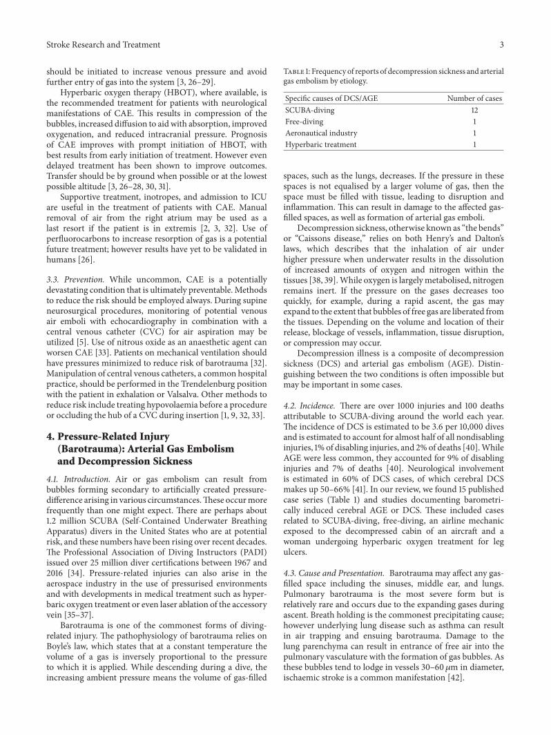
Table 1: Frequency of reports of decompression sickness and arterialgas embolism by etiology.
Specific causes of DCS/AGE Number of casesSCUBA-diving 12Free-diving 1Aeronautical industry 1Hyperbaric treatment 1
spaces, such as the lungs, decreases. If the pressure in thesespaces is not equalised by a larger volume of gas, then thespace must be filled with tissue, leading to disruption andinflammation. This can result in damage to the affected gas-filled spaces, as well as formation of arterial gas emboli.
Decompression sickness, otherwise known as “the bends”or “Caissons disease,” relies on both Henry’s and Dalton’slaws, which describes that the inhalation of air underhigher pressure when underwater results in the dissolutionof increased amounts of oxygen and nitrogen within thetissues [38, 39].While oxygen is largelymetabolised, nitrogenremains inert. If the pressure on the gases decreases tooquickly, for example, during a rapid ascent, the gas mayexpand to the extent that bubbles of free gas are liberated fromthe tissues. Depending on the volume and location of theirrelease, blockage of vessels, inflammation, tissue disruption,or compression may occur.
Decompression illness is a composite of decompressionsickness (DCS) and arterial gas embolism (AGE). Distin-guishing between the two conditions is often impossible butmay be important in some cases.
4.2. Incidence. There are over 1000 injuries and 100 deathsattributable to SCUBA-diving around the world each year.The incidence of DCS is estimated to be 3.6 per 10,000 divesand is estimated to account for almost half of all nondisablinginjuries, 1% of disabling injuries, and 2%of deaths [40].WhileAGE were less common, they accounted for 9% of disablinginjuries and 7% of deaths [40]. Neurological involvementis estimated in 60% of DCS cases, of which cerebral DCSmakes up 50–66% [41]. In our review, we found 15 publishedcase series (Table 1) and studies documenting barometri-cally induced cerebral AGE or DCS. These included casesrelated to SCUBA-diving, free-diving, an airline mechanicexposed to the decompressed cabin of an aircraft and awoman undergoing hyperbaric oxygen treatment for legulcers.
4.3. Cause and Presentation. Barotrauma may affect any gas-filled space including the sinuses, middle ear, and lungs.Pulmonary barotrauma is the most severe form but isrelatively rare and occurs due to the expanding gases duringascent. Breath holding is the commonest precipitating cause;however underlying lung disease such as asthma can resultin air trapping and ensuing barotrauma. Damage to thelung parenchyma can result in entrance of free air into thepulmonary vasculature with the formation of gas bubbles. Asthese bubbles tend to lodge in vessels 30–60 𝜇m in diameter,ischaemic stroke is a common manifestation [42].

4 Stroke Research and Treatment
Table 2: Features of decompression sickness and associated neurological deficits.
Types of DCS Symptoms and signs Speed of onset
Type 1 Musculoskeletal pain and cutaneous symptoms such as “marbling” or cutismarmorata
Begin quickly and build inintensity
Type 2
More serious affecting 1 or more of the following Immediate to delayed(i) Neurological: paraesthesia, ataxia, focal deficits, altered mental status. Spinal
cord is the commonest site of involvementDelayed presentation can resultin more serious injury due toinsidious onset and delay inreporting
(ii) Inner-ear: tinnitus, vertigo, ataxia, deafness(iii) Cardiopulmonary: dyspnoea, pain, “gasp” due to sudden hypoxia from
development of bubbles, cardiopulmonary collapse
4.4. Arterial Gas Embolism. Almost two-thirds of patientswith AGE have alterations of consciousness, with seizures,focal deficits, and coma accounting for the majority of pre-sentations [27, 43–45]. Patients with AGE develop symptomswithin minutes on surfacing in 80% of cases, or even duringascent. They do not require exposure to significant depthor time underwater to develop AGE. CT or MRI scanningmay show focal lesions or the gas emboli within the cerebralvasculature [27, 40, 42].
AGE has significant overlap with DCS and can presentsimultaneously, making the two conditions indistinguishablein many cases; however differentiation is often made basedon suddenness of symptom onset in AGE [40, 46, 47].
4.5. Decompression Sickness. Decompression sickness mayresult from medical-, altitude-, aerospace-, and free-diving-related activities [48]; however SCUBA-diving is the mostcommon cause. Dive profile and ascent time are impor-tant risk factors, as are increased age, obesity, dehydration,hypothermia, and the presence of a patent foramen ovale[27, 44, 45, 49].
Focal weakness, stupor, visual loss, gait disturbance, andvertigo are amongst the common manifestations of cerebralDCS [27, 34]. Motor weakness soon after a dive is very suspi-cious for DCS. It tends to present later than AGE, with 50%developing symptoms within 30–60mins, and 90% within 6hours [27, 40, 50]. The spinal cord is the most commonlyaffected organ which can result in limb weakness, potentiallybeing mistaken for cerebral DCS. Imaging shows lesions thatdo not enhance with contrast in 30–55% of cases; howevermany are normal; therefore diagnosis should be made on aclinical basis [43, 46, 51]. Features of decompression sicknessare presented in Table 2.
4.6. Treatment. Due to the significant amount of overlapbetween the two conditions, treatment is often identical in theinitial stages. Basic and advanced life support, 100% oxygen,rehydration, and Durant’s (Left lateral decubitus) and mildTrendelenburg positions should be initiated immediately torestore perfusion to the ischaemic brain. Immediate transferto the nearest recompression facility for hyperbaric oxygenshould be first priority, as early treatment is one of themain determinants of outcome [52–54]. Recompression withhyperbaric oxygen increases the concentration of oxygensupplied to tissues, accelerates the absorption of nitrogenemboli, and reduces the size of bubbles by Boyle’s law.
Studies have shown 75% of patients with severe DCSexperience full resolution of symptoms if treatment is ini-tiated within 12 hours, which falls to 57% afterwards [54].This is in contrast to patients with AGE, of whom 50%fully improve with hyperbaric oxygen treatment [55]. 88%of the patients in our review underwent recompressionwith hyperbaric oxygen. 37.5% of these had a full recovery,25% died, and the remaining 44% made partial recoveryat the time of writing. Patients with residual deficits arerecommended to receive repetitive treatment over severaldays to maximise their chances of improvement [36, 55].Adjunctive treatments such as steroids and prostacyclin arenot currently part of the treatment protocol due to lack ofevidence [56, 57].
Urgent transfer to a centre for treatment with hyperbaricoxygen is central to the treatment of both decompressionsickness and arterial gas embolism.
5. Missiles and Foreign Bodies
5.1. Background. Embolization of foreign bodies is a rareand often catastrophic occurrence. There has been a widevariety of materials reported as causing embolic phenomena,ranging from illicit drugs [58] and shotgun pellets [59–65].Missile embolization is a recognised component of militarytrauma [64]; however it has also been reported amongst thecivilian population [66]. Lower velocity, smaller projectilesused outside of military exercises may actually result in therisk of missile emboli being slightly higher amongst civilians[66].
Illicit drugs are frequently sold diluted and mixed withpotentially unknown and hazardous substances [58]. SomeIntravenous Drug users inject chemicals not intended fordilution. These vary from hypnotic medications to opiates.If the substance itself or the ingredients it is mixed with arenot completely dissolved, then there is a risk of emboliza-tion. Cerebral involvement may arise from advertent arterialinjection or the presence of a cardiac defect including patentforamen ovale (PFO).
5.2. Incidence. Missile emboli resulting from shotgun pellets,bullets, incendiary shrapnel, and other sources are rare.Military studies have shown that the rate of embolic phe-nomena from missile-induced vascular injury is 0.3–1.1%;however these do not account for cerebral emboli [67, 68].Studies show that intracerebral emboli have amortality rate of

Stroke Research and Treatment 5
(a) (b)
Figure 2: Cerebral infarction in injecting drug users. (a) CT scan showing infarct resulting from paradoxical embolus of injected Zopiclone.(b) MR DWI image of multiple emboli caused by inadvertent injection of diamorphine (Heroin) into left common carotid artery.
25–33% [69, 70].We found 9 cases describing cerebral missileemboli. Cerebral embolization of illicit material is extremelyrare, and we found only 2 documented cases in one report.One involved injection of a partially dissolved hypnoticwhichembolized via a cardiac defect and the other involved theinjection of a mixed opiate into the carotid artery, causingseizure and left hemiparesis [71] (Figure 2).
This is also potentially the case for medications admin-istered through central venous lines. One case of a strokeoccurring following administration of cancer chemotherapythrough a port-catheter terminating in the right atriumwas identified [72] in the presence of a PFO. This eventoccurred after the second cycle of a carboplatin containingchemotherapy regimen which is a known prothromboticagent and thus may have been due to a secondary thrombosisrather than the agent itself [73].
Spinal cord infarction following Cervical TransforaminalEpidural Steroid Injection is much more frequently reported,either by secondary thrombosis or by accidental extravasa-tion of particulates within the medications [74, 75]. Suchinjections have also been reported as a cause of cerebralinfarction but mechanism is unclear.
Angiographic intervention commonly results in foreignbody embolization, with one study stating up to 25% ofresected cerebral AV malformations showing evidence ofembolized foreign material after neurovascular intervention[76]. We found 7 series and cases documenting iatrogeniccauses for cerebral emboli, with materials including glue,autologous fat, cotton wool, and hydrophilic polymers. Thecommonest source is related to embolization of cerebral orcarotid aneurysms.
5.3. Presentation. Focal neurological deficits, sudden distur-bance of consciousness, or coma in the appropriate settingshould raise suspicion of cerebral foreign body embolus.
History of penetrating missile trauma, incongruence of entryand exit wounds, and symptoms out of keeping with thesuspected location of the missile could all suggest missileembolus. IVDUs who develop neurological complicationsfollowing injection or those that inject near arterial sites, suchas the neck or groin, should be investigated for potentialstroke caused by embolic debris. Patients undergoing proce-dures, particularly neurointerventional in nature, should bemonitored closely for potential embolization of particulatematter. Simultaneous echocardiography or Doppler US ofthe carotids can be used to screen for such material, andnew neurological deficits during or after the procedure wouldsuggest occurrence of an embolic event.
5.4. Treatment. Removal of a foreign body depends onpresence of symptoms, size, location, and risk of compli-cations. Arterial bullets are symptomatic in 80% of caseswhile those in the venous system are symptomatic in 1/3of the time. Complications of arterial bullets include distalischaemia, thrombosis, and further embolism but may alsocause extensive psychological disturbance [77]. Due to thehigh rate of mortality associated with cerebral foreign bodies,removal is recommended.
6. Discussion
Nonthrombotic, embolic stroke (NTES) is often overlookeddue to its rarity and the variety of clinical presentation, whichcontributes to difficulties in diagnosis and management.Treatment and outcomes are often based on individualcase reports and case series, meaning that there is a lackof a uniform approach to these patients. Collation of theavailable data and creation of a stroke register may enable thedevelopment of treatment strategies for these rarer forms ofstroke.

6 Stroke Research and Treatment
Nonthrombotic causes of embolic stroke are relativelyrare and certain pathological processes are likely to remainuncommon, that is, missile embolism. However, factors suchas the increasing availability of interventional and intravas-cular procedures may lead to a rise in the incidence of othertypes of NTES. CT-guided biopsies, AV node ablations, andcentral venous catheter insertions are all associated withcerebral air embolism. There is increasing awareness of therole right left shunts, particularly atrial septal defects andPFOs, which can be a risk in transmitting thrombi andother materials from the venous to arterial circulation, thusrisking paradoxical stroke.While reports of this occurring aresurprisingly infrequent, this may be because of the difficultiesin absolutely proving that the cause of the cerebral arteryocclusion was in fact a foreign body or substance withoutbeing able to obtain a sample of same from cerebral vessels.PFOs may affect 1 in 4 individuals and establishing causeand effect beyond doubt is difficult in such circumstances butthis may change with increased use of neurointerventionaltechniques in treating stroke. While PFO closure may be ofbenefit to some patients with recurrent stroke with no othercause found, current guidelines do not currently recommendroutine closure [78].
Knowledge of the potential for cerebral events duringand after these procedures is essential for early diagnosis andinitiation of prompt management. Pre- and postprocedureneurological checklists may flag high-risk procedures andidentify early those effected or at risk of cerebral complica-tions. As the neurological consequences of cerebral arterialocclusion by foreign body or substance may differ little fromthose resultant from thrombotic occlusion, physicians needto be conscious of the circumstances and timing of when theevent occurred and, in the era of potential neurointervention,be aware of potential therapeutic interventions.
Due to the small amount of available data, managementof these patients is infrequently uniform in nature. Promptinitiation of basic resuscitation and removal of the patho-logical cause is recommended. HBOT is now thought tobe effective in cases of air embolism and decompressionsickness and clinicians need to be aware of the symptomsand management of same (Table 2). Embolectomy has beenshown to be successful in cases of cervical and proximalcerebral artery occlusion and indeed embolectomy has beendemonstrated to improves outcomes in proximal arterialocclusion up to 12 hours from time of onset, in certain casesof thrombotic stroke [79, 80].This approachmay be extendedto the mechanical retrieval of foreign bodies and missiles;however, data is scarce. HBOT is a resource which is notreadily available in many locations; however with the risk ofiatrogenic air embolism and the potential for improvementwith treatment, consideration must be given to expand theavailability of this therapy [3, 26–28, 30, 31].
Nonthrombotic, embolic strokes are uncommon causesof cerebral stroke and can be easily overlooked. Knowledgeof the pathological processes and presentation is essentialto ensure that diagnoses are not missed. In the absenceof a substantial evidence base there is still validity in thesubmission and publication of short series and case reportsfor such events and in particular where anecdotally theremay
be a risk, for example, medical device failure and disintegra-tion, but there are no published reports of management oroutcome.
Conflicts of Interest
The authors declare that there are no conflicts of interestregarding the publication of this paper.
References
[1] T. M. Dudney and C. G. Elliott, “Pulmonary embolism fromamniotic fluid, fat, and air,” Progress in Cardiovascular Diseases,vol. 36, no. 6, pp. 447–474, 1994.
[2] M. B. King and K. R. Harmon, “Unusual forms of pulmonaryembolism,” Clinics in Chest Medicine, vol. 15, no. 3, pp. 561–580,1994.
[3] C. M. Muth and E. S. Shank, “Gas embolism,”The New EnglandJournal of Medicine, vol. 342, no. 7, pp. 476–482, 2000.
[4] P. G. Jorens, E. Van Marck, A. Snoeckx, and P. M. Parizel,“Nonthrombotic pulmonary embolism,” European RespiratoryJournal, vol. 34, no. 2, pp. 452–474, 2009.
[5] N. Shaikh and F. Ummunisa, “Acute management of vascularair embolism,” Journal of Emergencies, Trauma, and Shock, vol.2, no. 3, pp. 180–185, 2009.
[6] S. C. Palmon, L. E. Moore, J. Lundberg, and T. Toung, “Venousair embolism: a review,” Journal of Clinical Anesthesia, vol. 9, no.3, pp. 251–257, 1997.
[7] T. Gale and K. Leslie, “Anaesthesia for neurosurgery in thesitting position,” Journal of Clinical Neuroscience, vol. 11, no. 7,pp. 693–696, 2004.
[8] J. M. Raskin, E. Benjamin, and T. J. Iberti, “Venous airembolism: case report and review,” Mount Sinai Journal ofMedicine, vol. 52, no. 5, pp. 367–370, 1985.
[9] R. J. O’Quin and S. Lakshminarayan, “Venous air embolism,”JAMA Internal Medicine, vol. 142, no. 12, pp. 2173–2176, 1982.
[10] R. L. Hybels, “Venous air embolism in head and neck surgery,”The Laryngoscope, vol. 90, no. 6, part 1, pp. 946–954, 1980.
[11] L.W. Faberowski, S. Black, and J. P.Mickle, “Incidence of venousair embolism during craniectomy for craniosynostosis repair,”Anesthesiology, vol. 92, no. 1, pp. 20–23, 2000.
[12] G. I. Van Boxel, C. E. Hommers, I. Dash, A. J. Goodman, J.Green, and R. M. Orme, “Myocardial and cerebral infarctiondue to massive air embolism following endoscopic retrogradecholangiopancreatography (ERCP),” Endoscopy, vol. 42, supple-ment 2, pp. E80–E81, 2010.
[13] D. Chavalitdhamrong and P. V. Draganov, “Acute stroke due toair embolism complicating ERCP,” Endoscopy, vol. 45, supple-ment 2 UCTN, no. 2, pp. E177–E178, 2013.
[14] W. Jens, A. Lee, and M. Ibrahimi, “Imaging of a fatal airembolism from ERCP,” Neurology, vol. 86, no. 16, 2016.
[15] C. K. Baban, M. Murphy, T. Hennessy, and D. O’Hanlon, “Fatalcerebral air embolism following endoscopic evaluation of rectalstump,” BMJ Case Reports, 2013.
[16] M. V. Forlee, M. Grouden, D. J. Moore, and G. Shanik, “Strokeafter varicose vein foam injection sclerotherapy,” Journal ofVascular Surgery, vol. 43, no. 1, pp. 162–164, 2006.
[17] T.M. Leslie-Mazwi, L. L. Avery, and J. R. Sims, “Intra-arterial airthrombogenesis after cerebral air embolism complicating lowerextremity sclerotherapy,” Neurocritical Care, vol. 11, no. 2, pp.247–250, 2009.

Stroke Research and Treatment 7
[18] T. Chemmanam, D. Ghia, and C. Bladin, “Varicose vein sclero-therapy-an uncommon cause of stroke,” International Journal ofStroke, vol. 5, pp. 41-42, 2010.
[19] S. Adatia, V. Nambiar, R. Kapadia et al., “Acute ischemicstroke caused by paradoxical air embolism following injectionsclerotherapy for varicose veins,” Neurology India, vol. 61, no. 4,pp. 431–433, 2013.
[20] J. C. Leong andN.R. Johnston, “Visual loss following sclerother-apy for varicose veins,” BMJ Case Reports, 2011.
[21] C. Huang and J. Pik, “Tension pneumocephalus and oxygenemboli from hydrogen peroxide irrigation,” Journal of ClinicalNeuroscience, vol. 21, no. 2, pp. 323–325, 2014.
[22] S. J. Sherman, L. V. Boyer, andW. A. Sibley, “Cerebral infarctionimmediately after ingestion of hydrogen peroxide solution,”Stroke, vol. 25, no. 5, pp. 1065–1067, 1994.
[23] J. F. Gudmundsdottir, A. Geirsson, P. Hannesson, and T. Gud-bjartsson, “Major ischaemic stroke caused by an air embolismfrom a ruptured giant pulmonary bulla,” BMJ Case Reports, vol.2015, 2015.
[24] J. G. Heckmann, C. J. G. Lang, K. Kindler,W. Huk, F. J. Erbguth,and B. Neundorfer, “Neurologic manifestations of cerebral airembolism as a complication of central venous catheterization,”Critical Care Medicine, vol. 28, no. 5, pp. 1621–1625, 2000.
[25] S. Gordy and S. Rowell, “Vascular air embolism,” InternationalJournal of Critical Illness & Injury Science, vol. 3, no. 1, pp. 73–76,2013.
[26] M. A. Mirski, A. V. Lele, L. Fitzsimmons, and T. J. K. Toung,“Diagnosis and treatment of vascular air embolism,” Anesthesi-ology, vol. 106, no. 1, pp. 164–177, 2007.
[27] R. E. Moon, “Treatment of diving emergencies,” Critical CareClinics, vol. 15, no. 2, pp. 429–456, 1999.
[28] J. Sheasgreen, T. Terry, and J. R. Mackey, “Large-volume airembolism as a complication of augmented computed tomogra-phy: case report,” Canadian Association of Radiologists Journal,vol. 53, no. 4, pp. 199–201, 2002.
[29] D. Karaosmanoglu, S. O. Oktar, M. Arac, and G. Erbas, “Casereport: portal and systemic venous gas in a patient after lumbarpuncture,” British Journal of Radiology, vol. 78, no. 932, pp. 767–769, 2005.
[30] S. Imai, T. Tamada, M. Gyoten, T. Yamashita, and Y. Kajihara,“Iatrogenic venous air embolism caused by CT injector—froma risk management point of view,” Radiation Medicine, vol. 22,no. 4, pp. 269–271, 2004.
[31] S. Ohashi, H. Endoh, T. Honda, N. Komura, and K. Satoh,“Cerebral air embolism complicating percutaneous thin-needlebiopsy of the lung: complete neurological recovery after hyper-baric oxygen therapy,” Journal of Anesthesia &Clinical Research,vol. 15, no. 4, pp. 233–236, 2001.
[32] S. L. Orebaugh, “Venous air embolism: clinical and experimen-tal considerations,” Critical Care Medicine, vol. 20, no. 8, pp.1169–1177, 1992.
[33] P. J. Pronovost, A. W. Wu, and J. B. Sexton, “Acute decompen-sation after removing a central line: practical approaches toincreasing safety in the intensive care unit,” Annals of InternalMedicine, vol. 140, no. 12, pp. 1025–1033, 2004.
[34] TheProfessionalAssociation ofDiving Instructors (PADI.com),Worldwide Corporate Statistics 2017 Data for 2011-2016, https://www.padi.com/sites/default/files/2017-07/2017%20PADI%20WW%20Statistics.pdf.
[35] M. J. Hickey and C. L. Zanetti, “Delayed-onset cerebral arterialgas embolism in a commercial airline mechanic,” Aviation,
Space, and Environmental Medicine, vol. 74, no. 9, pp. 977–980,2003.
[36] B. P. Murphy, F. J. Harford, and F. S. Cramer, “Cerebral airembolism resulting from invasive medical procedures. Treat-ment with hyberbaric oxygen,” Annals of Surgery, vol. 201, no.2, pp. 242–245, 1985.
[37] P. Kale, B. Javed, N. Pednekar, R. Sahni, L. Resor, M. Tenneret al., “Stroke due to air embolism related to laser ablation ofaccessory vein,” Neurology, vol. 86, no. 16, 2016.
[38] R. C. Viadero, “Henry’s law,” inWater Encyclopedia, JohnWiley& Sons, 2005.
[39] L. J. Gillespie, “The Gibbs-Dalton law of partial pressures,”Physical Review A: Atomic, Molecular and Optical Physics, vol.36, no. 1, pp. 121–131, 1930.
[40] P. Buzzacott and Divers Alert Network, “DAN Annual Div-ing Report, 2012-2015 Edition,” 2015, http://www.ncbi.nlm.nih.gov/books/NBK344435/.
[41] A. Erde and C. Edmonds, “Decompression sickness: a clinicalseries,” Journal of Occupational Medicine, vol. 17, no. 5, pp. 324–328, 1975.
[42] H. D. Greer and E. W. Massey, “Neurologic injury fromundersea diving,”Neurologic Clinics, vol. 10, no. 4, pp. 1031–1045,1992.
[43] Y. Melamed, A. Shupak, and H. Bitterman, “Medical problemsassociated with underwater diving,”TheNew England Journal ofMedicine, vol. 326, no. 1, pp. 30–35, 1992.
[44] T. L. Clenney and L. F. Lassen, “Recreational scuba divinginjuries,” American Family Physician, vol. 53, no. 5, pp. 1761–1764, 1996.
[45] A. P. K. Dick and E. W. Massey, “Neurologic presentationof decompression sickness and air embolism in sport divers,”Neurology, vol. 35, no. 5, pp. 667–671, 1985.
[46] D. Cialoni, M. Pieri, C. Balestra, and A. Marroni, “Flying afterdiving: in-flight echocardiography after a scuba diving week,”Aviation, Space, and Environmental Medicine, vol. 85, no. 10, pp.993–998, 2014.
[47] R. T. Goldhahn, “Scuba diving deaths: a review and approachfor the pathologist,” Legal Medicine Annual, vol. 1976, pp. 109–132, 1977.
[48] J. D. Auten, M. A. Kuhne, H. M. Walker II, and H. O.Porter, “Neurologie decompression sickness following cabinpressure fluctuations at high altitude,” Aviation, Space, andEnvironmental Medicine, vol. 81, no. 4, pp. 427–430, 2010.
[49] M.Knauth, S. Ries, S. Pohimann et al., “Cohort study ofmultiplebrain lesions in sport divers: role of a patent foramen ovale,”British Medical Journal, vol. 314, no. 7082, pp. 701–705, 1997.
[50] T. J. Francis, R. R. Pearson, A. G. Robertson, M. Hodgson, A. J.Dutka, and E. T. Flynn, “Central nervous systemdecompressionsickness: latency of 1070 human cases,” Undersea BiomedicalResearch, vol. 15, 1988.
[51] H. S. Levin, F. C. Goldstein, K. Norcross, E. G. Amparo, F.C. Guinto, and J. T. Mader, “Neurobehavioral and magneticresonance imaging findings in two cases of decompressionsickness,” Aviation, Space, and Environmental Medicine, vol. 60,no. 12, pp. 1204–1210, 1989.
[52] K. Tetzlaff, E. S. Shank, and C. M. Muth, “Evaluation and man-agement of decompression illness—an intensivist’s perspective,”Intensive Care Medicine, vol. 29, no. 12, pp. 2128–2136, 2003.
[53] Y. Melamed, D. Sherman, D. Wiler-Ravell, and D. Kerem, “Thetransportable recompression rescue chamber as an alternativeto delayed treatment in serious diving accidents,” Aviation,

8 Stroke Research and Treatment
Space, and Environmental Medicine, vol. 52, no. 8, pp. 480–484,1981.
[54] M. H. Bennett, J. P. Lehm, S. J. Mitchell, and J. Wasiak,“Recompression and adjunctive therapy for decompressionillness,” Cochrane Database of Systematic Reviews, no. 5, articleCD005277, 2012.
[55] D. R. Leitch and R. D. Green, “Pulmonary barotrauma in diversand the treatment of cerebral arterial gas embolism,” Aviation,Space, and Environmental Medicine, vol. 57, no. 10, part 1, pp.931–938, 1986.
[56] D. E. Evans, P. W. Catron, J. J. McDermott, L. B. Thomas,A. I. Kobrine, and E. T. Flynn, “Effect of lidocaine afterexperimental cerebral ischemia induced by air embolism.,”Journal of Neurosurgery, vol. 70, no. 1, pp. 97–102, 1989.
[57] J. M. Hallenbeck, D. R. Leitch, A. J. Dutka, L. J. Greenbaum,andA. E.McKee, “Prostaglandin I2, indomethacin, and heparinpromote postischemic neuronal recovery in dogs,” Annals ofNeurology, vol. 12, no. 2, pp. 145–156, 1982.
[58] L. R. Caplan, C.Thomas, andG. Banks, “Central nervous systemcomplications of addiction to ‘T’s and Blues’,”Neurology, vol. 32,no. 6, pp. 623–628, 1982.
[59] R. Yaari, J. Ahmadi, and G. Y. Chang, “NeuroImages. Cerebralshotgun pellet embolism,” Neurology, vol. 54, no. 7, article 1487,2000.
[60] J. M. Sethi and B. Rozdilsky, “Internal carotid artery embolismby shotgun pellet,” Canadian Journal of Neurological Sciences /Journal Canadien des Sciences Neurologiques, vol. 5, no. 3, pp.325-326, 1978.
[61] C. S. Kase, R. L. White, T. L. Vinson, and R. P. Eichelberger,“Shotgun pellet embolus to the middle cerebral artery,” Neurol-ogy, vol. 31, no. 4, pp. 458–461, 1981.
[62] M. A. Dada, I. A. J. Loftus, and G. S. Rutherfoord, “Shotgunpellet embolism to the brain,”The American Journal of ForensicMedicine and Pathology, vol. 14, no. 1, pp. 58–60, 1993.
[63] L. B. da Costa, M. C.Wallace, andW.Montanera, “Shotgun pel-let embolization to the posterior cerebral circulation,”AmericanJournal of Neuroradiology, vol. 27, no. 2, pp. 261–263, 2006.
[64] A. J. Chapman and J. McClain, “Wandering missiles: autopsystudy,” Journal of Trauma-Injury Infection & Critical Care, vol.24, no. 7, pp. 634–637, 1984.
[65] T. Anda, K. Suyama, T. Kawano, and K. Mori, “Shotgun pelletembolus in the cerebral circulation via the internal carotidartery in the neck; a case report,” Journal of Neurological Surgery,vol. 20, no. 4, pp. 457–461, 1992.
[66] K. Lu, S. Gandhi,M. A.Qureshi, A. S.Wright, N. Kantathut, andT. P. Noeller, “Approach to management of intravascular missileemboli: review of the literature and case report,”Western Journalof Emergency Medicine, vol. 16, no. 4, pp. 489–496, 2015.
[67] N. M. Rich, G. J. Collins, C. A. Andersen, P. T. McDonald, L.Kozloff, and J. J. Ricotta, “Missile emboli,” Journal of Trauma-Injury Infection and Critical Care, vol. 18, no. 4, pp. 236–239,1978.
[68] G. Aidinian, C. J. Fox, T. E. Rasmussen, and D. L. Gillespie,“Varied presentations of missile emboli in military combat,”Journal of Vascular Surgery, vol. 51, no. 1, pp. 214–217, 2010.
[69] M. Massad and M. S. Slim, “Intravascular missile embolizationin childhood: report of a case, literature review, and recommen-dations for management,” Journal of Pediatric Surgery, vol. 25,no. 12, pp. 1292–1294, 1990.
[70] P. N. Symbas, A. L. Picone, C. R. Hatcher Jr., and S. E. Vlasis-Hale, “Cardiac missiles. A review of the literature and personalexperience,”Annals of Surgery, vol. 211, no. 5, pp. 639–648, 1990.
[71] R. Kelly and J. Harbison, “Cerebral infarction due to non-thrombotic paradoxical embolism in an intravenous drug user,”European Stroke Journal, no. 1(IS), article 147, 2016.
[72] D. Ahn, M. E. Brickner, and J. Dowell, “Embolic stroke sec-ondary to an indwelling catheter in a patient with a patentforamen ovale: a case report and review of the literature,”Clinical Advances in Hematology & Oncology, vol. 10, pp. 335–338, 2012.
[73] W. Saber, T. Moua, E. C. Williams et al., “Risk factors forcatheter-related thrombosis (CRT) in cancer patients: a patient-level data (IPD) meta-analysis of clinical trials and prospectivestudies,” Journal ofThrombosis andHaemostasis, vol. 9, no. 2, pp.312–319, 2011.
[74] J. Moon and H.-M. Kwon, “Spinal cord infarction after cervicaltransforaminal epidural steroid injection: case report and liter-ature review,” Case Reports in Neurology, vol. 9, pp. 1–5, 2017.
[75] B. Benny, P. Azari, and D. Briones, “Complications of cervicaltransforaminal epidural steroid injections,” American Journalof Physical Medicine & Rehabilitation/Association of AcademicPhysiatrists, vol. 89, no. 7, pp. 601–607, 2010.
[76] P. Shannon, J. M. Billbao, T. Marotta, and K. Terbrugge, “Inad-vertent foreign body embolization in diagnostic and therapeuticcerebral angiography,” American Journal of Neuroradiology, vol.27, no. 2, pp. 278–282, 2006.
[77] S. K. Gandhi, B. C. Marts, B. M. Mistry, J. W. Brown, R.M. Durham, and J. E. Mazuski, “Selective management ofembolized intracardiac missiles,” The Annals of Thoracic Sur-gery, vol. 62, no. 1, pp. 290–292, 1996.
[78] S. R. Messe, G. Gronseth, D. M. Kent et al., “Practice advisory:recurrent stroke with patent foramen ovale (update of practiceparameter): report of the guideline development, dissemi-nation, and implementation subcommittee of the americanacademy of neurology,” Neurology, vol. 87, no. 8, pp. 815–821,2016.
[79] R. A. Veg, J. L. Chan, T. I. Anene-Maidoh, M. M. Grimes, andJ. F. Reavey-Cantwell, “Mechanical thrombectomy for pediatricstroke arising from an atrial myxoma: case report,” Journal ofNeurosurgery: Pediatrics, vol. 15, no. 3, pp. 301–305, 2015.
[80] M. Goyal, B. K. Menon, W. H. Van Zwam et al., “Endovascularthrombectomy after large-vessel ischaemic stroke: a meta-analysis of individual patient data from five randomised trials,”The Lancet, vol. 387, no. 10029, pp. 1723–1731, 2016.

Submit your manuscripts athttps://www.hindawi.com
Stem CellsInternational
Hindawi Publishing Corporationhttp://www.hindawi.com Volume 2014
Hindawi Publishing Corporationhttp://www.hindawi.com Volume 2014
MEDIATORSINFLAMMATION
of
Hindawi Publishing Corporationhttp://www.hindawi.com Volume 2014
Behavioural Neurology
EndocrinologyInternational Journal of
Hindawi Publishing Corporationhttp://www.hindawi.com Volume 2014
Hindawi Publishing Corporationhttp://www.hindawi.com Volume 2014
Disease Markers
Hindawi Publishing Corporationhttp://www.hindawi.com Volume 2014
BioMed Research International
OncologyJournal of
Hindawi Publishing Corporationhttp://www.hindawi.com Volume 2014
Hindawi Publishing Corporationhttp://www.hindawi.com Volume 2014
Oxidative Medicine and Cellular Longevity
Hindawi Publishing Corporationhttp://www.hindawi.com Volume 2014
PPAR Research
The Scientific World JournalHindawi Publishing Corporation http://www.hindawi.com Volume 2014
Immunology ResearchHindawi Publishing Corporationhttp://www.hindawi.com Volume 2014
Journal of
ObesityJournal of
Hindawi Publishing Corporationhttp://www.hindawi.com Volume 2014
Hindawi Publishing Corporationhttp://www.hindawi.com Volume 2014
Computational and Mathematical Methods in Medicine
OphthalmologyJournal of
Hindawi Publishing Corporationhttp://www.hindawi.com Volume 2014
Diabetes ResearchJournal of
Hindawi Publishing Corporationhttp://www.hindawi.com Volume 2014
Hindawi Publishing Corporationhttp://www.hindawi.com Volume 2014
Research and TreatmentAIDS
Hindawi Publishing Corporationhttp://www.hindawi.com Volume 2014
Gastroenterology Research and Practice
Hindawi Publishing Corporationhttp://www.hindawi.com Volume 2014
Parkinson’s Disease
Evidence-Based Complementary and Alternative Medicine
Volume 2014Hindawi Publishing Corporationhttp://www.hindawi.com