Nonlinear dynamics of oscillators with bilinear hysteresis and sinusoidal excitation
A study of nuclear dynamics followingexcitation and ionization in small
molecules and weakly bound clusters
A DissertationPresented to the Faculty of Science and Technology
of Aarhus Universityin Partial Fulfillment of the Requirements
for the PhD Degree
byQingli Jing
September, 2018
Supervisor: Lars Bojer Madsen


English summary
Great advances in modern laser technologies have made the generationof intense femtosecond and attosecond light pulses a trivial task inlaboratory. The ultrashort pulses can induce electronic excitation andionization in atomic and molecular systems. In the molecular systems,nuclear dynamics, such as dissociation into two fragments, can alsotake place due to a change of the potential landscapes seen by thenuclei. Thus, the theoretical study of the interaction of moleculeswith ultrashort light pulses is challenging since both the electronicand nuclear dynamics should be taken into consideration.
The Monte Carlo wave packet (MCWP) approach can be appliedto study the nuclear dynamics following electronic excitation and ion-ization in small molecules and weakly bounded clusters. This methodis equivalent to the master equation in the Lindblad form in quantumoptics. In the MCWP method, we treat the interaction between thebound and continuum electronic states in a molecule as the interac-tion between an open system and its surroundings. This simplifica-tion greatly reduces the computational costs. In this thesis, we haveobtained the nuclear kinetic energy release (KER) spectra followingdissociative double ionization of H2 and triple ionization of Ne2. Ananalysis of these nuclear KER spectra helps to reveal the nuclear dy-namics during these processes.
i

Danish Resumé
Store fremskridt indenfor moderne laserteknologi har gjort det til entriviel opgave at generere intense femtosekund og attosekund lyspulseri laboratoriet. De ultrakorte pulser kan frembringe både elektron-iske excitationer og ionisering af atomare- og molekylære systemer.I molekylære systemer kan nuklear dynamik, såsom dissociation i tofragmenter, også induceres på grund af ændringer i potentialelandsk-abet. Derfor er den teoretiske undersøgelse af interaktionen mellemmolekyler og ultrakorte lyspulser udfordrende, da både den elektron-iske og nukleare dynamik skal tages i betragtning.
Monte Carlo-bølgepakken (MCWP) tilgangen kan anvendes til atstudere den nukleare dynamik efter elektronisk excitation og ioniser-ing i små molekyler og svagt bundne klynger. Denne metode svarer tilmasterligningen i Lindblad-form i kvanteoptik. I MCWP-metoden be-handler vi samspillet mellem de bundne og kontinuerte elektroniske til-stande i et molekyle som interaktionen mellem et åbent system og detsomgivelser. Denne forenkling reducerer i høj grad beregningsomkost-ningerne. I denne afhandling har vi fundet spektrene for udsendelse afnuklear kinetisk energi (KER) efter dissociativ dobbelt ionisering af H2og trippel ionisering af Ne2. En analyse af disse nukleare KER-spektrehjælper med at afsløre den nukleare dynamik under disse processer.
ii

Preface
This thesis summarizes the work done during my Ph.D. studies atDepartment of Physics and Astronomy, Aarhus University, under thesupervision of Professor Lars Bojer Madsen.
NotationAtomic units 4πε0 = ~ = me = e = 1 are used throughout this reportunless stated otherwise.
AcknowledgementsI want to express my great gratitude to my supervisor Lars BojerMadsen for him offering me the opportunity to conduct my PhD studiesunder his excellent guidance. He has a strong passion for physics andis very kind, patient, and considerate.
I would like to thank Elke Fasshauer, Jørgen Johansen Rørstad,Nikolaj Schrøder Wittig Ravn, and Alexander Holm Kiilerich for writ-ing the Danish resumé for this thesis. They also helped me to improvethe English summary. I would like to thank Lun Yue for giving part ofhis Fortran codes to accelerate the startup of my PhD project. I alsobenefited a lot from discussion with him about my project. I wouldlike to thank Chuan Yu for helping me solve a lot of technical problems
iii

when I write my thesis. I would like to show my great thanks to allthe group members. It is my pleasure to meet all of you.
I would like to thank the MEDEA project to provide me the fundingfor my PhD studies and to offer me the opportunity to become closerto many famous scientists. Thanks a lot to all the MEDEA colleagues.
I am really grateful for the two months’ visit of Fernando Martín’sgroup in Madrid. I really enjoyed working together with Alicia Pala-cios, Fernando Martin and the other colleagues in Madrid.
Great and great thanks to my family and friends to support mefrom the bottom of heart.
Finally, thank the beautiful world.
Qingli Jing,Aarhus, September, 2018.
iv

List of Abbreviations
KER kinetic energy releaseMCWP Monte Carlo wave packetFEL free electron laserHHG high-harmonic generationTDSE time-dependent Schrödinger equationTDFCC time-dependent Feshbash close couplingTDGASCI time-dependent generalized-active-space configuration-
interactionMCTDHF multiconfigurational time-dependent Hartree-FockCREI charge-resonance-enhanced ionizationTISE time-independent Schrödinger equationDES doubly excited stateICD interatomic (intermolecular) Coulombic decayETMD electron transfer mediated decay
v
Contents
Preface iii
Contents vi
I Overview 1
1 Introduction 2
2 TDSE for a molecular system 92.1 Laser-induced ionization . . . . . . . . . . . . . . . . . 92.2 Molecular Hamiltonian . . . . . . . . . . . . . . . . . . 102.3 Time-independent Schrödinger equation for the electronic
Hamiltonian . . . . . . . . . . . . . . . . . . . . . . . . 142.4 Time-dependent Schrödinger equation for the nuclear
motion . . . . . . . . . . . . . . . . . . . . . . . . . . . 15
3 the Monte Carlo wave packet approach 193.1 Equivalence with the master equation . . . . . . . . . . 193.2 Calculation strategy for the deterministic sampling method 233.3 Approximations and simplifications . . . . . . . . . . . 253.4 Applications of the MCWP approach . . . . . . . . . . 27
4 Dissociative single or double ionization of H2 29
vi

Contents
4.1 Implementation of the MCWP method to double ion-ization of H2 . . . . . . . . . . . . . . . . . . . . . . . . 29
4.2 Nuclear KER spectra at near- and mid-IR wavelengths 32Influences of laser parameters . . . . . . . . . . . . . . 33Influences of nuclear mass . . . . . . . . . . . . . . . . 41
4.3 Nuclear KER spectra in an XUV-pump-IR-probe setup 424.4 Nuclear KER spectra when autoionization from doubly
excited states is involved . . . . . . . . . . . . . . . . . 474.5 Concluding remarks . . . . . . . . . . . . . . . . . . . . 55
5 Dissociative triple ionization of Ne2 575.1 Implementation of the MCWP method to triple ioniza-
tion of Ne2 . . . . . . . . . . . . . . . . . . . . . . . . . 605.2 Nuclear KER spectra following triple ionization of Ne2
interacting with coherent XUV pulse . . . . . . . . . . 665.3 Nuclear KER spectra following triple ionization of Ne2
interacting with chaotic XUV pulses . . . . . . . . . . 735.4 Concluding remarks . . . . . . . . . . . . . . . . . . . . 74
6 Summary and outlook 77
II Publications 79
Paper I 80
Paper II 92
Bibliography 107
vii


Part I
Overview
1

Chapter 1Introduction
Molecules, which are composed by at least two atoms, are aboundingin the universe. They are stable because there are chemical bonds be-tween the constituent atoms. Due to the very small spatial scale ofa molecule, quantum effects become important. Thus, in theory, tocorrectly predict the behaviors of a molecule, one has to resort to thequantum methods. Compared with atoms, the motion of molecules aremore complex. Apart from translation in space, they can rotate andvibrate. It is known that the timescales for the dynamics in quantumsystems like atoms and molecules can be expected from the time-energyuncertainty relation ∆t∆E ≈ 1. The energy uncertainty ∆E, relatedto the discrete energy levels that are coherently populated, is depen-dent on the (reduced) mass of the considered object. Simply speaking,∆E is inversely proportional to the (reduced) mass of an object. Thus,the heavier the object is, the smaller ∆E is and the larger ∆t is. Asa result, the timescales for the vibrational and rotational dynamicsin a heavier molecule is larger than that in a lighter molecule. Eventhough the timescales for the nuclear dynamics are more or less differ-ent for different molecules, the rotation of molecules usually takes placeon a timescale of picoseconds (10−12 s) and the vibration usually ona timescale of femtoseconds (10−15 s). For the much lighter electrons,their dynamics occur on a much smaller timescale of attoseconds (10−18
s). To image and control such fast nuclear and electronic dynamics inmolecules, one can resort to femtosecond and attosecond light pulsesto obtain time-resolved spectroscopy.
2

Femtosecond pulses can be generated by femtosecond laser sources.For example, the versatile femtosecond titanium sapphire laser (Ti:Al2O2)can produce pulses within the visible and near-infrared spectrum [1–3]. To produce laser pulses at shorter ultraviolet [4] or longer mid-infrared wavelengths [5–7], one can utilize the nonlinear optical effectsin materials such as sum- and difference-frequency mixing. To gener-ate femtosecond pulses at x-ray wavelength, one strategy is to applyfemtosecond laser pulses in the visible or infrared light spectrum tomodulate the energy of electrons in an electron storage ring in syn-chrotrons [8, 9]. Apart from that, femtoseocond and subfemtoseocondx-ray pulses can be generated by tailoring of the bunch electron dis-tribution in a self-amplified spontaneous-emission free-electron laser(FEL) [10, 11].
The much shorter attosecond pulses can be produced via high-harmonic generation (HHG) by irradiating femtosecond laser pulseson a gas jet. One can obtain a train of attosecond pulses when thedriving laser pulse is multi-cycle [12–14]. An isolated attosecond pulsewould be obtained instead when the driving laser pulse is few-cycle [15–18]. Isolated attosecond pulses can be also produced with multi-cyclelaser pulses by applying optical gating techniques such as polarizationgating and two-color gating [19].
There have been many theoretical and experimental works in study-ing the interaction of molecules with femtosecond and attosecond lightpulses. Various phenomena can be observed in molecules upon inter-acting with ultrashort pulses, i.e.,
• non-adiabatic molecular alignment induced by femtosecondlaser pulses [20–22], the dynamic alignment of a molecule corre-sponds to a revival of a rotational wave packet at a rotationalperiod. The rotational wave packet in a given vibronic band iscreated by coherent excitation of an ensemble of rotational states.
• bond softening and bond hardening [23], a sufficiently stronglaser field can either weaken or strengthen the molecular bondsand thus dissociation or association can be induced.
• above-threshold dissociation [24], more photons are absorbedthan required to overcome the bond energy and the access energy
3

1. Introduction
appears as an ensemble of peaks spaced by a photon energy inthe kinetic energy release spectrum.
• dissociative ionization [25, 26], it describes a process that dis-sociation of a molecule is accompanied with the occurrence ofionization. The ionization can be described in different regimes,e.g., tunneling ionization, over-the-barrier ionization and single-or multi-photoionization. One can employ dissociative ioniza-tion in molecules to achieve a coherent control of chemical reac-tions [27].
• electron localization in molecular dissociation [28–30] due toa coherent superposition of electronic states in different symme-tries,
• laser-induced electron diffraction to image ultrafast molec-ular dynamics [31–33] , the field-ionized coherent electron wavepacket is accelerated by the laser field and its rescattering fromits parent molecular ion mimics the conventional electron diffrac-tion of an electron beam.
• electron charge migration in molecules via electron correla-tion [34, 35].
In addition to the above mentioned phenomena, anomalously high ion-ization at large internuclear separations resulting from charge-resonance-enhanced ionization (CREI) [36, 37] can be observed in some diatomicmolecular ion.
We are particularly interested in resolving and retrieving nucleardynamics in this thesis as control over nuclear dynamics is prerequisitefor realizing control of chemical reactions. Many techniques have beendeveloped to probe nuclear dynamics in molecules. For example, thevibrational wave packet in D+
2 was observed from the nuclear KERspectra of D+
2 by exploiting the correlation between the electronic andnuclear wave packets [38]. Apart from that, the pump-probe setup, bywhich the nuclear KER spectra were obtained as a function of delay,was widely used to image the ultrafast nuclear wave packet motionin real time [39–41]. It was also demonstrated that using the chirpin the HHG spectra allows information about nuclear dynamics on a
4
subfemtosecond timescale [42]. Moreover, the HHG spectra can beused to image molecular orbitals [43]. Nuclear dynamics in moleculescan be also reflected by the photoelectron spectra [44] and by theattosecond transient absorption spectra [45].
When we look back into the history, we can find that it was thestudy of light-matter interaction that eventually resulted in the estab-lishment of modern quantum mechanics. In the late 19th and early20th century, classical physics failed in explaining the Rayleigh–Jeanslaw in blackbody radiation and the photoelectric effect in metal. Thefirst problem, later known as ultraviolet catastrophe, was solved byMax Planck through assuming that the absorption or emission of elec-tromagnetic radiation by a blackbody can be only in the form of dis-crete packets called quanta. Soon after, the photoelectric effect wassuccessfully explained by Albert Einstein after introducing the hy-pothesis that light energy is carried in discrete packets called pho-ton. The discrete or quantized description was later extended to ma-terial systems and sparked the birth of the old quantum theory in theearly 20th century. This theory brought about strong brainstorms tothe scientists at that time and numerous breakthroughs promoted theemergence of quantum mechanics. Nowdays, quantum mechanics hasbecome a fundamental theory to describe microscopic systems.
The Hamiltonian is of fundamental importance in the formulationof a quantum theory. The Hamiltonian for a molecule interacting withan ultrashort pulse is time-dependent. Thus, one can perform a simula-tion of the quantum dynamics of the molecular system by solving thetime-dependent Schrödinger equation (TDSE). Many methods havebeen developed to solve the TDSE for a molecular system, e.g.,
• the time-dependent Feshbach close coupling (TDFCC)method, this is an ab initio method and the TDSE is solved usingthe spectral method, where the total wave function is expandedin the basis of electronic eigenstates by diagonalizing the field-free Hamiltonian. The Feshbash formalism introduces projectionoperators to project the total wave function onto non-resonantscattering and bound states, respectively. This method has beenused to study resonant dissociative photoionization of diatomicmolecules where singly or doubly excited states are involved [46–49].
5

1. Introduction
• themulticonfigurational time-dependent Hartree-Fock (MCT-DHF) method generalized to treat correlated electronic andmolecular dynamics in diatomic molecules [50], in this method,the correlated state of a multi-electron system is expressed by asuperposition of determinant of time-dependent orbitals.
• the time-dependent generalized-active-space configuration-interaction (TDGASCI) approach to study correlated ioniza-tion dynamics of diatomic molecules [51], in this method, themany-body wave function is expanded based on the configura-tion interaction and the determinantal space is restricted to areduced subspace.
• the Monte Carlo wave packet (MCWP) method applied inthis thesis, this method treats ionization as a decay process whereelectrons are gradually lost to the surroundings. Lack of informa-tion related to the continuum electrons, the MCWPmethod is es-pecially powerful in studying nuclear dynamics following doubleor multiple ionization of small molecules. This method was ini-tially developed for dissipative processes in quantum optics [52]and was applied to study dissociative double ionization of H2 orD2 [53–55] and O2 [56]. A good agreement between the resultsobtained by this method and by the experiments [57, 58] hasbeen achieved.
In addition to solving the TDSE, laser-induced coupled electron-nuclear dynamic can be obtained by solving quantum Liouville–vonNeumann equation with the semiclassical surface hopping method [59].
Outline of this thesisThis thesis presents theoretical studies on the nuclear dynamics follow-ing electronic excitation and ionization of small molecules and weaklybounded clusters by the MCWP approach. It is divided into two parts:Part One gives an overview of the discussed topics and Part Twoattaches our publications [60, 61]. We organize the remaining chaptersof Part One as follows:
6
• Started from the molecular Hamiltonian, Chapter 2 introducesthe TDSE for a molecular system exposed to external electro-magnetic fields.
• Chapter 3 provides a detailed description of the MCWP ap-proach including its equivalence with the master equation inquantum optics, its implementation under the deterministic sam-pling method and the approximations and simplifications it hasmade.
• Chapter 4 simulates single or double ionization of H2 when ex-posed to intense laser pulses by applying the MCWP approach.The interaction of H2 with different laser pulses including singleinfrared and XUV pulses and two pulses in a pump-probe set-ting are discussed. When the XUV pulses are applied, nucleardynamics in the singly or doubly excited states in H2 are induced.
• Chapter 5 extends the MCWP approach to simulate dissocia-tive triple ionization of the neon dimmer. A comparison betweenour simulation and a recent experiment in an XUV-pump-XUV-probe setup [62] is carried out.
• Chapter 6 gives a brief summary and conclusion of this thesisas well as a short outlook on future research.
7

Chapter 2TDSE for a molecular system
In this chapter, we will deduce the time-dependent Schrödinger equa-tion (TDSE) for the interaction of a molecule with intense laser pulsesstarting from the molecular Hamiltonian. Before that, we will firsthave a brief discussion of different ionization regimes in the following.
2.1 Laser-induced ionizationWhen an atom or molecule interacts with an intense laser pulse, ion-ization may take place since the external field is comparable to theinternal field that binds the outer electron. The ionization is sepa-rated into two regimes by the Keldysh parameter γ, i.e.,
γ =√
Ip2Up
, (2.1)
where Ip is the ionization potential and Up = is the pondermotiveenergy and is determined as
Up = F 2
4ω2 , (2.2)
where F is the electric field strength and ω is the frequency of theexternal field.
If γ . 1 is satisfied, the field-induced ionization can be describedas a tunneling process. When a strong electrostatic field is applied,
9
2. TDSE for a molecular system
the potential well felt by the electrons is suppressed to form a finitebarrier through which an electron can tunnel out. When the fieldstrength is sufficiently large (larger than a critical value), the finitebarrier is so suppressed that over-the-barrier ionization can take place,i.e., the electron can escape the nuclear core over the barrier.
When a strong oscillating laser field at a very low frequency isapplied, the potential well would change adiabatically following theinstantaneous field in a quasi-static picture. An electron can tunnelout through the barrier only if the tunneling time is smaller than half ofthe laser period, i.e., tunneling of an electron should be finished beforethe direction of electric field is reversed. Over-the-barrier ionizationcan be similarly induced by a sufficiently strong laser field at a verylow frequency.
If γ � 1 is satisfied, the ionization can be described as a mult-photon process: when a laser field at a sufficiently high frequency isapplied, the electron may absorb a large amount of energy, i.e., sev-eral photons, from the external field before it succeeds in escaping thenucleus due to the fast change of direction of the electric field. Whenthe frequency is extremely high, the ionization can be described as asingle- or double-photon process: absorption of one or two photonsfrom the external field is sufficient to support the energy for the elec-tron escaping the nuclear core.
2.2 Molecular HamiltonianHamiltonian is the starting point to perform a quantum-mechanicaldescription of a system. For a molecular system with Nn nuclei andNe electrons interacting with an external laser field, its time-dependentHamiltonian in the laboratory frame of reference is expressed as
Htotal(t) =Nn∑j
~p2Nj
2mNj+
Ne∑j
~p2ej
2 + 12
Nn∑j,k 6=j
ZjZk
|~Rj − ~Rk|+ 1
2
Ne∑j,k 6=j
1|~rj − ~rk|
−Nn,Ne∑j,k
Zj
|~Rj − ~rk|+ VI(t), (2.3)
where ~pNj = −i∇~Rj(~pej
= −i∇~rj) is the momentum operator of the
jth nucleus (electron), Zj and mNj denote the charge and mass of
10

2.2. Molecular Hamiltonian
the jth nucleus, and ~Rj (~rj) is the position vector of the jth nucleus(electron). On the right hand of Eq. (2.3), from left to right, thefirst term represents the kinetic energy operator of the Nn nuclei, thesecond term the kinetic energy operator of the Ne electrons, the thirdterm the Coulomb interaction between the Nn nuclei, the fourth termthe Coulomb interaction between the Ne electrons, the fifth term theCoulomb interaction between the Nn nuclei and Ne electrons and thelast term the laser-molecule interaction operator, which has differentforms in the length and velocity gauges, i.e.,
VI(t) =
~F (t) · (∑Nej ~rj −
∑Nnj Zj ~Rj) LG,∑Ne
j (~pej · ~A(t) + ~A2(t)2 ) +∑Nn
j (−Zj~pnj · ~A(t) + Z2j~A2(t)2 ) VG,
(2.4)where ~F (t) and ~A(t) are the electric field and vector potential of the ap-plied laser pulse, respectively. The laser-molecule interaction operatorin the above equation also implies the application of dipole approxima-tion. In this thesis, we will use the Hamiltonian in the length gauge.For many cases, it is more convenient to use the Hamiltonian in thecenter of mass frame instead of the Hamiltonian in Eq. (2.3). This isbecause the center of mass frame, which is independent of experimentalgeometries, can provide initiative pictures of the considered processes.The position vector of the center of mass in the laboratory frame is
~RCM =∑Nnj mNj ~Rj +∑Ne
j ~rj∑Nnj mNj +Ne
. (2.5)
The position vectors of the nuclei and electrons in the center of massframe are
~Rj = ~Rj − ~RCM, (2.6)~rj = ~rj − ~RCM. (2.7)
Exploiting the fact that the position of the center of mass in the cen-ter of mass frame is the origin of this frame, we obtain the followingrelation
~RCM = 0 =∑Nnj mNj ~Rj +∑Ne
j ~rj∑Nnj mNj +Ne
. (2.8)
11

2. TDSE for a molecular system
For a diatomic molecule, i.e., Nn = 2, the Coulomb interaction betweenthe two nuclei only depends on their relative position vector, i.e.,
~R = ~R1 − ~R2. (2.9)
Combining Eqs. (2.8-2.9) together, we can first express the positionvectors of the two nuclei in the center of mass frame, i.e., ~R1 and ~R2,by the relative position vector of the two nuclei ~R and the positionvectors of the electrons in the center of mass frame ~rj. By applyingthe relation in Eq.( 2.6), we can then represent the position vectors ofthe nuclei in the laboratory frame by the position vector of the center ofmass in the laboratory frame ~RCM, the relative positive vector betweenthe two nuclei ~R, and the position vectors of the electrons in the centerof mass frame ~rj, i.e,
~R1 =−∑Ne
j ~rj +mN2 ~R
mN1 +mN2+ ~RCM ≈
mN2 ~R
mN1 +mN2+ ~RCM (2.10)
~R2 =−∑Ne
j ~rj −mN1 ~R
mN1 +mN2+ ~RCM ≈
−mN1 ~R
mN1 +mN2+ ~RCM (2.11)
The approximations on the right hands of the above two equations arefairly reasonable due to the fact that the electron mass is much smallerthan that of the nuclei. Now we take the approximated position vectorsof the two nuclei in Eqs. (2.10-2.11) and the position vectors of theelectrons in Eq. (2.7) into Eq. (2.3), together with the relations
∇~R1= ∂ ~R
∂ ~R1∇~R + ∂ ~RCM
∂ ~R1∇~RCM
+∑j
∂~rj
∂ ~R1∇~rj
= ∇~R + mN1
mN1 +mN2 +Ne(∇~RCM
−∑j
∇~rj), (2.12)
∇~R2= ∂ ~R
∂ ~R2∇~R + ∂ ~RCM
∂ ~R2∇~RCM
+∑j
∂~rj
∂ ~R2∇~rj
= −∇~R + mN2
mN1 +mN2 +Ne(∇~RCM
−∑j
∇~rj), (2.13)
12

2.2. Molecular Hamiltonian
and
∇~rj= ∂~rj
∂~rj∇~rj
+Ne∑k 6=j
∂~rk∂~rj∇~rk
+ ∂ ~RCM
∂~rj∇~RCM
= ∇~rj−
Ne∑k
1mN1 +mN2 +Ne
∇~rk+ 1mN1 +mN2 +Ne
∇~RCM,
(2.14)
we can rewrite the Hamiltonian for a diatomic molecule with Ne elec-trons as a combination of the Hamiltonian for the center of massHCM(t) and the Hamiltonian describing the relative motion H(t), i.e.,
Htotal(t) = HCM(t)+H(t) = HCM(t)+TN+Te+VN+Ve+VNe+Vmp+VL(t).(2.15)
In the above equation, TN (Te) is the kinetic energy operator of thereduced nuclei (electrons), VN (Ve) is the Coulomb interaction betweenthe nuclei (electrons), VNe is the Coulomb interaction between the nu-clei and electrons, Vmp is the mass polarization term and VL(t) is thelaser-molecular interaction in the center of mass frame. The expres-sions for these operators in Eq. (2.15) can be found in the following,
HCM(t) = −i∇2
~RCM
2(mN1 +mN2 +Ne)+
Ne∑j
(Ne − Z1 − Z2)~F (t) · ~RCM
(2.16)
TN = −i∇2~R
2µ , Te =Ne∑j
−i∇2~rj
2me(2.17)
VN = = Z1Z2
R, Ve =
∑j,k<j
1|~rj − ~rk|
(2.18)
VNe =∑j
Z1
| mN2~RmN1+mN2
− ~rj|+∑j
Z2
| mN1~RmN1+mN2
+ ~rj|(2.19)
Vmp =∑j,k 6=j
∇~rj· ∇~rk
2(mN1 +mN2 +Ne)(2.20)
VL(t) = ~F (t) · [∑j
~rj(1 + Z1 + Z2
mN1 +mN2)− Z1mN2 − Z2mN1
mN1 +mN2~R]
(2.21)
13

2. TDSE for a molecular system
with
µ = mN1mN2
mN1 +mN2, me = mN1 +mN2 +Ne
mN1 +mN2 +Ne − 1 . (2.22)
The mass polarization term Vmp, which is purely dependent on theelectronic coordinates, is usually neglected because its denominatoris much larger than that of the electronic kinetic energy operator Te.Thus, it is reasonable to only include the other six terms of the Hamil-tonian for the relative motion, i.e, H(t) = TN+Te+VN+Ve+VNe+VL(t).The above equations altogether shows clearly to us that the Hamilto-nian for the center of mass HCM is only dependent on the positionvector ~RCM, while the Hamiltonian for the relative motion H(t) hasnoting to do with ~RCM. Thus, the total wave function |Ψtotal(t)〉 canbe written as a tensor product of the wave function for the center ofmass |ΨCM(t)〉 and the wave function for the relative motion |Ψ(t)〉,i.e.,
|Ψtotal(t)〉 = |ΨCM(t)〉 ⊗ |Ψ(t)〉. (2.23)
The center of mass mimics a particle with mass mN1 +mN2 +Ne andcharge Ne − Z1 − Z2 moving in the external field F (t). Its motiondoes not affect the relative motion of the system. As a result, to studythe dynamics in a molecule, we can directly take the Hamiltoniandescribing the relative motion H(t).
2.3 Time-independent Schrödingerequation for the electronicHamiltonian
Now that we have obtained the molecular Hamiltonian H(t), the nextstep is to solve the corresponding TDSE, i.e.,
i|Ψ(t)〉 = H(t)|Ψ(t)〉. (2.24)
Taking use of the fact that the electronic dynamics is usually muchfaster than the nuclear dynamics, we can separate the nuclear andelectronic motion by applying the Born-Oppenheimer approximation
14

2.4. Time-dependent Schrödinger equation for the nuclear motion
via using the following ansatz
|Ψ(t)〉 =∑m
|χm(t)〉⊗|ψelR,m〉 =
∑m
∫d~Rχm(~R, t)|ψel
R,m〉⊗|~R〉, (2.25)
where the completeness relation 1 =∫d~R|~R〉〈~R| is utilized and χm(~R, t) =
〈~R|χm(t)〉 is the nuclear wave function when the electronic state is|ψelR,m〉. The electronic wave functions can be obtained by solving
the time-independent Schrödinger equation for the field-free electronicHamiltonian Hel, i.e.,
Hel|ψelR,m〉 = Em(~R)|ψel
R,m〉, (2.26)
with Hel = Te +VN +Ve +VNe. We note that the electronic Hamiltonianand its corresponding eigenfunctions |ψel
R,m〉 and eigenvalues Em allhave a parametric dependence on the internuclear separation vectors~R. For a givenm, different internuclear separation vectors would resultin different Em(~R) values, which eventually form an energy surface.We will show in the following section that the energy surfaces areindeed the potential energy surfaces where the nuclei move on.
2.4 Time-dependent Schrödingerequation for the nuclear motion
In this section, we will focus on obtaining the evolution of time-dependentnuclear wave functions χm(~R, t). After first taking Eq. (2.25) intothe TDSE in Eq. (2.24) and then projecting both sides on the state〈ψel
R,m| ⊗ 〈~R|, we obtain the TDSE for the nuclear motion, i.e.,
iχm(~R, t) = (−∇2~R
2µ + Em(~R))χm(~R, t)
+∑j 6=m〈ψel
R,m|VL(t)|ψelR,j〉χj(~R, t)
−∑j
〈ψelR,m|∇2~R
2µ |ψelR,j〉χj(~R, t)
− 1µ〈ψel
R,m|∇~R|ψelR,j〉∇~Rχj(~R, t).
(2.27)
15
2. TDSE for a molecular system
Now it is time to implement the adiabatic approximation to simplifythe above equation by neglecting the last two vibronic coupling termson the right side of the equation, i.e.,
iχm(~R, t) = (−∇2~R
2µ +Em(~R))χm(~R, t)+∑j 6=m〈ψel
R,m|VL(t)|ψelR,j〉χj(~R, t).
(2.28)The adiabatic approximation is adopted throughout this thesis be-cause we do not pay attention to physics near avoided crossings, wherethis approximation breaks down. We note that for a homonuclear di-atomic molecule, the laser-molecular interaction operator is as simpleas VL(t) = β ~F (t) ·∑j ~rk according to Eq. (2.21) with β = 1+ Z1+Z2
mN1+mN2.
We can see from Eq (2.28) that the nuclear wave packet χm(~R, t)moves along its potential energy surfaces Em(~R) accompanied withlaser-induced couplings with the other electronic states. In principle,one should include the electronic states as many as possible to ob-tain the nuclear dynamics as accurate as possible. This means thatone has to take many calculation resources to solve a huge number ofcoupled equations according to Eq. (2.28). In this thesis, we takethe assumption that the molecules are rotationally frozen and thelaser polarization direction is parallel to the molecular axis. Thus,the three-dimensional coupled equations in Eq. (2.28) will be reducedto one-dimensional by replacing the internuclear separation vector ~Rby the scaler R. Besides, the potential energy surfaces are reduced topotential energy curves.
In this thesis, we are concerning about nuclear dynamics followingelectronic excitation and ionization in molecules. When the ionizationoccurs, a large number of continuum electronic states with continuouseigenenergies Em(R) + ε would be populated. Here ε is the energyof the continuum electron and Em(R) is the energy of the mth elec-tronic state in the remaining ion. Thus, the potential energy curves forthe continuum electronic states converging to the same mth electronicstate in the remaining ion are parallel to the potential energy curve forthe mth electronic state. It is an acceptable approximation to treatthe evolution of the nuclear wave packets along the potential energycurves for the continuum states converging to a given ionic state asan effective nuclear wave packet evolving along the potential energy
16
2.4. Time-dependent Schrödinger equation for the nuclear motion
curve for the ionic state. This simplification, as we shown in the nextchapter, is implemented by the MCWP approach where the nucleardynamics is main concern.
17


Chapter 3the Monte Carlo wave packet
approach
In this chapter, we will present a thorough discussion of the MCWP ap-proach. This method is developed to study multiple ionization of smallmolecules. It treats ionization as a decay process by phenomenologi-cally introducing a non-Hermitian term into the system Hamiltonian.We will start this chapter by presenting to readers the equivalence ofthe MCWP method with the master equations.
3.1 Equivalence with the masterequation
We can see from the previous chapter that the Hamiltonian H(t) for amolecule is Hermitian, so is the electronic Hamiltonian Hel. Thus,solving the TISE for the electronic Hamiltonian will result in realeigenvalues Em(~R) for the eigenstates |φel
R,m〉. Non-Hermitian Hamilto-nian, however, would lead to complex eigenvalues for the eigenstates,where the real parts correspond to the eigenenergies for the eigen-states and the imaginary parts are related to the decay rates of theeigenstates. Thus, the complex eigenenergy representation is usuallyused to obtain the lifetimes or decay rates of unstable states. Thesestates have a finite lifetime due to spontaneous relaxation processessuch as spontaneous emission and autoionization. Now we generalize
19

3. the Monte Carlo wave packet approach
the concept of instability to the states experiencing non-spontaneousdecay processes, e.g., ionization induced by laser. Thus, in this the-sis, for the considered states which undergo laser-induced ionization,their eigenenergies can be also constructed by complex numbers, i.e.,E ′m(~R) = Em(~R) − i
2Γm(~R), where Γm(~R) are the ~R- and state-dependent ionization rates. These complex eigenenergies indicate anew electronic Hamiltonian, i.e.,
H ′el = Hel −i
2∑m
∫d~RΓm(~R)|ψel
R,m〉〈ψelR,m| ⊗ |~R〉〈~R|, (3.1)
where the non-Hermitian term in Eq. (3.1) describes the laser-inducedionization from the involved bound electronic states |ψel
R,m〉. We notethat the laser-induced ionization only accounts for the coupings be-tween the bound and the continuum electronic states. To describe thecoherent couplings between the bound electronic states (coupings be-tween continuum states are neglected), we remain the use of the laser-molecule interaction operator VL(t). The new electronic Hamiltonianin Eq. (3.1), the nuclear kinetic operator TN and the laser-moleculeinteraction operator VL(t) altogether constitute a new non-HermitianHamiltonianH ′(t), which is the starting point of the MCWP approach,i.e.,
H ′(t) = TN +H ′el + VL(t)
= H(t)− i
2∑m
∫d~RΓm(~R)|ψel
R,m〉〈ψelR,m| ⊗ |~R〉〈~R|
= H(t)− i
2∑m
C†mCm. (3.2)
In the above equation, we rewrite the non-Hermitian term by intro-ducing the quantum jump operators Cm, which specify the transitionsfrom the |ψel
R,m〉 states in a given charge state to the |ψelR,n〉 states in
the charge state with an electron less. The jump operators may be dif-ferent for different ionization mechanisms. In this chapter, the jumpoperators for tunneling ionization are expressed as
Cm =∫d~R
√Γm(~R)
∑n
cn|ψelR,n〉〈ψel
R,m| ⊗ |~R〉〈~R|. (3.3)
A sum over n in Eq. (3.3) means the final state following tunnel ion-ization by the strong external field is a coherent superposition of the
20

3.1. Equivalence with the master equation
electronic states in the charge state with one electron less. For manycases, only including one electronic state in the charge state with oneelectron less in Eq. (3.3) is a good approximation, i.e., n = 1 andcn = 1. We note that the tunneling ionization rates of molecules arenot only dependent on the internuclear distances but also on the in-stantaneous external field. Thus, the tunneling ionization rates Γm(~R)are time-dependent.
The explicit operations of the MCWP approach at each time stepby using the stochastic sampling method are as follows:
1. Propagate the state |Ψ(t)〉 described by Eq. (2.25) using thesmall-time evolution operator U(t+ ∆t, t) = exp(−H ′(t)∆t), i.e,
|Ψ(t+ ∆t)〉 = U(t+ ∆t, t)|Ψ(t)〉. (3.4)
Here we choose a very small time step ∆t so that H ′(t) can beregarded as a constant within this small time interval.
2. Calculate the drop in the probability after a time step ∆t, i.e.,
dP = 〈Ψ(t)|Ψ(t)〉 − 〈Ψ(t+ ∆t)|Ψ(t+ ∆t)〉=∑m
dPm ≈∑m
〈Ψ(t)|C†mCm)|Ψ(t)〉∆t, (3.5)
where the above approximation is to the first order of ∆t.
3. Determine the quantum jump occurs or not by comparing dPwith a random number ε. If dP is smaller than ε, the jump cannot occur and the system remains in the following state
|Ψ(t+ ∆t)〉 = |Ψ(t+ ∆t)〉√(1− dP )
= U(t+ ∆t)|Ψ(t)〉√(1− dP )
. (3.6)
Otherwise, the jump occurs and the new state is
Ψ(t+ ∆t)〉 = Cm|Ψ(t)〉√dPm/∆t
, (3.7)
where the choice of the mth channel above depends on a com-parison of a new random number η with the branching ratiodPm/dP . For example, for the case of two states, i.e., m = 1, 2,if η is smaller than dP1/dP , then the jump occurs from |ψel
R,1〉state, otherwise, from the |ψel
R,2〉 state.
21

3. the Monte Carlo wave packet approach
4. Continue to the next time step from point 1.
The propagation from the initial state at the beginning to the finalstate at the end of the propagation time is called a realization. Weshould include many realizations and average over them to obtain thephysical evolution of a system.
We will show in the following the equivalence of the MCWP ap-proach with the master equation in the Lindblad form for a dissipativeprocess. The density operator at t+∆t is ρ(t+∆t) = |Ψ(t+∆t)〉〈Ψ(t+∆t)|. Statistically, the average value of this density operator can beexpressed as
ρ(t+ ∆t) = (1− dP ) |Ψ(t+ ∆t)〉〈Ψ(t+ ∆t|)1− dP
+∑m
dPmCm|Ψ(t)〉〈Ψ(t)|C†m
dPm/∆t, (3.8)
with the knowledge that there is a probability of (1 − dP ) that thesystem is in the |Ψ(t+∆t)〉√
1−dP state and there is a probability of dPm thatthe state is in the Cm|Ψ(t)〉√
dPm∆t state. By taking Eq. (3.6) into Eq. (3.8), wecan obtain the following equation after neglecting the (∆t)2 term, i.e.,
ρ(t+ ∆t) = (1− iH(t)∆t− 12∑m
C†mCm∆t)ρ(t)
× (1 + iH(t)∆t− 12C†mCm∆t) + ∆tCmρ(t)C†m
= ρ(t)− i[H(t), ρ(t)]∆t− 12{ρ(t), C†mCm}∆t
+ ∆tCm ¯ρ(t)C†m. (3.9)
Taking ˙ρ(t) = ρ(t+∆t)−ρ(t)∆t into the above equation, we can obtain the
master equation in the Lindblad form
i ˙ρ = [ρ, H(t)]− i
2{ρ, C†mCm}+ iCmρC
†m. (3.10)
22

3.2. Calculation strategy for the deterministic sampling method
3.2 Calculation strategy for thedeterministic sampling method
In the MCWP approach, the evolution equation for the nuclear wavefunctions in each charge state can be similarly obtained by projectingthe TDSE of the non-Hermitian Hamiltonian H ′(t) on the 〈ψel
R,m| ⊗〈~R| state. After applying the adiabatic approximation, the obtainedequation for the nuclear motion is similar to Eq. (2.28) except thatEm(~R) in Eq. (2.28) is replaced by Em(R)− i
2Γm(R, t), i.e.,
iχm(R, t) = (−∇2R
2µ + Em(R)− i
2Γm(R, t))χm(R, t)
+∑j 6=m〈ψel
R,m|VL(t)|ψelR,j〉χj(R, t). (3.11)
In the previous section, we have discussed the procedures for the im-plementation of the MCWP approach when the stochastic samplingmethod is applied. An alternative is to apply a much more simpli-fied deterministic method [63]. Here are the explicit procedures forobtaining the nuclear KER spectrum following double ionization of adiatomic molecule by using the deterministic sample strategy:
1. Propagate the nuclear wave packet along the potential energycurves for the electronic states involved according to Eq. (3.11)in the neutral molecule. The probability in the neutral chargestate Pn(t) is decreasing over time because of ionization. Thefirst jumps are assumed to occur at every time step. The proba-bility for a jump occurring at a given time step is the ionizationprobability within a small time interval ∆t, i.e.,
P1(t) = Pn(t)− Pn(t+ ∆t) =∑m
∫dR|χnm(R, t)|2−|χnm(R, t+∆t)|2,
(3.12)where χnm(R, t) is the nuclear wave packet evolving along thepotential energy curve for the |ψel,n
R,m〉 state.
2. When the first ionization occurs at t1, the initial nuclear wavepacket along the potential energy curve for the |ψel,s
R,k〉 state in
23

3. the Monte Carlo wave packet approach
the singly ionized molecule can be obtained by from Eq. (3.7)
χsk(R, t1) = Nck√
Γnm(R, t1)χnm(R, t1), (3.13)
where N is introduced to normalize total wave function in thesingly ionized system, i.e.,∑k
∫dR|χsk(R, t1)|2 = 1, and Γnm(R, t1)
is the R-dependent ionization rates from the |ψel,nR,m〉 state in the
neutral molecule to the singly charged ion at t1. The appear-ance of ck in Eq. (3.13) comes from the jump operator Cm inEq. (3.3). When several electronic states in the neutral moleculeare involved, there are several first ionization pathways since thefirst ionization can take place from each state in the neutralmolecule. The relative probability or branch ratio for a givenpathway whose ionization takes place from the |ψel,n
R,m〉 state is
P1m(t1) =∫dRΓnm(R, t1)|χnm(R, t1)|2∑
m
∫dRΓnm(R, t1)|χnm(R, t1)|2 . (3.14)
Thus, the probability for the first jump occurring at t1 from the|ψel,nR,m〉 state is P1(t1)× P1m(t1).
3. After the first jump at t1, the nuclear wave packet would evolvealong the potential energy curves for the electronic states in thesingly charged ion according to Eq. (3.11) by using the initialnuclear wave packet in Eq. (3.13) as the initial condition. Theprobability in the singly charged ion Ps(t) is decreasing over timedue to ionization. The probabilities for the second jumps occur-ring at t2 are similarly obtained by
P2(t2) = Ps(t2)−Ps(t2+∆t) =∑k
∫dR|χsk(R, t2)|2−|χsk(R, t2+∆t)|2.
(3.15)The initial nuclear wave packet χdj (R, t2) along the potential en-ergy curve for the |ψel,d
R,j 〉 state at t2 in the doubly ionized moleculeis described by
χdj (R, t2) = Mcj√
Γsk(R, t2)χsk(R, t2). (3.16)
In the above equation, a similar constant M is introduced tomake the total wave function normalized and Γsk(R, t2) represents
24

3.3. Approximations and simplifications
the ionization rates from the |ψel,sR,k〉 in the singly ionized molecule
to the doubly ionized system. Similarly, when there are severalsecond jump pathways, the relative probability or branch ratiofor the pathway whose ionization takes place from the |ψel,s
R,k〉 stateis
P2j(t2) =∫dRΓsk(R, t2)|χsk(R, t2)|2∑
k
∫dRΓsk(R, t2)|χsk(R, t2)|2 . (3.17)
Thus, the probability for the second jump occurring at t2 viafrom the |ψel,s
R,k〉 state are P2(t2)× P2m(t2).
4. Once the second ionization takes place at t2, the nuclear wavepacket would propagate along the potential energy curves for thestates in the doubly ionized system according to Eq. (3.11) byusing the nuclear wave packet in Eq. (3.16) as the initial condi-tion. The nuclear wave packets in the doubly ionized moleculeat the end of laser pulse, i.e., χdj (R, te) with j = 1, 2, ..., are pro-jected on the eigenstates χE,j(R) of the potential energy curvesfor the |ψel,d
R,j 〉 states to obtain the nuclear KER spectrum for adeterministic realization, i.e.,
pE(m, t1, k, t2) =∑j
|∫dRχE,j(R)χdj (R, te)|2. (3.18)
The final nuclear KER spectra can be obtained by summing overthe contributions from the realizations, also called trajectories,at different first and second jump times, and from different firstand second jump pathways, i.e.,
PE =∑
m,t1,k,t2
P1(t1)P1m(t1)P2(t2)P2k(t2)pE(m, t1, k, t2). (3.19)
3.3 Approximations and simplificationsWe have adopted a series of approximations and simplifications in theMCWP approach. We first summarize the approximations in the fol-lowing:
1. the dipole approximation, we have applied the dipole ap-proximation by using the laser-molecule interaction operator in
25

3. the Monte Carlo wave packet approach
Eq. (2.4). The dependence of the external field on the spatialcoordinates can be neglected when the scale of small moleculesis much smaller than the wavelength of the applied laser pulse.
2. theBorn-Oppenheimer approximation, we have applied thisapproximation to assume the total wave function as a productof the nuclear wave function and electronic wave functions, seeEq. (2.25). This is reasonable when the electronic dynamics ismuch faster than the nuclear dynamics.
3. the adiabatic approximation, this means that we ignore thechange of the electronic states on the nuclear motion, see Eq. (2.28).The electronic states remain the same order. For example, if thesystem is initially in the ground electronic state at a given inter-nuclear distance, when the nuclei move to another internucleardistance, the electronic state is still the ground state of the newelectronic Hamiltonian.
4. the Born-Markov approximation, when the electron is re-moved from the molecule, it is immediately absorbed by the(imaginary) detector so that rescattering of the electron by theexternal field is not included in our simulation. The unidirec-tional loss of electrons to the surroundings is described by thenon-Herimitian term in the Hamiltonian in Eq. (3.2).
5. the quasi-static approximation in obtaining the tunnelingionization rates, tunneling ionization rates in an slowly oscillat-ing field is assumed to be dependent on the instantaneous fieldstrength.
The simplifications are summarized as follows:
1. neglecting the mass-polarization term in the Hamiltonian, thisterm is much smaller than the electronic kinetic energy operatorand thus it can be neglected, see Eq. (2.15).
2. assuming the molecules are rotationally frozen, this assumptionis reasonable since the rotational motion of molecules are usuallymuch slower than the vibrational motion. It greatly simplify theproblem by reducing the three dimensional ~R to the scalar R.
26

3.4. Applications of the MCWP approach
3. assuming the laser polarization direction parallel to the molecularaxis throughout this thesis
3.4 Applications of the MCWPapproach
The MCWP approach was applied to to study dissociative double ion-ization of small diatomic molecules such as H2 interacting with laserpulses at visible or near-infrared wavelengths [53–55] and O2 interact-ing with UV pulses [56]. We have extended this method to study dis-sociative double ionization of H2 interacting with intense mid-infraredlaser pulses [60]. In addition, we have applied this method to studythe nuclear dynamics in the singly excited states in H2 by using XUV-pump-IR-probe spectroscopy [61]. This method can be further ex-tended to study triple ionization of small molecules, which is beyondthe capacities of many quantum methods, as shown in Chapter 5. Wecan easily apply this method to study multiple ionization in otherdiatomic molecules when the potential energy curves, electronic ion-ization rates and dipole moment functions are available in literature.
The MCWP approach is appropriate to simulate multiple ionizationof small molecules in the cost of lack of information for the electronsremoved from the molecular system. It provides a unique possibilityto conduct a trajectory analysis of the features in the nuclear KERspectrum to obtain the origins of the features.
27

Chapter 4Dissociative single or double
ionization of H2
In this chapter, we apply the MCWP approach to simulate dissociativesingle or double ionization of H2 when exposed intense laser pulses.Before we present the nuclear KER spectra following double ionizationof H2 at different laser parameters, we first explicitly describe how toobtain the individual trajectories by the MCWP method.
4.1 Implementation of the MCWPmethod to double ionization of H2
When we consider double ionization of H2, three charge states areinvolved, i.e., H2, H+
2 and H++2 : the first ionization takes the neutral
H2 to the singly charged H+2 and the second ionization takes H+
2 tothe doubly charged H++
2 . We first obtain the evolution of the nuclearpackets in the neutral H2 by solving Eq. (3.11). The probability in H2decreases over time due to laser-induced ionization. Different from thedeterministic method discussed in Chapter 3, where the ionization isassumed to occur at every time step, we attempt to adopt a smartersampling method to reduce the computational cost: we take advantageof the gradually decreased probability in H2 as a function time to pickup the first jumps. Here are the methods for different cases:
29

4. Dissociative single or double ionization of H2
1. If the ionization probabilities within a time interval ∆t aroundthe field extrema are locally largest. The jumps are assumed tooccur at the instants of the field extrema. They jumps are evenlyseparated in time by half a laser cycle and their probabilities arerepresented by the ionization probabilities within a small timeinterval ∆t according to Eq. (3.12). This sampling strategy togather the first jumps was applied in Ref. [53–55, 60].
2. If the locally largest ionization probabilities within a time interval∆t do not appear at the instants for the field extrema. The jumpsare still assumed to occur at the instants with locally largestionization probabilities within a small time interval. Since thesejump times are not necessarily equally separated in time, it isunfair to treat their probabilities as the ionization probabilitieswithin a time interval ∆t for all the jump. For a given jumpoccurring at t1, we can find the instants tL and tR for the twoadjacent locally smallest ionization probabilities within a timeinterval ∆t. tL and tR are left and right to t1, respectively. Theprobability for the jump at t1 is taken by the sum of the ionizationprobabilities within a time interval ∆t at the instants from tL totR, i.e., P1new(t1) = ∑tR
tL P1(t). This sampling method to pick upthe first jumps was applied in Ref. [61].
3. If the ionization probabilities within a small time interval donot dramatically change for a relatively long time, e.g., for anautoionization process, the jumps are simply assumed to occurat every several time steps. The probability for a given first jumpis the ionization probability within a small time interval at thatjump time.
Once the first ionization occurs, we continue to propagate the nuclearwave packets in H+
2 until the second ionization takes place. The nucleardynamics in H+
2 is induced thus we assume the second jumps takingplace at every several time steps. After the second ionization, thenuclear wave packets would propagate to large internuclear distancesdue to the repulsion energy of 1/R between two two protons. Toobtain the KER of the two protons, it is not necessary to project thenuclear wave packet in H++
2 at very large internuclear distances on the
30

4.1. Implementation of the MCWP method to double ionization ofH2
Coulomb waves χE(R) satisfying the following TISE, i.e.,
(TN + 1R
)χE(R) = EχE(R). (4.1)
Instead, we can project directly the normalized initial wave packet inH++
2 to the above Coulomb waves, since the probabilities of the individ-ual Coulomb waves remain unchanged over time during the evolutionof the nuclear wave packet in H++
2 according to the TDSE for thenuclear motion in H++
2
iχc(R, t) = ((TN + 1R
))χc(R, t). (4.2)
Summing over the contributions from the trajectories at different firstand second jump times according to Eq. (3.19), we can obtain thenuclear KER spectrum following double ionization H2 interacting withintense laser pulse. In the following part, the influences of the laserparameters such as peak intensity, pulse duration, central wavelengthas well as nuclear mass on the nuclear KER spectra, are investigatedfollowing double ionization of H2.
Numerically, within charge state, we solve Eq. (3.11) by applyingthe split-operator method [64, 65] on the short-time propagator, i.e.,
U(t+ ∆t, t) = exp(−iTN∆t2 )exp(−iV (R, t+ ∆t
2 )∆t)
× exp(−iTN∆t2 ), (4.3)
with TN = − 12µ
d2
dR2 and V (R, t) = M(R) + βD(R)F (t). The ma-trix M(R) is diagonal with diagonal elements Mmm(R) = Em(R) −i2Γm(R, t) and the matrix elements of D(R) are the electric dipole mo-ment functions Dmj(R) = 〈ψel
R,m|r|ψelR,j〉.
The size of our simulation box for dissociative ionization of H2is 40.96. The time step ∆t is 1 and the spatial step ∆R is 0.02.The matrix exp(−iTN ∆t
2 ) is diagonal in the momentum representation.Thus, the fast Fourier transformation from position to momentum andvice versa is implemented to speed up solving Eq. (3.11).
31

4. Dissociative single or double ionization of H2
4.2 Nuclear KER spectra at near- andmid-IR wavelengths
−35
−30
−25
−20
−15
−10
−5
0
5
10
0 2 4 6 8 10 12 14
laser pulse
CREI
E (
eV
)
R (a.u.)
H(1s)+H(1s)
H(1s)+p
p+p
(1sσg)2
1sσg
2pσu
1/R
Figure 4.1: Potential energy curves involved when we consider H2 in-teracts with laser pulses at IR wavelengths. They are the potentialenergy curves for the ground state in H2, for the ground 1sσg and firstexcited 2pσu states in H+
2 and the 1/R curve for the two protons inH++
2 , respectively.
Double ionization of the hydrogen molecule by femtosecond laserpulses at the near-infrared wavelengths has been studied for over twodecades [57, 66–69]. Due to the limited accessibility of the mid-infraredlaser sources, the interaction of H2 with laser pulses at mid-infraredwavelengths has barely been investigated [70]. Therefore, we apply theMCWP method to study dissociative double ionization of H2 interact-ing with femtosecond mid-infrared laser pulses [60]. Laser-inducedionization by infrared wavelengths works in the tunneling ionizationregime when the Keldysh parameter is smaller than 1. In H2, the firstexcited electronic state is well separated from the ground electronicstate: the energy separation between these two states is over 10 eV in
32

4.2. Nuclear KER spectra at near- and mid-IR wavelengths
the Franck-Condon region. Thus, laser-induced couplings between theground state and the excited states in H2 by the IR laser pulses can beneglected. It is reasonable to only include the ground electronic statein H2 in our simulation. After tunneling ionization from the groundstate in H2, the ground and first excited states in H+
2 , i.e., the 1sσg and2pσu states, can be coherently populated. Thus we can only includethe 1sσg and 2pσu states in H+
2 in our simulation. The applied laserpulse can also induce couplings between these two states and simul-taneously induce tunneling ionization from the two states leading toCoulomb explosion. In H++
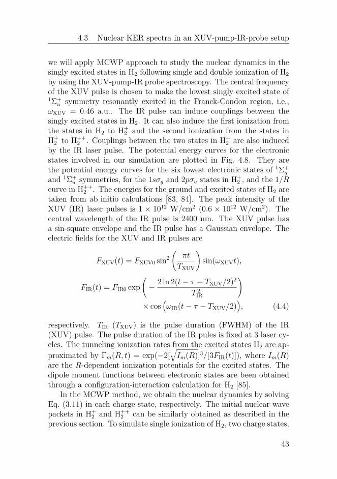
2 , the repulsion energy between two pro-tons is 1/R. The potential energy curves for the states involved in thesimulation are plotted in Fig. 4.1. The data for the potential energycurves for the electronic states in H2 and H+
2 are taken from Ref. [71].The dipole moment functions between the 1sσg and 2pσu states aretaken from Ref. [72]. The tunneling ionization rates from the groundstate in H2 are obtained by using the weak field asymptotic theory[73, 74]. The tunneling ionization rates from the two states in H+
2 areobtained by interpolation and extrapolation of the rates available inRef. [75]. Charge-resonance enhanced ionization (CREI) in the 2pσustate in H+
2 is observed in Ref. [75], e.g, enhanced ionization at aroundR = 7 a.u. and R = 11 a.u. for a field strength of 0.04 a.u.. Accordingto Eq. (3.7), the initial states in H+
2 can be expressed (without nor-malization) as cg
√Γh(R, t)χg(R, t1)|g〉+cg
√Γh(R, t)χu(R, t1)|u〉. Here
Γh(R, t) is the time-dependent ionization rate from the ground statein H2, and |g〉 and |u〉 denotes the 1sσg and 2pσu states. cg and cu aretake from Ref. [76]. The initial nuclear wave packet in H++
2 after thesecond jump can be easily obtained according to Eq. (3.16) by takingj = 1 since there is only one state in H++
2 .
Influences of laser parametersIn this section, we will investigate the influence of laser parameters onthe nuclear KER spectra following double ionization of H2. The exter-nal laser field has a Gaussian envelope and its electric field componentis
F (t) = FIR0 exp(− 2 ln 2(t− tc)2
T 2IR
)× cos
(ωIR(t− tc)
),
33

4. Dissociative single or double ionization of H2
where FIR0 is the peak field strength, TIR is the pulse duration (FWHM)and tc = 1.5TIR. We present the nuclear KER spectra for laser pulsesat four different wavelengths, i.e., 800, 1600, 3200 and 6400 nm, inFig. 4.2. The pulse duration TIR for the four laser pulses is fixed at64 fs and the peak intensity of these pulses is 6× 10¹³ W/cm2 (corre-sponding to a field strength of FIR0 = 0.0413). For all the wavelengths,we observe signal peaks at similar KER positions, i.e., KER ≈ 10 and4 eV. From the simple reflection principle, i.e., KER ≈ 1/R, we canrelate the formation of the peaks around these two KER values in thenuclear KER spectra to nuclear motion as follows:
1. the signals peaks at around 10 eV are mainly from the nuclearwave packets at around R = 27.2/10 = 2.72 a.u.. This positionis close to the outer turning point of the nuclear wave packetsevolving along the 1sσg curve in H+
2 when the initial nuclear wavepacket centers at around 1.4 a.u..
2. the signals peaks at around 4 eV are mainly from the nuclearwave packets at around R = 27.2/4 = 6.8 a.u., which is close tothe first CREI position at around R = 7 a.u. in the 2pσu statefor the considered peak intensity.
0
0.2
0.4
0.6
0.8
1
0 2 4 6 8 10 12 14
signal
KER (eV)
800 nm
1600 nm
3200 nm
6400 nm
Figure 4.2: Nuclear KER spectra following double ionization of H2interacting with laser pulses at four different wavelengths, i.e., 800,1600, 3200 and 6400 nm. The pulse duration (FWHM) is fixed at 64fs and the peak intensity is fixed at 6× 10¹³ W/cm2.
34
4.2. Nuclear KER spectra at near- and mid-IR wavelengths
We can also see from Fig. 4.2 that the relative intensities of the peaksat around 10 eV slightly increases when the wavelength is increasedfrom 800 to 6400 nm. This feature can be understood by the fact thata smaller part of the nuclear wave packet along the 2pσu curve in H+
2can reach the first CREI position of R = 7 for a longer wavelength.The one-photon resonance position between the 1sσg and 2pσu statesin H+
2 is larger for a longer wavelength. It is beyond the outer turningpoint of the 1sσg curve at around R = 3 for the infrared wavelength. Itis more unlikely for the nuclear wave packet along 1sσg curve reachingthe one-photon resonance position for a longer wavelength. Thus, for alonger wavelength, there is a smaller part of nuclear wave packet alongthe 2pσu curve at around R = 7, which comes from couplings between1sσg and 2pσu states in H+
2 at the one-photon resonance position.
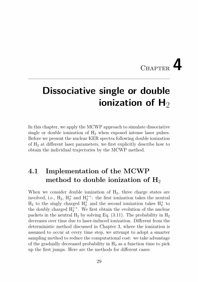
Figure 4.3: Results obtained by conducting a trajectory analysis tothe peaks at around 10 and 4 eV for the 1600 nm case in Fig. 4.2.Contributions from the individual trajectories at different first andsecond jump times, i.e, t1 and t2, to the peaks at around 10 eV (a) and4 eV (c). Transformation of (a) and (c) to (b) and (d), respectively,by replacing the t2 axis with a new t2 − t1 axis.
One unique possibility of the MCWP approach is to carry out atrajectory analysis to obtain the origin of the features of interest in the
35

4. Dissociative single or double ionization of H2
nuclear KER spectra. A trajectory analysis to a given feature in thenuclear KERR spectrum can tell us information about the dominanttrajectories including the first and second jump times, i.e., t1 and t2,and the states where the second jump takes place. In the following,we will conduct a trajectory analysis to the peaks at around 10 and4 eV for the 1600 nm case in Fig. 4.2. We show the contributions(probabilities) of the individual trajectories, whose second jumps takeplace from the 2pσu state in H+
2 , to the two peaks in Figs. 4.3(a) and(c), respectively. The much smaller contributions of the trajectorieswhose second jumps take place from the 1sσg state in H+
2 are notpresented here. In Figs. 4.3(a) and (c), the probabilities from thetrajectories are distributed along titled parallel lines, each of whichcorresponds to an identical evolution time in H+
2 , i.e., t2 − t1.The initial nuclear wave packets in H+
2 after the first jumps arevery similar in spite of different first jump times. This fact results inthat the nuclear dynamics in H+
2 is mainly determined by the evolutiontime in H+
2 . Therefore, we make a transformation of Figs. 4.3(a) and(c) to Figs. 4.3(b) and (d), respectively, by replacing the t2 axis witht2−t1. From Figs. 4.3(a) and (c), we can see that for the trajectories ata given t1, their probabilities oscillate as a function of t2 with a periodof half a laser cycle. The large (small) probability for a given trajectoryoriginates from that its second jump taking place at instants when theinstantaneous field strength is large (small) due to large (small) laser-induced ionization rates. For the trajectories at a given t2, we do notobserve a similar oscillatory behavior of the probabilities as a functiont1 since the first ionization is assumed to only take place at the instantsfor the field extrema.
It is clearly shown in Figs. 4.3(b) and (d) that the evolution timein H+
2 for the most dominant trajectories are around t2− t1 = 300 and1200 a.u. for the peak at 10 eV and around t2 − t1 = 1000 a.u. forthe peak at 4 eV. From the reflection principle, we aware that the twopeaks at 10 and 4 eV mainly comes from trajectories whose nuclearwave packets evolving along the 2pσu curve in H+
2 are at around R = 3and R = 7 when the second jumps occur. Thus, it takes the nuclearwave packet in H+
2 about 300 a.u. to reach the internuclear positionsof the outer turning point at around R = 3 for the first time and about1200 a.u. to reach the internuclear positions at around R = 3 for thesecond time. The difference between them, i.e., 1200− 300 = 900 a.u.,
36

4.2. Nuclear KER spectra at near- and mid-IR wavelengths
reflects that the vibrational period of the nuclear wave packet alongthe 1sσg state in H+
2 is around 900 a.u. = 22 fs. For the peak at 4 eV,it takes about 1000 a.u. for the nuclear wave packet to reach the firstCREI region at around R = 7.
We can read from Figs. 4.3(a) and (c) the information about themost dominant trajectory for the peak at 4 eV: the first jump occurs att1 = 3806 a.u. and the second jump occurs at t2 = 4719 a.u.. For thepeak at 10 eV, the information about the most dominant trajectoryis that the first jump takes place at t1 = 3938 a.u. and the secondjump at t2 = 4231 a.u.. The difference of the first times for these twotrajectories is half a laser cycle. We present in Figs. 4.4(a) and (b) theevolution of the nuclear wave packet along the 1sσg and 2pσu curves inH+
2 when the first jump takes place at t1 = 3806 a.u.. In addition, weshow in Figs. 4.4(c) and (d) the initial nuclear wave packets (withoutnormalization) in H++
2 for the second jumps taking place from the σgand 2pσu curves when the first jump occur at t1 = 3806 a.u.. We cansee that the probabilities of the initial nuclear wave packets for thetrajectories whose second jumps take place from the 2pσu state areseveral order larger than that for the trajectories whose second jumpstake place from the 1sσg state.
We can clearly see from Fig. 4.4(a) that the vibrational periodof the nuclear wave packet evolving along the 1sσg curve is around900 a.u. = 22 fs, which agrees very well to the result obtained fromthe above trajectory analysis. We can also see from Fig. 4.4(b) thenuclear wave packet evolving along the 2pσu curve can reach aroundR = 7 at t2 = 4719 a.u.. Due to the CREI at around R = 7, thelarge probabilities of the initial nuclear wave packet at around R = 7in H++
2 can be observed at t2 = 4719 a.u. in Fig. 4.4(d). It is thelarge signals at around R = 7 in Fig. 4.4(d) that eventually result inthe appearance of the peak at around 4 eV in the considered nuclearKER spectrum. The signals at around R = 3 in Fig. 4.4(d) leads tothe peak at around 10 eV in the nuclear KER spectrum.
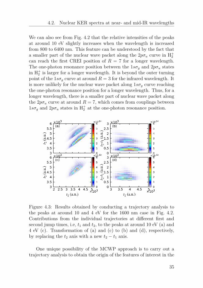
We will investigate the influence of the pulse duration of the ap-plied laser pulse on the nuclear KER spectra. We show the results inFig. 4.5 for two wavelengths of 1600 nm and 6400 nm. A systematicalstudy of the dependence on the duration for several wavelengths hasbeen performed in a recent article [60]. From Fig. 4.5, we can observethat the nuclear KER spectrum moves to smaller KER values when
37

4. Dissociative single or double ionization of H2
Figure 4.4: Evolution of the nuclear wave packets along the 1sσg (a)and 2pσu (b) curves in H+
2 when the first jump occurs at t1 = 3806a.u.. The initial nuclear wave packets in H++
2 for the second jumpstaking place from the 1sσg (c) and 2pσu (d) curves at varying secondjump times when the first jump occurs at t1 = 3806 a.u..
the pulse duration is increased. A larger pulse duration can induce theionization from H+
2 at larger internuclear distances when the nuclearwave packet evolves along the 2pσu curve in H+
2 . The ionization fromH+
2 at larger internuclear distances would result in signals at smallerKER values. We can also observe several peaks at similar KER posi-tions in the nuclear KER spectra at the same wavelength in Fig. 4.5.The KER of H++
2 is made up of two parts: the kinetic energy obtainedin H+
2 and the Coulomb repulsion energy between the two protons. Thekinetic energy obtained along the 2pσu curve in H+
2 is determined bythe position for the one- or three- photon resonance between the twostates in H+
2 and the position where the second jump takes place. Theinternuclear position for the one- or three-photon resonances betweenthe two states in H+
2 is only dependent on the wavelength. For H2interacting with relatively long pulses, there is a large probability thatthe second jumps take places at the CREI regions. The trajectorieswhose second jumps take place from the CREI regions would result in
38
4.2. Nuclear KER spectra at near- and mid-IR wavelengths
0
0.2
0.4
0.6
0.8
1
0 2 4 6 8 10 12 14
(b)
signal
KER (eV)
3 cycles 7 cycles11 cycles15 cycles19 cycles
0
0.2
0.4
0.6
0.8
1(a)
sign
al
Figure 4.5: Nuclear KER spectra following double ionization of H2when exposed to laser pulses at (a) 1600 nm and (b) 6400 nm fordifferent pulse durations. The pulse duration (FWHM) is expressedby the number of laser cycles. The peak intensity is fixed at 6 × 10¹³
W/cm2.
signals at similar KER positions for the pulses at the same wavelength,since both the kinetic energy obtained in H+
2 and the Coulomb repul-sion energy between the two protons are similar. The peaks at verylow KER values, i.e., about 1.5 eV, in Fig. 4.5 results from the trajec-tories whose the evolution time in H+
2 is sufficient long so that largeionization at internuclear distances beyond the CREI region of R = 11can take place. A trajectory analysis of this peak has been conductedin Ref. [60]. The peaks at around and 10 eV have similar origins aswe discussed before. For the 6400 nm case, the peaks at around 3eV are from trajectories whose nuclear wave packets in H+
2 reach thesecond CREI position at around R = 11 a.u. for the considered peakintensity.
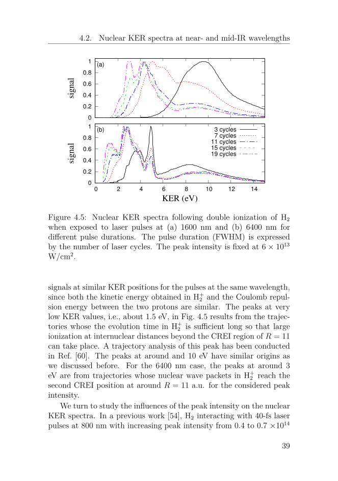
We turn to study the influences of the peak intensity on the nuclearKER spectra. In a previous work [54], H2 interacting with 40-fs laserpulses at 800 nm with increasing peak intensity from 0.4 to 0.7 ×10¹⁴
39
4. Dissociative single or double ionization of H2
0
0.2
0.4
0.6
0.8
1
0 2 4 6 8 10 12 14
(b)
signal
KER (eV)
0.60.91.21.51.8
0
0.2
0.4
0.6
0.8
1(a)
sign
al
Figure 4.6: Nuclear KER spectra following double ionization of H2when exposed to laser pulses at (a) 800 nm and (b) 3200 nm withthe peak intensities ranging from 0.6 to 1.8×10¹⁴ W/cm2. The pulsedurations (FWHM) are 40 fs for the 800 nm pulses and 53 fs for the3200 nm pulses, respectively.
W/cm2 was studied by the MCWP approach. Now we extend the studyof H2 interacting with pulses at much larger peak intensities for the 800nm wavelength and with pulses at a mid-IR wavelength of 3200 nm.The pulse durations for the 800 nm case is 15 laser cycles (40 fs) andfor the 3200 nm case, it is 5 laser cycles (40 fs). In Ref. [54], the nuclearKER spectra move to smaller KER values when the peak intensitiesare increased from 0.4 to 0.7 ×10¹⁴ W/cm2. The nuclear KER spectra,however, moves oppositely, i.e., to larger KER values, when we increasethe peak intensities from 0.9 to 1.8 ×10¹⁴ W/cm2 in Fig. 4.6(a). Thisalso applies to the mid-IR wavelength in Fig. 4.6(b). The shift of thenuclear KER spectra to smaller KER values in Ref. [54] is ascribedto the fact that an increase of the peak intensity of a laser pulse isequivalent to an increase of the pulse duration. This is true when theapplied laser field is relatively weak, i.e., the loss of the probability inH+
2 continues gradually until the end of the laser pulse. However, when
40

4.2. Nuclear KER spectra at near- and mid-IR wavelengths
the peak intensity is sufficiently large, the most loss of the probabilityin H+
2 takes place within a short time. Thus, for a larger peak intensity,the nuclear wave packet in H+
2 reaches smaller internuclear distanceswhen the second ionization occurs, which results in signals at largerKER values, see Fig. 4.6. A similar shift of the nuclear KER spectrato slightly larger KER values was observed when the peak intensitywas increased from 1 to 5 ×10¹⁴ W/cm2 in an experiment where D2interacting with 100-fs pulses at 600 nm was studied [77].
Influences of nuclear mass
0
0.2
0.4
0.6
0.8
1
0 2 4 6 8 10 12 14
signal
KER (eV)
H2, 5 cyclesH2, 10 cyclesH2, 21 cyclesD2, 5 cyclesD2, 10 cyclesD2, 21 cycles
Figure 4.7: Nuclear KER spectra following double ionization of H2 andD2 interacting with laser pulses at 3200 nm for three pulse durations,i.e., 5, 10 and 21 cycles. The peak intensity is fixed at 0.6×10¹⁴ W/cm2.
In addition to the laser parameters, the nuclear mass has an effecton the nuclear dynamics. Generally speaking, the heavier the nucleiare, the more slowly they can move, and the slower the nuclear dynam-ics is. We will study the isotope effect on the nuclear KER spectra.The isotope effect has been studied at near-IR wavelengths [57] and wewill extend the study to mid-IR wavelengths. We present the nuclearKER spectra following double ionization of H2 and D2 at a mid-IRwavelength of 3200 nm in Fig. 4.7 for three different pulse durations,i.e., 5, 10 and 21 cycles. At a given pules duration, the nuclear KERspectrum for D2 moves to larger KER values compared to that for H2.The shifts are more apparent for shorter pulse durations. The signals
41

4. Dissociative single or double ionization of H2
at around 3 and 4 eV in Fig. 4.7 mainly come from trajectories whosesecond jumps occur the two CREI regions at R = 7 and R = 11,respectively.
4.3 Nuclear KER spectra in anXUV-pump-IR-probe setup
−35
−30
−25
−20
−15
−10
−5
0
5
10
0 2 4 6 8 10 12 14
pump (XUV)probe (IR)
E (
eV
)
R (a.u.)
EF1Σg
+
GK1Σg
+
H−
H1Σg
+
P1Σg
+
Q1Σg
+
B1Σu
+
B’1
Σu+
B31Σu
+
B41Σu
+
B51Σu
+
B61Σu
+
H(1s)+H(1s)
H(1s)+p
p+p
H(1s)+H*
(1sσg)2
1sσg
2pσu
1/R
Figure 4.8: Potential energy curves for the electronic states as a func-tion of R. They are the curves for the six lowest states of 1Σ+
g and 1Σ+u
symmetries in H2, the curves for the ground and first excited states inH+
2 and the 1/R Coulomb in H++2 , respectively.
In the former section, we studied the interaction of H2 with singleIR laser pulses. Double ionization of H2 (D2) by laser pulses in a pump-probe setup has been studied by many works [60, 78, 79]. The pumppulse is applied to initiate the dynamics in molecules and the probepulse is applied to take the snapshots of the dynamics in real time.The XUV-pump-IR-probe setup has been applied to study nucleardynamics in the singly excited states in H2 by obtaining the nuclearKER spectra following single ionization of H2 [80–82]. In this section,
42
4.3. Nuclear KER spectra in an XUV-pump-IR-probe setup
we will apply MCWP approach to study the nuclear dynamics in thesingly excited states in H2 following single and double ionization of H2by using the XUV-pump-IR probe spectroscopy. The central frequencyof the XUV pulse is chosen to make the lowest singly excited state of1Σ+
u symmetry resonantly excited in the Franck-Condon region, i.e.,ωXUV = 0.46 a.u.. The IR pulse can induce couplings between thesingly excited states in H2. It can also induce the first ionization fromthe states in H2 to H+
2 and the second ionization from the states inH+
2 to H++2 . Couplings between the two states in H+
2 are also inducedby the IR laser pulse. The potential energy curves for the electronicstates involved in our simulation are plotted in Fig. 4.8. They arethe potential energy curves for the six lowest electronic states of 1Σ+
g
and 1Σ+u symmetries, for the 1sσg and 2pσu states in H+
2 , and the 1/Rcurve in H++
2 . The energies for the ground and excited states of H2 aretaken from ab initio calculations [83, 84]. The peak intensity of theXUV (IR) laser pulses is 1 × 10¹² W/cm2 (0.6 × 10¹² W/cm2). Thecentral wavelength of the IR pulse is 2400 nm. The XUV pulse hasa sin-square envelope and the IR pulse has a Gaussian envelope. Theelectric fields for the XUV and IR pulses are
FXUV(t) = FXUV0 sin2(
πt
TXUV
)sin(ωXUVt),
FIR(t) = FIR0 exp(− 2 ln 2(t− τ − TXUV/2)2
T 2IR
)× cos
(ωIR(t− τ − TXUV/2)
), (4.4)
respectively. TIR (TXUV) is the pulse duration (FWHM) of the IR(XUV) pulse. The pulse duration of the IR pules is fixed at 3 laser cy-cles. The tunneling ionization rates from the excited states H2 are ap-proximated by Γm(R, t) = exp(−2[
√Im(R)]3/[3FIR(t)]), where Im(R)
are the R-dependent ionization potentials for the excited states. Thedipole moment functions between electronic states are been obtainedthrough a configuration-interaction calculation for H2 [85].
In the MCWP method, we obtain the nuclear dynamics by solvingEq. (3.11) in each charge state, respectively. The initial nuclear wavepackets in H+
2 and H++2 can be similarly obtained as described in the
previous section. To simulate single ionization of H2, two charge states,
43

4. Dissociative single or double ionization of H2
Figure 4.9: Nuclear KER spectra following single ionization of H2 asa function of delay when an XUV-pump-IR-probe setup is applied.Contributions from the nuclear wave packets along the 1sσg (a) and2pσu (b) curves. The pulse duration of the XUV pulse is 3 cycles.
i.e., H2 and H+2 , are involved. Since there are 12 electronic states in H2
involved in our calculation, the matrix ofM(R) and V (R) in the smalltime propagator for H2 are 12 by 12. We will present the nuclear KERspectra as a function a delay following single and double ionizationof H2 by applying the second sampling method in Sec. 4.1 to gatherthe first jumps. The second jumps are assumed to take place at everyseveral time steps when we simulate double ionization of H2.
Single ionization channel
For single ionization of H2, a trajectory is specified by its first jumptime t1 and the state from which the first jump takes place. Thereare 12 first jump pathways. By projecting the nuclear wave packetsχm(R, te) (with m = g, u) at the end of the IR laser pulse along the1sσg and 2pσu curves in H+
2 for a given trajectory on the correspondingeigenstates χEm(R) for the two curves, one can obtain the nuclear KER
44

4.3. Nuclear KER spectra in an XUV-pump-IR-probe setup
spectrum of H+2 for that trajectory, i.e.,
pEm = |∫dRχEm(R)χm(R, t2)|2. (4.5)
with m = g, u. The total nuclear KER spectrum can be obtained bysumming over the nuclear KER spectra for all the individual trajecto-ries in H+
2 , i.e.,
PE(k, t1) =∑k,t1
P1(t1)P1k∑
m=g,upEm, (4.6)
where P1(t1) is the total jump probability for the first jumps occur-ring at t1 and P1k are the relative probabilities for different ionizationpathways. We should note that the first jump probability P1(t1) relieson the sampling method.
We show in Fig. 4.9 the nuclear KER spectra following single ion-ization of H2 as a function of delay when an XUV-pump-IR-probesetup is applied. The pulse duration of the applied XUV pulse is 3cycles. After absorption of a photon from the XUV pulse, the nuclearwave packets along the singly excited states of 1Σ+
u symmetry in H2 arecreated. The IR pulse couples the excited states together since theyare close with each other. The nuclear wave packets can move to largeinternuclear distances along the potential energy curves for the excitedstates in H2. For the nuclear wave packets at large internuclear dis-tances, the initial state in H+
2 following the first ionization would notprefer to one of two states in H+
2 since they are almost degenerate atlarge internuclear positions, i.e., |cg|2 ≈ |cu|2. Different from the dou-ble ionization channel, for the single ionization channel, contributionsto the nuclear KER spectra from the nuclear wave packets along the1sσg curve are comparable to that from the 2pσu curve [Figs. 4.9(a)and (b)].
From Fig. 4.9, we can see a periodical enhancement of the signalsat KER values smaller than 0.5 eV. This period is around 30 fs, whichresults from the vibrational motion of the nuclear wave packets alongthe singly-excited states in H2. There are several energy decreasingbranches at delays smaller than 40 fs at Figs. 4.9(a) and (b), showingthat the nuclear wave packets in the excited states move to largerinternuclear distances.
45

4. Dissociative single or double ionization of H2
Double ionization channel
×10−5
×10−4
×10−4
delay τ (fs)
KER
(eV)
ratio
0 20 40 60 80 100 120 1400.00
0.05
0.10
0.20
2
4
6
8
2
4
6
8
2
4
6
8
1 cycle
3 cycles
5 cycles
0.00.51.01.52.02.5
0.00.30.60.91.21.5
0.00.81.62.43.24.01 cycle (a)
3 cycles (b)
5 cycles (c)
(d)
Figure 4.10: Results taken from Ref. [61]: nuclear KER spectra fol-lowing double ionization of H2 within the XUV-pump-IR-probe setupwhen the XUV pulses are 1 (a), 3 (b) and 5 (c) cycles, respectively.(d) Ratios between the signals on the energy decreasing branches andthe total yield for the three XUV durations as a function of delay.
The MCWP approach is particularly useful in simulating multi-ple ionization process. In this section, the nuclear KER spectra as afunction of delay following double ionization of H2 are presented inFig. 4.10. The results are for three XUV pulse durations, which are
46
4.4. Nuclear KER spectra when autoionization from doubly excitedstates is involved
taken from our recent work [61]. We will summarize the main featuresin Fig. 4.10 and their origins in the following:
1. enhancements of the signals at KER values around 4 eV appear-ing at delays around 40, 70 and 100 fs. A detailed analysis inRef. [61] shows us that this 30-fs period corresponds to the vi-brational period of the nuclear wave packet evolving along thepotential energy curve for the B1Σ+
u state in H2.
2. periodical enhancements of the signals appearing at delays smallerthan 30 fs. This period is half an IR laser cycle. The structurebecomes clearer for larger XUV pulse durations and it originatesfrom the AC Stark effect. When the IR field is strong (weak) atthe overlap between the ultrashort XUV pulse and the IR pulse,the Stark shift of the B+Σ+
u state is large (small). Thus, the ex-citation from the ground state to the B+Σ+
u state in H2 is small(large) because the two states are off-resonant (resonant) withthe XUV pulse.
3. an energy decreasing branch moving from around 2 to 1 eV fordelays larger than 50 fs. The signals in this branch come from thenuclear wave packet reaching internuclear distances larger thanR = 15 a.u.. The nuclear wave packets at such large internucleardistances come from the dissociating part of the nuclear wavepacket along the potential energy curve for the B31Σ+
u state. Thisbranch becomes relatively less important for a larger XUV pulseduration, which results from that there is a relatively smallerpopulation transfer from the ground state to the B31Σ+
u statewhen the XUV pulse is larger.
4.4 Nuclear KER spectra whenautoionization from doubly excitedstates is involved
In the previous section, we investigated the nuclear dynamics when thesingly excited states in H2 are involved. Now we will study the influ-ences of the the doubly excited states in H2 on the nuclear dynamics.
47

4. Dissociative single or double ionization of H2
We still take the example of the simplest molecule of H2. A doublyexcited state is a state where two electrons in the system are excited si-multaneously. The doubly excited states in atoms stay beyond the firstionization threshold. In molecules, however, the doubly excited statesmay be below the first ionization threshold at certain internuclear dis-tance regions. The singly excited states in atoms and molecules candecay to the more stable states via the radiation or non-radiation re-laxation processes. In addition, vibrational autoionization [86, 87] maytake place from the very highly excited states in molecules by convert-ing the vibrational energy of the ion core to the kinetic energy of theoutgoing electron. The doubly excited states in atoms and moleculescan decay to more stable states by electron emission. This autoion-ization process may be a dominant relaxation process since it occursmuch faster than the radiation or non-radiation relaxation processes.
The evidences of doubly excited states in the hydrogen moleculeswere provided by electron impact experiments [88, 89]. The doubly ex-cited states can be also reached by absorption of one energetic photonfrom the external field [90, 91]. The doubly excited states in moleculescan not only autoionize but also dissociate into two neutral nuclei. Itwas the doubly excited states in H2 that resulted in the comparativelylarge ratio between H+ and H+
2 at the 26 eV photon energy [92]. Abinitio calculations have been conducted to obtain the potential energycurves and the corresponding autoionization rates for the doubly ex-cited states [93–96]. There are series of doubly excited states. Forexample, the Q1 series are the doubly excited states lying betweenthe first single-ionization threshold and the second single-ionizationthreshold and theQ2 series are made up of the doubly excited states be-yond the second single-ionization threshold and below the third single-ionization threshold.
In this thesis, we apply the MCWP approach to study the influenceof doubly excited states on the nuclear KER spectra of H+
2 followingphotoionization of H2 interacting with intense XUV pulses. The initialstate is the ground state in H2, i.e., X1Σ+
g (v = 0). Within the Frank-Condon region, the ionization potentials for the ground state in H2to the first and second single-ionization thresholds, i.e., the 1sσg and2pσu states in H+
2 , are around 18 and 30 eV, respectively. We show thepotential energy curves for the above mentioned states in Fig. 4.11.After absorbing one photon with an energy larger than 18 eV and
48
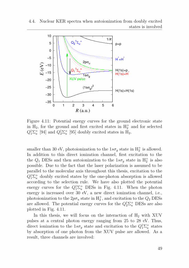
4.4. Nuclear KER spectra when autoionization from doubly excitedstates is involved
−35
−30
−25
−20
−15
−10
−5
0
5
10
0 1 2 3 4 5 6
XUV pulse
E (
eV
)
R (a.u.)
H(1s)+H(1s)
H(1s)+p
p+p
H*+H
*
H(1s)+H*
(1sσg)2
1sσg
2pσu
Q11Σu
+
Q21Σu
+1/R
Figure 4.11: Potential energy curves for the ground electronic statein H2, for the ground and first excited states in H+
2 and for selectedQ1
1Σ+u [94] and Q1
2Σ+u [95] doubly excited states in H2.
smaller than 30 eV, photoionization to the 1sσg state in H+2 is allowed.
In addition to this direct ionization channel, first excitation to thethe Q1 DESs and then autoionization to the 1sσg state in H+
2 is alsopossible. Due to the fact that the laser polarization is assumed to beparallel to the molecular axis throughout this thesis, excitation to theQ1
1Σ+u doubly excited states by the one-photon absorption is allowed
according to the selection rule. We have also plotted the potentialenergy curves for the Q1
1Σ+u DESs in Fig. 4.11. When the photon
energy is increased over 30 eV, a new direct ionization channel, i.e.,photoionization to the 2pσu state in H+
2 , and excitation to the Q2 DESsare allowed. The potential energy curves for the Q1
2Σ+u DESs are also
plotted in Fig. 4.11.In this thesis, we will focus on the interaction of H2 with XUV
pulses at a central photon energy ranging from 25 to 28 eV. Thus,direct ionization to the 1sσg state and excitation to the Q1
1Σ+u states
by absorption of one photon from the XUV pulse are allowed. As aresult, three channels are involved:
49

4. Dissociative single or double ionization of H2
• H2+~ω → H+2 (1sσg) + e−, direct ionization
• H2+~ω → H∗∗2 (Q1) → H+2 (1sσg) + e−, autoionization
• H2+~ω → H∗∗2 (Q1) → H(1s)+H∗, dissociation
We can see from Fig. 4.11 that the potential energy curves for the Q1DESs are repulsive and they cross with the potential energy curve forthe 1sσg state in H2 at certain critical internuclear positions. The nu-clear wave packets along the potential energy curves for the Q1 DESsmove very fast. Autoionization from the DESs takes place at inter-nuclear distances left to the crossing points. If there are nuclear wavepackets evolving along the potential energy curves for the Q1 DESs be-yond the crossing points, dissociation of H2 into two neutral H atoms,i.e., one is in the ground state and the other is excited, is allowed.
0
0.2
0.4
0.6
0.8
1
1.2
1.4
−2 0 2 4 6 8 10
signal
KER (eV)
25 eV
26 eV
27 eV
28 eV
Figure 4.12: Nuclear KER spectra following single ionization of H2when interacting with XUV pulses at different photon energies. Thesignals at negative energies corresponds to the vibrational distributionfor H2+ and the signals at positive energies to the proton kinetic energydistribution.
As we see in Fig. 4.11, the Q11Σ+
u states are very close to each other,the one-photon absorption would result in a coherent superposition ofthese states. The more states we include in our simulation, the moreaccurate results we would obtain. The following results, however, areobtained by only including the Q1
1Σ+u (1) state. This simplification
would greatly reduce our computational costs and it is a reasonable
50
4.4. Nuclear KER spectra when autoionization from doubly excitedstates is involved
approximation since these Q11Σ+
u states are close to each other at theFranck-Condon region; the dipole coupling strengths from the groundstate in H2 to the Q1
1Σ+u (1) state are several times larger than that for
the Q11Σ+
u (2) state [97]. It is also shown in Ref. [94] that the autoion-ization widths of the Q1
1Σ+u (1) state are several times larger than that
for the Q11Σ+
u (2) state. Thus, the influence of the Q11Σ+
u (1) state onthe nuclear KER spectra would be dominant.
We show in Fig. 4.12 the nuclear KER spectra following single ion-ization of H2 interacting with a 10 fs XUV pulses at four central photonenergies, i.e., 25, 26, 27 and 28 eV. These spectra are obtained by sum-ming the spectra for the individual trajectories. The spectrum for eachtrajectory is calculated according to Eq. (4.5), i.e., by projecting thenuclear wave packets along the 1sσg curve in H+
2 at the end of thepropagation time on the corresponding eigenstates for the 1sσg curve.We obtain the knowledge that the direct ionization cross section isabout one order of magnitude larger than the absorption cross sectionto the Q1
1Σ+u states for the considered photon energies [48]. The direct
ionization channel would mainly lead to bound H+2 signals at negative
energies in Fig. 4.12 and the relatively weak channel, i.e, autoioniza-tion from the DESs to the 1sσg state, can result in relatively large H+ p signals at relatively large KER values. This is because the nuclearwave packets gain kinetic energies through moving along the potentialenergy curves for the DESs. We will summarize the other requiredcalculation inputs in obtaining Fig. 4.12 in the following:
• The total photoionization cross section σp of H2 by photons atthe considered energies can be found in Ref. [48]. The photoion-ization rate thus can be obtained using the relation
Γp = σp × IXUV
ωXUV, (4.7)
where IXUV is the intensity of the XUV pulse.
• assuming the first jumps occurring at every 40 a.u..
Now let us have a more careful look at Fig. 4.12. For better com-parison, the amplitudes for the signals at -2.11 eV are rescaled to 1for the four cases. The signals at negative energies corresponds to thevibrational distribution for H2+ and the signals at positive energies
51
4. Dissociative single or double ionization of H2
corresponds to the proton kinetic energy distribution. When we in-crease the photon energy from 25 to 28 eV, the spectra at negativeenergies look the same. This similarity implies that the signals at neg-ative KER values are mainly from the direct ionization channel, wherethe photon energy only influences the absolute ionization cross sec-tion from H2 to H+
2 and has nearly no effect on initial the nuclear wavepackets in H+
2 after direct ionization. The spectra are quite different atpositive KER values. First, there are relatively more and more signalswhen the photon energy is increased. Second, the spectra are extendedto larger and larger KER values when the photon energy is increased.For example, when the XUV photon energy is 25 eV, a cutoff of 8 eVof spectrum is observed and when the XUV photon energy is 26 eV,we see a larger cutoff of 10 eV. The increase of the cutoff energy canbe understood by the fact that a higher photon energy means a largerenergy shared by the nuclei and autoionizing electron. The signals atpositive energies become relatively larger for larger photon energiesdue to the fact that the direct ionization cross section is reduced fora larger photon energy and the photon energy becomes closer to theresonant photon energy within the Franck-Condon region. In addition,there is a slight shift of the peak positions to larger energies when thephoton energy is increased: the peak for the 26 eV is at around 5 eVand for the 27 eV case at around 5.8 eV and for the 28 eV case ataround 6.5 eV.
Next we will compare our simulation results with the experimentalresults in Ref. [98] and with the simulation results in Ref. [99]. Similarwith the experimental results, there are relatively more dissociationsignals and the spectra tend to become broader for larger photon en-ergies and there are more structures at positive kinetic energies. Wenote, however, that our simulation results do not agree with the ex-periment [98] as well as the simulation in Ref. [99]. The discrepanciesmainly emerge at the positive kinetic energies. One discrepancy is thatwe have a longer tail at relatively large energies. This is because partof the the nuclear wave packets along the Q1
1Σ+u (1) curve would evolve
to internuclear distances larger than the cross point between this curveand the 1sσg curve and thus autoionization taking place near the crosspoint would result in a large KER value, which is indeed the cutoffenergy in our spectra. In fact, the cutoff energy can be obtained byusing the energy conservation, i.e., ωXUV = Ee + KER + IP + 1.5 eV,
52

4.4. Nuclear KER spectra when autoionization from doubly excitedstates is involved
where Ee is the energy for the autoionizing electron and IP is the ion-ization potential to the 1sσg state in H+
2 within the Franck-Condonregion. For example, the cutoff energy for the 25 eV case is aroundKER = 25−18.5 = 6.5 eV. While the experiment gave a smaller cutoffof at around 4 eV at this photon energy. There are also differencesfor the peak positions, e.g., we can not reproduce the peaks at around1 eV by our spectra, which may come from a underestimate of thecontributions from the DES. Further efforts should be taken to resolvethe discrepancies.
Figure 4.13: Evolution of the nuclear wave packets along the potentialenergy curve for the Q1Σ+
u (1) state when interacting with laser pulsesat 25 eV (a) and 28 eV (c). The probability as a function of timestaying in H2 for 25 eV (b) and 28 eV (d). In both (b) and (d), thesolid (black) curve is for the probability in H2 and the dashed (red)curve for that in the ground state in H2.
In Fig. 4.13, we show the evolution of the nuclear wave packetsalong the Q1
1Σ+u state for two cases, i.e., ωXUV = 25 and 28 eV. For
the both two cases, the nuclear wave packets move very quickly andreach beyond the cross point at around R = 4 in 400 a.u. (10 fs).The XUV pulse is as short as 10 fs, thus when t is larger than 400a.u., no excitation from the ground state to the Q1
1Σ+u state and no
53

4. Dissociative single or double ionization of H2
autoionization to the 1sσg state in H+2 . We also show in the left panel
the probability of staying in H2 and in the ground state of H2 as afunction of time for the two cases. For both two cases, the two curvesare very close to each other, which means that the direct ionizationdominants the ionization process. For the 28 eV case, there is visibledifference between the two probabilities and thus the autoionizationfrom the DES plays a more important role. This is consistent withour findings in Fig. 4.12 that there are more signals at positive KERvalues for larger photon energies.
0
0.2
0.4
0.6
0.8
1
0 5 10 15 20 25
signal
electron energy (eV)
25 eV
26 eV
27 eV
28 eV
Figure 4.14: Autoionizing electron spectra following single ionizationof H2 interacting with XUV pulses at different photon energies.
Apart from the nuclear KER spectra, we can obtain the kineticenergy spectra for the autoionizing electrons. We show the results forthe above mentioned four cases in Fig. 4.14. The spectra are obtainedby simply using the fact that autoionization at a specific R along theQ1
1Σ+u curve would result in an electron whose kinetic energy is the
energy difference between the DES state and the 1sσg state at thatposition, i.e., Ee = EDES(R) − Eg(R). The probability to produce anelectron at a given R is directly related to the initial nuclear wavepackets created along the 1sσg curve following autoionization at t1,i.e., P (R) = ΓDES(R)|χDES(R, t1)|2. By summing the spectra for allthe trajectories, we can get the kinetic energy spectrum for the au-toionizing electron. From Fig. 4.14, we can see that the autoionizingelectron kinetic energy spectra tend to move to larger kinetic energiesfor larger photon energies. This feature is due to a larger energy is
54

4.5. Concluding remarks
shared by the autoionizing electron and the nuclei for a larger photonenergy. Thus, as we discussed before, it actually is consistent witha larger cutoff KER value of the nuclear KER spectra in Fig. 4.14.The peak at a given KER position demonstrate that the nuclear wavepackets along the Q1
1Σ+u curve are largely decayed to the 1sσg state at
specific internuclear distances. For example, the peak at 11 eV for the28 eV case corresponds to large autoionization at around R = 1.7 a.u.,while for the 25 eV case peak at 9 eV corresponds to large autoioniza-tion at around R = 1.9 a.u..
4.5 Concluding remarksIn this chapter, we have applied the MCWP method to study the inter-action of H2 with intense laser pulses including single IR femtosecondpulses, combined pulses in the XUV-pump-IR-probe setting and singleXUV pulses. When we consider the interaction of H2 with intense IRpulses, tunneling ionization from H2 to H+
2 and from H+2 to H++
2 cantake place. Thus, we have obtained the nuclear KER spectra follow-ing the full break-up of H2, since our method is especially powerful intreating the double or multiple ionization processes. We have studiedthe influences of the laser parameters as well as the nuclear mass on thenuclear dynamics in H2 when exposed to laser pulses at IR wavelength.When we consider the interaction of H2 with XUV pulses, relativelylarge single-photon ionization or excitation would be induced from theground state in H2 when the XUV photon energy is larger than theionization potential or is comparable with the energy difference be-tween the excited electronic states and the ground state in H2. Wehave obtained and analyzed the nuclear KER spectra following doubleionization of H2 to resolve the nuclear dynamics in the singly excitedstates in H2 by using XUV-pump-IR-probe spectroscopy. By tuningthe photon energy, photoionization may be accompanied by the ex-citation of double excited states. Compared with the singly excitedstates, (when the singly excited states are very close to the ionizationthresholds, vibrational autoionization [86, 87] may also take place inmolecules, but we will do not study this process in this thesis), thereare autoionization from DESs due to the electron correlation, i.e., theenergy of one electron is transfered to the other electron to remove the
55

4. Dissociative single or double ionization of H2
other electron from the molecule. We have studied the influences ofthe exciation of the Q1 DESs and the autoionization from the DESs inH2 to the nuclear KER spectra following single ionization of H2. Moreefforts should be put into the study of the influence of the DESs onthe nuclear dynamics in H2 to resolve the discrepancies between oursimulation and the experiment. Further studies can be conducted onobtaining the lifetimes of the DESs by using the pump-probe spec-troscopy.
56

Chapter 5Dissociative triple ionization of
Ne2
The previous chapter shows us the application of the MCWP approachto study the nuclear dynamics following ionization and excitation ofmolecules by intense laser pulses. In this chapter, we will turn to studythe ultrafast dynamics in a new system, i.e., weakly bounded clusters.In the field of atomic and molecular physics, clusters refer to a bunchof atoms or molecules which are loosely bounded by weak attractionforces, i.e., Van der Waals forces or hydrogen bonds, between them.The size of clusters ranges from 2 to millions of atoms or molecules.The bonds in forming clusters are comparably weak compared withionic or covalent bonds in molecules. Thus, clusters are not as stableas molecules and are prone to break up under external interferences.The interactions between the constituent atoms and molecules in clus-ters are either repulsive or attractive, depending on the internucleardistances and the electronic states. Thus, similar to molecules, follow-ing excitation or ionization by a strong external field, nuclear dynamicsare induced in weakly bounded clusters. Different from molecules, dueto the weak interactions between individual atoms or molecules in clus-ters, the double or higher ionization thresholds for clusters are loweredthan that for the constituent atoms or molecules. Thus, unique elec-tronic decay channels are opened for weakly bounded clusters, e.g.,the interatomic (intermolecular) Coulombic decay (ICD) and electrontransfer mediated decay (ETMD) [100].
57
5. Dissociative triple ionization of Ne2
Before we go detailed to ICD or ETMD, we first give a short intro-duction of a multi-electron effect called the Auger decay, named afterP. V. Auger who independently discovered this effect after L. Meitner’sfirst discovery. The Auger decay is a phenomenon that the filling ofa deep inner-shell vacancy, e.g., a core-shell vacancy, within an atomis accompanied by the emission of another electron. The Auger ef-fect has been widely investigated in atoms and molecules [101–103].The core-shell vacancy in an atom can be created by the incidenceof very highly energetic photons or electrons to atomic or molecularsamples. After the emission of a core-shell electron, the remaining ionis highly excited and unstable, and thus would relax to more stablestates through an emission of an energetic photon or electron. Thiselectronic decay is energetically allowed since the highly excited ionicstate is above the lowest double ionization thresholds for the atom ormolecule. The electronic and radiational decays are competitive andthe former one would be more favorable since it is much faster thanthe latter case, typically on a timescale of femtoseconds.
However, if a inner-shell electron is kicked out from an atom, itis energetically forbidden for the Auger decay to take place. This isbecause even though the remaining ionic state is still excited, it is nowbelow the lowest double ionization thresholds for the atom or molecule.It becomes fascinating when the atom has weakly bounded neighbors,i.e., these atoms or molecules have formed clusters. The energy re-leased from filling the inner-shell vacancy can not support to removean outer-valence electron from the same site but it can supply the emis-sion of an outer-valence electron at the neighboring sites via a virtualphoton. This process is called interatomic (intermolecular) Coulombicdecay (ICD) due to the fact that it is an interatomic (intermolecular)phenomenon and ICD is driven by the Coulombic interaction betweenthe two electrons. For the ICD processes, two resultant positivelycharged particles would repel with each other via the Coulomb forcebetween them. This electronic decay mechanism was first theoreticallypredicted by [104] and later experimental evidences were found in Vander Waals clusters [105–107] and hydrogen bonded clusters [108–110].The direct ICD process is shown in Fig. 5.1. The ICD occurs at atimescale from a few to hundreds of femtoseconds, depending on thesize and type of clusters. Thus in some clusters where the ICD life-times are comparable to the timescales for the nuclear dynamics, it
58
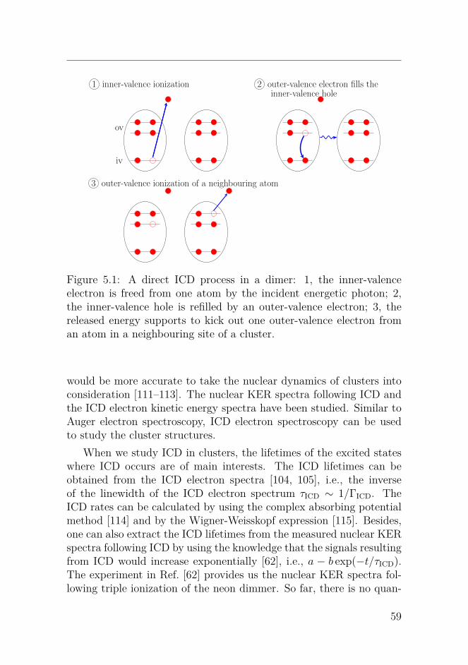
a b
1 inner-valence ionization
iv
ov
2 outer-valence electron fills theinner-valence hole
3 outer-valence ionization of a neighbouring atom
Figure 5.1: A direct ICD process in a dimer: 1, the inner-valenceelectron is freed from one atom by the incident energetic photon; 2,the inner-valence hole is refilled by an outer-valence electron; 3, thereleased energy supports to kick out one outer-valence electron froman atom in a neighbouring site of a cluster.
would be more accurate to take the nuclear dynamics of clusters intoconsideration [111–113]. The nuclear KER spectra following ICD andthe ICD electron kinetic energy spectra have been studied. Similar toAuger electron spectroscopy, ICD electron spectroscopy can be usedto study the cluster structures.
When we study ICD in clusters, the lifetimes of the excited stateswhere ICD occurs are of main interests. The ICD lifetimes can beobtained from the ICD electron spectra [104, 105], i.e., the inverseof the linewidth of the ICD electron spectrum τICD ∼ 1/ΓICD. TheICD rates can be calculated by using the complex absorbing potentialmethod [114] and by the Wigner-Weisskopf expression [115]. Besides,one can also extract the ICD lifetimes from the measured nuclear KERspectra following ICD by using the knowledge that the signals resultingfrom ICD would increase exponentially [62], i.e., a − b exp(−t/τICD).The experiment in Ref. [62] provides us the nuclear KER spectra fol-lowing triple ionization of the neon dimmer. So far, there is no quan-
59
5. Dissociative triple ionization of Ne2
tum theory applied to obtain the similar nuclear KER spectra as afunction of delays. Therefore, in this chapter, we will extend ourMCWP method to simulate this process, which is beyond the capacityof many theories, to help us to better understand the involved ICDprocesses in the neon dimmer. We will show in the following sectionthe implementation of the MCWP approach to treat triple ionization.
5.1 Implementation of the MCWPmethod to triple ionization of Ne2
In Chapter 3, we have presented in detail how to apply the MCWPapproach to simulate the process of double ionization of a moleculedue to its interaction with intense laser pulses. Now we will extendour method to simulate the process of triple ionization, i.e., two pho-toionizations and an autoionizaion via ICD, in Ne2. An XUV-pump-XUV-probe setup is applied to resolve the involved ICD dynamics inthe neon dimer. Four charge states are involved during this process,i.e., Ne2, Ne+
2 , Ne2+2 and Ne3+
2 . This triple ionization process is de-scribed in detail as follows: starting from the initial state, i.e., theground state of Ne2, the system would evolve and stay in Ne2 until
1 an 2s inner-valence electron is removed from one of the atom inNe2 by the XUV pump pulse,
2 nuclear dynamics is induced in the remaining ion, i.e., Ne+2 (1s−1).
At the same time, it can be further ionized by the XUV pumpor probe pulse to Ne2+
2 (1s−12p−1) or can undergo an ICD toNe2+
2 (2p−2) because the cation is highly excited,
3 nuclear dynamics in Ne2+2 (2p−2) or Ne2+
2 (1s−12p−1) is also in-duced. Besides, Ne2+
2 (2p−2) can be further ionized by absorp-tion of an XUV photon from the XUV pump or probe pulse toNe3+
2 (2p−3) and Ne2+2 (1s−12p−1) would also decay to Ne3+
2 (2p−3)via ICD.
We assume that the neon dimmer is rotationally frozen since the rota-tional timescale is much larger than that for the vibrational motion ofNe2. Thus, the evolution of the nuclear dynamics in each charge state
60

5.1. Implementation of the MCWP method to triple ionization ofNe2
can be obtained by solving the simplified one-dimensional equations,i.e., Eq. (3.11) for each charge state. To solve the above-mentionedequation(s) in the positive charge states, the initial conditions, i.e.,the initial nuclear wave packets after ionization, are required. Weknow that it is the quantum jumps, either in the manner of photoion-ization or autoionization, that connect different charge states together.Thus, applying the jump operators to the electronic states in a givencharge state according to Eq. (3.7), the initial nuclear wave packetin the charge state with one electron less at a given jump time canbe obtained. The potential energy curves for the electronic states in-volved in our simulation are plotted Fig. 5.2. They are extracted fromRef. [62].
−0.004
0
0.004
0.008
4 5 6 7 8 9
2Σu
+
2Σg
+
pump
ICD1
pump or probe
ICD2
R (a.u.)
Ne + Ne
Ne+(2p
−1) + Ne
+(2p
−1)
Ne+(1s
−1) + Ne
Ne2+
(2p−2
) + Ne+(2p
−1)
Ne2+
(1s−1
2p−1
) + Ne
46
48
50
52
E (
eV
)
92
96
100
104
E (
eV
)
Figure 5.2: Selected Born-Oppenheimer potential energy curves as afunction of the internuclear distance R for Ne2. From bottom to topat R = 5 a.u., they are the potential curves for the ground state inNe2, the 22Σ+
g and 22Σ+u inner-valance hole states in Ne+
2 , the two-siteouter-valence hole state in Ne2+
2 , the inner- and outer-valance vacancystate in Ne2+
2 and the two-site triple outer-valence vacancy state ofNe3+
2 , respectively.
As shown in Fig. 5.2, we only include the ground electronic state
61
5. Dissociative triple ionization of Ne2
in Ne2 since the excited states in Ne2 are far away from resonancewith the XUV pulses. This curve is very shallow which reflects theweak bounded feature of the neon dimer. In Ne+
2 , the 22Σ+u and 22Σ+
g
states are involved. Due to the different symmetry of these two states,the one-photon ionization would result in an incoherent superpositionof these two states. The couplings between the two states can beneglected because of the relatively large photon energy compared withthe energy separation between the two states. There are several two-site outer-valence vacancy states in Ne2+
2 . As they are nearly paralleland are close to each other, only considering one averaged state wouldcapture the important dynamics. It is also possible to open the channelof populating the single-site inner- and outer-valence vacancy statefrom the two inner-valence hole states in Ne+
2 by the XUV pulse. Forsimplicity, only one state is included. This state can also undergoan ICD to the triply ionized neon dimmer. There are several statesfor the triply ionized states and since they are nearly parallel to eachother we only include one for simplicity. Compared with the doubleionization case, the implementation of the MCWP approach to thetriple ionization process is more complicated since we have to includea third ionization channel into our simulation. Another aspect thatresults in the increase of the computational costs is that we have toevolve the nuclear wave packets long after the pulse until ICD removesmost of the population in the excited state.
The sampling strategy for the triple ionization process is summa-rized below. We have considered two pulses cases, the XUV pulseis a coherent pulse or a chaotic pulse with random noises. The for-mer case models the pulses from table-top lasers by high harmonicgeneration processes. Similar to the XUV pulses applied in Chapter4, the coherent pulses have a sin-square envelope and its field is ex-pressed by Eq. (4.4). The latter case models the pulses free electronlasers (FEL) in self-amplified spontaneous emission mode. For the firstjumps from Ne2 to Ne+
2 by photoionization, it is reasonable for bothcases to take the second sampling method in Chapter 4, i.e., assumingthe first jumps occurring at the instants whose ionization probabilitiesare locally largest. For the second jumps to Ne+
2 to Ne2+2 , however,
the photoinization is accompanied by the ICD which continues to takeplace long after the pulse. During the ICD, the nuclear wave packetsexperience a substantial change. Thus, the second sampling method
62

5.1. Implementation of the MCWP method to triple ionization ofNe2
in Chapter 4 is not appropriate to carry the information of the nucleardynamics. We instead choose the third sampling method in Chapter4, which assumes the jumps occurs at every several time steps. Thethird sampling method is similarly applied to pick up the trajectoriesfor the third jumps from Ne2+
2 to Ne3+2 . The nuclear wave packet in
Ne2 nearly does not change due to the fact that the initial state isthe ground state. Even though the coherent pulses have smooth en-velopes while the chaotic pulses do not, we can consider only one firstjump with its probability being the total ionization probability in Ne2due to the interaction with the pulses. Under this circumstance, thecomputational time for the chaotic pulses is not increased.
There have been plenty of works [116–118] which studied the fieldstatistics of the self-amplified spontaneous emission FEL sources. Dueto random noises, the coherence of the FEL pulses is broken and pulsesare different from shot to shot. Researchers show that the coherencetime of the FEL pulses is decreased and thus is equivalent to ultrashortpulses which is powerful to capture ultrafast dynamics [119]. In thefollowing, we will present how to use the partial coherence method [120]to model the chaotic pulses. First, the average frequency spectra forthe intensity of the pulses from many shots are obtained, i.e., I(ω).The average electric field spectra is the square root of that for theintensity, i.e., E(ω) =
√I(ω). Now it is time to introduce the random
noises by introducing random phases θn(ω), i.e.,
E1(ω) = E(ω) exp(−iθn(ω)). (5.1)
The temporal electric field E1(t) is obtained after performing a Fouriertransform of E1(ω). The obtained result E1(t) is totally incoherent andinfinitely long in the time domain. To have a limited pulse duration τpinstead, one has to multiply a temporal filtering function f(t) to E1(t).For example, we can choose a temporal Gaussian filtering function ifthe average temporal envelope has a Gaussian envelope. We assume aGaussian filtering function in our simulation and thus the electric fieldE(t) is
E(t) = exp(−2 ln 2(t− tc)2
τ 2p
)E1(t). (5.2)
with tc = 1.5τp. The constructed pulse after the above mentionedprocedures is partially coherent and this is the reason why this method
63
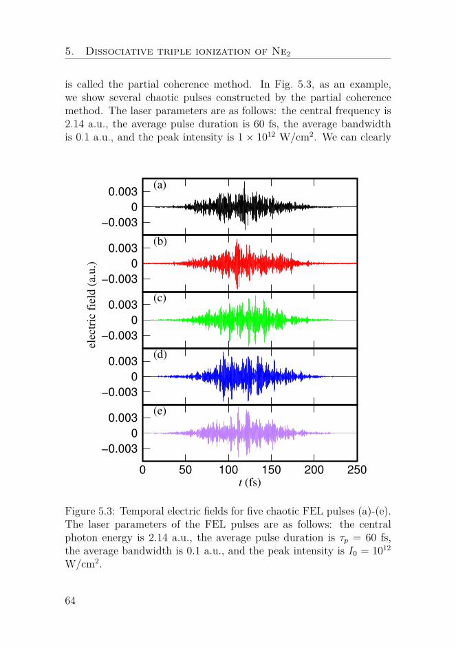
5. Dissociative triple ionization of Ne2
is called the partial coherence method. In Fig. 5.3, as an example,we show several chaotic pulses constructed by the partial coherencemethod. The laser parameters are as follows: the central frequency is2.14 a.u., the average pulse duration is 60 fs, the average bandwidthis 0.1 a.u., and the peak intensity is 1 × 10¹² W/cm2. We can clearly
−0.003
0
0.003(a)
−0.003
0
0.003(b)
−0.003
0
0.003(c)
−0.003
0
0.003(d)
−0.003
0
0.003
0 50 100 150 200 250
t (fs)
(e)
ele
ctr
ic f
ield
(a.u
.)
Figure 5.3: Temporal electric fields for five chaotic FEL pulses (a)-(e).The laser parameters of the FEL pulses are as follows: the centralphoton energy is 2.14 a.u., the average pulse duration is τp = 60 fs,the average bandwidth is 0.1 a.u., and the peak intensity is I0 = 1012
W/cm2.
64

5.1. Implementation of the MCWP method to triple ionization ofNe2
observe the fast variation of the field envelope for the chaotic pulses.That is the origin why we have to take more sampling points whentreating Ne2 interacting with the chaotic pulses.
The nuclear KER spectrum following triple ionization of Ne2 can beobtained by summering over the contributions from all the trajectories.Each trajectory is specified by its first, second and third jump times,i.e., t1, t2 and t3, and the states from which the first, second and thirdjumps takes. For the trajectories taking place at t1, t2 and t3, theionization probabilities for the first, second and third jumps are P1(t1),P2(t2) and P3(t3), respectively. When there are more than one jumppathways from one charge state to another charge state, the actualionization probability for the jth pathway for the kth jump, i.e., Pkj,should be a multiplication of the ionization probabilities Pk by therelative probability for that pathway, i.e., Pkj(tk) = Pk(tk) × Pkj(tk).Here the relative probability for the jth pathway is
Pkj(tk) =〈Ψ(tk)|C†kjCkj|Ψ(tk)〉∑j〈Ψ(tk)|C†kjCkj|Ψ(tk)〉
, (5.3)
Where |Ψ〉 is expressed by Eq. (2.25) and Ckj is the jump operatordescribing the jth jump pathway for the kth jump by Eq. (3.3). Thenuclear KER spectrum for each trajectory is obtained by projectingthe nuclear wave packets at the end of propagation, i.e., χ3(R, te), inNe3+
2 to its energy-normalized vibrational eigenstates χE(R) for thefinal potential energy curve, i.e.,
pEjkm(t1, t2, t3) = |∫dRχ3(R, te; j, k,m, t1, t2, t3)χE(R)|2 (5.4)
Thus, the total nuclear KER spectrum following triple ionization is
KER =∑
j,k,m,t1,t2,t3
P1jP2kP3mpEjkm(t1, t2, t3). (5.5)
The input data in our calculation are as follows. The photoioniza-tion cross section of the 2s inner-valence electron in Ne2 was approxi-mate by the photoionization cross section of the 2s electron in Ne [121],i.e., σ2s/2p = 0.36 Mb. There have been no works in investigating thephotoionization from Ne2+
2 (2p−2) to Ne3+2 (2p−3) and we approximate
this process by the photoionization of the neon atom [122]. Thus the
65

5. Dissociative triple ionization of Ne2
photoionization cross section from Ne2+2 (2p−2) to Ne3+
2 (2p−3) is ap-proximated by σ3p
2p = 8.08 Mb. We can find the ICD rates from thetwo excited states in Ne+
2 in Ref. [123] where they were calculatedby the complex absorbing potential method. Another ICD processfrom Ne2+
2 (1s−12s−1) to Ne3+2 (2p−3) has been not investigated in lit-
erature and the ICD rates are simply assume to be same with thefirst ICD process. The photonionization cross section from Ne+
2 (1s−1)to Ne2+
2 (1s−12s−1) is unknown and we make an initial guess, i.e.,σ2p
1s = 0.24 Mb and laser adjust it according to the comparison be-tween our simulation results and the experimental results.
Similar to the H2 case, we adopt the split-operator fast Fouriertransform method to solve Eq. (3.11) in each charge state numerically.We utilize a time step of ∆t = 4 a.u.. and a space step ∆R = 0.0215a.u. in our simulation. The reason why we can choose a relatively large∆t here is the photoionization involved in this triple ionization processfollows the relatively slow-varying temporal profile of the intensity ofthe XUV pulse. To avoid reflection from the box boundaries, we adopta simulation box from Rmin = 3.32 a.u. to Rmax = 91.15 a.u.. Forboth the coherent and chaotic pulse cases, the first jump occurs at theintensity maximum and the second and third jumps are assumed tooccur at every 40 time steps.
5.2 Nuclear KER spectra followingtriple ionization of Ne2 interactingwith coherent XUV pulse
In this section, we will present the results obtained by our method forNe2 interacting with coherent XUV pulses. According to the experi-ment, the pump and probe pulses are identical to each other exceptthe probe pulse is delayed by τ . The vector potential of the pumppulse is expressed by
A(t) ={A0 sin2( πt
TXUV) cos(ωXUVt) 0 ≤ t ≤ TXUV,
0 elsewhere, (5.6)
where TXUV is the pulse duration of the pump pulse and the electricfield can be obtained by using the relation F (t) = −∂A(t)
∂t. The peak
66
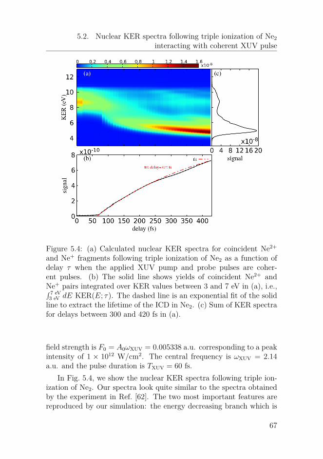
5.2. Nuclear KER spectra following triple ionization of Ne2interacting with coherent XUV pulse
Figure 5.4: (a) Calculated nuclear KER spectra for coincident Ne2+
and Ne+ fragments following triple ionization of Ne2 as a function ofdelay τ when the applied XUV pump and probe pulses are coher-ent pulses. (b) The solid line shows yields of coincident Ne2+ andNe+ pairs integrated over KER values between 3 and 7 eV in (a), i.e.,∫ 7 eV3 eV dE KER(E; τ). The dashed line is an exponential fit of the solidline to extract the lifetime of the ICD in Ne2. (c) Sum of KER spectrafor delays between 300 and 420 fs in (a).
field strength is F0 = A0ωXUV = 0.005338 a.u. corresponding to a peakintensity of 1 × 10¹² W/cm2. The central frequency is ωXUV = 2.14a.u. and the pulse duration is TXUV = 60 fs.
In Fig. 5.4, we show the nuclear KER spectra following triple ion-ization of Ne2. Our spectra look quite similar to the spectra obtainedby the experiment in Ref. [62]. The two most important features arereproduced by our simulation: the energy decreasing branch which is
67

5. Dissociative triple ionization of Ne2
directly related to the ICD process from Ne+2 (1s−1) to Ne2+
2 (2p−2) andthe delay-independent high energy branch centered at about 9 eV. Byconducting a trajectory and wave packet analysis (we do not show theresults here), we find the origins of the two branches. The signals at theenergy decreasing branch comes from the nuclear wave packets evolv-ing along the repulsive potential energy curve for the Ne2+
2 followingan ICD from Ne+
2 are later photoionized to Ne3+2 by the probe pulse.
Thus, if the pump probe delay is larger, then there is a longer timefor the nuclear wave packets moving to larger internuclear distancesin Ne2+
2 . This finally results in coincident Ne+ and Ne2+ signals atsmaller KER values. The high energy branch is from the triple ioniza-tion taking place within a short time, to say, within either the pumpor probe pulse. Apart from that, we can also observe the similaritythat there are more signals at delay close to 0 due to the constructiveinterference between the pump and probe pulse. We can also observediscrepancies between our results and the experimental results:
• the ratio of the signals between the low and high energy peaks inFig. 5.4(c), which come from the energy decreasing branch andthe high energy branch in Fig. 5.4(a), respectively, is overesti-mated
• the extracted lifetime in Fig. 5.4(b), i.e., 372 fs, is over two timeslarger than that extracted from the experimental spectra, i.e.,150 ± 50 fs.
It is known to us that the ICD lifetime of an excited state can beapproximated by the inverse of the ICD rate of the excited state. Thus,the discrepancy of the extracted ICD lifetimes between our simulationand the experiment clearly shows that the ICD rates from the twoexcited states in Ne+
2 , which is taken from Ref. [123] by the complexabsorbing potential method, is underestimated by a factor of around3 for the internuclear distances where the nuclear wave packets evolvealong the potential energy curves for these two excited states. To con-firm this, in Fig. 5.5, we show the the similar results as in Fig. 5.4(b) byusing new ICD rates which is 3 times larger than the rates in Ref. [123].We can see that the new extracted lifetime, i.e., 167 fs, is much closerto the counterpart extracted from the experiment.
68

5.2. Nuclear KER spectra following triple ionization of Ne2interacting with coherent XUV pulse
delay τ (fs)0 100 200 300 400
signal
×10-9
1.4
1.2
1.0
0.8
0.6
0.4
0.2
0
fit: τICD = 167 fs
Figure 5.5: Yields of Ne2+ and Ne+ pairs similar as Fig. 5.4(b) whenthe ICD rates are three times larger.
Now we have a closer look at the low energy peak at around 5eV and the high energy peak at around 9 eV Fig. 5.4(c). Comparedwith the ratio of the signals between the low and high energy peaks ob-tained in the experiment, our ratio indicates that the high (low) energybranches in Fig. 5.4 are relatively underestimated (overestimated) inour calculation. We can see that as the delay is increased, the signalsat the energy decreasing branch would be increased until at a certaindelay the signals saturate since the wave packets in the excited statesin Ne+
2 are entirely prompted to the two-site outer-valence vacancystates in Ne2+
2 via ICD. While in the high energy branch, the signalsat around 9 eV remain almost the same for delays larger than 250 fs.In fact, there are two channels that mainly result in the signals at thehigh energy branches, i.e.,
• the first photoionization by the pump (probe) pulse, a followingfast ICD to Ne2+
2 , and the second photoionization by the pump(probe) pulse
• the first photoionization by the pump (probe) pulse, the secondphotoionization by the pump (probe) pulse and the followingICD to Ne3+
2
69
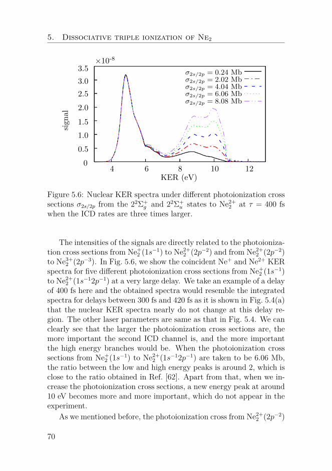
5. Dissociative triple ionization of Ne2
KER (eV)4 6 8 10 12
signal
×10-8
3.5
3.0
2.5
2.0
1.5
1.0
0.5
0
σ2s/2p = 0.24 Mbσ2s/2p = 2.02 Mbσ2s/2p = 4.04 Mbσ2s/2p = 6.06 Mbσ2s/2p = 8.08 Mb
Figure 5.6: Nuclear KER spectra under different photoionization crosssections σ2s/2p from the 22Σ+
g and 22Σ+u states to Ne2+
2 at τ = 400 fswhen the ICD rates are three times larger.
The intensities of the signals are directly related to the photoioniza-tion cross sections from Ne+
2 (1s−1) to Ne2+2 (2p−2) and from Ne2+
2 (2p−2)to Ne3+
2 (2p−3). In Fig. 5.6, we show the coincident Ne+ and Ne2+ KERspectra for five different photoionization cross sections from Ne+
2 (1s−1)to Ne2+
2 (1s−12p−1) at a very large delay. We take an example of a delayof 400 fs here and the obtained spectra would resemble the integratedspectra for delays between 300 fs and 420 fs as it is shown in Fig. 5.4(a)that the nuclear KER spectra nearly do not change at this delay re-gion. The other laser parameters are same as that in Fig. 5.4. We canclearly see that the larger the photoionization cross sections are, themore important the second ICD channel is, and the more importantthe high energy branches would be. When the photoionization crosssections from Ne+
2 (1s−1) to Ne2+2 (1s−12p−1) are taken to be 6.06 Mb,
the ratio between the low and high energy peaks is around 2, which isclose to the ratio obtained in Ref. [62]. Apart from that, when we in-crease the photoionization cross sections, a new energy peak at around10 eV becomes more and more important, which do not appear in theexperiment.
As we mentioned before, the photoionization cross from Ne2+2 (2p−2)
70

5.2. Nuclear KER spectra following triple ionization of Ne2interacting with coherent XUV pulse
0
0.2
0.4
0.6
0.8
1
4 6 8 10 12
signal
KER (eV)
8.08 Mb
20 Mb
30 Mb
40 Mb
50 Mb
Figure 5.7: Nuclear KER spectra following triple ionization of Ne2 ata 400-fs delay when the photoionization cross sections are taken 8.08,20, 30, 40 and 50 Mb, respectively.
to Ne3+2 (2p−3) is approximated by the photoionization cross section
from Ne+(2p−1) to Ne2+(2p−2). Actually, as we will show in the fol-lowing, the ratio of the signals between the low and high energy peakis, to some extent, not relevant to the photoionization cross sectionsfrom Ne2+
2 (2p−2) to Ne3+2 (2p−3). For example, the nuclear KER spec-
tra following triple ionization of Ne2 at the 400-fs delay for five differentphotoinization cross sections from Ne2+
2 (2p−2) to Ne3+2 (2p−3) are pre-
sented in Fig .5.7. To avoid the interferences from the second tripleionization channel, the results in Fig. 5.7 are obtained by only in-cluding the first triple ionization channel, i.e., first photoionization,then ICD and second photoionization. Thus, the almost identical fivecurves clearly show us that the choice of the photoinization cross sec-tions from Ne2+
2 to Ne3+2 has nothing to do with the discrepancy of the
experimental and theoretical ratios of the signals between the low andhigh energy peaks.
As a result, based on the above analysis, we know that the dis-crepancies between the simulation and the experiment comes from theunderestimated ICD rates and photoinization cross sections from Ne+
2to Ne2+
2 . We show in Fig. 5.8 the nuclear KER spectra following triple
71

5. Dissociative triple ionization of Ne2
Figure 5.8: (Nuclear KER spectra as a function of delay (a) and sumof the nuclear KER spectra for delays between 300 and 420 fs (b)following triple ionization of Ne2 when interacting with coherent laserpulses. The laser parameters are same as Fig. 5.4. While the ICD ratesare three times larger than that in Fig. 5.4 and the photoionizationcross section from Ne+
2 to Ne2+2 are taken as 6.06 Mb.
ionization of Ne2 when we take the ICD rates three times larger andthe photoionization cross sections from Ne+
2 to Ne2+2 are increased to
6.06 Mb. The laser parameters are identical to Fig. 5.4. We do notshow here the integrated signals from 3 to 7 eV as a function of delay,which is similar to Fig. 5.4(c), since it is quite similar to Fig. 5.5 be-cause of the same ICD rates are employed. Compared with Fig. 5.4,even though we have a better ratio of the signals between the lowand high energy peaks, we have also obtained relatively much largersignals at delays smaller than 70 fs due to the constructive coherencebetween the pump and probe pulses. The intensities of the signals atthese small delays, as we show in the next section, would be not sosignificant when chaotic pump and probe pulses are applied becauseof the reduced coherence time of the pulses.
72
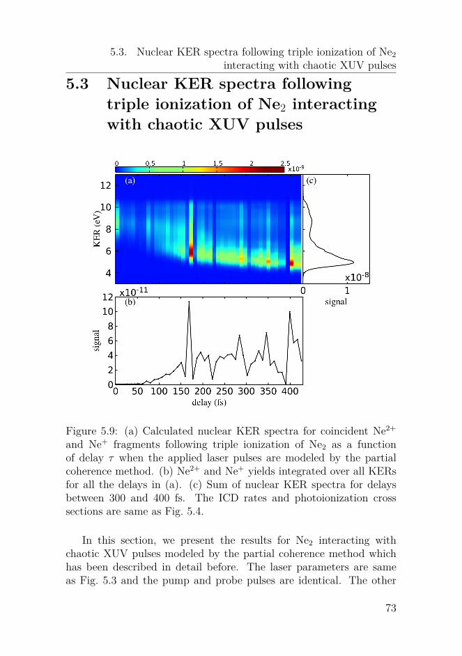
5.3. Nuclear KER spectra following triple ionization of Ne2interacting with chaotic XUV pulses
5.3 Nuclear KER spectra followingtriple ionization of Ne2 interactingwith chaotic XUV pulses
Figure 5.9: (a) Calculated nuclear KER spectra for coincident Ne2+
and Ne+ fragments following triple ionization of Ne2 as a functionof delay τ when the applied laser pulses are modeled by the partialcoherence method. (b) Ne2+ and Ne+ yields integrated over all KERsfor all the delays in (a). (c) Sum of nuclear KER spectra for delaysbetween 300 and 400 fs. The ICD rates and photoionization crosssections are same as Fig. 5.4.
In this section, we present the results for Ne2 interacting withchaotic XUV pulses modeled by the partial coherence method whichhas been described in detail before. The laser parameters are sameas Fig. 5.3 and the pump and probe pulses are identical. The other
73

5. Dissociative triple ionization of Ne2
parameters such as the ICD rates and the photoinization cross sec-tions are taken originally, i.e., same as Fig. 5.4. In Fig. 5.9(a), weshow the similar nuclear KER spectra, i.e., following triple ionizationof Ne2 by applying the chaotic pulses. Due to the fact that the XUVpules are different from shot to shot, the XUV pulse is modeled ateach delay. This is the reason the nuclear KER spectra in Fig. 5.9(a)as a function of delay is not as smooth as the coherent case. Similarly,we can observe the high energy branch centered around 9 eV and theenergy decreasing branch. Similar to Fig. 5.4(c), we have obtained asimilar ratio of the signals between the low and high energy peaks inFig. 5.9(c). Because of the variations of the signals originating fromthe random noises in Fig. 5.9(b), it is different to extract the ICDlifetime from Fig. 5.9(b). Extraction of the lifetime would be possibleonly if we average the nuclear KER spectra for many realizations.
Finally, we run a simulation where we take the new ICD rateswhich are three times larger than the original rates and assume newphotoionization cross sections of 6.06 Mb. The corresponding resultsare shown in Fig. 5.10. Similar to Fig. 5.9(a), the energy decreasingbranch and the high energy branch are reproduced. Apart from that,just as expected, we can clearly observe an enhancement of the highenergy branch in Fig. 5.10(a) compared with Fig. 5.9(a). Similar tothe coherent case in Fig. 5.8(c), a broadening of the high energy peakis also observed.
5.4 Concluding remarksIn this chapter, we have taken the example of Ne2 to study the nucleardynamics in weakly bounded clusters. Due to the relatively weak inter-action between the constituent atoms in clusters, new electronic decaychannels, such as the ICD process, become possible in weakly boundedclusters. The ICD process is an electron decay process where after re-moving an inner-valence electron from one atom in the cluster, theremaining cation is highly excited and thus can decay to more stablestates by emitting another electron. Impressed by a recent XUV-pump-XUV-probe experiment [62], we have investigated the ICD processesin Ne2 by applying the MCWP approach to obtain the nuclear KERspectra following triple ionization of Ne2. The involved triple ioniza-
74

5.4. Concluding remarks
Figure 5.10: Similar results as Fig. 5.9 where the R-dependent ICDrates are three times larger and the photoionization cross sections fromNe+
2 to Ne2+2 are 6.06 Mb.
tion process is as follows: one 2s inner-valence electron is first removedfrom Ne2 by absorbing one photon from the XUV pulse, then the sec-ond ionization takes the remaining Ne+
2 to Ne2+2 either via Ne+
2 (1s−1)to Ne2+
2 (1s−12p−1) by tje second photoionization or via Ne+2 (1s−1) to
Ne2+2 (2p−2) by ICD, and the third ionization takes Ne2+
2 (1s−12p−1) orNe2+
2 (2p−2) to Ne3+2 (2p−3) by ICD or the second photoionization. We
have obtained the nuclear KER spectra following the above mentionedtriple ionization of Ne2 and reproduced the two important features,i.e., the high energy branch and the energy decreasing branch in thenuclear KER spectra obtained by that experiments. Apart from that,an analysis of the discrepancies between our simulation and the ex-periment, i.e., the extracted ICD lifetime and the ratio of the signals
75

5. Dissociative triple ionization of Ne2
between the low and high energy peaks in the nuclear KER spectra atlarge delays, conveys us that the ICD rates obtained by the complexabsorbing method in Ref. [123] are underestimated by over two timesand we can make a more accurate estimate of the photoionization crosssections from Ne+
2 to Ne2+2 .
76
Chapter 6Summary and outlook
In this thesis, we have applied the MCWP approach to investigate thenuclear dynamics following ionization and excitation in small moleculesand weakly bound clusters through interacting with intense laser pulses.We have presented a detailed description of the implementation of theMCWP approach and its equivalence to the master equation in theLindblad form in Chapter 3. In Chapter 4, we mainly studied thedissociative double ionization of H2. We first studied the influencesof parameters including the central wavelength, pulse duration, peakintensity and nuclear mass on the nuclear KER spectrum followingfull break-up of H2 interacting with single IR laser pulses. Besides, weshowed the unique possibility of the MCWP approach by conductinga trajectory analysis to a given feature in a nuclear KER spectrum.Then we investigated the nuclear dynamics in the singly excited statesin H2 by using the XUV-pump-IR-probe spectroscopy. After that, westudied the nuclear dynamics following single ionization of H2 eithervia photoionization or autoionization from the doubly excited stateswhen exposed to XUV pulses. In Chapter 5, we extended the MCWPapproach to investigate triple ionization of the neon dimmer to studythe ICD process in Ne2 by using XUV-pump-XUV-probe spectroscopy.We have reproduced the nuclear KER spectra following triple ioniza-tion of Ne2 obtained by a recent experiment [62], which is beyond thecapability of the other quantum methods.
Throughout this thesis, we solve the simplified one-dimensionalcoupled equations for the motion of the nuclei since we assume the
77

6. Summary and outlook
molecule and cluster is rotational frozen. We can later take the ro-tational motion of the nuclei into consideration by solving the morecomplicated three-dimensional equations for the nuclear motion. Be-sides, the laser polarization directions are assumed to be parallel to themolecular axis and only the transition from the states of Σ symmetriesto the states of Σ symmetries are involved. When there are electricfield components at the directions perpendicular to the molecular axis,for example, circularly or elliptically polarized laser pulses, other tran-sitions from the states of a given symmetry to the states of differentsymmetries should be considered.
The present study of the influence of the doubly excited states onthe nuclear KER spectra is not sufficient. On the one aspect, there aresome discrepancies between our simulation and the results obtained bythe experiment and another theoretical method. We should put moreefforts on figuring out the origins of the discrepancies. On the otheraspect, we have only studied the interaction of H2 with single XUVpulses. An extended study of the interaction of H2 with laser pulsesin a pump-probe setup is worthy to carry out to map out the nucleardynamics from the nuclear KER spectra as a function of delay.
Apart from the ICD process studied in Chapter 5, we can apply theMCWP method to investigate other electronic decay processes, such asthe ETMD process, in weakly bounded clusters. For the ETMD pro-cess, the inner valence hole in one atom is filled by one outer valenceelectron in its neighboring atom and the energy released by this tran-sition supports to remove another electron from the neighboring atom.Different from ICD, after ETMD taking place, there is no hole in thefirst atom and there are two outer-valence holes in the second atom.Thus, there is no Coulomb explosion between the two atoms and theremaining ion may remain weakly bounded. As a result, one can ap-ply a similar XUV-pump-XUV-probe setup as applied in the ICD caseto study the nuclear dynamics within an ETMD process by obtainingthe nuclear KER spectra following triple ionization of a weakly boundcluster.
78

Part II
Publications
79

My First Paper
Paper I. PHYSICAL REVIEW A 94, 063402 (2016)
Title:Laser-induced dissociative ionization of H2 from the near-infrared to the mid-infrared regimeAuthors: Qingli Jing and Lars Bojer MadsenDOI: https://doi.org/10.1103/PhysRevA.94.063402
Reprinted with permission from the American Physical Society. Copy-right (2016) by the American Physical Society.
My contribution to this publication: (1) implementation of the theoret-ical calculations and (2) preparation of the manuscript (with figures).
80

PHYSICAL REVIEW A 94, 063402 (2016)
Laser-induced dissociative ionization of H2 from the near-infrared to the mid-infrared regime
Qingli Jing and Lars Bojer MadsenDepartment of Physics and Astronomy, Aarhus University, 8000 Aarhus C, Denmark
(Received 6 October 2016; published 2 December 2016)
We apply the Monte Carlo wave packet (MCWP) approach to investigate the kinetic energy release (KER)spectra of the protons following double ionization in H2 when interacting with laser pulses with centralwavelengths ranging from the near-infrared (IR) (800 nm) to the mid-IR (6400 nm) regions and with durationsof 3–21 laser cycles. We uncover the physical origins of the peaks in the nuclear KER spectra and ascribe themto mechanisms such as ionization following a resonant dipole transition, charge-resonance-enhanced ionization,and ionization in the dissociative limit of large internuclear distances. For relatively large pulse durations, i.e.,for 15 or more laser cycles at 3200 nm and 10 or more at 6400 nm, it is possible for the nuclear wave packetin H2
+ to reach very large separations. Ionization of this part of the wave packet results in peaks in the KERspectra with very low energies. These peaks give direct information about the dissociative energy in the 2pσu
potential energy curve of H2+ at the one- and three-photon resonances between the 2pσu and 1sσg curves in H2
+.With the MCWP approach, we perform a trajectory analysis of the contributions to the KER peaks and identifythe dominant ionization pathways. Finally, we consider a pump-probe scheme by applying two delayed pulsesto track the nuclear dynamics in a time-resolved setting. Low-energy peaks appear for large delays and theseare used to obtain the 2pσu dissociative energy values at the one-photon resonance between the 2pσu and 1sσg
curves in H2+ for different wavelengths.
DOI: 10.1103/PhysRevA.94.063402
I. INTRODUCTION
The significant advances made in femtosecond laser tech-nology have opened the possibility of controlling chemicalreactions with laser light [1–3]. When exposing molecules tointense laser pulses, strong interaction between molecules andthe external electromagnetic field may give rise to electronicexcitation or even result in ionization. Nuclear dynamics ishence often induced because of the interplay between thenuclei and electrons. A good example is the process ofdissociative ionization [4,5], a process in which dissociationis induced as a result of the removal of one or more electrons.Laser-induced dissociative ionization has received consider-able interest in the past two decades [6–8]. Thanks to the fastdevelopments in imaging techniques such as COLTRIMS [9]and VMI spectrometers [10], it is now possible to measure thekinetic energy of the ionic fragments or the liberated electronsor both. By analyzing the kinetic energy release (KER)spectra of the ionic fragments, information about the ionizationprocess can be obtained [11]. For example, the processesof charge-resonance-enhanced ionization (CREI) [12,13] andresonance-enhanced multiphoton ionization [14,15] both man-ifest themselves through the occurrence of characteristicpeaks in the nuclear KER spectra [16]. Different from themechanism of resonance-enhanced multiphoton ionization, forone-electron system like H2
+, CREI results from lowering theinternal barrier between the double wells at certain criticalinternuclear separations in the presence of a strong field, alongwith the creation of a pair of field-dressed charge-resonantstates.
In parallel to the advances in experimental techniques,many theoretical methods [17–19] have been developed andproven to work very well in describing a range of phenomenaassociated with the interaction of molecules and light. Amongthese methods, the Monte Carlo wave packet (MCWP)approach [20,21] performs well when describing the laser-induced dissipative dynamics. Especially in previous works
on H2 [22,23], and O2 [24], where the nuclear KER spectrawere of main concern, the simulations agreed very well withthe measurements. This approach, like many others, is devotedto solving the time-dependent Schrodinger equation (TDSE),but the problem is greatly simplified because the electronicdegrees of freedom are treated in an effective description. Atthe same time, the Markov approximation [25] is applied,which means that the flow of electrons from the molecularsystems to the surroundings is unidirectional and irreversible.The loss of electrons is encoded in jump operators, whichare responsible for jumps among different Hilbert spacesand constitute a non-Hermitian part of the total Hamiltonian.The non-Hermitian Hamiltonian implies that the norm of thesystem is decreasing over time. The drop of the norm overtime is controlled by the instantaneous ionization rates [26,27]of the system. If the ionization rates depend on the nuclearconfiguration, wave-packet motion is induced in the neutralmolecule. Inducing nuclear dynamics in this way is calledLochfraß [28–30] and this phenomenon is an integrated partof the MCWP simulations.
In the present MCWP approach to dissociative ionization,the jumps between different charge states and associatedHilbert spaces are described by ionization rates. While theevolution of the system within a given charge state is coherent,the rate treatment of the ionization step means that coherencebetween the remaining cation and the ionized electron is lost.Also we have no knowledge of the energy distribution of theoutgoing electron.
Recently there has been a shift in strong-field physicstoward the use of mid-infrared (IR) driving wavelengths [31],e.g., to image molecular structures by light-induced electrondiffraction [32,33] and to greatly increase the photon energiesof high-harmonic generation [34]. The interaction between themid-IR laser pulses and molecules can be treated in the qua-sistatic regime. The systematic investigation of the behaviorof the nuclear KER spectra with increasing wavelength from
2469-9926/2016/94(6)/063402(11) 063402-1 ©2016 American Physical Society
81
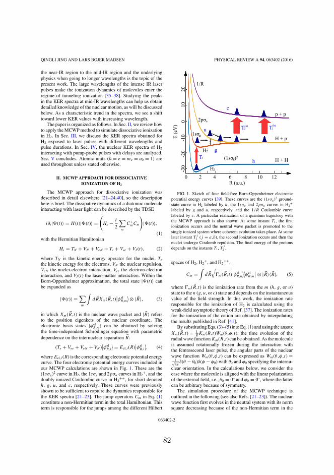
QINGLI JING AND LARS BOJER MADSEN PHYSICAL REVIEW A 94, 063402 (2016)
the near-IR region to the mid-IR region and the underlyingphysics when going to longer wavelengths is the topic of thepresent work. The large wavelengths of the intense IR laserpulses make the ionization dynamics of molecules enter theregime of tunneling ionization [35–38]. Studying the peaksin the KER spectra at mid-IR wavelengths can help us obtaindetailed knowledge of the nuclear motion, as will be discussedbelow. As a characteristic trend in the spectra, we see a shifttoward lower KER values with increasing wavelength.
The paper is organized as follows. In Sec. II, we review howto apply the MCWP method to simulate dissociative ionizationin H2. In Sec. III, we discuss the KER spectra obtained forH2 exposed to laser pulses with different wavelengths andpulse durations. In Sec. IV, the nuclear KER spectra of H2
interacting with pump-probe pulses with delays are analyzed.Sec. V concludes. Atomic units (� = e = me = a0 = 1) areused throughout unless stated otherwise.
II. MCWP APPROACH FOR DISSOCIATIVEIONIZATION OF H2
The MCWP approach for dissociative ionization wasdescribed in detail elsewhere [21–24,40], so the descriptionhere is brief. The dissipative dynamics of a diatomic moleculeinteracting with laser light can be described by the TDSE
i∂t |�(t)〉 = H (t)|�(t)〉 =(
Hs − i
2
∑m
C+mCm
)|�(t)〉,
(1)with the Hermitian Hamiltonian
Hs = TN + VN + VeN + Te + Vee + VI (t), (2)
where TN is the kinetic energy operator for the nuclei, Te
the kinetic energy for the electrons, VN the nuclear repulsion,VeN the nuclei-electron interaction, Vee the electron-electroninteraction, and VI (t) the laser-matter interaction. Within theBorn-Oppenheimer approximation, the total state |�(t)〉 canbe expanded as
|�(t)〉 =∑m
∫d �RXm( �R,t)
∣∣φelR,m
⟩ ⊗ | �R〉, (3)
in which Xm( �R,t) is the nuclear wave packet and | �R〉 refersto the position eigenkets of the nuclear coordinate. Theelectronic basis states |φel
R,m〉 can be obtained by solvingthe time-independent Schrodinger equation with parametricdependence on the internuclear separation �R:
(Te + Vee + VeN + VN )∣∣φel
R,i
⟩ = Eel,i(R)∣∣φel
R,i
⟩, (4)
where Eel,i(R) is the corresponding electronic potential energycurve. The four electronic potential energy curves included inour MCWP calculations are shown in Fig. 1. These are the(1sσg)2 curve in H2, the 1sσg and 2pσu curves in H2
+, and thedoubly ionized Coulombic curve in H2
++, for short denotedh, g, u, and c, respectively. These curves were previouslyshown to be sufficient to capture the dynamics responsible forthe KER spectra [21–23]. The jump operators Cm in Eq. (1)constitute a non-Hermitian term in the total Hamiltonian. Thisterm is responsible for the jumps among the different Hilbert
R (a.u.)E
(eV
)0 2 4 6 8 10 12
0201
001-
02-03-
H + H
H + p
p + p
H2
H2+
1/R
1sσ
2pσ
g
u
(1sσ )g 2h
u
c
gT1
T2(b)T2
(a)
FIG. 1. Sketch of four field-free Born-Oppenheimer electronicpotential energy curves [39]. These curves are the (1sσg)2 ground-state curve in H2 labeled by h, the 1sσg and 2pσu curves in H2
+
labeled by g and u, respectively, and the 1/R Coulombic curvelabeled by c. A particular realization of a quantum trajectory withthe MCWP approach is also shown: At some instant T1, the firstionization occurs and the neutral wave packet is promoted to thesingly ionized system where coherent evolution takes place. At somelater instant T
j
2 (j = a,b), the second ionization occurs and then thenuclei undergo Coulomb repulsion. The final energy of the protonsdepends on the instants T1, T
j
2 .
spaces of H2, H2+, and H2
++,
Cm =∫
d �R√
�m( �R,t)∣∣φel
R,n
⟩⟨φel
R,m
∣∣ ⊗ | �R〉〈 �R|, (5)
where �m( �R,t) is the ionization rate from the m (h, g, or u)state to the n (g, u, or c) state and depends on the instantaneousvalue of the field strength. In this work, the ionization rateresponsible for the ionization of H2 is calculated using theweak-field asymptotic theory of Ref. [37]. The ionization ratesfor the ionization of the cation are obtained by interpolatingthe results published in Ref. [41].
By substituting Eqs. (3)–(5) into Eq. (1) and using the ansatzXm( �R,t) = 1
RKm(R,t)Wm(θ,φ,t), the time evolution of the
radial wave function Km(R,t) can be obtained. As the moleculeis assumed rotationally frozen during the interaction withthe femtosecond laser pulse, the angular parts of the nuclearwave function Wm(θ,φ,t) can be expressed as Wm(θ,φ,t) =
1√4π
δ(θ − θ0)δ(φ − φ0) with θ0 and φ0 specifying the internu-clear orientation. In the calculations below, we consider thecase where the molecule is aligned with the linear polarizationof the external field, i.e., θ0 = 0◦ and φ0 = 0◦, where the lattercan be arbitrary because of symmetry.
The simulation procedure of the MCWP technique isoutlined in the following (see also Refs. [21–23]). The nuclearwave function first evolves in the neutral system with its normsquare decreasing because of the non-Hermitian term in the
063402-2
82

LASER-INDUCED DISSOCIATIVE IONIZATION OF H . . . PHYSICAL REVIEW A 94, 063402 (2016)
Hamiltonian. At each time step, the drop in the norm squareis compared with a random number (between 0 and 1), whichdetermines whether the jump from one Hilbert space to anothertakes place or not. If the drop in the norm square is largerthan the random number, ionization occurs and thus the wavefunction jumps, is renormalized, and then evolves in the newHilbert space, otherwise, ionization cannot occur and thewave function needs to be renormalized in the neutral system.The comparison between the drop in the norm square and arandom number continues until the first jump occurs. Similarly,whether the second jump takes place or not is controlled bycomparing the drop in the norm square of the nuclear wavefunction in H2
+ with a new random number (between 0 and1). The difference compared to the first jump is that thereare two pathways for the second jump to the doubly ionizedsystem, from the g state curve and from the u state curve,which means that another random number (between 0 and 1)should be introduced to decide which pathway to choose. As aresult, a nuclear wave function after emitting two electrons isobtained stochastically, with the first jump occurring at someinstant of time T1 and the second jump at some instant of latertime T2 (Fig. 1). The nuclear wave packet after two jumpsKc(R,T2) holds all the information of the nuclei. For example,the nuclear kinetic energies can be obtained by projecting thewave packet on Coulomb waves KE(R),
EKERm (T1,T2) =
∣∣∣∣∫
KE(R)Kc(R,T2)dR
∣∣∣∣2
, (6)
with m = g, u.Equation (6) only gives the KER signal for one stochastic
event occurring at specific T1 and T2. We refer to such aspecific realization of jump times and dynamics as a quantumtrajectory. The total nuclear KER spectra of the dissociativeionization process can be obtained by averaging over allthe stochastic events. In the present case of dissociativedouble ionization, the computational effort can, however,be dramatically reduced through eliminating all the randomnumbers by applying a completely deterministic approach. Tothis end, the two jumps which are dressed with probabilitiesare assumed to take place at every time step. Actually,the probability for each jump is proportional to the dropin the norm square at that jump time. This is reasonable becausethe larger the drop in the norm square, the more likely it is fora jump to occur. Summing over all the weighted events, thenuclear KER spectra can be obtained from the formula,
EKERtot =
∑T1,T2
P1P12
∑m=g,u
P (m|{T1,T2})EKERm (T1,T2). (7)
In the above equation, P1 = −d(Nh(t))/dt is the probabilitydensity for the first jump with Nh(t) representing the normsquare of the nuclear wave function of the h state; P12 =−d(Ng(t) + Nu(t))/dt is the conditional probability densityof the second jump for a given first jump with Ng(t) andNu(t) representing the norm square of the nuclear wavefunction of the g state and the u state, respectively; andP (m|{T1,T2}) = 〈�|C+
mCm|�〉/〈�|C+g Cg + C+
u Cu|�〉 is theconditional probability density from the m (g or u) state forgiven first and second jump times (see also Ref. [40]).
As shown in Fig. 1, the initial nuclear wave function isassumed to be the ground state of the (1sσg)2 potential energy
curve of H2. The applied laser pulse with the polarization axisparallel to the molecular axis first kicks out the first electronand then induces nuclear motion in the two potential energycurves of the H2
+ system. Meanwhile, there is electronicdipole coupling between the 1sσg and 2pσu states. Finally, thenuclei experience Coulomb repulsion after emitting the secondelectron. Thus the nuclei pick up energy from two terms, oneis the dissociative kinetic energy in the singly ionized systemand the other is the Coulomb repulsion energy in the doublyionized system. The time-dependent Hamiltonian, includingthe non-Hermitian part representing the interaction betweenstates in different Hilbert spaces as well as the electronicdipole coupling between states within a given Hilbert space,originates from the external laser field. As a result and as shownin the next section, the kinetic energy is closely related to theparameters of the applied laser pulse, such as peak intensity,wavelength, and pulse duration.
Numerically, within each Hilbert space, we solve the TDSEby applying the split-operator method [42] on the short-timepropagator, i.e.,
U (t + t,t) = exp
(−iTN
t
2
)exp
[−iV
(R,t + t
2
)t
]
× exp
(−iTN
t
2
). (8)
Obviously, it is the term V (R,t) that determines how thewave packet evolves in each Hilbert space. For example, inH2
+, the 2 × 2 matrix representation of V (R,t) is Vij (R,t) =Eel,i(R)δij−i�i(R,t)/2δij +βDij (R)F (t) (i,j =g,u), whereβ = 1 + 1/(2mp + 1) with mp the proton mass, F (t) is theelectric field, and Dij (R) is the electronic dipole momentfunction between i and j states along the direction ε of thelinear polarization, i.e., Dij (R) = −〈φel
R,i |�r · ε|φelR,j 〉. We use
the explicit expression for Dgu(R) = R
2√
1−[(1+R+R2/3)e−R ]2−
1(2+1.4R) given in Ref. [43]. The size of our simulation boxis 40.96. The time step t is 0.1 and the spatial step R is0.02. The operators exp(−iTN
t2 ) and exp[−iV (R,t + t
2 )t]are diagonal in the momentum and position representations,respectively, thus a fast-Fourier-transform algorithm is appliedin the implementation.
III. WAVELENGTH AND PULSE DURATIONDEPENDENCE OF KER SPECTRA FOR H2
INTERACTING WITH A SINGLE LASER PULSE
In this section, we simulate the process of dissociativedouble ionization of ground-state H2 interacting with singlelaser pulses. The calculated proton KER spectra for differentwavelengths (ranging from 800 to 6400 nm) and pulsedurations (ranging from 3 to 21 laser cycles) are shown inFig. 2. In our calculation, the laser pulses are of Gaussianshape, and the pulse durations are expressed in terms of thenumber of laser cycles within the FWHM of the intensityprofile of the applied laser pulses. The laser intensity of6 × 1013 W/cm2 is chosen to be similar to the one in theexperiment of Ref. [44]. If there are specific internuclearpositions from which a significant part of the second ionizationtakes place, distinctive peaks occur in the nuclear KER spectra.
063402-3
83
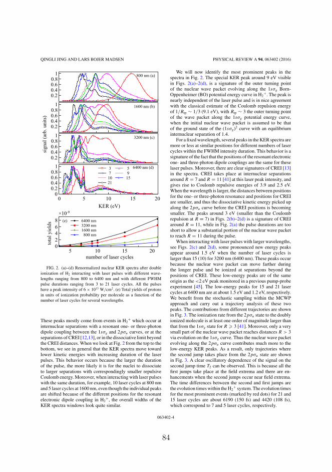
QINGLI JING AND LARS BOJER MADSEN PHYSICAL REVIEW A 94, 063402 (2016)
0.20.40.60.8
1 800 nm (a)
3 57 910 1521
0.20.40.60.8
1 1600 nm (b)
3200 nm (c)
00.20.40.60.8
1
0 5 10 15 20
0.20.40.60.8
1
KER (eV)
sign
al(a
rb.u
nits
)
6400 nm (d)
02468
number of laser cycles5 10 15 20
6400 nm3200 nm1600 nm
800 nm
(e)
tota
lyie
lds
×10-9
FIG. 2. (a)–(d) Renormalized nuclear KER spectra after doubleionization of H2 interacting with laser pulses with different wave-lengths ranging from 800 to 6400 nm and with different FWHMpulse durations ranging from 3 to 21 laser cycles. All the pulseshave a peak intensity of 6 × 10¹³ W/cm2. (e) Total yields of protonsin units of ionization probability per molecule as a function of thenumber of laser cycles for several wavelengths.
These peaks mostly come from events in H2+ which occur at
internuclear separations with a resonant one- or three-photondipole coupling between the 1sσg and 2pσu curves, or at theseparations of CREI [12,13], or in the dissociative limit beyondthe CREI distances. When we look at Fig. 2 from the top to thebottom, we see in general that the KER spectra move towardlower kinetic energies with increasing duration of the laserpulses. This behavior occurs because the larger the durationof the pulse, the more likely it is for the nuclei to dissociateto larger separations with correspondingly smaller repulsiveCoulomb energy. Moreover, when interacting with laser pulseswith the same duration, for example, 10 laser cycles at 800 nmand 5 laser cycles at 1600 nm, even though the individual peaksare shifted because of the different positions for the resonantelectronic dipole coupling in H2
+, the overall widths of theKER spectra windows look quite similar.
We will now identify the most prominent peaks in thespectra in Fig. 2. The special KER peak around 9 eV visiblein Figs. 2(a)–2(d), is a signature of the outer turning pointof the nuclear wave packet evolving along the 1sσg Born-Oppenheimer (BO) potential energy curve in H2
+. The peak isnearly independent of the laser pulse and is in nice agreementwith the classical estimate of the Coulomb repulsion energyof 1/Rtp ∼ 1/3 (9.1 eV), with Rtp ∼ 3 the outer turning pointof the wave packet along the 1sσg potential energy curve,when the initial nuclear wave packet is assumed to be thatof the ground state of the (1sσg)2 curve with an equilibriuminternuclear separation of 1.4.
For a fixed wavelength, several peaks in the KER spectra aremore or less at similar positions for different numbers of lasercycles within the FWHM intensity duration. This behavior is asignature of the fact that the positions of the resonant electronicone- and three-photon dipole couplings are the same for theselaser pulses. Moreover, there are clear signatures of CREI [13]in the spectra. CREI takes place at internuclear separationsaround R = 7 and R = 11 [41] at this laser peak intensity, andgives rise to Coulomb repulsive energies of 3.9 and 2.5 eV.When the wavelength is larger, the distances between positionsfor the one- or three-photon resonance and positions for CREIare smaller, and thus the dissociative kinetic energy picked upalong the 2pσu curve before the CREI positions is becomingsmaller. The peaks around 3 eV (smaller than the Coulombrepulsion at R = 7) in Figs. 2(b)–2(d) is a signature of CREIaround R = 11, while in Fig. 2(a) the pulse durations are tooshort to allow a substantial portion of the nuclear wave packetto reach R = 11 during the pulse.
When interacting with laser pulses with larger wavelengths,see Figs. 2(c) and 2(d), some pronounced new energy peaksappear around 1.5 eV when the number of laser cycles islarger than 15 (10) for 3200 nm (6400 nm). These peaks occurbecause the nuclear wave packet can move farther duringthe longer pulse and be ionized at separations beyond thepositions of CREI. These low-energy peaks are of the sameorigin as the <2 eV peak monitored in a previous pump-probeexperiment [45]. The low-energy peaks for 15 and 21 lasercycles at 6400 nm are at about 1.5 eV and 1.2 eV, respectively.We benefit from the stochastic sampling within the MCWPapproach and carry out a trajectory analysis of these twopeaks. The contributions from different trajectories are shownin Fig. 3. The ionization rate from the 2pσu state to the doublyionized molecule is at least one order of magnitude larger thanthat from the 1sσg state for R � 3 [41]. Moreover, only a verysmall part of the nuclear wave packet reaches distances R > 3via evolution on the 1sσg curve. Thus the nuclear wave packetevolving along the 2pσu curve contributes much more to thelow-energy KER peaks. As a result, only trajectories wherethe second jump takes place from the 2pσu state are shownin Fig. 3. A clear oscillatory dependence of the signal on thesecond jump time T2 can be observed. This is because all thefirst jumps take place at the field extrema and there are en-hancements when the second jumps occur near field extrema.The time differences between the second and first jumps arethe evolution times within the H2
+ system. The evolution timesfor the most prominent events (marked by red dots) for 21 and15 laser cycles are about 6190 (150 fs) and 4420 (108 fs),which correspond to 7 and 5 laser cycles, respectively.
063402-4
84

LASER-INDUCED DISSOCIATIVE IONIZATION OF H . . . PHYSICAL REVIEW A 94, 063402 (2016)
FIG. 3. Contributions from different trajectories to the low-energy peak around (a) 1.2 eV in Fig. 2(d) when interacting witha pulse with 21 laser cycles at 6400 nm, and (b) 1.5 eV in Fig. 2(d)when interacting with a pulse with 15 laser cycles at 6400 nm. Thered dots mark the trajectories that give the largest contributions. Thelaser pulses are indicted by the grey lines and the parameters are asin Fig. 2(d).
In this manner, the MCWP approach allows us to find thetypical time spent in the H2
+ system, i.e., the time needed toionize H2
+, for each particular feature in the KER spectra.These times are long enough for the nuclear wave packetsto reach quite large internuclear separations (R > 20) alongthe dissociative 2pσu potential energy curve. As we will nowshow, the difference [2 laser cycles for the two peaks at 1.2 and1.5 eV in Fig. 2(d)] between the two evolution times allows usto obtain the dissociative kinetic energy Ed for the low-energypeaks.
One can see from Fig. 1 that there is nearly no variationin the 2pσu potential energy curve when R > 8. Thus thedissociative kinetic energies of the low-energy protons are
almost the same in spite of different numbers of laser cyclesand ionization from different internuclear separations largerthan 8. We can therefore estimate the dissociation energies inthe following way. We consider two equations Ed + 1/R21 =1.2 eV and Ed + 1/R15 = 1.5 eV, with R21 and R15 denotingthe positions where the second ionization takes place wheninteracting with laser pulses with 21 and 15 laser cycles,respectively. In combination with the use of the relationsEd = 1
2mv2 and vt = R21 − R15, valid at R > 8, we obtain thedissociative kinetic energies along the 2pσu potential energycurve from both the positions of the one- and three-photonresonances between the 2pσu and 1sσg curves. In theseequations, v is the velocity corresponding to Ed , m here isthe reduced mass of the nuclei, and t is the evolution timedifference for the 1.5 and 1.2 eV peaks. By substituting theevolution time difference of 2 laser cycles into the aboveequations, two physical solutions of the dissociative kineticenergy are obtained. One is 0.12 eV corresponding to thedissociative kinetic energy via the one-photon resonance. Theother is 0.47 eV which corresponds to the dissociative kineticenergy after the three-photon resonance. Thus the potentialenergies at the one- and three-photon resonances can beexpected as −13.6 + 0.12 = −13.48 eV and −13.6 + 0.47 =−13.13 eV, where −13.6 eV is the energy of the dissociationlimit. Both the energies obtained are quite close to the energiesin the 2pσu curve of −13.46 and −13.24 eV at the one- andthree-photon resonances as listed in Table I. In a similar way,we can obtain the potential energies of the 2pσu curve at theone- and three-photon resonances between the 2pσu and 1sσg
curves for 3200 nm.A detailed analysis of the KER spectra in Fig. 2 is conducted
in the following with reference to Table I. The four processesshown in Fig. 4 are expected to play an important role for thefinal nuclear KER spectra. They represent enhanced ionizationat the two CREI positions after the one- and three-photonresonant dipole coupling between the 1sσg and the 2pσu
states in H2+ at smaller internuclear distances. In principle,
ionization at the one- and three-photon resonances shouldalso result in peaks close to their Coulomb repulsion energiessince the dissociative kinetic energy is much smaller for thesetwo cases. To better understand the origin of the peaks inFig. 2, the positions of the one-photon resonance, R1, and thethree-photon resonance, R3, are given in Table I for severalwavelengths. In addition, the potential energies in the 2pσu
potential energy curve (Eu1 and Eu3) and in the Coulombpotential energy curve for H2
++ at R1 and R3 are given.When the wavelength is 800 nm, as expected from Table I,
there should be some enhancement in the proton yields ataround 8.3, 5.7, 6.6, 5.36, 4.63, and 3.4 eV. They result fromenhanced ionization at the three-photon resonance and theone-photon resonance as well as from the four processespictured in Fig. 4. In Fig. 2(a), the peaks around 4.7 eVmainly come from enhanced ionization at the CREI positionof R = 7 via the one-photon resonance, which can be verifiedby the following process: The dissociative kinetic energyis 13.43 − 12.69 = 0.74 eV, which is the difference in the2pσu potential energy curve between R3 = 3.28 and R = 7.Adding this kinetic energy to the Coulomb repulsion energy1/7 (3.89 eV) at R = 7, the total kinetic energy of the proton is4.63 eV. Similarly, the peaks around 5.3 eV in Fig. 2(a) can be
063402-5
85

QINGLI JING AND LARS BOJER MADSEN PHYSICAL REVIEW A 94, 063402 (2016)
TABLE I. Data used for the analysis of the nuclear KER spectra following dissociative double ionization of H2. The 750 nm case will beconsidered in Sec. IV. Here ω denotes the laser angular frequency. R1 and R3 are the positions of the one- and three-photon resonances betweenthe 2pσu and 1sσg curves in H2
+, respectively. Eu1 and Eu3 are the corresponding potential energies in the 2pσu potential energy curve at R1
and R3. a0 is the Bohr radius.
λ (nm) �ω (eV) R1(a0) R3(a0) Eu1 (eV) Eu3 (eV) 1/R1(eV) 1/R3(eV)
800 1.55 4.74 3.28 −12.69 −10.7 5.7 8.31600 0.775 5.64 4.22 −13.14 −12.23 4.8 6.43200 0.388 6.5 5.12 −13.35 −12.91 4.2 5.36400 0.194 7.32 6 −13.46 −13.24 3.7 4.5750 1.653 4.67 3.19 −12.63 −10.47 5.82 8.53
related to enhanced ionization at the CREI position of R = 11via the three-photon resonance, which are quite close to theexpected 2.89 + 2.47 = 5.36 eV. The peaks around 7.3 eVare indeed from the combined contributions from at least twoprocesses. These are enhanced ionization at the three-photonresonance (8.3 eV) and at the CREI position of R = 7 viathe three-photon resonance (6.6 eV). These two relative broadpeaks may give a peak at around (8.3 + 6.6) / 2 = 7.45 eV.The peaks around 6.2 eV are mainly from the combination ofenhanced ionization at the one-photon resonance (5.7 eV) andat the CREI position of R = 7 via the three-photon resonance(6.6 eV).
When the wavelength is 1600 nm, Table I and reasoning asin the 800 nm case above shows that enhancements around6.4, 4.8, 4.2, 3.0, 5.1, and 3.9 eV are expected to appearin the spectra shown in Fig. 2(b). As a result, the peaksaround 6.4 eV mainly come from enhanced ionization via thethree-photon resonance. The peaks around 5 eV are mainlyfrom enhanced ionization via the one-photon resonance andenhanced ionization at R = 7 after the three-photon resonance.The peaks around 4.3 eV are mainly from enhanced ionization
(a) (b)
(c) (d)
R=7
R=7 R=11
R=11
FIG. 4. Sketch of four processes of enhanced ionization at thetwo CREI positions of R = 7 and R = 11 (denoted by the outermostright arrows in the figures) after one- and three-photon resonancetransitions. (a) and (b) CREI at R = 7 and R = 11 after the three-photon resonance. (c) and (d) CREI at R = 7 and R = 11 after theone-photon resonance.
at the CREI position of R = 7 and enhanced ionization at R =11 via the three-photon resonance. Besides, large ionizationat the CREI position of R = 11 through the one-photonresonance leads to the peaks around 3 eV.
As the wavelength increases to 3200 nm, the expectedenhancements in the spectra are around 5.3, 4.2, 4.0, 2.8,4.4, and 3.2 eV, respectively (Table I). The ionization at thethree-photon resonance can result in peaks around 5.3 eV.The peaks around 4.0 eV mainly come from large ionizationat the one-photon resonance or at the CREI position of R = 7via the one-photon resonance or even from enhanced ionizationat R = 7 after the three-photon resonance. The peaks around2.6 eV come from large ionization at the CREI positionof R = 11 after the one-photon resonance, while the peaksaround 3 eV are from enhanced ionization at R = 11 after thethree-photon resonance. Further increasing the wavelength to6400 nm, there should be enhancement in the proton yieldsaround five kinetic energies [4.5, 3.7, 2.6, 4.1, and 2.9 eV(Table I)]. There would be no events at R = 7 after theone-photon resonance, because the position of the one-photonresonance R = 7.32 is larger than R = 7. The one-photon andthree-photon resonances give peaks around 3.6 and 4.9 eV.Enhanced ionization at the CREI position of R = 11 from theone- and three-photon resonances gives the respective peaksaround 2.6 and 2.9 eV.
All of the plots in Figs. 2(a)–2(d) are renormalized to betterobserve the common features in the spectra. There are largedifferences in the proton yields for the pulses. In Fig. 2(e),the total yields of protons as a function of the number oflaser cycles under several wavelengths are shown in units ofionization probability per molecule, which are also the unitsfor the nuclear KER spectra below. When the peak intensity ofthe laser pulse is fixed, the duration of the pulse does influencethe total yields. The larger the duration, the larger the yield.Besides, for the same durations, e.g., 10 laser cycles at 1600 nmand 5 laser cycles at 3200, the yields are nearly identical.
IV. DELAY DEPENDENCE OF KER SPECTRA FOR H2
INTERACTING WITH TWO PULSES
When molecules interact with two or more laser pulses, dy-namics of the nuclei as well as dynamics of the photoelectronscan be studied as a function of the time delay between thepulses. Usually, the first pulse is the pump which stimulateselectronic excitation and nuclear motion. The following pulseacts as the probe and is used to detect the nuclear or electronicwave packets. The pump-probe technique has been widely
063402-6
86

LASER-INDUCED DISSOCIATIVE IONIZATION OF H . . . PHYSICAL REVIEW A 94, 063402 (2016)
0 5 10 15 20KER (eV)
0
0.02
0.04
0.06
0.08
0.1
0.12
0.14
0.16
sign
al
delay-60 fsdelay-70 fs
(b)
(a)
FIG. 5. (a) Nuclear KER distributions as a function of time delaybetween pump and probe pulses. The peak intensity of the pump(probe) pulse is 4 × 10¹⁴ W/cm2 (6 × 10¹³ W/cm2). The two pulseshave the same wavelength (750 nm) and the same duration (3 lasercycles). The data are plotted to the power of 0.2 in order to gain alarger visibility of the structures and after that the signal in the insertwas multiplied by a factor of 3. (b) Cuts from (a) before multiplyingby a factor 3 at time delays of 60 and 70 fs.
used to study ultrafast dynamics of both electrons and nucleiand we refer the readers to Refs. [11,29,44,46–48] for recentexamples in H2. In Fig. 5(a), we plot the simulated KERdistributions as a function of time delay between pump andprobe laser pulses with their polarization axes parallel to themolecular axis. To test the performance of our approach, theparameters of the two laser pulses are chosen according toRef. [48]: The central wavelength and the FWHM duration ofthe two pulses are 750 nm and 3 laser cycles, respectively. Thepump laser has a peak intensity of 4 × 10¹⁴ W/cm2 while theprobe has a peak intensity of 6 × 10¹³ W/cm2. We find a goodagreement between Fig. 5(a) and the experimental results ofRef. [48]. In particular, the high-energy branch around 10 eV
and the energy-decreasing branch from 6 to 2 eV for increasingtime delay are reproduced.
In Fig. 5(a), the channel with KER around 10 eV mainlycomes from double ionization of H2 by the more intense pumppulse. This channel shows no delay dependence when the delayis larger than 20 fs. However, for delays smaller than 20 fs,oscillation of the KER distributions with a period of one lasercycle is observed. This oscillation is a signature of constructiveand destructive interference between the two pulses when thetwo pulses overlap in time. The channel with KER in the2–6 eV range has a strong delay dependence and is due tothe ionization of H2
+ by the relatively weak probe pulse. Themain energy-decreasing branch from 6 to 2 eV for increasingdelay is a result of the nuclear wave packet moving along the2pσu potential energy curve in H2
+.In addition, there are two much weaker energy-decreasing
branches. A zoom of part of the branches is shown in the insertto the right of Fig. 5(a). These branches can be shown to resultfrom the oscillatory movement of the nuclear wave packetalong the 1sσg potential energy curve in H2
+: The durationbetween the first dominant decreasing branch starting at atime delay around 15 fs and the second branch highlighted inthe insert is around 20–25 fs corresponding to a full oscillatorymotion in the 1sσg curve.
To extract properties of the 2pσu potential energy curve, weresort to solving the equations of the dissociative kinetic energyas discussed in Sec. III. We use the peak positions of 2.17 and2.34 eV for the two low-energy peaks at time delays of 60 and70 fs [Fig. 5(b)]. With this difference in evolution time of 10 fs,we obtain the dissociative kinetic energy of about 0.93 eV. Thusthe 2pσu potential energy at the one-photon resonance positionfor 750 nm is about −13.6 + 0.93 = −12.67 eV, which is veryclose to the expectation of −12.63 eV from Table. I.
As the wavelength of the laser pulses becomes larger,more structures in the KER spectra are expected, since thelarger laser duration can activate more ionization pathways.In Fig. 6(a), we plot the KER distributions as a function ofdelay, when the wavelengths of both pulses are 1600 nm whilethe other parameters are as in Fig. 5. Processes similar tothe ones in Fig. 5(a) can be seen in Fig. 6(a), for example,interference between the two pulses at time delays shorterthan 30 fs and movement of the nuclear wave packet along the2pσu potential energy curve (branch decreasing from 5 eV).Three peaks around 10 eV are observed. The origin of thesepeaks is discussed below. As shown in Fig. 6(b), when thedelay is 140 fs, the low-energy peak of the KER spectra isaround 1.2 eV, and when the delay is 80 fs, the low-energypeak of the KER spectra is around 1.75 eV. By solving similarequations as in Sec. III, we obtain a dissociative kinetic energyof 0.42 eV, which is used to obtain the potential energyalong the 2pσu curve at the one-photon resonance position for1600 nm. The result of −13.6 + 0.42 = −13.18 eV is in verygood agreement with the energy of −13.14 eV from Table. I.
A trajectory analysis can help to clearly identify how thepeaks in Fig. 6(b) are formed. In Fig. 7, contributions fromdifferent trajectories to the four peaks (8.5, 9.8, 13, and 1.4 eV)in the KER spectra when the delay is 122 fs in Fig. 6(b) areshown. As in Fig. 3, a periodical dependence of the signalwith the second jump time T2 is clearly seen. The three peaksaround 8.5, 9.8, and 13 eV are mainly from double ionization
063402-7
87

QINGLI JING AND LARS BOJER MADSEN PHYSICAL REVIEW A 94, 063402 (2016)
0 5 10 15 20KER (eV)
0
0.05
0.1
0.15
sign
al
delay-80 fsdelay-122 fsdelay-140 fs
(b)
(a)
FIG. 6. (a) Nuclear KER distributions as a function of timedelay between pump and probe pulses. The peak intensity of thepump (probe) pulse is 4 × 10¹⁴ W/cm2 (6 × 10¹³ W/cm2). Both thewavelengths are 1600 nm and the duration of both pulses is 3 lasercycles. The data are plotted to the power of 0.2 in order to gain alarger visibility of the structures. (b) Cuts from (a) at time delays of80, 122, and 140 fs.
induced by the pump pulse alone: The signals around 13 eVmainly come from trajectories in which the evolution time inH2
+ (difference between the first and second jump times) isabout half a laser cycle; the signals around 9.8 eV are fromtrajectories where the evolution time is around one laser cycle;and the signals around 8.5 eV are from trajectories wherethe evolution time is about one and a half laser cycles. Thelow-energy peak around 1.4 eV is from trajectories where thefirst ionization is induced by the pump pulse and the second bythe probe pulse. In this manner, as was the case in Sec. III, theMCWP approach offers a very direct look into the time scaleof the ionization dynamics associated with a given feature inthe KER spectra. In Fig. 7, we also notice that the first jumpsmainly occur at the same time around 750 (18.3 fs) for the three
FIG. 7. Contributions from different trajectories to the four peaks(8.5, 9.8, 13, and 1.4 eV) in the KER spectra in Fig. 6(b) when thedelay is 122 fs. The distributions in the small T2 ∼ 1000 range resultin three high-energy peaks around 8.5, 9.8, and 13 eV while thedistributions in the large T2 ∼ 6000 range give the low-energy peakaround 1.4 eV. The two pulses are also shown by the grey line in thetop of the figure. To show the distributions clearly in a single figure,signals for T2 ∼ 6000 have been multiplied by a factor of 5000.
high-energy peaks. The pump field is so strong that nearly allof the singly ionized molecules would be further ionized intwo laser cycles.
The evolution of the wave packet could be quite differentwhen the wavelengths of the two pulses are different. In Fig. 8,
FIG. 8. Nuclear KER distributions as a function of time delaybetween the pump and probe pulses. The peak intensity of the pump(probe) pulse is 4 × 10¹⁴ W/cm2 (6 × 10¹³ W/cm2). The pump(probe) wavelength is 800 nm (6400 nm). The duration of both pulsesis 3 laser cycles. The data are plotted to the power of 0.2 in order togain a larger visibility of the structures.
063402-8
88

LASER-INDUCED DISSOCIATIVE IONIZATION OF H . . . PHYSICAL REVIEW A 94, 063402 (2016)
tota
lyie
lds
0
0.5
1
2
2
2
3
KER (eV)0 1 2 3 4 5
750 nm pump, 750 nm probe800 nm pump, 6400 nm probe
×10-4
FIG. 9. Total KER spectra integrated over all delays and shownas a function of KER energy. The red dashed line is for Fig. 5 andand the black solid line for Fig. 8, respectively.
the nuclear KER spectra of H2 interacting with two pulseswith different wavelengths are shown. The central wavelengthof the pump pulse is 800 nm and that of the probe pulse is6400 nm. They both contain three laser cycles. Because theprobe pulse is much longer, there are more structures andmore yields in the KER spectra mainly induced by the probepulse. Even when the delay reaches almost 70 fs, the overlapbetween the two pulses still plays a role in the final KERspectra, as is seen from the delay dependence of the 10 eVchannel. Moreover, there are more structures around the largekinetic energy channel at small delays because the effectiveduration of the pump pulse is becoming larger. Because of thecomplicated structures in the energy-decreasing branch (from5 to 2 eV) in Fig. 8, the low-energy peaks in this case arenot used to obtain the dissociative kinetic energy after theone-photon resonance.
The pronounced energy-decreasing branch in Fig. 8 is,however, useful for extracting information about CREI. Thetotal KER spectra integrated over all delays in Figs. 5 and 8 areshown in Fig. 9, which shows a clear signature of the presenceof the enhanced ionization. For both cases, the peaks around3.6 eV mainly come from ionization at the CREI positionof R = 11 after the one-photon resonance between the 2pσu
and 1sσg curves in H2+, while the energy-decreasing branch
around 2.6 eV for delays larger than ∼40 fs represents nuclearKER spectra resulting from ionization from R > 11. In theory,there should be peaks around 4.6 eV because of large ionizationaround the other CREI position at R = 7 after the one-photonresonance between the 2pσu and 1sσg curves in H2
+. However,they are not as clear as the peaks around 3.6 eV. A possiblecause is that the enhanced ionization at around R = 7 results in
a broader energy peak than that at around R = 11 because thereis a larger relative change in the Coulomb repulsion energyat smaller internuclear distances than at larger internucleardistances for a given R.
The above discussion shows how the wavelength, pulseduration, as well as delay of the laser pulses determine thenuclear KER spectra. The dynamics of the nuclear wavepackets can be extracted when these parameters are wellchosen. The ionization induced by the probe pulse afterthe one-photon resonance resulting from the pump pulselargely contributes to the energy-decreasing branch of theKER spectra. This delay-dependent channel will be morepronounced if the duration or the intensity of the probe pulseis increased. Meanwhile, when increasing the duration of thelaser pulses, CREI would also show up more clearly on theaveraged spectra integrated over all delays.
V. SUMMARY AND CONCLUSION
We calculated the nuclear KER spectra of H2 interactingwith laser pulses with central wavelengths ranging fromthe near-IR regime to the mid-IR regime. With increasingwavelength, we observe a shift in the nuclear kinetic energydistribution toward lower energies. A detailed analysis of thepeaks in the KER spectra was carried out. Some of the peaksare from ionization taking place at the one- or three-photonresonance between the 1sσg and 2pσu curves in the H2
+system. Others are from CREI after the one- and three-photonresonances, or from a combination of these processes. Bothfor the cases of a single laser pulse and for two time-delayedpulses, characteristic low-energy peaks were observed whenthe duration or the delay of the pulses is large, e.g., for asingle pulse with 21 laser cycles at 6400 nm and for twopump-probe pulses at 1600 nm with delay of 122 fs. The mainphysical origin of the low-energy peaks is ionization occurringat instants of field extrema when the nuclear wave packethas reached separations beyond the CREI position of R = 11.For the single-pulse case, the low-energy peak is a result ofionization after dissociation via the one- and three-photonresonances between the 1sσg and 2pσu states. For the twopulse case, it mainly originates from ionization followingdissociation via the one-photon resonance between the twostates. In the pump-probe simulation, by integrating the KERspectra with all delays, the effect of CREI was shown in theKER spectra. We also showed how to extract the dissociativeenergies of the 2pσu curve in H2
+ following excitation atthe one- or three- photon resonances at 750, 1600, and 6400nm. Finally, we illustrated in a few selected cases how theMCWP approach allows a trajectory analysis helping withthe identification of dominant breakup pathways including anassessment of typical durations between the ionization eventsforming the characteristic features in the KER spectra.
ACKNOWLEDGMENTS
We thank Lun Yue for help with numerical issues. Thenumerical results presented in this work were obtained atthe Centre for Scientific Computing, Aarhus. This work wassupported by the European Union Horizon 2020 research
063402-9
89

QINGLI JING AND LARS BOJER MADSEN PHYSICAL REVIEW A 94, 063402 (2016)
and innovation program under the Marie Sklodowska-CurieGrant Agreement No. 641789 MEDEA (Molecular ElectronDynamics Investigated by Intense Fields and Attosecond
Pulses), the European Research Council StG (Project No.277767-TDMET), and the VKR Center of Excellence, QUS-COPE.
[1] L. Zhu, V. Kleiman, X. Li, S. P. Lu et al., Coherent laser controlof the product distribution obtained in the photoexcitation of HI,Science 270, 77 (1995).
[2] A. H. Zewail, Femtochemistry: Atomic-scale dynamics of thechemical bond, J. Phys. Chem. A 104, 5660 (2000).
[3] I. V. Hertel and W. Radloff, Ultrafast dynamics in isolatedmolecules and molecular clusters, Rep. Prog. Phys. 69, 1897(2006).
[4] D. Rapp, P. Englander-Golden, and D. D. Briglia, Cross sectionsfor dissociative ionization of molecules by electron impact,J. Chem. Phys. 42, 4081 (1965).
[5] K. Codling and L. J. Frasinski, Dissociative ionization of smallmolecules in intense laser fields, J. Phys. B: At., Mol. Opt. Phys.26, 783 (1993).
[6] S. Chelkowski, C. Foisy, and A. D. Bandrauk, Electron-nucleardynamics of multiphoton H2
+ dissociative ionization in intenselaser fields, Phys. Rev. A 57, 1176 (1998).
[7] J. H. Posthumus, The dynamics of small molecules in intenselaser fields, Rep. Prog. Phys. 67, 623 (2004).
[8] L. Yue and L. B. Madsen, Dissociation and dissociativeionization of H2
+ using the time-dependent surface flux method,Phys. Rev. A 88, 063420 (2013).
[9] T. Jahnke, A. Czasch, M. S. Schoffler, S. Schossler, A. Knapp,M. Kasz, J. Titze, C. Wimmer, K. Kreidi, R. E. Grisenti, A.Staudte, O. Jagutzki, U. Hergenhahn, H. Schmidt-Bocking, andR. Dorner, Experimental Observation of Interatomic Coulom-bic Decay in Neon Dimers, Phys. Rev. Lett. 93, 163401(2004).
[10] L. Struder, S. Epp, D. Rolles, R. Hartmann, P. Holl, G. Lutz,H. Soltau, R. Eckart, C. Reich, K. Heinzinger et al., Large-format, high-speed, X-ray pnCCDs combined with electronand ion imaging spectrometers in a multipurpose chamber forexperiments at 4th generation light sources, Nucl. Instrum.Methods Phys. Res. A 614, 483 (2010).
[11] U. Thumm, T. Niederhausen, and B. Feuerstein, Time-seriesanalysis of vibrational nuclear wave-packet dynamics in D2
+,Phys. Rev. A 77, 063401 (2008).
[12] A. D. Bandrauk and J. Ruel, Charge-resonance-enhanced ion-ization of molecular ions in intense laser pulses: Geometric andorientation effects, Phys. Rev. A 59, 2153 (1999).
[13] T. Zuo and A. D. Bandrauk, Charge-resonance-enhanced ion-ization of diatomic molecular ions by intense lasers, Phys. Rev.A 52, R2511 (1995).
[14] B. Yang, M. Saeed, L. F. DiMauro, A. Zavriyev, and P. H.Bucksbaum, High-resolution multiphoton ionization and disso-ciation of H2 and D2 molecules in intense laser fields, Phys. Rev.A 44, R1458 (1991).
[15] A. Apalategui and A. Saenz, Multiphoton ionization of thehydrogen molecule H2, J. Phys. B: At., Mol. Opt. Phys. 35,1909 (2002).
[16] S. Chelkowski, A. D. Bandrauk, A. Staudte, and P. B. Corkum,Dynamic nuclear interference structures in the Coulomb ex-plosion spectra of a hydrogen molecule in intense laser fields:
Reexamination of molecular enhanced ionization, Phys. Rev. A76, 013405 (2007).
[17] J. Caillat, J. Zanghellini, M. Kitzler, O. Koch, W. Kreuzer, and A.Scrinzi, Correlated multielectron systems in strong laser fields:A multiconfiguration time-dependent Hartree-Fock approach,Phys. Rev. A 71, 012712 (2005).
[18] J. L. Sanz-Vicario, H. Bachau, and F. Martın, Time-dependenttheoretical description of molecular autoionization produced byfemtosecond XUV laser pulses, Phys. Rev. A 73, 033410 (2006).
[19] F. Calegari, D. Ayuso, A. Trabattoni, L. Belshaw, S. DeCamillis, S. Anumula, F. Frassetto, L. Poletto, A. Palacios, P.Decleva, J. B. Greenwood, F. Martın, and M. Nisoli, Ultrafastelectron dynamics in phenylalanine initiated by attosecondpulses, Science 346, 336 (2014).
[20] J. Dalibard, Y. Castin, and K. Mølmer, Wave-Function Approachto Dissipative Processes in Quantum Optics, Phys. Rev. Lett. 68,580 (1992).
[21] H. A. Leth, L. B. Madsen, and K. Mølmer, Monte Carlo WavePacket Theory of Dissociative Double Ionization, Phys. Rev.Lett. 103, 183601 (2009).
[22] H. A. Leth, L. B. Madsen, and K. Mølmer, Monte Carlo wavepacket approach to dissociative multiple ionization in diatomicmolecules, Phys. Rev. A 81, 053409 (2010).
[23] H. A. Leth, L. B. Madsen, and K. Mølmer, Dissociative doubleionization of H2 and D2: Comparison between experiment andMonte Carlo wave packet calculations, Phys. Rev. A 81, 053410(2010).
[24] H. A. Leth and L. B. Madsen, Dissociative multiple ionizationof diatomic molecules by extreme-ultraviolet free-electron-laserpulses, Phys. Rev. A 83, 063415 (2011).
[25] H.-P. Breuer, E.-M. Laine, J. Piilo, and B. Vacchini, Colloquium:Non-Markovian dynamics in open quantum systems, Rev. Mod.Phys. 88, 021002 (2016).
[26] M. Vafaee, H. Sabzyan, Z. Vafaee, and A. Katanforoush,Detailed instantaneous ionization rate of H2
+ in an intense laserfield, Phys. Rev. A 74, 043416 (2006).
[27] G. L. Yudin and M. Y. Ivanov, Nonadiabatic tunnel ionization:Looking inside a laser cycle, Phys. Rev. A 64, 013409 (2001).
[28] E. Goll, G. Wunner, and A. Saenz, Formation of Ground-StateVibrational Wave Packets in Intense Ultrashort Laser Pulses,Phys. Rev. Lett. 97, 103003 (2006).
[29] T. Ergler, B. Feuerstein, A. Rudenko, K. Zrost, C. D. Schroter, R.Moshammer, and J. Ullrich, Quantum-Phase Resolved Mappingof Ground-State Vibrational D2 Wave Packets via SelectiveDepletion in Intense Laser Pulses, Phys. Rev. Lett. 97, 103004(2006).
[30] J. Forster, E. Plesiat, A. Magana, and A. Saenz, Imaging ofthe umbrella motion and tunneling in ammonia molecules bystrong-field ionization, Phys. Rev. A 94, 043405 (2016).
[31] B. Wolter, M. G. Pullen, M. Baudisch, M. Sclafani, M. Hemmer,A. Senftleben, C. D. Schroter, J. Ullrich, R. Moshammer, andJ. Biegert, Strong-Field Physics with Mid-IR Fields, Phys. Rev.X 5, 021034 (2015).
063402-10
90
LASER-INDUCED DISSOCIATIVE IONIZATION OF H . . . PHYSICAL REVIEW A 94, 063402 (2016)
[32] C. I. Blaga, J. Xu, A. D. DiChiara, E. Sistrunk, K. Zhang, P.Agostini, T. A. Miller, L. F. DiMauro, and C. D. Lin, Imag-ing ultrafast molecular dynamics with laser-induced electrondiffraction, Nature 483, 194 (2012).
[33] M. Meckel, D. Comtois, D. Zeidler, A. Staudte, D. Pavicic,H. C. Bandulet, H. Pepin, J. C. Kieffer, R. Dorner, D. M.Villeneuve, and P. B. Corkum, Laser-induced electron tunnelingand diffraction, Science 320, 1478 (2008).
[34] T. Popmintchev, M.-C. Chen, D. Popmintchev, P. Arpin, S.Brown, S. Alisauskas, G. Andriukaitis, T. Balciunas, O. D.Mucke, A. Pugzlys, A. Baltuska, B. Shim, S. E. Schrauth, A.Gaeta, C. Hernandez-Garcıa, L. Plaja, A. Becker, A. Jaron-Becker, M. M. Murnane, and H. C. Kapteyn, Bright coherentultrahigh harmonics in the keV X-ray regime from mid-infraredfemtosecond lasers, Science 336, 1287 (2012).
[35] X.-M. Tong, Z. X. Zhao, and C.-D. Lin, Theory of moleculartunneling ionization, Phys. Rev. A 66, 033402 (2002).
[36] X. Urbain, B. Fabre, E. M. Staicu-Casagrande, N. de Ruette,V. M. Andrianarijaona, J. Jureta, J. H. Posthumus, A. Saenz,E. Baldit, and C. Cornaggia, Intense-Laser-Field Ionization ofMolecular Hydrogen in the Tunneling Regime and its Effect onthe Vibrational Excitation of H2
+, Phys. Rev. Lett. 92, 163004(2004).
[37] O. I. Tolstikhin, T. Morishita, and L. B. Madsen, Theoryof tunneling ionization of molecules: Weak-field asymptoticsincluding dipole effects, Phys. Rev. A 84, 053423 (2011).
[38] J. Svensmark, O. I. Tolstikhin, and L. B. Madsen, Theoryof dissociative tunneling ionization, Phys. Rev. A 93, 053426(2016).
[39] P. J. Lindstrom and W. G. Mallard, NIST Chemistry Webbook,NIST Standard Reference 69, June 2005, National Institute ofStandards and Technology, Gaithersburg MD, http://webbook-.nist.gov.
[40] H. A. Leth, Dissociative ionization. A study using the MonteCarlo wave packet approach, Ph.D. dissertation, Aarhus Univer-sity, 2011.
[41] M. Plummer and J. F. McCann, Field-ionization rates of thehydrogen molecular ion, J. Phys. B: At., Mol. Opt. Phys. 29,4625 (1996).
[42] M. Feit, J. Fleck, and A. Steiger, Solution of the Schrodingerequation by a spectral method, J. Comput. Phys. 47, 412(1982).
[43] J. T. Lin and T. F. Jiang, Photodissociation of H2+ in intense
chirped laser fields, Phys. Rev. A 63, 013408 (2000).[44] A. Staudte, D. Pavicic, S. Chelkowski, D. Zeidler, M. Meckel,
H. Niikura, M. Schoffler, S. Schossler, B. Ulrich, P. P. Rajeev,T. Weber, T. Jahnke, D. M. Villeneuve, A. D. Bandrauk, C. L.Cocke, P. B. Corkum, and R. Dorner, Attosecond Strobing ofTwo-Surface Population Dynamics in Dissociating H2
+, Phys.Rev. Lett. 98, 073003 (2007).
[45] H. Niikura, D. M. Villeneuve, and P. B. Corkum, Controllingvibrational wave packets with intense, few-cycle laser pulses,Phys. Rev. A 73, 021402(R) (2006).
[46] I. A. Bocharova, H. Mashiko, M. Magrakvelidze, D. Ray, P.Ranitovic, C. L. Cocke, and I. V. Litvinyuk, Direct Coulomb-explosion imaging of coherent nuclear dynamics induced byfew-cycle laser pulses in light and heavy hydrogen, Phys. Rev.A 77, 053407 (2008).
[47] A. S. Alnaser, X. M. Tong, T. Osipov, S. Voss, C. M. Maharjan,P. Ranitovic, B. Ulrich, B. Shan, Z. Chang, C. D. Lin, andC. L. Cocke, Routes to Control of H2 Coulomb Explosion inFew-Cycle Laser Pulses, Phys. Rev. Lett. 93, 183202 (2004).
[48] H. Xu, F. He, D. Kielpinski, R. T. Sang, and I. V. Litvinyuk,Experimental observation of the elusive double-peak structurein R-dependent strong-field ionization rate of H2
+, Sci. Rep. 5,13527 (2015).
063402-11
91
My Second Paper
Paper II. PHYSICAL REVIEW A 97, 043426 (2018)
Title:Monte Carlo wave-packet approach to trace nuclear dynam-ics in molecular excited states by XUV-pump–IR-probe spec-troscopyAuthors: Qingli Jing, Roger Y. Bello, Fernando Martín, Alicia Pala-cios, and Lars Bojer MadsenDOI: https://doi.org/10.1103/PhysRevA.97.043426
My contribution to this publication: (1) implementation of the theoret-ical calculations and (2) preparation of the manuscript (with figures).
92
PHYSICAL REVIEW A 97, 043426 (2018)
Monte Carlo wave-packet approach to trace nuclear dynamics in molecularexcited states by XUV-pump–IR-probe spectroscopy
Qingli Jing,1 Roger Y. Bello,2 Fernando Martín,2,3,4 Alicia Palacios,2,5 and Lars Bojer Madsen1
1Department of Physics and Astronomy, Aarhus University, 8000 Aarhus C, Denmark2Departamento de Química, Universidad Autónoma de Madrid, 28049 Madrid, Spain
3Instituto Madrileño de Estudios Avanzados en Nanociencia, Cantoblanco, 28049 Madrid, Spain4Condensed Matter Physics Center, UAM, 28049 Madrid, Spain
5Institute for Advanced Research in Chemical Sciences, Universidad Autónoma de Madrid, 28049 Madrid, Spain
(Received 13 December 2017; revised manuscript received 26 March 2018; published 25 April 2018)
Recent research interests have been raised in uncovering and controlling ultrafast dynamics in excited neutralmolecules. In this work we generalize the Monte Carlo wave packet (MCWP) approach to XUV-pump–IR-probeschemes to simulate the process of dissociative double ionization of H2 where singly excited states in H2 areinvolved. The XUV pulse is chosen to resonantly excite the initial ground state of H2 to the lowest excitedelectronic state of 1�u
+ symmetry in H2 within the Franck-Condon region. The delayed intense IR pulse couplesthe excited states of 1�u
+ symmetry with the nearby excited states of 1�g+ symmetry. It also induces the first
ionization from H2 to H2+ and the second ionization from H2
+ to H+ + H+. To reduce the computational costs inthe MCWP approach, a sampling method is proposed to determine in time the dominant ionization events fromH2 to H2
+. By conducting a trajectory analysis, which is a unique possibility within the MCWP approach, theorigins of the characteristic features in the nuclear kinetic energy release spectra are identified for delays rangingfrom 0 to 140 fs and the nuclear dynamics in the singly excited states in H2 is mapped out.
DOI: 10.1103/PhysRevA.97.043426
I. INTRODUCTION
For molecular systems, due to interactions between nucleiand electrons, the electronic dynamics directly induced bythe applied laser pulses is usually accompanied by nucleardynamics. For example, dissociation of molecules can beinduced by electronic excitation or ionization [1,2]. Manypioneering works have exploited ultrashort laser pulses toreveal and control the breakup dynamics including ionizationand dissociation dynamics in molecules, where the appliedlaser pulses can be either single laser pulses or two laser pulsesin a pump-probe configuration. For instance, 40-fs laser pulseswith wavelengths tunable from 800 to 1850 nm were employedto probe the nuclear dynamics in H2
+ by recollision excitationafter tunneling ionization from H2 [3,4]. Under the same recol-lision excitation mechanism, carrier-envelope stabilized few-cycle near-IR laser pulses were exploited to achieve electronlocalization, which is a crucial precondition in realizing controlof chemical reaction dynamics, in molecular dissociation ofH2
+ [5]. More efficient control over electron localization in thesame system was later realized in several dissociation channelsinvolving recollision excitation and bond-softening mecha-nisms by using few-cycle [6] or 50-fs two-color [7] mid-IRlaser pulses. In addition, by using IR laser pulses with varying
Published by the American Physical Society under the terms of theCreative Commons Attribution 4.0 International license. Furtherdistribution of this work must maintain attribution to the author(s)and the published article’s title, journal citation, and DOI.
wavelengths, intensities, or pulse durations [8–10], nucleardynamics in H2
+ were resolved by studying the nuclear kineticenergy release (KER) spectra following double ionization, i.e.,full breakup of H2 after removal of two electrons.
Pump-probe spectroscopy is powerful in uncovering andcontrolling ultrafast dynamics taking place in molecules, es-pecially since the emergence of attosecond laser sources. Ingeneral, the pump pulse is used to initiate molecular dynamicsand the probe pulse is used to take snapshots of the dynamics inreal time as a function of the controlled delay. Recently, therehas been a considerable amount of work devoted to revealingand controlling ultrafast dynamics in molecules by usingXUV-pump–IR-probe spectroscopy. For instance, control overelectron localization in dissociation of H2
+ was achieved bychanging the delay between an attosecond pulse train (APT)and a many-cycle IR laser pulse [11,12]. Instead of applyingan APT, exploiting a single isolated attosecond pulse as thepump pulse was also able to realize control over electronlocalization in the same H2 system [13]. Apart from controllingelectron localization, this XUV-IR scenario is especially usefulin monitoring the nuclear dynamics in molecular excited states.For example, this scenario was applied to characterize thesingly excited molecular dynamics by using the ionizationsignal [14–16].
For most of the works in the XUV-IR scenario (e.g.,Refs. [14–16]) only the single-ionization channel is open,which limits the information about the ultrafast dynamicsobtained by the measurements. More information can beobtained if double- or multiple-ionization channels are open.For example, former studies of the double-ionization channelof H2 due to interaction with intense IR laser pulses have
2469-9926/2018/97(4)/043426(13) 043426-1 Published by the American Physical Society
93
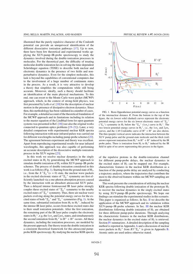
JING, BELLO, MARTÍN, PALACIOS, AND MADSEN PHYSICAL REVIEW A 97, 043426 (2018)
illustrated that the purely repulsive character of the Coulombpotential can provide an unequivocal identification of thedifferent dissociative ionization pathways [17]. Up to now,there have been few theoretical and experimental works ap-plying the XUV-pump–IR-probe spectroscopy to study thedynamics involved during the double-ionization processes inmolecules. For the theoretical part, the difficulty of treatingmolecular double ionization lies in solving the time-dependentSchrödinger equation (TDSE) to describe both nuclear andelectronic dynamics in the presence of two fields and non-perturbative dynamics. Even for the simplest molecules, thistask is beyond the capabilities of conventional computers dueto the involvement of a huge number of continuum statesin the process. As a result, it is very attractive to developa theory that simplifies the computations while still beingaccurate. Moreover, ideally, such a theory should facilitatean identification of the main physical mechanisms. To thisend, one can resort to the Monte Carlo wave packet (MCWP)approach, which, in the context of strong-field physics, wasfirst presented by Leth et al. [10] for the description of nuclearmotion in the presence of dissipative electron dynamics. Sincethen, this methodology has been validated in a number of cases.For example, a detailed account of the theoretical foundation ofthe MCWP approach and its limitations including its relationto the master equation of the Lindblad form for open quantumsystems was presented in Ref. [18] (see also the original worksconnected to quantum optics problems [19–21]). Later, a verydetailed comparison with experimental nuclear KER spectrafollowing interaction with near-infrared pulses was carried outfor different wavelengths including pump-probe schemes [22].The agreement between experiment and theory was excellent.Apart from reproducing experimental results for near infraredwavelengths, this approach was also capable of performingan accurate description of the dissociative multiple-ionizationprocess in the XUV regime [23].
In this work we resolve nuclear dynamics in the singlyexcited states in H2 by generalizing the MCWP approach tosimulate double ionization of H2 within XUV-pump–IR-probeschemes. The process of double ionization considered in thiswork is as follows (Fig. 1). Starting from the ground state of H2,i.e., from the X 1�g
+(v = 0) state, the nuclear wave packetsin the excited electronic states of 1�u
+ symmetry are first ef-ficiently launched via a one-photon absorption process causedby the interaction with an ultrashort attosecond XUV pulse.Then a delayed intense femtosecond IR laser pulse stronglycouples these excited states of 1�u
+ symmetry to the nearbyexcited states of 1�g
+ symmetry. Thus, there are nuclear wavepackets evolving along the potential energy curves for the ex-cited states of both 1�g
+ and 1�u+ symmetries (Fig. 1). At the
same time, substantial ionization from H2 to H2+, induced by
the intense IR laser pulse, occurs from these excited states dueto their small ionization potentials. After the first ionization,the remaining part of the IR laser pulse couples the electronicstates in H2
+, e.g, the 1sσg and 2pσu states, and simultaneouslythe second ionization from H2
+ to H+ + H+ occurs. All thesedynamics, including the ionization processes, are modeled bythe present approach. Our theory treats the double ionization ina consistent theoretical framework for this attosecond pump-probe KER spectroscopy. By studying the nuclear KER spectra
FIG. 1. Born-Oppenheimer potential energy curves as a functionof the internuclear distance R. From the bottom to the top of thefigure, the six lowest solid (dashed) curves represent the electronicpotential energy curves for the six lowest electronic states of 1�g
+
(1�u+) symmetry in H2 below the 2�g
+ (1sσg) curve in H2+. The
two lowest potential energy curves in H2+, i.e., the 1sσg and 2pσu
curves, and the 1/R Coulombic curve of H+ + H+ are also shown.The thin (purple) vertical arrow indicates the interaction between theXUV pump pulse and the ground-state molecule and the thick (red)arrows represent ionization from H2
+ to H+ + H+ induced by the IRprobe pulse. There is ionization from H2 to H2
+ induced by the IRfield in spite of no arrow representing this process in the figure.
of the repulsive protons in the double-ionization channelfor different pump-probe delays, the nuclear dynamics inthe excited states of H2 can be mapped out. For example,characteristic features in the nuclear KER distribution as afunction of the pump-probe delay are analyzed by conductinga trajectory analysis, where the trajectories that contribute themost to the observed features within our MCWP sampling areidentified.
This work presents the consideration of utilizing the nuclearKER spectra following double ionization of the prototype H2
to recover the nuclear dynamics in the singly excited statesby using XUV-pump–IR-probe spectroscopy. We hope ourfindings can stimulate the experimental studies in this scenario.This paper is organized as follows. In Sec. II we describe theapplication of the MCWP approach and its validation withinXUV-pump–IR-probe schemes. In Sec. III the nuclear KERdistributions following double ionization of H2 are obtainedfor three different pump-pulse durations. Through analyzingthe characteristic features in the nuclear KER distributions,the nuclear dynamics in the excited states of H2 is retrieved.Section IV summarizes. A detailed description of the samplingmethod is reported in Appendix A and a discussion of nuclearwave packets in H2
+ from B3 1�u+ is given in Appendix B.
Atomic units are used unless otherwise stated.
043426-2
94

MONTE CARLO WAVE-PACKET APPROACH TO TRACE … PHYSICAL REVIEW A 97, 043426 (2018)
II. METHOD
A. The MCWP approach
The MCWP approach has been thoroughly described inRefs. [10,18,22–26]. In this work we extend it to obtain thenuclear KER spectrum of the protons following double ion-ization of H2 in the two-color XUV-pump–IR-probe schemewhere singly excited states in H2 are involved. When we usethe MCWP approach to describe double ionization of H2, threesystems are involved, i.e., the neutral H2, H2
+, and H22+. For
a specific system containing electrons, due to its interactionwith the applied external field, ionization may occur whichtakes the specific system to the system with one electron less.Meanwhile, the electron removed from the system is now in thecontinuum states. The MCWP approach regards the integratedinfluence of the continuum states as the surroundings of agiven system. Thus, the unidirectional loss of electrons froma given system to its surroundings can be treated by addinga non-Hermitian term to the system Hamiltonian. For a givensystem, the Hamiltonian is
H = Hs − i
2
∑m
C†mCm. (1)
The equivalence of the constructed non-Hermitian Hamilto-nian (1) to the Lindblad form master equation for an opensystem is shown in Ref. [18]. In Eq. (1), Cm is a jump operator,which specifies the transition from the electronic state |φel
R,m〉in a given charge state to the electronic states |φel
R,n〉 in thesystem with one electron less [10,18], i.e.,
Cm =∑
n
∫d �R
√�m( �R,t)Mn( �R,t)
∣∣φelR,n
⟩⟨φel
R,m
∣∣ ⊗ | �R〉〈 �R|,
(2)
where | �R〉 is the position eigenket of the nuclear coordi-nate and �m( �R,t) is the ionization rate for the moleculewhen the electron (electrons) is (are) in the |φel
R,m〉 state.
The term∑
n Mn( �R,t)|φelR,n〉 in Eq. (2) represents the field-
dressed ground state in the new charge state, which is alinear combination of the field-free electronic states |φel
R,n〉.For example, following a quantum jump from H2, the ex-pression for Mn( �R,t) in H2
+ can be found in Ref. [27]. Theabove-mentioned electronic states, which have a parametricdependence on the internuclear distance �R, can be obtainedby solving the time-independent Schrödinger equation ofthe electronic Hamiltonian Hel at fixed �R, i.e., Hel|φel
R,m〉 =Eel
m( �R)|φelR,m〉. Since the influence of the continuum states is
integrated in the non-Hermitian term in Eq. (1), the electronicstates involved in the jump operator in Eq. (2) are boundstates. The electronic Hamiltonian Hel is related to the systemHamiltonian Hs through Hs = TN + Hel + VL(t), where TN isthe nuclear kinetic energy operator and VL(t) = −�μ · �F (t) isthe light-molecule interaction operator. In this operator, �μ isdipole operator of the electrons and �F (t) = �FXUV(t) + �FIR(t)is the combination of the electric fields of the applied pump andprobe pulses. In this work, both pulses are linearly polarizedparallel to the molecular axis. For the XUV pulse, its vector
potential is described by a sin2 envelope, i.e.,
AXUV(t) = AXUV0 sin2
(πt
TXUV
)sin(ωXUVt), (3)
where TXUV is the XUV pulse duration and ωXUV is the centralfrequency of the XUV pulse. The instantaneous field strengthof the XUV pulse is obtained from the relation FXUV(t) =−dAXUV(t)/dt , where AXUV0ωXUV is its peak field strength.For the time-delayed femtosecond IR laser pulse, the electricfield profile is Gaussian, i.e.,
FIR(t) = FIR0 exp
(− 2 ln 2(t − τ − TXUV/2)2
T 2IR
)
× cos[ωIR(t − τ − TXUV/2)], (4)
where FIR0 is the peak field strength, τ the time delay betweenthe two pulses, ωIR the central frequency of the IR field, andTIR the FWHM duration of the temporal intensity profile of theIR laser pulse. In this work, both the first and second ionizationevents of H2 are overwhelmingly induced by the IR laser pulse,i.e., the XUV pulse is relatively weak with its central photonenergy well below the first ionization threshold of H2. Fordissociative double ionization of H2, the three involved chargestates correspond to H2, H2
+, and H+ + H+, respectively.For a specific charge state, the total wave function |(t)〉
satisfies the TDSE i|(t)〉 = H (t)|(t)〉, with H (t) fromEq. (1) and |(t)〉 expressed using the ansatz
|(t)〉 =∑m
∫d �R χm( �R,t)|φel
R,m〉 ⊗ | �R〉, (5)
where χm( �R,t) is the nuclear wave function corresponding tothe electronic state |φel
R,m〉. Similar to the jump operator inEq. (2), the total wave function in Eq. (5) is also a coherentsuperposition of bound electronic states. Substituting Eq. (5)into the TDSE and projecting on the electronic eigenstate〈φel
R,m|, we obtain a system of coupled equations for the nuclearwave functions
iχm( �R,t) =(
TN + Eelm( �R) − i
2�m( �R,t)
)χm( �R,t)
+∑k �=m
Vmk( �R,t)χk( �R,t), (6)
with Vmk( �R,t) = 〈φelR,m|VL(t)|φel
R,k〉 = −〈φelR,m| �μ|φel
R,k〉 · �F (t),where we have neglected nonadiabatic couplings. The last termin Eq. (6) describes the influence from the dipole couplingsbetween the |φel
R,m〉 state and the other states within a givensystem, e.g., in either H2 or H2
+. Equation (6) shows thatthe nuclear wave packet χm( �R,t) evolves along its electronicpotential energy surface Eel
m( �R) accompanied by coherentcoupling with the other bound electronic states in the samecharge state and ionization loss to the adjacent charge state.
We assume that the molecule is aligned along the linearpolarization direction of �F (t). This assumption reduces thethree-dimensional coupled equations to one-dimensional equa-tions and the equations of motion within a given charge state
043426-3
95
JING, BELLO, MARTÍN, PALACIOS, AND MADSEN PHYSICAL REVIEW A 97, 043426 (2018)
can be written in matrix-vector form as
i∂
∂t
⎡⎢⎢⎢⎣
· · ·χm(R,t)
· · ·χk(R,t)
· · ·
⎤⎥⎥⎥⎦ =
⎡⎢⎢⎢⎢⎣
· · · · · · · · · · · · · · ·· · · − d2
dR2 + Eelm(R) − i
2�m(R,t) · · · Vmk(R,t) · · ·· · · · · · · · · · · · · · ·· · · Vkm(R,t) · · · − d2
dR2 + Eelk (R) − i
2�k(R,t) · · ·· · · · · · · · · · · · · · ·
⎤⎥⎥⎥⎥⎦
⎡⎢⎢⎢⎣
· · ·χm(R,t)
· · ·χk(R,t)
· · ·
⎤⎥⎥⎥⎦, (7)
where each χm(R,t) is expanded in a primitive basis for the R
degree of freedom. To obtain the complete nuclear dynamicsfor double ionization of H2, we solve Eq. (7) in three chargestates corresponding to H2, H2
+, and H+ + H+, respectively.It is the quantum jump that transfers nuclear motion in a givencharge state to nuclear motion in the charge state with oneelectron less. Thus, in our model, we have the first jump fromH2 to H2
+ following the first ionization and the second jumpfrom H2
+ to H+ + H+ following the second ionization. InFig. 1 the relevant Born-Oppenheimer potential energy curvesare plotted. The energies corresponding to the ground andexcited states of the neutral molecule are taken from accuratecorrelated ab initio calculations [28,29]. The cation energiesare obtained from exact simulations using a one-electrondiatomic molecule code [30]. The figure shows the potentialenergy curves for the six lowest electronic states of 1�g
+ and1�u
+ symmetries in H2, the potential energy curves for thetwo lowest electronic states in H2
+, and the 1/R Coulombiccurve of H+ + H+. The dipole moment functions betweenelectronic states entering the couplings Vmk(R,t) have beenobtained through a configuration-interaction calculation for H2
by using a procedure similar to that described in Ref. [31] andtaken from the literature for H2
+ [32].To make Eq. (7) solvable, it is necessary to have an estimate
of the state-resolved ionization rates �m(R,t). In H2, theR-dependent ionization potentials Im(R) of the consideredsingly excited states are much smaller than the ionizationpotential of the ground state (Fig. 1). Thus, in order to ensureionization from the excited potential energy curves to work inthe tunneling regime, the central wavelength of the IR laserpulse is chosen to be long enough to satisfy the correspondingionization conditions, i.e., the IR photon energy ωIR shouldbe smaller than the ionization potential for the highest-lyingexcited state in H2 in Fig. 1 and the Keldysh parameterγ for this state should be smaller than 1 [33]. Apart fromnumerically solving the TDSE [34], methods such as themolecular Ammosov-Delone-Krainov theory [35,36] and theweak-field asymptotic theory [37–41] have been developedto obtain analytical formulas for the tunneling ionizationrates for molecules. The tunneling ionization rates from thetwo lowest electronic states in H2
+, i.e, the 1sσg and 2pσu
states, were ab initio values taken from Ref. [42]. Becauseof the charge-resonance-enhanced ionization (CREI), for thepeak probe intensity used in this work, the R-dependentionization rates from the 2pσu state are relatively large atinternuclear distances around R = 7 and 11 a.u. Due to lack ofmore accurate published results, the tunneling ionization ratesinduced by the IR laser pulse from the excited states |φel
R,m〉 inH2 are approximated by the exponential factor in the tunnelingformulas, i.e, �m(R,t) = exp{−2[
√2Im(R)]3/[3FIR(t)]}. We
apply the split-operator method to the short-time propagator
to solve Eq. (7) for H2 and H2+ as specified by the quantum
jumps from H2 to H2+ and from H2
+ to H+ + H+ (see below).The practical application of the MCWP approach contains
the following three steps. (i) In H2, starting from the groundstate of H2 (X 1�g
+,v = 0), we first evolve the initial nuclearwave packet along the potential energy curves in H2 (weinclude 12 curves in this case) under the influence of the pumpand probe pulses. The overall probability of staying in H2,i.e., PH2 (t) = ∑
m
∫dR|χm(R,t)|2, decreases over time since
the IR pulse induces ionization. The first jumps from H2 toH2
+ are assumed to have a chance to occur at each timestep and the ionization probability P1(t) within a small timeinterval t around t during the presence of the IR field isthe drop in the probability PH2 (t) at adjacent time steps, i.e.,P1(t) = PH2 (t) − PH2 (t + t). For a given first jump time t1,there are 12 individual pathways corresponding to jumps fromthe 12 involved potential energy curves to H2
+ and the relativeprobability from the potential energy curve for the |φel
R,m〉 stateis P1m(t1) = 〈(t1)|C+
mCm|(t1)〉/∑m〈(t1)|C+
mCm|(t1)〉.(ii) After the first ionization, the initial state in H2
+ afterthe first jump at t1 from the |φel
R,m〉 state is Cm|(t1)〉 =∑k=g,u
∫dR
√�m(R,t1)Mk(R)χm(R,t1)|φel
R,k〉 ⊗ |R〉. Here g
and u denote the 1sσg and 2pσu states in H2+, respectively,
and Mk(R) describes the field-dressed ground state in H2+
and it is taken from Ref. [27]. Inserting this normalizedinitial nuclear wave packet into Eq. (7), we obtain the evo-lution of the nuclear wave packets along the 1sσg and 2pσu
curves under the influence of the IR pulse. The ionizationprobability within a time interval t around t for the sec-ond jump from H2
+ to H+ + H+ is P2(t ; t1) = PH2+ (t ; t1) −
PH2+ (t + t ; t1), where PH2
+(t ; t1) = ∑g,u
k
∫dR|χk(R,t)|2 is
the overall probability of being in H2+ at time t . For a
given second jump time t2, the relative probabilities for thesecond jumps taking place from the two pathways, i.e., fromthe 1sσg and 2pσu curves to H+ + H+, are P2k(t2; t1) =〈(t2)|C+
k Ck|(t2)〉/∑k〈(t2)|C+
k Ck|(t2)〉 with k = g oru. (iii) After the second ionization, since there is only theCoulombic curve in H+ + H+, the term Mm in Eq. (2) forthe second jump operators is unity, thus the initial wave packetin H+ + H+, corresponding to the case where the second jumptakes place from the 1sσg (2pσu) curve, is, before normal-ization, χc(R,t) = √
�g(R,t2)χg(R,t2) [√
�u(R,t2)χu(R,t2)].After removing two electrons from H2, the IR pulse has noinfluence on the evolution of the nuclear wave packets in H+ +H+ and the nuclear KER spectra are obtained by projecting therenormalized nuclear wave packets χc(R,t2) in H+ + H+ onthe energy-normalized Coulomb waves KE(R), i.e.,
PE,mk(t2; t1) =∣∣∣∣∫
dR KE(R)χc(R,t2)
∣∣∣∣2
, (8)
043426-4
96
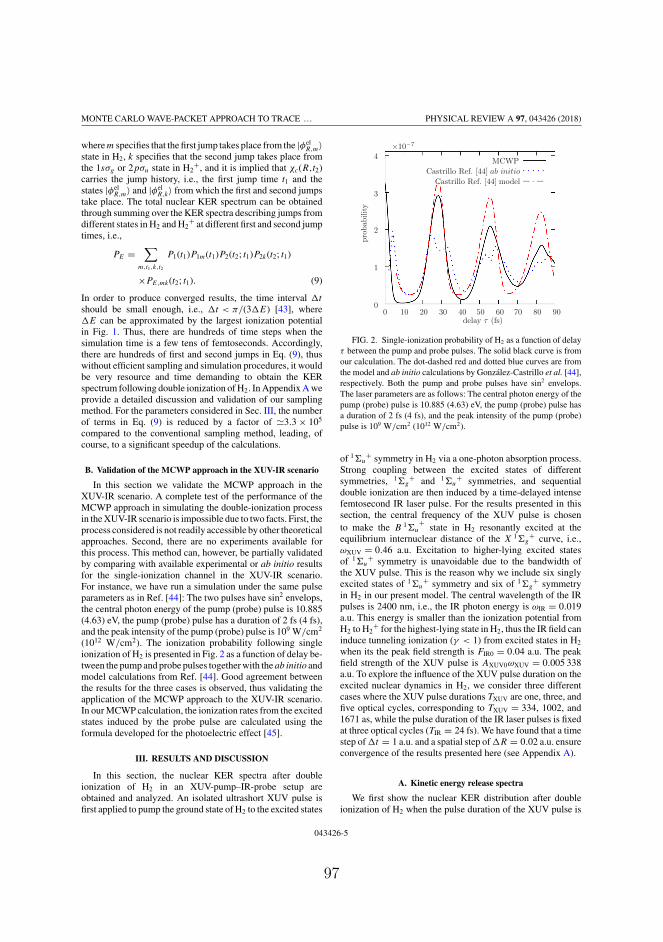
MONTE CARLO WAVE-PACKET APPROACH TO TRACE … PHYSICAL REVIEW A 97, 043426 (2018)
where m specifies that the first jump takes place from the |φelR,m〉
state in H2, k specifies that the second jump takes place fromthe 1sσg or 2pσu state in H2
+, and it is implied that χc(R,t2)carries the jump history, i.e., the first jump time t1 and thestates |φel
R,m〉 and |φelR,k〉 from which the first and second jumps
take place. The total nuclear KER spectrum can be obtainedthrough summing over the KER spectra describing jumps fromdifferent states in H2 and H2
+ at different first and second jumptimes, i.e.,
PE =∑
m,t1,k,t2
P1(t1)P1m(t1)P2(t2; t1)P2k(t2; t1)
×PE,mk(t2; t1). (9)
In order to produce converged results, the time interval t
should be small enough, i.e., t < π/(3 E) [43], where E can be approximated by the largest ionization potentialin Fig. 1. Thus, there are hundreds of time steps when thesimulation time is a few tens of femtoseconds. Accordingly,there are hundreds of first and second jumps in Eq. (9), thuswithout efficient sampling and simulation procedures, it wouldbe very resource and time demanding to obtain the KERspectrum following double ionization of H2. In Appendix A weprovide a detailed discussion and validation of our samplingmethod. For the parameters considered in Sec. III, the numberof terms in Eq. (9) is reduced by a factor of �3.3 × 10⁵
compared to the conventional sampling method, leading, ofcourse, to a significant speedup of the calculations.
B. Validation of the MCWP approach in the XUV-IR scenario
In this section we validate the MCWP approach in theXUV-IR scenario. A complete test of the performance of theMCWP approach in simulating the double-ionization processin the XUV-IR scenario is impossible due to two facts. First, theprocess considered is not readily accessible by other theoreticalapproaches. Second, there are no experiments available forthis process. This method can, however, be partially validatedby comparing with available experimental or ab initio resultsfor the single-ionization channel in the XUV-IR scenario.For instance, we have run a simulation under the same pulseparameters as in Ref. [44]: The two pulses have sin2 envelops,the central photon energy of the pump (probe) pulse is 10.885(4.63) eV, the pump (probe) pulse has a duration of 2 fs (4 fs),and the peak intensity of the pump (probe) pulse is 109 W/cm2
(1012 W/cm2). The ionization probability following singleionization of H2 is presented in Fig. 2 as a function of delay be-tween the pump and probe pulses together with the ab initio andmodel calculations from Ref. [44]. Good agreement betweenthe results for the three cases is observed, thus validating theapplication of the MCWP approach to the XUV-IR scenario.In our MCWP calculation, the ionization rates from the excitedstates induced by the probe pulse are calculated using theformula developed for the photoelectric effect [45].
III. RESULTS AND DISCUSSION
In this section, the nuclear KER spectra after doubleionization of H2 in an XUV-pump–IR-probe setup areobtained and analyzed. An isolated ultrashort XUV pulse isfirst applied to pump the ground state of H2 to the excited states
×10−7
delay τ (fs)
pro
bability
MCWP
Castrillo Ref. [44] ab initio
Castrillo Ref. [44] model
0 10 20 30 40 50 60 70 80 900
1
2
3
4
FIG. 2. Single-ionization probability of H2 as a function of delayτ between the pump and probe pulses. The solid black curve is fromour calculation. The dot-dashed red and dotted blue curves are fromthe model and ab initio calculations by González-Castrillo et al. [44],respectively. Both the pump and probe pulses have sin2 envelops.The laser parameters are as follows: The central photon energy of thepump (probe) pulse is 10.885 (4.63) eV, the pump (probe) pulse hasa duration of 2 fs (4 fs), and the peak intensity of the pump (probe)pulse is 109 W/cm2 (1012 W/cm2).
of 1�u+ symmetry in H2 via a one-photon absorption process.
Strong coupling between the excited states of differentsymmetries, 1�g
+ and 1�u+ symmetries, and sequential
double ionization are then induced by a time-delayed intensefemtosecond IR laser pulse. For the results presented in thissection, the central frequency of the XUV pulse is chosento make the B 1�u
+ state in H2 resonantly excited at theequilibrium internuclear distance of the X 1�g
+ curve, i.e.,ωXUV = 0.46 a.u. Excitation to higher-lying excited statesof 1�u
+ symmetry is unavoidable due to the bandwidth ofthe XUV pulse. This is the reason why we include six singlyexcited states of 1�u
+ symmetry and six of 1�g+ symmetry
in H2 in our present model. The central wavelength of the IRpulses is 2400 nm, i.e., the IR photon energy is ωIR = 0.019a.u. This energy is smaller than the ionization potential fromH2 to H2
+ for the highest-lying state in H2, thus the IR field caninduce tunneling ionization (γ < 1) from excited states in H2
when its the peak field strength is FIR0 = 0.04 a.u. The peakfield strength of the XUV pulse is AXUV0ωXUV = 0.005 338a.u. To explore the influence of the XUV pulse duration on theexcited nuclear dynamics in H2, we consider three differentcases where the XUV pulse durations TXUV are one, three, andfive optical cycles, corresponding to TXUV = 334, 1002, and1671 as, while the pulse duration of the IR laser pulses is fixedat three optical cycles (TIR = 24 fs). We have found that a timestep of t = 1 a.u. and a spatial step of R = 0.02 a.u. ensureconvergence of the results presented here (see Appendix A).
A. Kinetic energy release spectra
We first show the nuclear KER distribution after doubleionization of H2 when the pulse duration of the XUV pulse is
043426-5
97
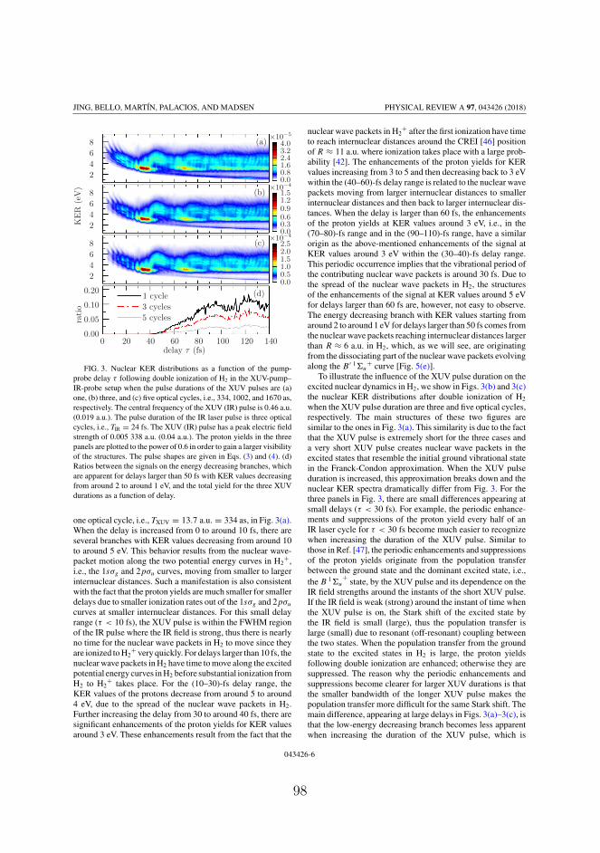
JING, BELLO, MARTÍN, PALACIOS, AND MADSEN PHYSICAL REVIEW A 97, 043426 (2018)
×10−5
×10−4
×10−4
delay τ (fs)
KE
R(e
V)
ratio
0 20 40 60 80 100 120 1400.00
0.05
0.10
0.20
2
4
6
8
2
4
6
8
2
4
6
8
1 cycle
3 cycles
5 cycles
0.00.51.01.52.02.5
0.00.30.60.91.21.5
0.00.81.62.43.24.0(a)
(b)
(c)
(d)
FIG. 3. Nuclear KER distributions as a function of the pump-probe delay τ following double ionization of H2 in the XUV-pump–IR-probe setup when the pulse durations of the XUV pulses are (a)one, (b) three, and (c) five optical cycles, i.e., 334, 1002, and 1670 as,respectively. The central frequency of the XUV (IR) pulse is 0.46 a.u.(0.019 a.u.). The pulse duration of the IR laser pulse is three opticalcycles, i.e., TIR = 24 fs. The XUV (IR) pulse has a peak electric fieldstrength of 0.005 338 a.u. (0.04 a.u.). The proton yields in the threepanels are plotted to the power of 0.6 in order to gain a larger visibilityof the structures. The pulse shapes are given in Eqs. (3) and (4). (d)Ratios between the signals on the energy decreasing branches, whichare apparent for delays larger than 50 fs with KER values decreasingfrom around 2 to around 1 eV, and the total yield for the three XUVdurations as a function of delay.
one optical cycle, i.e., TXUV = 13.7 a.u. = 334 as, in Fig. 3(a).When the delay is increased from 0 to around 10 fs, there areseveral branches with KER values decreasing from around 10to around 5 eV. This behavior results from the nuclear wave-packet motion along the two potential energy curves in H2
+,i.e., the 1sσg and 2pσu curves, moving from smaller to largerinternuclear distances. Such a manifestation is also consistentwith the fact that the proton yields are much smaller for smallerdelays due to smaller ionization rates out of the 1sσg and 2pσu
curves at smaller internuclear distances. For this small delayrange (τ < 10 fs), the XUV pulse is within the FWHM regionof the IR pulse where the IR field is strong, thus there is nearlyno time for the nuclear wave packets in H2 to move since theyare ionized to H2
+ very quickly. For delays larger than 10 fs, thenuclear wave packets in H2 have time to move along the excitedpotential energy curves in H2 before substantial ionization fromH2 to H2
+ takes place. For the (10–30)-fs delay range, theKER values of the protons decrease from around 5 to around4 eV, due to the spread of the nuclear wave packets in H2.Further increasing the delay from 30 to around 40 fs, there aresignificant enhancements of the proton yields for KER valuesaround 3 eV. These enhancements result from the fact that the
nuclear wave packets in H2+ after the first ionization have time
to reach internuclear distances around the CREI [46] positionof R ≈ 11 a.u. where ionization takes place with a large prob-ability [42]. The enhancements of the proton yields for KERvalues increasing from 3 to 5 and then decreasing back to 3 eVwithin the (40–60)-fs delay range is related to the nuclear wavepackets moving from larger internuclear distances to smallerinternuclear distances and then back to larger internuclear dis-tances. When the delay is larger than 60 fs, the enhancementsof the proton yields at KER values around 3 eV, i.e., in the(70–80)-fs range and in the (90–110)-fs range, have a similarorigin as the above-mentioned enhancements of the signal atKER values around 3 eV within the (30–40)-fs delay range.This periodic occurrence implies that the vibrational period ofthe contributing nuclear wave packets is around 30 fs. Due tothe spread of the nuclear wave packets in H2, the structuresof the enhancements of the signal at KER values around 5 eVfor delays larger than 60 fs are, however, not easy to observe.The energy decreasing branch with KER values starting fromaround 2 to around 1 eV for delays larger than 50 fs comes fromthe nuclear wave packets reaching internuclear distances largerthan R ≈ 6 a.u. in H2, which, as we will see, are originatingfrom the dissociating part of the nuclear wave packets evolvingalong the B ′ 1�u
+ curve [Fig. 5(e)].To illustrate the influence of the XUV pulse duration on the
excited nuclear dynamics in H2, we show in Figs. 3(b) and 3(c)the nuclear KER distributions after double ionization of H2
when the XUV pulse duration are three and five optical cycles,respectively. The main structures of these two figures aresimilar to the ones in Fig. 3(a). This similarity is due to the factthat the XUV pulse is extremely short for the three cases anda very short XUV pulse creates nuclear wave packets in theexcited states that resemble the initial ground vibrational statein the Franck-Condon approximation. When the XUV pulseduration is increased, this approximation breaks down and thenuclear KER spectra dramatically differ from Fig. 3. For thethree panels in Fig. 3, there are small differences appearing atsmall delays (τ < 30 fs). For example, the periodic enhance-ments and suppressions of the proton yield every half of anIR laser cycle for τ < 30 fs become much easier to recognizewhen increasing the duration of the XUV pulse. Similar tothose in Ref. [47], the periodic enhancements and suppressionsof the proton yields originate from the population transferbetween the ground state and the dominant excited state, i.e.,the B 1�u
+ state, by the XUV pulse and its dependence on theIR field strengths around the instants of the short XUV pulse.If the IR field is weak (strong) around the instant of time whenthe XUV pulse is on, the Stark shift of the excited state bythe IR field is small (large), thus the population transfer islarge (small) due to resonant (off-resonant) coupling betweenthe two states. When the population transfer from the groundstate to the excited states in H2 is large, the proton yieldsfollowing double ionization are enhanced; otherwise they aresuppressed. The reason why the periodic enhancements andsuppressions become clearer for larger XUV durations is thatthe smaller bandwidth of the longer XUV pulse makes thepopulation transfer more difficult for the same Stark shift. Themain difference, appearing at large delays in Figs. 3(a)–3(c), isthat the low-energy decreasing branch becomes less apparentwhen increasing the duration of the XUV pulse, which is
043426-6
98

MONTE CARLO WAVE-PACKET APPROACH TO TRACE … PHYSICAL REVIEW A 97, 043426 (2018)
because the smaller bandwidth makes direct excitation fromthe ground state to the B ′ 1�u
+ state (responsible for thisbranch) less likely. We plot in Fig. 3(d) the ratios between thesignals in the energy decreasing branches and the total protonyields for the three XUV pulse durations, which clearly showthat the energy decreasing branch is relatively less importantfor a larger XUV pulse duration.
B. Trajectory and wave-packet analysis
Now we concentrate on identifying the origin of the above-outlined characteristic structures in Fig. 3. We first conducta trajectory analysis for a specific characteristic feature inFig. 3(a) as an example to show the strength of the MCWPapproach in obtaining physical insights, including the state-and time-resolved information, of a given feature in the nuclearKER spectrum. Conducting a trajectory analysis is a uniquepossibility in the MCWP approach and it can tell us theindividual contributions from all the involved trajectories tothe signal for a specific KER value at a specific delay inthe nuclear KER distribution. Here a trajectory means thecomplete dynamics leading to the final nuclear wave packetalong the Coulombic curve. A trajectory is specified by fourparameters: the first and second jump times t1 and t2 and thestates from which the first and second jumps take place. Thus,by performing the trajectory analysis, we can, for any givenfeature in the KER spectra, identify the propagation in theneutral H2, the first jump time from the H2 to the H2
+ system,the propagation in H2
+, and the final second jump from H2+
to the Coulomb explosion channel. It is interesting to analyzethe nuclear KER spectra for τ ≈ 71.5 fs in Fig. 3(a) since atleast two important structures are apparent for this value of thetime delay τ , e.g., the enhancements of the proton yields atKER values around 3 eV and low-energy decreasing branchstarting from τ ≈ 50 fs. When we carry out an analysis of thecontributions to the signal at a given KER value in a nuclearKER spectrum, it is possible to obtain the origin of the signalsat other KER values at the same time. This is because thesignals for all the KER values in the same nuclear spectrumare formed from the same collection of trajectories with eachtrajectory giving its individual contribution to a particularfeature. Thus, we present in Figs. 4(b)–4(d) the results obtainedafter a trajectory analysis to the signal for a KER equal to 3 eVat τ = 71.5 fs in Fig. 3(a). Figure 4(b) shows the respectiveprobabilities for the first jump taking place from the states inH2 for the considered energy (3.0 eV). We can see that thefirst jumps from the HH 1�g
+ and B3 1�u+ states make
the largest contributions to the signal. In Figs. 4(c) and 4(d),the contributions from the trajectories for the first jump takingplace from the HH 1�g
+ and B3 1�u+ states and the second
jumps from the 2pσu state are presented as a function of t1and t2, respectively. Contributions from the trajectories forthe second jumps taking place from the 1sσg state are notpresented since they give much smaller contributions becausethe ionization rates from the 1sσg curve are about two ordersof magnitude smaller than those from the 2pσu curve.
For a fixed first jump time, a clear periodic dependenceof the dominant trajectories on the second jump time t2 isobserved in Figs. 4(c) and 4(d). These dominant trajectoriescorrespond to ionization events taking place at instants around
×10-8
×10-8
×10-8
t2 (a.u.) t2 (a.u.)
E(a
.u.)
t 1(a
.u.)
t 1(a
.u.)
R (a.u.)
2000 3000 4000 2000 3000 4000
2000
2500
0
0.5
1.0
1.5
-0.75-0.70
-0.65-0.60
-0.55
(a)
(b)
(c)
B31Σ+u
(d)
HH1Σ+
g
0 5 10 15 20
1.2
0.8
0.4
0
1.2
0.8
0.4
0
B1Σ+u
B’1Σ+u
B31Σ+u
B41Σ+u
B51Σ+u
B61Σ+u
X1Σ+g
EF1Σ+g
GK1Σ+g
HH1Σ+
gP1Σ+
g
O1Σ+g
X1Σ
+ g
EF
1Σ
+ g
GK
1Σ
+ g
HH
1Σ
+ g
P1Σ
+ g
O1Σ
+ g
B1Σ
+ u
B’1Σ
+ u
B31Σ
+ u
B41Σ
+ u
B51Σ
+ u
B61Σ
+ u
FIG. 4. Trajectory analysis for the enhancement of the protonyields at a specific point in Fig. 3(a): with the KER equal to 3 eVand τ = 71.5 fs. (a) For better visibility, the potential energy curvesfor the 12 states of 1�g
+ and 1�u+ symmetries in H2 are plotted within
the energy region between −0.5 and −0.8 eV. (b) Contributions fromdifferent states in H2 to the signal of the considered energy. Alsoshown are the trajectories making contributions to the signal fromthe (c) HH 1�g
+ and (d) B3 1�u+ states. The other laser parameters
are as follows: ωXUV = 0.46 a.u., ωIR = 0.019 a.u., TXUV = 13.7 a.u.(334 as), and TIR = 992 a.u. (24 fs). The peak electric field strengthsare 0.005 338 and 0.04 a.u. for the XUV and the IR fields, respectively.The pulse shapes are given in Eqs. (3) and (4).
the field extrema with large second ionization rates from the2pσu curve in H2
+. For the HH 1�g+ state, the most important
trajectory corresponds to the first jump occurring at 2193 a.u.(53 fs) and the second jump at around 3039 a.u. (74 fs). For theB3 1�u
+ state, the most important trajectory is the first jumpoccurring at 2193 a.u. (53 fs) and the second jump at around2889 a.u. (70 fs). The time difference between the first and sec-ond jump times for the two cases (21 and 17 fs) is the evolutiontime of the nuclear wave packets in H2
+. The enhancements ataround 3.0 eV in Fig. 3(a) are mainly from ionization eventsoccurring at the CREI position of R ≈ 11 a.u. from the 2pσu
curve in H2+. These evolution times in H2
+ imply that thecontributing nuclear wave packets move slowly along the 2pσu
curve. This can be verified by the relatively small kineticenergies for these two trajectories before the second ionizationtaking place, which is the difference between the final kineticenergy and the Coulomb repulsion energy at around R ≈11 a.u., i.e., 3 eV − (1/11) a.u. × 27.21 eV/a.u. = 0.53 eV.Thus, the kinetic energies of the contributing nuclear wavepackets at t1 = 2193 a.u. (53 fs) obtained by moving alongthe HH 1�g
+ and B3 1�u+ curves are very small and the
internuclear distances of the contributing nuclear wave packetsalong these two curves at t1 = 2193 a.u. (53 fs) are quite large.
043426-7
99

JING, BELLO, MARTÍN, PALACIOS, AND MADSEN PHYSICAL REVIEW A 97, 043426 (2018)
×10-5
×10-4
×10-6
×10-4
t (a.u.) t (a.u.)
R(a
.u.)
R(a
.u.)
R(a
.u.)
0 1000 2000 0 1000 2000
3000 4000 5000 3000 4000 5000
2
4
6
8
102
4
6
8
10
0
10
20
30(a)
(c)
(e)
(b)
(d)
(f)
0510152025
01
2
3
4
0
0.1
0.2
0.30.8
0.6
0.4
0.2
0
8
6
4
2
0
16
128
40
FIG. 5. Evolution of the nuclear wave packets |χm(R,t)|2 alongthe (a) 1sσg and (b) 2pσu curves in H2
+ from the HH 1�g+ state when
the first jump occurs at t1 = 2193 a.u. (53 fs), which corresponds tothe first jump time t1 of the most important trajectory in Fig. 4(c).Also shown is the evolution of the nuclear wave packets along the(c) B 1�u
+, (d) EF 1�g+, (e) B ′ 1�u
+, and (f) HH 1�g+ curves in
H2. The laser parameters are identical to those in Fig. 4.
To confirm this speculation, we show in Figs. 5(a) and5(b) the evolution of the nuclear wave packets along the 1sσg
and 2pσu curves in H2+ when the first jump takes place at
t1 = 2193 a.u. (53 fs) from the HH 1�g+ state. As seen from
Fig. 4(b), the B3 1�u+ state is as important as the HH 1�g
+
state. The nuclear wave packets motion along the two H2+
curves for the first jumps occurring from the B31�u+ state
at t1 = 2193 a.u. can be obtained and analyzed in the sameway as the wave packets from the HH 1�g
+ state. We havefound that the main features of the evolution of the nuclearwave packets in H2
+ are very similar irrespective of whetherthe intermediate HH 1�g
+ and B3 1�u+ states are used and
therefore here we only show the results associated with theHH 1�g
+ state in Figs. 5(a) and 5(b) (see Appendix B for theresults associated with the B3 1�u
+ state). Now let us returnto Fig. 5(b), where we can clearly see that the nuclear wavepacket moving along the 2pσu curve, starting from around R ≈6.2 a.u., reaches internuclear distances around the CREIposition of R ≈ 11 a.u. at around t2 = 3039 a.u. (74 fs).
We can also see from Fig. 5(b) that the nuclear wavepacket at t2 = 3039 a.u. is distributed over a very broad rangeof internuclear distances, i.e., from R = 5 to 15 a.u. Thus,apart from the above-mentioned nuclear wave packet at aroundR = 11 a.u., the other part of the nuclear wave packet wouldresult in different structures in the nuclear KER spectrum. Thismeans that a detailed analysis of this trajectory is worthwhile.Because of the R-dependent ionization rates out of the 2pσu
curve in H2+, nuclear wave packets along the 2pσu curve
located at internuclear distances with larger ionization rates aremore likely to be ionized to H2
2+. Thus, for a given trajectory,
t2 (a.u.)
R(a
.u.)
2500 3000 3500 40000
5
10
15
20×10−5
0
1
2
3
4
5
6
FIG. 6. Initial nuclear wave packets in H22+ at different second
jump times for the second jumps occurring from the 2pσu state inH2
+, i.e., |χc(R,t2)|2, when the first jump occurs from the HH 1�g+
state at t1 = 2193 a.u.
the initial nuclear wave packet in H22+ is dependent not only on
the nuclear wave packet in H2+ at its second jump time t2 but
also on the instantaneous ionization rates from H2+ to H2
2+ att2, as discussed in Sec. II. In Fig. 6 we show the initial nuclearwave packets in H2
2+ at different second jump times for thesecond jumps occurring from the 2pσu state in H2
+, whenthe first jump occurs from the HH 1�g
+ state in H2 at t1 =2193 a.u. We can see that for the most dominant trajectorydiscussed above, i.e., for the trajectory where the first jumptakes place from the HH 1�g
+ state at t1 = 2193 a.u. andthe second jump from the 2pσu state at t2 = 3039 a.u., thereare enhancements of the nuclear wave packet densities ataround R = 5, 10, and 15 a.u. By applying the reflectionapproximation, i.e., KER ≈ 1
R, such enhancements would
result in KER signals peaking at around 5.5, 3, and 2 eV forthis considered delay, respectively, which are consistent withthe features in the nuclear KER spectrum for the 71.5-fs delayin Fig. 3(a).
To obtain knowledge of the initial wave packets in H2+ we
plot the evolution of the nuclear wave packets in H2 alongthe HH 1�g
+ state in Fig. 5(f). The two regions with largeprobabilities at around t ≈ 1500 a.u. (37 fs) and t ≈ 2000 a.u.(49 fs) are from large dipole couplings between this state andthe adjacent B ′ 1�u
+ state. The evolution of the nuclear wavepackets in the B ′ 1�u
+ state is plotted in Fig. 5(e). We cansee that the population of the nuclear wave packets in theHH 1�g
+ state is out of phase with that in the B ′ 1�u+ state
for these two regions, i.e., t ≈ 1500 and 2000 a.u., directlyshowing a time-dependent population transfer between thesetwo states. That is, at certain instants of time when there is amaximum of population in the B 1�u
+ state [Fig. 5(e)], thereis a minimum of population in the HH 1�g
+ state [Fig. 5(f)]and vice versa. To find the origin of the regions with largeprobabilities in Figs. 5(e) and 5(f), time evolution behaviorsof the nuclear wave packets along the B 1�u
+ and EF 1�g+
curves are also presented in Figs. 5(c) and 5(d). By examiningthe out-of-phase behavior of the nuclear wave packets inFigs. 5(c) and 5(d), it is clear that the large probabilitiesat internuclear distances R ≈ 6 a.u. originate from the largedipole coupling from the dominant B 1�u
+ state to the adjacent
043426-8
100

MONTE CARLO WAVE-PACKET APPROACH TO TRACE … PHYSICAL REVIEW A 97, 043426 (2018)
EF 1�g+ state due to a one-photon resonance occurring. A
similar out-of-phase behavior of the nuclear wave packets inthe EF 1�g
+ and B ′ 1�u+ states for time larger than t =
2000 a.u. can be observed by comparing Figs. 4(d) and 4(e).Thus, the enhancements of the proton yields at KER valuesaround 3.0 eV for the 71.5-fs delay in Fig. 3(a) are mainly fromthe first jumps taking place at around 2193 a.u. (53 fs) fromthe HH 1�g
+ state through a population transfer process, i.e.,B 1�u
+ → EF 1�g+ → B ′ 1�u
+ → HH 1�g+ at internu-
clear distances around R ≈ 6 a.u. Moreover, the enhancementsat KER values around 3.0 eV for the three delay ranges inFig. 3(a) are consistent with the moving nuclear wave packetsalong the B 1�u
+ curve periodically reaching its outer turningpoint around every 30 fs.
Applying the reflection principle, the nuclear wave packetsat around R = 5.5 a.u. along the 2pσu curve in H2
+ [Fig. 5(b)]at around t2 = 3000 a.u. (73 fs) result in the enhancements ofthe proton yields at KER values around 5 eV for the 71.5-fsdelay in Fig. 3(a), i.e., 1
5.5 × 27.2 ≈ 5 eV. In fact, for delayslarger than 30 fs in Fig. 3(a), it is also the nuclear wave packetsat around R = 5.5 a.u. that result in the enhancements of theproton yields at KER values around 5 eV. Moreover, the struc-ture for KER increasing from 3 eV to 5 eV and then decreasingback to 3 eV in the (40–60)-fs delay range in Fig. 3(a) manifeststhe motion of the nuclear wave packet along the B ′ 1�u
+ curvein H2 when the nuclear wave packets along the B 1�u
+ curveare away from the outer turning points. The 20-fs delay range isconsistent with the vibrational period of the nuclear wavepackets along the B ′ 1�u
+ curve [Fig. 5(e)]. Apart from that,the fast moving nuclear wave packets starting at around R =8 a.u. in Fig. 5(b), which originate from the dissociating partof the nuclear wave packets in the B 1�u
+ curve [Fig. 5(e)],later reach distances around R = 15 a.u. at instants of timewhen the instantaneous field strength becomes maximal. Fromthe reflection principle, the nuclear wave packet located ataround R = 15 a.u. results in the appearance of the signalsat KER values around 1
15 × 27.2 eV = 1.8 eV for the 71.5-fsdelay. This KER value is decreased (increased) for a (smaller)larger delay because the dissociation nuclear wave packet inthe B ′ 1�u
+ state would reach larger (smaller) internucleardistances. Thus, energy decreasing branches show up in Fig. 3.As a result, one can benefit greatly from conducting a trajectoryanalysis to a given feature in the nuclear KER spectrum inuncovering its origin and the origins of other features in theconsidered nuclear KER spectrum. By conducting an analysissimilar to that for the other characteristic structures in thenuclear KER distribution in Fig. 3, a thorough understandingof the formation of the structures can be achieved.
IV. CONCLUSION
We have extended the MCWP approach to simulate thefragmentation upon double ionization of H2 in an XUV-pump–IR-probe setup where the ultrafast dynamics of singly excitedstates is involved. We have applied the MCWP approach to thisXUV-IR scenario after providing a validation of this method inthis scenario. This theoretical study accounts for the ionizationinduced by the intense IR pulse from the excited states in H2,
reached from the ground state of H2 by an XUV pulse, andagain the ionization from H2
+ to H+ + H+. We have obtainedthe nuclear KER distributions for three different pump-probeschemes using XUV pulses of durations of one, three, and fiveoptical cycles, respectively, to excite H2 for the pump-probedelays ranging from 0 to 140 fs. The nuclear dynamics inmolecular excited states is reflected in the characteristicfeatures in the nuclear KER distributions and, for instance,the periodic nuclear motion in the B 1�u
+ state is mapped outby trajectory and wave-packet analysis. Apart from that, theStark effect can play a role in determining the efficiency ofpopulation transfer from the ground state to the excited states inH2 for time delays τ < 30 fs. The present MCWP approach canbe applied to XUV-pump–IR-probe attosecond spectroscopyof any molecule to resolve and control ultrafast dynamics in ex-cited states through laser-induced double-ionization processes,provided the Born-Oppenheimer potential energy curves indifferent charge states, the dipole moment functions betweendifferent states, and the state-resolved ionization rates areavailable.
ACKNOWLEDGMENTS
This work was supported by the European UnionHorizon 2020 research and innovation program under theMarie Sklodowska-Curie Grant Agreement No. 641789MEDEA, the Villum Kann Rasmussen Center of Excellence,QUSCOPE, and MINECO Project No. FIS2016-77889-R(Spain). A.P. acknowledges a Ramón y Cajal contract fromthe Ministerio de Economia y Competitividad (Spain). TheMCWP results presented in this work were obtained at theCentre for Scientific Computing, Aarhus, and the dipole tran-sitions moments at the CCC-UAM computer center, Madrid.
APPENDIX A: SAMPLING METHOD
In this appendix we give a detailed description and vali-dation of the MCWP sampling method for XUV-pump–IR-probe spectroscopy. Within the MCWP approach, in order toreproduce experimental results, the time step t and the spacestep R should be chosen small enough. We have found that t = 1 a.u. and R = 0.02 a.u. give converged results for theprocesses studied in this work. Apart from that, in order tocapture the features of the nuclear motion at large internucleardistances, such as the CREI [46] regions for the 2pσu state,and also to reduce the reflection from the boundaries, thesimulation box should be large enough, e.g., Rbox � 40 a.u.For the approach in this work, the first jumps from H2 to H2
+
take place at every time step and the second jumps take placefrom H2
+ to H+ + H+ at every time step after the first jumps.In addition, the first jumps have 12 pathways and the secondjumps have two pathways, corresponding to the number ofstates included in H2 and H2
+, respectively. Thus, for an IRpulse with a duration of 24 fs (992 a.u.), there are at least12 × 2 × 9923/3 terms in Eq. (9). The reduction of the hugenumber of terms in Eq. (9) to a computationally more feasiblelevel is the task of our sampling method.
In fact, the sampling method in our previous works involv-ing only IR fields [10,18,22,24–26] is very resource and timesaving, since it assumes that the first jumps occur at the field
043426-9
101
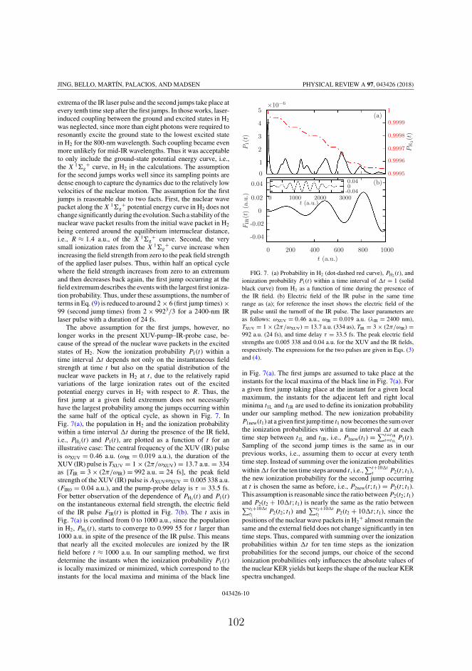
JING, BELLO, MARTÍN, PALACIOS, AND MADSEN PHYSICAL REVIEW A 97, 043426 (2018)
extrema of the IR laser pulse and the second jumps take place atevery tenth time step after the first jumps. In those works, laser-induced coupling between the ground and excited states in H2
was neglected, since more than eight photons were required toresonantly excite the ground state to the lowest excited statein H2 for the 800-nm wavelength. Such coupling became evenmore unlikely for mid-IR wavelengths. Thus it was acceptableto only include the ground-state potential energy curve, i.e.,the X 1�g
+ curve, in H2 in the calculations. The assumptionfor the second jumps works well since its sampling points aredense enough to capture the dynamics due to the relatively lowvelocities of the nuclear motion. The assumption for the firstjumps is reasonable due to two facts. First, the nuclear wavepacket along the X 1�g
+ potential energy curve in H2 does notchange significantly during the evolution. Such a stability of thenuclear wave packet results from the initial wave packet in H2
being centered around the equilibrium internuclear distance,i.e., R ≈ 1.4 a.u., of the X 1�g
+ curve. Second, the verysmall ionization rates from the X 1�g
+ curve increase whenincreasing the field strength from zero to the peak field strengthof the applied laser pulses. Thus, within half an optical cyclewhere the field strength increases from zero to an extremumand then decreases back again, the first jump occurring at thefield extremum describes the events with the largest first ioniza-tion probability. Thus, under these assumptions, the number ofterms in Eq. (9) is reduced to around 2 × 6 (first jump times) ×99 (second jump times) from 2 × 9923/3 for a 2400-nm IRlaser pulse with a duration of 24 fs.
The above assumption for the first jumps, however, nolonger works in the present XUV-pump–IR-probe case, be-cause of the spread of the nuclear wave packets in the excitedstates of H2. Now the ionization probability P1(t) within atime interval t depends not only on the instantaneous fieldstrength at time t but also on the spatial distribution of thenuclear wave packets in H2 at t , due to the relatively rapidvariations of the large ionization rates out of the excitedpotential energy curves in H2 with respect to R. Thus, thefirst jump at a given field extremum does not necessarilyhave the largest probability among the jumps occurring withinthe same half of the optical cycle, as shown in Fig. 7. InFig. 7(a), the population in H2 and the ionization probabilitywithin a time interval t during the presence of the IR field,i.e., PH2 (t) and P1(t), are plotted as a function of t for anillustrative case: The central frequency of the XUV (IR) pulseis ωXUV = 0.46 a.u. (ωIR = 0.019 a.u.), the duration of theXUV (IR) pulse is TXUV = 1 × (2π/ωXUV) = 13.7 a.u. = 334as [TIR = 3 × (2π/ωIR) = 992 a.u. = 24 fs], the peak fieldstrength of the XUV (IR) pulse is AXUVωXUV = 0.005 338 a.u.(FIR0 = 0.04 a.u.), and the pump-probe delay is τ = 33.5 fs.For better observation of the dependence of PH2 (t) and P1(t)on the instantaneous external field strength, the electric fieldof the IR pulse FIR(t) is plotted in Fig. 7(b). The t axis inFig. 7(a) is confined from 0 to 1000 a.u., since the populationin H2, PH2 (t), starts to converge to 0.999 55 for t larger than1000 a.u. in spite of the presence of the IR pulse. This meansthat nearly all the excited molecules are ionized by the IRfield before t ≈ 1000 a.u. In our sampling method, we firstdetermine the instants when the ionization probability P1(t)is locally maximized or minimized, which correspond to theinstants for the local maxima and minima of the black line
t (a.u.)
t (a.u.)
P1(t
)
PH
2(t
)
FIR
(t)
(a.u
.)
×10−6
0 200 400 600 800 1000
0 1000 2000 3000-0.0400.04
-0.04
-0.02
0
0.02
0.04
0
1
2
3
4
5(a)
(b)
0.9995
0.9996
0.9997
0.9998
0.9999
1
FIG. 7. (a) Probability in H2 (dot-dashed red curve), PH2 (t), andionization probability P1(t) within a time interval of t = 1 (solidblack curve) from H2 as a function of time during the presence ofthe IR field. (b) Electric field of the IR pulse in the same timerange as (a); for reference the inset shows the electric field of theIR pulse until the turnoff of the IR pulse. The laser parameters areas follows: ωXUV = 0.46 a.u., ωIR = 0.019 a.u. (λIR = 2400 nm),TXUV = 1 × (2π/ωXUV) = 13.7 a.u. (334 as), TIR = 3 × (2π/ωIR) =992 a.u. (24 fs), and time delay τ = 33.5 fs. The peak electric fieldstrengths are 0.005 338 and 0.04 a.u. for the XUV and the IR fields,respectively. The expressions for the two pulses are given in Eqs. (3)and (4).
in Fig. 7(a). The first jumps are assumed to take place at theinstants for the local maxima of the black line in Fig. 7(a). Fora given first jump taking place at the instant for a given localmaximum, the instants for the adjacent left and right localminima t1L and t1R are used to define its ionization probabilityunder our sampling method. The new ionization probabilityP1new(t1) at a given first jump time t1 now becomes the sum overthe ionization probabilities within a time interval t at eachtime step between t1L and t1R, i.e., P1new(t1) = ∑t=t1R
t=t1LP1(t).
Sampling of the second jump times is the same as in ourprevious works, i.e., assuming them to occur at every tenthtime step. Instead of summing over the ionization probabilitieswithin t for the ten time steps around t , i.e.,
∑t+10 tt P2(t ; t1),
the new ionization probability for the second jump occurringat t is chosen the same as before, i.e., P2new(t ; t1) = P2(t ; t1).This assumption is reasonable since the ratio between P2(t2; t1)and P2(t2 + 10 t ; t1) is nearly the same as the ratio between∑t2+10 t
t2P2(t2; t1) and
∑t2+10 tt2
P2(t2 + 10 t ; t1), since thepositions of the nuclear wave packets in H2
+ almost remain thesame and the external field does not change significantly in tentime steps. Thus, compared with summing over the ionizationprobabilities within t for ten time steps as the ionizationprobabilities for the second jumps, our choice of the secondionization probabilities only influences the absolute values ofthe nuclear KER yields but keeps the shape of the nuclear KERspectra unchanged.
043426-10
102

MONTE CARLO WAVE-PACKET APPROACH TO TRACE … PHYSICAL REVIEW A 97, 043426 (2018)
KER (eV)
sign
al
2 4 6 8 10 120
0.4
0.8
1.2
1.6×10⁻⁸
FIG. 8. Nuclear KER spectra obtained through applying the orig-inal sampling method (solid black curve) and the sampling methodpresented in this work (dashed red curve). The PE on the verticalaxis labels the probability to produce a signal at a given energy. Thelaser parameters are identical to those for Fig. 7(a) except that a 0.5-fspump-probe delay is used.
In order to validate our sampling method, we have con-ducted comparisons between results obtained from this andthe original sampling method. As an example, the nuclearKER spectra obtained by these two sampling methods arepresented in Fig. 8. The laser parameters are identical to thosein Fig. 7(a) except that the pump-probe delay is τ = 0.5 fs.The excellent agreement between the two curves in Fig. 8clearly shows that the sampling method presented herein worksvery well. Our method can reduce the number of terms inEq. (9) from about 12 × 2 × 9923/3 to about 12 × 2 × 10 ×99. Thus, we apply the MCWP approach with this much moreefficient sampling method to obtain the results presented inSec. III.
t (a.u.)
R(a
.u.)
R(a
.u.)
2500 3000 3500 4000 4500 50000
5
10
15
20
25
300
5
10
15
20
25
30(a)
(b)
0
0.1
0.2
0.3
0
0.2
0.4
0.6
FIG. 9. Evolution of the nuclear wave packets along the (a) 1sσg
and (b) 2pσu curves in H2+ when the first jump occurs from the
B3 1�u+ state in H2 at t1 = 2193 a.u.
APPENDIX B: NUCLEAR WAVE PACKETSIN H2
+ FROM B3 1�u+
In this appendix we show in Fig. 9 the nuclear wave packetsalong the 1sσg and 2pσu states in H2
+ when the first jumpoccurs at t1 = 2193 a.u. from the B3 1�u
+ state in H2. Wecan clearly see that the evolution of the nuclear wave packetsfor the first jump occurring from the B3 1�u
+ state (Fig. 9)is very similar to that for the first jump occurring from theHH 1�g
+ state [Figs. 5(a) and 5(b)]. This means that at agiven second jump time t2, the trajectory whose first jumptakes place at t1 = 2193 a.u. from the B3 1�u
+ state wouldresult in a nuclear KER spectrum similar to that associatedwith the trajectory whose first jump takes place at t1 =2193 a.u. from the HH 1�g
+ state.
[1] N. R. Isenor, V. Merchant, R. S. Hallsworth, and M. C.Richardson, CO2 laser-induced dissociation of SiF4 moleculesinto electronically excited fragments, Can. J. Phys. 51, 1281(1973).
[2] K. Codling and L. J. Frasinski, Dissociative ionization of smallmolecules in intense laser fields, J. Phys. B 26, 783 (1993).
[3] H. Niikura, F. Légaré, R. Hasbani, A. D. Bandrauk, M. Y. Ivanov,D. M. Villeneuve, and P. B. Corkum, Sub-laser-cycle electronpulses for probing molecular dynamics, Nature (London) 417,917 (2002).
[4] H. Niikura, F. Légaré, R. Hasbani, M. Y. Ivanov, D. M.Villeneuve, and P. B. Corkum, Probing molecular dynamics withattosecond resolution using correlated wave packet pairs, Nature(London) 421, 826 (2003).
[5] M. F. Kling, C. Siedschlag, A. J. Verhoef, J. I. Khan, M. Schultze,T. Uphues, Y. Ni, M. Uiberacker, M. Drescher, F. Krausz, andM. J. J. Vrakking, Control of electron localization in moleculardissociation, Science 312, 246 (2006).
[6] I. Znakovskaya, P. von den Hoff, G. Marcus, S. Zherebtsov, B.Bergues, X. Gu, Y. Deng, M. J. J. Vrakking, R. Kienberger,F. Krausz, R. de Vivie-Riedle, and M. F. Kling, Subcycle Con-trolled Charge-Directed Reactivity with Few-Cycle MidinfraredPulses, Phys. Rev. Lett. 108, 063002 (2012).
[7] V. Wanie, H. Ibrahim, S. Beaulieu, N. Thiré, B. E. Schmidt,Y. Deng, A. S. Alnaser, I. V. Litvinyuk, X.-M. Tong, andF. Légaré, Coherent control of D2/H2 dissociative ionizationby a mid-infrared two-color laser field, J. Phys. B 49, 025601(2015).
[8] A. S. Alnaser, X. M. Tong, T. Osipov, S. Voss, C. M. Maharjan,P. Ranitovic, B. Ulrich, B. Shan, Z. Chang, C. D. Lin, andC. L. Cocke, Routes to Control of H2 Coulomb Explosion inFew-Cycle Laser Pulses, Phys. Rev. Lett. 93, 183202 (2004).
[9] A. Staudte, D. Pavičić, S. Chelkowski, D. Zeidler, M. Meckel,H. Niikura, M. Schöffler, S. Schössler, B. Ulrich, P. P. Rajeev,T. Weber, T. Jahnke, D. M. Villeneuve, A. D. Bandrauk, C. L.Cocke, P. B. Corkum, and R. Dörner, Attosecond Strobing of
043426-11
103
JING, BELLO, MARTÍN, PALACIOS, AND MADSEN PHYSICAL REVIEW A 97, 043426 (2018)
Two-Surface Population Dynamics in Dissociating H2+, Phys.
Rev. Lett. 98, 073003 (2007).[10] H. A. Leth, L. B. Madsen, and K. Mølmer, Monte Carlo Wave
Packet Theory of Dissociative Double Ionization, Phys. Rev.Lett. 103, 183601 (2009).
[11] K. P. Singh, F. He, P. Ranitovic, W. Cao, S. De, D. Ray, S. Chen,U. Thumm, A. Becker, M. M. Murnane, H. C. Kapteyn, I. V.Litvinyuk, and C. L. Cocke, Control of Electron Localizationin Deuterium Molecular Ions using an Attosecond Pulse Trainand a Many-Cycle Infrared Pulse, Phys. Rev. Lett. 104, 023001(2010).
[12] F. Kelkensberg, W. Siu, J. F. Pérez-Torres, F. Morales, G.Gademann, A. Rouzée, P. Johnsson, M. Lucchini, F. Calegari,J. L. Sanz-Vicario, F. Martín, and M. J. J. Vrakking, AttosecondControl in Photoionization of Hydrogen Molecules, Phys. Rev.Lett. 107, 043002 (2011).
[13] G. Sansone, F. Kelkensberg, J. Pérez-Torres, F. Morales,M. F. Kling, W. Siu, O. Ghafur, P. Johnsson, M. Swoboda,E. Benedetti et al., Electron localization following attosec-ond molecular photoionization, Nature (London) 465, 763(2010).
[14] A. González-Castrillo, A. Palacios, H. Bachau, and F. Martín,Clocking Ultrafast Wave Packet Dynamics in MoleculesThrough UV-Induced Symmetry Breaking, Phys. Rev. Lett. 108,063009 (2012).
[15] A. R. Bainbridge, J. Harrington, A. Kirrander, C. Cacho,E. Springate, W. A. Bryan, and R. S. Minns, VUV exci-tation of a vibrational wavepacket in D2 measured throughstrong-field dissociative ionization, New J. Phys. 17, 103013(2015).
[16] F. P. Sturm, X. M. Tong, A. Palacios, T. W. Wright, I.Zalyubovskaya, D. Ray, N. Shivaram, F. Martín, A. Belkacem,P. Ranitovic, and T. Weber, Mapping and controlling ultrafastdynamics of highly excited H2 molecules by VUV-IR pump-probe schemes, Phys. Rev. A 95, 012501 (2017).
[17] T. Ergler, A. Rudenko, B. Feuerstein, K. Zrost, C. D. Schröter,R. Moshammer, and J. Ullrich, Time-Resolved Imaging andManipulation of H2 Fragmentation in Intense Laser Fields, Phys.Rev. Lett. 95, 093001 (2005).
[18] H. A. Leth, L. B. Madsen, and K. Mølmer, Monte Carlo wavepacket approach to dissociative multiple ionization in diatomicmolecules, Phys. Rev. A 81, 053409 (2010).
[19] J. Dalibard, Y. Castin, and K. Mølmer, Wave-Function Approachto Dissipative Processes in Quantum Optics, Phys. Rev. Lett. 68,580 (1992).
[20] K. Mølmer, Y. Castin, and J. Dalibard, Monte Carlo wave-function method in quantum optics, J. Opt. Soc. Am. B 10, 524(1993).
[21] H. J. Carmichael, An Open Systems Approach to Quantum Optics(Springer, Berlin, 1993).
[22] H. A. Leth, L. B. Madsen, and K. Mølmer, Dissociative doubleionization of H2 and D2: Comparison between experiment andMonte Carlo wave packet calculations, Phys. Rev. A 81, 053410(2010).
[23] H. A. Leth and L. B. Madsen, Dissociative multiple ionizationof diatomic molecules by extreme-ultraviolet free-electron-laserpulses, Phys. Rev. A 83, 063415 (2011).
[24] H. A. Leth, Dissociative ionization—A study using the MonteCarlo wave packet approach, Ph.D. thesis, Aarhus University,2011.
[25] Q. Jing and L. B. Madsen, Laser-induced dissociative ionizationof H2 from the near-infrared to the mid-infrared regime, Phys.Rev. A 94, 063402 (2016).
[26] Q. Jing, Monte Carlo wave packet approach to dissociativedouble ionization of H2 interacting with intense laser pulses,M.Sc. thesis, Aarhus University, 2016.
[27] H. Kono, Y. Sato, Y. Fujimura, and I. Kawata, Identification ofthe doorway states to ionization of molecules in intense laserfields, Laser Phys. 13, 883 (2003).
[28] L. Wolniewicz and K. Dressler, The EF , GK , and HH 1�g+
states of hydrogen. Improved ab initio calculation of vibrationalstates in the adiabatic approximation, J. Chem. Phys. 82, 3292(1985).
[29] G. Staszewska and L. Wolniewicz, Adiabatic energies of excited1�u states of the hydrogen molecule, J. Mol. Spectrosc. 212, 208(2002).
[30] J. D. Power, Fixed nuclei two-centre problem in quantummechanics, Philos. Trans. R. Soc. London Ser. A 274, 663(1973).
[31] A. Palacios, H. Bachau, and F. Martín, Excitation and ionizationof molecular hydrogen by ultrashort vuv laser pulses, Phys.Rev. A 75, 013408 (2007).
[32] J. T. Lin and T. F. Jiang, Photodissociation of H2+ in intense
chirped laser fields, Phys. Rev. A 63, 013408 (2000).[33] L. V. Keldysh, Ionization in the field of a strong electromagnetic
wave, Sov. Phys. JETP 20, 1307 (1965).[34] M. Awasthi, Y. V. Vanne, A. Saenz, A. Castro, and P. Decleva,
Single-active-electron approximation for describing moleculesin ultrashort laser pulses and its application to molecular hydro-gen, Phys. Rev. A 77, 063403 (2008).
[35] X. M. Tong, Z. X. Zhao, and C. D. Lin, Theory of moleculartunneling ionization, Phys. Rev. A 66, 033402 (2002).
[36] X. M. Tong and C. D. Lin, Empirical formula for static fieldionization rates of atoms and molecules by lasers in the barrier-suppression regime, J. Phys. B 38, 2593 (2005).
[37] O. I. Tolstikhin, T. Morishita, and L. B. Madsen, Theoryof tunneling ionization of molecules: Weak-field asymptoticsincluding dipole effects, Phys. Rev. A 84, 053423 (2011).
[38] L. B. Madsen, O. I. Tolstikhin, and T. Morishita, Applicationof the weak-field asymptotic theory to the analysis of tunnel-ing ionization of linear molecules, Phys. Rev. A 85, 053404(2012).
[39] O. I. Tolstikhin, L. B. Madsen, and T. Morishita, Weak-fieldasymptotic theory of tunneling ionization in many-electronatomic and molecular systems, Phys. Rev. A 89, 013421 (2014).
[40] A. I. Dnestryan and O. I. Tolstikhin, Integral-equation approachto the weak-field asymptotic theory of tunneling ionization,Phys. Rev. A 93, 033412 (2016).
[41] L. B. Madsen, F. Jensen, A. I. Dnestryan, and O. I. Tolstikhin,Structure factors for tunneling ionization rates of molecules:General Hartree-Fock-based integral representation, Phys.Rev. A 96, 013423 (2017).
[42] M. Plummer and J. F. McCann, Field-ionization rates of thehydrogen molecular ion, J. Phys. B 29, 4625 (1996).
[43] M. R. Hermann and J. A. Fleck, Split-operator spectral methodfor solving the time-dependent Schrödinger equation in sphericalcoordinates, Phys. Rev. A 38, 6000 (1988).
[44] A. González-Castrillo, J. F. Pérez-Torres, A. Palacios, and F.Martín, Probing vibrational wave packets in molecular excitedstates, Theor. Chem. Acc. 128, 735 (2011).
043426-12
104

MONTE CARLO WAVE-PACKET APPROACH TO TRACE … PHYSICAL REVIEW A 97, 043426 (2018)
[45] H. A. Bethe and E. E. Salpeter, Quantum Mechanics of One-and Two-Electron Atoms (Springer Science + Business Media,New York, 2012).
[46] T. Zuo and A. D. Bandrauk, Charge-resonance-enhanced ion-ization of diatomic molecular ions by intense lasers, Phys.Rev. A 52, R2511 (1995).
[47] P. Ranitovic, X.-M. Tong, B. Gramkow, S. De, B. De-Paola, K. Singh, W. Cao, M. Magrakvelidze, D. Ray, I.Bocharova et al., IR-assisted ionization of helium by attosec-ond extreme ultraviolet radiation, New J. Phys. 12, 013008(2010).
043426-13
105

Bibliography
[1] U. Keller, G. W. ’tHooft, W. H. Knox, and J. E. Cunningham,“Femtosecond pulses from a continuously self-starting passivelymode-locked Ti:sapphire laser,” Opt. Lett. 16, 1022 (1991).
[2] A. Stingl, C. Spielmann, F. Krausz, and R. Szipöcs, “Genera-tion of 11-fs pulses from a Ti:sapphire laser without the use ofprisms,” Opt. Lett. 19, 204 (1994).
[3] A. Stingl, M. Lenzner, C. Spielmann, F. Krausz, and R. Szipöcs,“Sub-10-fs mirror-dispersion-controlled Ti:sapphire laser,” Opt.Lett. 20, 602 (1995).
[4] J. Ringling, O. Kittelmann, F. Noack, G. Korn, and J. Squier,“Tunable femtosecond pulses in the near vacuum ultraviolet gen-erated by frequency conversion of amplified Ti:sapphire laserpulses,” Opt. Lett. 18, 2035 (1993).
[5] M. R. X. de Barros and P. C. Becker, “Two-color synchronouslymode-locked femtosecond Ti:sapphire laser,” Opt. Lett. 18, 631(1993).
[6] C.-H. Pai, Y.-Y. Chang, L.-C. Ha, Z.-H. Xie, M.-W. Lin, J.-M.Lin, Y.-M. Chen, G. Tsaur, H.-H. Chu, S.-H. Chen, J.-Y. Lin,J. Wang, and S.-Y. Chen, “Generation of intense ultrashort mid-infrared pulses by laser-plasma interaction in the bubble regime,”Phys. Rev. A 82, 063804 (2010).
107

Bibliography
[7] E. A. Stepanov, A. A. Lanin, A. A. Voronin, A. B. Fedotov, andA. M. Zheltikov, “Solid-State Source of Subcycle Pulses in theMidinfrared,” Phys. Rev. Lett. 117, 043901 (2016).
[8] A. A. Zholents and M. S. Zolotorev, “Femtosecond X-Ray Pulsesof Synchrotron Radiation,” Phys. Rev. Lett. 76, 912 (1996).
[9] R. W. Schoenlein, S. Chattopadhyay, H. H. W. Chong,T. E. Glover, P. A. Heimann, C. V. Shank, A. A. Zho-lents, and M. S. Zolotorev, “Generation of FemtosecondPulses of Synchrotron Radiation,” Science 287, 2237 (2000),http://science.sciencemag.org/content/287/5461/2237.full.pdf .
[10] V. Ayvazyan, N. Baboi, I. Bohnet, R. Brinkmann, M. Castellano,P. Castro, L. Catani, S. Choroba, A. Cianchi, M. Dohlus, et al.,“Generation of GW Radiation Pulses from a VUV Free-ElectronLaser Operating in the Femtosecond Regime,” Phys. Rev. Lett.88, 104802 (2002).
[11] P. Emma, K. Bane, M. Cornacchia, Z. Huang, H. Schlarb, G. Stu-pakov, and D. Walz, “Femtosecond and Subfemtosecond X-RayPulses from a Self-Amplified Spontaneous-Emission–Based Free-Electron Laser,” Phys. Rev. Lett. 92, 074801 (2004).
[12] P. Antoine, A. L’Huillier, and M. Lewenstein, “Attosecond PulseTrains Using High–Order Harmonics,” Phys. Rev. Lett. 77, 1234(1996).
[13] P. M. Paul, E. S. Toma, P. Breger, G. Mullot,F. Augé, P. Balcou, H. G. Muller, and P. Agostini,“Observation of a Train of Attosecond Pulses fromHigh Harmonic Generation,” Science 292, 1689 (2001),http://science.sciencemag.org/content/292/5522/1689.full.pdf .
[14] J. Mauritsson, P. Johnsson, E. Gustafsson, A. L’Huillier, K. J.Schafer, and M. B. Gaarde, “Attosecond Pulse Trains Gener-ated Using Two Color Laser Fields,” Phys. Rev. Lett. 97, 013001(2006).
[15] C. Spielmann, N. H. Burnett, S. Sartania, R. Koppitsch,M. Schnürer, C. Kan, M. Lenzner, P. Wobrauschek, and
108
Bibliography
F. Krausz, “Generation of Coherent X-rays in the Water WindowUsing 5-Femtosecond Laser Pulses,” Science 278, 661 (1997),http://science.sciencemag.org/content/278/5338/661.full.pdf .
[16] X. Feng, S. Gilbertson, H. Mashiko, H. Wang, S. D. Khan,M. Chini, Y. Wu, K. Zhao, and Z. Chang, “Generation ofIsolated Attosecond Pulses with 20 to 28 Femtosecond Lasers,”Phys. Rev. Lett. 103, 183901 (2009).
[17] G. Sansone, E. Benedetti, F. Calegari, C. Vozzi, L. Avaldi,R. Flammini, L. Poletto, P. Villoresi, C. Altucci, R. Velotta,S. Stagira, S. De Silvestri, and M. Nisoli, “IsolatedSingle-Cycle Attosecond Pulses,” Science 314, 443 (2006),http://science.sciencemag.org/content/314/5798/443.full.pdf .
[18] M. Hentschel, R. Kienberger, C. Spielmann, G. A. Rei-der, N. Milosevic, T. Brabec, P. Corkum, U. Heinzmann,M. Drescher, and F. Krausz, “Attosecond metrology,” Nature414, 509 (2001).
[19] H. Mashiko, S. Gilbertson, C. Li, S. D. Khan, M. M. Shakya,E. Moon, and Z. Chang, “Double Optical Gating of High-OrderHarmonic Generation with Carrier-Envelope Phase StabilizedLasers,” Phys. Rev. Lett. 100, 103906 (2008).
[20] J. H. Posthumus, J. Plumridge, M. K. Thomas, K. Codling, L. J.Frasinski, A. J. Langley, and P. F. Taday, “Dynamic and geomet-ric laser-induced alignment of molecules in intense laser fields,”Journal of Physics B: Atomic, Molecular and Optical Physics 31,L553 (1998).
[21] M. Tsubouchi, B. J. Whitaker, L. Wang, H. Kohguchi, andT. Suzuki, “Photoelectron Imaging on Time-Dependent Molec-ular Alignment Created by a Femtosecond Laser Pulse,” Phys.Rev. Lett. 86, 4500 (2001).
[22] S. De, I. Znakovskaya, D. Ray, F. Anis, N. G. Johnson, I. A.Bocharova, M. Magrakvelidze, B. D. Esry, C. L. Cocke, I. V.Litvinyuk, and M. F. Kling, “Field-Free Orientation of COMolecules by Femtosecond Two-Color Laser Fields,” Phys. Rev.Lett. 103, 153002 (2009).
109
Bibliography
[23] L. J. Frasinski, J. H. Posthumus, J. Plumridge, K. Codling, P. F.Taday, and A. J. Langley, “Manipulation of Bond Hardening inH+
2 by Chirping of Intense Femtosecond Laser Pulses,” Phys.Rev. Lett. 83, 3625 (1999).
[24] A. Giusti-Suzor, X. He, O. Atabek, and F. H. Mies, “Above-threshold dissociation of H+
2 in intense laser fields,” Phys. Rev.Lett. 64, 515 (1990).
[25] C. Cornaggia, J. Lavancier, D. Normand, J. Morellec, P. Agos-tini, J. P. Chambaret, and A. Antonetti, “Multielectron dissocia-tive ionization of diatomic molecules in an intense femtosecondlaser field,” Phys. Rev. A 44, 4499 (1991).
[26] S. Chelkowski, C. Foisy, and A. D. Bandrauk, “Electron-nucleardynamics of multiphoton H+
2 dissociative ionization in intenselaser fields,” Phys. Rev. A 57, 1176 (1998).
[27] A. Assion, T. Baumert, M. Bergt, T. Brixner, B. Kiefer,V. Seyfried, M. Strehle, and G. Gerber, “Control ofChemical Reactions by Feedback-Optimized Phase-ShapedFemtosecond Laser Pulses,” Science 282, 919 (1998),http://science.sciencemag.org/content/282/5390/919.full.pdf .
[28] M. F. Kling, C. Siedschlag, A. J. Verhoef, J. I. Khan,M. Schultze, T. Uphues, Y. Ni, M. Uiberacker, M. Drescher,F. Krausz, and M. J. J. Vrakking, “Control of Electron Lo-calization in Molecular Dissociation,” Science 312, 246 (2006),http://science.sciencemag.org/content/312/5771/246.full.pdf .
[29] G. Sansone, F. Kelkensberg, J. Pérez-Torres, F. Morales,M. F. Kling, W. Siu, O. Ghafur, P. Johnsson, M. Swoboda,E. Benedetti, et al., “Electron localization following attosecondmolecular photoionization,” Nature 465, 763 (2010).
[30] K. P. Singh, F. He, P. Ranitovic, W. Cao, S. De, D. Ray, S. Chen,U. Thumm, A. Becker, M. M. Murnane, H. C. Kapteyn, I. V.Litvinyuk, and C. L. Cocke, “Control of Electron Localizationin Deuterium Molecular Ions using an Attosecond Pulse Trainand a Many-Cycle Infrared Pulse,” Phys. Rev. Lett. 104, 023001(2010).
110

Bibliography
[31] C. I. Blaga, J. Xu, A. D. DiChiara, E. Sistrunk, K. Zhang,P. Agostini, T. A. Miller, L. F. DiMauro, and C. Lin, “Imagingultrafast molecular dynamics with laser-induced electron diffrac-tion,” Nature 483, 194 (2012).
[32] M. Meckel, D. Comtois, D. Zeidler, A. Staudte, D. Pavičić,H. C. Bandulet, H. Pépin, J. C. Kieffer, R. Dörner,D. M. Villeneuve, and P. B. Corkum, “Laser-Induced Elec-tron Tunneling and Diffraction,” Science 320, 1478 (2008),http://science.sciencemag.org/content/320/5882/1478.full.pdf .
[33] M. Lein, “Molecular imaging using recolliding electrons,” J.Phys. B: At., Mol. Opt. Phys. 40, R135 (2007).
[34] L. Cederbaum and J. Zobeley, “Ultrafast charge migration byelectron correlation,” Chemical Physics Letters 307, 205 (1999).
[35] P. M. Kraus, B. Mignolet, D. Baykusheva, A. Rupenyan,L. Horný, E. F. Penka, G. Grassi, O. I. Tolstikhin, J. Schneider,F. Jensen, L. B. Madsen, A. D. Bandrauk, F. Remacle, and H. J.Wörner, “Measurement and laser control of attosecond chargemigration in ionized iodoacetylene,” Science 350, 790 (2015),http://science.sciencemag.org/content/350/6262/790.full.pdf .
[36] T. Zuo and A. D. Bandrauk, “Charge-resonance-enhanced ion-ization of diatomic molecular ions by intense lasers,” Phys. Rev.A 52, R2511 (1995).
[37] S. Chelkowski, A. Conjusteau, T. Zuo, and A. D. Bandrauk,“Dissociative ionization of H2
+ in an intense laser field: Charge-resonance-enhanced ionization, Coulomb explosion, and har-monic generation at 600 nm,” Phys. Rev. A 54, 3235 (1996).
[38] H. Niikura, F. Légaré, R. Hasbani, M. Y. Ivanov, D. Villeneuve,and P. Corkum, “Probing molecular dynamics with attosecondresolution using correlated wave packet pairs,” Nature 421, 826(2003).
[39] T. Ergler, A. Rudenko, B. Feuerstein, K. Zrost, C. D. Schröter,R. Moshammer, and J. Ullrich, “Spatiotemporal Imaging of Ul-
111
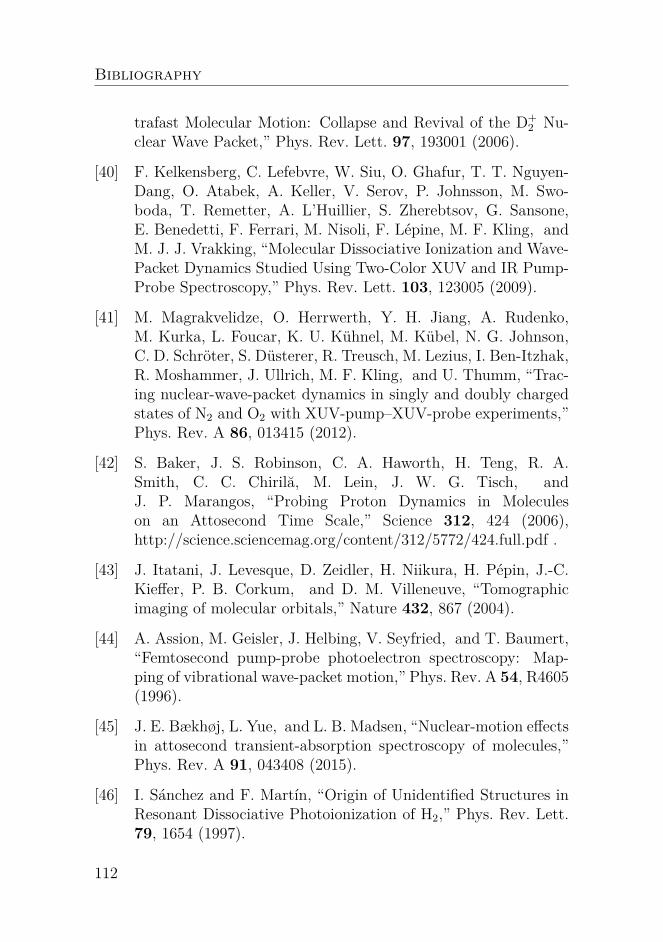
Bibliography
trafast Molecular Motion: Collapse and Revival of the D+2 Nu-
clear Wave Packet,” Phys. Rev. Lett. 97, 193001 (2006).
[40] F. Kelkensberg, C. Lefebvre, W. Siu, O. Ghafur, T. T. Nguyen-Dang, O. Atabek, A. Keller, V. Serov, P. Johnsson, M. Swo-boda, T. Remetter, A. L’Huillier, S. Zherebtsov, G. Sansone,E. Benedetti, F. Ferrari, M. Nisoli, F. Lépine, M. F. Kling, andM. J. J. Vrakking, “Molecular Dissociative Ionization and Wave-Packet Dynamics Studied Using Two-Color XUV and IR Pump-Probe Spectroscopy,” Phys. Rev. Lett. 103, 123005 (2009).
[41] M. Magrakvelidze, O. Herrwerth, Y. H. Jiang, A. Rudenko,M. Kurka, L. Foucar, K. U. Kühnel, M. Kübel, N. G. Johnson,C. D. Schröter, S. Düsterer, R. Treusch, M. Lezius, I. Ben-Itzhak,R. Moshammer, J. Ullrich, M. F. Kling, and U. Thumm, “Trac-ing nuclear-wave-packet dynamics in singly and doubly chargedstates of N2 and O2 with XUV-pump–XUV-probe experiments,”Phys. Rev. A 86, 013415 (2012).
[42] S. Baker, J. S. Robinson, C. A. Haworth, H. Teng, R. A.Smith, C. C. Chirilă, M. Lein, J. W. G. Tisch, andJ. P. Marangos, “Probing Proton Dynamics in Moleculeson an Attosecond Time Scale,” Science 312, 424 (2006),http://science.sciencemag.org/content/312/5772/424.full.pdf .
[43] J. Itatani, J. Levesque, D. Zeidler, H. Niikura, H. Pépin, J.-C.Kieffer, P. B. Corkum, and D. M. Villeneuve, “Tomographicimaging of molecular orbitals,” Nature 432, 867 (2004).
[44] A. Assion, M. Geisler, J. Helbing, V. Seyfried, and T. Baumert,“Femtosecond pump-probe photoelectron spectroscopy: Map-ping of vibrational wave-packet motion,” Phys. Rev. A 54, R4605(1996).
[45] J. E. Bækhøj, L. Yue, and L. B. Madsen, “Nuclear-motion effectsin attosecond transient-absorption spectroscopy of molecules,”Phys. Rev. A 91, 043408 (2015).
[46] I. Sánchez and F. Martín, “Origin of Unidentified Structures inResonant Dissociative Photoionization of H2,” Phys. Rev. Lett.79, 1654 (1997).
112
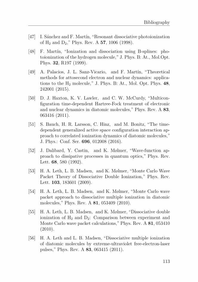
Bibliography
[47] I. Sánchez and F. Martín, “Resonant dissociative photoionizationof H2 and D2,” Phys. Rev. A 57, 1006 (1998).
[48] F. Martín, “Ionization and dissociation using B-splines: pho-toionization of the hydrogen molecule,” J. Phys. B: At., Mol.Opt.Phys. 32, R197 (1999).
[49] A. Palacios, J. L. Sanz-Vicario, and F. Martín, “Theoreticalmethods for attosecond electron and nuclear dynamics: applica-tions to the H2 molecule,” J. Phys. B: At., Mol. Opt. Phys. 48,242001 (2015).
[50] D. J. Haxton, K. V. Lawler, and C. W. McCurdy, “Multicon-figuration time-dependent Hartree-Fock treatment of electronicand nuclear dynamics in diatomic molecules,” Phys. Rev. A 83,063416 (2011).
[51] S. Bauch, H. R. Larsson, C. Hinz, and M. Bonitz, “The time-dependent generalized active space configuration interaction ap-proach to correlated ionization dynamics of diatomic molecules,”J. Phys.: Conf. Ser. 696, 012008 (2016).
[52] J. Dalibard, Y. Castin, and K. Mølmer, “Wave-function ap-proach to dissipative processes in quantum optics,” Phys. Rev.Lett. 68, 580 (1992).
[53] H. A. Leth, L. B. Madsen, and K. Mølmer, “Monte Carlo WavePacket Theory of Dissociative Double Ionization,” Phys. Rev.Lett. 103, 183601 (2009).
[54] H. A. Leth, L. B. Madsen, and K. Mølmer, “Monte Carlo wavepacket approach to dissociative multiple ionization in diatomicmolecules,” Phys. Rev. A 81, 053409 (2010).
[55] H. A. Leth, L. B. Madsen, and K. Mølmer, “Dissociative doubleionization of H2 and D2: Comparison between experiment andMonte Carlo wave packet calculations,” Phys. Rev. A 81, 053410(2010).
[56] H. A. Leth and L. B. Madsen, “Dissociative multiple ionizationof diatomic molecules by extreme-ultraviolet free-electron-laserpulses,” Phys. Rev. A 83, 063415 (2011).
113
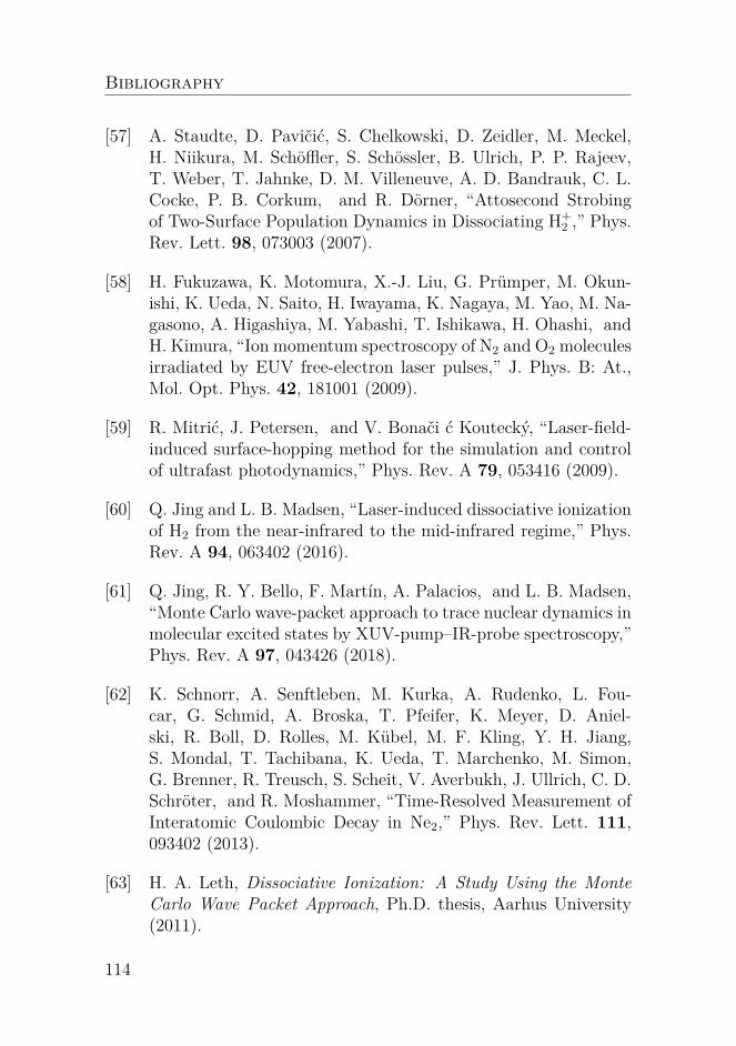
Bibliography
[57] A. Staudte, D. Pavičić, S. Chelkowski, D. Zeidler, M. Meckel,H. Niikura, M. Schöffler, S. Schössler, B. Ulrich, P. P. Rajeev,T. Weber, T. Jahnke, D. M. Villeneuve, A. D. Bandrauk, C. L.Cocke, P. B. Corkum, and R. Dörner, “Attosecond Strobingof Two-Surface Population Dynamics in Dissociating H+
2 ,” Phys.Rev. Lett. 98, 073003 (2007).
[58] H. Fukuzawa, K. Motomura, X.-J. Liu, G. Prümper, M. Okun-ishi, K. Ueda, N. Saito, H. Iwayama, K. Nagaya, M. Yao, M. Na-gasono, A. Higashiya, M. Yabashi, T. Ishikawa, H. Ohashi, andH. Kimura, “Ion momentum spectroscopy of N2 and O2 moleculesirradiated by EUV free-electron laser pulses,” J. Phys. B: At.,Mol. Opt. Phys. 42, 181001 (2009).
[59] R. Mitrić, J. Petersen, and V. Bonači ć Koutecký, “Laser-field-induced surface-hopping method for the simulation and controlof ultrafast photodynamics,” Phys. Rev. A 79, 053416 (2009).
[60] Q. Jing and L. B. Madsen, “Laser-induced dissociative ionizationof H2 from the near-infrared to the mid-infrared regime,” Phys.Rev. A 94, 063402 (2016).
[61] Q. Jing, R. Y. Bello, F. Martín, A. Palacios, and L. B. Madsen,“Monte Carlo wave-packet approach to trace nuclear dynamics inmolecular excited states by XUV-pump–IR-probe spectroscopy,”Phys. Rev. A 97, 043426 (2018).
[62] K. Schnorr, A. Senftleben, M. Kurka, A. Rudenko, L. Fou-car, G. Schmid, A. Broska, T. Pfeifer, K. Meyer, D. Aniel-ski, R. Boll, D. Rolles, M. Kübel, M. F. Kling, Y. H. Jiang,S. Mondal, T. Tachibana, K. Ueda, T. Marchenko, M. Simon,G. Brenner, R. Treusch, S. Scheit, V. Averbukh, J. Ullrich, C. D.Schröter, and R. Moshammer, “Time-Resolved Measurement ofInteratomic Coulombic Decay in Ne2,” Phys. Rev. Lett. 111,093402 (2013).
[63] H. A. Leth, Dissociative Ionization: A Study Using the MonteCarlo Wave Packet Approach, Ph.D. thesis, Aarhus University(2011).
114

Bibliography
[64] M. Feit, J. Fleck, and A. Steiger, “Solution of the Schrödingerequation by a spectral method,” J. Comput. Phys. 47, 412(1982).
[65] P. Schwendner, F. Seyl, and R. Schinke, “Photodissociation ofAr+
2 in strong laser fields,” Chem. Phys. 217, 233 (1997), dy-namics of Driven Quantum Systems.
[66] T. D. G. Walsh, F. A. Ilkov, S. L. Chin, F. Châteauneuf, T. T.Nguyen-Dang, S. Chelkowski, A. D. Bandrauk, and O. Atabek,“Laser-induced processes during the Coulomb explosion of H2 ina Ti-sapphire laser pulse,” Phys. Rev. A 58, 3922 (1998).
[67] A. S. Alnaser, X. M. Tong, T. Osipov, S. Voss, C. M. Maharjan,P. Ranitovic, B. Ulrich, B. Shan, Z. Chang, C. D. Lin, andC. L. Cocke, “Routes to Control of H2 Coulomb Explosion inFew-Cycle Laser Pulses,” Phys. Rev. Lett. 93, 183202 (2004).
[68] S. Saugout, E. Charron, and C. Cornaggia, “H2 double ionizationwith few-cycle laser pulses,” Phys. Rev. A 77, 023404 (2008).
[69] S. Saugout and C. Cornaggia, “Temporal separation of H2double-ionization channels using intense ultrashort 10−fs laserpulses,” Phys. Rev. A 73, 041406 (2006).
[70] V. Wanie, H. Ibrahim, S. Beaulieu, N. Thiré, B. E. Schmidt,Y. Deng, A. S. Alnaser, I. V. Litvinyuk, X.-M. Tong, and F. Lé-garé, “Coherent control of D2 /H2 dissociative ionization by amid-infrared two-color laser field,” J. Phys. B: At., Mol. Opt.Phys. 49, 025601 (2016).
[71] T. Sharp, “Potential-energy curves for molecular hydrogen andits ions,” Atomic Data and Nuclear Data Tables 2, 119 (1970).
[72] J. T. Lin and T. F. Jiang, “Photodissociation of H+2 in intense
chirped laser fields,” Phys. Rev. A 63, 013408 (2000).
[73] O. I. Tolstikhin, T. Morishita, and L. B. Madsen, “Theory oftunneling ionization of molecules: Weak-field asymptotics in-cluding dipole effects,” Phys. Rev. A 84, 053423 (2011).
115

Bibliography
[74] O. I. Tolstikhin, L. B. Madsen, and T. Morishita, “Weak-field asymptotic theory of tunneling ionization in many-electronatomic and molecular systems,” Phys. Rev. A 89, 013421 (2014).
[75] M. Plummer and J. F. McCann, “Field-ionization rates of thehydrogen molecular ion,” J. Phys. B: At., Mol. Opt. Phys. 29,4625 (1996).
[76] H. Kono, Y. Sato, Y. Fujimura, and I. Kawata, “Identificationof the doorway states to ionization of molecules in intense laserfields,” Laser physics 13, 883 (2003).
[77] I. V. Litvinyuk, A. S. Alnaser, D. Comtois, D. Ray, A. T. Hasan,J.-C. Kieffer, and D. M. Villeneuve, “Wavelength-dependentstudy of strong-field Coulomb explosion of hydrogen,” New J.Phys. 10, 083011 (2008).
[78] T. Ergler, A. Rudenko, B. Feuerstein, K. Zrost, C. D. Schröter,R. Moshammer, and J. Ullrich, “Time-Resolved Imaging andManipulation of H2 Fragmentation in Intense Laser Fields,”Phys. Rev. Lett. 95, 093001 (2005).
[79] Y. H. Jiang, A. Rudenko, J. F. Pérez-Torres, O. Herrwerth,L. Foucar, M. Kurka, K. U. Kühnel, M. Toppin, E. Plésiat,F. Morales, F. Martín, M. Lezius, M. F. Kling, T. Jahnke,R. Dörner, J. L. Sanz-Vicario, J. van Tilborg, A. Belkacem,M. Schulz, K. Ueda, T. J. M. Zouros, S. Düsterer, R. Treusch,C. D. Schröter, R. Moshammer, and J. Ullrich, “Investigatingtwo-photon double ionization of D2 by XUV-pump–XUV-probeexperiments,” Phys. Rev. A 81, 051402 (2010).
[80] A. R. Bainbridge, J. Harrington, A. Kirrander, C. Cacho,E. Springate, W. A. Bryan, and R. S. Minns, “VUV excitationof a vibrational wavepacket in D 2 measured through strong-fielddissociative ionization,” New J. Phys. 17, 103013 (2015).
[81] Y. H. Jiang, A. Rudenko, J. F. Pérez-Torres, O. Herrwerth,L. Foucar, M. Kurka, K. U. Kühnel, M. Toppin, E. Plésiat,F. Morales, F. Martín, M. Lezius, M. F. Kling, T. Jahnke,R. Dörner, J. L. Sanz-Vicario, J. van Tilborg, A. Belkacem,
116
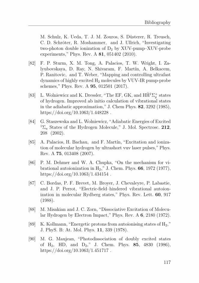
Bibliography
M. Schulz, K. Ueda, T. J. M. Zouros, S. Düsterer, R. Treusch,C. D. Schröter, R. Moshammer, and J. Ullrich, “Investigatingtwo-photon double ionization of D2 by XUV-pump–XUV-probeexperiments,” Phys. Rev. A 81, 051402 (2010).
[82] F. P. Sturm, X. M. Tong, A. Palacios, T. W. Wright, I. Za-lyubovskaya, D. Ray, N. Shivaram, F. Martín, A. Belkacem,P. Ranitovic, and T. Weber, “Mapping and controlling ultrafastdynamics of highly excited H2 molecules by VUV-IR pump-probeschemes,” Phys. Rev. A 95, 012501 (2017).
[83] L. Wolniewicz and K. Dressler, “The EF, GK, and HH1Σ+g states
of hydrogen. Improved ab initio calculation of vibrational statesin the adiabatic approximation,” J. Chem Phys. 82, 3292 (1985),https://doi.org/10.1063/1.448228 .
[84] G. Staszewska and L. Wolniewicz, “Adiabatic Energies of Excited1Σu States of the Hydrogen Molecule,” J. Mol. Spectrosc. 212,208 (2002).
[85] A. Palacios, H. Bachau, and F. Martín, “Excitation and ioniza-tion of molecular hydrogen by ultrashort vuv laser pulses,” Phys.Rev. A 75, 013408 (2007).
[86] P. M. Dehmer and W. A. Chupka, “On the mechanism for vi-brational autoionization in H2,” J. Chem. Phys. 66, 1972 (1977),https://doi.org/10.1063/1.434154 .
[87] C. Bordas, P. F. Brevet, M. Broyer, J. Chevaleyre, P. Labastie,and J. P. Perrot, “Electric-field–hindered vibrational autoion-ization in molecular Rydberg states,” Phys. Rev. Lett. 60, 917(1988).
[88] M. Misakian and J. C. Zorn, “Dissociative Excitation of Molecu-lar Hydrogen by Electron Impact,” Phys. Rev. A 6, 2180 (1972).
[89] K. Kollmann, “Energetic protons from autoionising states of H2,”J. PhyS. B: At. Mol. Phys. 11, 339 (1978).
[90] M. G. Maujean, “Photodissociation of doubly excited statesof H2, HD, and D2,” J. Chem. Phys. 85, 4830 (1986),https://doi.org/10.1063/1.451717 .
117

Bibliography
[91] Y. M. Chung, E. Lee, T. Masuoka, and J. A. R. Samson, “Disso-ciative photoionization of H2 from 18 to 124 eV,” J. Chem. Phys.99, 885 (1993), https://doi.org/10.1063/1.465352 .
[92] R. Browning and J. Fryar, “Dissociative photoionization of H2and D2 through the 1sσg ionic state,” J. Phys. B: At. Mol. Phys.6, 364 (1973).
[93] S. L. Guberman, “The doubly excited autoionizing statesof H2,” The Journal of Chemical Physics 78, 1404 (1983),https://doi.org/10.1063/1.444882 .
[94] I. Sánchez and F. Martín, “The doubly excited statesof the H2 molecule,” J. Chem. Phys. 106, 7720 (1997),https://doi.org/10.1063/1.473773 .
[95] I. Sánchez and F. Martín, “Doubly excited autoionizingstates of H2 above the second ionization threshold: theQ2 resonance series,” J. Chem. Phys. 110, 6702 (1999),https://doi.org/10.1063/1.478576 .
[96] J. Fernández and F. Martín, “Autoionizing 1Σ+u and 1Πu states
of H2 above the third and fourth ionization thresholds,” J. Phys.sB: At., Mol. Opt. Phys. 34, 4141 (2001).
[97] I. B. Jr and C. E. Bielschowsky, “Doubly excited states of molecu-lar hydrogen: theoretical absorption and photodissociation crosssections,” J. Phys. B: At., Mol. Opt. Phys. 33, 1713 (2000).
[98] K. Ito, R. I. Hall, and M. Ukai, “Dissociative photoionizationof H2 and D2 in the energy region of 25–45 eV,” J. Chem. Phys.104, 8449 (1996), https://doi.org/10.1063/1.471594 .
[99] J. L. Sanz-Vicario, H. Bachau, and F. Martín, “Time-dependenttheoretical description of molecular autoionization produced byfemtosecond xuv laser pulses,” Phys. Rev. A 73, 033410 (2006).
[100] K. Sakai, S. Stoychev, T. Ouchi, I. Higuchi, M. Schöffler,T. Mazza, H. Fukuzawa, K. Nagaya, M. Yao, Y. Tamenori, A. I.Kuleff, N. Saito, and K. Ueda, “Electron-Transfer-Mediated De-cay and Interatomic Coulombic Decay from the Triply IonizedStates in Argon Dimers,” Phys. Rev. Lett. 106, 033401 (2011).
118

Bibliography
[101] W. Eberhardt, G. Kalkoffen, and C. Kunz, “Measurement ofthe Auger Decay after Resonance Excitation of Xe 4d and Kr 3dResonance Lines,” Phys. Rev. Lett. 41, 156 (1978).
[102] J. M. Schins, P. Breger, P. Agostini, R. C. Constantinescu, H. G.Muller, G. Grillon, A. Antonetti, and A. Mysyrowicz, “Observa-tion of Laser-Assisted Auger Decay in Argon,” Phys. Rev. Lett.73, 2180 (1994).
[103] M. L. Knotek and P. J. Feibelman, “Ion Desorption by Core-HoleAuger Decay,” Phys. Rev. Lett. 40, 964 (1978).
[104] L. S. Cederbaum, J. Zobeley, and F. Tarantelli, “Giant Inter-molecular Decay and Fragmentation of Clusters,” Phys. Rev.Lett. 79, 4778 (1997).
[105] S. Marburger, O. Kugeler, U. Hergenhahn, and T. Möller, “Ex-perimental Evidence for Interatomic Coulombic Decay in NeClusters,” Phys. Rev. Lett. 90, 203401 (2003).
[106] T. Jahnke, A. Czasch, M. S. Schöffler, S. Schössler, A. Knapp,M. Käsz, J. Titze, C. Wimmer, K. Kreidi, R. E. Grisenti,A. Staudte, O. Jagutzki, U. Hergenhahn, H. Schmidt-Böcking,and R. Dörner, “Experimental Observation of InteratomicCoulombic Decay in Neon Dimers,” Phys. Rev. Lett. 93, 163401(2004).
[107] G. Öhrwall, M. Tchaplyguine, M. Lundwall, R. Feifel, H. Berg-ersen, T. Rander, A. Lindblad, J. Schulz, S. Peredkov, S. Barth,S. Marburger, U. Hergenhahn, S. Svensson, and O. Björne-holm, “Femtosecond Interatomic Coulombic Decay in Free NeonClusters: Large Lifetime Differences between Surface and Bulk,”Phys. Rev. Lett. 93, 173401 (2004).
[108] T. Jahnke, H. Sann, T. Havermeier, K. Kreidi, C. Stuck,M. Meckel, M. Schöffler, N. Neumann, R. Wallauer, S. Voss,et al., “Ultrafast energy transfer between water molecules,” Nat.Phys. 6, 139 (2010).
119

Bibliography
[109] E. F. Aziz, N. Ottosson, M. Faubel, I. V. Hertel, and B. Win-ter, “Interaction between liquid water and hydroxide revealed bycore-hole de-excitation,” Nature 455, 89 (2008).
[110] M. Mucke, M. Braune, S. Barth, M. Förstel, T. Lischke, V. Ul-rich, T. Arion, U. Becker, A. Bradshaw, and U. Hergenhahn, “Ahitherto unrecognized source of low-energy electrons in water,”Nat. Phys. 6, 143 (2010).
[111] S. Scheit, L. S. Cederbaum, and H.-D. Meyer, “Time-dependentinterplay between electron emission and fragmentation in theinteratomic Coulombic decay,” J. Chem. Phys. 118, 2092 (2003),https://doi.org/10.1063/1.1531996 .
[112] R. Santra, J. Zobeley, L. S. Cederbaum, and N. Moiseyev, “Inter-atomic Coulombic Decay in van der Waals Clusters and Impactof Nuclear Motion,” Phys. Rev. Lett. 85, 4490 (2000).
[113] N. Moiseyev, R. Santra, J. Zobeley, and L. S. Ceder-baum, “Fingerprints of the nodal structure of autoionizingvibrational wave functions in clusters: Interatomic Coulom-bic decay in Ne dimer,” J. Chem. Phys. 114, 7351 (2001),https://doi.org/10.1063/1.1361070 .
[114] R. Santra, L. Cederbaum, and H.-D. Meyer, “Electronic decay ofmolecular clusters: non-stationary states computed by standardquantum chemistry methods,” Chemical Physics Letters 303,413 (1999).
[115] V. Averbukh, I. B. Müller, and L. S. Cederbaum, “Mechanismof Interatomic Coulombic Decay in Clusters,” Phys. Rev. Lett.93, 263002 (2004).
[116] E. Saldin, E. Schneidmiller, and M. Yurkov, “Statistical proper-ties of radiation from VUV and X-ray free electron laser,” Opt.Commun. 148, 383 (1998).
[117] E. L. Saldin, E. A. Schneidmiller, and M. V. Yurkov, “Statisticaland coherence properties of radiation from x-ray free-electronlasers,” New J. Phys. 12, 035010 (2010).
120

Bibliography
[118] S. Krinsky and R. L. Gluckstern, “Analysis of statistical corre-lations and intensity spiking in the self-amplified spontaneous-emission free-electron laser,” Phys. Rev. ST Accel. Beams 6,050701 (2003).
[119] K. Meyer, C. Ott, P. Raith, A. Kaldun, Y. Jiang, A. Senftleben,M. Kurka, R. Moshammer, J. Ullrich, and T. Pfeifer, “NoisyOptical Pulses Enhance the Temporal Resolution of Pump-ProbeSpectroscopy,” Phys. Rev. Lett. 108, 098302 (2012).
[120] T. Pfeifer, Y. Jiang, S. Düsterer, R. Moshammer, and J. Ullrich,“Partial-coherence method to model experimental free-electronlaser pulse statistics,” Opt. Lett. 35, 3441 (2010).
[121] K. Codling, R. G. Houlgate, J. B. West, and P. R. Woodruff,“Angular distribution and photoionization measurements on the2p and 2s electrons in neon,” J. Phys. B: At. Mol. Phys. 9, L83(1976).
[122] A. K. Pradhan, “Photo-ionization cross-sections of Ne II, Ne IIIand Ne IV,” Monthly Notices of the Royal Astronomical Society190, 5P (1980).
[123] S. Scheit, V. Averbukh, H.-D. Meyer, N. Moiseyev,R. Santra, T. Sommerfeld, J. Zobeley, and L. S.Cederbaum, “On the interatomic Coulombic decay inthe Ne dimer,” J. Chem. Phys. 121, 8393 (2004),https://aip.scitation.org/doi/pdf/10.1063/1.1794654 .
121