A solid pseudopapillary tumor of the pancreas treated with
4
CASE REPORT Open Access A solid pseudopapillary tumor of the pancreas treated with laparoscopic distal pancreatectomy and splenectomy: a case report and review of the literature Athanasios Marinis * , Georgios Anastasopoulos, Georgios Polymeneas Abstract Introduction: Laparoscopic distal pancreatectomy has been described for more than a decade now and has been considered technically feasible, safe, and with reproducible outcomes. It seems to exhibit several benefits of minimally invasive surgery and should be performed in carefully selected patients. Case presentation: We report the case of a 55-year-old Greek woman with a solid pseudopapillary tumor of the tail of the pancreas. She underwent a laparoscopic distal pancreatectomy and splenectomy. The histopathologic examination finally revealed a cystic-solid pseudopapillary neoplasm of the pancreas. Solid pseudopapillary tumors of the pancreas are rare and affect predominantly young women. These tumors are of unclear pathogenesis and low malignancy, and surgical resection offers an excellent chance for long-term survival. Conclusion: This case report indicates that in selected centers and for selected patients, laparoscopic distal pancreatectomy is feasible. The benign characteristics of these tumors make them ideal for laparoscopic excision. Introduction Laparoscopic resection of the pancreas was initially described experimentally in the early 1990s [1]. The first laparoscopic pancreatoduodenectomy was reported in 1994 by Gagner et al. [2]. Laparoscopic distal pancrea- tectomy (with or without splenectomy), on the contrary, may be well suited to the laparoscopic approach, is tech- nically easier because no anastomosis is required, and is more widely accepted [3]. Solid pseudopapillary tumors (SPTs) of the pancreas are rare and affect predominantly young women. These tumors are of unclear pathogenesis and low malignancy, and surgical resection offers an excellent chance for long-term survival [4]. The larger review contains 718 well-documented cases and confirms the characteristics of these tumors [4]. Clinically, they appear as slowly growing masses with or without pain; however, these tumors may appear with other rare symptoms [5]. It is not uncommon, though, to arrive at the final diagnosis only by pathology several weeks after the operation [6,7]. The benign characteristics of these tumors make them ideal for laparoscopic excision [8-10]. We present the case of a woman who successfully underwent a laparoscopic distal pancreatectomy and splenectomy for an SPT of the pancreatic tail, and we review the English literature to clarify the safety and efficiency of laparoscopic distal pancreatectomy. Case presentation A 55-year-old Greek woman was referred to our clinic for the management of a cystic lesion located in the tail of the pancreas. The lesion was discovered incidentally during her staging workup with abdominal ultrasound for invasive ductal adenocarcinoma of the left breast 16 months ago. Just after a modified radical left mastect- omy had been performed, we further investigated the pancreatic lesion with a magnetic resonance imaging (MRI) scan, which revealed a space-occupying cystic lesion of maximum diameter of 5 cm located in the tail of the pancreas with calcifications of the wall and a * Correspondence: [email protected] Second Department of Surgery, Aretaieion University Hospital, 76 Vassilisis, Sofia’s Ave, 11528, Athens, Greece Marinis et al. Journal of Medical Case Reports 2010, 4:387 http://www.jmedicalcasereports.com/content/4/1/387 JOURNAL OF MEDICAL CASE REPORTS © 2010 Marinis et al; licensee BioMed Central Ltd. This is an Open Access article distributed under the terms of the Creative Commons Attribution License (http://creativecommons.org/licenses/by/2.0), which permits unrestricted use, distribution, and reproduction in any medium, provided the original work is properly cited.
Transcript of A solid pseudopapillary tumor of the pancreas treated with
CASE REPORT Open Access
A solid pseudopapillary tumor of the pancreastreated with laparoscopic distal pancreatectomyand splenectomy: a case report and review ofthe literatureAthanasios Marinis*, Georgios Anastasopoulos, Georgios Polymeneas
Abstract
Introduction: Laparoscopic distal pancreatectomy has been described for more than a decade now and has beenconsidered technically feasible, safe, and with reproducible outcomes. It seems to exhibit several benefits ofminimally invasive surgery and should be performed in carefully selected patients.
Case presentation: We report the case of a 55-year-old Greek woman with a solid pseudopapillary tumor of thetail of the pancreas. She underwent a laparoscopic distal pancreatectomy and splenectomy. The histopathologicexamination finally revealed a cystic-solid pseudopapillary neoplasm of the pancreas. Solid pseudopapillary tumorsof the pancreas are rare and affect predominantly young women. These tumors are of unclear pathogenesis andlow malignancy, and surgical resection offers an excellent chance for long-term survival.
Conclusion: This case report indicates that in selected centers and for selected patients, laparoscopic distalpancreatectomy is feasible. The benign characteristics of these tumors make them ideal for laparoscopic excision.
IntroductionLaparoscopic resection of the pancreas was initiallydescribed experimentally in the early 1990s [1]. The firstlaparoscopic pancreatoduodenectomy was reported in1994 by Gagner et al. [2]. Laparoscopic distal pancrea-tectomy (with or without splenectomy), on the contrary,may be well suited to the laparoscopic approach, is tech-nically easier because no anastomosis is required, and ismore widely accepted [3].Solid pseudopapillary tumors (SPTs) of the pancreas
are rare and affect predominantly young women. Thesetumors are of unclear pathogenesis and low malignancy,and surgical resection offers an excellent chance forlong-term survival [4]. The larger review contains 718well-documented cases and confirms the characteristicsof these tumors [4]. Clinically, they appear as slowlygrowing masses with or without pain; however, thesetumors may appear with other rare symptoms [5]. It is
not uncommon, though, to arrive at the final diagnosisonly by pathology several weeks after the operation [6,7].The benign characteristics of these tumors make themideal for laparoscopic excision [8-10].We present the case of a woman who successfully
underwent a laparoscopic distal pancreatectomy andsplenectomy for an SPT of the pancreatic tail, and wereview the English literature to clarify the safety andefficiency of laparoscopic distal pancreatectomy.
Case presentationA 55-year-old Greek woman was referred to our clinicfor the management of a cystic lesion located in the tailof the pancreas. The lesion was discovered incidentallyduring her staging workup with abdominal ultrasoundfor invasive ductal adenocarcinoma of the left breast16 months ago. Just after a modified radical left mastect-omy had been performed, we further investigated thepancreatic lesion with a magnetic resonance imaging(MRI) scan, which revealed a space-occupying cysticlesion of maximum diameter of 5 cm located in the tailof the pancreas with calcifications of the wall and a
* Correspondence: [email protected] Department of Surgery, Aretaieion University Hospital, 76 Vassilisis,Sofia’s Ave, 11528, Athens, Greece
Marinis et al. Journal of Medical Case Reports 2010, 4:387http://www.jmedicalcasereports.com/content/4/1/387 JOURNAL OF MEDICAL
CASE REPORTS
© 2010 Marinis et al; licensee BioMed Central Ltd. This is an Open Access article distributed under the terms of the Creative CommonsAttribution License (http://creativecommons.org/licenses/by/2.0), which permits unrestricted use, distribution, and reproduction inany medium, provided the original work is properly cited.
central cystic component (Figure 1). Besides the chemo-,radio-, and hormonal therapy she received for her breastcancer, her past medical history also included hypothyr-oidism under hormone-replacement therapy.The findings in the physical examination were unre-
markable. Blood investigation and tumor markers (CEA,CA 15-3, CA 19-9, and CA 125) were within normallimits. The possibility of a mucinous cystic neoplasm ofthe pancreas was considered, and a laparoscopic distalpancreatectomy and a splenectomy were chosen.
TechniqueThe first distal pancreatectomy in pigs was described bySoper et al. [1] in 1994; 2 years later, Gagner [11]reported his first five cases of spleen-preserving laparo-scopic distal pancreatectomy for insulinoma.In our case, the patient was placed in a modified lithot-
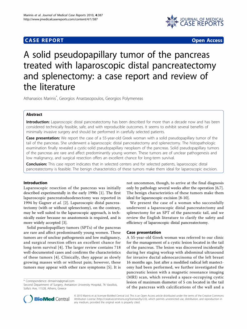
omy position, with the surgeon standing between the legsof the patient. We used a five-port technique, placing a12 mm trocar left paramedian at about the level of theumbilicus, a 10 mm-trocar in the left upper quadrant ofthe abdomen on the anterior axillary line, a 10 mm-trocarin the subxiphoid region, a 5 mm-trocar in the left hypo-chondrium on the midclavicular line, and a 5 mm-trocarin the right hypochondrium in the midclavicular line aswell (Figure 2). Pneumoperitoneum was established withthe open Hasson technique through the 12 mm parame-dian port. Exploratory laparoscopy did not reveal macro-scopically evident intra-abdominal metastases. We used a30-degree laparoscope and an ultrasonic dissector (Ultra-Cision; Ethicon, Endosurgery). After entering the lessersac, we identified the splenic artery at the upper border of
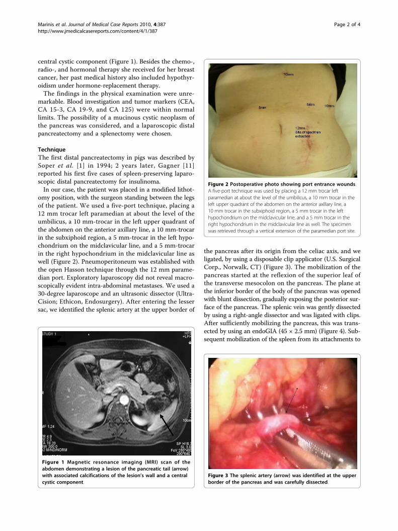
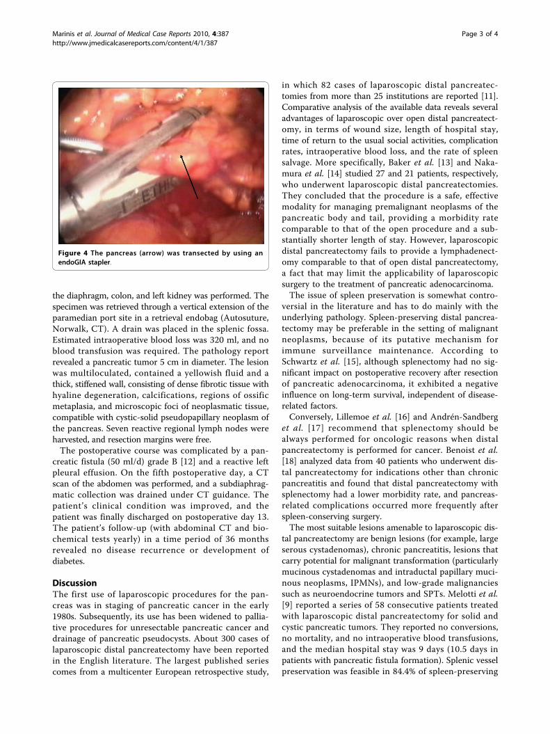
the pancreas after its origin from the celiac axis, and weligated, by using a disposable clip applicator (U.S. SurgicalCorp., Norwalk, CT) (Figure 3). The mobilization of thepancreas started at the reflexion of the superior leaf ofthe transverse mesocolon on the pancreas. The plane atthe inferior border of the body of the pancreas was openedwith blunt dissection, gradually exposing the posterior sur-face of the pancreas. The splenic vein was gently dissectedby using a right-angle dissector and was ligated with clips.After sufficiently mobilizing the pancreas, this was trans-ected by using an endoGIA (45 × 2.5 mm) (Figure 4). Sub-sequent mobilization of the spleen from its attachments to
Figure 1 Magnetic resonance imaging (MRI) scan of theabdomen demonstrating a lesion of the pancreatic tail (arrow)with associated calcifications of the lesion’s wall and a centralcystic component.
Figure 2 Postoperative photo showing port entrance wounds.A five-port technique was used by placing a 12 mm trocar leftparamedian at about the level of the umbilicus, a 10 mm trocar in theleft upper quadrant of the abdomen on the anterior axillary line, a10 mm trocar in the subxiphoid region, a 5 mm trocar in the lefthypochondrium on the midclavicular line, and a 5 mm trocar in theright hypochondrium in the midclavicular line as well. The specimenwas retrieved through a vertical extension of the paramedian port site.
Figure 3 The splenic artery (arrow) was identified at the upperborder of the pancreas and was carefully dissected.
Marinis et al. Journal of Medical Case Reports 2010, 4:387http://www.jmedicalcasereports.com/content/4/1/387
Page 2 of 4
the diaphragm, colon, and left kidney was performed. Thespecimen was retrieved through a vertical extension of theparamedian port site in a retrieval endobag (Autosuture,Norwalk, CT). A drain was placed in the splenic fossa.Estimated intraoperative blood loss was 320 ml, and noblood transfusion was required. The pathology reportrevealed a pancreatic tumor 5 cm in diameter. The lesionwas multiloculated, contained a yellowish fluid and athick, stiffened wall, consisting of dense fibrotic tissue withhyaline degeneration, calcifications, regions of ossificmetaplasia, and microscopic foci of neoplasmatic tissue,compatible with cystic-solid pseudopapillary neoplasm ofthe pancreas. Seven reactive regional lymph nodes wereharvested, and resection margins were free.The postoperative course was complicated by a pan-
creatic fistula (50 ml/d) grade B [12] and a reactive leftpleural effusion. On the fifth postoperative day, a CTscan of the abdomen was performed, and a subdiaphrag-matic collection was drained under CT guidance. Thepatient’s clinical condition was improved, and thepatient was finally discharged on postoperative day 13.The patient’s follow-up (with abdominal CT and bio-chemical tests yearly) in a time period of 36 monthsrevealed no disease recurrence or development ofdiabetes.
DiscussionThe first use of laparoscopic procedures for the pan-creas was in staging of pancreatic cancer in the early1980s. Subsequently, its use has been widened to pallia-tive procedures for unresectable pancreatic cancer anddrainage of pancreatic pseudocysts. About 300 cases oflaparoscopic distal pancreatectomy have been reportedin the English literature. The largest published seriescomes from a multicenter European retrospective study,
in which 82 cases of laparoscopic distal pancreatec-tomies from more than 25 institutions are reported [11].Comparative analysis of the available data reveals severaladvantages of laparoscopic over open distal pancreatect-omy, in terms of wound size, length of hospital stay,time of return to the usual social activities, complicationrates, intraoperative blood loss, and the rate of spleensalvage. More specifically, Baker et al. [13] and Naka-mura et al. [14] studied 27 and 21 patients, respectively,who underwent laparoscopic distal pancreatectomies.They concluded that the procedure is a safe, effectivemodality for managing premalignant neoplasms of thepancreatic body and tail, providing a morbidity ratecomparable to that of the open procedure and a sub-stantially shorter length of stay. However, laparoscopicdistal pancreatectomy fails to provide a lymphadenect-omy comparable to that of open distal pancreatectomy,a fact that may limit the applicability of laparoscopicsurgery to the treatment of pancreatic adenocarcinoma.The issue of spleen preservation is somewhat contro-
versial in the literature and has to do mainly with theunderlying pathology. Spleen-preserving distal pancrea-tectomy may be preferable in the setting of malignantneoplasms, because of its putative mechanism forimmune surveillance maintenance. According toSchwartz et al. [15], although splenectomy had no sig-nificant impact on postoperative recovery after resectionof pancreatic adenocarcinoma, it exhibited a negativeinfluence on long-term survival, independent of disease-related factors.Conversely, Lillemoe et al. [16] and Andrén-Sandberg
et al. [17] recommend that splenectomy should bealways performed for oncologic reasons when distalpancreatectomy is performed for cancer. Benoist et al.[18] analyzed data from 40 patients who underwent dis-tal pancreatectomy for indications other than chronicpancreatitis and found that distal pancreatectomy withsplenectomy had a lower morbidity rate, and pancreas-related complications occurred more frequently afterspleen-conserving surgery.The most suitable lesions amenable to laparoscopic dis-
tal pancreatectomy are benign lesions (for example, largeserous cystadenomas), chronic pancreatitis, lesions thatcarry potential for malignant transformation (particularlymucinous cystadenomas and intraductal papillary muci-nous neoplasms, IPMNs), and low-grade malignanciessuch as neuroendocrine tumors and SPTs. Melotti et al.[9] reported a series of 58 consecutive patients treatedwith laparoscopic distal pancreatectomy for solid andcystic pancreatic tumors. They reported no conversions,no mortality, and no intraoperative blood transfusions,and the median hospital stay was 9 days (10.5 days inpatients with pancreatic fistula formation). Splenic vesselpreservation was feasible in 84.4% of spleen-preserving
Figure 4 The pancreas (arrow) was transected by using anendoGIA stapler.
Marinis et al. Journal of Medical Case Reports 2010, 4:387http://www.jmedicalcasereports.com/content/4/1/387
Page 3 of 4

procedures, and pancreatic fistula formation occurred in27.5% of all cases.In our case, the histopathologic examination finally
revealed a cystic-solid pseudopapillary neoplasm of thepancreas. This rare neoplasm accounts for 1% to 2% ofall exocrine pancreatic tumors, can have either benignor malignant behaviour, and was first described byFrantz in 1959 [19]. SPT is a very uncommon pancreatictumor that affects mainly women (F/M ratio, 10:1). Themean age at its appearance is 21.97 years (ranging from2 to 85 years). Most of the patients are young (~22% areyounger than 18 years), but a considerable 6% of thepatients are older than 51 years [7].Concerning the surgical approach, many techniques
are used. The low grade of malignancy of this tumorhas led some surgeons to perform simple enucleation ofthe neoplasm. However, distal pancreatectomy withsplenic preservation or pancreatoduodenectomy,depending on the location of the tumor, represents theprocedure of choice. In general, the prognosis, even inthe case of a malignant SPT with metastasis, is favor-able. Some patients with “unresectable” tumors or thosewith hepatic metastasis have survived more than 10years after the operation [7].
ConclusionLaparoscopic distal pancreatectomy is feasible in institu-tions with advanced laparoscopic surgical experience,shares the advantages of minimally invasive surgery, andshould be performed in carefully selected patients. SPTsare the ideal pancreatic tumors for the laparoscopicapproach because of their low malignancy and theirexcellent prognosis.
ConsentWritten informed consent was obtained from the patientfor publication of this case report and accompanyingimages. A copy of the written consent is available forreview by the Editor-in-Chief of this journal.
Authors’ contributionsGA analyzed and interpreted the data from the patient’s medical file; GAand AM drafted the manuscript; GA, AM, and GP critically revised themanuscript and gave final approval for manuscript publication.
Competing interestsThe authors declare that they have no competing interests.
Received: 30 January 2010 Accepted: 29 November 2010Published: 29 November 2010
References1. Soper NJ, Brunt LM, Dunnegan DL, Meiniger TA: Laparoscopic distal
pancreatectomy in the porcine model. Surg Endosc 1994, 8:57-60.2. Gagner M, Pomp A: Laparoscopic pylorus-preserving
pancreatoduodenectomy. Surg Endosc 1994, 8:408-410.
3. Palanivelu C, Shetty R, Jani K, Sendhikumar K, Rajan PS, Maheshkumar GS:Laparoscopic distal pancreatectomy: results of a prospectivenonrandomized study from a tertiary center. Surg Endosc 2007,21:373-377.
4. Papavramidis T, Papavramidis S: Solid pseudopapillary tumors of thepancreas: review of 718 patients reported in English literature. J Am CollSurg 2005, 6:965-972.
5. Apostolidis S, Papavramidis TS, Zatagias A, Michalopoulos A,Papadopoulos VN, Paramythiotis D, Harlaftis N: Hematemesis, a very rarepresentation of solid pseudo-papillary tumors of the pancreas: a casereport. J Med Case Rep 2008, 2:271.
6. Klimstra DS, Wenig BM, Heffess CS: Solid-pseudopapillary tumor of thepancreas: a typically cystic carcinoma of low malignant potential. SeminDiagn Pathol 2000, 17:66-80.
7. Papavramidis TS, Pliakos I, Michalopoulos N, Karayanopoulou G, Kesisoglou I,Tzioufa V, Papavramidis S: Asolid pseudo-papillary tumor of the pancreas.Ann Gastroenterol 2008, 21:242-244.
8. Carricaburu E, Enezian G, Bonnard A, Berrebi D, Belarbi N, Huot O, Aigrain Y,de Lagausie P: Laparoscopic distal pancreatectomy for Frantz’s tumor ina child. Surg Endosc 2003, 17:2028-2031.
9. Melotti G, Butturini G, Piccoli M, Casetti L, Bassi C, Mullineris B,Lazzaretti MG, Pederzoli P: Laparoscopic distal pancreatectomy: results ona consecutive series of 58 patients. Ann Surg 2007, 246:77-82.
10. Kang CM, Kim HG, Kim KS, Choi JS, Lee WJ, Kim BR: Laparoscopic distalpancreatectomy for solid pseudopapillary neoplasm of the pancreas:report of two cases. Hepatogastroenterology 2007, 54:1053-1056.
11. Mabrut JY, Fernandez-Cruz L, Azagra JS, Bassi C, Delvaux G, Weerts J, et al:Hepatobiliary and pancreatic section (HBPS) of the Royal Belgian Societyof Surgery; Belgian group for Endoscopic Surgery (BGES); Club Coelio:laparoscopic pancreatic resection: results of a multicenter Europeanstudy of 127 patients. Surgery 2005, 137:597-605.
12. Bassi C, Dervenis C, Butturini G, Fingerhut A, Yeo C, Izbicki J,Neoptolemos J, Sarr M, Traverso W, Buchler M, for the International StudyGroup on Pancreatic Fistula Definition: Postoperative pancreatic fistula: aninternational study group (ISGPF) definition. Surgery 2005, 138:8-13.
13. Baker MS, Bentrem DJ, Ujiki MB, Stocker S, Talamonti MS: A prospectivesingle institution comparison of peri-operative outcomes forlaparoscopic and open distal pancreatectomy. Surgery 2009, 146:635-643.
14. Nakamura Y, Uchida E, Aimoto T, Matsumoto S, Yoshida H, Tajiri T: Clinicaloutcome of laparoscopic distal pancreatectomy. J Hepatobiliary PancreatSurg 2009, 16:35-41.
15. Schwartz RE, Harisson LE, Conlon KC, Klimstra DS, Brennan MF: The impactof splenectomy on outcomes after resection of pancreaticadenocarcinoma. J Am Coll Surg 1999, 188:516-521.
16. Lillemoe KD, Kaushal S, Cameron JL, Sohn TA, Pitt HA, Yeo CJ: Distalpancreatectomy: indications and outcomes in 235 patients. Ann Surg1999, 229:693-698.
17. Andren-Sandberg A, Wagner M, Tihanyi T, Lofgren P, Fries H: Technicalaspects of left-sided pancreatic resection for cancer. Dig Surg 1999,16:305-312.
18. Benoist S, Dugue L, Sauvanet A, Valverde A, Mauvais F, Paye F, Farges O,Belghiti J: Is there a role of preservation of the spleen in distalpancreatectomy? J Am Coll Surg 1999, 188:255-260.
19. Frantz VK: Tumors of the pancreas. Atlas of tumor pathology Washington,DC: Armed Forces Institute of Pathology; 1953, 32-33.
doi:10.1186/1752-1947-4-387Cite this article as: Marinis et al.: A solid pseudopapillary tumor of thepancreas treated with laparoscopic distal pancreatectomy andsplenectomy: a case report and review of the literature. Journal ofMedical Case Reports 2010 4:387.
Marinis et al. Journal of Medical Case Reports 2010, 4:387http://www.jmedicalcasereports.com/content/4/1/387
Page 4 of 4
![Solid and Cystic Pseudopapillary Tumor of the Pancreas: A Case …€¦ · Cystic tumors of the pancreas are often misdiagnosed as pseu docysts and are inappropriately managed [8].](https://static.fdocuments.net/doc/165x107/5f6d9c61a7374f61f46d815b/solid-and-cystic-pseudopapillary-tumor-of-the-pancreas-a-case-cystic-tumors-of.jpg)