A smallest 6 kda metalloprotease, mini-matrilysin, in living world: a ...
13
RESEARCH Open Access A smallest 6 kda metalloprotease, mini-matrilysin, in living world: a revolutionary conserved zinc- dependent proteolytic domain- helix-loop-helix catalytic zinc binding domain (ZBD) Wei-Hsuan Yu 1* , Po-Tsang Huang 1,2 , Kuo-Long Lou 1,2,3,4 , Shuan-Su C Yu 1 and Chen Lin 1 Abstract Background: The Aim of this study is to study the minimum zinc dependent metalloprotease catalytic folding motif, helix B Met loop-helix C, with proteolytic catalytic activities in metzincin super family. The metzincin super family share a catalytic domain consisting of a twisted five-stranded β sheet and three long α helices (A, B and C). The catalytic zinc is at the bottom of the cleft and is ligated by three His residues in the consensus sequence motif, HEXXHXXGXXH, which is located in helix B and part of the adjacent Met turn region. An interesting question is - what is the minimum portion of the enzyme that still possesses catalytic and inhibitor recognition?” Methods: We have expressed a 60-residue truncated form of matrilysin which retains only the helix B-Met turn-helix C region and deletes helix A and the five-stranded β sheet which form the upper portion of the active cleft. This is only 1/4 of the full catalytic domain. The E. coli derived 6 kDa MMP-7 ZBD fragments were purified and refolded. The proteolytic activities were analyzed by Mca-Pro-Leu-Gly-Leu-Dpa-Ala-Arg-NH2 peptide assay and CM-transferrin zymography analysis. SC44463, BB94 and Phosphoramidon were computationally docked into the 3day structure of the human MMP7 ZBD and TAD and thermolysin using the docking program GOLD. Results: This minimal 6 kDa matrilysin has been refolded and shown to have proteolytic activity in the Mca-Pro-Leu -Gly-Leu-Dpa-Ala-Arg-NH2 peptide assay. Triton X-100 and heparin are important factors in the refolding environment for this mini-enzyme matrilysin. This minienzyme has the proteolytic activity towards peptide substrate, but the hexamer and octamer of the mini MMP-7 complex demonstrates the CM-transferrin proteolytic activities in zymographic analysis. Peptide digestion is inhibited by SC44463, specific MMP7 inhibitors, but not phosphorimadon. Interestingly, the mini MMP-7 can be processed by autolysis and producing ~ 6 ~ 7 kDa fragments. Thus, many of the functions of the enzyme are retained indicating that the helix B-Met loop-helix C is the minimal functional “domain” found to date for the matrixin family. Conclusions: The helix B-Met loop-helix C folding conserved in metalloprotease metzincin super family is able to facilitate proteolytic catalysis for specific substrate and inhibitor recognition. The autolysis processing and producing 6 kDa mini MMP-7 is the smallest metalloprotease in living world. Keywords: Matrilysin, Zinc-dependent proteolytic domain, Catalytic zinc binding domain, Helix-loop-helix, SC44463 * Correspondence: [email protected] 1 Institute of Biochemistry and Molecular Biology, College of Medicine, National Taiwan University, Ren-Ai Road, Taipei, Taiwan Full list of author information is available at the end of the article © 2012 Yu et al.; licensee BioMed Central Ltd. This is an Open Access article distributed under the terms of the Creative Commons Attribution License (http://creativecommons.org/licenses/by/2.0), which permits unrestricted use, distribution, and reproduction in any medium, provided the original work is properly cited. Yu et al. Journal of Biomedical Science 2012, 19:54 http://www.jbiomedsci.com/content/19/1/54
Transcript of A smallest 6 kda metalloprotease, mini-matrilysin, in living world: a ...

Yu et al. Journal of Biomedical Science 2012, 19:54http://www.jbiomedsci.com/content/19/1/54
RESEARCH Open Access
A smallest 6 kda metalloprotease, mini-matrilysin,in living world: a revolutionary conserved zinc-dependent proteolytic domain- helix-loop-helixcatalytic zinc binding domain (ZBD)Wei-Hsuan Yu1*, Po-Tsang Huang1,2, Kuo-Long Lou1,2,3,4, Shuan-Su C Yu1 and Chen Lin1
Abstract
Background: The Aim of this study is to study the minimum zinc dependent metalloprotease catalytic folding motif,helix B Met loop-helix C, with proteolytic catalytic activities in metzincin super family. The metzincin super family share acatalytic domain consisting of a twisted five-stranded β sheet and three long α helices (A, B and C). The catalytic zinc is atthe bottom of the cleft and is ligated by three His residues in the consensus sequence motif, HEXXHXXGXXH, which islocated in helix B and part of the adjacent Met turn region. An interesting question is - what is the minimum portion ofthe enzyme that still possesses catalytic and inhibitor recognition?”
Methods: We have expressed a 60-residue truncated form of matrilysin which retains only the helix B-Met turn-helix Cregion and deletes helix A and the five-stranded β sheet which form the upper portion of the active cleft. This is only 1/4of the full catalytic domain. The E. coli derived 6 kDa MMP-7 ZBD fragments were purified and refolded. The proteolyticactivities were analyzed by Mca-Pro-Leu-Gly-Leu-Dpa-Ala-Arg-NH2 peptide assay and CM-transferrin zymography analysis.SC44463, BB94 and Phosphoramidon were computationally docked into the 3day structure of the human MMP7 ZBDand TAD and thermolysin using the docking program GOLD.
Results: This minimal 6 kDa matrilysin has been refolded and shown to have proteolytic activity in the Mca-Pro-Leu-Gly-Leu-Dpa-Ala-Arg-NH2 peptide assay. Triton X-100 and heparin are important factors in the refolding environmentfor this mini-enzyme matrilysin. This minienzyme has the proteolytic activity towards peptide substrate, but thehexamer and octamer of the mini MMP-7 complex demonstrates the CM-transferrin proteolytic activities inzymographic analysis. Peptide digestion is inhibited by SC44463, specific MMP7 inhibitors, but not phosphorimadon.Interestingly, the mini MMP-7 can be processed by autolysis and producing~ 6~ 7 kDa fragments. Thus, many of thefunctions of the enzyme are retained indicating that the helix B-Met loop-helix C is the minimal functional “domain”found to date for the matrixin family.
Conclusions: The helix B-Met loop-helix C folding conserved in metalloprotease metzincin super family is able tofacilitate proteolytic catalysis for specific substrate and inhibitor recognition. The autolysis processing and producing6 kDa mini MMP-7 is the smallest metalloprotease in living world.
Keywords: Matrilysin, Zinc-dependent proteolytic domain, Catalytic zinc binding domain, Helix-loop-helix, SC44463
* Correspondence: [email protected] of Biochemistry and Molecular Biology, College of Medicine,National Taiwan University, Ren-Ai Road, Taipei, TaiwanFull list of author information is available at the end of the article
© 2012 Yu et al.; licensee BioMed Central Ltd. This is an Open Access article distributed under the terms of the CreativeCommons Attribution License (http://creativecommons.org/licenses/by/2.0), which permits unrestricted use, distribution, andreproduction in any medium, provided the original work is properly cited.

Yu et al. Journal of Biomedical Science 2012, 19:54 Page 2 of 13http://www.jbiomedsci.com/content/19/1/54
BackgroundMatrix metalloproteases, or matrixins, are a family of cal-cium and zinc-dependent metalloenzymes that degrade agreat variety of extracellular matrix components. Thematrixins are part of a large family, the “metzincin” family,which includes Family M10 (matrixins and serralysins)and Family M12 (astacins and reprolysins). All four groupsshare the consensus zinc-binding sequence and Met-turn,which forms the base of the binding pocket. These 4groups are not closely related by sequence identity, and itis their protein fold that first led to their assignment to asingle metzincin group. The active centers have in com-mon the characteristic HEXXHXXGXXH zinc-bindingmotif located in helix-2 (Table 1) [1]. Three histidine resi-dues serve as zinc ligands, and the glutamic acid residuepolarizes a water molecule involved in the nucleophilic at-tack on the scissile peptide bond. The mutation of thisglutamic acid residue in MMP-7 can lead to reduction ofspecific activity up to 1000-fold [2]. Another interestingfeature is the integrity of the 12 Å wide catalytic grooveproduced by the combination of the five –stranded αsheet, the catalytic β helix II, and the Met-turn loop(Figure 1). Instead of using cysteine disulfide bridging, twocalcium and one or two structural zinc atoms are requiredto maintain the correct architecture. Two homologousEF-hand calcium-binding motifs and the triple His zinc-binding motif stabilize the five-stranded β sheet [3]. Thethird calcium in the X-ray crystal structure of the TIMP-1/MMP-3 complex was located adjacent to the Met-turn[4,5]. The specific importance of this structural calcium isstill unrevealed. The fourth-strand of this β sheet contri-butes the major contact area for docking the naturalMMP inhibitors, the TIMPs [6,7].An interesting question is - what is the minimum por-
tion of the enzyme that still possesses catalytic activity?”It is known that in most MMPs, not only the propeptidebut also the hemopexin domain can be deleted withouteffect on activity (e.g., stromelysin). Matrilysin, MMP-7,is already “truncated”, and the active form consists only
Table 1 Sequence alignment of two representatives ofeach subfamily of the metzincins
Zinc-binding Signature Met-turn
ADAM TAAHQLGHVFNMLHDNSK PLSTSRHVMAPVMAHVD
Astacin TIIHELMHAIGFYHEHT EDYQYYSIMHYGKYSFS
Adamalysin II TMAHELGHNLGMEHDGK LRGASLCIMRPGLTPGR
Matrilysin AATHELGHSLGMGHSSD HSSDPNAVMYPTYGNGDOnly the sections containing the zinc-binding histidine residues and the Met-loop areshown. The histidine zinc-ligands, the catalytically active glutamic acidresidue, the residue directly following the third His ligand, the conserved Metresidue, and the two positions after the Met are in.a Pseudomonas aeruginosa alkaline proteinase.b 3.4.24.11 protease.PDB code: (ADAM: 2RJP[20], Astacin: 1QJI[21], Adamlysin II: 2AIG[22],HumanMMP-7: 1MMR[3], P. aeruginosa alkaline protease: 1AKL[23], 3.4.24.11protease: 1THL[10].)
of a 180 residue catalytic domain. However, in the gelati-nases this catalytic domain is divided by the insertion of3 fibronectin-like repeats. The division leaves the activezinc and sixty residues on the carboxyl side of the‘break’. We decided to employ recombinant protein techni-ques to generate the truncated form of rat matrilysin whichis lacking the five-stranded β sheet (N-domain) and toaddress the following questions concerning the resulting60-mer. First, is the helix B-Met-turn-helix C the minimumdomain required for activity; second, does this minimal do-main still possess the original substrate recognition; andthird, in the absence of the five-stranded-β-sheet, canmatrilysin complex with its native inhibitor, TIMP, and syn-thetic inhibitors?
MethodsExpression and purification of recombinant rat matrilysinzinc-binding domain (ZBD) domain proteins in Escherichiacoli BL 21(DE3) cellsThe rat cDNA containing zinc-binding domain (a.a. 212–267) insert corresponding to human cDNA containingzinc-binding domain (a.a. 188–247) of rat matrilysin wassynthesized by using a pair of primers, Zn-UMP 5’TCA-CATATGGGAGTGAACTTCCTGTTT3’ and the Sp6primer which is from the SP6 promoter region ofpGEM3Zf(+) in one-step PCR and sub cloned into theNde1 and BamH1 sites of PET3a vector (Novae). ThecDNA insert of the zinc-binding domain (ZBD) expres-sion construct was completely sequenced by using theSequenase Version 2.0 Kit (U.S. Biochemical Corp.). Theexpression plasmid was used to transform E. coli BL21(DE3) cells (Novagen). Cells were cultured for 6 h(OD600=0.2 ~ 0.4) followed by 2 hrs with IPTG. Cellswere collected, passed through a French press and centri-fuged at 13,000 rpm for 20 mines. The pellets werewashed 3X with 10 ml of inclusion-body wash solution(0.01% Triton X-100, 50 mM Tris, pH7.5). The pelletswere suspended in 8 M urea, 0.2 M NaCl, 50 mM Tris,pH 7.5, 0.02% Na azide. After 48 hrs at 4 °C, the samplewas centrifuged. The soluble fraction was fractionated bypassing through a P30 molecular sieve chromatographycolumn (Pharmacia) in 8 M urea, 0.2 M NaCl, 50 mMTris, pH 7.5, 0.02% Na azide to partially separate the highmolecular weight and the 6 kDa ZBD, and then the frac-tions contained 6 kDa ZBD were combined and applied toa 2 ml zinc chelate-Sepharose 6LB column (Pharmacia).This was washed with 30 ml buffer containing 8 M urea,0.2 M Nacl, 50 mM Tris, pH 7.5, 0.02% Na azide. Thecollected fractions were subjected to high resolution SDS-PAGE analysis and followed by silver staining.
High resolution SDS-PAGEThe stock solutions for high resolution SDS-PAGE wereprepared as follows: Acryl/Bis (100 ml) contains 25.0 g

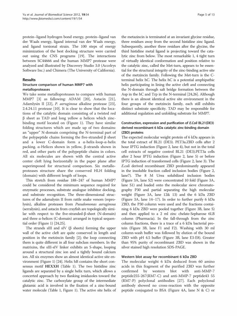
Figure 1 Structure comparison of Human MMP7 with metalloproteases. Details of the PDB structure codes of related proteins are as followed:ADAM: 2RJP, Astacin: 1QJI, Adamlysin II: 2AIG, Human MMP-7: 1MMR, P. aeruginosa alkaline protease: 1AKL, 3.4.24.11 protease: 1THL. It is interesting tonotice the locations of the catalytic domain consisting of a red twisted β sheet as TAD and long yellow α helices which zinc-binding motif located on. Allthe structures are viewed in approximately the same orientation for better comparisons. TADs are presented in red ribbons, and catalytic HLH werepresented in yellow ribbon. The main chains of the six metalloproteases are represented with ribbons in different colors. The Important motifs, such ascalcium, zinc and their binding residues, and inhibitors are presented with ball-and-stick style. The side-chains of crucial basic residues discussed in the textare shown as colored sticks.
Yu et al. Journal of Biomedical Science 2012, 19:54 Page 3 of 13http://www.jbiomedsci.com/content/19/1/54
acrylamide and 0.25 g Bis; lower buffer (100 ml) contains12.1 g Tris (1 M), 3.76 g Glycine (0.5 M), 2.0 g SDS (4%),pH 9.0; upper buffer (25 ml) contains 1.51 g Tris, pH 6.7,1.4 ml 0.5 M EDTA, and 0.7 g SDS; running buffer(300 ml) contains 3.63 g Tris, 3.38 g Glycine, 0.3 g SDS, pH8,45, self-set, pre-SDS. For resolving the molecular weightranging between 2–100 kDa proteins, a 15% high resolutionrunning gel was prepared by mixing 6.0 ml Acryl/lBis,2.0 ml lower buffer, 1.0 ml H2O, 1.0 ml 50% Glycerol, 10 μl;TEMED, 100 μl; 10% APS. The mixture with running gelsolution was poured into the gel cast, leaving a 2.5 cmspace from the top for 30 minutes. The stacking gel solu-tion containing a mixture of 1.6 ml Acryl/Bis, 1.4 ml upperbuffer, 6.0 ml H2O, 1.0 ml 50% Glycerol, 15 μl TEMED,100 μl 10% APS was poured. The stacking gel was polyme-rized for another 30 minutes. Samples were loaded into thewells and run at 90 V in the presence of full strengthrunning buffer in the upper chamber, and 1; 1 dilution ofrunning buffer in the lower chamber.
Refolding of zinc binding domainThe purified and denatured (8 M urea) 6 kDa ZBD wasdiluted drop wise 10-fold in ice-cold refolding buffer
(20 mM Acetate, pH 5.6, 10 mM CaCl2, 1 μm ZnCl2,0.2 mg/ml heparin and 0.05% Triton X-100). After 30minutes, the sample was centrifuged at 14,000 rpm for10 minutes to remove the insoluble portion. Storage ofsoluble refolded protein was at −70 °C in 18% Glycerol.
Western blotThe protein samples were separated by SDS-PAGE, trans-ferred to nitrocellulose membranes (BioRad). Then 0.1%Tween-20 (TTBS) containing 5% non-fat milk was used toblock the membrane for 3 hrs at 24 °C. The first antibodies(e.g., RM7-C and RM7-P) were applied at 4 °C overnight.After 3X washing with TTBS/milk, the second antibody (e.g., goat anti-rabbit IgG-alkaline phosphatase) was appliedfor 2 hrs. Then the transblot was washed 3X with TTBS/milk and stained with NBT/BCIP (Pierce).
Mca-peptide assayFor assay the proteolytic activities for the E Coli derivedpurified refolded recombinant mini-Matrilysin(ZBD) pro-teins, Enzyme assays were conducted at 37 °C using50 mM Tris–HCl buffer, pH 7.5, containing 100 mMNaCl and 10 mM CaCl2,1nM Zinc Chloride, 0.1% tween

Yu et al. Journal of Biomedical Science 2012, 19:54 Page 4 of 13http://www.jbiomedsci.com/content/19/1/54
20 plus Brij 35 in a total volume of 2.5 ml. The substrate,Mca-Pro-Leu-Gly-Leu-Dpa-Ala-Arg-NH2 (λex= 380 nm,λem=460 nm) was first dissolved in DMSO and the solu-tion was diluted to give a final concentration of 0.1 mM,using the buffer described above. The fluorescence of 7-amino-4-methyl-coumarin produced was monitored usinga Hitachi fluorescence spectrofluorimeter, model MPF-2A, equipped with a recorder. The measurements were car-ried out with excitation at 380 nm and emission at460 nm. The fluorescence spectrofluorimeter was set tozero with substrate in assay buffer. For each enzyme, theinitial rate of cleavage of Mca-Pro-Leu-Gly-Leu-Dpa-Ala-Arg-NH2, measured over 10 to 15 minutes, Stoppedassays were made with the 0.1 M sodium acetate, pH 4.0at different time points of interest [8]. The fluorescencespectrofluorimeter was standardized so that at 10 μM so-lution of MCA in 0.1% DMSO gave 1.0 relative fluore-scence unit. 7-amino-4-methyl-coumarine is used as thereference compound to make a standard curve to quanti-tate the hydrolysis products from the reactions. The H-Pro-Phe-Arg-AMC peptide is a non-cleavable substrate asnegative background control to standardize the instrument.
Amc-peptide assayEnzyme assays were conducted at 37 °C in 50 mMTris–HCl buffer, pH 7.5, containing 100 mM NaCl and10 mM CaCl2 in a total volume of 2.5 ml. The sub-strate, H-Pro-Phe-Arg-AMC was first dissolved inDMSO and the solution was diluted to give a final con-centration of 0.1 mM in the buffer described above.The reaction was started by the addition of 10 μl of enzymeand the fluorescence of 7-amino-4-methyl-coumarinproduced was monitored using a fluorescence spectro-fluorimeter. The measurements were carried out with exci-tation at 380 nm and emission at 460 nm. The fluorescencespectrofluorimeter was standardized so that at 10 μM solu-tion of MCA in 0.1% DMSO gave 1.0 relative fluorescenceunit. 7-amino-4-methyl-coumarine is used as the referencecompound to make a standard curve to quantitate thehydrolysis products from the reaction.
N-TIMP-1 and synthetic inhibitors inhibition assaySynthetic fluorogenic peptide substrate, Mca-Pro-Leu-Gly-Leu-Dpa-Ala-Arg-NH2, for MMP-7 [8] was used toassay the ability of N-TIMP-1 and several synthetic inhi-bitors, BB94 and SC44463 to inhibit recombinant ZBD.Assay of the inhibition of recombinant ZBD and humanactive MMP-7 by recombinant full length TIMP-1, N-TIMP-1(gift from Dr. Keith Brew [6,9]) and syntheticinhibitors was carried out by pre incubating the MMP(0.1 to 1 nM) and TIMP (a series of concentrations) in50 mM Tris–HCl, pH 7.5, containing 0.15 M NaCl,10 mM CaCl2 and 0.02% Brij at 37 °C for 1 hr. The timerequired for equilibration was determined by following
the progress of inhibition after mixing MMPs and TIMPat concentrations in the range used in the assay. Analiquot (60 μl) of substrate (15 μM) was then added to540 μl of the pre incubated MMP-TIMP mixture and ac-tivity was measured at 37 °C by following product re-lease with time.
CM-transferrin zymographyCM-transferrin (bovine), transferrin was reduced andcarboxymethylated as described by Nagase (1995). CM-transferrin (0.3 mg/ml) was embedded in 12.5% SDS–PAGE gel. Purified & refolded 6 kDa MMP-7 ZBD wastreated with sample buffer without dithiothreitol at roomtemperature and electrophoreses until the dye-front wasnear the bottom of the gel. Each gel was washed 5 timeswith 50 ml 2.5% Triton X-100, 50 mM Tris, pH 7.5, 4 °C,20 min each, to remove SDS and then 3 times with 50 mlbuffer plus 5 mMCaCl2. The gel was washed with 50 ml in-cubation buffer (50 mM Tris, pH 7.5, 5 mM CaCl2) andthen incubated in 50 ml this buffer with added proteaseinhibitors (50 mM each of Z-Phe-chloromethylketone,Tosyl-Phe-chloromethylketone (ZPCK) and amino ethylbenzenesulfonyl fluoride (PMSF)) for 18 h, 37 °C, withgentle shaking. Gels were stained with 0.1% Coomassie bluein 40% MeOH, 10% acetic acid, for 50 min, and destainedwith 7% acetic acid. The gels were scanned and digitizedwith a UVP analyzer and two dimensional intensities weredetermined with the Gelbase program (Ultra VioletProducts, Upland, CA).
Molecular docking of SC44463, BB94 and phosphoramidonSC44463, BB94 and phosphoramidon docking and dataanalysis. BB94 is selected for positive binding and phos-phoramidon for negative control on MMP7. The 3D struc-tures of the human MMP7 protease [3] (PDB code:1MMR) and thermolysin [10] (EC#: 3.4.24.11) ((PDB code:1THL) were retrieved from the Protein Data Bank.PRODRG program (http://davapc1.bioch.dundee.ac.uk/programs/prodrg/) were used to generate the coordinateand topology files of SC44463 [11,12], BB94 [13-15] andPhosphoramidon [16,17]. SC44463, BB94 and Phosphora-midon were computationally docked into the 3D structureof the human MMP7 protease and thermolysin using thedocking program GOLD [18] (version 2.1.2; GeneticOptimization for Ligand Docking, CCDC, Cambridge, UK)or PPDOCK program (http://140.112.135.49/ppdock/).GOLD operates with a genetic search algorithm and allowsfor complete ligand and partial-binding site flexibility [18].Because SC44463, BB94 and Phosphoramidon may be theinhibitors of the catalytic centers which zinc-binding motiflocated in helix-2, we defined the binding site to the inhibi-tor as the putative znic catalytic pocket. The results wereranked by GOLD’s scoring function which is a molecularmechanics-like function with four terms based on
Yu et al. Journal of Biomedical Science 2012, 19:54 Page 5 of 13http://www.jbiomedsci.com/content/19/1/54
protein–ligand hydrogen bond energy, protein–ligand vander Waals energy, ligand internal van der Waals energy,and ligand torsional strain. The 100 steps of energyminimization of the best docking structure were carriedout using the CNS programs [19]. The interactionsbetween SC44666 and the human MMP7 protease wereanalyzed and illustrated by Discovery Studio 3.0 (AccelrysSoftware Inc.) and Chimera (The University of California).
ResultsStructure comparison of human MMP7 withmetalloproteasesWe take some metalloproteases to compare with humanMMP7 [3] as fallowing: ADAM [20], Astacin [21],Adamlysin II [22], P. aeruginosa alkaline protease [23],2.4.24.11 protease [10]. It is clear to show that the loca-tions of the catalytic domain consisting of a twisted redβ sheet as TAD and long yellow α helices which zinc-binding motif located on (Figure 1). They have similarfolding structures which are made up of two domains:an “upper” N-domain comprising the N-terminal part ofthe polypeptide chains forming the five-stranded β sheetand a lower C-domain form a α-helix-loop-α-helixpacking. α-Helices shown in yellow, β-strands shown inred, and other parts of the polypeptide chains in colors.All six molecules are shown with the central activecenter cleft lying horizontally in the paper plane aftersuperimposed for structural comparison. Six metallo-proteases structure share the conserved HLH folding(domain) with different length of loops.This stretch from residue 188–247 of human MMP7
could be considered the minimum sequence required forenzymatic processes, substrate analogue inhibitor docking,and substrate recognition. The three-dimensional struc-tures of the adamalysin II from rattle snake venom (repro-lysin), alkaline proteases from Pseudomonas aeruginosa(serralysin), and astacin from crayfish are topologically simi-lar with respect to the five-stranded-β-sheet (N-domain)and three α-helices (C-domain) arranged in typical sequen-tial order (Figure 1) [24,25].The strands sIII and sIV (β sheets) forming the upper
wall of the active cleft are quite conserved in length andposition in the metzincin family [2]; the loop connectingthem is quite different in all four subclass members. In thematrixins, the sIII-sIV linker exhibits an S-shape, loopingaround a structural zinc ion and a tightly bound calciumion. All six enzymes show an almost identical active site en-vironment (Figure 1) [24]. Helix hB contains the short con-sensus motif HEXXH (Table 1). The two histidine zincligands are separated by a single helix turn, which allows aconcerted approach by two flanking imidazoles toward thecatalytic zinc. The carboxylate group of the intermediateglutamic acid is involved in the fixation of a zinc-boundwater molecule (Table 1, Figure 1). The active site helix of
the metazincin is terminated at an invariant glycine residue,three residues away from the second histidine zinc ligand.Subsequently, another three residues after the glycine, thethird histidine metal ligand is projecting toward the cata-lytic zinc from below. The most remarkable 1, 4 tight turnof virtually identical conformation and position relative tothe catalytic zinc, called the Met-turn, appears to be essen-tial for the structural integrity of the zinc-binding active siteof the metzincin family. Following the Met-turn is the C-terminal helix hC. The helix hC is a potential amphipathichelix participating in lining the active cleft and connectingthe N-domain through salt bridge formation between theAsp in the hC and Trp in the N-terminal [24,26]. Althoughthere is an almost identical active site environment in thefour groups of the metzincin family, each still exhibitsdistinct substrate specificity. TAD may be responsible foradditional regulation and unfolding substrate for MMP7.
Construction, expression and purification of E.Coli BL21(DE3)derived recombinant 6 kDa catalytic zinc-binding domain(ZBD) proteinsThe expected molecular weight protein of 6 kDa appears inthe total extract of BL21 (DE3). PET3a.ZBD cells after 2hour IPTG induction (Figure 2, lane 4), but not in the totalcell extracts of negative control BL21 (DE3).PET3a cellsafter 2 hour IPTG induction (Figure 2, lane 5) or beforeIPTG induction of transformed cells (Figure 2, lane 3). TheE.coli derived recombinant ZBD predominantly appearedin the insoluble fraction called inclusion bodies (Figure 2,lane7). The 8 M Urea solubilized inclusion bodies(Figure 3A, lane S2) were concentrated 10 fold (Figure 3A,lane S1) and loaded onto the molecular sieve chromato-graphy P30 and partial separating the high molecularweight (Figure 3A, lane 12& 13) and the 6 kDa ZBD(Figure 3A, lane 14–17). In order to further purify 6 kDaZBD, the P30 column were used and the fractions contai-ning 6 kDa ZBD were pooled together (Figure 3B, lane S)and then applied to a 2 ml zinc chelate-Sepharose 6LBcolumn (Pharmacia). In the fall-through from the zinccolumn fractions, there is a trace of a 6 kDa bacterial pro-tein (Figure 3B, lane F1 and F2). Washing with 30 mlcolumn-wash buffer was followed by elution of the boundZBD with pH 4.5 buffer (Figure 3B, lane E3-E8). Greaterthan 95% purity of recombinant ZBD was shown in thesilver stained high resolution SDS-PAGE.
Western blot assay for recombinant 6 kDa ZBDThe molecular weight 6 kDa deduced from 60 aminoacids in this fragment of the purified ZBD was furtherconfirmed by western blot with anti-MMP-7peptide255-267(RM7-C) and anti-MMP-7 peptide43 55(RM7-P) polyclonal antibodies [27]. Each polyclonalantibody showed no cross-reaction with the oppositepeptide conjugated to BSA (Figure 4A, lane N & C) or

Figure 2 The E. coli BL21 (DE3). PET3a ZBD Expression Profileof Recombinant ZBD. E. coli BL21 (DE3). PET3a ZBD transformedcells were grown at 37 0 C to OD600 reading 0.2 ~ 0.4, 10ul of totalcell extracts from 200 μl of cells solibilized in 1% SDS sample bufferas control for before IPTG induction were prepared as methoddescribed (lane 3). After 2 hour 0.8 mM IPTG induction, 10ul of totalcell extracts from 100 ul of cells were prepared as after IPTGincubation (lane 4). 10ul of total cell extracts from 100 μl of 2 hoursIPTG inducing PET 3a mock transformation BL21 (DE3) cells wasprepared as negative control (lane 5). The pellet after French pressand inclusion bodies 0.1% triton X-100 wash steps were prepared(lane 7). Lane 1: Molecular weight standard; Lane2 and 6: Blank. Figure 3 Purification of Recombinant ZBD. Panel A: shows the
silver staining for the high resolution SDS-PAGE analysis for thepurification fraction profile from the P30 molecular sievechromatography. The 8 M Urea 50 mM Tris pH 7.5 solubilizedinclusion bodies (lane S2) was concentrated 10 fold (laneS1) as thestart material before loading onto P30 column. 15 μl of the fractionsgave OD280 reading were subjected to high resolution SDS-PAGEanalysis (lane 10–17). The combined fractions #14-#15 (lane p) wereconcentrated 10 fold (lane pc). Panel B: shows the silver staining forthe high resolution SDS-PAGE analysis for the purification fractionprofile from the fractions zinc-chelate chromatography. Combiningthe partial purified fractions containing ZBD from P30 collumn (lane S)were subjected to the zinc-chelate affinity. The unbound fall-throughfractions (lane F1 and F2) and the wash buffer fractions are shown(lane W1 and W2). The bound ZBD was eluted with pH 4.5 buffer andthe low pH eluted fractions (lane E3-E10).
Yu et al. Journal of Biomedical Science 2012, 19:54 Page 6 of 13http://www.jbiomedsci.com/content/19/1/54
the BSA carrier (Figure 4B, lane B). This purified 6 kDazinc binding region of rat matrilysin can only react withanti-MMP-7 peptide255-257 polyclonal antibody (RM7-C) (Figure 4A, right panel lane Z), but not anti-MMP-7peptidere43-55(RM7-P) (Figure 4A, left panel lane Z).This immunodetection confirmed that the zinc-chelateaffinity-purified 6 kDa protein is the expected recombinantZBD protein and the fact that there is no trace of cross-reactivity shown by using anti-MMP-7 peptide43-55 indi-cates that 6 kDa proteins are not derived from the N-terminal part of rat matrilysin, instead of being producedby autolysis post activation (Figure 4A, right panel lane A).
Triton X-100 and heparin are accessory factors foldingrecombinant ZBD in vitroUnlike the inclusion bodies of recombinant proMMP-7 oractive MMP-7 which can be partially purified by severalwashing with Tris 50 mM pH 7.5, 0.25% Triton X-100 and5 mM DTT, the inclusion bodies of recombinant ZBD areeasily solubilized by 0..25% Triton X-100 and demonstrateMMP activity (data not shown). This is quite a uniqueproperty of the inclusion bodies of recombinant ZBD. Inmost cases of recombinant proteins over-expressed in BL21(DE3), it usually requires strong chaotropic reagent, such as4~8 M Urea or 4 M Guanidine, to solubilize the inclusionbodies. Surprisingly, we found that 0.05% Triton X-100 isenough to solubilize the inclusion bodies of recombinant
ZBD (data not shown). This raised the possibility thatTriton X-100 might also be an accessory factor in the fol-ding process of ZBD, especially since it is frequently addedto various MMPs as a stabilizer. Interestingly, 0.25% TritonX-100 had been used in the zymography procedure to re-move the SDS and assist folding in gels. My previousstudies also showed that heparin can enhance the acti-vities of MMPs by stabilizing the enzymes. The resultfrom the refolding test showed that Triton X-100 was abetter accessory folding factor than heparin, but thecombination of 0.05% Triton X-100 and 0.3 mg/mlheparin gave the best refolding activities in the Mca-peptide assay (Figure 5A). The instability appeared tobe the major factor concerned in the refolding process,

Figure 4 Western Blot Analysis for ZBD. The polyclonalantibodies, anti-propeptide (RM7-P) and anti-C-terminal peptide(RM7-C), were used to detect the final purified fraction of ZBD.Panel A left: The transblot was reacted with RM7-P anti serumwhich specifically recognizes the proenzyme (lane P1 and P2) andthe peptide22-34 conjugated BSA (lane N); Panel A right : Thetransblot was reacted with RM7-C anti serum which recognizes allpopulations of recombinant MMP-7 and the peptide238-247conjugated BSA (lane C), except for the peptide22-34 conjugated toBSA (lane N). Both antibodies have no cross-reactivities for the BSAcarrier (laneB) or each other. Panel B: The silver staining for the highresolution SDS-PAGE shows the sample loading profile. Mw:Molecular weight standard; P1 and P2: proMMP-7; A: all populationsof activated MMP-7. Z: recombinant CZBD; N: peptide22-34conjugated BSA; C: the peptide238-247 conjugated BSA. B: BSA.
Figure 5 Combination of 0.05% Triton and 0.2 mg/ml heparingive the optimal refolding activities to cleave the syntheticcoumarin-labelled peptide substrate, Mca-Pro-Leu-Gly-Leu-Dpa-Ala-Arg-NH2. Panel A: Shows the refolded ZBD activities increased indose-dependent manner. In the absence of the refolding accessoryfactors, Triton X-100 and heparin. The significant reduced activities inthe high-concentration (> 100 μg/ml) was observed which could bedue to autolysis. Panel B: Under 37 °C incubation for 18 hours, TritonX-100 and heparin can prevent the activity loss. (All experiments wererepeated at two batch of purification and refolding preparation anddata collected from a representative experiments).
Yu et al. Journal of Biomedical Science 2012, 19:54 Page 7 of 13http://www.jbiomedsci.com/content/19/1/54
because in the absence of the accessory folding factorwhen the concentration of ZBD reached 40 ng/perassay, the activities of ZBD decreased dramatically. Inthe next test, after challenging with 18 hrs 37 °C pre in-cubation, the loss of activity of refolded enzyme foreach refolding condition was measured by the Mca-peptide assay. There is only 13% of total activity lost in thepresence of Triton X-100 and heparin. Heparin seemed tobe a more important factor and did a better job than Triton
X-100, 23% vs 37% lost of total activity (Figure 5B). In theabsence of both factors, 52% of total activity was lost after18 hrs 37 °C pre incubation (Figure 5B).
The helix-loop-helix, 6 kDa ZBD, provides sufficientstructure for substrate and inhibitor recognitionsThe activity was determined by hydrolysis of the coumarinlabeled-peptide, Mca-Pro-Leu-Gly-Leu-Dpa-Ala-Arg-NH2which has been reported to be a very sensitive substrate formatrilysin. Another peptide substrate, H-Pro-Phe-Arg-AMC, which has been reported a sensitive substrate forplasminogen was used to test the substrate specificity of6 kDa ZBD. The results revealed that the activities are in-creasing in dose-dependent manner against Mca peptide

Yu et al. Journal of Biomedical Science 2012, 19:54 Page 8 of 13http://www.jbiomedsci.com/content/19/1/54
(Figure 6A), but not Amc-peptide (data not shown). Thebuffer alone or enzyme itself did not give any significantintrinsic fluorescence reading. The kcat/Km is about106 M-1 s-1determined by hydrolysis of Mca-peptide sub-strate, which is about 400~ 500 fold less than the wildtype activity. Interestingly, according to the X-ray crystalstructure even though there is no the third calciumrevealed in the catalytic zinc-binding domain, 5 mMcalcium and 1 μM zinc are required for optimal catalyticactivity. Refolded 6 kDa ZBD continued to catalyze thehydrolysis of the fluorogenic peptide substrate over theentire time of the assay. The activity increased in a time-dependent manner (Figure 6B), but the substrate aloneshowed no digestion. However wild-type matrilysin cancompletely digest all the substrate within 2 hrs. Although6 kDa ZBD was able to cleave the peptide substrate and
Figure 6 Mca-Pro-Leu-Gly-Leu-Dpa-Ala-Arg-NH2 assay for characterizrefolded ZBD shows increasing enzymatic activity in dose-dependent mannsituation. Panel B: approximately 6 ng/ml refolded ZBD shows the increasiduring overnight incubation. Panel C: Recombinant ZBD can be inhibitedSC44463 and CoCl2, but not b6 250 nM Phosphoramidon. (All experimentsand data collected from a representative experiments).
kept its substrate specificity it is much less efficient thanthe full length active matrilysin. This result stronglysuggests that the C-domain, helix-Met-turn-helix, lowerpart of the active matrilysin is the minimum structuralelement for substrate recognition and catalytic activitiesand the N-domain, five-stranded β sheet, upper portion isre1uired for efficient substrate hydrolysis. The proteinsubstrate, CM-transferrin, cleavages seem dependent onthe hexamer and octamer formations of 6 kDa in the pre-sence of heparin sulfate and triton X-100 revealed in CM-transferrin zymographic analysis (Figure 7) [27,28]
6 kDa ZBD is inhibited by EDTA and synthetic inhibitors, butN-TIMP-2 inhibits ZBD less effective than it does wild typeThis activity could be inhibited by 10 mM EDTA, 1 mMCoCl2 and MMP substrate analogue synthetic inhibitors,
ation of refolded ZBD. Panel A: Under the optimized conditions, theer. No significant activity loss was found in the high concentrationng activity during the time course study and no significant activity lossby 10 nM EDTA, 1 mM CoCl2 and synthetic inhibitors, 50 nM BB94 &were repeated at two batch of purification and refolding preparation

Yu et al. Journal of Biomedical Science 2012, 19:54 Page 9 of 13http://www.jbiomedsci.com/content/19/1/54
SC44463 and BB94, but not by the 24.11 metalloendopepti-dase (neprilysin) inhibitor, phosphoramidon, or serine pro-tease inhibitors, such as ZPCK [29]. The concentrations ofSC44463 and BB94 required inhibiting 50% of the enzymeactivities were about 5 nM. This value is similar to the IC50for active MMP-7 (Figure 7C). Full length TIMP-1 or N-TIMP-1 did not inhibit ZBD as efficiently as full-lengthactive matrilysin. However, a 25-fold higher concentrationof N-TIMP-2 can completely inhibit ZBD (data notshown).
Molecular docking of SC44666SC44463, (N4-hydroxy-N1-[1 S [(4-methoxphenyl)me-thyl]-2-(methylamino)-2 -oxoethyl]- 2R-(2-methylpropyl)butane-diamide), A powerful synthetic inhibitor of Matri-lysin and collagenases is reported to inhibit ovulation inperfused rat ovaries at 25 nM [30]. From our previousstudy, sc44463 is a potent inhibitor with IC50 ~10nM[30,31]. We decide to use sc44463 to perform the dockingstudies for Zinc-Dependent Proteolytic Domain- Helix-Loop-Helix Catalytic Zinc Binding Domain (ZBD) forMMP-7.Phosphoramidon were not good for docking into MMP7
as negative control for selectivity. SC44463 can successfullydock into MMP7 in multiple residues (in pink) Ala187,Ala184, Phe185, His 124, Ala 183 which contribute up to70% contact inter phase compared to the whole enzyme. InBB94 case, the results of docking only shown weakerhydrophobic interaction than SC44463 with TAD domain(Figure 8 & 9B,3). There is no pi-pi interaction betweenBB94 and MMP7 as pi-pi interaction between SC44463and Tyr73 in MMP-7 (Figure 8). The hydroxamate group,HONHCO-CH2C-, of sc44463 contributes to occupy thecatalytic zinc to prevent from ionizing H2O molecules.MMP7,a zinc dependent metallopotease, the proteolyticcatalytic divalent cation zinc in the active center is involvedin the peptide bonds cleavage through the transient
Figure 7 The polymerization of the 6 kDa ZBD of MMP-7 in pentametowards to the CM-transferrin substrate in CM-transferrin zymographco-polymerized with SDS-PAGE as a substrate gel for analyzing the MMP-7
electron acceptor for the peptide bond attack by the ionizedH2O which Glu-121 donate a pair of electron (Figure 8A).In this study, we try to define the minienzyme of metzin-
cin super-family. The minienzyme of human MMP7 asZBD can fold and maintain its catalytic activity. Due to theabove description, the mini-enzyme can provide the plat-form for performing screen or virtual screen to discoverbroad range of inhibitors. Nevertheless, the discovery ofhighly selective small molecule protease inhibitors across awide variety of protease is still achievable. For example,neprilysin (neutral endopeptidase 24.11; NEP) inhibitor,phosphoramidon, obtained by analogs with methyl or ethylsubstitutions, was relatively not potent [32]. The dockingsimulation of SC44463 revealed that this compoundblocked the direction of the peptide chain in contact withthe active site is in the reverse direction of that seen incomplex of stromelysin with synthetic inhibitors [33]. Asshown in Figure 8, we found that the SC44463 coordinatedthe zinc ion and formed pi-pi interaction with side chainsof His-124 (HLH) and Tyr-73 (TAD), the oxygen on themain chains of Glu-121 (HLH) of human MMP-7. Inaddition, Ala-83 (TAD), Ala-85 (TAD), Ala-87 (TAD)donated hydrophobic interaction to SC44463. Thus, thedrug docking data of human MMP-7 indicated thatSC44463 was a good lead compound for the inhibiting theactivity of human MMP-7 through its competitive actionwith peptide chain pocket. In the discovery and develop-ment of metzincin inhibitors, some inhibitors have beenapproved and successfully targeted to different therapeutictargets in various protease [34]. Our results presented hereshow that SC44463 was able to suppress the proteolytic ac-tivity of human MMP-7 and 6 kDa mini MMP-7. Based onthe modeling results, further refinement of the potency andselectivity of SC44463 can be further studied by the modifi-cations of some critical functional groups of SC44463 forrational analog- based searching more potent specific inhi-bitors for this helix B Met loop-helix C folding domain.
r and Octamer demonstrate the significant proteolytic activitiesic assay. 300 μg of craboxylmethylated transferrin (CM-transferrin) wasactivities in situ.

Figure 8 Molecular docking of SC44463 in Human MMP7. (A) A 2D diagram of the details of the SC44463 binding environment. The SC44463 wasdocked onto Human MMP7. Illustration of amino acid contacted to the SC44463 in Human MMP7. SC44463 contacted to TAD (57–114) in the left part(Tyr73, Ala83, Ala85, Ala87) and to HLH (114–173) in the right part (His120, Glu 121, His124, His138, Znic ion). (B) Overall illustration of molecular docking ofSC44463 in Human MMP7. HLH shown in yellow, TAD shown in red, and other parts of the polypeptide chains in rice white. The binding complex isshown in the standard orientation, i.e. with the cental active center cleft lying horizontally in the paper plane. Zinc ion, three binding histidines andSC44463 are presented with ball and stick model. (C) TAD and HLH of binding complex are shown as 30% transparency solid surface colored with surfacecharge. SC44463 is presented with stick and the complex in rice white ribbon. (D) Only HLH of binding complex are shown as 30% transparency solidsurface colored with surface charge. It is interesting to notice that the major contact between SC44463 and Human MMP7 are in the HLH area with saltbridge, hydrogen bond and pi-pi interaction. SC44463 is presented with stick and the complex in rice white ribbon.
Yu et al. Journal of Biomedical Science 2012, 19:54 Page 10 of 13http://www.jbiomedsci.com/content/19/1/54
Therefore, we speculate that SC44463 can be a good leadcompound to develop more potent selective inhibitor forMMP-7.
DiscussionA widely-accepted concept is that the full-length active do-main of MMP is the minimum structure required for fullcatalytic activity and affinity for TIMP. This appears to betrue, but the present studies show considerable residual ac-tivity in the truncated enzyme. The characteristic consensusmotif HEXXHXXGXXH and the Met-turn motif (Table 1),P/Y-XMBX (B: Bulky residues), are the major elementsarranged in the common topological structure helix-loop-helix forming the lower half C-terminal portion of the 60residue domain. [25,35-37]. by adding the upper half five-stranded β sheet (N-domain) (Figure 1), the active centercleft is completed (Figure 8C-D). The exact function of theC-portion which possesses the major catalytic elements isstill unknown. It is important to address the molecular and
structural function of this helix-Met turn-helix motif, whichcontains the catalytic zinc element. In evolution, this funda-mental building block was augmented by additional genewith the metzincin super-family to give rise ultimately tothe present four subclasses. In this report, the foldedrecombinant ZBD corresponding to the C-portion demon-strates that it possesses the abilities of substrate recognition,catalytic hydrolysis and inhibitor binding (Figure 8A & 9).At this point, it will be fair to call this ZBD fragment a zincbinding domain with catalytic function [35].However, the reasons for the loss of catalytic hydrolysis
efficiency compared to full-length active enzyme could bethat the five-stranded β sheet (N-domain) is important incontributing hydrophobicity to the active cleft. A commonfeature for the active center of most enzymes is the non-polar environment in the active center. The hypothesis, asintroduced by Cohen et al. 1970 and Crosby et al. 1970[38,39], suggested that enzyme active sites becomes basi-cally non-polar after removal of water molecules and that

Figure 9 Comparison docking results of SC44463 in Human MMP7 and in 3.4.24.11 protease and other protease drugs. (A) The structureof the SC44463, BB94 amd Phosphoramidon is presented as colored stick model. (B) The SC44463 was docked onto human MMP7. Illustration of threehistidine (His120, His124, His138) of human MMP7 in SC44463-Huaman MMP-7 complex. SC44463 is presented as rice white stick model and HLH ofhuman MMP7 is shown as yellow ribbon. CCT-Thermolysin complex (PDB code: 1THL). The N-(<1-[(2 S)-2-carboxy-4-phenylbutyl]cyclopentyl> carbonyl)-L-tryptophan (CCT) as 3.4.24.11 protease inhibitor is presented as rice white stick model and SC44463 is presented as sky bluestick model. It should be notice that SC44463 could not dock in to 3.4.24.11 protease very well. The huamn MMP7 is presented with ribbon model,HLH shown in yellow, TAD shown in red. The docking result of SC44463-Huaman MMP-7 complex and BB94 shows that the pi-pi and hydrophobicinteraction of SC44463 is better than BB94. The SC44463 is presented as sky blue stick model and BB94 is presented as rice white stick model.
Yu et al. Journal of Biomedical Science 2012, 19:54 Page 11 of 13http://www.jbiomedsci.com/content/19/1/54
such non-polar sites help in accelerating enzymatic reac-tions. Applying this hypothetical concept of desolvationKcat contribution to the situation of this “naked” activecenter of 6 kDa ZBD, it is not difficult to imagine that theloss of the N-domain five stranded β sheet makes the ac-tive center of C-domain completely exposed to a polar en-vironment which does not favor the desolvation processand reduces the catalytic hydrolysis efficiency by two tothree orders of magnitude. The contribution of N-domaincould be offering the hydrophobicity to the active cleftand helping the desolvation process during the catalytichydrolysis of substrate. 0.025% Triton X-100 could takethe place of the N-domain function, which enhances thehydrophobicity of the active center, helps the desolvationprocess and enhances the catalytic hydrolysis. Interest-ingly, ZBD can only digest the intact protein substrate inpolymer forms, such as CM-transferrin, but can digest
peptides efficiently. One explanation is that this evidenceimplies that the “naked” active centers of ZBD are buriedinside the non-polar environment of Triton X-100 micelleand the peptide substrates can be more easily infused intothis micelle and become more accessible for ZBD tohydrolyze than intact protein substrates do. The other ex-planation is that the helix-loop-helix zinc binding catalyticmotif forming the smallest peptide bound cleavage foldingunit, however, the rest part of active MMP-7(~19 kDa)facilitates protein-protein interactions to induce conform-ational changes to expose the cleavable peptide regionwhich could be buried in the intact protein substrates.However, this is purely hypothetical explanation. Anotherinteresting observation regarding the requirement for cal-cium for activity, which is contradictory to the X-ray crys-tal structure of active matrilysin [3] needs to be furtheraddressed in the near future.

Yu et al. Journal of Biomedical Science 2012, 19:54 Page 12 of 13http://www.jbiomedsci.com/content/19/1/54
ConclusionsThe helix B-Met loop-helix C folding conserved in metallo-protease metzincin super family is able to facilitate proteo-lytic catalysis for specific peptide substrate and inhibitorrecognition. Triton X-100 plus heparin sulfate is critical forthe refolding this 6 kDa helix B-Met loop-helix C domainand also reveal the functional active hexamer and octamerof the mini MMP-7 complex in CM-transferrin zymogra-phy. 6 kDa mini MMP-7 through autolysis of proMMP-7 isthe smallest metalloprotease in living world.
Competing interestsAll authors declare that they have no competing interests.
AcknowledgmentsThis work was supported in part by the Taiwanese NSC funding NSC-100-2325-b002056 for WHY. We thank Prof. J. Frederick Woessner of Departmentof Biochemistry and Molecular Biology, University of Miami School ofMedicine for guiding and help.
Author details1Institute of Biochemistry and Molecular Biology, College of Medicine,National Taiwan University, Ren-Ai Road, Taipei, Taiwan. 2Graduate Instituteof Oral Biology, College of Medicine, National Taiwan University, Ren-Ai Road,Taipei, Taiwan. 3NTU-DRCP Lectures and Core for Membrane Proteins, Centerfor Biotechnology, National Taiwan University, Chang Sing Street, Taipei,Taiwan. 4Institute of Biotechnology, National Taiwan University, Chang SingStreet, Taipei, Taiwan.
Authors’ contributionsWei-Hsuan Yu designs the experiments, collection, and analysis of theexperimental data. And writes the manuscript. Po-Tsang Huang and Kuo-Long Lou design and process the docking experiments, write and presentthe structural relative sections. Shuan-Su C Yu and Chen Lin carried out themolecular cloning, preparation of purified and refolding recombinant 6 kDamini MMP-7 and drafted the manuscript. All authors read and approved thefinal manuscript.
Received: 14 March 2012 Accepted: 29 May 2012Published: 29 May 2012
References1. Bode W, Gomis-Ruth FX, Stockler W: Astacins, serralysins, snake venom
and matrix metalloproteinases exhibit identical zinc-bindingenvironments (HEXXHXXGXXH and Met-turn) and topologies and shouldbe grouped into a common family, the 'metzincins'. FEBS Lett 1993,331:134–140.
2. Cha J, Auld DS: Site-directed mutagenesis of the active site glutamate inhuman matrilysin: investigation of its role in catalysis. Biochemistry 1997,36:16019–16024.
3. Browner MF, Smith WW, Castelhano AL: Matrilysin-inhibitor complexes:common themes among metalloproteases. Biochemistry 1995,34:6602–6610.
4. Gomis-Ruth FX, Gomez-Ortiz M, Vendrell J, Ventura S, Bode W, et al: Crystalstructure of an oligomer of proteolytic zymogens: detailedconformational analysis of the bovine ternary complex and implicationsfor their activation. J Mol Biol 1997, 269:861–880.
5. Gomis-Ruth FX, Maskos K, Betz M, Bergner A, Huber R, et al: Mechanism ofinhibition of the human matrix metalloproteinase stromelysin-1 byTIMP-1. Nature 1997, 389:77–81.
6. Huang W, Meng Q, Suzuki K, Nagase H, Brew K: Mutational study of theamino-terminal domain of human tissue inhibitor of metalloproteinases1 (TIMP-1) locates an inhibitory region for matrix metalloproteinases.J Biol Chem 1997, 272:22086–22091.
7. Van Doren SR, Kurochkin AV, Hu W, Ye QZ, Johnson LL, et al: Solutionstructure of the catalytic domain of human stromelysin complexed witha hydrophobic inhibitor. Protein Sci 1995, 4:2487–2498.
8. Knight CG, Willenbrock F, Murphy G: A novel coumarin-labelled peptidefor sensitive continuous assays of the matrix metalloproteinases.FEBS Lett 1992, 296:263–266.
9. Meng Q, Malinovskii V, Huang W, Hu Y, Chung L, et al: Residue 2 of TIMP-1is a major determinant of affinity and specificity for matrixmetalloproteinases but effects of substitutions do not correlate withthose of the corresponding P1' residue of substrate. J Biol Chem 1999,274:10184–10189.
10. Holland DR, Barclay PL, Danilewicz JC, Matthews BW, James K: Inhibition ofthermolysin and neutral endopeptidase 24.11 by a novel glutaramidederivative: X-ray structure determination of the thermolysin-inhibitorcomplex. Biochemistry 1994, 33:51–56.
11. Cruwys SC, Davies DE, Pettipher ER: Co-operation between interleukin-1and the fibrinolytic system in the degradation of collagen by articularchondrocytes. Br J Pharmacol 1990, 100:631–635.
12. Creemers LB, Jansen ID, Docherty AJ, Reynolds JJ, Beertsen W, et al:Gelatinase A (MMP-2) and cysteine proteinases are essential for thedegradation of collagen in soft connective tissue. Matrix Biol 1998,17:35–46.
13. Kazes I, Elalamy I, Sraer JD, Hatmi M, Nguyen G: Platelet release oftrimolecular complex components MT1-MMP/TIMP2/MMP2: involvementin MMP2 activation and platelet aggregation. Blood 2000, 96:3064–3069.
14. Aoki T, Sato D, Li Y, Takino T, Miyamori H, et al: Cleavage of apolipoproteinE by membrane-type matrix metalloproteinase-1 abrogates suppressionof cell proliferation. J Biochem 2005, 137:95–99.
15. Yang Z, Kyriakides TR, Bornstein P: Matricellular proteins as modulators ofcell-matrix interactions: adhesive defect in thrombospondin 2-nullfibroblasts is a consequence of increased levels of matrixmetalloproteinase-2. Mol Biol Cell 2000, 11:3353–3364.
16. Fernandez-Patron C, Stewart KG, Zhang Y, Koivunen E, Radomski MW, et al:Vascular matrix metalloproteinase-2-dependent cleavage of calcitoningene-related peptide promotes vasoconstriction. Circ Res 2000,87:670–676.
17. Jeyabalan A, Novak J, Danielson LA, Kerchner LJ, Opett SL, et al: Essentialrole for vascular gelatinase activity in relaxin-induced renal vasodilation,hyperfiltration, and reduced myogenic reactivity of small arteries.Circ Res 2003, 93:1249–1257.
18. Jones G, Willett P, Glen RC, Leach AR, Taylor R: Development andvalidation of a genetic algorithm for flexible docking. J Mol Biol 1997,267:727–748.
19. Brunger AT, Adams PD, Clore GM, DeLano WL, Gros P, et al: Crystallography& NMR system: A new software suite for macromolecular structuredetermination. Acta Crystallogr D Biol Crystallogr 1998, 54:905–921.
20. Mosyak L, Georgiadis K, Shane T, Svenson K, Hebert T, et al: Crystalstructures of the two major aggrecan degrading enzymes, ADAMTS4and ADAMTS5. Protein Sci 2008, 17:16–21.
21. Riek R, Precheur B, Wang Y, Mackay EA, Wider G, et al: NMR structure ofthe sea urchin (Strongylocentrotus purpuratus) metallothionein MTA.J Mol Biol 1999, 291:417–428.
22. Gomis-Ruth FX, Meyer EF, Kress LF, Politi V: Structures of adamalysin II withpeptidic inhibitors. Implications for the design of tumor necrosis factoralpha convertase inhibitors. Protein Sci 1998, 7:283–292.
23. Miyatake H, Hata Y, Fujii T, Hamada K, Morihara K, et al: Crystal structure ofthe unliganded alkaline protease from Pseudomonas aeruginosaIFO3080 and its conformational changes on ligand binding.J Biochem 1995, 118:474–479.
24. Stocker W, Bode W: Structural features of a superfamily of zinc-endopeptidases: the metzincins. Curr Opin Struct Biol 1995, 5:383–390.
25. Stocker W, Grams F, Baumann U, Reinemer P, Gomis-Ruth FX, et al: Themetzincins–topological and sequential relations between the astacins,adamalysins, serralysins, and matrixins (collagenases) define asuperfamily of zinc-peptidases. Protein Sci 1995, 4:823–840.
26. Bode W, Grams F, Reinemer P, Gomis-Ruth FX, Baumann U, et al: Themetzincin-superfamily of zinc-peptidases. Adv Exp Med Biol 1996,389:1–11.
27. Yu WH, Woessner JF Jr: Heparan sulfate proteoglycans as extracellulardocking molecules for matrilysin (matrix metalloproteinase 7).J Biol Chem 2000, 275:4183–4191.
28. Yu WH, Woessner JF Jr: Heparin-enhanced zymographic detection ofmatrilysin and collagenases. Anal Biochem 2001, 293:38–42.

Yu et al. Journal of Biomedical Science 2012, 19:54 Page 13 of 13http://www.jbiomedsci.com/content/19/1/54
29. Zieske LR, Hsi KL, Chen L, Yuan PM: Structural determination of theessential serine and glycosylation sites of carboxypeptidase P.Arch Biochem Biophys 1992, 295:76–83.
30. Butler TA, Zhu C, Mueller RA, Fuller GC, Lemaire WJ, et al: Inhibition ofovulation in the perfused rat ovary by the synthetic collagenaseinhibitor SC 44463. Biol Reprod 1991, 44:1183–1188.
31. Butler TA, Woessner JF Jr: Gelatinases and endogenous inhibitors in thepreovulatory rat ovary. Ann N Y Acad Sci 1994, 732:444–446.
32. Adler M, Nicholson JD, Starks DF, Kane CT, Cornille F, et al: Evaluation ofphosphoramidon and three synthetic phosphonates for inhibition ofbotulinum neurotoxin B catalytic activity. J Appl Toxicol 1999,19(Suppl 1):S5–S11.
33. Tronrud DE, Monzingo AF, Matthews BW: Crystallographic structuralanalysis of phosphoramidates as inhibitors and transition-state analogsof thermolysin. Eur J Biochem 1986, 157:261–268.
34. Lazure C: The peptidase zymogen proregions: nature's way of preventingundesired activation and proteolysis. Curr Pharm Des 2002, 8:511–531.
35. Tallant C, Marrero A, Gomis-Ruth FX: Matrix metalloproteinases: fold andfunction of their catalytic domains. Biochim Biophys Acta 2010,1803:20–28.
36. Guan HH, Goh KS, Davamani F, Wu PL, Huang YW, et al: Structures of twoelapid snake venom metalloproteases with distinct activities highlightthe disulfide patterns in the D domain of ADAMalysin family proteins.J Struct Biol 2010, 169:294–303.
37. Gomis-Ruth FX: Structural aspects of the metzincin clan ofmetalloendopeptidases. Mol Biotechnol 2003, 24:157–202.
38. Cohen SG, Vaidya VM, Schultz RM: Active site of alpha-chymotrypsinactivation by association-desolvation. Proc Natl Acad Sci USA 1970,66:249–256.
39. Crosby J, Stone R, Lienhard GE: Mechanisms of thiamine-catalyzedreactions. Decarboxylation of 2-(1-carboxy-1-hydroxyethyl)-3,4-dimethylthiazolium chloride. J Am Chem Soc 1970, 92:2891–2900.
doi:10.1186/1423-0127-19-54Cite this article as: Yu et al.: A smallest 6 kda metalloprotease, mini-matrilysin, in living world: a revolutionary conserved zinc-dependentproteolytic domain- helix-loop-helix catalytic zinc binding domain(ZBD). Journal of Biomedical Science 2012 19:54.
Submit your next manuscript to BioMed Centraland take full advantage of:
• Convenient online submission
• Thorough peer review
• No space constraints or color figure charges
• Immediate publication on acceptance
• Inclusion in PubMed, CAS, Scopus and Google Scholar
• Research which is freely available for redistribution
Submit your manuscript at www.biomedcentral.com/submit