
A simple procedure for retrieval of a cement-retained implant-supported crown: a case report
4
1 QUINTESSENCE INTERNATIONAL doi: ??.????/j.qi.a?????? A simple procedure for retrieval of a cement-retained implant-supported crown: A case report Muaiyed Mahmoud Buzayan, BDS, MClinDent 1 /Wan Adida Azina Binti Mahmood, BDS, MDSc 2 / Norsiah Binti Yunus, BDS, MSc 3 Retrieval of cement-retained implant prostheses can be more demanding than retrieval of screw-retained prostheses. This case report describes a simple and predictable procedure to locate the abutment screw access openings of cement- retained implant-supported crowns in cases of fractured ceramic veneer. A conventional periapical radiography image was captured using a digital camera, transferred to a comput- er, and manipulated using Microsoft Word document software to estimate the location of the abutment screw access. (Quintessence Int 201#;##:1–4; doi: ##.####/j.qi.a#####) Key words: cement-retained, ceramic veneer fracture, implant-supported prosthesis PROSTHODONTICS Muaiyed Mahmoud Buzayan to retrieve a cement-retained prosthesis without affect- ing the implant abutment and restoration compared to a screw-retained implant restoration. 2,8 Unlike in natural abutment teeth, conventional cements do not chemi- cally adhere to metallic abutments. However, the appropriate choice of cement should be made to pro- vide adequate crown retention on the implant abut- ment and at the same time allow for retrievability. 8-10 In view of the many reports of abutment screw loosening and ceramic veneer fracture, 11 various techniques have been described in the literature to simplify the retrieval of cement-retained implant crown restorations. Some contingency plans to allow the identification of screw access location and hence easy retrieval include incor- porating a retrieval slot in the design 1,3 and staining the occlusal surface of the ceramic restoration to indicate the abutment screw location. 12 Crown sectioning at the midfacial surface to break the cement seal before the sectioned crown is retrieved, 13 however, involved pro- longed chairside time. A more common method is to locate the screw access by drilling and perforating a section of the restoration using a bur. 14 The ability to Implant-supported prostheses can be either screw- or cement-retained, 1,2 and the choice of retention means depends on the clinician’s preference, the available interridge space, esthetics, and cost. 2 Predictable retrievability of implant-retained restorations is another factor to be considered as a part of patient care, 2,3 where for maintenance purposes, the prosthesis may need to be retrieved on many occasions. Screw reten- tion allows easier retrievability; however, the range of benefits of cement-retained prostheses includes better seating of the superstructure/framework, 4 less screw loosening, 5 fewer problems related to occlusal screw holes, 6 and fewer problems with ceramic strength issues. 7 In terms of retrievability, it is more demanding 1 Lecturer, Department of Prosthodontics, Dental Faculty, University Of Tripoli, Tripoli, Libya. 2 Associate Professor, Department of Prosthetic Dentistry, Faculty of Dentistry, University of Malaya, Kuala Lumpur, Malaysia. 3 Professor, Department of Prosthetic Dentistry, Faculty of Dentistry, University of Malaya, Kuala Lumpur, Malaysia. Correspondence: Dr Muaiyed Mahmoud Buzayan, Department of Pros- thodontics, Dental Faculty, University Of Tripoli, Tripoli, Libya. Email: [email protected]
-
Upload
university-malayas-dental-sciences-research -
Category
Documents
-
view
219 -
download
3
Transcript of A simple procedure for retrieval of a cement-retained implant-supported crown: a case report

1
Q U I N T E S S E N C E I N T E R N AT I O N A L
doi: ??.????/j.qi.a??????
A simple procedure for retrieval of a cement-retained implant-supported crown: A case reportMuaiyed Mahmoud Buzayan, BDS, MClinDent1/Wan Adida Azina Binti Mahmood, BDS, MDSc2/Norsiah Binti Yunus, BDS, MSc3
Retrieval of cement-retained implant prostheses can be more demanding than retrieval of screw-retained prostheses. This case report describes a simple and predictable procedure to locate the abutment screw access openings of cement-retained implant-supported crowns in cases of fractured
ceramic veneer. A conventional periapical radiography image was captured using a digital camera, transferred to a comput-er, and manipulated using Microsoft Word document software to estimate the location of the abutment screw access. (Quintessence Int 201#;##:1–4; doi: ##.####/j.qi.a#####)
Key words: cement-retained, ceramic veneer fracture, implant-supported prosthesis
PROSTHODONTICS
Muaiyed Mahmoud Buzayan
to retrieve a cement-retained prosthesis without aff ect-
ing the implant abutment and restoration compared to
a screw-retained implant restoration.2,8 Unlike in natural
abutment teeth, conventional cements do not chemi-
cally adhere to metallic abutments. However, the
appropriate choice of cement should be made to pro-
vide adequate crown retention on the implant abut-
ment and at the same time allow for retrievability.8-10 In
view of the many reports of abutment screw loosening
and ceramic veneer fracture,11 various techniques have
been described in the literature to simplify the retrieval
of cement-retained implant crown restorations. Some
contingency plans to allow the identifi cation of screw
access location and hence easy retrieval include incor-
porating a retrieval slot in the design1,3 and staining the
occlusal surface of the ceramic restoration to indicate
the abutment screw location.12 Crown sectioning at the
midfacial surface to break the cement seal before the
sectioned crown is retrieved,13 however, involved pro-
longed chairside time. A more common method is to
locate the screw access by drilling and perforating a
section of the restoration using a bur.14 The ability to
Implant-supported prostheses can be either screw- or
cement-retained,1,2 and the choice of retention means
depends on the clinician’s preference, the available
interridge space, esthetics, and cost.2 Predictable
retrievability of implant-retained restorations is another
factor to be considered as a part of patient care,2,3
where for maintenance purposes, the prosthesis may
need to be retrieved on many occasions. Screw reten-
tion allows easier retrievability; however, the range of
benefi ts of cement-retained prostheses includes better
seating of the superstructure/framework,4 less screw
loosening,5 fewer problems related to occlusal screw
holes,6 and fewer problems with ceramic strength
issues.7 In terms of retrievability, it is more demanding
1 Lecturer, Department of Prosthodontics, Dental Faculty, University Of Tripoli, Tripoli, Libya.
2 Associate Professor, Department of Prosthetic Dentistry, Faculty of Dentistry, University of Malaya, Kuala Lumpur, Malaysia.
3 Professor, Department of Prosthetic Dentistry, Faculty of Dentistry, University of Malaya, Kuala Lumpur, Malaysia.
Correspondence: Dr Muaiyed Mahmoud Buzayan, Department of Pros-thodontics, Dental Faculty, University Of Tripoli, Tripoli, Libya. Email: [email protected]

2
Q U I N T E S S E N C E I N T E R N AT I O N A L
Buzayan et al
doi: ??.????/j.qi.a??????
identify the approximate location of the screw access
opening in cement-retained implant-supported crowns
may eliminate laborious intraoral crown sectioning.
The purpose of this article is to describe a simple
and undemanding procedure making use of readily
available conventional periapical radiography to locate
the screw access opening of ceramic implant-sup-
ported crowns with fractured ceramic veneer. The
image was loaded onto a computer, and using readily
available software, the abutment screw location was
estimated by measuring the mesiodistal dimension of
the crown in relation to the adjacent teeth.
CLINICAL CASE
A 21-year-old female patient presented to the Depart-
ment of Prosthetic Dentistry, 3 months after a metal-
ceramic implant-supported crown was cemented,
replacing the mandibular right second premolar. She
was concerned with the crown restoration, which was
gradually chipping off , and had no other associated
symptoms (Fig 1). The patient’s dental record indicated
that a 4.5-mm diameter (bone level; SuperLine, Implan-
tium) implant had been inserted in the mandibular
right second premolar edentulous area. The metal-
ceramic crown was cemented using provisional
cement (TempBond, Kerr). A straight abutment was
used in this case. The most likely cause for the ceramic
veneer fracture in the present case was unsupported
ceramic as a result of an undercontoured and poorly
designed metal coping. The treatment plan included
replacement of the damaged crown and recementa-
tion of a new metal-ceramic crown on the existing
implant abutment (Fig 1).
Fig 2 Periapical radiograph of the implant-abutment junction and the cemented metal ceramic-crown before editing.
Fig 3 With the help of the ruler, the image was enlarged such that the implant shoulder measured 4.5 cm on the screen.
Fig 1 Partial veneer fracture of implant crown replacing the mandibular right second premolar with an exposed metal coping.
3
Q U I N T E S S E N C E I N T E R N AT I O N A L
Buzayan et al
doi: ??.????/j.qi.a??????
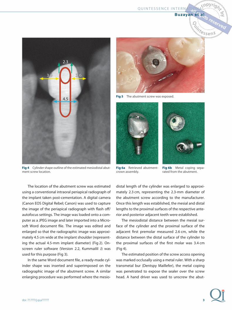
The location of the abutment screw was estimated
using a conventional intraoral periapical radiograph of
the implant taken post-cementation. A digital camera
(Canon EOS Digital Rebel; Canon) was used to capture
the image of the periapical radiograph with fl ash off /
autofocus settings. The image was loaded onto a com-
puter as a JPEG image and later imported into a Micro-
soft Word document fi le. The image was edited and
enlarged so that the radiographic image was approxi-
mately 4.5 cm wide at the implant shoulder (represent-
ing the actual 4.5-mm implant diameter) (Fig 2). On-
screen ruler software (Version 2.2, Kummailil J) was
used for this purpose (Fig 3).
In the same Word document fi le, a ready-made cyl-
inder shape was inserted and superimposed on the
radiographic image of the abutment screw. A similar
enlarging procedure was performed where the mesio-
distal length of the cylinder was enlarged to approxi-
mately 2.3 cm, representing the 2.3-mm diameter of
the abutment screw according to the manufacturer.
Once this length was established, the mesial and distal
lengths to the proximal surfaces of the respective ante-
rior and posterior adjacent teeth were established.
The mesiodistal distance between the mesial sur-
face of the cylinder and the proximal surface of the
adjacent fi rst premolar measured 2.6 cm, while the
distance between the distal surface of the cylinder to
the proximal surfaces of the fi rst molar was 3.4 cm
(Fig 4).
The estimated position of the screw access opening
was marked occlusally using a metal ruler. With a sharp
transmetal bur (Dentspy Maillefer), the metal coping
was penetrated to expose the sealer over the screw
head. A hand driver was used to unscrew the abut-
Fig 4 Cylinder shape outline of the estimated mesiodistal abut-ment screw location.
Fig 5 The abutment screw was exposed.
Fig 6a Retrieved abutment-crown assembly.
Fig 6b Metal coping sepa-rated from the abutment.
2.3
3.4 2.6
4.5

4
Q U I N T E S S E N C E I N T E R N AT I O N A L
Buzayan et al
doi: ??.????/j.qi.a??????
ment-crown assembly, which was easily separated
once out of the mouth (Figs 5 and 6). A new crown res-
toration was fabricated and cemented in place.
DISCUSSION
One advantage of this radiographic technique over
other methods that utilize a photographic image15,16 is
that the intraoral periapical radiograph can be made
available even after cementation. With the technique of
Figueras-Alvarez et al,15 two digital photographs of the
defi nitive cast precementation are required, indicating
that the procedure needs to be performed routinely
before the prosthesis is cemented. With the technique
of Daher and Morgano,16 taking digital photographs of
the patient is time-consuming for both the patient and
the dental offi ce staff , and it needs to be performed
routinely precementation. The present technique also
requires information on the implant system used,
which can easily be obtained from the website or prod-
uct catalogue.
The two-dimensional approach with this technique,
however, may provide limited information as to the
buccolingual position of the screw access opening.
While a three-dimensional radiographic imaging would
provide such information, such equipment is not read-
ily available in all dental clinics.
CONCLUSION
A simple and undemanding procedure for locating the
abutment screw access to allow abutment retrieval was
described using readily available information on the
implant system and the postcementation periapical
radiograph. The implant abutment radiographic image
was captured on a digital camera and the image was
manipulated using Word document software to esti-
mate the screw access location on the crown. This
technique can be performed by anyone with a com-
puter, without the need for special equipment or soft-
ware.
REFERENCES 1. Prestpino V, Ingber A, Kravitz J, Whitehead GM. A practical approach for
retrieving cement-retained, implant-supported restorations. Quintessence Dent Technol 2001;24:182–187.
2. Misch CE. Contemporary Implant Dentistry. St Louis: Mosby 1993:651–685.
3. Schweitzer DM, Berg RW, Mancia GO. A technique for retrieval of cement-retained implant-supported prostheses. J Prosthet Dent 2011;106:134–138.
4. Pietrabissa R, Gionso L, Quaglini V, Di Martino E, Simion M. An in vitro study on compensation of mismatch of screw versus cement-retained implant sup-ported fi xed prostheses. Clin Oral Implants Res 2000;11:448–457.
5. Wood MR, Vermilyea SG; Committee on research in fi xed prosthodontics of the Academy of Fixed Prosthodontics. A review of selected dental literature on evidence-based treatment planning for dental implants: report of the Committee on research in fi xed prosthodontics of the Academy of Fixed Pros-thodontics. J Prosthet Dent 2004;92:447–462.
6. Taylor TD, Agar JR. Twenty years of progress in implant prosthodontics. J Prosthet Dent 2002;88:89–95.
7. Torrado E, Ercoli C, Al Mardini M, et al. A comparison of the porcelain fracture resistance of screw-retained and cement-retained implant-supported metal-ceramic crowns. J Prosthet Dent 2004;91:532–537.
8. Hebel KS, Gajjar RC. Cement-retained versus screw-retained implant restor-ations: achieving optimal occlusion and esthetics in implant dentistry. J Prosthet Dent 1997;77:28–35.
9. Breeding LC, Dixon DL, Bogacki MT, Tietge JD. Use of luting agents with an implant system: part I. J Prosthet Dent 1992;68:737–741.
10. Mehl C, Harder S, Wolfart M, Kern M, Wolfart S. Retrievability of implant-retained crowns following cementation. Clin Oral Implants Res 2008;19:1304–1311.
11. Goodacre CJ, Bernal G, Rungcharassaeng K, Kan JY. Clinical complications with implants and implant prostheses. J Prosthet Dent 2003;90:121–132.
12. Schwedhelm ER, Raigrodski AJ. A technique for locating implant abutment screws of posterior cement-retained metal-ceramic restorations with ceramic occlusal surfaces. J Prosthet Dent 2006;95:165–167.
13. Rajeev Gupta P, Verma R. A simple technique for removing cement retained implant prosthesis in case of abutment screw loosening: a case report. Indian J Dent Sci 2009;1:38–41.
14. Doerr J. Simplifi ed technique for retrieving cemented implant restorations. J Prosthet Dent 2002;88:352–353.
15. Figueras-Alvarez O, Cedeño R, Cano-Batalla J, Cabratosa-Termes J. A method for registering the abutment screw position of cement-retained implant res-torations. J Prosthet Dent 2010;104:60–62.
16. Daher T, Morgano SM. The use of digital photographs to locate implant abut-ment screws for implant-supported cement-retained restorations. J Prosthet Dent 2008;100:238–239.


![Evaluation of Two Different Attachment Systems Used with ... · Implant-retained overdentures is extremely valuable [4,5]. Rehabilitation with mandibular implant-tissue-supported](https://static.fdocuments.net/doc/165x107/5e6a2f66f71f2340c57bdc1c/evaluation-of-two-different-attachment-systems-used-with-implant-retained-overdentures.jpg)


