A Review Study on Macrolides Isolated from Cyanobacteria...macrolides isolated from cyanobacteria,...
19
marine drugs Review A Review Study on Macrolides Isolated from Cyanobacteria Mengchuan Wang 1 , Jinrong Zhang 1, *, Shan He 1 and Xiaojun Yan 2 1 School of Marine Sciences, Laboratory of Marine Natural Products, Ningbo University, Ningbo 315211, China; [email protected] (M.W.); [email protected] (S.H.) 2 Key Laboratory of Applied Marine Biotechnology of Ministry of Education, Ningbo University, Ningbo 315211, China; [email protected] * Correspondence: [email protected]; Tel./Fax: +86-574-8760-0458 Academic Editor: Anake Kijjoa Received: 27 January 2017; Accepted: 24 April 2017; Published: 26 April 2017 Abstract: Cyanobacteria are rich sources of structurally-diverse molecules with promising pharmacological activities. Marine cyanobacteria have been proven to be true producers of some significant bioactive metabolites from marine invertebrates. Macrolides are a class of bioactive compounds isolated from marine organisms, including marine microorganisms in particular. The structural characteristics of macrolides from cyanobacteria mainly manifest in the diversity of carbon skeletons, complexes of chlorinated thiazole-containing molecules and complex spatial configuration. In the present work, we systematically reviewed the structures and pharmacological activities of macrolides from cyanobacteria. Our data would help establish an effective support system for the discovery and development of cyanobacterium-derived macrolides. Keywords: cyanobacteria; macrolides; secondary metabolites; bioactivity 1. Introduction Cyanobacteria, also known as blue-green algae, including cyanobacteria from terrestrial, freshwater and marine ecosystems, are a group of ancient photosynthetic prokaryotes. As defensive chemicals, structurally-diverse secondary metabolites from cyanobacteria have been proven to greatly contribute to successful survival and reproduction of cyanobacteria in changing, complex and diverse environments during the long-lasting evolutionary process [1]. At present, hundreds of compounds with important bioactivities have been isolated from terrestrial or marine cyanobacteria [2]. Macrolides are a class of important bioactive compounds, which are commonly found in marine organisms, including cyanobacteria [3]. Some marine macrolides are promising candidates for future applications in medicine. For example, bryostatin-1 shows potent antitumor activity in phase I cancer clinical trials [4]. Macrolide antibiotics, such as erythromycin and polyene macrolides, have been employed for widespread application of severe bacterial infections [5]. Structurally-diverse macrolides from cyanobacteria often contain unique and unusual substituents, including chlorinated residues, thiazole residues [6] or pyran residues [7]. Macrolides usually exhibit potent antitumor or antibacterial activities [8]. In addition, cyanobacteria have great potentials as sustainable sources for production of bioactive macrolides because of their rapid growth, genetic tractability and cultivable property [2]. Although cyanobacteria possess cultivable properties similar to those of microorganisms, cyanobacteria have attracted far less attention than microorganisms. In the present review paper, we systematically summarized the structures and bioactivities of macrolides isolated from cyanobacteria, and over 50 references were cited. Up to the end of 2016, a total of 64 macrolide compounds have been isolated from cyanobacteria, including 49 macrolides from marine cyanobacteria and 15 macrolides from terrestrial cyanobacteria, most of which are mainly from Mar. Drugs 2017, 15, 126; doi:10.3390/md15050126 www.mdpi.com/journal/marinedrugs
Transcript of A Review Study on Macrolides Isolated from Cyanobacteria...macrolides isolated from cyanobacteria,...

marine drugs
Review
A Review Study on Macrolides Isolatedfrom Cyanobacteria
Mengchuan Wang 1, Jinrong Zhang 1,*, Shan He 1 and Xiaojun Yan 2
1 School of Marine Sciences, Laboratory of Marine Natural Products, Ningbo University, Ningbo 315211,China; [email protected] (M.W.); [email protected] (S.H.)
2 Key Laboratory of Applied Marine Biotechnology of Ministry of Education, Ningbo University,Ningbo 315211, China; [email protected]
* Correspondence: [email protected]; Tel./Fax: +86-574-8760-0458
Academic Editor: Anake KijjoaReceived: 27 January 2017; Accepted: 24 April 2017; Published: 26 April 2017
Abstract: Cyanobacteria are rich sources of structurally-diverse molecules with promisingpharmacological activities. Marine cyanobacteria have been proven to be true producers of somesignificant bioactive metabolites from marine invertebrates. Macrolides are a class of bioactivecompounds isolated from marine organisms, including marine microorganisms in particular.The structural characteristics of macrolides from cyanobacteria mainly manifest in the diversityof carbon skeletons, complexes of chlorinated thiazole-containing molecules and complex spatialconfiguration. In the present work, we systematically reviewed the structures and pharmacologicalactivities of macrolides from cyanobacteria. Our data would help establish an effective supportsystem for the discovery and development of cyanobacterium-derived macrolides.
Keywords: cyanobacteria; macrolides; secondary metabolites; bioactivity
1. Introduction
Cyanobacteria, also known as blue-green algae, including cyanobacteria from terrestrial,freshwater and marine ecosystems, are a group of ancient photosynthetic prokaryotes. As defensivechemicals, structurally-diverse secondary metabolites from cyanobacteria have been proven to greatlycontribute to successful survival and reproduction of cyanobacteria in changing, complex and diverseenvironments during the long-lasting evolutionary process [1]. At present, hundreds of compoundswith important bioactivities have been isolated from terrestrial or marine cyanobacteria [2]. Macrolidesare a class of important bioactive compounds, which are commonly found in marine organisms,including cyanobacteria [3]. Some marine macrolides are promising candidates for future applicationsin medicine. For example, bryostatin-1 shows potent antitumor activity in phase I cancer clinicaltrials [4]. Macrolide antibiotics, such as erythromycin and polyene macrolides, have been employedfor widespread application of severe bacterial infections [5]. Structurally-diverse macrolides fromcyanobacteria often contain unique and unusual substituents, including chlorinated residues, thiazoleresidues [6] or pyran residues [7]. Macrolides usually exhibit potent antitumor or antibacterialactivities [8]. In addition, cyanobacteria have great potentials as sustainable sources for productionof bioactive macrolides because of their rapid growth, genetic tractability and cultivable property [2].Although cyanobacteria possess cultivable properties similar to those of microorganisms, cyanobacteriahave attracted far less attention than microorganisms.
In the present review paper, we systematically summarized the structures and bioactivities ofmacrolides isolated from cyanobacteria, and over 50 references were cited. Up to the end of 2016, a totalof 64 macrolide compounds have been isolated from cyanobacteria, including 49 macrolides frommarine cyanobacteria and 15 macrolides from terrestrial cyanobacteria, most of which are mainly from
Mar. Drugs 2017, 15, 126; doi:10.3390/md15050126 www.mdpi.com/journal/marinedrugs
Mar. Drugs 2017, 15, 126 2 of 19
Lyngbya, Scytonema and Oscillatoria. It has been reported that most of these cyanobacterium-derivedmacrolides possess several noticeable bioactivities, including antitumor, antibacterial, antimalarial andtoxicity to animals. This review summarizes new macrolides derived from cyanobacteria, providinguseful information in the further discovery of novel cyanobacterial macrolides.
2. Anti-Neoplastic Property of Cyanobacterium-Derived Macrolides on Different Cell Lines
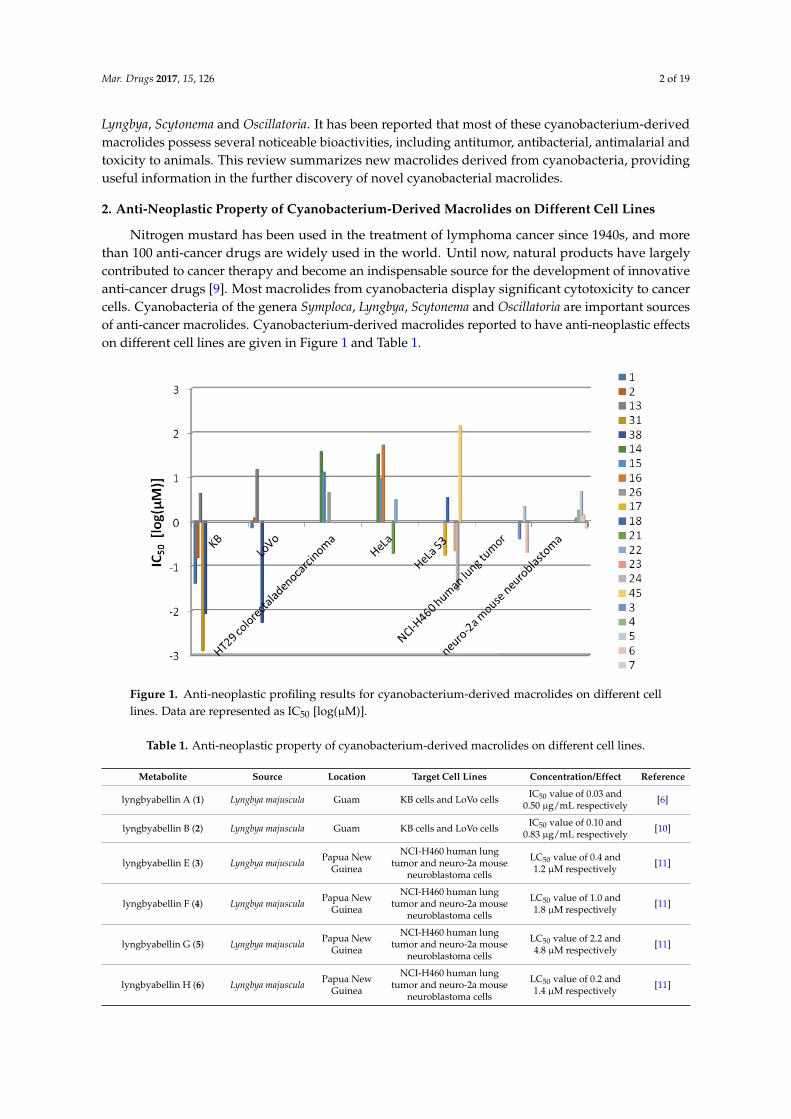
Nitrogen mustard has been used in the treatment of lymphoma cancer since 1940s, and morethan 100 anti-cancer drugs are widely used in the world. Until now, natural products have largelycontributed to cancer therapy and become an indispensable source for the development of innovativeanti-cancer drugs [9]. Most macrolides from cyanobacteria display significant cytotoxicity to cancercells. Cyanobacteria of the genera Symploca, Lyngbya, Scytonema and Oscillatoria are important sourcesof anti-cancer macrolides. Cyanobacterium-derived macrolides reported to have anti-neoplastic effectson different cell lines are given in Figure 1 and Table 1.
Mar. Drugs 2017, 15, 126 2 of 18
mainly from Lyngbya, Scytonema and Oscillatoria. It has been reported that most of these
cyanobacterium‐derived macrolides possess several noticeable bioactivities, including antitumor,
antibacterial, antimalarial and toxicity to animals. This review summarizes new macrolides derived
from cyanobacteria, providing useful information in the further discovery of novel cyanobacterial
macrolides.
2. Anti‐Neoplastic Property of Cyanobacterium‐Derived Macrolides on Different Cell Lines
Nitrogen mustard has been used in the treatment of lymphoma cancer since 1940s, and more
than 100 anti‐cancer drugs are widely used in the world. Until now, natural products have largely
contributed to cancer therapy and become an indispensable source for the development of innovative
anti‐cancer drugs [9]. Most macrolides from cyanobacteria display significant cytotoxicity to cancer
cells. Cyanobacteria of the genera Symploca, Lyngbya, Scytonema and Oscillatoria are important sources
of anti‐cancer macrolides. Cyanobacterium‐derived macrolides reported to have anti‐neoplastic
effects on different cell lines are given in Figure 1 and Table 1.
Figure 1. Anti‐neoplastic profiling results for cyanobacterium‐derived macrolides on different cell
lines. Data are represented as IC50 [log(μM)].
Table 1. Anti‐neoplastic property of cyanobacterium‐derived macrolides on different cell lines.
Metabolite Source Location Target Cell Lines Concentration/Effect Reference
lyngbyabellin A (1) Lyngbya
majuscula Guam
KB cells and
LoVo cells
IC50 value of 0.03 and
0.50 μg/mL respectively [6]
lyngbyabellin B (2) Lyngbya
majuscula Guam
KB cells and
LoVo cells
IC50 value of 0.10 and
0.83 μg/mL respectively [10]
lyngbyabellin E (3) Lyngbya
majuscula
Papua New
Guinea
NCI‐H460 human
lung tumor and
neuro‐2a mouse
neuroblastoma cells
LC50 value of 0.4 and
1.2 μM respectively [11]
lyngbyabellin F (4) Lyngbya
majuscula
Papua New
Guinea
NCI‐H460 human
lung tumor and
neuro‐2a mouse
neuroblastoma cells
LC50 value of 1.0 and
1.8 μM respectively [11]
lyngbyabellin G (5) Lyngbya
majuscula
Papua New
Guinea
NCI‐H460 human
lung tumor and
neuro‐2a mouse
neuroblastoma cells
LC50 value of 2.2 and
4.8 μM respectively [11]
lyngbyabellin H (6) Lyngbya
majuscula
Papua New
Guinea
NCI‐H460 human
lung tumor and
LC50 value of 0.2 and
1.4 μM respectively [11]
Figure 1. Anti-neoplastic profiling results for cyanobacterium-derived macrolides on different celllines. Data are represented as IC50 [log(µM)].
Table 1. Anti-neoplastic property of cyanobacterium-derived macrolides on different cell lines.
Metabolite Source Location Target Cell Lines Concentration/Effect Reference
lyngbyabellin A (1) Lyngbya majuscula Guam KB cells and LoVo cells IC50 value of 0.03 and0.50 µg/mL respectively [6]
lyngbyabellin B (2) Lyngbya majuscula Guam KB cells and LoVo cells IC50 value of 0.10 and0.83 µg/mL respectively [10]
lyngbyabellin E (3) Lyngbya majuscula Papua NewGuinea
NCI-H460 human lungtumor and neuro-2a mouse
neuroblastoma cells
LC50 value of 0.4 and1.2 µM respectively [11]
lyngbyabellin F (4) Lyngbya majuscula Papua NewGuinea
NCI-H460 human lungtumor and neuro-2a mouse
neuroblastoma cells
LC50 value of 1.0 and1.8 µM respectively [11]
lyngbyabellin G (5) Lyngbya majuscula Papua NewGuinea
NCI-H460 human lungtumor and neuro-2a mouse
neuroblastoma cells
LC50 value of 2.2 and4.8 µM respectively [11]
lyngbyabellin H (6) Lyngbya majuscula Papua NewGuinea
NCI-H460 human lungtumor and neuro-2a mouse
neuroblastoma cells
LC50 value of 0.2 and1.4 µM respectively [11]

Mar. Drugs 2017, 15, 126 3 of 19
Table 1. Cont.
Metabolite Source Location Target Cell Lines Concentration/Effect Reference
lyngbyabellin I (7) Lyngbya majuscula Papua NewGuinea
NCI-H460 human lungtumor and neuro-2a mouse
neuroblastoma cells
LC50 value of 1.0 and0.7 µM respectively [11]
lyngbyabellin N (10) Moorea bouillonii PalmyraAtoll, USA
H-460 human lungcarcinoma and HCT116colon cancer cell lines
IC50 value of0.0048–1.8 µM and15 µM respectively
[12]
lyngbyaloside B (13) Lyngbya sp. Palau KB cells and LoVo cells IC50 value of 4.3 and15 µM respectively [13]
2-epi-lyngbyalosid (14) Lyngbya bouillonii Apra Harbor,Guam
HT29 colorectaladenocarcinoma and
HeLa cells
IC50 value of 38 and33 µM respectively [14]
18E-lyngbyaloside C (15) Lyngbya bouillonii Apra Harbor,Guam
HT29 colorectaladenocarcinoma and
HeLa cells
IC50 value of 13 and9.3 µM respectively [14]
18Z-lyngbyaloside C (16) Lyngbya bouillonii Apra Harbor,Guam
HT29 colorectaladenocarcinoma and
HeLa cells
IC50 value of >100 µMand 53 µM respectively [14]
biselyngbyaside (17) Lyngbya sp. TokunoshimaIsland, Japan HeLa S3 cells IC50 value of 0.1 µg/mL [15]
biselyngbyaside B (18) Lyngbya sp. TokunoshimaIsland, Japan HeLa S3 cells and HL60 cells IC50 value of 3.5 and
0.82 µM respectively [16]
biselyngbyaside E (21) Lyngbya sp. IshigakiIsland, Japan HeLa and HL60 cells IC50 value of 0.19 and
0.071 µM respectively [17]
biselyngbyaside F (22) Lyngbya sp. IshigakiIsland, Japan HeLa and HL60 cells IC50 value of 3.1 and
0.66 µM respectively [17]
biselyngbyolide A (23) Lyngbya sp. TokunoshimaIsland, Japan HeLa S3 cells and HL60 cells IC50 value of 0.22 and
0.027 µM respectively [18]
biselyngbyolide B (24) Lyngbya sp. IshigakiIsland, Japan HeLa S3 cells and HL60 cells IC50 value of 0.028 and
0.0027 µM respectively [19]
caylobolide A (25) Lyngbya majuscula Bahamian human colon tumor cellsHCT 116 IC50 values of 9.9 µM [20]
caylobolide B (26) Phormidium spp. Florida USAHT29 colorectal
adenocarcinoma and HeLacervical carcinoma cells
IC50 value of 4.5 and12.2 µM respectively [21]
swinholide A (27) Symploca cf. sp. Fiji several cancer cell lines IC50 values of0.37 nM–1.0 µM [22]
ankaraholide A (28) Geitlerinema sp. Madagascar NCI-H460, Neuro-2a cellsand MDA-MB-435 cells
IC50 value of 119, 262and 8.9 nM respectively [22]
scytophycin A (30) Scytonemapseudohofmanni Oahu, Hawaii human carcinoma of
nasopharynx Cell (KB cells) IC50 value of 1 ng/mL [23]
scytophycin B (31) Scytonemapseudohofmanni Oahu, Hawaii KB cells IC50 value of 1 ng/mL [23]
scytophycins C-E (32–34) Scytonemapseudohofmanni Oahu, Hawaii KB cells IC50 value of
10–100 ng/mL [23]
6-hydroxyscytophycinB (35)
Scytonemamirabile cultured KB cells and LoVo cells
MICs of 1–5 and10–50 ng/mLrespectively
[23]
19-O-demethylscytophycin
C (36)
Scytonemaburmanicurn cultured KB cells and LoVo cells
MICs of 1–5 and10–50 ng/mLrespectively
[23]
6-hydroxy-7-O-methylscytophycin E (37)
Scytonemaocellatum cultured KB cells and LoVo cells
MICs of 1–5 and10–50 ng/mLrespectively
[23]
tolytoxin (38)Tolypothrix
conglutinata var.colorata
FanningIsland KB cells and LoVo cells IC50 value of 8.4 and
5.3 nM respectively [24]
debromoaplysiatoxin (39) Lyngbya majuscula MarshallIslands
P-388 lymphocytic mouseleukemia weak [25]
lyngbouilloside (44) Lyngbya bouillonii Papua NewGuinea neuroblastoma cells IC50 value of 17 µM [26]

Mar. Drugs 2017, 15, 126 4 of 19
Table 1. Cont.
Metabolite Source Location Target Cell Lines Concentration/Effect Reference
koshikalide (45) Lyngbya sp. MiePrefecture HeLa S3 cells IC50 value of 42 µg/mL [27]
sanctolide A (46) Oscillatoria sancta cultured HT-29 and MDA-MB-435cell lines nd a [28]
acutiphycin (47) Oscillatoriaacutissima
Manoa ValleyOahu, Hawaii KB cells and NIH/3T3 cells ED50 < 1 µg/mL [29]
20,21-didehydroacutiphycin(48)
Oscillatoriaacutissima
Manoa ValleyOahu, Hawaii KB cells and NIH/3T3 cells ED50 < 1 µg/mL [29]
polycavernoside D (49) Okeania sp. Puerto Rican H-460 human lung cancercell lines EC50 value of 2.5 µM [30]
bastimolide A (50) Okeania hirsuta Panama Vero cells IC50 value of 2.1 µM [31]
nuiapolide (51)colonial
cyanobacterium(071905-NII-01)
Hawaii Jurkat cells and cancerous Tlymphocytes anti-chemotactic activity [32]
a Not determined.
A series of cytotoxic marine cyanobacterial metabolites, named lyngbyabellins (1–11) possessingthiazole residues and chlorine substituents, have been isolated from the cyanobacterial genus Lyngbya(Figure 2). Isolated from the marine cyanobacterium Lyngbya majuscula collected from Guam,lyngbyabellin A (1) exhibits potent in vitro cytotoxicity against human carcinoma of nasopharynxCell (KB cells) and LoVo cells with IC50 values of 0.03 and 0.50 µg/mL, respectively [6]. The analogof lyngbyabellin A (1), lyngbyabellin B (2), was isolated from the same strain of Lyngbya majuscula.Compared with lyngbyabellin A (1), lyngbyabellin B (2) is slightly less cytotoxic to KB and LoVocells with IC50 values of 0.10 and 0.83 µg/mL, respectively [10]. Five analogs of lyngbyabellinA (1), including lyngbyabellins E-I (3–7), are produced from the same strain of Lyngbya majusculaharvested in Papua New Guinea. To the best of our knowledge, lyngbyabellins E-I (3–7) have potentin vitro cytotoxicity against human lung tumor (NCI-H460) and mouse neuroblastoma (neuro-2a)cells. Lyngbyabellin E (3) and lyngbyabellin H (6) display significant cytotoxicity to NCI-H460(LC50 values of 0.4 and 0.2 µM, respectively) and neuro-2a cells (LC50 values of 1.2 and 1.4 µM,respectively). Lyngbyabellins F-G (4–5) and lyngbyabellin I (7) are slightly less cytotoxic to NCI-H460(LC50 values of 1.0, 2.2 and 1.0 µM, respectively) and neuro-2a cells (LC50 values of 1.8, 4.8 and 0.7 µM,respectively) [11]. The marine cyanobacterium Moorea bouillonii (formerly Lyngbya bouillonii) collectedfrom Palmyra Atoll affords four analogs of lyngbyabellin A (1), lyngbyabellins K (8), L (9), N (10) and7-epi-lyngbyabellin L (11). Lyngbyabellin N (10) shows variable cytotoxicity to H-460 human lungcarcinoma (IC50 = 0.0048–1.8 µM) and potent in vitro cytotoxicity against the HCT116 colon cancercell line (IC50 = 40.9 ± 3.3 nM). This result could perhaps be explained by the solubility problem oflyngbyabellin N (10). The nitrogen-containing side chain (leucine statine residue) of lyngbyabellin N(11) may be the basic structural feature for its cytotoxic activity [12].
Mar. Drugs 2017, 15, 126 4 of 18
in (39) majuscula Islands mouse leukemia
lyngbouilloside
(44)
Lyngbya
bouillonii
Papua New
Guinea neuroblastoma cells IC50 value of 17 μM [26]
koshikalide (45) Lyngbya sp. Mie Prefecture HeLa S3 cells IC50 value of 42 μg/mL [27]
sanctolide A (46) Oscillatoria
sancta cultured
HT‐29 and
MDA‐MB‐435 cell
lines
nd a [28]
acutiphycin (47) Oscillatoria
acutissima
Manoa Valley
Oahu, Hawaii
KB cells and
NIH/3T3 cells ED50 < 1 μg/mL [29]
20,21‐didehydroac
utiphycin (48)
Oscillatoria
acutissima
Manoa Valley
Oahu, Hawaii
KB cells and
NIH/3T3 cells ED50 < 1 μg/mL [29]
polycavernoside D
(49) Okeania sp. Puerto Rican
H‐460 human lung
cancer cell lines EC50 value of 2.5 μM [30]
bastimolide A (50) Okeania hirsuta Panama Vero cells IC50 value of 2.1 μM [31]
nuiapolide (51)
colonial
cyanobacterium
(071905‐NII‐01)
Hawaii
Jurkat cells and
cancerous T
lymphocytes
anti‐chemotactic activity [32]
a Not determined.
A series of cytotoxic marine cyanobacterial metabolites, named lyngbyabellins (1–11)
possessing thiazole residues and chlorine substituents, have been isolated from the cyanobacterial
genus Lyngbya (Figure 2). Isolated from the marine cyanobacterium Lyngbya majuscula collected
from Guam, lyngbyabellin A (1) exhibits potent in vitro cytotoxicity against human carcinoma of
nasopharynx Cell (KB cells) and LoVo cells with IC50 values of 0.03 and 0.50 μg/mL, respectively [6].
The analog of lyngbyabellin A (1), lyngbyabellin B (2), was isolated from the same strain of Lyngbya
majuscula. Compared with lyngbyabellin A (1), lyngbyabellin B (2) is slightly less cytotoxic to KB
and LoVo cells with IC50 values of 0.10 and 0.83 μg/mL, respectively [10]. Five analogs of
lyngbyabellin A (1), including lyngbyabellins E‐I (3–7), are produced from the same strain of
Lyngbya majuscula harvested in Papua New Guinea. To the best of our knowledge, lyngbyabellins
E‐I (3–7) have potent in vitro cytotoxicity against human lung tumor (NCI‐H460) and mouse
neuroblastoma (neuro‐2a) cells. Lyngbyabellin E (3) and lyngbyabellin H (6) display significant
cytotoxicity to NCI‐H460 (LC50 values of 0.4 and 0.2 μM, respectively) and neuro‐2a cells (LC50
values of 1.2 and 1.4 μM, respectively). Lyngbyabellins F‐G (4–5) and lyngbyabellin I (7) are slightly
less cytotoxic to NCI‐H460 (LC50 values of 1.0, 2.2 and 1.0 μM, respectively) and neuro‐2a cells (LC50
values of 1.8, 4.8 and 0.7 μM, respectively) [11]. The marine cyanobacterium Moorea bouillonii
(formerly Lyngbya bouillonii) collected from Palmyra Atoll affords four analogs of lyngbyabellin A
(1), lyngbyabellins K (8), L (9), N (10) and 7‐epi‐lyngbyabellin L (11). Lyngbyabellin N (10) shows
variable cytotoxicity to H‐460 human lung carcinoma (IC50 = 0.0048–1.8 μM) and potent in vitro
cytotoxicity against the HCT116 colon cancer cell line (IC50 = 40.9 ± 3.3 nM). This result could
perhaps be explained by the solubility problem of lyngbyabellin N (10). The nitrogen‐containing
side chain (leucine statine residue) of lyngbyabellin N (11) may be the basic structural feature for its
cytotoxic activity [12].
Figure 2. Cont.

Mar. Drugs 2017, 15, 126 5 of 19
Mar. Drugs 2017, 15, 126 5 of 18
Figure 2. Chemical structures of Compounds 1–11.
Several 16‐membered glycoside macrolides, termed lyngbyalosides, are produced from
various species of the cyanobacterial genus Lyngbya (Figure 3). The marine Lyngbya bouillonii,
collected from Laing Island, afford lyngbyaloside (12) [8]. Lyngbyaloside B (13), isolated from
marine cyanobacterium Lyngbya sp., which was collected from Palaua, shows weak cytotoxicity
against KB cells and LoVo cells with IC50 values of 4.3 and 15 μM, respectively [13]. The total
synthesis of lyngbyaloside B (13) has been reported by Fuwa et al. [33]. Three analogs of
lyngbyaloside (12), including 2‐epi‐lyngbyaloside (14), 18E‐lyngbyaloside C (15) and
18Z‐lyngbyaloside C (16), were isolated from the marine cyanobacterium Lyngbya bouillonii,
collected from Apra Harbor, Guam. Cytotoxicity assays revealed that these macrolides possess
weak to moderate cytotoxicity against the human colorectal adenocarcinoma cell line HT29 and
HeLa cervical carcinoma cells. 18E‐lyngbyaloside C (15) is more cytotoxic toward HT29 colorectal
Figure 2. Chemical structures of Compounds 1–11.
Several 16-membered glycoside macrolides, termed lyngbyalosides, are produced from variousspecies of the cyanobacterial genus Lyngbya (Figure 3). The marine Lyngbya bouillonii, collected fromLaing Island, afford lyngbyaloside (12) [8]. Lyngbyaloside B (13), isolated from marine cyanobacteriumLyngbya sp., which was collected from Palaua, shows weak cytotoxicity against KB cells and LoVocells with IC50 values of 4.3 and 15 µM, respectively [13]. The total synthesis of lyngbyalosideB (13) has been reported by Fuwa et al. [33]. Three analogs of lyngbyaloside (12), including2-epi-lyngbyaloside (14), 18E-lyngbyaloside C (15) and 18Z-lyngbyaloside C (16), were isolated fromthe marine cyanobacterium Lyngbya bouillonii, collected from Apra Harbor, Guam. Cytotoxicity assaysrevealed that these macrolides possess weak to moderate cytotoxicity against the human colorectal

Mar. Drugs 2017, 15, 126 6 of 19
adenocarcinoma cell line HT29 and HeLa cervical carcinoma cells. 18E-lyngbyaloside C (15) is morecytotoxic toward HT29 colorectal adenocarcinoma and HeLa cervical carcinoma cells (IC50 values of13 and 9.3 µM, respectively) than 2-epi-lyngbyaloside (14) (IC50 values of 38 and 33 µM, respectively).18E-Lyngbyaloside C (15) is approximately five-fold more cytotoxic than 18Z-lyngbyaloside C (16)(IC50 values of >100 µM and 53 µM, respectively) [14]. The total synthesis of lyngbyaloside C has alsobeen accomplished [34].
Mar. Drugs 2017, 15, 126 6 of 18
adenocarcinoma and HeLa cervical carcinoma cells (IC50 values of 13 and 9.3 μM, respectively) than
2‐epi‐lyngbyaloside (14) (IC50 values of 38 and 33 μM, respectively). 18E‐Lyngbyaloside C (15) is
approximately five‐fold more cytotoxic than 18Z‐lyngbyaloside C (16) (IC50 values of >100 μM and
53 μM, respectively) [14]. The total synthesis of lyngbyaloside C has also been accomplished [34].
Figure 3. Chemical structures of Compounds 12–16.
Another distinct class of 18‐membered ring glycoside macrolides has been isolated from the
cyanobacterial genus Lyngbya (Figure 4). Biselyngbyaside (17) was discovered through a
bioassay‐guided screening for cytotoxic compounds from cyanobacterium Lyngbya sp. collected
from Okinawa Prefecture, Japan. Biselyngbyaside (17) shows a broad spectrum of cytotoxicity
against human solid tumor cell lines, especially for HeLa S3 cells with an IC50 value of 0.1 μg/mL
[15], and its total synthesis was completed [35]. Extensive efforts toward finding cytotoxic natural
products have resulted in the isolation of three analogs of biselyngbyaside (17), named
biselyngbyasides B–D (18–20), from the marine cyanobacterium Lyngbya sp. Biselyngbyaside B (18)
displays significant cytotoxicity against HeLa S3 and HL60 cells (IC50 values of 3.5 and 0.82 μM,
respectively, using thapsigargin as a positive control drug). In addition, biselyngbyasides B‐D
(18–20) induced apoptosis of cancer cells by inhibiting calcium influx into the endoplasmic
reticulum and increasing the concentration of intracellular calcium [16]. Two analogs of
biselyngbyaside (17), biselyngbyasides E (21) and F (22), were isolated from the marine
cyanobacterium Lyngbya sp. collected from Ishigaki Island, Japan. In vitro cell cytotoxicity assays
showed that biselyngbyaside E (21) has higher cytotoxicity against HeLa and HL60 cells (IC50 values
of 0.19 and 0.071 μM, respectively) than biselyngbyaside F (22) (IC50 values of 3.1 and 0.66 μM,
respectively). Based on the trisubstituted olefin geometry, the presence and absence of the sugar
moiety are crucial for the biological activities [17].
Like a cytotoxic biselyngbyaside‐related macrolide, biselyngbyolide A (23) was isolated from
the marine cyanobacterium Lyngbya sp. harvested from Tokunoshima Island, Japan.
Biselyngbyolide A (23) shows strong cytotoxicity against HeLa S3 cells and HL60 cells with IC50
values of 0.22 and 0.027 μM, respectively [18]. Biselyngbyolide B (24) was also isolated from the
same strain of Lyngbya sp. and displays significant inhibitory effects on growth of HeLa S3 cells and
HL60 cells (IC50 values of 0.028 and 0.0027 μM, respectively, using thapsigargin as a positive control
drug). Moreover, biselyngbyolide B (24) has 3–100‐fold more potent apoptosis‐inducing activity
than biselyngbyaside (17) [16,19].
Figure 3. Chemical structures of Compounds 12–16.
Another distinct class of 18-membered ring glycoside macrolides has been isolated fromthe cyanobacterial genus Lyngbya (Figure 4). Biselyngbyaside (17) was discovered through abioassay-guided screening for cytotoxic compounds from cyanobacterium Lyngbya sp. collectedfrom Okinawa Prefecture, Japan. Biselyngbyaside (17) shows a broad spectrum of cytotoxicity againsthuman solid tumor cell lines, especially for HeLa S3 cells with an IC50 value of 0.1 µg/mL [15], and itstotal synthesis was completed [35]. Extensive efforts toward finding cytotoxic natural products haveresulted in the isolation of three analogs of biselyngbyaside (17), named biselyngbyasides B–D (18–20),from the marine cyanobacterium Lyngbya sp. Biselyngbyaside B (18) displays significant cytotoxicityagainst HeLa S3 and HL60 cells (IC50 values of 3.5 and 0.82 µM, respectively, using thapsigarginas a positive control drug). In addition, biselyngbyasides B-D (18–20) induced apoptosis of cancercells by inhibiting calcium influx into the endoplasmic reticulum and increasing the concentration ofintracellular calcium [16]. Two analogs of biselyngbyaside (17), biselyngbyasides E (21) and F (22),were isolated from the marine cyanobacterium Lyngbya sp. collected from Ishigaki Island, Japan.In vitro cell cytotoxicity assays showed that biselyngbyaside E (21) has higher cytotoxicity againstHeLa and HL60 cells (IC50 values of 0.19 and 0.071 µM, respectively) than biselyngbyaside F (22)(IC50 values of 3.1 and 0.66 µM, respectively). Based on the trisubstituted olefin geometry, the presenceand absence of the sugar moiety are crucial for the biological activities [17].
Like a cytotoxic biselyngbyaside-related macrolide, biselyngbyolide A (23) was isolated from themarine cyanobacterium Lyngbya sp. harvested from Tokunoshima Island, Japan. Biselyngbyolide A (23)shows strong cytotoxicity against HeLa S3 cells and HL60 cells with IC50 values of 0.22 and 0.027 µM,respectively [18]. Biselyngbyolide B (24) was also isolated from the same strain of Lyngbya sp. anddisplays significant inhibitory effects on growth of HeLa S3 cells and HL60 cells (IC50 values of 0.028

Mar. Drugs 2017, 15, 126 7 of 19
and 0.0027 µM, respectively, using thapsigargin as a positive control drug). Moreover, biselyngbyolideB (24) has 3–100-fold more potent apoptosis-inducing activity than biselyngbyaside (17) [16,19].Mar. Drugs 2017, 15, 126 7 of 18
OO
OR O
biselyngbyaside (17) R=
biselyngbyolide B (24) R=H
OO
OR OH O
biselyngbyaside B (18) R=
biselyngbyolide A (23) R=H
O
OH
HO
HOMeO
O
OH
HO
HOMeO
Figure 4. Chemical structures of Compounds 17–24.
A novel 36‐membered macrolactone, caylobolide A (25), was isolated from Bahamian
cyanobacterium Lyngbya majuscula, which contains an unprecedented repeating unit, an adjoining
pentad of 1,5‐diols and a 1,3,5‐triol (Figure 5). In vitro cytotoxicity assay showed that caylobolide A
(25) possesses potent cytotoxicity against human colon tumor cells HCT‐116 with an IC50 value of
9.9 μM [20], and its total synthesis has been accomplished [36]. Caylobolide B (26) was isolated from
the marine cyanobacterium Phormidium spp. collected from Key West, Florida, and it exhibits strong
cytotoxicity against HT29 colorectal adenocarcinoma (IC50 value of 4.5 μM) and HeLa cervical
carcinoma cells (IC50 value of 12.2 μM) [21].
Figure 5. Chemical structures of Compounds 25 and 26.
Figure 4. Chemical structures of Compounds 17–24.
A novel 36-membered macrolactone, caylobolide A (25), was isolated from Bahamiancyanobacterium Lyngbya majuscula, which contains an unprecedented repeating unit, an adjoiningpentad of 1,5-diols and a 1,3,5-triol (Figure 5). In vitro cytotoxicity assay showed that caylobolideA (25) possesses potent cytotoxicity against human colon tumor cells HCT-116 with an IC50 valueof 9.9 µM [20], and its total synthesis has been accomplished [36]. Caylobolide B (26) was isolatedfrom the marine cyanobacterium Phormidium spp. collected from Key West, Florida, and it exhibitsstrong cytotoxicity against HT29 colorectal adenocarcinoma (IC50 value of 4.5 µM) and HeLa cervicalcarcinoma cells (IC50 value of 12.2 µM) [21].

Mar. Drugs 2017, 15, 126 8 of 19
Mar. Drugs 2017, 15, 126 7 of 18
OO
OR O
biselyngbyaside (17) R=
biselyngbyolide B (24) R=H
OO
OR OH O
biselyngbyaside B (18) R=
biselyngbyolide A (23) R=H
O
OH
HO
HOMeO
O
OH
HO
HOMeO
Figure 4. Chemical structures of Compounds 17–24.
A novel 36‐membered macrolactone, caylobolide A (25), was isolated from Bahamian
cyanobacterium Lyngbya majuscula, which contains an unprecedented repeating unit, an adjoining
pentad of 1,5‐diols and a 1,3,5‐triol (Figure 5). In vitro cytotoxicity assay showed that caylobolide A
(25) possesses potent cytotoxicity against human colon tumor cells HCT‐116 with an IC50 value of
9.9 μM [20], and its total synthesis has been accomplished [36]. Caylobolide B (26) was isolated from
the marine cyanobacterium Phormidium spp. collected from Key West, Florida, and it exhibits strong
cytotoxicity against HT29 colorectal adenocarcinoma (IC50 value of 4.5 μM) and HeLa cervical
carcinoma cells (IC50 value of 12.2 μM) [21].
Figure 5. Chemical structures of Compounds 25 and 26. Figure 5. Chemical structures of Compounds 25 and 26.
Swinholide A (27), originally isolated from the marine sponge Theonella swinhoei, was isolatedfrom the marine cyanobacterium cf. Symploca sp. collected from Fiji and was found to stronglyinhibit the growth of several tumor cell lines with IC50 values ranging from 0.37 nM to 1.0 µM [22].Two swinholide-based glycosylated macrolides, named ankaraholides A,B (28,29), were isolatedfrom two field collections of marine cyanobacteria (Figure 6). Ankaraholide A (28) exhibits strongantiproliferative activity against NCI-H460, Neuro-2a and MDA-MB-435 cell lines with IC50 values of119, 262 and 8.9 nM, respectively. Ankaraholide A (28) inhibits proliferation of A-10 cells by inducingcomplete loss of the filamentous (F)-actin during the cell extending process when the concentration ofankaraholide A (28) reaches 30 nM [22].
Mar. Drugs 2017, 15, 126 8 of 18
Swinholide A (27), originally isolated from the marine sponge Theonella swinhoei, was isolated
from the marine cyanobacterium cf. Symploca sp. collected from Fiji and was found to strongly
inhibit the growth of several tumor cell lines with IC50 values ranging from 0.37 nM to 1.0 μM [22].
Two swinholide‐based glycosylated macrolides, named ankaraholides A,B (28,29), were isolated
from two field collections of marine cyanobacteria (Figure 6). Ankaraholide A (28) exhibits strong
antiproliferative activity against NCI‐H460, Neuro‐2a and MDA‐MB‐435 cell lines with IC50 values
of 119, 262 and 8.9 nM, respectively. Ankaraholide A (28) inhibits proliferation of A‐10 cells by
inducing complete loss of the filamentous (F)‐actin during the cell extending process when the
concentration of ankaraholide A (28) reaches 30 nM [22].
Figure 6. Chemical structures of Compounds 27–29.
A family of potent cytotoxic natural products, scytophycins A–E (30–34), was isolated from the
terrestrial cyanobacterium Scytonema pseudohofmanni [37]. Scytophycins A (30) and B (31) display
significant cytotoxicity against KB cells (IC50 value of 1 ng/mL), while scytophycins C‐E (32–34) are
less cytotoxic to KB cells (IC50 values ranging from 10 to 100 ng/mL) than scytophycin A (30) [23].
Total synthesis of scytophycin C (32) has been completed [38]. Screening of cyanobacteria leads to
the discovery of three analogs of scytophycins, including 6‐hydroxyscytophycin B (35),
19‐O‐demethylscytophycin C (36) and 6‐hydroxy‐7‐O‐methylscytophycin E (37) (Figure 7). These
compounds (35–37) show strong inhibitory effect on the growth of KB (MIC values ranging from 1
to 5 ng/mL) and LoVo cells (MIC values ranging from 10 to 50 ng/mL) [23]. The cytotoxic tolytoxin
(38) was isolated from terrestrial cyanobacterium Tolypothrix conglutinata, collected from Fanning
Island [39], and displays excellent cytotoxicity against LoVo and KB cells with IC50 values of 8.4 and
5.3 nM, respectively [24].
Figure 6. Chemical structures of Compounds 27–29.
A family of potent cytotoxic natural products, scytophycins A–E (30–34), was isolated from theterrestrial cyanobacterium Scytonema pseudohofmanni [37]. Scytophycins A (30) and B (31) displaysignificant cytotoxicity against KB cells (IC50 value of 1 ng/mL), while scytophycins C-E (32–34)are less cytotoxic to KB cells (IC50 values ranging from 10 to 100 ng/mL) than scytophycin A(30) [23]. Total synthesis of scytophycin C (32) has been completed [38]. Screening of cyanobacterialeads to the discovery of three analogs of scytophycins, including 6-hydroxyscytophycin B (35),19-O-demethylscytophycin C (36) and 6-hydroxy-7-O-methylscytophycin E (37) (Figure 7). Thesecompounds (35–37) show strong inhibitory effect on the growth of KB (MIC values ranging from 1 to5 ng/mL) and LoVo cells (MIC values ranging from 10 to 50 ng/mL) [23]. The cytotoxic tolytoxin(38) was isolated from terrestrial cyanobacterium Tolypothrix conglutinata, collected from FanningIsland [39], and displays excellent cytotoxicity against LoVo and KB cells with IC50 values of 8.4 and5.3 nM, respectively [24].

Mar. Drugs 2017, 15, 126 9 of 19
Mar. Drugs 2017, 15, 126 8 of 18
Swinholide A (27), originally isolated from the marine sponge Theonella swinhoei, was isolated
from the marine cyanobacterium cf. Symploca sp. collected from Fiji and was found to strongly
inhibit the growth of several tumor cell lines with IC50 values ranging from 0.37 nM to 1.0 μM [22].
Two swinholide‐based glycosylated macrolides, named ankaraholides A,B (28,29), were isolated
from two field collections of marine cyanobacteria (Figure 6). Ankaraholide A (28) exhibits strong
antiproliferative activity against NCI‐H460, Neuro‐2a and MDA‐MB‐435 cell lines with IC50 values
of 119, 262 and 8.9 nM, respectively. Ankaraholide A (28) inhibits proliferation of A‐10 cells by
inducing complete loss of the filamentous (F)‐actin during the cell extending process when the
concentration of ankaraholide A (28) reaches 30 nM [22].
Figure 6. Chemical structures of Compounds 27–29.
A family of potent cytotoxic natural products, scytophycins A–E (30–34), was isolated from the
terrestrial cyanobacterium Scytonema pseudohofmanni [37]. Scytophycins A (30) and B (31) display
significant cytotoxicity against KB cells (IC50 value of 1 ng/mL), while scytophycins C‐E (32–34) are
less cytotoxic to KB cells (IC50 values ranging from 10 to 100 ng/mL) than scytophycin A (30) [23].
Total synthesis of scytophycin C (32) has been completed [38]. Screening of cyanobacteria leads to
the discovery of three analogs of scytophycins, including 6‐hydroxyscytophycin B (35),
19‐O‐demethylscytophycin C (36) and 6‐hydroxy‐7‐O‐methylscytophycin E (37) (Figure 7). These
compounds (35–37) show strong inhibitory effect on the growth of KB (MIC values ranging from 1
to 5 ng/mL) and LoVo cells (MIC values ranging from 10 to 50 ng/mL) [23]. The cytotoxic tolytoxin
(38) was isolated from terrestrial cyanobacterium Tolypothrix conglutinata, collected from Fanning
Island [39], and displays excellent cytotoxicity against LoVo and KB cells with IC50 values of 8.4 and
5.3 nM, respectively [24].
Mar. Drugs 2017, 15, 126 9 of 18
O O O
O
N
O
O O O
OOR2
O
H
OHtolytoxin (38) R1=OH R2=Me
R1
6-hydroxyscytophycin B (35) R1=OH R2=H
scytophycin B (31) R1=H R2=H
Figure 7. Chemical structures of Compounds 30–38.
Debromoaplysiatoxin (39) was isolated from the marine cyanobacterium Lyngbya majuscula,
collected from Hawaii [40], and shows potent cytotoxicity against mouse lymphocytic leukemia
P‐388 [25]. Four analogs of debromoaplysiatoxin (39), including oscillatoxin A (40),
19,21‐dibromooscillatoxin A (41), 19‐bromoaplysiatoxin (42) and 21‐bromooscillatoxin A (43), were
isolated from a mixture of marine cyanobacteria Oscillatoria nigroviridis and Schizothrix calcicola from
Enewetak Island (Figure 8). These compounds (41–43) contain the same 14‐membered macrocycle
as debromoaplysiatoxin (39), but they are bromine‐containing macrolides [41]. A 14‐membered
glycosidic macrolide, lyngbouilloside (44), was isolated from the marine cyanobacterium Lyngbya
bouillonii, harvested from Papua New Guinea. It displays a modest cytotoxicity against
neuroblastoma cells with an IC50 value of 17 μM [26]. Another 14‐membered macrolide, koshikalide
(45), was isolated from the marine cyanobacterium Lyngbya sp., collected from Mie Prefecture,
Japan, and shows slight cytotoxicity against HeLa S3 cells with an IC50 value of 42 μg/mL [27]. In
addition, the total synthesis of koshikalide (45) has been achieved by exploiting a novel convergent
strategy [42]. A 14‐membered marine macrolide, sanctolide A (46), containing a rare N‐methyl
enamide and a 2‐hydroxyisovaleric acid, was obtained from the culture of cyanobacterium
Oscillatoria sancta. It is cytotoxic against HT‐29 and MDA‐MB‐435 cell lines [28], and its total
synthesis was achieved [43].
Figure 7. Chemical structures of Compounds 30–38.
Debromoaplysiatoxin (39) was isolated from the marine cyanobacterium Lyngbya majuscula,collected from Hawaii [40], and shows potent cytotoxicity against mouse lymphocyticleukemia P-388 [25]. Four analogs of debromoaplysiatoxin (39), including oscillatoxin A (40),19,21-dibromooscillatoxin A (41), 19-bromoaplysiatoxin (42) and 21-bromooscillatoxin A (43), wereisolated from a mixture of marine cyanobacteria Oscillatoria nigroviridis and Schizothrix calcicola fromEnewetak Island (Figure 8). These compounds (41–43) contain the same 14-membered macrocycle asdebromoaplysiatoxin (39), but they are bromine-containing macrolides [41]. A 14-membered glycosidicmacrolide, lyngbouilloside (44), was isolated from the marine cyanobacterium Lyngbya bouillonii,harvested from Papua New Guinea. It displays a modest cytotoxicity against neuroblastoma cellswith an IC50 value of 17 µM [26]. Another 14-membered macrolide, koshikalide (45), was isolatedfrom the marine cyanobacterium Lyngbya sp., collected from Mie Prefecture, Japan, and shows slightcytotoxicity against HeLa S3 cells with an IC50 value of 42 µg/mL [27]. In addition, the total synthesisof koshikalide (45) has been achieved by exploiting a novel convergent strategy [42]. A 14-membered

Mar. Drugs 2017, 15, 126 10 of 19
marine macrolide, sanctolide A (46), containing a rare N-methyl enamide and a 2-hydroxyisovalericacid, was obtained from the culture of cyanobacterium Oscillatoria sancta. It is cytotoxic against HT-29and MDA-MB-435 cell lines [28], and its total synthesis was achieved [43].
Mar. Drugs 2017, 15, 126 9 of 18
O O O
O
N
O
O O O
OOR2
O
H
OHtolytoxin (38) R1=OH R2=Me
R1
6-hydroxyscytophycin B (35) R1=OH R2=H
scytophycin B (31) R1=H R2=H
Figure 7. Chemical structures of Compounds 30–38.
Debromoaplysiatoxin (39) was isolated from the marine cyanobacterium Lyngbya majuscula,
collected from Hawaii [40], and shows potent cytotoxicity against mouse lymphocytic leukemia
P‐388 [25]. Four analogs of debromoaplysiatoxin (39), including oscillatoxin A (40),
19,21‐dibromooscillatoxin A (41), 19‐bromoaplysiatoxin (42) and 21‐bromooscillatoxin A (43), were
isolated from a mixture of marine cyanobacteria Oscillatoria nigroviridis and Schizothrix calcicola from
Enewetak Island (Figure 8). These compounds (41–43) contain the same 14‐membered macrocycle
as debromoaplysiatoxin (39), but they are bromine‐containing macrolides [41]. A 14‐membered
glycosidic macrolide, lyngbouilloside (44), was isolated from the marine cyanobacterium Lyngbya
bouillonii, harvested from Papua New Guinea. It displays a modest cytotoxicity against
neuroblastoma cells with an IC50 value of 17 μM [26]. Another 14‐membered macrolide, koshikalide
(45), was isolated from the marine cyanobacterium Lyngbya sp., collected from Mie Prefecture,
Japan, and shows slight cytotoxicity against HeLa S3 cells with an IC50 value of 42 μg/mL [27]. In
addition, the total synthesis of koshikalide (45) has been achieved by exploiting a novel convergent
strategy [42]. A 14‐membered marine macrolide, sanctolide A (46), containing a rare N‐methyl
enamide and a 2‐hydroxyisovaleric acid, was obtained from the culture of cyanobacterium
Oscillatoria sancta. It is cytotoxic against HT‐29 and MDA‐MB‐435 cell lines [28], and its total
synthesis was achieved [43].
Mar. Drugs 2017, 15, 126 10 of 18
Figure 8. Chemical structures of Compounds 39–46.
Two cytotoxic marcolides, acutiphycin (47) and 20,21‐didehydroacutiphycin (48), were isolated
from freshwater cyanobacterium Oscillatoria acutissima, collected from Manoa Valley, Oahu, and
possess strong cytotoxicity against KB and NIH/3T3 cells (ED50 < 1 μg/mL), as well as Lewis lung
carcinoma [29]. A rare marine toxin, polycavernoside D (49), was obtained from the marine Okeania
sp. collected from the Caribbean (Figure 9). The discovery of polycavernoside D, for the first time,
provides a conclusive proof that these lethal toxins (polycavernosides) have, in fact, a
cyanobacterial origin rather than other marine organisms. Polycavernoside D (49) displays
cytotoxicity against the H‐460 human lung cancer cell line in a dose‐dependent manner, with an
EC50 value of 2.5 μM [30]. Bastimolide A (50), isolated from the marine Okeania hirsuta from
Bastimentos Park, Panama, is a rare 40‐membered polyhydroxy macrolide consisting of one
1,3‐diol, one 1,3,5‐triol, six 1,5‐diols and one tert‐butyl group. Bastimolide A (50) exhibits strong
cytotoxicity against Vero cells with an IC50 value of 2.1 μM [31].
A rare 40‐membered macrolactone, nuiapolide (51), was isolated from Niihau marine
cyanobacterium. As a polyhydroxylated macrolide, nuiapolide (51) contains a rare tert‐butyl
carbinol residue, and it displays anti‐chemotactic activity against Jurkat cells and cancerous T
lymphocytes and can trigger a predominant G2/M phase shift in the cell cycle [32].
O
O
OO
HOO
OO
MeOOOMeO
HOOH OMe
polycavernoside D (49)
Figure 8. Chemical structures of Compounds 39–46.
Two cytotoxic marcolides, acutiphycin (47) and 20,21-didehydroacutiphycin (48), were isolatedfrom freshwater cyanobacterium Oscillatoria acutissima, collected from Manoa Valley, Oahu, andpossess strong cytotoxicity against KB and NIH/3T3 cells (ED50 < 1 µg/mL), as well as Lewis lungcarcinoma [29]. A rare marine toxin, polycavernoside D (49), was obtained from the marine Okeania sp.collected from the Caribbean (Figure 9). The discovery of polycavernoside D, for the first time,provides a conclusive proof that these lethal toxins (polycavernosides) have, in fact, a cyanobacterialorigin rather than other marine organisms. Polycavernoside D (49) displays cytotoxicity against theH-460 human lung cancer cell line in a dose-dependent manner, with an EC50 value of 2.5 µM [30].Bastimolide A (50), isolated from the marine Okeania hirsuta from Bastimentos Park, Panama, is a rare40-membered polyhydroxy macrolide consisting of one 1,3-diol, one 1,3,5-triol, six 1,5-diols and onetert-butyl group. Bastimolide A (50) exhibits strong cytotoxicity against Vero cells with an IC50 valueof 2.1 µM [31].
A rare 40-membered macrolactone, nuiapolide (51), was isolated from Niihau marinecyanobacterium. As a polyhydroxylated macrolide, nuiapolide (51) contains a rare tert-butyl carbinolresidue, and it displays anti-chemotactic activity against Jurkat cells and cancerous T lymphocytes andcan trigger a predominant G2/M phase shift in the cell cycle [32].

Mar. Drugs 2017, 15, 126 11 of 19
Mar. Drugs 2017, 15, 126 10 of 18
Figure 8. Chemical structures of Compounds 39–46.
Two cytotoxic marcolides, acutiphycin (47) and 20,21‐didehydroacutiphycin (48), were isolated
from freshwater cyanobacterium Oscillatoria acutissima, collected from Manoa Valley, Oahu, and
possess strong cytotoxicity against KB and NIH/3T3 cells (ED50 < 1 μg/mL), as well as Lewis lung
carcinoma [29]. A rare marine toxin, polycavernoside D (49), was obtained from the marine Okeania
sp. collected from the Caribbean (Figure 9). The discovery of polycavernoside D, for the first time,
provides a conclusive proof that these lethal toxins (polycavernosides) have, in fact, a
cyanobacterial origin rather than other marine organisms. Polycavernoside D (49) displays
cytotoxicity against the H‐460 human lung cancer cell line in a dose‐dependent manner, with an
EC50 value of 2.5 μM [30]. Bastimolide A (50), isolated from the marine Okeania hirsuta from
Bastimentos Park, Panama, is a rare 40‐membered polyhydroxy macrolide consisting of one
1,3‐diol, one 1,3,5‐triol, six 1,5‐diols and one tert‐butyl group. Bastimolide A (50) exhibits strong
cytotoxicity against Vero cells with an IC50 value of 2.1 μM [31].
A rare 40‐membered macrolactone, nuiapolide (51), was isolated from Niihau marine
cyanobacterium. As a polyhydroxylated macrolide, nuiapolide (51) contains a rare tert‐butyl
carbinol residue, and it displays anti‐chemotactic activity against Jurkat cells and cancerous T
lymphocytes and can trigger a predominant G2/M phase shift in the cell cycle [32].
O
O
OO
HOO
OO
MeOOOMeO
HOOH OMe
polycavernoside D (49)
Mar. Drugs 2017, 15, 126 11 of 18
Figure 9. Chemical structures of Compounds 47–51.
3. Antibacterial Activity
Some macrolides, such as erythromycin and azithromycin, have shown excellent antibacterial
activity and are widely used in clinical practice of various types of bacterial infections [44]. Some
macrolides from cyanobacteria also show good antibacterial activities. Cyanobacterium‐derived
macrolides with antimicrobial properties are listed in Table 2.
Scytophycins C–E (32–34) were isolated from the terrestrial cyanobacterium Scytonema
pseudohofmanni, collected from Oahu, Hawaii, and were shown to exhibit weak antibacterial activity
[37]. Three analogs of scytophycin C (32), including 6‐hydroxyscytophycin B (35),
19‐O‐demethylscytophycin C (36) and 6‐hydroxy‐7‐O‐methylsctophycin E (37), were isolated from
the cyanobacteria S. mirabile, S. burmanicurn and S. ocellatum, respectively. These macrolides (35–37)
display antifungal activity against Aspergillus oryzae, Candida albicans, Penicillium notatum and
Saccharomyces cerevisiae [23]. The cytotoxin, tolytoxin (38), was isolated from the terrestrial
cyanobacterium Tolypothrix conglutinata, collected from Fanning Island [39], and was found to exhibit
potent antifungal activity against various yeasts and filamentous fungi (MICs of 0.25–8 nM) [24].
A bioactive marcolide, 7‐OMe‐scytophycin B (52), was identified from a culture of a marine
cyanobacterium and was found to exhibit antifungal activity against Candida albicans HAMbI 484
and Candida guilliermondii HAMBI 257 with MIC values of 0.40 and 0.80 mM and IC50 values of 0.19
and 0.23 mM, respectively [45]. Two 40‐membered macrolactones, amantelides A,B (53,54), are
composed of a 1,3‐diol and contiguous 1,5‐diol units and a tert‐butyl substituent. These compounds
were isolated from a Guam cyanobacterium belonging to the family Oscillatoriales (Figure 10).
Amantelide A (53) shows a broad spectrum of inhibitory effects on the growth of both eukaryotic
and prokaryotic cells. The growth of the fungi Lindra thalassiae and Fusarium sp. is completely
inhibited when the concentration of amantelide A (53) is 62.5 μg/mL. When the concentration of
amantelide B (54) is 6.25 μg/mL, the growth of the fungus Dendryphiella salina is completely
inhibited [46].
Figure 9. Chemical structures of Compounds 47–51.
3. Antibacterial Activity
Some macrolides, such as erythromycin and azithromycin, have shown excellent antibacterialactivity and are widely used in clinical practice of various types of bacterial infections [44]. Somemacrolides from cyanobacteria also show good antibacterial activities. Cyanobacterium-derivedmacrolides with antimicrobial properties are listed in Table 2.
Scytophycins C–E (32–34) were isolated from the terrestrial cyanobacterium Scytonemapseudohofmanni, collected from Oahu, Hawaii, and were shown to exhibit weak antibacterialactivity [37]. Three analogs of scytophycin C (32), including 6-hydroxyscytophycin B (35),19-O-demethylscytophycin C (36) and 6-hydroxy-7-O-methylsctophycin E (37), were isolated fromthe cyanobacteria S. mirabile, S. burmanicurn and S. ocellatum, respectively. These macrolides(35–37) display antifungal activity against Aspergillus oryzae, Candida albicans, Penicillium notatumand Saccharomyces cerevisiae [23]. The cytotoxin, tolytoxin (38), was isolated from the terrestrialcyanobacterium Tolypothrix conglutinata, collected from Fanning Island [39], and was found to exhibitpotent antifungal activity against various yeasts and filamentous fungi (MICs of 0.25–8 nM) [24].

Mar. Drugs 2017, 15, 126 12 of 19
Table 2. Antibacterial and antifungal macrolides.
Metabolite Source Location Target Concentration/Effect Reference
6-hydroxyscytophycinB (35) Scytonema mirabile cultured
Fungus (1) Aspergillus oryzae(2) Candida albicans(3) Penicillium notatum(4) Saccharomyces cerevisiae
nd a [23]
19-O-demethylscytophycin
C (36)Scytonema burmanicurn cultured
Fungus (1) Aspergillus oryzae(2) Candida albicans(3) Penicillium notatum(4) Saccharomyces cerevisiae
nd a [23]
6-hydroxy-7-O-methylscytophycin
E (37)Scytonema ocellatum cultured
Fungus (1) Aspergillus oryzae(2) Candida albicans(3) Penicillium notatum(4) Saccharomyces cerevisiae
nd a [23]
tolytoxin (38) Tolypothrix conglutinatavar. colorata
FanningIsland
Fungi Penicillium notatumand Rhizoctonia solani 1165
MIC value of 0.25 nMrespectively [24]
7-OMe-scytophycinB (52) Anabaena sp. cultured
Fungus Candida albicansHAMBI 484 and Candidaguilliermondii HAMBI 257
MIC values of 0.40 and0.80 mM respectively;IC50 value of 0.19 and0.23 µM respectively
[45]
amantelide A (53) Oscillatoriales Guam Fungi Lindra thalassiae andFusarium sp.
totally inhibited of62.5 µg/mL [46]
amantelide B (54) Oscillatoriales Guam Fungus Dendryphiella salina totally inhibited of6.25 µg/mL [46]
a Not determined.
A bioactive marcolide, 7-OMe-scytophycin B (52), was identified from a culture of a marinecyanobacterium and was found to exhibit antifungal activity against Candida albicans HAMbI 484 andCandida guilliermondii HAMBI 257 with MIC values of 0.40 and 0.80 mM and IC50 values of 0.19 and0.23 mM, respectively [45]. Two 40-membered macrolactones, amantelides A,B (53,54), are composedof a 1,3-diol and contiguous 1,5-diol units and a tert-butyl substituent. These compounds were isolatedfrom a Guam cyanobacterium belonging to the family Oscillatoriales (Figure 10). Amantelide A(53) shows a broad spectrum of inhibitory effects on the growth of both eukaryotic and prokaryoticcells. The growth of the fungi Lindra thalassiae and Fusarium sp. is completely inhibited when theconcentration of amantelide A (53) is 62.5 µg/mL. When the concentration of amantelide B (54) is6.25 µg/mL, the growth of the fungus Dendryphiella salina is completely inhibited [46].
Mar. Drugs 2017, 15, 126 11 of 18
Figure 9. Chemical structures of Compounds 47–51.
3. Antibacterial Activity
Some macrolides, such as erythromycin and azithromycin, have shown excellent antibacterial
activity and are widely used in clinical practice of various types of bacterial infections [44]. Some
macrolides from cyanobacteria also show good antibacterial activities. Cyanobacterium‐derived
macrolides with antimicrobial properties are listed in Table 2.
Scytophycins C–E (32–34) were isolated from the terrestrial cyanobacterium Scytonema
pseudohofmanni, collected from Oahu, Hawaii, and were shown to exhibit weak antibacterial activity
[37]. Three analogs of scytophycin C (32), including 6‐hydroxyscytophycin B (35),
19‐O‐demethylscytophycin C (36) and 6‐hydroxy‐7‐O‐methylsctophycin E (37), were isolated from
the cyanobacteria S. mirabile, S. burmanicurn and S. ocellatum, respectively. These macrolides (35–37)
display antifungal activity against Aspergillus oryzae, Candida albicans, Penicillium notatum and
Saccharomyces cerevisiae [23]. The cytotoxin, tolytoxin (38), was isolated from the terrestrial
cyanobacterium Tolypothrix conglutinata, collected from Fanning Island [39], and was found to exhibit
potent antifungal activity against various yeasts and filamentous fungi (MICs of 0.25–8 nM) [24].
A bioactive marcolide, 7‐OMe‐scytophycin B (52), was identified from a culture of a marine
cyanobacterium and was found to exhibit antifungal activity against Candida albicans HAMbI 484
and Candida guilliermondii HAMBI 257 with MIC values of 0.40 and 0.80 mM and IC50 values of 0.19
and 0.23 mM, respectively [45]. Two 40‐membered macrolactones, amantelides A,B (53,54), are
composed of a 1,3‐diol and contiguous 1,5‐diol units and a tert‐butyl substituent. These compounds
were isolated from a Guam cyanobacterium belonging to the family Oscillatoriales (Figure 10).
Amantelide A (53) shows a broad spectrum of inhibitory effects on the growth of both eukaryotic
and prokaryotic cells. The growth of the fungi Lindra thalassiae and Fusarium sp. is completely
inhibited when the concentration of amantelide A (53) is 62.5 μg/mL. When the concentration of
amantelide B (54) is 6.25 μg/mL, the growth of the fungus Dendryphiella salina is completely
inhibited [46].
Mar. Drugs 2017, 15, 126 12 of 18
Figure 10. Chemical structures of Compounds 52–54.
Table 2. Antibacterial and antifungal macrolides.
Metabolite Source Location Target Concentration/Effect Reference
6‐hydroxyscytophycin B
(35)
Scytonema
mirabile cultured
Fungus (1) Aspergillus oryzae
(2) Candida albicans
(3) Penicillium notatum
(4) Saccharomyces cerevisiae
nd a [23]
19‐O‐demethylscytophyc
in C (36)
Scytonema
burmanicurn cultured
Fungus (1) Aspergillus oryzae
(2) Candida albicans
(3) Penicillium notatum
(4) Saccharomyces cerevisiae
nd a [23]
6‐hydroxy‐7‐O‐methylsc
ytophycin E (37)
Scytonema
ocellatum cultured
Fungus (1) Aspergillus oryzae
(2) Candida albicans
(3) Penicillium notatum
(4) Saccharomyces cerevisiae
nd a [23]
tolytoxin (38)
Tolypothrix
conglutinata var.
colorata
Fanning
Island
Fungi Penicillium notatum
and Rhizoctonia solani 1165
MIC value of
0.25 nM respectively [24]
7‐OMe‐scytophycin B
(52) Anabaena sp. cultured
Fungus Candida albicans
HAMBI 484 and Candida
guilliermondii HAMBI 257
MIC values of
0.40 and 0.80 mM
respectively; IC50
value of 0.19 and
0.23 μM respectively
[45]
amantelide A (53) Oscillatoriales Guam Fungi Lindra thalassiae and
Fusarium sp.
totally inhibited of
62.5 μg/mL [46]
amantelide B (54) Oscillatoriales Guam Fungus Dendryphiella salina totally inhibited of
6.25 μg/mL [46]
a Not determined.
4. Effects of Cyanobacterium‐Derived Macrolides on Animals
Toxin‐producing cyanobacterial blooms are a potential health risk for other living organisms,
including humans [47]. Cyanobacterium‐derived macrolides show toxicity to animals, such as brine
shrimp and mice. The effects of cyanobacterium‐derived macrolides on fauna are described in Table 3.
The cytotoxic macrolactone, lyngbyabellin A (1), exhibits potent toxicity to mice in vivo trials
(lethal dose of 2.4 to 8.0 mg/kg; sublethal dose of 1.2 to 1.5 mg/kg) [6]. Tolytoxin (38) is highly toxic
to mice with a sublethal dose (ip) of 1.5 mg /kg [24].
A 14‐membered macrolide, sanctolide A (48), shows high toxicity toward the brine shrimp
with an LC50 value of 23.5 μM [28]. A 10‐membered ring macrolide, gloeolactone (55), was isolated
from the cyanobacterium Gloeotrichia sp., harvested in Clark Canyon Reservoir (Figure 11).
Gloeolactone (55) exhibits weak toxicity to brine shrimp. All brine shrimps are dead when the
concentration of gloeolactone (55) is 125 μg/mL [48]. Phormidolide (56) was isolated from the
marine cyanobacterium Phormidium sp. cultured in Indonesia and was found to exhibit very high
toxicity (LC50 value of 1.5 μM) in the brine shrimp test [49].
A symmetrical macrolide dimer, cyanolide A (57), was obtained from the marine
cyanobacterium Lyngbya bouillonii collected from Papua New Guinea. Cyanolide A (57) displays
potent molluscicidal activity against the snail vector Biomphalaria glabrata with an LC50 value of 1.2 μM.
Figure 10. Chemical structures of Compounds 52–54.

Mar. Drugs 2017, 15, 126 13 of 19
4. Effects of Cyanobacterium-Derived Macrolides on Animals
Toxin-producing cyanobacterial blooms are a potential health risk for other living organisms,including humans [47]. Cyanobacterium-derived macrolides show toxicity to animals, such as brineshrimp and mice. The effects of cyanobacterium-derived macrolides on fauna are described in Table 3.
Table 3. Effects of cyanobacterium-derived macrolides on animals.
Metabolite Source Location Target Fauna Impacts Reference
lyngbyabellin A (1) Lyngbyamajuscula Guam mice LD50 value of
1.2–1.5 mg/kg [6]
tolytoxin (38) Scytonemapseudohofmanni cultured mice LD50 value of
1.5 mg/kg [24]
sanctolide A (48) Oscillatoriasancta cultured brine shrimp LD50 value of
23.5 µM [28]
gloeolactone (55) Gloeotrichia sp. Clark CanyonReservoir brine shrimp 100% killed at
125 µg/mL [48]
phormidolide (56) Phormidium sp. Sulawesi,Indonesia brine shrimp LD50 value of
1.5 µM [49]
cyanolide A (57) Lyngbyabouillonii
Papua NewGuinea
snail vectorBiomphalaria glabrata
LD50 value of1.2 µM [50]
The cytotoxic macrolactone, lyngbyabellin A (1), exhibits potent toxicity to mice in vivo trials(lethal dose of 2.4 to 8.0 mg/kg; sublethal dose of 1.2 to 1.5 mg/kg) [6]. Tolytoxin (38) is highly toxic tomice with a sublethal dose (ip) of 1.5 mg /kg [24].
A 14-membered macrolide, sanctolide A (48), shows high toxicity toward the brine shrimp withan LC50 value of 23.5 µM [28]. A 10-membered ring macrolide, gloeolactone (55), was isolated fromthe cyanobacterium Gloeotrichia sp., harvested in Clark Canyon Reservoir (Figure 11). Gloeolactone(55) exhibits weak toxicity to brine shrimp. All brine shrimps are dead when the concentration ofgloeolactone (55) is 125 µg/mL [48]. Phormidolide (56) was isolated from the marine cyanobacteriumPhormidium sp. cultured in Indonesia and was found to exhibit very high toxicity (LC50 value of1.5 µM) in the brine shrimp test [49].
Mar. Drugs 2017, 15, 126 13 of 18
Cyanolide A (57) can be used as a new, potent molluscicidal agent to effectively control the spread
of schistosomiasis [50]. Its total synthesis has been accomplished [51].
Figure 11. Chemical structures of Compounds 55–57.
Table 3. Effects of cyanobacterium‐derived macrolides on animals.
Metabolite Source Location Target fauna Impacts Reference
lyngbyabellin A (1) Lyngbya majuscula Guam mice LD50 value of
1.2–1.5 mg/kg [6]
tolytoxin (38) Scytonema
pseudohofmanni cultured mice
LD50 value of
1.5 mg/kg [24]
sanctolide A (48) Oscillatoria sancta cultured brine shrimp LD50 value of
23.5 μM [28]
gloeolactone (55) Gloeotrichia sp. Clark Canyon
Reservoir brine shrimp
100% killed at
125 μg/mL [48]
phormidolide (56) Phormidium sp. Sulawesi, Indonesia brine shrimp LD50 value of
1.5 μM [49]
cyanolide A (57) Lyngbya bouillonii Papua New Guinea snail vector
Biomphalaria glabrata
LD50 value of
1.2 μM [50]
5. Other Bioactivity
Cyanobacterium‐derived macrolides with rich chemical diversity show various important
bioactivities (Table 4). The macrolide biselyngbyaside (17), isolated from the marine
cyanobacterium Lyngbya sp., has been investigated for its effects on osteoclast differentiation and
function. Biselyngbyaside (17) inhibits RANKL‐induced osteoclastogenesis by inhibiting the
expression of c‐Fos and NFATc1 in mouse monocytic RAW264 cells. Therefore, biselyngbyaside
(17) is a potentially promising compound with therapeutic and preventive activities against
bone‐lytic diseases [52]. A toxic cyanobacterial macrolide, debromoaplysiatoxin (39), has been
found to cause severe cutaneous inflammation in humans and other animals after topical
application [25].
A rare 40‐membered polyhydroxy macrolide, bastimolide A (50), exhibits high selectivity and
antimalarial activity against four drug‐resistant malaria parasite strains, including TM90‐C2A,
TM90‐C2B, W2 and TM91‐C235, with IC50 values of 80, 90, 140 and 270 nM, respectively. It has been
proven that bastimolide A (50) is a potentially promising antimalarial lead compound with high
selectivity and antimalarial activity against drug‐resistant strains [31]. Malyngolide dimer (58) was
isolated from the marine cyanobacterium Lyngbya majuscula collected from Panama and was shown
Figure 11. Chemical structures of Compounds 55–57.

Mar. Drugs 2017, 15, 126 14 of 19
A symmetrical macrolide dimer, cyanolide A (57), was obtained from the marine cyanobacteriumLyngbya bouillonii collected from Papua New Guinea. Cyanolide A (57) displays potent molluscicidalactivity against the snail vector Biomphalaria glabrata with an LC50 value of 1.2 µM. Cyanolide A (57)can be used as a new, potent molluscicidal agent to effectively control the spread of schistosomiasis [50].Its total synthesis has been accomplished [51].
5. Other Bioactivity
Cyanobacterium-derived macrolides with rich chemical diversity show various importantbioactivities (Table 4). The macrolide biselyngbyaside (17), isolated from the marine cyanobacteriumLyngbya sp., has been investigated for its effects on osteoclast differentiation and function.Biselyngbyaside (17) inhibits RANKL-induced osteoclastogenesis by inhibiting the expression ofc-Fos and NFATc1 in mouse monocytic RAW264 cells. Therefore, biselyngbyaside (17) is a potentiallypromising compound with therapeutic and preventive activities against bone-lytic diseases [52].A toxic cyanobacterial macrolide, debromoaplysiatoxin (39), has been found to cause severe cutaneousinflammation in humans and other animals after topical application [25].
Table 4. Other bioactivity of cyanobacterium-derived macrolides.
Metabolite Source Location Biological Activity Reference
biselyngbyaside (17) Lyngbya sp. OkinawaPrefecture Japan
osteoclast differentiationand function [52]
debromoaplysiatoxin (39) Lyngbya majuscula Enewetak Atoll,Marshall Islands
produce an irritant pustularfolliculitis in humans and cause asevere cutaneous inflammatory
reaction in the rabbit and inhairless mice
[25]
bastimolide A (50) Okeania hirsuta Caribbean coastof Panama
Plasmodium falciparum TM90-C2A,TM90-C2B, W2, TM91-C235 (IC50
values of 80, 90, 140 and270 nM respectively)
[31]
malyngolide dimer (58) Lyngbya majuscula Panama Plasmodium falciparum (IC50 valuesof 19 µM) [53]
tanikolide dimer (59) Lyngbya majuscula Madagascar SIRT2 (IC50 = 176 nM to 2.4 µM) [54]
palmyrolide A (60) Leptolyngbya cf.Oscillatoria sp. Palmyra Atoll
suppression of calcium influx incerebrocortical neurons (IC50
value of 3.70 µM) sodium channelblocking activity in neuro-2a cells
(IC50 value of 5.2 µM)
[55]
cocosolide (61) Symploca sp. Guam inhibition of IL-2 production andT-cell proliferation [7]
A rare 40-membered polyhydroxy macrolide, bastimolide A (50), exhibits high selectivityand antimalarial activity against four drug-resistant malaria parasite strains, including TM90-C2A,TM90-C2B, W2 and TM91-C235, with IC50 values of 80, 90, 140 and 270 nM, respectively. It has beenproven that bastimolide A (50) is a potentially promising antimalarial lead compound with highselectivity and antimalarial activity against drug-resistant strains [31]. Malyngolide dimer (58) wasisolated from the marine cyanobacterium Lyngbya majuscula collected from Panama and was shown toexhibit moderate antimalarial activity against chloroquine-resistant Plasmodium falciparum (W2) withan IC50 value of 19 µM [53].
A novel SIRT2-selective inhibitor, tanikolide dimer (59), was isolated from marine cyanobacteriumLyngbya majuscula collected from Madagascar, and it possesses a symmetrical dimer, which has beenelucidated by comparison of the natural and synthetic stereoisomers using chiral GC-MS (Figure 12).The tanikolide dimer (59) is a potent and selective SIRT2 inhibitor with an IC50 value of 176 nM [54].

Mar. Drugs 2017, 15, 126 15 of 19
Mar. Drugs 2017, 15, 126 14 of 18
to exhibit moderate antimalarial activity against chloroquine‐resistant Plasmodium falciparum (W2)
with an IC50 value of 19 μM [53].
A novel SIRT2‐selective inhibitor, tanikolide dimer (59), was isolated from marine
cyanobacterium Lyngbya majuscula collected from Madagascar, and it possesses a symmetrical
dimer, which has been elucidated by comparison of the natural and synthetic stereoisomers using
chiral GC‐MS (Figure 12). The tanikolide dimer (59) is a potent and selective SIRT2 inhibitor with an
IC50 value of 176 nM [54].
An unusually stabilized neuroactive macrolide, palmyrolide A(60), was isolated, via an
assay‐based screening program for new neuroactive compounds from cyanobacteria Leptolyngbya cf.
and Oscillatoria spp. harvested in Palmyra Atoll. Palmyrolide A (60) contains a rare N‐methyl
enamide and an intriguing tert‐butyl group, and it can potently inhibit Ca2+ oscillations in murine
cerebrocortical neuronal cells with an IC50 value of 3.70 μM. Moreover, palmyrolide A (60) can
significantly block the sodium channel activity of neuro‐2a cells (IC50 value of 5.2 μM) without
appreciable cytotoxicity. The above intriguing experimental results suggest that palmyrolide A (60)
could be a promising drug candidate for further pharmacological exploration [55], and its total
synthesis has been completed [56].
A dimeric macrolide, cocosolide (61), was isolated from the marine cyanobacterium Symploca
sp. from Guam, and it strongly inhibits IL‐2 production in both T‐cell receptor‐dependent and
independent manners. Both the presence of the sugar moiety and the integrity of the dimeric
structure ensure the functionality of cocosolide (61). In addition, the total synthesis of cocosolide
(61) has been accomplished [7].
Three novel nitrogen‐containing macrolides, laingolide (62) [57], laingolide A (63) and
madangolide (64) [58], have been identified from the marine cyanobacterium Lyngbya bouillonii
harvested in Laing Island, Papua‐New Guinea (Figure 12). The structures of these macrolides
(62–64) contain a lactone ring of 15, 15 and 17 members, respectively [58].
Figure 12. Chemical structures of Compounds 58–64. Figure 12. Chemical structures of Compounds 58–64.
An unusually stabilized neuroactive macrolide, palmyrolide A(60), was isolated, via anassay-based screening program for new neuroactive compounds from cyanobacteria Leptolyngbya cf.and Oscillatoria spp. harvested in Palmyra Atoll. Palmyrolide A (60) contains a rare N-methyl enamideand an intriguing tert-butyl group, and it can potently inhibit Ca2+ oscillations in murine cerebrocorticalneuronal cells with an IC50 value of 3.70 µM. Moreover, palmyrolide A (60) can significantly blockthe sodium channel activity of neuro-2a cells (IC50 value of 5.2 µM) without appreciable cytotoxicity.The above intriguing experimental results suggest that palmyrolide A (60) could be a promising drugcandidate for further pharmacological exploration [55], and its total synthesis has been completed [56].
A dimeric macrolide, cocosolide (61), was isolated from the marine cyanobacterium Symploca sp.from Guam, and it strongly inhibits IL-2 production in both T-cell receptor-dependent and independentmanners. Both the presence of the sugar moiety and the integrity of the dimeric structure ensurethe functionality of cocosolide (61). In addition, the total synthesis of cocosolide (61) has beenaccomplished [7].
Three novel nitrogen-containing macrolides, laingolide (62) [57], laingolide A (63) andmadangolide (64) [58], have been identified from the marine cyanobacterium Lyngbya bouilloniiharvested in Laing Island, Papua-New Guinea (Figure 12). The structures of these macrolides (62–64)contain a lactone ring of 15, 15 and 17 members, respectively [58].
6. Conclusions
Cyanobacteria are rich sources of various natural products with unprecedented pharmacologicaland biological activities. Up to the end of 2016, a total of 64 macrolide compounds have beenisolated from cyanobacteria, including 49 macrolides from marine cyanobacteria and 15 macrolides

Mar. Drugs 2017, 15, 126 16 of 19
from terrestrial cyanobacteria. More than half of the cyanobacterium-derived macrolides, a totalof 36 compounds, were isolated from the cyanobacterial genus Lyngbya species, particularly fromLyngbya majuscula. Most of these cyanobacterium-derived macrolides possess several noticeablebioactivities, including antitumor, antibacterial and antimalarial. The overwhelming majority ofcyanobacteria derived macrolides (1–51) display in vitro antitumor activity. Secondary metabolitesof cyanobacteria are widely evaluated for their antitumor effects since many metabolites ofcyanobacteria have exhibited potent antitumor activities. Some of these macrolides, includingtolytoxin (38), bastimolide A (50) and tanikolide dimer (59), exhibited surprisingly strong bioactivity,thus representing potential new drug lead compounds, which are worthy of further research onsynthesis and pharmacological activity. The total synthesis of 10 bioactive macrolides, such ascocosolide, has been achieved with a great deal of efforts. The research on the total synthesisof macrolides will promote pharmacologic research and create new opportunities to undertakeresearch in drug discovery, medicine design and large-scale manufacturing. At present, threescholars, including Luesch, Moore and Gerwick, have greatly contributed to the discovery ofnew macrolides from cyanobacteria. Cyanobacteria have great potentials as sustainable sourcesfor the production of bioactive metabolites because of their rapid growth, genetic tractability andcultivable property. Although cyanobacteria possess the cultivable properties similar to those ofmicroorganisms, cyanobacteria have attracted far less attention than microorganisms. More effortsshould be devoted to improving the production of bioactive metabolites in cyanobacteria via cultivationdesign, metabolic engineering together with efficient isolation. In addition, the programs for drugdiscovery from cyanobacteria, including the Panama International Cooperative Biodiversity Group(ICGB) program, might facilitate and enhance drug discovery from cyanobacteria. A systematicreview on macrolides from cyanobacteria would help establish an effective support system for thediscovery and development of cyanobacterium-derived macrolides, and such a support system couldalso facilitate collection, purification and identification of bioactive macrolides, leading to improvebioactivity assay, synthesis, data analysis and information technology.
Acknowledgments: This work was financially supported by the National Natural Science Foundation ofChina (41406163), the 863 Program of China (2013AA092902), the Ningbo Marine Algae Biotechnology Team(2011B81007), the Li Dak Sum Yip Yio Chin Kenneth Li Marine Biopharmaceutical Development Fund, theNational 111 Project of China, the Scientific Research Foundation for Returned Scholars of ZJHRSS and theK.C. Wong Magna Fund in Ningbo University.
Conflicts of Interest: The authors declare no conflict of interest.
References
1. Capper, A.; Erickson, A.A.; Ritson-Williams, R.; Becerro, M.A.; Arthur, K.A.; Paul, V.J. Palatability andchemical defences of benthic cyanobacteria to a suite of herbivores. J. Exp. Mar. Biol. Ecol. 2016, 474, 100–108.[CrossRef]
2. Tan, L.T. Bioactive natural products from marine cyanobacteria for drug discovery. Phytochemistry 2007, 68,954–979. [CrossRef] [PubMed]
3. Napolitano, J.G.; Daranas, A.H.; Norte, M.; Fernández, J.J. Marine macrolides, a promising source ofantitumor compounds. Anti-Cancer Agent Med. Chem. 2009, 9, 122–137. [CrossRef]
4. Kollár, P.; Rajchard, J.; Balounová, Z.; Pazourek, J. Marine natural products: bryostatins in preclinical andclinical studies. Pharm. Biol. 2014, 52, 237–242. [CrossRef] [PubMed]
5. Belakhov, V.V.; Garabadzhiu, A.V. Polyene macrolide antibiotics: mechanisms of inactivation, ways ofstabilization, and methods of disposal of unusable drugs (review). Russ. J. Gen. Chem. 2015, 85, 2985–3001.[CrossRef]
6. Luesch, H.; Yoshida, W.Y.; Moore, R.E.; Paul, V.J.; Mooberry, S.L. Isolation, structure determination, andbiological activity of lyngbyabellin A from the marine cyanobacterium Lyngbya majuscula. J. Nat. Prod. 2000,63, 611–615. [CrossRef] [PubMed]

Mar. Drugs 2017, 15, 126 17 of 19
7. Gunasekera, S.P.; Li, Y.; Ratnayake, R.; Luo, D.M.; Lo, J.; Reibenspies, J.H.; Xu, Z.S.; Clare-Salzler, M.J.; Ye, T.;Paul, V.J.; Luesch, H. Discovery, total synthesis and key structural elements for the immunosuppressiveactivity of cocosolide, a symmetrical glycosylated macrolide dimer from marine cyanobacteria. Chem. Eur. J.2016, 22, 8158–8166. [CrossRef] [PubMed]
8. Klein, D.; Braekman, J.C.; Daloze, D.; Hoffmann, L.; Demoulin, V. Lyngbyaloside, a novel2,3,4-Tri-O-methyl-6-deoxy-r-mannopyranoside macrolide from Lyngbya bouillonii (Cyanobacteria).J. Nat. Prod. 1997, 60, 1057–1059. [CrossRef]
9. DeVita, V.T.J.; Rosenberg, S.A. Two hundred years of cancer research. N. Engl. J. Med. 2012, 366, 2207–2214.[CrossRef] [PubMed]
10. Luesch, H.; Yoshida, W.Y.; Moore, R.E.; Paul, V.J. Isolation and structure of the cytotoxin lyngbyabellin B andabsolute configuration of lyngbyapeptin A from the marine cyanobacterium Lyngbya majuscula. J. Nat. Prod.2000, 63, 1437–1439. [CrossRef] [PubMed]
11. Han, B.N.; McPhail, K.L.; Gross, H.; Goeger, D.E.; Mooberry, S.L.; Gerwick, W.H. Isolation and structureof five lyngbyabellin derivatives from a Papua New Guinea collection of the marine cyanobacteriumLyngbya majuscula. Tetrahedron 2005, 61, 11723–11729. [CrossRef]
12. Choi, H.; Mevers, E.; Byrum, T.; Valeriote, F.A.; Gerwick, W.H. Lyngbyabellins K–N from Two Palmyra AtollCollections of the marine cyanobacterium Moorea bouillonii. Eur. J. Org. Chem. 2012, 5141–5150. [CrossRef][PubMed]
13. Luesch, H.; Yoshida, W.Y.; Harrigan, G.G.; Doom, J.P.; Moore, R.E.; Paul, V.J. Lyngbyaloside B, a newglycoside macrolide from a palauan marine cyanobacterium, Lyngbya sp. J. Nat. Prod. 2002, 65, 1945–1948.[CrossRef] [PubMed]
14. Matthew, S.; Salvador, L.A.; Schupp, P.J.; Paul, V.J.; Luesch, H. Cytotoxic halogenated macrolides andmodified peptides from the apratoxin-producing marine cyanobacterium Lyngbya bouillonii from Guam.J. Nat. Prod. 2010, 73, 1544–1552. [CrossRef] [PubMed]
15. Teruya, T.; Sasaki, H.; Kitamura, K.; Nakayama, T.; Suenaga, K. Biselyngbyaside, a macrolide glycoside frommarine cyanobacterium Lyngbya sp. Org. Lett. 2009, 11, 2421–2424. [CrossRef] [PubMed]
16. Morita, M.; Ohno, O.; Teruya, T.; Yamori, T.; Inuzuka, T.; Suenaga, K. Isolation and structures ofbiselyngbyasides B, C, and D from the marine cyanobacterium Lyngbya sp., and the biological activities ofbiselyngbyasides. Tetrahedron 2012, 68, 5984–5990. [CrossRef]
17. Watanabe, A.; Ohno, O.; Morita, M.; Inuzuka, T.; Suenaga, K. Structures and biological activities of novelbiselyngbyaside analogs isolated from the marine cyanobacterium Lyngbya sp. Bull. Chem. Soc. Jpn. 2015, 88,1256–1264. [CrossRef]
18. Morita, M.; Ohno, O.; Suenaga, K. Biselyngbyolide A, a novel cytotoxic macrolide from the marinecyanobacterium Lyngbya sp. Chem. Lett. 2012, 41, 165–167. [CrossRef]
19. Ohno, O.; Watanabe, A.; Morita, M.; Suenaga, K. Biselyngbyolide B, a novel ER stress-inducer isolated fromthe marine cyanobacterium Lyngbya sp. Chem. Lett. 2014, 43, 287–289. [CrossRef]
20. MacMillan, J.B.; Molinski, T.F. Caylobolide A, a unique 36-membered macrolactone from a BahamianLyngbya majuscula. Org. Lett. 2002, 4, 1535–1538. [CrossRef] [PubMed]
21. Salvador, L.A.; Paul, V.J.; Luesch, H. Caylobolide B, a macrolactone from symplostatin 1-producing marinecyanobacteria phormidium spp. from Florida. J. Nat. Prod. 2010, 73, 1606–1609. [CrossRef] [PubMed]
22. Andrianasolo, E.H.; Gross, H.; Goeger, D.; Musafija-Girt, M.; McPhail, K.; Leal, R.M.; Mooberry, S.L.;Gerwick, W.H. Isolation of swinholide A and related glycosylated derivatives from two field collections ofmarine cyanobacteria. Org. Lett. 2005, 7, 1375–1378. [CrossRef] [PubMed]
23. Carmeli, S.; Moore, R.E.; Patterson, G.M.L. Tolytoxin and new scytophycins from three species of Scytonema.J. Nat. Prod. 1990, 53, 1533–1542. [CrossRef] [PubMed]
24. Patterson, G.M.L.; Carmeli, S. Biological effects of tolytoxin (6-hydroxy-7-O-methyl-scytophycin b), a potentbioactive metabolite from cyanobacteria. Arch. Microbiol. 1992, 157, 406–410. [CrossRef] [PubMed]
25. Mynderse, J.S.; Moore, R.E.; Kashiwagi, M.; Norton, T.R. Antileukemia activity in the Osciliatoriaceae:Isolation of debromoaplysiatoxin from Lyngbya. Science 1977, 196, 538–540. [CrossRef] [PubMed]
26. Tan, L.T.; Márquez, B.L.; Gerwick, W.H. Lyngbouilloside, a novel glycosidic macrolide from the marinecyanobacterium Lyngbya bouillonii. J. Nat. Prod. 2002, 65, 925–928. [CrossRef] [PubMed]
27. Iwasaki, A.; Teruya, T.; Suenaga, K. Isolation and structure of koshikalide, a 14-membered macrolide fromthe marine cyanobacterium Lyngbya sp. Tetrahedron Lett. 2010, 51, 959–960. [CrossRef]

Mar. Drugs 2017, 15, 126 18 of 19
28. Kang, H.S.; Krunic, A.; Orjala, J. Sanctolide A, a 14-membered PK-NRP hybrid macrolide from the culturedcyanobacterium Oscillatoria sancta (SAG 74.79). Tetrahedron Lett. 2012, 53, 3563–3567. [CrossRef] [PubMed]
29. Barchi, J.J.; Moore, R.E.; Patterson, G.M.L. Acutiphycin and 20,21-didehydroacutiphycin, new antineoplasticagents from the cyanophyte Oscillatoria acutissima. J. Am. Chem. Soc. 1984, 106, 8193–8197. [CrossRef]
30. Navarro, G.; Cummings, S.; Lee, J.; Moss, N.; Glukhov, E.; Valeriote, F.A.; Gerwick, L.; Gerwick, W.H.Isolation of polycavernoside D from a marine cyanobacterium. Environ. Sci. Technol. Lett. 2015, 2, 166–170.[CrossRef]
31. Shao, C.L.; Linington, R.G.; Balunas, M.J.; Centeno, A.; Boudreau, P.; Zhang, C.; Engene, N.; Spadafora, C.;Mutka, T.S.; Kyle, D.E.; Gerwick, L.; Wang, C.Y.; Gerwick, W.H. Bastimolide A, a potent antimalarialpolyhydroxy macrolide from the marine cyanobacterium Okeania hirsute. J. Org. Chem. 2015, 80, 7849–7855.[CrossRef] [PubMed]
32. Mori, S.; Williams, H.; Cagle, D.; Karanovich, K.F.; Horgen, D.; Smith, R.; Watanabe, C.M.H. Macrolactonenuiapolide, isolated from a Hawaiian marine cyanobacterium, exhibits anti-chemotactic activity. Mar. Drugs2015, 13, 6274–6290. [CrossRef] [PubMed]
33. Fuwa, H.; Yamagata, N.Y.; Okuaki, Y.T.; Ogata, Y.Y.; Saito, A.; Sasaki, M. total synthesis and completestereostructure of a marine macrolide glycoside, (-)-lyngbyaloside B. Chem. Eur. J. 2016, 22, 6815–6829.[CrossRef] [PubMed]
34. Chang, C.; Stefan, E.; Taylo, R.E. Total synthesis and structural reassignment of lyngbyaloside C highlightedby intermolecular ketene esterification. Chem. Eur. J. 2015, 21, 10681–10686. [CrossRef] [PubMed]
35. Chandrasekhar, S.; Rajesh, G.; Naresh, T. Enantioselective synthesis of the C5–C23 segment ofbiselyngbyaside. Tetrahedron Lett. 2013, 54, 252–255. [CrossRef]
36. Yadav, J.S.; Swapnil, N.; Venkatesh, M.; Prasad, A.R. Studies directed toward the synthesis of caylobolide A:convergent synthesis of C21–C40 subunit. Tetrahedron Lett. 2014, 55, 1164–1167. [CrossRef]
37. Ishibashi, M.; Moore, R.E.; Patterson, G.M.L. Scytophycins, cytotoxic and antimycotic agents from thecyanophyte Scytonema pseudohofmanni. J. Org. Chem. 1986, 51, 5300–5306. [CrossRef]
38. Cui, J.; Watanabe, T.; Shibasaki, M. Catalytic asymmetric synthesis of key intermediate for scytophycin C.Tetrahedron Lett. 2016, 57, 446–448. [CrossRef]
39. Moore, R.E. Constituents of blue-green algae. In Marine Natural Products; Scheuer, P.J., Ed.; Academic Press:New York, NY, USA, 1981; Volume 4, pp. 1–52.
40. Solomon, A.E.; Stoughton, R.B. Dermatitis from purified sea algae toxin (debromoaplysiatoxin).Arch. Dermatol. 1978, 114, 1333–1335. [CrossRef] [PubMed]
41. Mynderse, J.S.; Moore, R.E. Toxins from Blue-Green algae: Structures ofoscillatoxin A and three relatedbromine-containing toxins. J. Org. Chem. 1978, 43, 2301–2303. [CrossRef]
42. Kunifuda, K.; Iwasaki, A.; Nagamoto, M.; Suenaga, K. Total synthesis and absolute configuration ofkoshikalide. Tetrahedron Lett. 2016, 57, 3121–3123. [CrossRef]
43. Yadav, J.S.; Suresh, B.; Srihari, P. Stereoselective total synthesis of the marine macrolide sanctolide A. Eur. J.Org. Chem. 2015, 5856–5863. [CrossRef]
44. Frayman, K.; Robinson, P. Macrolide therapy in cystic fibrosis: New developments in clinical use.Clin. Investig. 2013, 3, 1179–1186. [CrossRef]
45. Shishido, T.K.; Humisto, A.; Jokela, J.; Liu, L.W.; Wahlsten, M.; Tamrakar, A.; Fewer, D.P.; Permi, P.;Andreote, A.P.D.; Fiore, M.F.; Sivonen, K. Antifungal compounds from cyanobacteria. Mar. Drugs 2015, 13,2124–2140. [CrossRef] [PubMed]
46. Salvador-Reyes, L.A.; Sneed, J.; Paul, V.J.; Luesch, H. Amantelides A and B, polyhydroxylated macrolideswith differential broad-spectrum cytotoxicity from a guamanian marine cyanobacterium. J. Nat. Prod. 2015,78, 1957–1962. [CrossRef] [PubMed]
47. Wood, R. Acute animal and human poisonings from cyanotoxin exposure—A review of the literature.Environ. Int. 2016, 9, 276–282. [CrossRef] [PubMed]
48. Stierle, D.B.; Stierle, A.A.; Bugni, T.; Loewen, G. Gloeolactone, a new epoxy lactone from a blue-green alga.J. Nat. Prod. 1998, 61, 251–252. [CrossRef] [PubMed]
49. Williamson, R.T.; Boulanger, A.; Vulpanovici, A.; Roberts, M.A.; Gerwick, W.H. Structure and absolutestereochemistry of phormidolide, a new toxic metabolite from the marine cyanobacterium phormidium sp.J. Org. Chem. 2002, 67, 7927–7936. [CrossRef] [PubMed]

Mar. Drugs 2017, 15, 126 19 of 19
50. Pereira, A.R.; McCue, C.F.; Gerwick, W.H. Cyanolide A, a glycosidic macrolide with potent molluscicidalactivity from the Papua New Guinea cyanobacterium Lyngbya bouillonii. J. Nat. Prod. 2010, 73, 217–220.[CrossRef] [PubMed]
51. Bates, R.W.; Lek, T.G. A synthesis of cyanolide A by intramolecular oxa-michael addition. Synthesis 2014, 46,1731–1738. [CrossRef]
52. Yonezawa, T.; Mase, N.; Sasaki, H.; Teruya, T.; Hasegawa, S.; Cha, B.Y.; Yagasaki, K.; Suenaga, K.; Nagai, K.;Woo, J.T. Biselyngbyaside, isolated from marine cyanobacteria, inhibits osteoclastogenesis and inducesapoptosis in mature osteoclasts. J. Cell. Biochem. 2012, 113, 440–448. [CrossRef] [PubMed]
53. Gutiérrez, M.; Tidgewell, K.; Capson, T.L.; Engene, N.; Almanza, A.; Schemies, J.; Jung, M.; Gerwick, W.H.Malyngolide Dimer, a bioactive symmetric cyclodepside from the panamanian marine cyanobacteriumLyngbya majuscula. J. Nat. Prod. 2010, 73, 709–711. [CrossRef] [PubMed]
54. Gutierrez, M.; Andrianasolo, E.H.; Shin, W.K.; Goeger, D.E.; Yokochi, A.; Schemies, J.; Jung, M.;France, D.; Cornell-Kennon, S.; Lee, E. Structural and synthetic investigations of tanikolide dimer, a SIRT2selective inhibitor, and tanikolideseco-acid from the Madagascar marine cyanobacterium Lyngbya majuscula.J. Org. Chem. 2009, 74, 5267–5275. [CrossRef] [PubMed]
55. Pereira, A.R.; Cao, Z.Y.; Engene, N.; Soria-Mercado, I.E.; Murray, T.F.; Gerwick, W.H. Palmyrolide A,an Unusually Stabilized Neuroactive Macrolide from Palmyra Atoll Cyanobacteria. Org. Lett. 2010, 12,4490–4493. [CrossRef] [PubMed]
56. Sudhakar, G.; Reddy, K.J.; Nanubolu, J.B. Total synthesis of palmyrolide A and its 5,7-epi isomers. Org. Lett.2010, 12, 4490–4493. [CrossRef]
57. Klein, D.; Braekman, J.C.; Daloze, D. Laingolide, a novel 15-membered macrolide from Lyngbya bouillonii(cyanophyceae). Tetrahedron Lett. 1996, 37, 7519–7520. [CrossRef]
58. Klein, D.; Braekman, J.C.; Daloze, D.; Hoffmann, L.; Castillo, G.; Demoulin, V. Madangolide and laingolide A,two novel macrolides from Lyngbya bouillonii (Cyanobacteria). J. Nat. Prod. 1999, 62, 934–936. [CrossRef][PubMed]
© 2017 by the authors. Licensee MDPI, Basel, Switzerland. This article is an open accessarticle distributed under the terms and conditions of the Creative Commons Attribution(CC BY) license (http://creativecommons.org/licenses/by/4.0/).