A Review of the Odontogenic Keratocyst and Report of a Case
7
_____________________________________________________________________________________________________ *Corresponding author: E-mail: [email protected]; Journal of Advances in Medicine and Medical Research 29(8): 1-7, 2019; Article no.JAMMR.48290 ISSN: 2456-8899 (Past name: British Journal of Medicine and Medical Research, Past ISSN: 2231-0614, NLM ID: 101570965) A Review of the Odontogenic Keratocyst and Report of a Case M. Chandra Sekhar 1 , D. Ayesha Thabusum 1* , M. Charitha 1 , G. Chandrasekhar 1 and M. Shalini 1 1 Department of Oral Medicine and Radiology, Government Dental College and Hospital, Kadapa, India. Authors’ contributions This work was carried out in collaboration among all authors. Authors MCS, DAT, MC, GC and MS studied the case and evaluated. Authors MCS and DAT designed the case study and wrote the first draft of the manuscript. All authors managed the literature searches. All authors read and approved the final manuscript. Article Information DOI: 10.9734/JAMMR/2019/v29i830107 Editor(s): (1) Dr. Sandra Aparecida Marinho, Professor, Paraíba State University, (Universidade Estadual da Paraíba - UEPB), Campus, Brazil. (2) Dr. James Anthony Giglio, Adjunct Clinical Professor, Oral and Maxillofacial Surgery, School of Dentistry, Virginia Commonwealth University, Virginia, USA. Reviewers: (1) Fábio Wildson Gurgel Costa, Federal University of Ceará, Brazil. (2) Manas Bajpai, NIMS Dental College, India. (3) Olalere Omoyosola Gbolahan, University of Ibadan, Nigeria. Complete Peer review History: http://www.sdiarticle3.com/review-history/48290 Received 24 January 2019 Accepted 12 April 2019 Published 25 April 2019 ABSTRACT The Odontogenic keratocyst (OKC) is a developmental, non – inflammatory chronic cystic lesion, on radiograph it may be unilocular or multi locular OKC is a cyst of odontogenic origin, usually asymptomatic with an aggressive clinical behavior including a high recurrence rate and tendency to invade bone and adjacent soft tissues. Diagnosis is based on the clinical history, clinical appearance, and radiographs and histology. A case of odontogenic keratocyst involving the ramus of the mandible is presented in this article emphasizing on the characteristics and various features of OKC. Keywords: Odontogenic keratocyst; keratocyst odontogenic tumor. Case Study
Transcript of A Review of the Odontogenic Keratocyst and Report of a Case

_____________________________________________________________________________________________________ *Corresponding author: E-mail: [email protected];
Journal of Advances in Medicine and Medical Research 29(8): 1-7, 2019; Article no.JAMMR.48290 ISSN: 2456-8899 (Past name: British Journal of Medicine and Medical Research, Past ISSN: 2231-0614, NLM ID: 101570965)
A Review of the Odontogenic Keratocyst and Report of a Case
M. Chandra Sekhar1, D. Ayesha Thabusum1*, M. Charitha1, G. Chandrasekhar1
and M. Shalini1
1Department of Oral Medicine and Radiology, Government Dental College and Hospital, Kadapa, India.
Authors’ contributions
This work was carried out in collaboration among all authors. Authors MCS, DAT, MC, GC and MS
studied the case and evaluated. Authors MCS and DAT designed the case study and wrote the first draft of the manuscript. All authors managed the literature searches. All authors read and approved
the final manuscript.
Article Information
DOI: 10.9734/JAMMR/2019/v29i830107 Editor(s):
(1) Dr. Sandra Aparecida Marinho, Professor, Paraíba State University, (Universidade Estadual da Paraíba - UEPB), Campus, Brazil.
(2) Dr. James Anthony Giglio, Adjunct Clinical Professor, Oral and Maxillofacial Surgery, School of Dentistry, Virginia Commonwealth University, Virginia, USA.
Reviewers: (1) Fábio Wildson Gurgel Costa, Federal University of Ceará, Brazil.
(2) Manas Bajpai, NIMS Dental College, India. (3) Olalere Omoyosola Gbolahan, University of Ibadan, Nigeria.
Complete Peer review History: http://www.sdiarticle3.com/review-history/48290
Received 24 January 2019 Accepted 12 April 2019 Published 25 April 2019
ABSTRACT
The Odontogenic keratocyst (OKC) is a developmental, non – inflammatory chronic cystic lesion, on radiograph it may be unilocular or multi locular OKC is a cyst of odontogenic origin, usually asymptomatic with an aggressive clinical behavior including a high recurrence rate and tendency to invade bone and adjacent soft tissues. Diagnosis is based on the clinical history, clinical appearance, and radiographs and histology. A case of odontogenic keratocyst involving the ramus of the mandible is presented in this article emphasizing on the characteristics and various features of OKC.
Keywords: Odontogenic keratocyst; keratocyst odontogenic tumor.
Case Study

Sekhar et al.; JAMMR, 29(8): 1-7, 2019; Article no.JAMMR.48290
2
1. INTRODUCTION
Odontogenic keratocyst is a distinctive form of developmental odontogenic cyst that deserves a special consideration because of its specific clinical behavior and histopathologic features. The term odontogenic keratocyst was first given by philipsen in 1956 [1]. OKC’s most commonly occur in the second and third decades of life and show a slight predilection for males (males to female ratio 1.3:1). The recent WHO classification categorizes OKC as a developmental non-inflammatory odontogenic cyst that arises from the cell rests of dental lamina
[2]. Majority of the OKC’s occur in the
mandible, most commonly in the angle- ascending ramus region.
The clinical and radiographic features of OKC are indefinite; while some may be associated with pain, swelling or drainage, most of them are asymptomatic. OKC’s commonly occur in the tooth bearing areas (82%) and some of the cases show an association with atleast one impacted tooth (27% in mandibular third molar) [3]. Here we report a case of odontogenic keratocyst associated with an unerupted mandibular third molar.
2. CASE REPORT
The patient was a 50-year-old female who presented to our department with a chief-complaint of a slowly enlarging swelling, purulent discharge, and bad taste since 4 months duration from the left posterior region of the mandible. She was also having difficulty while swallowing. Her past medical history was negative for major systemic diseases. She has been blind for the past 15 years.
Clinical examination revealed a slight facial asymmetry due to a swelling over the region of
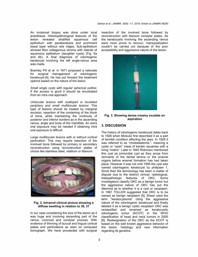
the left mandibular angle. Swelling dimensions were approximately 4 x 3.5 cm antero-posteriorly and 3.5 x 3 cm supero-inferiorly (Fig. 1). The swelling was non-tender and firm in consistency. Skin over the swelling was normal. On intra oral examination there was obliteration of buccal vestibule of the molar teeth (Fig. 2). Overlying surface of the swelling was of same colour as that of surrounding mucosa. On palpation it was firm in consistency and non tender, on application of pressure a white creamy exudate oozed out of the area distal to 37. Aspiration of the swelling yielded a cream coloured hazy fluid (Fig. 3). Based on the clinical findings and aspiration of the lesion (FNAC), a provisional diagnosis of odontogenic keratocyst was made, with a differential diagnosis of dentigerous cyst and ameloblastoma. A panoramic radiograph revealed a well-defined radiolucency on the left ramus of the mandible which was approximately 4x2x1 cm in size, oval In shape, extending anteriorly from distal aspect of 38 to posteriorly to 0.5 cm below the condyle; radiopaque scalloping margins with uniform radiolucency, expansion of inferior border of the mandible at the left angle region, and the inferior alveolar nerve canal was displaced inferiorly (Fig. 4). The bone was intact with no evidence of fracture. Computed tomography revealed a cyst-like radiolucency with scalloped borders and cortical perforations were seen (Fig. 5a & 5b).
Radiographic findings were suggestive of odontogenic keratocyst with a differential diagnosis of unicystic ameloblastoma, and odontogenic myxoma.
Fig. 1. Extra oral clinical photograph showing swelling in the left lower half of the face
An incisional biopsy was done uanesthesia. Histolopathological features of the lesion revealed stratified squamous cell epithelium with parakeratosis and prominent basal layer without rete ridges. Subshowed fibro collagenous stroma with islands of squamous epithelium (daughter cysts) (Figand 6b). A final diagnosis of odontogenic keratocyst involving the left anglewas made.
Bramley PA et al. in 1971 proposed a rationale for surgical management of odontogenic keratocyst
[4]. He has put forward the treatment
options based on the nature of the lesion. Small single cysts with regular spherical outlineIf the access is good it should be enucleatfrom an intra oral approach. Unilocular lesions with scalloped or loculated periphery and small multilocular lesionstype of lesions should be treated by marginal excision; resection of the containing of the blof bone, while maintaining the continuity of posterior and inferior borders as in the ascending ramus, angle and body of the mandible. An extra oral exposure may be needed if obtaining intra oral exposure is difficult. Large multilocular lesions with or without cortical perforation: This may require resection of the involved bone followed by primary or secondary reconstruction using reconstruction plates of choice like stainless steel, vitallium or titanium.
Fig. 2. Intraoral clinical picture showing a
diffuse swelling in relation to 36, 37
In our case considering the size of the lesion as it was huge and involving ascending part of the ramus, coronoid and condylar process. With evidence of thinning of buccal and liplates and perforations as seen on computed tomograph. We have proceeded with surgical
Sekhar et al.; JAMMR, 29(8): 1-7, 2019; Article no.
3
An incisional biopsy was done under local anesthesia. Histolopathological features of the
revealed stratified squamous cell ratosis and prominent
basal layer without rete ridges. Sub-epithelium showed fibro collagenous stroma with islands of squamous epithelium (daughter cysts) (Fig. 6a
diagnosis of odontogenic keratocyst involving the left angle-ramus area
in 1971 proposed a rationale for surgical management of odontogenic
. He has put forward the treatment options based on the nature of the lesion.
Small single cysts with regular spherical outline: access is good it should be enucleated
Unilocular lesions with scalloped or loculated periphery and small multilocular lesions: This type of lesions should be treated by marginal excision; resection of the containing of the block of bone, while maintaining the continuity of posterior and inferior borders as in the ascending ramus, angle and body of the mandible. An extra oral exposure may be needed if obtaining intra
or without cortical : This may require resection of the
involved bone followed by primary or secondary reconstruction using reconstruction plates of choice like stainless steel, vitallium or titanium.
Intraoral clinical picture showing a diffuse swelling in relation to 36, 37
In our case considering the size of the lesion as it was huge and involving ascending part of the ramus, coronoid and condylar process. With evidence of thinning of buccal and lingual cortical plates and perforations as seen on computed tomograph. We have proceeded with surgical
resection of the involved bone followed by reconstruction with titanium condylar plates. As the keratocysts involving the ascending ramus were more prone to reoccur, marsupialization couldn’t be carried out because of the poor accessibility and aggressive nature of the lesion.
Fig. 3. Showing dense creamy exudate on
aspiration
3. DISCUSSION The history of odontogenic keratocyst dates back to 1826 when Mickuliz first described it as a part of familial condition affecting the jaws. In 1926 it was referred to as “cholesteatoma.”cystic or “open” mass of keratin squames with a living “matrix”. Later in 1945 Robinson mentioned this cyst as primordial cyst as they arose from remnants of the dental lamina or the enamel organs before enamel formation has had taken place. However it was not until 1956 the cyst was named odontogenic keratocyst by philipsen 1. Since then the terminology has been a mattedispute due to the distinct clinical, radiological, histopathologic features of OKC. Some investigators classify OKC as a benign tumor but the aggressive nature of OKC has put the dilemma as to whether it is a cyst or neoplasm. In 1967 TOLLER suggested that OKC is to be named as benign neoplasm [5]. Shear used the term “keratocystoma” citing the aggressive nature of the odontogenic keratocyst and finally labeled it as a benign cystic neoplasm OKC was reclassified and renamed as keratocystic odontogenic tumor (KCOT) in the WHO classification of head and neck tumors in 2005 [6]. Redesignation of the OKC as the KCOT is based on the well known aggressive behavior of the lesion, histology and new information regarding its genetics.
; Article no.JAMMR.48290
resection of the involved bone followed by reconstruction with titanium condylar plates. As the keratocysts involving the ascending ramus
to reoccur, marsupialization couldn’t be carried out because of the poor accessibility and aggressive nature of the lesion.
. Showing dense creamy exudate on
The history of odontogenic keratocyst dates back Mickuliz first described it as a part
of familial condition affecting the jaws. In 1926 it was referred to as “cholesteatoma.”- meaning a cystic or “open” mass of keratin squames with a living “matrix”. Later in 1945 Robinson mentioned
dial cyst as they arose from remnants of the dental lamina or the enamel organs before enamel formation has had taken place. However it was not until 1956 the cyst was named odontogenic keratocyst by philipsen 1. Since then the terminology has been a matter of dispute due to the distinct clinical, radiological, histopathologic features of OKC. Some investigators classify OKC as a benign tumor but the aggressive nature of OKC has put the dilemma as to whether it is a cyst or neoplasm.
d that OKC is to be Shear used the
term “keratocystoma” citing the aggressive nature of the odontogenic keratocyst and finally labeled it as a benign cystic neoplasm OKC was reclassified and renamed as keratocystic
tumor (KCOT) in the WHO classification of head and neck tumors in 2005 [6]. Redesignation of the OKC as the KCOT is based on the well known aggressive behavior of the lesion, histology and new information

Fig. 4. Panoramic radiograph showed a well defined radiolucency in relation to left molar
Fig. 5a and 5b. Computed tomography revealed expansile corticated and scalloped cystic
(a) Fig. 6a. Histopathologic section shows stratified squamous epithelium showing parakeratosis
with prominent Fig. 6b. Histopathologic section shows sub
Sekhar et al.; JAMMR, 29(8): 1-7, 2019; Article no.
4
g. 4. Panoramic radiograph showed a well defined radiolucency in relation to left molar ramus area
. Computed tomography revealed expansile corticated and scalloped cystic
lesion
(b)
Histopathologic section shows stratified squamous epithelium showing parakeratosisprominent basal layer without rete ridges
section shows sub-epithelium islands of squamous epithelium (daughter cysts)
; Article no.JAMMR.48290
g. 4. Panoramic radiograph showed a well defined radiolucency in relation to left molar
. Computed tomography revealed expansile corticated and scalloped cystic
Histopathologic section shows stratified squamous epithelium showing parakeratosis
epithelium islands of squamous epithelium
(a)
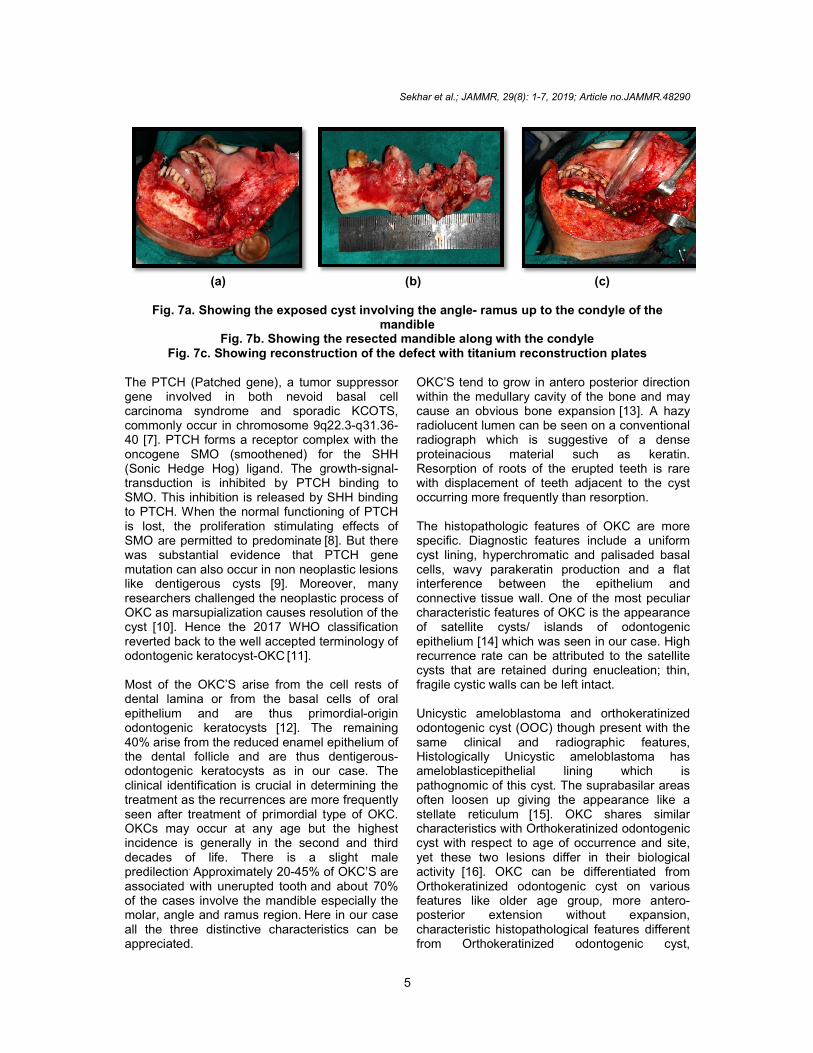
Fig. 7a. Showing the exposed cyst involving the angle
Fig. 7b. Showing Fig. 7c. Showing reconstruction of the defect with titanium rec
The PTCH (Patched gene), a tumor suppressor gene involved in both nevoid basal cell carcinoma syndrome and sporadic KCOTS, commonly occur in chromosome 9q2240 [7]. PTCH forms a receptor complex with the oncogene SMO (smoothened) for the SHH (Sonic Hedge Hog) ligand. The growthtransduction is inhibited by PTCSMO. This inhibition is released by SHH binding to PTCH. When the normal functioning of PTCis lost, the proliferation stimulating effects of SMO are permitted to predominatewas substantial evidence that PTCH gene mutation can also occur in non neoplastic lesions like dentigerous cysts [9]. Moreovresearchers challenged the neoplastic process of OKC as marsupialization causes resolution of the cyst [10]. Hence the 2017 WHO classification reverted back to the well accepted terminology of odontogenic keratocyst-OKC [11]. Most of the OKC’S arise from the cell rests of dental lamina or from the basal cells of oral epithelium and are thus primordialodontogenic keratocysts [12]. The remaining 40% arise from the reduced enamel epithelium of the dental follicle and are thus dentigerousodontogenic keratocysts as in our case. The clinical identification is crucial in determining the treatment as the recurrences are more frequently seen after treatment of primordial type of OKC. OKCs may occur at any age but the highest incidence is generally in the second and third decades of life. There is a slight male predilection. Approximately 20-45% of OKC’S are associated with unerupted tooth
and about 70%
of the cases involve the mandible especially the molar, angle and ramus region. Here in our caall the three distinctive characteristics can be appreciated.
Sekhar et al.; JAMMR, 29(8): 1-7, 2019; Article no.
5
(b) (c)
the exposed cyst involving the angle- ramus up to the condyle of the mandible
Showing the resected mandible along with the condyle reconstruction of the defect with titanium reconstruction plates
, a tumor suppressor gene involved in both nevoid basal cell carcinoma syndrome and sporadic KCOTS, commonly occur in chromosome 9q22.3-q31.36-
H forms a receptor complex with the SMO (smoothened) for the SHH
og) ligand. The growth-signal-CH binding to
SMO. This inhibition is released by SHH binding en the normal functioning of PTCH
is lost, the proliferation stimulating effects of SMO are permitted to predominate
[8]. But there
was substantial evidence that PTCH gene mutation can also occur in non neoplastic lesions
Moreover, many researchers challenged the neoplastic process of OKC as marsupialization causes resolution of the
. Hence the 2017 WHO classification reverted back to the well accepted terminology of
arise from the cell rests of dental lamina or from the basal cells of oral epithelium and are thus primordial-origin
The remaining 40% arise from the reduced enamel epithelium of the dental follicle and are thus dentigerous- odontogenic keratocysts as in our case. The clinical identification is crucial in determining the treatment as the recurrences are more frequently seen after treatment of primordial type of OKC. OKCs may occur at any age but the highest
lly in the second and third decades of life. There is a slight male
45% of OKC’S are and about 70%
of the cases involve the mandible especially the Here in our case
all the three distinctive characteristics can be
OKC’S tend to grow in antero posterior direction within the medullary cavity of the bone and may cause an obvious bone expansionradiolucent lumen can be seen on a conventional radiograph which is suggestive of a dense proteinacious material such as keratin. Resorption of roots of the erupted teeth is rarewith displacement of teeth adjacent to the cyst occurring more frequently than resorption. The histopathologic features of OKC are more specific. Diagnostic features include a uniform cyst lining, hyperchromatic and palisaded basal cells, wavy parakeratin production and a flat interference between the epithelium and connective tissue wall. One of the most peculiar characteristic features of OKC is the appearance of satellite cysts/ islands of odontogenic epithelium [14] which was seen in our case. High recurrence rate can be attributed to the satellite cysts that are retained during enucleation; thin, fragile cystic walls can be left intact. Unicystic ameloblastoma and orthokeratinized odontogenic cyst (OOC) though present with the same clinical and radiographic features, Histologically Unicystic ameloblastoma has ameloblasticepithelial lining which is pathognomic of this cyst. The suprabasilar areas often loosen up giving the appearance like a stellate reticulum
[15]. OKC shares similar
characteristics with Orthokeratinized odontogeniccyst with respect to age of occurrence and site, yet these two lesions differ in their biological activity [16]. OKC can be differentiated from Orthokeratinized odontogenic cyst on various features like older age group, more anteroposterior extension without expansion, characteristic histopathological features different from Orthokeratinized odontogenic cyst,
; Article no.JAMMR.48290
(c)
to the condyle of the
onstruction plates
OKC’S tend to grow in antero posterior direction within the medullary cavity of the bone and may cause an obvious bone expansion [13]. A hazy radiolucent lumen can be seen on a conventional radiograph which is suggestive of a dense proteinacious material such as keratin. Resorption of roots of the erupted teeth is rare
with displacement of teeth adjacent to the cyst tly than resorption.
The histopathologic features of OKC are more specific. Diagnostic features include a uniform cyst lining, hyperchromatic and palisaded basal cells, wavy parakeratin production and a flat interference between the epithelium and
ive tissue wall. One of the most peculiar characteristic features of OKC is the appearance
of odontogenic which was seen in our case. High
recurrence rate can be attributed to the satellite d during enucleation; thin,
fragile cystic walls can be left intact.
Unicystic ameloblastoma and orthokeratinized odontogenic cyst (OOC) though present with the same clinical and radiographic features, Histologically Unicystic ameloblastoma has
icepithelial lining which is pathognomic of this cyst. The suprabasilar areas often loosen up giving the appearance like a
OKC shares similar characteristics with Orthokeratinized odontogenic cyst with respect to age of occurrence and site, yet these two lesions differ in their biological
OKC can be differentiated from Orthokeratinized odontogenic cyst on various features like older age group, more antero-
hout expansion, characteristic histopathological features different from Orthokeratinized odontogenic cyst,

Sekhar et al.; JAMMR, 29(8): 1-7, 2019; Article no.JAMMR.48290
6
parakeratinized layer, high recurrence rate, association with basal cell nervous syndrome. The treatment options for OKC’S range from simple conservative treatment like enucleation, (with or without curettage), or marsupialization. Marsupialization is a technique relying on incomplete removal of the cyst lining. Opening a window into the cyst forms an invagination of the oral cavity or the maxillary antrum.It relies on the principle that decompression halts expansion of the cyst and appositional growth of bone occurs, and the former cyst lumen becomes smaller with time. Many modification of the procedure have been taken place over the time like usage of decompression tubes, marsupialization catheter. One such method has been demonstrated by COSTA F.W.G et al where he used a segment of polyethylene suction tube, prepared according to the radiographic size of the lesion. Using a disposable needle, a hole is drilled near the extremity, large enough to allow the passage of a 0.8-mm orthodontic stainless steel wire. With the aid of a needle holder, one end of the wire is shaped into a loop and the other end is inserted through the hole in the tube, pulled back, and twisted. The tooth crown is etched with acid and the loop is attached to the dental surface with composite resin. Advantage of this technique over other methods was that it provides greater stability and minimizes the need for additional surgical interventions, when compared to traditional methods where decompression devices were attached to the surrounding structures with sutures. This provides insufficient stability in case of surgical wound dehiscence. Poor adjustment increases the likelihood of device-related complications
[17].
Though conservative treatment preserves the anatomic structures they have a high risk for recurrence. The aggressive treatment includes peripheral ostectomy, chemical curettage with Carnoys solution, or enbloc resection. Carnoy's solution is composed of 3 ml of chloroform, 6 ml of absolute ethanol, 1 ml of glacial acetic acid, and 1 g of ferric chloride, is often used as a complementary treatment of lesions with high recurrence rates such as odontogenic keratocyst. The action of this solution is given by chemical cauterization, promoting a superficial necrosis of about 1.5 mm of depth after 5 minutes of bone cavities exposure. In a study conducted by Albuquerque et al on surgical treatment with or without carnoy’s solution in aggressive tumors of odontogenic origin the authors found a beneficial effect of the Camoy’s solution in reducing the
recurrence rate in several cases of jaw aggressive odontogenic tumors. This emphasizes the importance of carnoy’s solution when used in conjunction with conservative procedures like enucleation [18]. The problem of using Carnoy’s solution in bone cavity is its effect on the inferior alveolar nerve (if denuded or is free in the cavity after procedure).while the neurotoxic effects of carnoys solution on inferior alveolar nerve is still being debated [19], the chloroform used in the carnoy’s solution has carcinogenic potential. Hence, carnoy’s solution must be used with caution in order to avoid the deleterious effects on the nerve and surrounding tissues.
In the present case the surgical intervention included hemimandibulectomy (Fig. 7a, 7b) followed by reconstruction with titanium condylar plates (Fig. 7c).
4. CONCLUSION
Odontogenic keratocyst is a unique entity among odontogenic cysts, due to its varied clinical, radiological and histopathologic features. Hence the correlation of histopathologic findings with clinical and radiographic features is of paramount importance to achieve a definitive diagnosis, as most of these lesions have a prognostically different biologic behaviours and the final diagnosis helps to proceed with the appropriate treatment procedure.
CONSENT
As per international standard or university standard, patient’s consent has been collected and preserved by the author(s).
ETHICAL APPROVAL
As per international standard or university standard written ethical approval has been collected and preserved by the author(s).
COMPETING INTERESTS
Authors have declared that no competing interests exist.
REFERENCES
1. Philipsen HP. Omkeratocyst (kolesteatomer) kaekberne. Tandlaegeabladet. 1956;60:963-81.
2. Haring JI, Van Dis ML. Odontogenic Keratocysts, A clinical, radiographic and

Sekhar et al.; JAMMR, 29(8): 1-7, 2019; Article no.JAMMR.48290
7
histopathologic study. Oral Med Oral Surg Oral Pathol. 1988;66:145-53.
3. Mortazavi H, Baharvand M. Jaw lesions associated with impacted tooth: A radiographic diagnostic guide. Journal of Imaging Sci Dent. 2016;46(3):147–157.
4. Bramley PA. Odontogenic cysts treatment of the cyts of the jaws. Proc. Roy. Soc. Med. 1971;64.
5. Toller P. Origin and growth of cysts of the jaws. Ann R Coll Surg Engl. 1967;40(5): 306-36.
6. Philipsen HP. Keratocystic odontogenic tumour. In: Barnes L, Eveson JW, Reichart P, Sidransky D, Editors. World Health Organization Classification of Tumours: Pathology and Genetics of Head and Neck Tumours. Lyon, France: IARC Press. 2005;306.
7. Hemavathy S, Roy S. Follicular odontogenic keratocyst mimicking dentigerous cyst-report of two cases. Arch Oral Sci Res. 2011;1:100-3.
8. Chaudhary D, Bhargava M, Aggarwal S. Keratocystic odontogenic tumour-a case report with review of literature. Indian J Stomatol. 2012;3:66-9.
9. Pavelic B, Levant S. PTCH gene alteration in dentigerous cyst. Journal of Oral Pathol Med. 2001;30:569-76.
10. Soluk Tekkeşin, Merva M, Wright John. The world health organization classification of odontogenic lesions: A summary of the changes of the 2017 (4th) Edition. Turkish Journal of Pathology. 2013;34. DOI: 10.5146/tjpath.2017.01410
11. Passi, Deepak, Singhal, Deepika. Odontogenic Keratocyst (OKC) or Keratocystic Odontogenic Tumor (KCOT). Journey of okc from cyst to tumor to cyst again: Comprehensive review with recent updates on WHO classification.
International Journal of Current Research. 2017;9:54080-54086.
12. Marx, Stern. Oral and maxillofacial pathology: A rational for diagnosis and treatment. 2nd Edition. 2012;616-631.
13. Pogrel MA. The Keratocystic Odontogenic tumor oral maxillofacial. Surg Clin N Am. 2013;25(1):21–30.
14. Shear M, The aggressive nature of the odontogenic Keratocyst: Is it a benign cystic neoplasm? Part 1. Clinical and early experimental evidence of aggressive behavior. Oral Oncol. 2002;38:219-26.
15. Bajpai M, Agarwal D, Bhalla A, Kumar M, Garg M, Kumar M. Multilocular unicystic ameloblastoma of mandible. Case Rep Dent. 2013;2013:835892.
16. Sarvaiya B, Vadera H, Sharma V, Bhad K, Patel Z, Thakkar M. Orthokeratinised odontogenic cyst of the mandible: A rare case report with systemic review. J Int Soc Prev Community Dent. 2014;4(1):71–76.
17. Costa FWG, Carvalho FSR, Chaves F. Nobre, Soares ECS. A suitable device for cystic lesions close to the tooth-bearing areas of the jaws. Journal of Oral and Maxillofacial Surgery. 2014;72:96-98.
18. Albuquerque AFM, Silva PGB, Bezerra TMM, Aleves APNN, Pereira KMA, Ribeiro TR, Soares ECS, Costa FWG. Surgical treatment with or without the use of carnoy solution in aggressive tumors of odontogenic origin: A systematized critical literature review. International Journal of Clinical Dentistry. 2015;9:2.
19. Jitka Levorova, Vladimir Machon, Pavel Grill, Dusan Hirjak, Rene Foltan. Keratocystic odontogenic tumour with extraosseal spread: Evaluation of the effect carnoy’s solution. Prague Medical Report. 2015;116(4)303–313.
_________________________________________________________________________________ © 2019 Sekhar et al.; This is an Open Access article distributed under the terms of the Creative Commons Attribution License (http://creativecommons.org/licenses/by/4.0), which permits unrestricted use, distribution, and reproduction in any medium, provided the original work is properly cited.
Peer-review history: The peer review history for this paper can be accessed here:
http://www.sdiarticle3.com/review-history/48290