a retro-ocular teratoma containing pinealomatous tissue in a young
12
258 A RETRO-OCULAR TERATOMA CONTAINING PINEALOMATOUS TISSUE IN A YOUNG CHICKEN J. G. CAMPBELL From the British Empire Cancer Campaign Unit, A.R.C. Poultry Research Centre, Edinburgh Received for publication April 12, 1962 THE specimen was obtained during an extended study of spontaneous tumours of an embryonal nature occurring in " broiler " chickens which are bred for rapid growth, fed intensively and reared in an environment designed to exploit to the full the resulting increased growth rate. This tumour is one of a series which will be reported at a later date in a study on environmental carcinogenesis. Mean- while, it is considered that the case is sufficiently interesting to warrant a separate and more detailed description. A search of the literature shows that reports of cranial teratomas in the lower animals are extremely rare. Only two have been described, occurring intracranially in rabbits (Margulies, 1901; Shima, 1908) the first being presumably of hypo- physeal origin, while that of the second was not specified. Schlotthauer and Kernohan (1935) reported a pinealoma unassociated with any tridermal histoid structures, in a silver fox. Teratomas of the gonads, particularly in the fowl, are of course well known. Most of these have been produced experimentally by the intra-testicular injection of zinc salts, (Michalowsky, 1926; Bagg, 1936; Falin, 1941; Carleton, Friedman and Bomze, 1953), or copper salts (Falin and Anissimowa, 1940), although spontaneous teratomas have been recorded by Mashar (1932) and Campbell (1951). All of these avian teratomas have been confined to the testis. Even teratomas of the gonads, as distinct from those in other sites, are very uncommon in mammals, with the exception of those occurring in the testis of the horse. For further details the reader should consult Willis (1960). MATERIALS AND METHODS The specimen was received preserved in formal-saline, and consisted of the right eye of a six-week old Arbor Acres " broiler " male chick to which was attached on the posterior-medial aspect a firm partially cystic ovoid growth (25 x 22 x 20 mm.) the long axis of which was directed somewhat ventrally. Information was subsequently received that the state of the brain and pineal had not been ascertained. The tumour had been sliced open, and a large cyst con- taining a clear jelly-like substance was present at the posterior pole. The eye appeared to be intact as also was the optic nerve. After photography the gross specimen was transected in the same plane as the original incision. At this stage it was obvious that the tumour was only loosely attached to the selera by con- nective tissue. One half was wax-embedded after the usual processing, and a small portion of the other half was retained in formal saline. The remainder was post-
Transcript of a retro-ocular teratoma containing pinealomatous tissue in a young

258
A RETRO-OCULAR TERATOMA CONTAINING PINEALOMATOUSTISSUE IN A YOUNG CHICKEN
J. G. CAMPBELLFrom the British Empire Cancer Campaign Unit, A.R.C. Poultry Research Centre,
Edinburgh
Received for publication April 12, 1962
THE specimen was obtained during an extended study of spontaneous tumoursof an embryonal nature occurring in " broiler " chickens which are bred for rapidgrowth, fed intensively and reared in an environment designed to exploit to thefull the resulting increased growth rate. This tumour is one of a series whichwill be reported at a later date in a study on environmental carcinogenesis. Mean-while, it is considered that the case is sufficiently interesting to warrant a separateand more detailed description.
A search of the literature shows that reports of cranial teratomas in the loweranimals are extremely rare. Only two have been described, occurring intracraniallyin rabbits (Margulies, 1901; Shima, 1908) the first being presumably of hypo-physeal origin, while that of the second was not specified. Schlotthauer andKernohan (1935) reported a pinealoma unassociated with any tridermal histoidstructures, in a silver fox. Teratomas of the gonads, particularly in the fowl,are of course well known. Most of these have been produced experimentally bythe intra-testicular injection of zinc salts, (Michalowsky, 1926; Bagg, 1936;Falin, 1941; Carleton, Friedman and Bomze, 1953), or copper salts (Falin andAnissimowa, 1940), although spontaneous teratomas have been recorded byMashar (1932) and Campbell (1951). All of these avian teratomas have beenconfined to the testis.
Even teratomas of the gonads, as distinct from those in other sites, are veryuncommon in mammals, with the exception of those occurring in the testis of thehorse. For further details the reader should consult Willis (1960).
MATERIALS AND METHODSThe specimen was received preserved in formal-saline, and consisted of the
right eye of a six-week old Arbor Acres " broiler " male chick to which wasattached on the posterior-medial aspect a firm partially cystic ovoid growth(25 x 22 x 20 mm.) the long axis of which was directed somewhat ventrally.Information was subsequently received that the state of the brain and pineal hadnot been ascertained. The tumour had been sliced open, and a large cyst con-taining a clear jelly-like substance was present at the posterior pole. The eyeappeared to be intact as also was the optic nerve. After photography the grossspecimen was transected in the same plane as the original incision. At this stageit was obvious that the tumour was only loosely attached to the selera by con-nective tissue. One half was wax-embedded after the usual processing, and a smallportion of the other half was retained in formal saline. The remainder was post-

RETRO-OCULAR TERATOMA IN CHICKEN
fixed in Susa before wax embedding. Sections were cut at 6 It. at a number oflevels and stained with haematoxylin and eosin, Mallory's trichrome, phospho-tungstic acid-haematoxylin (PTAH), Einarsen's chrome alum gallocyanin forNissl's substance, Best's stain for glycogen, periodic acid-Schiff (PAS) for glycogenand acid muco-polysaccharide, thionin blue for mucin, and frozen formal-fixedsections were stained by Bielchowsky's method for neuro-fibrils. Serial sectionsand reconstructions were not attempted, but sections were studied at variouslevels through the tumour.
RESULTS
A low-power survey of a section of the tumour taken about the middle showsa quite complicated structure. Fig. 1 illustrates this in diagrammatic form. Atthe anterior and ventral aspects there is a mass of neural tissue which containsseveral slit-like irregular spaces, and occupies about a third of the total tumour.The remainder is composed largely of plain muscle mainly disposed in circularmanner around two prominent irregular cavities plus a number of smaller ones,all lined with glandular epithelium. Ventral to the smaller of the two main cavities,at the junction with the neural component, is a little group of tubular glands anda network of rather ill-defined capillaries. The largest cavity has two villiformprojections from the wall, and was originally filled with a clear viscous fluid, nowrepresented by a precipitated amorphous fibrinous material. The villiform struc-tures are not present in deeper sections. Patches of lymphoid tissue are presentadjacent to and in the sub-epithelial connective tissue. The opposite (posterio-dorsal) side of the large cavity has, in between the plain muscle bundles constitutingits wall, a plaque of cartilage and osteoid, and a small island of bone, betweenwhich lies another region of capillaries similar to those mentioned above. Pos-terior to these tissues, the wall of the tumour is composed of a fairly extensivezone of striated muscle, beyond which is fat and the thin limiting capsule. Bloodvessels are especially prominent in the villiform processes and below anothersmaller patch of peripheral fat.
A section of normal lachrymal gland lies in the connective tissue between thetumour and the dorso-lateral aspect of the eye. The following tissues derivedfrom the three primitive germ layers are represented in this tumour:
Ectoderm Mesoderm EntodermEpendymal cells Cartilage Mucus secreting cellsNerve tissue and cells Bone Intestinal epitheliumEpithelial cyst Muscle Respiratory tract ? epithelium
FatConnective tissueLymphoid tissue
It is not necessary to describe all these different tissues in detail. However, theirdisposition and the nature of the epithelium, neural tissue, and presumptive lungshow many points of interest, and will be reported more fully.
The neural componentAlthough there are no sharp histological divisions in this part, a transition
occurs from a relatively undifferentiated peripheral region which is thickest ad-jacent to the eye, to a central portion showing a more organised structure.
259

J. G. CAMPBELL
The peripheral part is characterised by three main cell types. Firstly, thereare large cells with vesicular nuclei and prominent nucleoli, whose cytoplasmcontains Nissl's substance. Secondly, there are numerous cells with balloonednuclei which contain glycogen, stainable even in paraffin sections by Best's method,and by PAS, and removable by previous diastase treatment; and finally, thereare many small cells of the glial type, with compact darkly staining nuclei (Fig. 2).
The central region contains numerous slit-like cavities, lined with ependymaand surrounded by trabeculae of pale neuro-epithelial cells. This structure isreminiscent of the pars nervosa of the hypophysis (Fig. 3).
Apart from this, most of the neural tissue is composed of the three cell typesalready described, with considerable areas showing a characteristic grouping ofglial and ependymal cells about small vessels and cavities, resulting in a distinctlypinealomatous appearance (Fig. 4). PTAH stained preparations show the epen-dymal cells particularly well, with their typical long polarized processes endingin branched tufts or swellings about a small vessel or cavity (Fig. 5). Frozensections stained by Bielchowsky's method, reveal the presence of large numbersof neuro-fibrils (Fig. 6, 7).
EXPLANATION OF PLATES
FIG. 1.-Diagram of structure of the bisected retro-ocular tumour. x 8* 5.FIG. 2.-Neurons containing Nissl's granules, glial cells, and vacuolated nuclei containing
glycogen. (From Region A in the diagram.) Einarsen's stain. x360.FIG. 3.-Trabeculated neuro-epithelial region (Region B) showing ependymal-lined cavities.Note resemblance to pars nervosa tissue. H. and E. x 90.
FIG. 4.-Detail of pinealomatous region. Note the radiate arrangement of ependymal cells,and the condensation of dark staining glial cells. (Region anterior to B.) x280.
FIG. 5. Ependymal cells, showing polarisation of their processes and, bottom right, a terminaltuft to a process. (Region posterior to B.) Frozen section, Bielchowsky. x 1150.
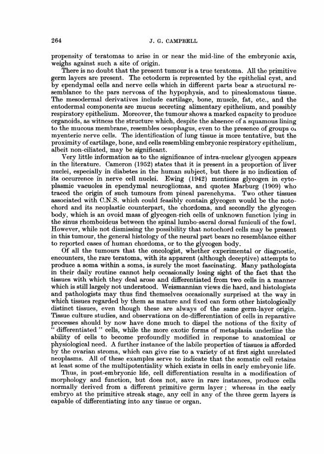
FIG. 6.-Oil-immersion photograph of a frozen section from the pinealomatous region, showinga pear-shaped neuron, and the aggregation of glial cells and ependymal processes about an
out-of-focus capillary. (Region anterior to A.) Bielchowsky. x 1150.FIG. 7.-Another region of the pinealoma, showing diversity of cell types and the abundance
of fibrils. (Region anterior to A.) Frozen section, Bielchowsky. x 900.FIG. 8.-Mucus-secreting intestinal epithelium lining a main cavity in the glandular portion.
There is a perfect sub-mucous coat and muscularis mucosae. (Region between A and C).Trichrome. x 90.
FIG. 9. One of the two villiform structures extending into the major lumen of the glandularpart. (Region C.) Note absence of muscle, vascularity and the modified investing epithelium(Region C.) An epithelial cyst, resembling a feather follicle, is at left centre. H. and E.x21.
FIG. 10. Base of the second "villus", showing collagenous and elastic stroma, and thetransition of the epithelium near the top to a type resembling corneal-scleral junctionepithelium as it continues down. (Region between A and C.) x 90.
FIG. 11.-Lymphoid tissue, a group of glands resembling embryonic bronchial epithelium,and a capillary network resembling embryo lung. (Region A.) H. and E. x 115.
FIG. 12.-" Embryonic lung tissue ", associated with glandular epithelium and cartilage.(Region D.) H. andE. x140.
FIG. 13.-A field containing, from left to right, fat, a small focus of epithelial cells, connectivetissue, osteoid, bone, and possible bronchial epithelium. (Region posterior to D.) H. and E.x90.
KEREY TO FIGURE 1
1. Pinealomatous tissue. 6. Blood vessels.2. Plain muscle. 7." Lung".3. Connective tissue. 8. Intestinal epithelium.4. Fat. 9. Bone.5. Cartilage. 10. Striated muscle.
260

BRITISH JOURNAL OF CANCER.
Campbell.
r-2
"In
......
VOl. XVI, NO. 2.

Vol. XVI, No. 2.
iiVW A,s _ t.
BRITISH JOURNAL OF CANCER.
* s:
4
-'. .i.-:s i-
::::..
.s 1. '......s }: ]*;ffi w *,. '
6 7
Campbell.
2i
II
i.
.1k .:
1....; i

BRITISH JOURNAL OF CANCER.
t0
2~~~~~~~~~~~~~-. ~,
I'sv <, w _ _~-4.--.i s
niSS.g , .:.s'If
13
Campbell.
VOl. XVI, NO. 2.
I:.$

RETRO-OCULAR TERATOMA IN CHICKEN
The glandular epitheliumNearly all the cavities except those in the neural part and a few small slit-like
spaces, presumably lymph channels, are lined with a single layer of epithelialcells. These vary from a flattened type investing the aforementioned villi, to atall pale columnar mucus-secreting type lining the cavities and the proximal partof the glandular evaginations arising therefrom (Fig. 8). Not all of these cellsare simultaneously active mucus secretors, as thionin blue and PAS stainedmaterial shows. The nuclei are basal and are very dense and crescent shaped.Small isolated glands are often associated with duct-like structures, which may beeither simple, or much branched, and are usually not actively secreting mucus.The transition between active and resting cells is abrupt. The two prominentvilliform processes which project into the lumen of the largest cavity are coveredwith an epithelium continuous with the mucus-secreting lining (Fig. 9) butshowing a transition near the villus base to cells which resemble conjunctivalepithelium at the corneal-palpebral junction (Fig. 10) and which later becomeflattened as the free tip of the villus is approached. The core of the villi is com-posed of vascular connective tissue. Glandular epithelium wherever situated isassociated with a connective tissue submucous layer, external to which are thininner and thick outer circular coats of plain muscle interspersed with connectivetissue. Small discrete clusters of pale nuclei resembling the cells of the myentericplexus lie in a thin fibrous matrix between the muscle coats.
Respiratory epitheliumA few minute cysts and tubular glands are lined with several layers of baso-
philic cells resembling embryonic bronchial epithelium. These cells do not appearto be ciliated, although typical basal bodies are present near the free border.The cysts are, for the most part, in the vicinity of the cartilage plaques and thepresumptive embryonic lung, and they are usually associated with lymphoidtissue. The " lung " varies from a loose network of epithelial tissue, to an openplexus of thin-walled tubes, some of which contain a few red blood cells. Althoughthe interpretation of these tubes is open to doubt, owing to the variable structure,it is thought not unreasonable to suggest tentatively a resemblance to lungcapillaries (Fig. 11, 12).
Muscle componentsAs already mentioned, the plain muscle is confined to the posterior and dorsal
two-thirds of the tumour, and is mainly arranged in a circular manner around theglandular cavities. Small isolated bundles of cells also occur scattered in a hap-hazard manner throughout the connective tissue stroma.
The striated muscle is present as a fairly broad band external to the cartilage,bone and " respiratory epithelium " (Fig. 13), and is otherwise unremarkable.
The connective tissueApart from the specialised glial fibres in the neural portion, the connective
tissue is confined to the capsule, which is not intact at the anterior portion, andto the remainder of the tumour, where it forms a submucous layer of the stromain general. It is mainly of an undifferentiated embryonic type with plump palenuclei and abundant cytoplasm, but showing little mitotic activity. That ill-vesting the neural part resembles meningeal tissue.
261

J. G. CAMPBELL
Lymphoid tissueSmall widely scattered foci of lymphoid tissue occur in association with glands
or glandular epithelium. A few aggregations are also present in the neural part ofthe tumour. The majority of the cells are mature lymphocytes, the remainderbeing plasma cells and reticulum cells.
Epithelial cystThe small cyst visible in Fig. 9 is composed of cuboidal ectodermal cells,
containing numerous minute brownish-black cytoplasmic granules showing adistinct aggregation at the free border. The lumen of the cyst contains a plugof dead epithelial cells. There is thus some resemblance to a feather follicle.
DISCUSSION
The pineal body or epiphysis cerebri is represented in the vertebrates from thecyclostomes upwards. In the lamprey (Petromyzon) it shows the greatest degreeof organization, being a double structure, consisting of parapineal (parietal organ)and pineal, both of which approach the dorsal surface of the skull and form welldefined eye-cups at the distal extremities. At their proximal connection with thediencephalon, they are definitely glandular. In the selachians, the glandularstructure is predominant and only the parapineal forms an eye-cup. In teleoststhe eye-cup is lost, but they show a marked glandular structure of pineal and para-pineal, both of which apparently secrete into the cerebro-spinal fluid. In amphibiathe pineal, and in reptiles the parietal organ is modified for light reception, andin Sphenodon, the parietal organ is a particularly well developed eye-cup. Fossilsaurian skulls have a dorsal foramen, which presumably housed the pineal or" third eye ", and this is still present in the parietal bones of many lizards. InRana temporaria, the pineal is visible beneath the skin in the interocular region asthe organ of Stieda. Histologically this is a degenerate organ, being an irregularmass of epithelial cells some of which resemble retinal rods. There is evidence,however, that in this animal the pineal organ still functions as a light receptor(Heerd and Dodt, 1961).
The only member of the class Aves in which the development of the pinealhas been studied in any detail is the domestic fowl, and here Lillie (1952) statesthat it is first discernible as an evagination from the roof of the diencephalon, atabout the 30-35 somite stage (approximately 60 hours). It subsequently growsout as a long narrow tube, dilated distally and giving off numerous hollow buds.There is no parietal organ. In the adult fowl the pineal body is situated behindthe thalami as a small body, triangular in section and attached to the roof of thethird ventricle. Its capsule is provided by the pia mater, which extends into theorgan to form trabeculae surrounding hollow intercommunicating cords of neuro-epithelial cells. The majority of these are glial cells resembling astrocytes, butthe lumen of the cord is lined with ependymal cells. The general appearance isgland-like, although no morphological evidence of secretion can be seen, andphysiological evidence of secretion in birds and other animals is inconclusive.Thus Foa (1914) found that pinealectomy had no effect on female chicks, butretarded the growth of male chicks until they were 2-3 months old, when theybecame normal in weight. Kozelka (1932) found no effects in chicks after implant-
262

RETRO-OCULAR TERATOMA IN CHICKEN
ation experiments, but Shellabarger (1952) found that pinealectomy inhibitedtestis growth in young chicks, and that this could be restored to normal by theinjection of lyophilized beef pineal. Lowenstein (1952), while showing that extractsof pineal were not physiologically inert, remarked on the fact that those workerswho have obtained positive experimental evidence of activity in chicks are agreedthat this activity is confined to males, It is perhaps not without significancethat pineal teratomas are practically confined to the human male, and that "aboutone half of the cases show precocious sexual and bodily development " (Bochnerand Scarff, 1938).
The present tumour almost certainly arose from pineal tissue. The possibilityof a hypophyseal origin is remote, in view of the extreme rarity of tumours of thepars nervosa in man, and the total absence of reports in lower animals. Olderreports of teratomas in the human pituitary are not now accepted, the view beingthat they were craniopharyngiomas showing variable metaplasia (Saphir, 1959).Infiltration of the posterior lobe of the human hypophysis with pinealomatouscells has been recorded by Walton (1949), who also discusses the so-called" ectopic or parapineal teratomas ", and concludes that the evidence for theirpineal origin is inconclusive. Although there was no opportunity in the presentcase to examine the cranial cavity and its contents, the information providedshowed that there were no nervous symptoms, i.e. that the growth was probablyalmost entirely confined to the orbit, resulting in a severe proptosis. This beingso, we are faced with the problem of how it gained access to the orbital space.
There appear to be two alternative ways of spread from the pineal region,one being forward between the cerebral hemispheres, down and back to the opticnerves, and so to the optic foramen in the hinder end of the interorbital septum;while an alternative route would seem to be laterally between the cerebral hemi-sphere and optic lobe to the optic tracts and the foramen. Unfortunately there isno direct evidence for either. It is possible that an intra-cranial tumour grew as
thin subarachnoid strands of tissue along one or other of these routes, to expandas soon as the orbital cavity was reached. Alternatively it may have arisen de novo
from an orbital locus of developmentally displaced pluripotential cells. At firstsight there is no reason why a group of such cells should not form pinealomatoustissue amongst other tissue. It seems more likely, however, that the tumourarose from an intracranial site; the literature oln human pineal teratomas recordsthat these very rare tumours frequently have a layer of either normal or adeno-matous pineal tissue in juxtaposition (Frankl-Hochwart, 1909; Getzeit, 1896,cited by Ewing, 1942). Willis (1958) states that in humans, almost all intra-cranial teratomas arise in the vicinity of the pineal gland, quadrigeminal plateor walls of the posterior part of the third ventricle, including the pituitary region.In those human cases where an intra-cranial pinealoma was found together witha normal pineal gland, Ewing (1942) suggested that the tumour may have arisenfrom other primitive outpocketings from the diencephalon, e.g. the pre-commisuralorgans of the mid-habenular corpusculum parietale. No reference to a retro-ocular teratoma associated with pinealomatous tissue in man could be found,although about 20 human orbital teratomas have been reported during the last100 years. Burnier and Salles (1945), quoted by Harbert (1949), collected 17 ofthese references, and see also Kamal (1954) and Winter (1960). Although a strictlyorbital origin cannot be excluded in the present case, the association with pinealo-matous tissue, not recorded in teratomas similarly sited in man, and the known
263
J. G. CAMPBELL
propensity of teratomas to arise in or near the mid-line of the embryonic axis,weighs against such a site of origin.
There is no doubt that the present tumour is a true teratoma. All the primitivegerm layers are present. The ectoderm is represented by the epitheial cyst, andby ependymal cells and nerve cells which in different parts bear a structural re-semblance to the pars nervosa of the hypophysis, and to pinealomatous tissue.The mesodermal derivatives include cartilage, bone, muscle, fat, etc., and theentodermal components are mucus secreting alimentary epithelium, and possiblyrespiratory epithelium. Moreover, the tumour shows a marked capacity to produceorganoids, as witness the structure which, despite the absence of a squamous liningto the mucous membrane, resembles oesophagus, even to the presence of groups o.myenteric nerve cells. The identification of lung tissue is more tentative, but theproximity of cartilage, bone, and cells resembling embryonic respiratory epithelium,albeit non-ciliated, may be significant.
Very little information as to the significance of intra-nuclear glycogen appearsin the literature. Cameron (1952) states that it is present in a proportion of livernuclei, especially in diabetes in the human subject, but there is no indication ofits occurrence in nerve cell nuclei. Ewing (1942) mentions glycogen in cyto-plasmic vacuoles in ependymal neurogliomas, and quotes Marburg (1909) whotraced the origin of such tumours from pineal parenchyma. Two other tissuesassociated with C.N.S. which could feasibly contain glycogen would be the noto-chord and its neoplastic counterpart, the chordoma, and secondly the glycogenbody, which is an ovoid mass of glycogen-rich cells of unknown function lying inthe sinus rhomboideus between the spinal lumbo-sacral dorsal funiculi of the fowl.However, while not dismissing the possibility that notochord cells may be presentin this tumour, the general histology of the neural part bears no resemblance eitherto reported cases of human chordoma, or to the glycogen body.
Of all the tumours that the oncologist, whether experimental or diagnostic,encounters, the rare teratoma, with its apparent (although deceptive) attempts toproduce a soma within a soma, is surely the most fascinating. Many pathologistsin their daily routine cannot help occasionally losing sight of the fact that thetissues with which they deal arose and differentiated from two cells in a mannerwhich is still largely not understood. Weismannian views die hard, and histologistsand pathologists may thus find themselves occasionally surprised at the way inwhich tissues regarded by them as mature and fixed can form other histologicallydistinct tissues, even though these are always of the same germ-layer origin.Tissue culture studies, and observations on de-differentiation of cells in reparativeprocesses should by now have done much to dispel the notions of the fixity of" differentiated" cells, while the more exotic forms of metaplasia underline theability of cells to become profoundly modified in response to anatomical orphysiological need. A further instance of the labile properties of tissues is affordedby the ovarian stroma, which can give rise to a variety of at first sight unrelatedneoplasms. All of these examples serve to indicate that the somatic cell retainsat least some of the multipotentiality which exists in cells in early embryonic life.
Thus, in post-embryonic life, cell differentiation results in a modification ofmorphology and function, but does not, save in rare instances, produce cellsnormally derived from a different primitive germ layer; whereas in the earlyembryo at the primitive streak stage, any cell in any of the three germ layers iscapable of differentiating into any tissue or organ.
264

RETRO-OCULAR TERATOMA IN CHICKEN 265
It is this fact which has led to the hypothesis that teratomas arise from tissueprimordia which have somehow become physiologically segregated in early em-bryonic life, thus escaping the influence of the dorsal lip primary organiser in theblastopore at the stage of gastrulation. (In the avian embryo, the primitive streakcorresponds to an elongated blastopore.) The earlier this segregation takes place,the greater the capacity for self-differentiation of the particular blastoderm cellsinvolved. These, in extreme cases, may possess all the potentialities of the fertil-ized ovum, and consequently, in the presence of organizers, but no individuationfield, expresses them in an organoid but otherwise completely chaotic manner(see Needham, 1950, for discussion). This is the view accepted by Willis (1958)in preference to the numerous alternative hypotheses, and in this he is in accordwith Nicholson (1950). Certainly it is the only theory of the origin of teratomaswhich satisfactorily explains the absence of organisation and their marked ten-dency to occur in pre-axial median or closely paramedian sites.
SUMMARY
A unique case is recorded of a retro-ocular teratoma associated with pinealo-matous tissue, which occurred in a six-week old male " broiler " chick. All threeprimitive germ layers are represented, and a variety of tissues and histioid struc-tures are described in some detail. Our knowledge of the origin and possiblefunction of the pineal in vertebrates is briefly discussed, as also is the significanceof tumours arising therefrom.
I am indebted to Dr. W. P. Blount, F.R.C.V.S., and to Mr. D. A. P. Grattan,M.R.C.V.S., of British Oil and Cake Mills Ltd., for sending the specimen and forsubsequent information regarding the post-mortem findings. All expenses forthis investigation were borne by the British Empire Cancer Campaign.
REFERENCESBAGG, J. J.-(1936) Amer. J. Cancer, 26, 69.BOCHNER, S. J. AND SCARFF, J. E.-(1938) Arch. Sury. Lond., 36, 303.BURNIER, P. AND SALLES, M.-(1945) Ary. In8t. Perido Burnier., 7, 114. (Quoted by
Harbert, 1949.)CAMERON, G. R.-(1952) 'Pathology of the Cell '. Edinburgh (Oliver and Boyd).CAMPBELL, J. G.-(1951) Brit. J. Cancer, 5, 69.CARLETON, R. L., FRIEDMAN, N. B. AND BOMZE, E. J.-(1953) Cancer, 6, 464.EWING, J.-(1942) 'Neoplastic Diseases'. London (W. B. Saunders Co.).FALIN, L. I.-(1941) Z. mrikr.-anat. Forsch., 49, 193.Idem AND ANISSIMOWA, W. M.-(1940) Z. Krebsforsch., 50, 339.FoA, C.-(1914) Arch. ital. biol., 61, 79.FRANKL-HOCHWART-(1909) Cited by Ewing, J.-(1942). 'Neoplastic Diseases'.GETZEIT, I. D.-(1896) f London (W. B. Saunders Co.).HARBERT, F.-(1949) Arch. Ophthal., N.Y., 42, 451.HEERD, E. AND DODT, E.-(1961) Pflug. Arch. ges. Physiol., 274, 33.KAMAL, A.-(1954) Bull. Soc. ophthal. Egypte, 47, 195.KOzELKA, A. W.-(1932) Proc. Soc. exp. Biol. N.Y., 30, 842.LTTTIE, F. R.-(1952) 'Development of the Chick'. New York (Henry Holt & Co.).LOWENSTEIN, M. G.-(1952) Exp. Med. Surg., 10, 135.

266 J. G. CAMPBELL
MARBURG, O.-(1909) Arb. neurol. In8t. (In8t. Anat. Physiol. ZentNerv.) Univ. Wien, 17,217. (Cited by Ewing, 1942.)
MARGULIES, A.-(1901) Neurol. Zbl., 20, 1026.MAsAnR, U.-(1932) Virchows Arch., 285, 155.MICIIALOWSKY, I.-(1926) Zbl. allg. Path. path. Anat., 38, 585.NEEDHAM, J.-(1950) 'Biochemistry and Morphogenesis'. London (Cambridge Uni-
versity Press).NIcHOLSON, G. W. DE P.-(1950) 'Studies on Tumour Formation'. London (Butter-
worth & Co.).SAPHIR, O.-(1959) 'A Text of Systemic Pathology'. Vol. II. Grune and Stratton.SCHLOTTHAUER, C. F. AND KERNOHAN, J. W.-(1935) Amer. J. Cancer, 24, 350.SHELTABAARGIER, C. J.-(1952) Endocrinology, 51, 152.SiHmA, R.-(1908) Neurol. Zbl., 27, 889.WALTON, K.-(1949) J. Path. Bact., 61, 11.Wis, R. A.-(1958) 'The Borderland of Embryology and Pathology'. London
(Butterworth & Co.).-(1960) 'The Pathology of Tumours'. 3rd Edition. Lon-don (Butterworth & Co.).
WINTER, F. C.-(1960) Arch. Ophthal., N. Y., 64, 163.