A Rare Complication of Rhinoplasty: A Case...
11
Transcript of A Rare Complication of Rhinoplasty: A Case...
1
A Rare Complication of Rhinoplasty: A Case Report
Alarfaj, Ahmed M.D,
Ahmed Mohammed Al Arfaj, M.D:
Associate Professor, Division of Facial Plastic Surgery, Dept. of ENT & HNS, King
Abdulaziz University Hospital, King Saud University, Riyadh, Saudi Arabia.
Address for Correspondence:
Dept. of ENT & HNS, King Abdulaziz University Hospital, King Saud University,
P.O.Box- 245, Riyadh-11411.
Tel: +966-11-4775735.
Fax: +966-11-4774857.
Email: [email protected]
This report was accepted for presentation in the 11th International Symposium of Facial Plastic
Surgery of the American Academy of Facial Plastic Surgery, May 27-31, 2014
New York, NY, USA.
2
ABSTRACT / SUMMARY
A 25 year old man presented with nasal obstruction and nasal deformity and was planned for open
septo-rhinoplasty. In the immediately post operative period, he developed ptosis, fixation of the
pupil and globe of the right eye, and loss of vision. Condition did not improve even 3 months post-
operatively. We go through the possible causes of blindness and its literature review with regards to
rhinoplasty.
Key Words: septorhinoplasty, complication, orbital.
3
INTRODUCTION
Elective rhinoplasty is a common procedure worldwide. Although these have been several
documented complications for this procedure[1]
. Transient and permanent blindness as a
complication post elective rhinoplasty has only been reported twice[2,3]
. Vascular insult was
proposed by Cheney et al[3]
secondary to retrograde flow of vasoconstrictor agent in blood flow, as a
result of forceful injection in the septal region.
CASE HISTORY
A 25 year old man presented to the clinic with the complaint of nasal obstruction and nasal
deformity. He had no previous significant medical or surgical history. He was planned for open
technique of septorhinoplasty. Routine laboratory investigations were done which included CBC,
PT, PTT, differential count and blood urea creatinine which were all in the normal range. He
underwent the procedure under general anesthesia. Vasoconstrictor (1:100000 Epinephrine with 1%
Xylocaine) was injected for the sites of columellar incision & marginal incision and bilateral
osteotomy sites and on the dorsum. An inverted V- columellar incision extending to bilateral
marginal was done and flap elevated, hump resection done, spreader grafts were placed bilaterally
following open septoplasty and bilateral low-low lateral osteotomy. Tip work with trans/intra and
inter-domal sutures was done. Blood loss during the procedure was minimal and no complications
were observed during the surgery. Mean blood pressure during the procedure was 50mm of Hg. The
patient was extubated following surgery and shifted to the recovery room.
In the recovery room, he was noticed to have fully dilated right pupil which were not reacting to
light. There was normal functioning of the left eye. Bedside ophthalmology consultation was done
in the recovery room and the initial impression was possible local anaesthesia (LA) infiltration to
4
the right orbital apex region with a recommendation of CT scan to rule out any possible bony defect
or haematoma formation. The CT scan with contrast was done immediately with no clear
abnormality in the orbital cavity or its borders. There was no evidence of intra-orbital or retrobulbar
haemorrhage. After the patient was completely awake following general anaesthesia, it was
observed that he had a fixed right globe, ptosis with mild peri-orbital ecchymosis. He did not
complaint of pain. There was no light perception, the conjunctiva and cornea were both clear.
Fundal examination was normal. Laboratory investigation were repeated to look for any
abnormality including CBC, PT, PTT, differential count and INR with no significant changes. A
neurology consultation was also taken and suggested an MRI and MRV which was done the
following day. MRI showed possible right thrombophletitis of cavernous sinus plus engorgement of
right superior ophthalmic vein (Fig.1). The neuro-ophthalmologist was consulted and he diagnosed
the case as right orbital apex syndrome (OAS) due to possible right cavernous sinus
thrombophletitis possibly during forceful injection during infiltration.
Immediate post-operatively the patient was started on IV cefuroxime 2 gm which was changed to
oral in 2 days and IV Hydrocortisone 250mg every 6 hour for 24 hours and aspirin 80 mg tablets.
The neurologist and neuro-ophthalmologist could not correlate the cause of the complication to any
surgical step. The surgeon had not done any new technique or experienced anything unusual during
the procedure. In the latest examination 3 months post-op, the patient still had complaint of loss of
vision and ptosis of right eye with minimal to no changes of his ophthalmic findings from the
previous visit though he was very happy with the shape of his nose. He still has no perception of
light in the right eye with 6mm non-reactive pupils and severe optic neuropathy.
DISCUSSION
Cavernous sinus lie on the side of the body of sphenoid, extending from the apex of petrous part of
5
the temporal bone to the medial end of the superior orbital fissure. The following cranial nerves lie
in its lateral wall: oculomotor (3rd
), trochlear (4th
), ophthalmic and maxillary division of trigeminal
nerve V. Internal carotid artery; abducent nerve and carotid sympathetic plexus lie within the cavity
of cavernous sinus. Cavernous sinus have tributarier and communications. Anteriorly, ophthalmic
veins (connect it with the facial veins in the face) and the sphenoparietal sinus. Posteriorly, superior
petrosal sinus (connected it with transverse sinus) and inferior petrosal sinus (connects it with the
internal jugular veins) medially anterior and posterior intercavernous sinuses (connect the 2
cavernous sinuses). Superiorly it is connected to superficial middle cerebral vein and cerebral veins.
Inferiorly it is connected to emissary veins through the cerebral canal and foramen ovule. The blood
flow in all the tributarier and communicators are reversible due to absence of venous valves.
Cavernous sinus communicate to midface veins via (1) superior ophthalmic vein and deep facial
veins, (2) pterygroid plexus and emissary veins through the foramen ovule.
The complete orbital apex syndrome (OAS) is the association of lesion of the 3rd
, 4th
and
ophthalmic division of the 5th
cranial nerve (V1) with optic neuropathy. Proptosis is common. The
superior orbital fissure syndrome (SOFS) and cavernous sinus syndrome (CSS) can produce similar
clinical picture. Orbital apex, superior orbital fissure and cavernous sinus are anatomically close to
each other so syndromes have been used to describe anatomical location of disease process.
However, the etiology, diagnostic evaluation and management are similar and hence grouped under
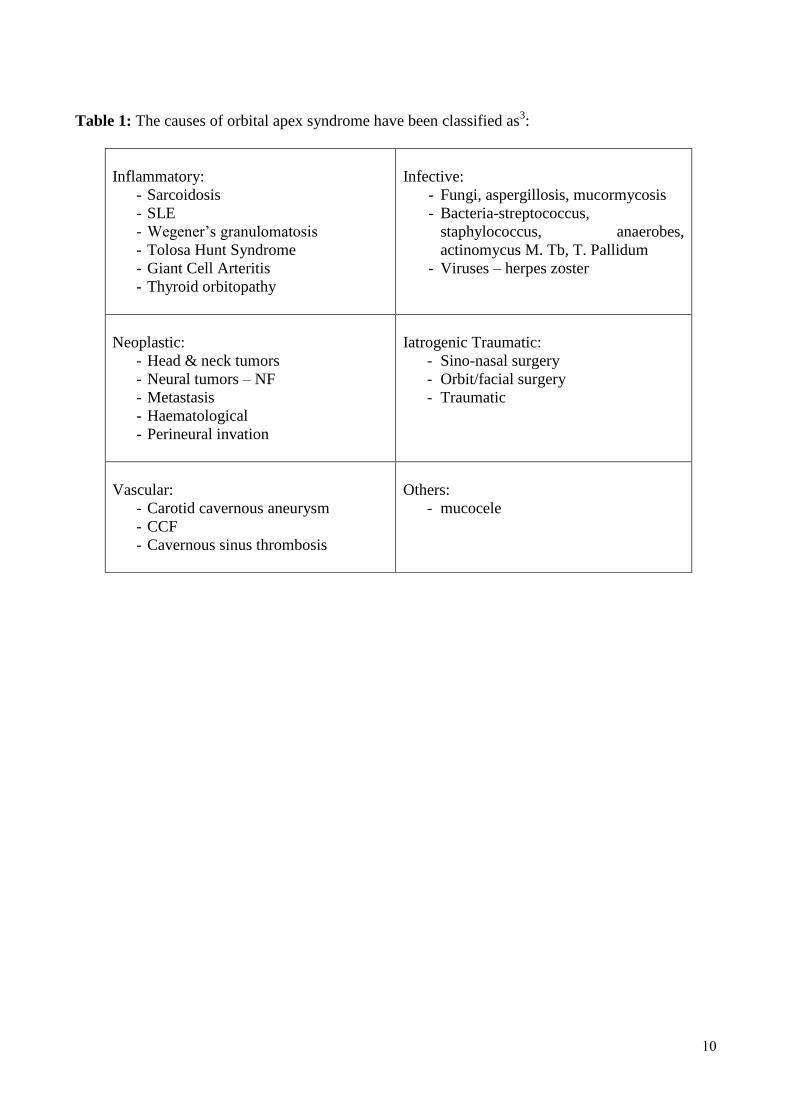
orbital apex syndrome. The causes of OAS are described in Table 1.
There is no definitive documented cause for all the signs and symptoms that have been encountered
in this particular case. J. Awad et.al[4]
, postulated that when epinephrine was injected under pressure
into the tissue surrounding the inferior turbinate, there will be retrograde flow through the anterior
6
ethmoidal artery into the ophthalmic artery, which causes likely vasospasm of the end arteries to the
optic nerve and retina. This hypo perfusion induces the patient’s optic neuropathy and unfortunately
there is no treatment available in the late stages, even with corticosteroids and vasodilators as it is
an ischemic (not an inflammatory) cause for the patient’s visual loss and therefore, corticosteroids
will not help. The most common probable reasons have been involvement of the retinal artery or the
caveneous sinus[5.6]
. Cavernous sinus involvement is through the possibility of retrograde flow of
the epinephrine during forceful injection through the valve less angular veins and the ophthalmic
veins to the cavernous sinus which could lead to cavernous sinus thrombosis or vasoconstriction in
the venous system which would in turn lead to hypo-perfusion in the arterial system.
Elective rhinoplasty is a common procedure. Although these have been several documented
complications for this procedure[1]
. Blindness as a complication post elective rhinoplasty has only
been reported once[2]
. Vascular insult was proposed by Cheney et al[3]
secondary to retrograde flow
of vasoconstrictor agent in blood flow as a result of forceful injection in the septal region.
Dubach et. Al[7]
showed histologically that forceful infiltration will flow into the blood vessels and
not be restricted to subperichondrial plane as intended by hydrodissection.
In our case, forceful injection for hydrodissection could have leaked into the vascular channels. This
has caused narrowing of the cavernous sinus. Another contributing factr is the hypotensive
anaesthesia during the surgery. This theory has been documented by MRV which showed an
element of venous involvement in the superior ophthalmic vein and cavernous sinus. This is the first
case in literature documented by MRV. Although all measures were taken by anti-inflammatory and
anti-coagulant therapy the sequels of vascular insult cannot be avoided. Unfortunately it is an
7
irreversible condition. A subclinical cavernous sinus thrombophelbitis prior to surgery however
could not be ruled out.
CONCLUSION
This case is a result of vascular insult as evident by MRV. In contrast to complications arising from
direct mechanical trauma vascular problems may be very difficult to prevent or predict. However it
is reasonable to make the following recommendations. (1) vasoconstrictive agents should be used in
as small doses as possible; (2) the injection of vasoconstrictor should be performed slowly with low
pressure; (3) hydrodissection using normal saline instead of vasoconstrictive agents; (4) the patient
should be closely observed in the postoperative period for at least 24 hours; 5) blindness, although
very rare, should be informed in the consent for septhorhinoplasty.
ACKNOWLEDGEMENT
I would like to acknowledge Dr. Yasin. S .Subhan and Dr. Tareq Al Otaibi for their role in the
research of this article.
FUNDING
Not sponsored in the study design, in the collection, analysis and interpretation of data; in the
writing of the manuscript; and in the decision to submit the manuscript for publication.
8
REFERENCES
1. Rettinger G., GMS Current Topics in ORL-HNS 2007. 2007; 6 Doc.08.
2. Wind J., Blindness as a Complication of Rhinoplasty. Arch Otolaryngology Head Neck
Surg; 1988 July, 114(5): 581.
3. Cheney ML., Blair PA., Blindness as a Complication of Rhinoplasty. Arch Otolaryngology
Head Neck Surg; 1987 July, 113(7): 768-769.
4. J. Awad, A. Awad, Y. Wong, S. Thomas, Unilateral Visual Loss after a Nasal Airway
Surgery. Clin Med Insights Case Rep. 2013; 6: 119–123. Published online 2013 June 25.
5. G. Nageswar Rao, Khageswar Rout, and Arttatrana Pal. Central Retinal Artery Occlusion
and Third Cranial Nerve Palsy Following Nasal Septoplasty. Case Rep OOphthalmol. 2012
Sep-Dec; 3(3): 321-326.
6. Rettinger G., Christ P., Meythales FH., Blindness Caused by Central Retinal Artery
Occlusion Following Nasal Septum Correction. HND 1990; 38:105-109.
7. P. Dubach, G. Mantokoudis, Y. Bans, G. Herrmann, M. Caversaccio. Hydrodissection for
subperichondrial septoplasty – an experimental anatomical study. Rhinology,48, 195-200,
2010.

9
FIGURES AND LEGEND
Fig 1. Fig 2.
Fig. 1: Scan show obstruction in the vessel on the affected side (red arrow) as compared to the
normal vessel as the opposite side (green arrow).
Fig. 2: MRV shows the typical bead sign/hour glass appearance of the superior ophthalmic vein due
to surrounding soft tissue oedema.
11
Table 1: The causes of orbital apex syndrome have been classified as3:
Inflammatory:
- Sarcoidosis
- SLE
- Wegener’s granulomatosis
- Tolosa Hunt Syndrome
- Giant Cell Arteritis
- Thyroid orbitopathy
Infective:
- Fungi, aspergillosis, mucormycosis
- Bacteria-streptococcus,
staphylococcus, anaerobes,
actinomycus M. Tb, T. Pallidum
- Viruses – herpes zoster
Neoplastic:
- Head & neck tumors
- Neural tumors – NF
- Metastasis
- Haematological
- Perineural invation
Iatrogenic Traumatic:
- Sino-nasal surgery
- Orbit/facial surgery
- Traumatic
Vascular:
- Carotid cavernous aneurysm
- CCF
- Cavernous sinus thrombosis
Others:
- mucocele