A prospective observational study to determine the...
95
1 A prospective observational study to determine the incidence of perioperative complications during percutaneous nephrolithotomy (PCNL) surgery and the various risk factors predisposing to them.
Transcript of A prospective observational study to determine the...

1
A prospective observational study to determine the
incidence of perioperative complications during
percutaneous nephrolithotomy (PCNL) surgery and the
various risk factors predisposing to them.

2
A DISSERTATION SUBMITTED IN PARTIAL FULFILLMENT OF
THE RULES AND REGULATIONS FOR THE MD BRANCH X,
ANAESTHESIA EXAMINATION OF THE TAMILNADU
DR. M.G.R. MEDICAL UNIVERSITY, TO BE HELD IN AUGUST 2010.

3
Certificate of Bonafide work
C E R T I F I C A T E
This is to certify that the dissertation entitled “A prospective observational
study to determine the incidence of perioperative complications during
percutaneous nephrolithotomy (PCNL) surgery and the various risk
factors predisposing to them” is the bonafide original work of Dr. Suma
Mary Thampi towards the M.D. Branch-X (Anaesthesia) Degree
Examination of the Tamil Nadu Dr. M.G.R University, Chennai, to be
conducted in August , 2010.
Signature of the HOD Signature of the guide
Dr Sarah Ninan Prof and HOD, Dept of Anaesthesia, CMCH, Vellore Tamil Nadu
4
Acknowledgement
It gives me immense pleasure to express my heartfelt and profound sense of gratitude to
my respected teacher and guide, Dr. Sarah Ninan for her valuable suggestions,
meticulous guidance, support and encouragement in doing this study.
I am grateful to Dr. L. Jeslin, my co-investigator for all his help in conducting this study.
I am also grateful to all my colleagues in the Department of Anaesthesia for all the
support received in preparing this dissertation and throughout my two year course in
Anaesthesia.
I also thank Ms. Anitha and technical staff at the Department of Anaesthesia for their
kind assistance.
I would also like to thank Mr. Prasanna and the Department of Clinical Epidemiology
who helped me with the analysis of the data.
I am grateful to my husband and my parents for their moral support and encouragement
throughout my studies.
I am grateful to God for his blessings on this study for its smooth completion.
Last, but not the least, I thank all my patients for their co-operation in this study.

5
TABLE OF CONTENTS
Contents Page Numbers
1) AIMS AND OBJECTIVES 6
2) ABSTRACT 7
3) LITERATURE REVIEW 9
4) METHODOLOGY 41
5) RESULTS 45
6) DISCUSSION 66
7) LIMITATIONS 74
8) CONCLUSIONS 75
9) BIBLIOGRAPHY 76
10) ANNEXURE 81

6
AIMS AND OBJECTIVES OF THE STUDY
AIM:
To assess the peri-operative anaesthetic complications of Percutaneous Nephrolithotomy
(PCNL)
OBJECTIVES:
i) To detect the incidence of cardiovascular changes namely hypotension and arrhythmias
during the surgery
ii) To detect the incidence of hypothermia during the surgery
iii) To detect the incidence of bleeding requiring intra-operative blood transfusion
iv) To detect the incidence of acidosis resulting from the procedure
v) To detect the incidence of any other complication that may result due to the surgery
vi) To determine risk factors associated with each of the above complication
7
ABSTRACT
Background: Percutaneous nephrolithotomy (PCNL) is a relatively non-invasive
surgical alternative available for removal of kidney stones. The traditional approach to
renal stones was through open surgical procedures that required general anesthesia and
long convalescence. Although, PCNL is a minimal invasive technique, it carries a
potential risk of complications. These include bleeding, anaemia, hypotension,
hyponatremia, hypothermia, injury to vital organs like pleural tear bowel perforation,
infection, and septic shock.
PCNL is a very common surgery that is being carried out in our institution.
However, till date the there has been no study on the profile of complications that have
been observed perioperatively. In literature, though the listed complications do not
actually quantify their incidence. Considering the frequency of cases and complications
seen, we feel that it would be a very useful exercise, since knowing the relative incidence
of complications will help institute the appropriate monitoring techniques and so, in the
better perioperative management.
Methods: Data was collected on 60 patients who underwent elective PCNL surgery. This
included monitoring of blood pressure, heart rate, nasopharyngeal temperature,
saturation, end-tidal carbon-di-oxide, arterial blood gas (ABG), the volume and
temperature of irrigation fluid, the temperature in the operating. The collected data was

8
analyzed to find out the complications that occur during the procedure and to determine
their incidence and risk factors associated with them. Data was analyzed using SPSS
version 16. Chi-square test was the test of significance in the study. Odds Ratio was
calculated and a p-value less than 0.05 was considered statistically significant.
Results: A total of 60 patients were studied of whom 42 were females and 18 males.40
patients were ASA grade 1 and 20 were ASA grade 2.The incidence of hypothermia and
acidosis was 60% and 26.7%, respectively. The incidence of hypothermia and
temperature of the irrigating fluid had statistically significant association with a p-value
of 0.026.The incidence of acidosis was significantly associated with the volume of
irrigating fluid with a p-value 0.025.The risk of acidosis was increased with hypothermia
(p-value 0.02 and OR 7.00)
Conclusion: The most common complication observed intraoperatively was
hypothermia, followed by acidosis and sepsis. Other complications that occurred were
excessive bleeding and hydrothorax. Electrolytes imbalance and cardiovascular changes
were not observed as major complications. The most significant risk factor associated
with hypothermia was found to be the usage of cold irrigating fluid.

9
LITERATURE REVIEW

10
URETERIC CALCULI-AN INTRODUCTION
Urinary calculi are solid particles in the urinary system. They may cause pain,
nausea, vomiting, hematuria, and, possibly, chills and fever from secondary infection.
Diagnosis is based on urinalysis and radiological imaging, usually noncontrast helical
CT. Treatment is with analgesics, antibiotics for infection, and, sometimes,
extracorporeal shock wave lithotripsy or endoscopic procedures.
About 1/1000 adults is hospitalized annually in the US because of urinary
calculi, which are also found in about 1% of all autopsies. Up to 12% of men and 5% of
women will develop a urinary calculus by age 70. Calculi vary from microscopic
crystalline foci to calculi several centimeters in diameter.(Figure 1) A large calculus,
called staghorn calculus, can fill an entire renal calyceal system.
Etiology
About 85% of calculi in the US are composed of Ca, mainly Ca oxalate. 10%
are uric acid; 2% are cystine; and the remainder is Mg ammonium phosphate (struvite).
Pathophysiology
Urinary calculi may remain within the renal parenchyma or renal pelvis or be
passed into the ureter and bladder. During passage, calculi irritate the ureter and may
become lodged, obstructing urine flow and causing hydroureter and sometimes
hydronephrosis. Common areas of lodgment include the ureteropelvic junction, the distal
ureter (at the level of the iliac vessels), and the ureterovesical junction. Typically, a
calculus must have a diameter > 5 mm to become lodged. Calculi ≤ 5 mm are likely to
pass spontaneously.

11
Table 1 : Composition of Urinary Calculi. Composition Percentage of All Calculi Common Causes
Calcium oxalate 70
Hypercalciuria
Hyperparathyroidism
Hypocitruria
Renal tubular acidosis
Calcium phosphate
15
Hypercalciuria
Hyperparathyroidism
Hypocitruria
Renal tubular acidosis Cystine 2 Cystinuria Magnesium ammonium
phosphate (struvite) 3 UTI caused by urea-
splitting bacteria Uric acid 10 Hyperuricosuria
Increased urine acidity
Figure 1 : Pictorial representation of sites of renal and ureteric stones.

12
Even partial obstruction causes decreased glomerular filtration, which may persist
briefly after the calculus has passed. With hydronephrosis and elevated glomerular
pressure, renal blood flow declines, further worsening renal function. Generally,
however, permanent renal dysfunction occurs only after about 28 days of complete
obstruction.
Secondary infection can occur with long-standing obstruction, but most patients
with Ca-containing calculi do not have infected urine.
Symptoms and Signs
Even large calculi remaining in the renal parenchyma or renal pelvis are usually
asymptomatic unless they cause obstruction. Symptoms, such as severe pain, often
accompanied by nausea and vomiting, and sometimes gross hematuria, usually occur
when calculi pass into the ureter, cause obstruction, or both. Pain (renal colic) is of
variable intensity but is typically excruciating and intermittent, often occurs cyclically,
and lasts 20 to 60 min. Nausea and vomiting is common. Pain in the flank or kidney area
that radiates across the abdomen suggests upper ureteral or renal pelvic obstruction. Pain
that radiates along the course of the ureter into the genital region suggests lower ureteral
obstruction. Suprapubic pain along with urinary urgency and frequency suggests a distal
ureteral, ureterovesical, or bladder calculus.
On examination, patients may be in obvious extreme discomfort, often ashen
and diaphoretic. Patients with renal colic may be unable to lie still and may pace, writhe,
or constantly shift position. The abdomen may be somewhat tender on the affected side
as palpation increases pressure in the already-distended ureter, but peritoneal signs

13
(guarding, rebound, rigidity) are lacking. For some patients, the first symptom is
hematuria or either gravel or a calculus in the urine. Other patients may have symptoms
of a UTI, such as fever, dysuria, or cloudy or foul-smelling urine. For most stones < 2 cm
in diameter, ESWL may be the procedure of choice because morbidity as measured by
blood loss, pain, fever, and postoperative stay was significantly less with ESWL.
TREATMENT-OPTIONS AND INDICATIONS
Some stones < 2 cm in diameter should still be considered for PCNL.(TABLE
2A,2B and 2C). These include cystine stones > 1 cm in diameter (especially if multiple),
because cystine is not easily fragmented with ESWL. In addition, some stones residing in
dependent calices may be better candidates for PCNL. If the calyx is dilated, the
likelihood of residual fragments following ESWL is high [25]. Thus, if rendering the
patient entirely stone-free is a high priority (such as for struvite) PCNL should be
performed instead of ESWL.
Very few stones larger than 3 cm can be treated successfully with ESWL alone.
This includes most staghorn calculi. Paik et al, in their study, reported of 13 non-staghorn
cases in this category, 77070 required further treatment, only 29070 were rendered stone-
free, and 57070 had to be considered treatment failures. For these reasons, large stones
and especially staghorn calculi are initially best approached percutaneously.(1) The most
important indications of PCNL are stone size >2.5cm, resistance to ESWL, lower pole
calyx stones with long and thin infundibulum and oblique infundibulo-pelvic angle. (2)

14
TABLE 2A : TREATMENT RECOMMENDATIONS
TABLE 2B : TREATMENT RECOMMENDATIONS
TABLE 2C : TREATMENT RECOMMENDATIONS

15
Performing the percutaneous procedure first is advisable for a number of reasons.
Advances in ESWL and expertise in endourological procedures have diminished the role
of open surgery in the management of patients with renal and ureteral calculi. The
indications for open surgery in the 1990s included complex stone burden, failure of
ESWL or endourological treatment, anatomic abnormalities such as infundibular stenosis,
renal calyceal diverticulum or concomitant ureteropelvic junction obstruction requiring
surgery and morbid obesity.(1)
Indications for PCNL
-Renal calculi greater than 2.5 cms in diameter.
-stone with composition inappropriate for ESWL
-Complete or partial staghorn calculus.
-Renal malformations like, pelvi - ureteric junction, infundibular stenosis obstruction
-cystine calculi greater than 1.5 cm
-Failure of ESWL.
-Body habitus unsuitable for ESWL like morbid obesity.
An ideal patient for PCNL is one who has two functioning kidneys and 2 cm stone in one
of the kidneys with extra renal pelvis with mild to moderate hydronephrosis.
History of PCNL
The approach to upper urinary calculi has been revolutionized since the early
1980s with the introduction of ESWL and the popularization of PCNL. Percutaneous
stone extraction was first described in 1941 by Rupel and Brown after a surgical
nephrostomy was created(3). Subsequently, in the late 1970s, however, PCNL, a

16
minimally invasive technique for the treatment of renal calculi, was introduced.(4) PCNL
as a primary procedure was described by Fernstrom and Johannson in 1976(5). However,
PCNL was not widely accepted until the mid-1980s.(3, 5). Although this technique also
relied on general anesthesia, it allowed effective and safe removal of most renal and some
ureteral stones with shorter postoperative recovery times, and quickly became a widely
accepted treatment option for renal calculi(6). However, PCNL frequency diminished
with the introduction of extracorporeal shock wave lithotripsy (ESWL) in the early
1980s(7). In recent years, as clinical experience with ESWL revealed its limitations, the
role of PNL for treating urolithiasis was redefined (8, 9).
Today, ESWL represents the initial treatment modality in approximately 90% of
patients with urinary tract stones, and is done predominantly on an outpatient basis.
Although ESWL has eclipsed all other methods of treatment for urinary tract stones, there
are situations in which it may not be beneficial. Nearly 6% of patients experience an
inadequate ESWL result, and may require endourologic or operative stone removal.
Pregnancy and the presence of a coagulopathy are contraindications to ESWL. Large
calculi and patients with distal ureteral stricture or obstruction may not be candidates for
ESWL monotherapy. Patients with calcium oxalate monohydrate, calcium phosphate, or
cystine calculi, which are resistant to fracture with ESWL, may require alternate
techniques. PCNL alone or and open surgical intervention remain alternative treatment
options for these patients with ESWL.(4)

17
RECENT TRENDS
The approach to upper urinary tract stones has changed from the sandwich PCNL
and SWL combination therapy to PCNL monotherapy.(3) In recent years, evolving trends
in the endoscopic management of complex renal calculi have resulted in improved stone
free status of patients treated with PCNL at our institution.
One of the trends has been increased usage of upper-pole access to obtain better
visibility of the collecting system, as upper-pole access gives better alignment with the
renal axis. The other advantage of the upper-pole approach is that it gives better access to
the UPJ and the upper third of the ureter when necessary. In the event that the stone is in
a horseshoe kidney, upper-pole access is crucial, as the abnormal anatomic reflection of
the peritoneum results in bowel positioning around the lower pole, making lower-pole
access dangerous.
The second trend in technique has been that all percutaneous access is obtained by
the urologist as part of a single-stage procedure. The urologist is most familiar with
intrarenal anatomy and the capabilities and limitations of available surgical instruments.
Furthermore, if additional access is necessary, the urologist will not have to depend on
the availability of a radiologist to create a second access.
The third trend in the technique has been the use of flexible nephroscopes after the
rigid nephroscope with ultrasonic lithotripsy has reached its limits in removing the stone.
Flexible nephroscopy allows the urologist to inspect the entire collecting system as well
as to remove residual stone debris using various endourologic adjuncts such as stone-
retrieval baskets, graspers, wires, and high-pressure irrigants. On occasion, laser

18
lithotripsy has been used through the flexible nephroscope to fragment a larger residual
stone. Generally, residual stones >2 cm in transverse diameter would require an
additional access and tract dilation for removal.
The fourth trend in PCNL in our institution has been the increased usage of
secondary PCNL, performed at the time of nephrostomy tube removal, to ensure stone-
free status.
The fifth and last trend has been the decreased reliance on SWL after PCNL. With
more thorough stone removal by endoscopic means, the role of SWL after PCNL has
decreased significantly. In our earlier reported series,16 41.4% of our patients required
SWL post-PCNL, but in our current series, only 21% required SWL. This trend has
resulted in fewer procedures, shorter hospitalization, and less cost to our patients.(3)
ADVANTAGE S OF PCNL
Most urologists believe that this operation is better than open surgery due to
decreases the length of stay, less morbidity, less pain and more preserved kidney
function(10).When compared to traditional open stone surgery, PCNL is associated with
lower morbidity and mortality and heightened patient acceptance because of its less
invasive nature (11-13) This technique has been demonstrated to be applicable to the
removal of a wide variety of upper urinary tract calculi, including staghorn calculi(12,
14), and may also be utilized for the treatment of obstructive lesions in the upper urinary
tract such as ureteropelvic junction (UPJ) narrowing and ureteral strictures [13]. The
technique has been used to remove calculi from calyceal diverticula and allows for
simultaneous obliteration via fulguration of these cavities [14].

19
TECHNIQUE OF PCNL
Initial step of PCNL is cystoscopy and placement of an open ended or ureteric catheter on
the side of the stone and injecting dye which will give the configuration of the pelvi-
calyceal system. The patient is placed in prone position. Proper PCN tract is critical to the
success in removing stones. Two important considerations are safety and access. Always
use a posterolateral approach. Use middle or inferior calyx for pelvic or upper calyceal
stone or direct access to a calyceal diverticular stone. Do not make a tract directly into the
pelvis but to go through posterior calyx end on so that parenchymal hold helps in
maintaining the tract.
Standard Puncture (Fig. 2)
The puncture is made with needle and help of "C" Arm. Once the needle is in
pelvicalyceal system, a J tipped, Teflon coated movable core guide wire is negotiated into
the renal pelvis and across the pelviureteric Junction into the ureter, a second guide wire
called the "safety wire" is also passed into ureter, if wire can not be passed into ureter
than it can be coiled into the calyces so that the guide wire does not get pulled out of the
kidney accidentally.
Tract Dilatation (Fig. 3)
To check if guide wire is in proper position with the rigid portion of wire across the renal
parenchyma, fascia and the body layers. Dilatation need to be done under fluoroscopy to
see that it is along the guide wire and it should not be bent during the process of
dilatation. Several options are available to enlarge the tract to a size of about 34 FR.

20
Figure 2 : Standard Puncture
Figure 3 : Tract Dilatation

21
Sequential Amplatz dilators, telescopic metal dilators or high pressure balloon. Balloon
dilation is less traumatic and results in less bleeding but expensive. At the end Amplatz
sheath is positioned over the last dilator to the appropriate site as this will be the conduit
for further instrumentation.
Stone Removal (Figs. 4,5)
As with all types of surgery, a clear field is a prerequisite to definitive treatment, after the
dilatation visibility may be poor due to blood clots. Renal pelvis is flushed with irrigant
through the sheath or from below by open ended catheter. Once the stone is visible,
endoscopy and fluoroscopy are used to decide how it should be removed. If stone is small
it can be grasped with rigid forceps and extracted intact. Larger stones require
fragmentation before removal and various techniques are available - electrohydrolic,
ultrasonic and laser lithotripsy. Electrohydraulic is a bipolar probe that creates a spark
when fired, which will vaporize liquid, producing a gas bubble and shock wave that
fragments the calculi. Ultrasonic lithotripsy uses mechanical vibrations at a frequency
above 17 cycles/Sec or 17 KHZ. Vibrations are created by applying alternating current to
a ceramic crystal which then expands and contracts; this vibration is transmitted to the tip
of probe where it causes formation of bubbles and produce cavitation on contact with
stone. Suction on the back of the probe removes small fragments. Ultrasound probe does
not damage renal tissue if it accidentally touches it. These probes are rigid hence cannot
be used with flexible instruments.

22
Fig. 4: X-ray with Amplatz sheath in position.
Fig.5: Nephroscope

23
Laser lithotripsy: Recently Holmium laser has been used to fragment stone, due to its
cost it is not used in all the centers. Laser is capable of fragmenting even the hardest
calcium oxalate m monohydrate and cystine calculi. In some cases where the stone is
large or has extension into different calyces than additional tract may be required in about
15% of cases. Multiple tracts help to achieve stone free Kidney.
COMPLICATIONS OF PCNL ( Table 3,4)
PCNL has acceptably low morbidity. Michel et al presented a total
complication rate of up to 83% following PNL (15). Overall significant complications
associated with PCNL include acute loss of kidney, colon injury, hydrothorax,
pneumothorax, prolonged leak, sepsis, vascular injury, and has an estimated rate of
15%(7-27%,CI 95%)(16) Complications during or after PNL may be present with an
overall complication rate of
up to 83%, including extravasation (7.2%),transfusion (11.2–17.5%), and fever (21.0–
32.1%), whereas major complications, such as septicemia (0.3–4.7%) and colonic (0.2–
0.8%) or pleural injury (0.0–3.1%) are rare.(15) The most common minor complications
of PCNL are pain (49%), fever (30%), urinary infection (11%), and renal colic (4%).The
most common major complications of PCNL are septicemia (4.1%) and bleeding
requiring blood transfusion (2.7%)(17, 18)
Similarly, Tefekli et al, in a retrospective review of 811 PCNL reported a total of
255 perioperative complications (29.2%)(19) This is the first published series trying to
classify PNL complications according to the modified Clavien system.
The modified Clavien system(Table 3) has been proposed to grade perioperative
complications. According to the modified Clavien classification system, perioperative

24
complications were stratified into five grades. Grade 1 defined all events that, if left
untreated, would have a spontaneous resolution or needed a simple bedside intervention.
Grade 2 complications required specific medication, including antibiotics and blood
transfusion. Grade 3 complications necessitated surgical, endoscopic, or radiological
intervention (3a without general anesthesia, 3b under general anesthesia). Neighboring
organ injuries and organ failures were classified as grade 4, and death was considered a
grade 5 complication (19, 20)
Table 3:Classification of surgical complications according to the modified Clavien system Grade 1 : Any deviation from the normal postoperative course without the need for pharmacologic treatment or surgical, endoscopic, and radiological interventions. Allowed therapeutic regimens are drugs as antiemetics, antipyretics, analgesics, diuretics, electrolytes, and physiotherapy. This grade also includes wound infections opened at the bedside. Grade 2 : Complications requiring pharmacologic treatment with drugs other than such allowed for grade 1 complications. Blood transfusions and total parenteral nutrition are also included. Grade 3 : Complications requiring surgical, endoscopic, or radiological intervention. Grade 3a : Intervention not under general anesthesia Grade 3b : Intervention under general anesthesia Grade 4 : Life-threatening complications (including central nervous system complications) requiring intensive care unit stay Grade 4a: Single organ dysfunction (including dialysis) Grade 4b: Multiorgan dysfunction Grade 5 : Death of the patient

25
Table 4 Complications during PCNL.
Sepsis
Incidence
In the immediate postoperative period after percutaneous stone removal, body
temperature elevations are common and often attributed to the release of inflammatory
mediators. Fever is one of the most worrisome and serious complications of PCNL
because of the high possibility of bacteremia or endotoxemia. Postoperative fever defined
the body temperature ‡38_C persisting still after 48 h
postoperatively (21).The incidence of fever has been reported as21–32.1% of the
cases (15)
In the setting of percutaneous nephrolithotomy (PCNL), concern is heightened
because bacteremia can be induced by the surgical manipulation and stone

26
fragmentation.(22) The incidence of postoperative fever and infection after PCNL has
been well documented
in various studies. In these studies, the postoperative fever and bacteriuria was reported
between 20–35% and 0–19%, respectively(22, 23) In fact, reports of both postoperative
fevers and bacteremia are as high as 37% in some series(24, 25).However, some series
report a much lower incidence of sepsis ranging from 0.3% to 4.3%(15, 26)
Diagnosis of sepsis
Bacteremia is the presence of pathogenic microorganisms in the blood, which can
lead to septicemia, which is the clinical syndrome caused by bacterial infection in the
blood, confirmed by positive blood cultures, and accompanied by systemic response to
the infection. SIRS is defined by at least two of the following:
-fever (>38c) or hypothermia (<36c)
-tachycardia >90bpm on non- beta blocked patients
-tachypnoea > 20 respiratory rate or PaCO2 >
-WBC count >12,000/mm3 or <4000/ mm3 or >10% band forms (oxford hand book of
urology)
Pathophysiology

27
The source of infection always comes from the stone itself(27). The incidence of
fever is significantly higher in cases with infected urinary stones than in those with sterile
stones (28).Most fever developed within 24 hours following the operation although all of
the patients had preoperative and postoperative prophylaxis antibiotics. Predisposing
factors to fever and sepsis include a preexisting untreated UTI, renal insufficiency,
sturvite or staghorn calculi, long-lasting operation and high amount or high pressure of
irrigation fluid used during PNL.(29)
Rao et al confirmed a statistically significant association between pre-operative
bacteriuria and the development of postoperative bacteraemia (bacteraemia in 37% of
patients with a positive MSSU compared to 8% in those with a negative urine culture pre-
operatively)(24, 30)The degree of bleeding from PCNL showed more injured renal
parenchyma or tear of vessels that increased the risk of bacteremia or endotoxemia.
Leakage of infected fluid from the kidney into the retro peritoneum can also be
reabsorbed slowly back into circulation and may cause infection in postoperative
period(31, 32)
Bleeding
Incidence
Bleeding is a major concern during percutaneous nephrolithotomy (PCNL),
especially with the use of multiple tracts. Although most bleeding associated with PCNL

28
can be managed conservatively, approximately 0.8% of patients require
angioembolization to control intractable bleeding (18, 33)
Etiology
The causes of bleeding were direct injury to blood vessels, laceration of the kidney
during operation especially with the technique of percutaneous tract creation(34),multiple
nephrostomy tracts and size of the tract as reported by Kukreja R (34, 35) Injury to blood
vessels could occur at anytime so intraoperative bleeding in the presented patients
seemed to relate with intraoperative hypothermia, duration of operation and volume of
irrigation fluid.(26) Excessive bleeding can occur during needle passage, tract dilation,
and nephroscopy or during the postoperative period.
Stoller et al, in their study on 127 PCNL patients estimated that the average blood
loss for uncomplicated 1-stage single puncture percutaneous nephrolithotomy was 2.8
gm./dl. Kukreja et al reported an average hemoglobin drop of 1.68 +/- 1.23
g/dLhemoglobin(36). Blood transfusions and a postoperative decrease in hemoglobin
level were combined to estimate total blood loss. Multiple punctures and/or renal pelvic
perforation were associated with a 2-fold greater blood loss. Half of the expected blood
loss occurred in patients with a preexisting nephrostomy tract. Diabetes, multiple-tract
procedures, prolonged operative time, and the occurrence of intraoperative complications
are associated with significantly increased blood loss.(34).Calculus morphology, location,
composition and length did not affect total blood loss, nor did the number of fragments or
stone-containing calices. Other factors, such as puncture site, type of fascial dilation,
hypertension, renal insufficiency, infection, previous open renal surgery or previous

29
extracorporeal shock wave lithotripsy, also did not affect total estimated blood loss.
Factors such as age, hypertension, renal insufficiency, urinary infection, the degree of
hydronephrosis, stone bulk, and the function of the ipsilateral renal unit did not have any
effect on the blood loss (34, 36)
Associated arteriosclerosis in patients with diabetes and hypertension may make
such patients more prone to bleeding after the initial trauma of tract formation. However,
hypertension was not associated with hemorrhage in our series. Although multivariate
analysis revealed that the presence of diabetes caused significant blood loss, univariate
analysis did not reveal any statistically significant difference between those with and
without diabetes. This paradox can be explained by either the small number of diabetic
patients in our series or the interaction of diabetes with other significant variables on
multivariate analysis.(18)
Bleeding during PCNL results from injury to the renal vessels. Venous bleeding
occurring during these procedures can usually be eradicated by transient balloon
tamponade of the tract. Arterial bleeding presents with different characteristics than
venous bleeding. The reported incidence of arterial injuries ranges from 0.9% to 3% after
percutaneous procedures.(37).If arterial bleeding does not subside after tamponade
measures, these patients can be treated successfully with angioembolization.
Blood transfusion

30
The rate of blood transfusion has been previously reported in various studies with
data ranging from 7%–23% (34, 36, 38). Stoller et al reported an incidence of 4% in
non-anemic patients(36). Srirangam et al supported the principle that patients having
multiple punctures are more likely to require a blood transfusion.(30, 34) . Preoperative
hemoglobin, multiple tracts, stone size, and total blood loss were significant in predicting
perioperative blood transfusion requirement (34, 36) On the basis of this evidence,
maneuvers that may reduce blood loss and transfusion rate include ultrasound-guided
access, use of Amplatz or balloon dilatation systems, reducing the operative time, and
staging the procedure in cases of a large stone burden or intraoperative complications(34)
Puncture site
The site of kidney puncture is a vital part of a successful PCNL. The choice of
access tract is based on the ability to provide good visibility of the stone bearing area and
a point of entry with minimal risk of injury to adjacent organs. Additionally, the access
tract should provide a trajectory projecting without torque or angulation into the
infundibulum and renal pelvis.(39) Typically, a subcostal puncture is used; however, in
certain circumstances, a supracostal approach may be desirable for improved access, e.g.
with staghorn and proximal ureteric stones Access via the superior posterior calyx offers
optimal exposure to staghorn calculi as well as multiple calculi in the superior and
inferior calyceal groups, renal pelvis, and upper ureter, and is therefore generally
preferred by urologists (Figures-2 and 3).However, prior publications on the subject have
suggested a higher rate of complications with 11th and 12th intercostal approach (39, 40)
. The drawback of supracostal punctures is an increased incidence of intrathoracic
complications as well as a higher rate of spleen and hepatic injury (41,42). Supracostal

31
approach is favorable in the therapy of upper calices calculi, but the risk of pneumothorax
and hydrothorax should be considered. Hydrothorax and pneumothorax can be hard to
recognize in these patients mainly because of the prone position(43)
Access to the posterior upper pole calyx affords an almost straight path to the
renal pelvis, upper ureter and both anterior and posterior inferior calyceal groups
(Figures-1 to 4). Even the posterior interpolar calyx may be accessible via this path
without significant angulation.(39) Access via the posterior superior calyceal group
makes calculi in the renal pelvis, upper ureter, and anterior and posterior inferior calyceal
groups accessible, and thus makes this an almost universal access route. Only
the superior anterior calyceal group and anterior and sometimes posterior interpolar
calyceal groups cannot be reached easily via this entry and hence may mandate
separate punctures and access routes if calculi are harbored in these regions(44, 45) .
Lang et al,in their study on routes of access, reported that access to the upper pole by the
intercostal route resulted in 1 pneumothorax, 1 arterio-calyceal fistula and 3 AV fistulae
in 111 patients (Table-3). Via a subcostal access route, we recorded 2 pneumothoraces, 1
AV fistula, 1 pseudoaneurysm, 1 ruptured UPJ, and 4 perforated ureters in 119 patients
(Table-5). The ratio of complication to no complication was significant (p = 0.0395). A
high incidence of atelectasis (n = 13) in the subcostal access group was also noted
(Table-6).(39)
Intercostal access via the posterior superior calyx offers the best trajectory via
infundibulum to pelvis, UPJ and inferior calyx. The supra 11th rib approach has a
particularly high rate of complications. Munver et al. reviewed their complications from
supracostal punctures for PNL access in 240 patients. The overall complication rate for

32
supracostal access tracts was 16.3% compared with 4.5% for infracostal access.
Punctures above the 11th rib resulted in a tremendously higher intrathoracic complication
rate (34.6%) compared to the supra 12th rib access (1.4%), a fact that corroborates the
strategy of avoiding this high approach if possible Pneumo and hemothorax, and
calyceal-pleural fistulae have been reported in up to 23.1% (46) The possibility of both a
transthoracic and transpleural trajectory of this type of access tract, despite attempts to
attain a high position of the lung by puncturing during the expiration phase, predisposes
to these complications(40, 46) . The incidence of hydropneumothorax occurring with
intercostal access has been reported at a rate of 4% to 15.3 %, with subcostal access, 0%
to 1.4% (46, 47). Similarly, large pleural effusions were reported in 8% to 12.5% with
intercostals approach, but virtually absent with subcostal access (40, 45, 47) . Moreover,
on the basis of anatomic considerations, the intercostal access route might have a higher
chance for injury to anterior segmental vessels or even anterior and posterior divisional
arteries(48)

33
Table 5: Major complications at various access sites.
Table 6: Minor complications at various access sites.

34
Fluid absorption
Mechanism
The procedure of PCNL requires the use of large amounts of irrigating fluids
continuously. Systemic absorption of this fluid can occur when there is disruption of the
pelvicaliceal system(49).Absorption can also occur via the vessels that open up during
tract dilatation or also during stone disintegration. Another route for fluid absorption is
leakage of fluid into peritoneal space(50).
Estimation ( Table 7)
Fluid absorption can be estimated by measuring the expired breath ethanol
concentration (EBEC) with the help of Alcosensor, a device that is directly connected to
the endotracheal tube. EBEC can be converted to the amount of irrigating fluid absorbed
using a standard formula(32).Routine hemogram and electrolyte examination will also
help in assessing the amount of fluid absorbed.(32)

35
Table 7 :Formula for calculating the irrigating fluid absorbed.

36
Factors affecting fluid absorption.
The volume of fluid absorbed increases with the amount of irrigation and
duration of procedure. Placement of an Amplatz sheath with subsequent reduced pressure
in the pelvicaliceal system reduced the amount of fluid absorption.(32)
Malhotra SK, et al reported that fluid absorption occurred in 78% of the PCNL
patients and 28% had volume absorption more than 1 liter. The study also showed that
volume of fluid absorption depended on volume of irrigation fluid, irrigation time and
rate of irrigation. Mean of fluid absorption was 696.7 + 603 (0-1916.2) ml, absorption
volumes were over 1 liter when the patients received more than 20 liters of irrigation
fluid(51)
Methods to reduce fluid absorption include reducing the height of the irrigating
bag, and use of Amplatz open drainage sheath to keep the pelvicaliceal pressure
low.Large stones, if done in a staged manner also reduce the amount of fluid
absorbed(49).
Hypothermia
Hypothermia is defined as a core temperature of < 36o C. Normal core is
temperature is 37 C; maintained within +0.2C (interthreshold range) by an efficient
thermoregulatory system; sweating at upper end and vasoconstriction at lower end.
37
Peripheral temperature of the limbs and skin is considerably less than the core; the
difference maintained by vasoconstriction. Anaesthesia adversely affects system and
interthreshold range increases from 0.2 to 4o C & patient cools. It mainly results from
anaesthetic-induced inhibition of thermoregulatory control and exposure to cold
operating room environment. High volume fluid administration also accelerate loss of
heat to the environment(52).
Hypothermia may be classified as mild (32°C – 35°C), moderate (28°C – 32°C)
and severe (<28°C) (52, 53)
Perioperative hypothermia develops in three distinct phases: (1) anaesthetic-
induced vasodilatation during induction of anaesthesia results in core-to-peripheral
redistribution of body heat and decreases core temperature 1–1.5°C during the first hour
of general anaesthesia; (2) subsequently core temperature decreases linearly as heat loss
to the environment exceeds metabolic heat production; (3) after 3–5 h of anaesthesia,
core temperature often stops decreasing. This core temperature plateau results from
reactivation of thermoregulatory vasoconstriction which decreases cutaneous heat loss
and constrains metabolic heat to the core thermal compartment.
Using a high volume of room temperature irrigation fluids could produce heat
loss in the presented patients by some intravascular absorption through open injured vein
and some from soaking internal organs after leakage into the perinephric space. Forced
air warming blankets did not reduce hypothermia due to the small area of effective
contact with the bodies. the major side effect of hypothermia in the presented patients
was slow recovery from anesthesia and muscle relaxant.(26) Hypothermia might also
38
cause postoperative shivering, cardiovascular complication, wound healing, infection and
bleeding (54, 55)
Cardiovascular changes
Vorrakitpokatorn et al reported cardiovascular changes (more than + 20% of the
base line) in 57.1% of the patients during the perioperative period(26). Hypertension
caused by hypervolemia from irrigation fluid absorption and/or from hormonal changes
was found in 29.4% (51, 56). The report of Atici et al showed significant increase in
ACTH, renin and aldosterone during surgery of PCNL that might cause an increase in
blood pressure(57)
Organ injury
Pleural injury
Vorrakitpokatorn et al found 3/128 (2.3%) of the patients developed dyspnea in
the early postoperative period. All of these patients were suspected pleural tear by the
surgeon intraoperatively(26). In another study to assess retrospectively the safety and
efficacy of the supracostal approach in percutaneous nephrolithotomy (PCNL) by Kekre
et al, among 862 patients who underwent PCNL, supracostal puncture was performed in
102 patients. They reported an incidence of 9.8% of pleural violation in the form of

39
hydrothorax, pneumothorax, or hydropneumothorax. All of these patients were managed
successfully by intercostal chest tube drainage(58)
Splenic injury
Splenic injury in the course of percutaneous nephrolithotomy is extremely rare. It
occurs in cases of stone in the left kidney (59).Shah et al reported two cases of splenic
injury that occurred during puncture of the 10th intercostal-space for PCNL. One of these
patients presented with hypotension on day 5 after discharge from the hospital. Both
patients needed emergency laparotomy, and one of them required splenectomy for
management of the injury(60).
Intestinal injuries
Iatrogenic colon injury is an uncommon but serious complication. Diagnosis is
sometimes delayed, and treatment strategies are still controversial, including conservative
management, colostomy, or primary repair. The incidence of intestinal injuries in PCNL
has been reported to vary from 0.35% to 1%.(61) High risk patients for colon injuries are
young, lean males with minimal retroperitoneal fat, in whom a retro renal colon is more
likely(62).
Electrolyte imbalances

40
Electrolyte imbalance in the postoperative period was reported as a minor
complication without statistical significance (p > 0.05) by Vorrakitpokatorn et al and not
related to the volume of irrigation fluid(26). Similar results were also observed by
Koroglu A et al, Atici S, et al and Moorthy et al (10, 57, 63). These mechanisms are still
not clear, they may be caused by complicated factors result from hormonal changes
during operation(57),homeostasis of the body or anything else that should be followed by
further study. Extravasation of irrigant fluid and infection, may predispose to electrolyte
imbalance which can be life threatening. Ghai et al reported a case of massive
extravasation of irrigant fluid producing severe metabolic acidosis, with life threatening
electrolyte imbalance(64)
An alternative reason for the low incidence of electrolyte imbalance maybe that
the irrigating fluid used in PCNL is normally saline since diathermy is rarely used, as
opposed to TURP, and as a result, electrolyte imbalances are less common than in
TURP(32).
,

41
MATERIALS AND METHODS
Type of study: A prospective cohort study.
Study setting: The study was conducted in Department of Anaesthesia, Christian
Medical College Hospital, Vellore, a 2500 bedded academic medical centre in South
India with an average of 1815 inpatients and approximately 4000 out- patient visits every
day.
Duration of study: November 2009 to March 2010
Inclusion criteria: All ASA Grade 1 and Grade 2 patients aged between 18 and 65 years
undergoing elective PCNL surgery in Christian Medical College, a tertiary care centre.
Exclusion criteria: Any patient age <17 yrs or >65 years or falling into ASA grade 3, 4, 5 or
undergoing another procedure along with PCNL
Methodology: A total of 60 patients were included in the study during the study period
from November 2009 to March 2010. Informed consent was taken from all patients.
Intraoperative monitoring of blood pressure, heart rate, nasopharyngeal temperature,
saturation, end-tidal carbon-di-oxide were done. These were observed prior to and during
the procedure till the end of the surgery. A baseline arterial blood gas (ABG) sample was
collected prior to start of the procedure for comparison of any changes that would occur
42
post operatively. Intra operatively, the volume and temperature of irrigation fluid used
was noted. The temperature in the operating room was also be noted during the
procedure. At the end of the surgery, patients’ temperature was noted and a repeat ABG
sample was collected. Changes in haematocrit, electrolytes, and lactates from baseline
were noted to assess blood loss, dilutional anaemia, dilutional hyponatremia, and
acidosis.
Sample size:
Was calculated based on the formula n = (4pq)/d2, where
p, the expected prevalence
q=100- p
and d, the precision of study.
The value of “p” was determined as 57 from a previous study reported in literature. The
precision of the study was fixed at 10. Applying the numbers, a sample size of 98 was
reached. However, only 60 patients were enrolled in the study.
Data analysis: Data entry was done using the Statistical Package for the Social Sciences
(SPSS) software package (version 16). Data is presented as mean + standard deviation,
frequency percentages or median and range. Descriptive statistics were calculated using
SPSS software. Univariate analysis was performed between categorical variables using
Chi-square test. Confidence intervals (CI) were calculated. Continuous variables were
compared using T-test. Odds ratio (OR) was presented as measures of risk. All reported p

43
values are two-sided and a ‘p’ value less than 0.05 was considered statistically
significant.
The study design and methods were approved by the Fluid Research Committee,
Christian Medical College, Vellore
Standardisation of anaesthesia: All patients were brought into the operating room and a
wide-bore peripheral intravenous access was established. After preoxygenation, all
patients were induced with 5mg/kg body wt of thiopentone, 2mg/kg body wt of fentanyl
and 1mg/kg body wt of vecuronium. Intubation was done using appropriate size cuffed
oral endotracheal tubes and fixed after confirming equal air entry. Anaesthesia was
maintained using a 50-50 mixture of air-O2 and isoflurane. Intra-operative analgesia was
using morphine upto 0.15mg/kg body wt. Fluid management was guided by fasting and
maintenance requirements, patient’s vital signs and the attending anaesthetist’s
assessment of bloodloss. At the end of the procedure anaesthetic gases were discontinued,
and patient reversed with neostigime and glycopyrrolate and extubated.
Data sources and measurements:
i) Arrhythmia: continuous three lead ECG monitoring was done during the procedure
and determined as defined above
ii) Hypotension: continuous invasive arterial blood pressure monitoring was done during
the procedure
iii) Hypothermia: temperature was measured via a nasopharyngeal temperature probe
inserted after the induction of anaesthesia and kept in place till extubation

44
v) Sepsis: the parameters defining sepsis were monitored and a diagnosis of sepsis was
made if any two of the four criteria wevi) Blood loss was calculated from the difference
in hemoglobin calculated from the pre procedure baseline ABG and post procedure ABG
vii) Volume of irrigation fluid: was measured as number of bags of fluid used multiplied
by 2 litres, which is the volume of one bag
viii) Temperature of fluid used: was determined as cold or warmed fluids
Definitions:
i) Arrhythmia was defined as bradycardia: heart rate <60 bpm,
tachycardia: heart rate>100 bpm.
Or any other rhythm changes noted on ECG monitoring
ii) Hypotension was defined as a fall in mean blood pressure to less than 15% of the
baseline value.
iii) Hypothermia was defined as a nasopharyngeal temperature recording of < 35 °C.
iv) Sepsis was defined as when two or more of these criteria are present:
a)Body temperature <36°C or >38°C
b)Heart rate > 90 beats per minute
c)Tachypnoea > 20 breaths per minute; or, an arterial PCO2 < 32 mmHg
d) WBC less than 4000 cells/mm³ (4 x 10⁹ cells/L) or greater than 12,000
cells/mm³ (12 x 109 cells/L); or the presence of greater than 10% band forms
v) Blood loss: as amount of blood lost during the procedure.

45
RESULTS
1) Population Demographics:
Population Demographics (N=60)
Mean Age(SD)
39.9(10.53)
Males(%)
Females(%)
42(70%)
18(30%)
The mean age of the population studied was 39.90 years with a standard deviation +
10.53 years.
The patients ranged from a minimum age of 15 years to a maximum age of 65 years.
Of the 60 patients in the study, 18 were females (30%) and 42 were males(70%)

46
2) Co-morbidity Profile
40 out of 60 patients(66.7%) had no comorbid conditions existing.
The most common comorbid condition present was hypertension with 14 out of 60
patients(23.3%).The others were Type 2 Diabetes Mellitus 8 out of 60(13.3%),chronic
renal failure and bronhial asthma/COPD both with a prevalence of 3.3%(2 out of
60).None of the patients had Ischaemic heart disease.
16 patients had only any one comorbid illness while 4 patients had multiple comorbid
illness (presence of 2 or more than 2 comorbid illness) existing.
47
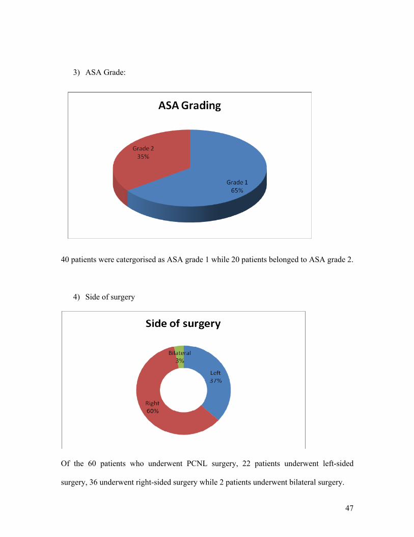
3) ASA Grade:
40 patients were catergorised as ASA grade 1 while 20 patients belonged to ASA grade 2.
4) Side of surgery
Of the 60 patients who underwent PCNL surgery, 22 patients underwent left-sided
surgery, 36 underwent right-sided surgery while 2 patients underwent bilateral surgery.

48
5) Staghorn Calculus
16 out of 60 patients were diagnosed to have staghorn calculus.
6) Temparature of Irrigating Fluid:
The irrigating fluid was warmed and used in 72% of the cases(43 out of 60),while cold
irrigating fluid was used in 28% of the cases(17 out of 60 patients)

49
7) Perioperative period characteristics.
Characteristic Mean(SD)
Mean Blood pressure(mmHg) 86.27(16.14)
Mean temperature(Celsius) 36.08(.41)
Mean heart rate(bpm) 82.27(16.48)
Mean Haematocrit(%) 38.38(7.68)
Mean Ambient temperature in
theatre(Celsius)
22.62(1.18)
Mean duration of surgery (hrs) 2.30(.833)
Mean volume of Intravenous fluid
used(litres)
1.52(.84)
Median volume of Irrigating fluid
used(litres)
24(13.87)
The mean baseline blood pressure was 86.27 mmHg with a std deviation of 16.14mmHg
and the mean baseline heart rate was 82.27bpm with a std deviation of 16.48bpm.The
mean baseline temperature was 36.08 degree Celsius with a standard deviation of
0.41degree Celsius.The mean haematocrit was 38.38 % with a standard deviation of
7.68%.The mean duration of surgery was 2.30 hours with a standard deviation of 0.83
hours.The mean volume of intravenous fluid used was 1.52 litres with a standard
deviation of 0.84.The volume of irrigating fluid used ranged from 6 litres to 84 litres with
a median value of 24 litres

50
B) OUTCOME MEASURES
Hypotension and Bradycardia
Of the 60 patients,only 7 patients patients had documented bradycardia
intraopeartively.None of the patients had and episodes of arrythmia.
On the other hand,39 out of 60 patients(65%) had a drop in blood pressure at some point
of time intraoperatively,while 21 patients (35%) never had any hypotension.

51
Of those who had intraoperative hypotension,the majority of patients,56.7%(34 of 60)had
hypotension after induction till the time of turning prone.Only 1 patient had hypotension
lasting through the duration of the surgery.4 patients had hypotension even after the
surgery was over till the time of extubation.
Blood requirement

52
6 out of 60 patients (10%) had recieved intraoperative transfusion,while 90% of
patients(54 out of 60) did not require transfusion .
Other Complications
The most common complication that occured intraoperatively was noted to be
hypothermia with 60%(36 out of 60) of patients becoming hypothermic during the
surgery.
The next most common complication was acidosis with 16 out of 60 aptients(26.7%)
observed to be acidotic by the end of the surgery.
53
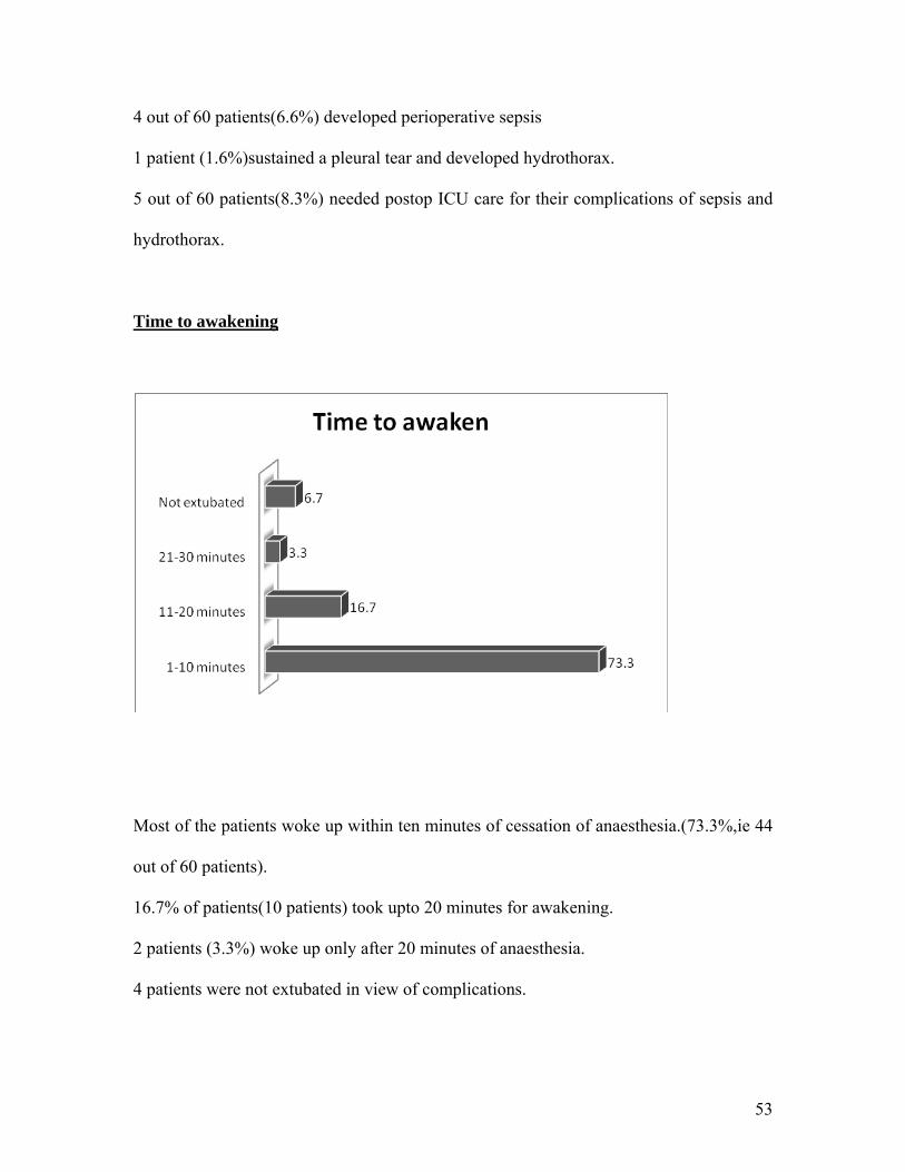
4 out of 60 patients(6.6%) developed perioperative sepsis
1 patient (1.6%)sustained a pleural tear and developed hydrothorax.
5 out of 60 patients(8.3%) needed postop ICU care for their complications of sepsis and
hydrothorax.
Time to awakening
Most of the patients woke up within ten minutes of cessation of anaesthesia.(73.3%,ie 44
out of 60 patients).
16.7% of patients(10 patients) took upto 20 minutes for awakening.
2 patients (3.3%) woke up only after 20 minutes of anaesthesia.
4 patients were not extubated in view of complications.

54
C) RISK FACTORS
I)HYPOTHERMIA
Hypothermia and CRF
Of 2 pateints who had Chronic Renal Failure,1 developed intraoperative hypothermia
while the other did not. As such,the results were not statistically different.p-value 0.76.
Hypothermia and Diabetes Mellitus
3
33
5
19
DM No DM
Hypothermia
No Hypothermia
Of 8 patients who had Diabetes, only 3 developed intra-operative hypothermia.
With a p-value of 0.163, this association was not significant.
1
35
1
23
0
10
20
30
40
CRF No CRF
Hypothermia
No Hypothermia

55
Hypothermia and duration of surgery
Hypothermia P value
Present Absent .077
Duration of
surgery(hours)
2 hrs 45 min 2 hrs 6 min
The mean duration of surgery in those patients who developed hypothermia was 2.45
hrs as compared to 2.06 hrs in those who did not. With a p-value of 0.077, this was not
significant.
Hypothermia and Temperature of Irrigating fluid
22
3
14
21
0
5
10
15
20
25
Hypothermia No hypothermia
Cold
Warm
Of 36 patients who developed hypothermia, 22 patients had been irrigated with cold
fluids. Of the 24 patients who were irrigated with warm fluid, only 3 developed
hypothermia. With a p-value of 0.026 and an Odds Ratio (OR) of 0.22, this result was
statistically significant; indicating that the risk of hypothermia was increased when cold
irrigating fluid was used as opposed to warm fluid.

56
Hypothermia and delayed awakening
0%
10%
20%
30%
40%
50%
60%
70%
80%
90%
100%
Hypothermia No Hypothermia
2816
55
1 12 2
Not extubated
21‐30 min
11‐20 min
1‐10 min
There was no statistically significant difference in the awakening time between those who
had hypothermia and those who did not. P-value 0.82
Hypothermia and volume of irrigating fluid
Hypothermia P value
Present Absent .083
Volume of
irrigating
fluid(Mean)
in Litres
29.42 23.08

57
The mean volume of irrigating fluid used in patients who had hypothermia was 29.42
litres while it was 23.08 litres in those who did not. With a p-value of 0.083, this was not
significant.
Hypothermia and ambient temperature
Hypothermia P value
Present Absent .193
Ambient
temperature
of theatre(°C)
22.46 22.87
The ambient temperature inside the operating theatre did not have any statistically
significant association with hypothermia, with a mean temperature of around 22°C in
those who had hypothermia as well as those who did not. p-value 0.193
Hypothermia and volume of IV fluid
Hypothermia P value
Present Absent .148
Total IV
fluid(Litres)
1.65 1.33
Though the total volume of Intravenous fluids administered was higher in those who
developed hypothermia when compared to those who did not, this observation was not
statistically significant. p-value 0.148

58
II) ACIDOSIS
Acidosis and CRF
1
15
1
43
CRF No CRF
Acidosis
No Acidosis
Of the 2 patients who had CRF,1 patient developed acidosis,while the other did
not.With a p-value of 0.45, this association was not significant.
Acidosis and Diabetes Mellitus
2
14
6
38
05
10152025303540
DM No DM
AcidosisNo Acidosis
There was no statistically significant association between DM and intra-operative
acidosis. p-value 0.9

59
Acidosis and duration of surgery
Acidosis No Acidosis P value
0.6 Mean
duration of
surgery(hrs)
2 hrs 39 min 2 hrs 26 min
There was no significant difference among those who developed acidosis and those who
did not with respect to the duration of surgery, with a mean duration of 2.39 hrs in the
former group and 2.26 hrs in the latter group. p-value 0.6
Acidosis and volume of irrigating fluid
Acidosis No Acidosis P value
.025 Volume of
irrigating
fluid(Mean)
in Litres
33.5 24.48
The mean volume of irrigating fluid was significantly higher in those patients who
developed acidosis (33.5 litres), as compared to those who did not develop acidosis (24.4
litres).This result was statistically significant. p-value 0.025

60
Acidosis and hypothermia
0
5
10
15
20
25
Hypothermia No Hypothermia
14
2
22 22
Acidosis
No Acidosis
Among the 16 patients who developed acidosis, 14 also had hypothermia. This
association was significant with a p-value of 0.02.An Odds Ratio of 7.00 indicated that
the presence of hypothermia was a risk factor for developing acidosis.
Acidosis and delayed awakening
0
5
10
15
20
25
30
35
Acidosis No Acidosis
11
33
3
7
1 13
1
1‐10min
11‐20 min
21‐30 min
Not Extubated
There was no statistically significant association between the presence or absence of
acidosis and the time to awaken after anaesthesia.p-value0.88

61
Acidosis and volume of IV fluid
0 0.5 1 1.5
Normal Saline
Ringer Lactate
Colloid
1.13
0.41
0.34
0.87
0.8
0.18
No Acidosis
Acidosis
The mean volume of intravenous fluid (IVF) administered in those patients who
developed acidosis was higher than in those who did not (1.65 litres and 1.33 litres
respectively). However, this difference was not statistically significant.
Comparing the different IV fluids, patients who were administered more Normal
Saline (NS) developed acidosis. This difference was statistically significant. P-value
0.01.Those who were given more NS-based colloids as opposed to Ringer Lactate
showed a trend towards significance in developing acidosis. p-value 0.09.
62
III) Sepsis
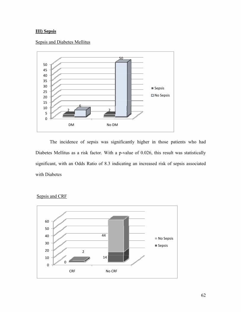
Sepsis and Diabetes Mellitus
05
101520253035404550
DM No DM
2 26
50
Sepsis
No Sepsis
The incidence of sepsis was significantly higher in those patients who had
Diabetes Mellitus as a risk factor. With a p-value of 0.026, this result was statistically
significant, with an Odds Ratio of 8.3 indicating an increased risk of sepsis associated
with Diabetes
Sepsis and CRF
0
10
20
30
40
50
60
CRF No CRF
2
140
44No Sepsis
Sepsis

63
The presence of CRF was a significant predictor in the development of sepsis.p-value
<0.01,with an Odds Ratio 28 indicating the increased risk of developing sepsis with CRF
Sepsis and stone size
Sepsis No sepsis P value
Mean stone
size(cm)
2.3 2.7 .59
There was no significant association between the size of the renal calculi and the
incidence of sepsis. The mean stone size was around 2.5 cm in both the groups. p-value
0.59
Sepsis and hypothermia
0
5
10
15
20
25
30
35
40
Hypothermia No Hypothermia
Sepsis
No Sepsis
There was no statistically significant association between sepsis and the presence or
absence of hypothermia.p-value 0.67
64
IV) Blood transfusion
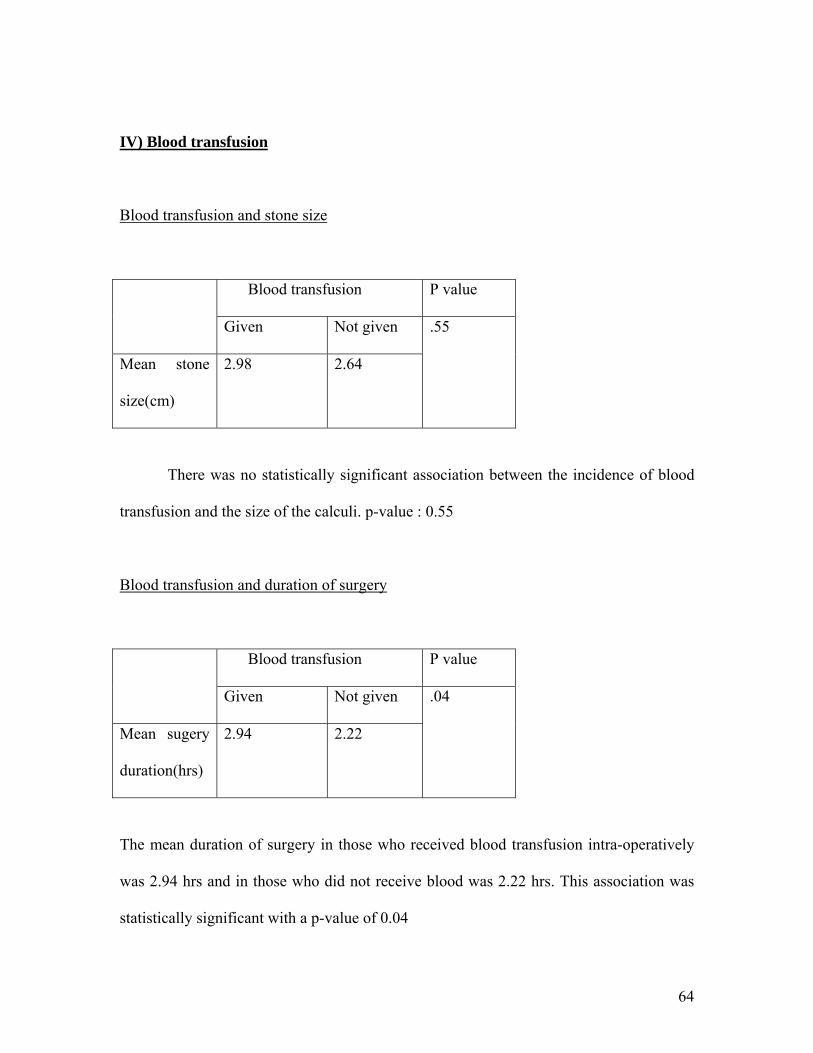
Blood transfusion and stone size
Blood transfusion P value
Given Not given .55
Mean stone
size(cm)
2.98 2.64
There was no statistically significant association between the incidence of blood
transfusion and the size of the calculi. p-value : 0.55
Blood transfusion and duration of surgery
Blood transfusion P value
Given Not given .04
Mean sugery
duration(hrs)
2.94 2.22
The mean duration of surgery in those who received blood transfusion intra-operatively
was 2.94 hrs and in those who did not receive blood was 2.22 hrs. This association was
statistically significant with a p-value of 0.04

65
Blood transfusion and baseline haematocrit
Blood transfusion P value
Given Not given <.01
Baseline hct
(%)
26.5 39.7
In the patients who received blood transfusion intra-operatively, the baseline haematocrit
was 26.5%.In those patients who did not receive a transfusion, it was 39.7%.This
association was statistically significant. p-value <0.01
The mean blood loss was 380ml (SD 78 ml)
The average drop in haematocrit in our study was 4.14 % (SD .64).

66
DISCUSSION
In the current day practice, Percutaneous Nephrolithotomy (PCNL) is a very common
treatment option available in the management of renal stones. Compared to the earlier
treatment option of open surgery, it is associated with less number of complications. This
leads to lesser rates of morbidity and lesser hospital stay, and thus is preferred to open
surgery.
However, PCNL procedure is not with out its own set of complications. All these
complications, though listed out in literature, have not very well been quantified. In our
institution, which is a tertiary care centre hospital, an average of 25 PCNL surgeries are
being done every month. In this scenario, we felt it would be worthwhile to know the
profile of complications and factors associated with them. This would help to institute
appropriate measures to try and limit their incidence and thus provide better quality
health care.
In our study, a total of 60 patients who underwent elective PCNL surgery was studied.
Majority of patients were ASA grade 1 and there was a predominance of males in the
study.

67
COMPLICATIONS OF PCNL
In our study the most common intraoperative complication encountered was hypothermia
and the second most common complication was acidosis.60% of our patients were
documented to have hypothermia and 26.7% of the patients had acidosis as evidenced by
an Arterial Blood Gas at the end of the procedure. Studies done earlier have not found
hypothermia or acidosis as a major complication.
In 2007, Michel et al presented a total complication rate of up to 83% following PCNL,
in which hypothermia or acidosis did not feature at all(15).They presented their findings
as major and minor complications, of which minor complications were more common.
Bleeding requiring transfusion was listed as the most common complication. Others
included insignificant bleeding, pain and fever(15).
Two other studies done in 1998 and 2007 by Havel et al and Turna et al respectively
project most common major complications of PCNL as septicemia (4.1%) and bleeding
requiring blood transfusion(2.7%)(17, 18)
HYPOTHERMIA
In our study, we found hypothermia to be the most common complication that occurred
during PCNL surgery. Our study had a 60% incidence of hypothermia. We found a
statistically significant positive correlation between the presence of hypothermia and the

68
temperature of irrigating fluid used. We also observed that there was a correlation
between the volume of irrigating fluid used and duration of surgery with hypothermia.
However these values were not statistically significant which could possibly be due to the
small number of sample size studied.
In 1997 a study done by Al-Shammare et al, also found a positive correlation between
hypothermia and the duration of surgery. They reported in their series of 9 children that,
when the duration of surgery exceeded 150 minutes, 2 children had hypothermia. In our
study the mean duration of surgery was 150 minutes (2.30 hrs).
We also compared the incidence of hypothermia with the ambient temperature in the
operating room. The mean ambient temperature in the operating room in our study was
22.6°C. However, we found no positive correlation between the two. This could probably
be because we took adequate measures to cover the patient well soon after induction of
anaesthesia to prevent losing body temperature to the environment. We also used forced
air warming devices. Inspite of this, the single most important factor that contributed to
patients developing hypothermia was the use of cold irrigating fluids.
We also compared the presence of hypothermia and the volume of intravenous fluid used.
There was no positive correlation. Presence of CRK or DM also did not seem to add any
further risk to the development of hypothermia. Even though hypothermia is known to
delay awakening from anaesthesia, in our study we found no positive correlation between

69
the two. One reason for this could be the time lines set for assessing time to awaken after
the discontinuation of anaesthetic gases.
SEPSIS
In our study, the incidence of sepsis was 6.6%.This is comparable to earlier done studies.
Michel et al in their series reported an incidence of 0.3%-4.7% of sepsis.
Vorrakitpokatorn et al have reported that in their series, they encountered sepsis with an
incidence of 0.9% to 4.7%. However some studies have reported a much higher
incidence. Rao et al, in their series has reported as high an incidence as 37% for
postoperative bacteremia (24).
In our study, we analysed the incidence of sepsis with respect to the presence of DM
preoperatively. We found that the presence of DM had a positive correlation to the
incidence of sepsis. In fact, the presence of DM tends to increase the risk of developing
sepsis (Odds Ratio 8.3).
On analysis of incidence of sepsis with respect to presence of CRF preoperatively, we
found a significant relationship between the two with the presence of CRF increasing the
risk for perioperative development of sepsis(OR 28).This is comparable to a study done
by Skolarikos et al, who described an increased incidence of sepsis with presence of
preoperative Type II DM and renal insufficiency(29).They also reported that increased
pressure of irrigating fluids increased the risk of sepsis.In our study we did not measure

70
the pressure of irrigating fluids. Rao et al also reported stone size and staghorn calculi to
increase the incidence of sepsis (24). However, in our study we did not find any positive
association between stone size and the incidence of sepsis.
HYPOTENSION
Earlier studies report cardiovascular changes of hypotension and hypertension as a major
complication of PCNL. Vorrakitpokatorn et al, in their series in 2006 reported an
incidence of 57.1% of intraoperative hypotension (26). In our study also, intraoperative
hypotension was found to have an incidence of 65%(39 out of 60 patients).However, on
further analysis, we found that the period when hypotension occurred most (in terms of
frequency and degree) was soon after induction of anaesthesia. 34 out of the 39 patients
had a drop in blood pressure readings during this time. This could be due to intravenous
and inhalational anaesthetic agents, as well as a lack of surgical stimulus. This therefore
cannot be considered as a major complication of PCNL surgery per se as this would
occur during any surgery under anaesthesia.
On the other hand, hypotension due to the PCNL procedure would be expected to occur
after the commencement of surgery and present till the end of surgery. The incidence of
this group in our study was 6.7%.These patients had significant hypotension and only
1.7% of these reached back to normal preoperative levels for extubation.

71
Hypertension has also been reported as a major cardiovascular event that occurred during
PCNL surgery. This has been attributed to the hypervolemia caused by absorption of
irrigation fluid. However in our study we did not find any significant hypertension in
patients at the end of the procedure.
BLEEDING AND BLOOD TRANSFUSION
Michel et al has reported the incidence of bleeding during PCNL surgery to vary from
0.6%-1.4% (15).In our study the blood loss was estimated from post-operative decrease
in haematocrit and blood transfusion. The mean blood loss estimated was 380 ml (SD 78
ml).The average drop in haematocrit in our study was 4.14 % (SD .64). This is similar to
previously done studies. Kukreja et al reported an average hemoglobin drop of 1.68 +/-
1.23 g / dL hemoglobin (36)
Most of the times, the bleeding that occurs during surgery have been managed
conservatively. The incidence of bleeding requiring transfusion has been reported to be
varying from 7%-23% in various studies (34, 36, 38).Michel et al reported an incidence
less than 8% their series (15). The transfusion rate in our study was higher. 10% of our
patients had received intraoperative blood transfusion. The rate of bleeding requiring
interventions has been reported to be less than 1%.Though 5% of our patients could not
have complete stone-free retrieval due to abandonment of surgery because of excessive
bleeding; none of them needed any intervention for it other than blood transfusion.

72
Kukreja et al reported the incidence of blood transfusion to be related to the duration of
surgery and intraoperative complications (34). We compared the incidence of blood
transfusion with the duration of surgery the stone size and baseline haematocrit. We
found statistically significant association between the incidence of blood transfusion and
the duration of surgery as well as baseline haematocrit value. However, there was no
significant association between size of the calculus and the incidences of blood
transfusion. These results are comparable to reports in literature.
PLEURAL TEAR
Pleural tear has been reported as a major complication of PCNL surgery. Michel et al,
reported an incidence of 2.3% - 3.1 %( 15). In our study we had 1 patient out of 60, ie;
1.6% who developed a hydrothorax after pleural injury due to a supracostal puncture
route. There were no patients who developed a pleural injury after subcostal puncture
route. This is comparable to studies done in literature which report a 0%-1.4% incidence
of hydrothorax or pneumothorax after subcostal access and a 4%-15% incidence with
supracostal route puncture. There was also no incidence of pleural effusion after
subcostal access.
OTHER ORGAN INJURY
Splenic laceration and bowel perforation are other visceral organ injuries listed as major
complications of PCNL procedure in literature. In our study, none of the patients had any

73
visceral organ injury. Splenic laceration has been reported to be very rare after PCNL
surgery and is reported to have increased likelihood after a 10th rib supracostal puncture.
The incidence of bowel perforation, namely colonic injuries have been reported to be
0.3%-0.8%

74
LIMITATIONS
1) The predominance of ASA grade 1 patients in the study itself would reduce the
incidence of morbidity associated with the surgery.
2) The recommended target of sample size could not be reached during the time period of
the study. This could have influenced the rate of complications in the study.
3) The amount of blood lost could not be estimated accurately because the loss is always
mixed with irrigation fluid. Replacement of blood loss was guided by visual assessment
of loss and by patient’s vital signs.

75
CONCLUSIONS
1) The most common complication observed intraoperatively was hypothermia, followed
by acidosis and sepsis.
2) The most significant risk factor associated with hypothermia was found to be the usage
of cold irrigating fluid..
3) The presence of hypothermia and increased use of intravenous Normal Saline
contributed to higher incidence of acidosis
4) Electrolytes imbalance and cardiovascular changes were not observed as major
complications.
5) An increased incidence of sepsis was seen in the presence of Diabetes Mellitus ad
renal insufficiency.
76
BIBLIOGRAPHY
1. Paik ML, Wainstein MA, Spirnak JP, Hampel N, Resnick MI. Current indications for open stone surgery in the treatment of renal and ureteral calculi. J Urol. 1998 Feb;159(2):374-8; discussion 8-9. 2. Falahatkar S KI, Roshan ZA, Golshahi M, Emadi SA. Decreasing the complications of PCNL with alternative techniques including complete supine PCNL and subcostal approach. Pak J Med Sci. 2009;25(3):353-8. 3. Wong MY. Evolving technique of percutaneous nephrolithotomy in a developing country: Singapore General Hospital experience. J Endourol. 1998 Oct;12(5):397-401. 4. Gravenstein D. Extracorporeal shock wave lithotripsy and percutaneous nephrolithotomy. Anesthesiol Clin North America. 2000 Dec;18(4):953-71. 5. Fernstrom I, Johansson B. Percutaneous pyelolithotomy. A new extraction technique. Scand J Urol Nephrol. 1976;10(3):257-9. 6. Mohta M, Bhagchandani T, Tyagi A, Pendse M, Sethi AK. Haemodynamic, electrolyte and metabolic changes during percutaneous nephrolithotomy. Int Urol Nephrol. 2008;40(2):477-82. 7. Chaussy C, Schmiedt E, Jocham D, Brendel W, Forssmann B, Walther V. First clinical experience with extracorporeally induced destruction of kidney stones by shock waves. J Urol. 1982 Mar;127(3):417-20. 8. Segura JW. The role of percutaneous surgery in renal and ureteral stone removal. J Urol. 1989 Mar;141(3 Pt 2):780-1. 9. Rassweiler J, Sentker L, Seemann O, Hatzinger M, Stock C, Frede T. Heilbronn laparoscopic radical prostatectomy. Technique and results after 100 cases. Eur Urol. 2001 Jul;40(1):54-64. 10. Koroglu A, Togal T, Cicek M, Kilic S, Ayas A, Ersoy MO. The effects of irrigation fluid volume and irrigation time on fluid electrolyte balance and hemodynamics in percutaneous nephrolithotripsy. Int Urol Nephrol. 2003;35(1):1-6. 11. Brannen GE, Bush WH, Correa RJ, Gibbons RP, Elder JS. Kidney stone removal: percutaneous versus surgical lithotomy. J Urol. 1985 Jan;133(1):6-12. 12. Clayman RV, Surya V, Miller RP, Castaneda-Zuniga WR, Smith AD, Hunter DH, et al. Percutaneous nephrolithotomy: extraction of renal and ureteral calculi from 100 patients. J Urol. 1984 May;131(5):868-71. 13. Preminger GM, Clayman RV, Hardeman SW, Franklin J, Curry T, Peters PC. Percutaneous nephrostolithotomy vs open surgery for renal calculi. A comparative study. JAMA. 1985 Aug 23-30;254(8):1054-8. 14. Segura JW, Patterson DE, LeRoy AJ, Williams HJ, Jr., Barrett DM, Benson RC, Jr., et al. Percutaneous removal of kidney stones: review of 1,000 cases. J Urol. 1985 Dec;134(6):1077-81. 15. Michel MS, Trojan L, Rassweiler JJ. Complications in percutaneous nephrolithotomy. Eur Urol. 2007 Apr;51(4):899-906; discussion
77
16. Vicentini FC, Gomes CM, Danilovic A, Neto EA, Mazzucchi E, Srougi M. Percutaneous nephrolithotomy: Current concepts. Indian J Urol. 2009 Jan;25(1):4-10. 17. Havel D, Saussine C, Fath C, Lang H, Faure F, Jacqmin D. Single stones of the lower pole of the kidney. Comparative results of extracorporeal shock wave lithotripsy and percutaneous nephrolithotomy. Eur Urol. 1998;33(4):396-400. 18. Turna B, Nazli O, Demiryoguran S, Mammadov R, Cal C. Percutaneous nephrolithotomy: variables that influence hemorrhage. Urology. 2007 Apr;69(4):603-7. 19. Tefekli A, Ali Karadag M, Tepeler K, Sari E, Berberoglu Y, Baykal M, et al. Classification of percutaneous nephrolithotomy complications using the modified clavien grading system: looking for a standard. Eur Urol. 2008 Jan;53(1):184-90. 20. Dindo D, Demartines N, Clavien PA. Classification of surgical complications: a new proposal with evaluation in a cohort of 6336 patients and results of a survey. Ann Surg. 2004 Aug;240(2):205-13. 21. O'Grady NP, Barie PS, Bartlett JG, Bleck T, Garvey G, Jacobi J, et al. Practice guidelines for evaluating new fever in critically ill adult patients. Task Force of the Society of Critical Care Medicine and the Infectious Diseases Society of America. Clin Infect Dis. 1998 May;26(5):1042-59. 22. Cadeddu JA, Chen R, Bishoff J, Micali S, Kumar A, Moore RG, et al. Clinical significance of fever after percutaneous nephrolithotomy. Urology. 1998 Jul;52(1):48-50. 23. Dogan HS, Sahin A, Cetinkaya Y, Akdogan B, Ozden E, Kendi S. Antibiotic prophylaxis in percutaneous nephrolithotomy: prospective study in 81 patients. J Endourol. 2002 Nov;16(9):649-53. 24. Rao PN, Dube DA, Weightman NC, Oppenheim BA, Morris J. Prediction of septicemia following endourological manipulation for stones in the upper urinary tract. J Urol. 1991 Oct;146(4):955-60. 25. Lee WJ, Smith AD, Cubelli V, Badlani GH, Lewin B, Vernace F, et al. Complications of percutaneous nephrolithotomy. AJR Am J Roentgenol. 1987 Jan;148(1):177-80. 26. Vorrakitpokatorn P, Permtongchuchai K, Raksamani EO, Phettongkam A. Perioperative complications and risk factors of percutaneous nephrolithotomy. J Med Assoc Thai. 2006 Jun;89(6):826-33. 27. Viville C. [Percutaneous nephrolithotomy (PCNL): evaluation of 250 PCNL by the same operator]. Prog Urol. 1993 Apr;3(2):238-51. 28. Takeuchi H, Ueda M, Nonomura M, Hida S, Oishi K, Higashi Y, et al. [Fever attack in percutaneous nephrolithotomy and transurethral ureterolithotripsy]. Hinyokika Kiyo. 1987 Sep;33(9):1357-63. 29. Skolarikos A, de la Rosette J. Prevention and treatment of complications following percutaneous nephrolithotomy. Curr Opin Urol. 2008 Mar;18(2):229-34. 30. Srirangam SJ, Darling R, Stopford M, Neilson D. Contemporary practice of percutaneous nephrolithotomy: review of practice in a single region of the UK. Ann R Coll Surg Engl. 2008 Jan;90(1):40-4. 31. Gehring H, Nahm W, Zimmermann K, Fornara P, Ocklitz E, Schmucker P. Irrigating fluid absorption during percutaneous nephrolithotripsy. Acta Anaesthesiol Scand. 1999 Mar;43(3):316-21. 32. Kukreja RA, Desai MR, Sabnis RB, Patel SH. Fluid absorption during percutaneous nephrolithotomy: does it matter? J Endourol. 2002 May;16(4):221-4.
78
33. Kessaris DN, Bellman GC, Pardalidis NP, Smith AG. Management of hemorrhage after percutaneous renal surgery. J Urol. 1995 Mar;153(3 Pt 1):604-8. 34. Kukreja R, Desai M, Patel S, Bapat S. Factors affecting blood loss during percutaneous nephrolithotomy: prospective study. J Endourol. 2004 Oct;18(8):715-22. 35. Davidoff R, Bellman GC. Influence of technique of percutaneous tract creation on incidence of renal hemorrhage. J Urol. 1997 Apr;157(4):1229-31. 36. Stoller ML, Wolf JS, Jr., St Lezin MA. Estimated blood loss and transfusion rates associated with percutaneous nephrolithotomy. J Urol. 1994 Dec;152(6 Pt 1):1977-81. 37. Patterson DE, Segura JW, LeRoy AJ, Benson RC, Jr., May G. The etiology and treatment of delayed bleeding following percutaneous lithotripsy. J Urol. 1985 Mar;133(3):447-51. 38. Stoller ML, Lee KL, Schwartz BF, Viele MK. Autologous blood use in percutaneous nephrolithotomy. Urology. 1999 Sep;54(3):444-9. 39. Lang EK, Thomas R, Davis R, Colon I, Cheung W, Sethi E, et al. Risks and benefits of the intercostal approach for percutaneous nephrolithotripsy. Int Braz J Urol. 2009 May-Jun;35(3):271-81; discussion 81-3. 40. Radecka E, Brehmer M, Holmgren K, Magnusson A. Complications associated with percutaneous nephrolithotripsy: supra- versus subcostal access. A retrospective study. Acta Radiol. 2003 Jul;44(4):447-51. 41. Robert M, Maubon A, Roux JO, Rouanet JP, Navratil H. Direct percutaneous approach to the upper pole of the kidney: MRI anatomy with assessment of the visceral risk. J Endourol. 1999 Feb;13(1):17-20. 42. Gupta R, Kumar A, Kapoor R, Srivastava A, Mandhani A. Prospective evaluation of safety and efficacy of the supracostal approach for percutaneous nephrolithotomy. BJU Int. 2002 Dec;90(9):809-13. 43. Munshi CA, Bardeen-Henschel A. Hydropneumothorax after percutaneous nephrolithotomy. Anesth Analg. 1985 Aug;64(8):840-2. 44. Hentschel H, Janitzky V, Weirich T. [Percutaneous nephrolithotomy - always effective and free of complications?]. Aktuelle Urol. 2007 May;38(3):232-6. 45. Sukumar S, Nair B, Ginil KP, Sanjeevan KV, Sanjay BH. Supracostal access for percutaneous nephrolithotomy: less morbid, more effective. Int Urol Nephrol. 2008;40(2):263-7. 46. Munver R, Delvecchio FC, Newman GE, Preminger GM. Critical analysis of supracostal access for percutaneous renal surgery. J Urol. 2001 Oct;166(4):1242-6. 47. Yadav R, Aron M, Gupta NP, Hemal AK, Seth A, Kolla SB. Safety of supracostal punctures for percutaneous renal surgery. Int J Urol. 2006 Oct;13(10):1267-70. 48. Sampaio FJ, Aragao AH. Anatomical relationship between the intrarenal arteries and the kidney collecting system. J Urol. 1990 Apr;143(4):679-81. 49. Rao PN. Fluid absorption during urological endoscopy. Br J Urol. 1987 Aug;60(2):93-9. 50. Sinclair JF, Hutchison A, Baraza R, Telfer AB. Absorption of 1.5% glycine after percutaneous ultrasonic lithotripsy for renal stone disease. Br Med J (Clin Res Ed). 1985 Sep 14;291(6497):691-2. 51. Malhotra SK, Khaitan A, Goswami AK, Gill KD, Dutta A. Monitoring of irrigation fluid absorption during percutaneous nephrolithotripsy: the use of 1% ethanol as a marker. Anaesthesia. 2001 Nov;56(11):1103-6.

79
52. Abelha FJ, Castro MA, Neves AM, Landeiro NM, Santos CC. Hypothermia in a surgical intensive care unit. BMC Anesthesiol. 2005 Jun 6;5:7. 53. Kempainen RR, Brunette DD. The evaluation and management of accidental hypothermia. Respir Care. 2004 Feb;49(2):192-205. 54. Sessler DI. Complications and treatment of mild hypothermia. Anesthesiology. 2001 Aug;95(2):531-43. 55. Cereda M, Maccioli GA. Intraoperative temperature monitoring. Int Anesthesiol Clin. 2004 Spring;42(2):41-54. 56. Konrad C, Gerber H, Schupfer G, Jenzer S, Schmucki O. Detection of fluid volume absorption by end-tidal alcohol monitoring in patients undergoing endoscopic renal pelvic surgery. J Clin Anesth. 1999 Aug;11(5):386-90. 57. Atici S, Zeren S, Aribogan A. Hormonal and hemodynamic changes during percutaneous nephrolithotomy. Int Urol Nephrol. 2001;32(3):311-4. 58. Kekre NS, Gopalakrishnan GG, Gupta GG, Abraham BN, Sharma E. Supracostal approach in percutaneous nephrolithotomy: experience with 102 cases. J Endourol. 2001 Oct;15(8):789-91. 59. Kondas J, Szentgyorgyi E, Vaczi L, Kiss A. Splenic injury: a rare complication of percutaneous nephrolithotomy. Int Urol Nephrol. 1994;26(4):399-404. 60. Shah HN, Hegde SS, Mahajan AP, Sodha H, Shah R, Bansal M. Splenic injury: rare complication of percutaneous nephrolithotomy: report of two cases with review of literature. J Endourol. 2007 Aug;21(8):919-22. 61. Korkes F, Lopes Neto AC, Lucio J, 2nd, Bezerra CA, Wroklawski ER. Management of colon injury after percutaneous renal surgery. J Endourol. 2009 Apr;23(4):569-73.

80
ANNEXURE I
PATIENT INFORMATION SHEET The surgery you are undergoing will be under general anaesthesia. Under anaesthesia, it is routine to
monitor your heart rate, blood pressure, saturation, etc to ensure a safe recovery.
A study is being conducted here wherein a few additional monitoring will be established along with the
routine ones to try and identify some complication that are expected from the sugery and anaesthesia.
The complications that are being studied include changes in heart rate, blood pressure, temperature and
sodium levels, bleeding injury to other organs and infection. This study is being done so that these
changes can be understood better and a better level of care can be given to all the patients undergoing
this procedure. For this additional monitoring of temperature and two blood samples need to be done
under anaesthesia, one at the beginning of the surgery and one at the end.
By enrolling in this study, there will be no potential risk or discomfort. There will be no change in the
procedure done or other treatment which may be necessary. You will not have to incur any additional
cost in treatment.
Also, by refusing to be enrolled in the study, there will be no change in the level of care that will be
given to you.
All the information collected during this study will be kept confidential.

81
ANNEXURE II
INFORMED CONSENT FORM
Study Title: prospective study on the hemodynamic and metabolic changes during percutaneous
nephrolithotomy
Study Number:
Subject’s Name: Age: (i)I confirm that I have read and understood the information sheet dated _________ for the above study and have had the opportunity to ask questions (ii) I understand that the Sponsor of the clinical trial, others working on the Sponsor’s behalf, the Ethics Committee and the regulatory authorities will not need my permission to look at my health records both in respect of the current study and any further research that may be conducted in relation to it, even if I withdraw from the trial. I agree to this access. However, I understand that my identity will not be revealed in any information released to third parties or published. (iii) I agree not to restrict the use of any data or results that arise from this study provided such a use is only for scientific purpose(s)
(iv) I agree to take part in the above study
Signature of the Subject/Legally Acceptable Representative: Date: Signatory’s Name: Signature of the Investigator: Date: Study Investigator’s Name: Signature of the Witness: Date: Name of the Witness:

82
ANNEXURE III
DATA ENTRY PROFORMA
PERIOPERATIVE COMPLICATIONS DURING PCNL
Name: Serial no: Weight: Height: Hosp no: Age: Sex:: Co-morbidities : DM / CRF / BA / COPD / HTN BASELINE VALUES Mean Blood Pressure(MBP):
MBP + 15% : ______ to______________ (Please mention as a range -15% to +15%)
Body Temperature : Heart Rate :
ABG value:
pH
pCO2
pO2
HCO3
BE
Na
K
Hct
Lact
INTRA-OP VALUES Mean BP values < or > 15% of baseline (Please note the value each time and if vasopressor was used, the drug). E - Ephedrine P -Phenylephrine N-Noradranaline A-Adranaline D-Dopamine

83
Any bradycardia(<60 bpm): (please note the lowest value in each episode and circle if treated
Arrythmias intraop? Yes / No
If yes intervention done? Temperature of irrigating fluid: Total no of irrigating bags used : Lowest body temperature noted during the procedure : Ambient temperature of theatre ( at end of procedure before switching off AC): Duration of surgery(from time of RGP): Volume of IVF used( in litres)
NS: RL: Colloids:
Was blood transfused periop? Other complications of Surgery
Pleural tear: Y / NSepsis: Y / N Any other:
POST-OP VALUES ABG value:
pH
pCO2
pO2
HCO3
BE
Na
K
Hct
Lact

ptname Hospno age sex ComoDM ComoHTN ComoIHD ComoCRF ComoBA asa wt
Mohammed Nazimmuddin537174d 41 2 1 1 1 1 1 1 75
raju borthakur 618072d 46 2 1 2 1 1 1 2 47
dawa lama 570719d 39 2 1 1 1 1 1 1 54
vinod lal 626315d 33 2 1 1 1 1 1 1 63
ratna davi 619044d 55 1 2 1 1 1 1 2 69
Raju Borthakur 618072d 46 2 1 1 1 1 1 1 47
Mukur Chandra Das 623798d 31 2 1 1 1 1 1 1 70
Asma Khatoon 623016d 44 1 1 1 1 1 1 1 65
vinod 626315d 33 2 1 1 1 1 1 1 67
bablu samantha 628241d 37 2 1 1 1 1 1 1 68
Debashish Gope 633127d 33 2 1 1 1 1 1 1 60
Arumugan 626372d 56 2 1 1 1 1 1 1 68
Mahesh Kumar 617034d 32 2 1 2 1 1 1 2 70
mahesh kumar 617034d 32 2 1 2 1 1 1 2 70
Md Meraj alam 633124d 18 2 1 1 1 1 1 1 54
prabati das 577913d 40 2 1 1 1 1 1 2 46
bharath prasad 630617d 52 2 1 1 1 1 1 1 64
vasanthi 432005c 28 2 1 2 1 1 1 2 58
mohd badruddin 642747d 42 1 1 2 1 1 1 2 75
nazma khan 624214d 47 2 2 1 1 1 1 2 68
Visalatchi 062540d 44 2 2 1 1 1 1 2 55
Rana Das 641467d 62 2 1 1 1 1 1 1 62
Santanu Sinha 641117d 32 2 1 1 1 1 1 1 57
Maliga 862814c 50 1 2 2 1 2 1 2 50
Mohammed Jamaluddin 619111d 35 2 1 1 1 1 1 1 45
Shamzuddin 634694d 38 2 1 1 1 1 1 1 60
Cleopatra Marbaniang 649705d 49 1 1 1 1 1 1 1 40
Bir Pratap Singh 592771d 15 2 1 1 1 1 1 1 54
Khurshida Yesmin 562507d 46 1 1 2 1 1 1 2 55
Salina Khatoon 587790d 38 1 1 1 1 1 1 1 67
Anil Chandra 969791b 45 1 1 1 1 1 1 1 45
Animesh Maiti 584390d 28 2 1 1 1 1 1 1 54
Sameer 64138d 21 2 1 1 1 1 1 1 50
Irom Tomba Singh 605204d 57 2 2 2 1 1 1 2 60
Gita Das 586163d 45 1 1 1 1 1 1 1 54

Rafikul Islam Khurun 615995d 51 2 1 1 1 1 1 1 65
Majoo Sahoo 487697b 41 1 1 1 1 1 1 1 46
Saroj Mishra 615098d 40 2 1 1 1 1 1 1 74
Sabuj Mukherjee 537186d 26 2 1 1 1 1 1 1 68
Saidul Islam 562771d 46 2 1 1 1 1 1 1 68
Purnima Hudi 506778d 33 1 1 1 1 1 2 2 54
Man bahadur 583506d 43 2 1 2 1 1 1 2 56
Usharani Mondal 532310d 25 1 1 1 1 1 1 1 46
Dawa Lama 570719d 39 1 1 1 1 1 1 1 60
Sukumar Gope 577533d 59 2 1 2 1 1 1 2 80
Subrata Malakar 515915d 65 2 1 1 1 1 1 1 65
Vajravelu 563220d 45 2 2 2 1 1 1 2 54
Bidya sagar 566337d 26 2 1 1 1 1 1 1 65
Sanjib das 503336d 36 2 1 1 1 1 1 1 64
bharati devi 585438d 38 1 1 1 1 1 1 1 52
Selima khatun 587790d 38 1 1 1 1 1 1 1 58
Mezibur Rehman 587237d 35 2 1 1 1 1 1 1 78
Satbir Singh 594195d 39 2 1 2 1 1 1 2 82
Rinku saha 598034d 30 1 1 1 1 1 2 2 67
Kaushik 602135d 36 1 1 1 1 1 1 1 70
Pratap 592377d 52 2 2 2 1 2 1 2 78
Kamala devi 573310d 44 1 1 2 1 1 1 2 68
David Tirwa 578271d 53 2 2 1 1 1 1 2 35
Ramesh chanden 567192d 28 2 1 1 1 1 1 1 54
Ajay kumar 36 2 1 1 1 1 1 1 45

sidesurgery staghorn basembp basehr basetemp pha pco2a po2a hco3a bea na1 k1
1 1 95 81 35.70 7.38 43.00 79.00 25.40 0.30 137 3.90
2 2 87 64 35.60 7.40 41.00 92.00 25.40 0.60 138 3.70
2 2 103 76 35.70 7.45 36.00 127.00 25.00 1.00 139 3.80
1 1 92 83 35.40 7.37 43.00 133.00 24.90 -0.60 140 3.40
1 1 91 92 36.70 7.35 41.00 95.00 28.50 4.50 135 4.00
2 2 75 106 36.40 7.44 37.00 84.00 25.80 1.10 136 3.70
2 1 75 100 36.50 7.41 46.00 90.00 27.80 3.70 135 3.70
2 1 89 83 36.50 7.28 43.00 112.00 20.20 -6.30 138 3.10
1 1 76 76 36.90 7.43 32.60 195.00 22.90 -2.20 139 3.30
1 1 66 69 36.40 7.42 40.00 188.00 26.00 1.30 139 3.40
1 2 96 78 35.40 7.40 39.00 327.00 24.20 -0.60 139 3.40
2 1 108 104 36.40 7.40 37.00 215.00 28.20 4.90 133 4.30
2 1 101 92 35.80 7.45 38.00 212.00 26.40 2.40 136 3.70
1 1 85 96 36.30 7.36 51.00 249.00 27.00 2.50 137 3.00
2 1 65 110 36.50 7.45 36.00 100.00 25.00 1.00 138 4.10
2 2 88 111 36.80 7.45 35.00 137.00 24.30 0.30 138 3.60
1 1 95 90 36.00 7.40 45.00 307.00 28.50 3.90 135 3.70
1 1 85 94 35.90 7.41 32.00 301.00 20.30 -4.30 137 3.60
1 2 97 57 36.20 7.34 40.00 268.00 21.60 -4.20 138 3.30
2 1 90 84 36.50 7.41 44.00 296.00 27.90 3.30 136 3.90
1 1 97 103 36.30 7.31 40.00 270.00 20.10 -6.20 147 3.70
2 1 90 79 36.60 7.32 44.00 106.00 22.70 -3.40 144 3.70
2 2 85 65 36.30 7.41 44.00 278.00 27.90 3.30 136 3.90
3 1 78 72 35.40 7.30 53.00 190.00 26.10 -0.30 140 4.10
1 2 79 56 35.50 7.35 35.00 269.00 19.30 -6.30 146 3.40
3 2 83 89 35.70 7.45 35.00 444.00 24.30 0.30 139 3.70
2 1 80 82 35.50 7.38 38.00 392.00 23.20 -2.30 136 4.30
1 1 89 69 35.60 7.44 40.00 95.00 27.20 2.80 137 3.60
1 2 81 94 36.10 7.40 43.00 163.00 26.60 1.50 137 4.00
2 1 112 86 37.00 7.47 39.00 69.00 28.40 4.40 137 3.80
2 2 78 73 35.60 7.39 43.00 70.00 26.00 1.00 136 3.50
2 1 123 102 35.50 7.44 40.00 103.00 27.20 3.00 136 3.70
2 1 90 105 35.60 7.37 41.00 96.00 23.70 -1.60 138 3.60
2 1 100 70 36.50 7.40 51.00 229.00 31.60 6.80 136 3.80
2 2 79 64 35.70 7.33 47.00 189.00 24.80 -1.50 142 3.40

2 1 2 86 35.90 7.43 39.00 245.00 25.90 1.50 135 3.80
2 2 93 98 36.00 7.37 39.00 178.00 22.00 -2.80 140 3.60
2 2 91 84 36.50 7.34 44.00 209.00 23.70 -2.10 139 3.60
2 1 104 86 36.50 7.43 38.00 119.00 25.70 1.00 136 3.60
1 2 87 82 35.70 7.43 42.00 98.00 27.90 3.60 139 3.70
2 1 83 109 36.70 7.43 36.00 109.00 24.90 0.10 137 3.40
2 1 78 78 36.00 7.37 32.00 205.00 22.00 1.00 138 3.50
2 1 95 93 36.60 7.44 38.00 91.00 25.80 1.60 136 3.60
2 2 101 56 36.20 7.42 37.00 112.00 24.00 -0.50 138 2.50
1 1 103 68 36.20 7.37 35.00 212.00 23.00 -0.30 139 3.40
2 1 85 78 35.60 7.38 40.00 91.00 23.70 -1.30 138 3.80
2 1 104 11 36.20 7.42 41.00 82.00 26.60 1.90 136 3.10
1 1 67 72 36.10 7.37 35.00 150.00 24.00 0.40 139 3.70
2 1 78 86 35.90 7.38 42.00 124.00 23.00 0.20 140 3.80
1 1 64 92 36.50 7.39 44.00 195.00 22.00 -0.50 136 3.40
2 1 74 88 36.00 7.43 37.00 163.00 27.00 0.30 139 3.70
1 1 84 78 36.10 7.41 38.00 250.00 25.00 -0.20 137 3.90
2 1 98 80 35.80 7.37 35.00 140.00 22.00 0.40 140 3.30
2 1 78 88 36.10 7.39 42.00 167.00 25.00 0.20 138 3.60
2 1 82 70 35.90 7.40 36.00 183.00 26.00 2.00 136 3.80
2 1 94 76 35.90 7.29 36.00 190.00 28.00 -3.00 135 4.50
2 1 90 90 35.80 7.37 43.00 280.00 23.00 0.80 139 4.00
1 1 84 80 36.10 7.36 38.00 95.00 24.00 0.80 138 3.70
1 1 72 68 36.20 7.38 42.00 120.00 22.00 0.60 137 3.50
1 1 82 84 36.00 7.39 42.00 167.00 25.00 0.20 138 3.60

hct1 lac1 stoneloc stonesize route bradycardia hypotension arrythmia fluidtemp litres bodytemp ambienttemp
46.00 0.80 1 2.50 1 1 1 1 1 14 34.90 20.00
37.00 0.60 1 2.00 1 1 2 1 1 28 33.00 20.90
33.00 0.40 1 2.00 1 1 1 1 1 10 35.10 22.00
42.00 1.30 1 1.00 1 1 1 1 1 44 33.10 22.00
35.00 1.80 1 3.00 1 1 1 1 1 26 35.80 23.00
24.00 0.50 1 5.00 1 1 2 1 1 18 34.20 20.00
45.00 0.80 1 2.00 1 1 2 1 1 12 34.10 20.00
37.00 1.10 2 5.40 1 1 1 1 1 9 35.70 22.00
24.30 0.80 3 2.40 1 1 1 1 1 18 35.50 24.00
40.00 1.10 1 7.00 1 1 1 1 1 32 35.70 25.00
38.00 2.10 1 3.00 1 2 1 1 1 16 35.60 23.00
47.00 1.10 1 2.40 1 1 2 1 1 20 36.20 22.00
28.00 2.20 1 2.00 1 2 2 1 1 20 33.70 22.00
35.00 2.10 1 2.00 1 1 2 1 1 30 34.60 25.00
50.00 1.30 1 3.00 1 1 2 1 1 24 35.60 22.00
30.00 1.10 1 2.00 1 1 1 1 1 20 35.50 24.00
35.00 2.90 1 2.00 1 1 2 1 1 20 35.00 20.40
30.00 1.30 1 1.50 1 1 1 1 1 24 34.60 24.00
39.00 1.20 1 5.00 1 1 2 1 1 84 33.60 24.00
40.00 0.60 1 2.00 1 1 2 1 1 26 36.10 22.00
38.00 2.00 1 1.80 1 1 2 1 1 16 35.50 22.50
52.00 0.80 1 2.00 1 1 2 1 1 22 35.90 22.50
49.00 1.00 1 1.00 1 1 1 1 1 20 35.80 22.50
30.00 0.60 4 2.00 1 1 4 1 1 70 35.10 21.00
44.00 0.70 1 7.00 1 1 2 1 1 46 34.80 23.00
42.00 0.80 1 3.00 1 1 2 1 1 34 34.40 23.00
30.00 0.40 1 2.00 1 1 2 1 1 22 35.00 23.50
49.00 0.70 1 1.50 1 1 1 1 1 34 35.00 22.00
37.00 0.60 1 4.50 1 1 2 1 1 36 35.90 23.00
11.80 0.80 1 2.50 1 1 2 1 1 40 36.40 22.00
51.00 1.10 1 2.50 1 1 2 1 1 18 35.00 20.00
46.00 1.90 1 1.80 1 1 4 1 1 20 35.10 22.00
42.00 1.70 1 2.00 1 1 1 1 1 20 35.60 23.00
41.00 1.30 1 1.50 1 2 2 1 1 40 34.20 24.00
39.00 0.80 1 2.50 1 2 4 1 1 20 34.70 22.00

39.00 1.20 1 2.00 1 1 2 1 1 20 34.70 24.00
39.00 1.30 1 2.50 1 1 2 1 1 28 35.30 24.00
55.00 1.50 1 2.00 1 1 2 1 2 38 34.30 23.00
49.00 0.80 1 1.90 1 1 1 1 2 30 34.50 22.00
45.00 0.70 1 1.00 1 1 2 1 1 36 34.20 24.00
38.00 1.20 1 3.00 1 1 2 1 1 46 33.80 23.00
40.00 0.90 1 3.00 1 2 2 1 2 12 34.70 22.00
34.00 0.40 1 1.50 1 1 1 1 2 22 36.00 23.00
35.00 0.70 1 2.80 1 2 2 1 1 38 34.30 21.30
42.00 0.50 1 2.50 1 1 2 1 2 7 35.50 24.00
44.00 1.00 1 2.80 1 1 2 1 1 36 35.60 24.00
45.00 1.20 1 2.00 1 1 2 1 1 6 35.80 22.40
42.00 0.20 1 1.50 1 1 1 1 2 10 35.40 24.00
40.00 0.50 1 1.50 2 2 3 1 2 33 33.20 23.00
33.00 0.50 1 5.00 1 1 2 1 1 40 33.30 22.50
36.00 0.20 1 2.50 1 1 2 1 2 18 34.60 23.00
41.00 0.10 1 3.00 1 1 1 1 2 32 34.00 23.00
39.00 0.60 1 3.00 1 1 2 1 2 26 34.20 22.00
34.00 0.10 1 3.50 1 1 1 1 2 30 34.30 23.00
36.00 0.50 1 2.00 1 1 1 1 2 14 35.00 23.00
27.00 1.00 2 3.00 1 1 4 1 2 34 33.40 22.00
30.00 0.40 1 4.20 1 1 2 1 2 38 34.00 24.00
34.00 0.60 1 5.00 1 1 2 1 2 24 34.20 23.00
35.00 0.20 1 1.00 1 1 1 1 2 12 35.00 22.00
34.00 0.10 1 3.50 1 1 1 1 2 30 34.30 23.00

dursurgery ivfns ivfrl ivfcolloid blood pltear sepsis icu phb pco2b po2b hco3b
2.00 1.00 0.50 0.00 1 1 1 1 7.39 39.00 226.00 23.90
2.40 1.00 0.00 0.50 1 1 1 1 7.32 40.00 245.00 20.60
2.00 1.00 0.00 0.00 1 1 1 1 7.38 44.00 313.00 26.00
3.40 0.50 0.50 1.00 1 1 1 1 7.25 48.00 331.00 21.00
2.30 1.00 0.00 0.00 1 1 1 1 7.40 40.00 181.00 2.90
3.48 1.00 0.50 0.50 2 1 1 1 7.31 42.30 137.00 20.60
3.00 1.00 0.00 0.00 1 1 1 1 7.40 43.00 234.00 26.60
1.23 1.00 0.50 0.00 1 1 1 1 7.29 36.00 256.00 17.30
2.45 1.00 0.00 0.50 2 1 1 1 7.31 42.90 188.00 21.00
2.40 0.50 1.20 0.00 1 1 1 1 7.33 52.00 338.00 25.50
2.00 0.50 0.50 0.50 1 1 1 1 7.28 52.00 435.00 22.90
2.20 1.00 0.00 0.00 1 1 1 1 7.40 32.90 239.00 24.60
2.25 2.00 1.00 0.00 1 1 1 1 7.26 56.00 401.00 25.10
3.00 0.50 1.20 0.00 1 1 1 1 7.29 51.00 335.00 23.10
2.30 0.25 0.00 0.00 1 1 1 1 7.45 32.00 260.00 22.20
2.00 1.00 0.00 0.50 1 1 1 1 7.43 36.00 269.00 23.90
1.30 1.00 5.00 0.00 1 1 1 1 7.34 51.00 419.00 25.80
1.30 1.00 0.00 0.00 1 1 1 1 7.22 51.00 388.00 20.90
4.00 1.50 0.50 0.00 1 1 1 1 7.23 49.00 214.00 20.50
3.50 1.50 0.00 0.50 1 1 1 1 7.28 55.00 155.00 25.80
1.45 2.00 0.00 0.00 1 1 1 1 7.36 41.00 242.00 23.20
1.45 0.50 0.00 0.00 1 1 1 1 7.53 32.00 28.70 26.70
1.20 1.00 0.00 0.00 1 1 1 1 7.35 50.00 219.00 27.60
4.50 1.00 0.50 0.50 2 1 2 2 7.33 42.00 185.00 22.50
3.00 1.00 0.50 0.00 1 1 1 1 7.30 46.00 98.00 22.60
2.03 0.50 1.00 0.50 2 1 1 1 7.41 37.00 256.00 23.50
1.30 2.00 0.00 1.00 1 1 1 1 7.22 52.00 430.00 20.00
3.00 1.00 0.50 0.00 1 1 1 1 7.43 32.00 332.00 23.00
2.15 1.00 0.50 0.00 1 1 1 1 7.19 67.00 77.00 25.60
2.20 0.50 0.50 0.50 2 1 1 1 7.34 45.00 220.00 24.30
2.00 0.50 0.00 0.50 1 1 1 1 7.36 41.00 127.00 23.20
3.00 1.00 0.50 0.50 1 1 2 2 7.40 39.00 367.00 24.20
1.50 1.00 0.00 0.00 1 1 1 1 7.33 41.00 284.00 21.60
2.50 1.50 0.00 0.00 1 1 1 1 7.33 51.00 261.00 26.90
1.35 0.50 0.50 0.00 1 1 2 2 7.32 45.00 271.00 23.20

2.00 0.00 1.00 0.50 1 1 1 1 7.39 43.00 532.00 26.00
1.50 0.50 0.50 0.00 1 1 1 1 7.33 39.00 293.00 20.60
2.30 1.50 0.00 0.00 1 1 1 1 7.24 53.00 279.00 22.70
1.50 1.00 1.00 0.00 1 1 1 1 7.40 37.00 120.00 25.00
1.40 0.70 0.00 0.00 1 1 1 1 7.43 42.00 98.00 27.40
2.00 0.70 0.00 0.00 1 1 1 1 7.26 46.00 283.00 20.30
1.50 0.50 0.00 0.00 1 1 1 2 7.35 33.00 290.00 21.00
1.50 1.00 1.00 0.00 1 1 1 1 7.45 32.00 304.00 22.20
2.50 2.00 0.00 1.00 1 1 1 1 7.23 49.00 213.00 20.50
2.50 0.50 0.50 0.00 1 1 1 1 7.37 35.00 212.00 22.00
1.10 0.50 1.00 0.00 1 1 1 1 7.19 61.00 458.00 20.30
2.00 0.50 0.50 0.00 1 1 1 1 7.34 49.00 239.00 26.40
1.20 0.50 0.50 0.00 1 1 1 1 7.35 37.00 340.00 22.00
3.30 1.00 0.50 1.00 1 2 1 2 7.29 54.00 100.00 36.00
4.00 1.50 0.50 0.50 1 1 1 1 7.30 35.00 350.00 20.00
2.30 0.50 0.50 0.00 1 1 1 1 7.35 32.00 287.00 24.00
3.00 1.00 0.00 0.00 1 1 1 1 7.35 30.00 265.00 24.00
3.30 0.50 0.50 1.00 1 1 1 1 7.34 38.00 259.00 22.00
3.00 0.50 0.00 0.50 1 1 1 1 7.33 42.00 300.00 21.00
1.50 1.00 0.00 0.00 1 1 1 1 7.38 43.00 290.00 23.00
3.00 1.00 0.00 0.50 2 1 2 2 7.29 38.00 254.00 16.00
4.00 1.00 0.00 0.50 1 1 1 1 7.33 40.00 190.00 20.00
2.20 1.00 0.00 0.00 1 1 1 1 7.34 40.00 204.00 23.00
1.30 0.50 0.00 0.00 1 1 1 1 7.35 42.00 280.00 24.00
3.00 0.50 0.00 0.50 1 1 1 1 7.33 42.00 300.00 21.00

beb na2 k2 hct2 lac2 delawake comments diff bloss
-1.40 136 3.80 34.00 0.80 1 uneventful 12.00 13.50
-5.50 141 3.40 23.00 0.90 1 stopped d/t excess bleeding 14.00 13.16
0.90 138 3.60 38.00 0.60 1 -5.00 -4.56
-6.10 137 3.90 29.00 2.00 1 13.00 13.84
1.40 135 4.80 32.00 2.70 1 3.00 3.71
-4.40 140 3.90 27.80 0.60 1 -3.80 -4.14
1.80 135 3.70 39.00 1.70 1 6.00 6.00
-9.30 138 3.40 39.00 1.30 2 -2.00 -2.05
-4.10 140 3.70 24.00 0.90 1 0.30 0.50
0.60 139 3.40 39.00 2.20 2 post op urine leak from nephrostomy sit 1.00 1.03
-2.30 138 3.50 32.00 1.70 1 6.00 6.17
0.10 137 4.20 42.00 1.30 1 5.00 4.58
-2.00 138 3.60 29.00 1.50 2 -1.00 -1.47
-2.50 137 4.00 35.00 4.00 1 0.00 0.00
-1.80 137 4.00 43.00 2.30 1 7.00 4.88
-0.40 137 3.90 25.00 1.30 3 prolonged waking 5.00 5.02
1.70 136 4.20 33.00 2.30 1 2.00 2.26
-6.60 137 3.30 28.00 0.90 1 2.00 2.40
-7.10 133 4.80 43.00 0.70 3 prolonged waking -4.00 -4.39
-0.90 138 3.40 36.00 1.00 1 4.00 4.29
-2.20 134 3.80 36.00 2.00 1 2.00 1.78
4.00 134 4.00 50.00 1.30 2 2.00 1.46
2.00 136 3.90 45.00 1.30 1 4.00 2.91
-3.20 136 4.00 26.00 4.80 5 icu for hypotensio 4.00 4.29
-3.80 138 3.70 42.00 1.90 1 2.00 1.26
-1.10 138 3.90 36.00 0.80 1 6.00 5.54
-6.40 141 4.10 27.00 0.50 1 3.00 2.53
-1.80 141 3.60 13.90 1.50 1 multiple direct punctures.bloody field.sx abandoned 35.10 36.16
-2.60 136 3.90 39.00 2.10 1 -2.00 -1.74
-1.40 136 4.00 24.00 1.20 1 post op fever settled with conservative mx -12.20 -27.40
-2.20 136 4.30 38.00 1.10 1 13.00 7.89
-0.60 137 4.00 42.00 2.50 5 postop sepsis 4.00 2.95
4.30 136 4.40 37.00 2.00 2 5.00 3.80
1.00 138 4.00 39.00 1.10 1 2.00 1.80
-3.00 142 3.50 37.00 1.10 5 postop sepsis 2.00 1.71

1.00 136 4.10 38.00 1.40 1 1.00 1.01
-5.30 137 4.20 37.00 1.80 1 2.00 1.45
-4.70 140 3.50 32.00 1.20 1 23.00 23.48
2.00 140 3.60 48.00 0.80 2 1.00 0.84
3.10 139 3.70 45.00 0.70 1 0.00 0.00
-6.50 139 3.60 33.00 1.10 1 5.00 4.56
-0.60 136 3.60 35.00 -0.90 1 5.00 4.48
-1.80 137 3.50 26.00 0.50 1 8.00 7.36
-7.50 144 3.40 36.00 1.20 1 -1.00 -1.01
0.50 139 3.60 42.00 0.50 1 0.00 0.00
-6.00 135 5.50 43.00 1.40 1 1.00 0.90
0.10 137 3.20 38.00 0.90 1 7.00 5.47
-0.60 136 3.90 37.00 0.80 2 5.00 4.94
-6.00 139 4.20 33.00 2.00 5 hydrothorax ventilated for 2 days. 7.00 7.36
-4.00 138 4.50 27.00 1.60 1 6.00 6.24
-2.00 139 4.00 32.00 1.00 1 4.00 4.09
-0.80 140 4.00 38.00 1.00 1 3.00 3.55
0.60 139 3.90 35.00 1.30 1 4.00 5.32
-0.20 137 4.10 29.00 0.80 2 5.00 6.38
-0.50 139 4.30 32.00 1.60 1 4.00 4.94
-6.00 140 4.80 21.00 3.00 2 excessive bleeding intraop,sx not completed,postop sepsis6.00 11.70
0.40 139 4.00 24.00 2.00 1 6.00 9.07
-0.60 140 3.90 29.00 1.50 1 5.00 3.33
0.20 138 3.80 33.00 0.80 1 2.00 1.91
-0.20 137 4.10 29.00 0.80 2 5.00 4.29

acidosis hypothemia TOTALFLU
0.00 1.00 1.50
0.00 1.00 1.50
0.00 0.00 1.00
1.00 1.00 2.00
0.00 0.00 1.00
0.00 1.00 2.00
0.00 1.00 1.00
1.00 0.00 1.50
0.00 0.00 1.50
0.00 0.00 1.70
1.00 0.00 1.50
0.00 0.00 1.00
1.00 1.00 3.00
1.00 1.00 1.70
0.00 0.00 0.25
0.00 0.00 1.50
0.00 1.00 6.00
1.00 1.00 1.00
1.00 1.00 2.00
1.00 0.00 2.00
0.00 0.00 2.00
0.00 0.00 0.50
0.00 0.00 1.00
0.00 0.00 2.00
0.00 1.00 1.50
0.00 1.00 2.00
1.00 1.00 3.00
0.00 1.00 1.50
1.00 0.00 1.50
0.00 0.00 1.50
0.00 1.00 1.00
0.00 0.00 2.00
0.00 0.00 1.00
0.00 1.00 1.50
0.00 1.00 1.00

0.00 1.00 1.50
0.00 0.00 1.00
1.00 1.00 1.50
0.00 1.00 2.00
0.00 1.00 0.70
1.00 1.00 0.70
0.00 1.00 0.50
0.00 0.00 2.00
1.00 1.00 3.00
0.00 0.00 1.00
1.00 0.00 1.50
0.00 0.00 1.00
0.00 0.00 1.00
1.00 1.00 2.50
0.00 1.00 2.50
0.00 1.00 1.00
0.00 1.00 1.00
0.00 1.00 2.00
0.00 1.00 1.00
0.00 1.00 1.00
1.00 1.00 1.50
0.00 1.00 1.50
0.00 1.00 1.00
0.00 1.00 0.50
0.00 1.00 1.00