A PILOT STUDY OF EXTUBATION FROM MECHANICAL …
55
A PILOT STUDY OF EXTUBATION FROM MECHANICAL VENTILATION AND THE EFFECT OF CHEST PHYSIOTHERAPY INTERVENTION Winnifred Keabecoe Ngubeni A research report submitted to the Faculty of Health Sciences, University of the Witwatersrand, Johannesburg, in partial fulfillment for the degree Masters of Science in Physiotherapy Johannesburg, 2008
Transcript of A PILOT STUDY OF EXTUBATION FROM MECHANICAL …
A PILOT STUDY OF EXTUBATION FROM
MECHANICAL VENTILATION AND THE EFFECT
OF CHEST PHYSIOTHERAPY INTERVENTION
Winnifred Keabecoe Ngubeni
A research report submitted to the Faculty of Health Sciences, University of the
Witwatersrand, Johannesburg, in partial fulfillment for the degree Masters of Science in
Physiotherapy
Johannesburg, 2008
i
DECLARATION
I, Winnifred Keabecoe Ngubeni, declare that this research report is my own work. It is being
submitted for the degree Masters of Science in Physiotherapy at the University of the
Witwatersrand, Johannesburg. It has not been submitted before any degree or examination
at this or any other University.
………………………………..
……………day of ………………………..2008
ii
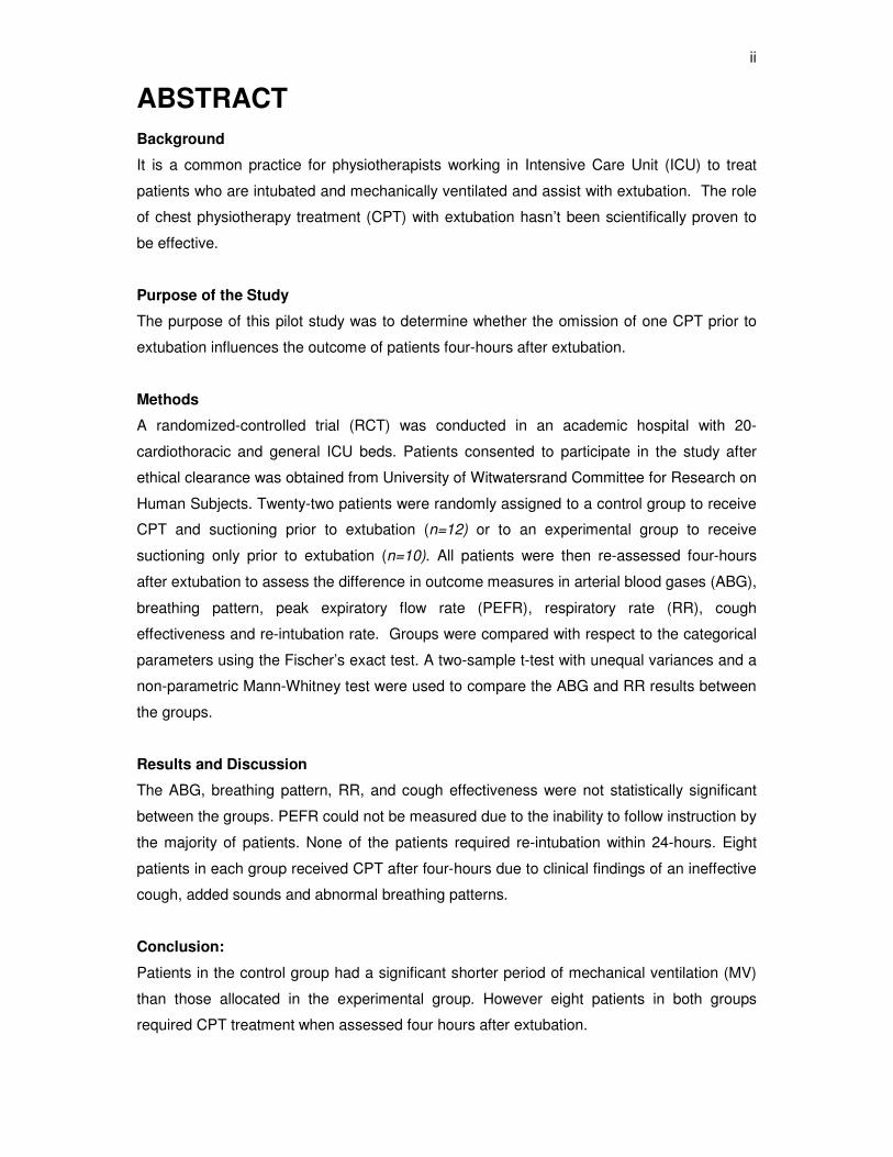
ABSTRACT
Background
It is a common practice for physiotherapists working in Intensive Care Unit (ICU) to treat
patients who are intubated and mechanically ventilated and assist with extubation. The role
of chest physiotherapy treatment (CPT) with extubation hasn’t been scientifically proven to
be effective.
Purpose of the Study
The purpose of this pilot study was to determine whether the omission of one CPT prior to
extubation influences the outcome of patients four-hours after extubation.
Methods
A randomized-controlled trial (RCT) was conducted in an academic hospital with 20-
cardiothoracic and general ICU beds. Patients consented to participate in the study after
ethical clearance was obtained from University of Witwatersrand Committee for Research on
Human Subjects. Twenty-two patients were randomly assigned to a control group to receive
CPT and suctioning prior to extubation (n=12) or to an experimental group to receive
suctioning only prior to extubation (n=10). All patients were then re-assessed four-hours
after extubation to assess the difference in outcome measures in arterial blood gases (ABG),
breathing pattern, peak expiratory flow rate (PEFR), respiratory rate (RR), cough
effectiveness and re-intubation rate. Groups were compared with respect to the categorical
parameters using the Fischer’s exact test. A two-sample t-test with unequal variances and a
non-parametric Mann-Whitney test were used to compare the ABG and RR results between
the groups.
Results and Discussion
The ABG, breathing pattern, RR, and cough effectiveness were not statistically significant
between the groups. PEFR could not be measured due to the inability to follow instruction by
the majority of patients. None of the patients required re-intubation within 24-hours. Eight
patients in each group received CPT after four-hours due to clinical findings of an ineffective
cough, added sounds and abnormal breathing patterns.
Conclusion:
Patients in the control group had a significant shorter period of mechanical ventilation (MV)
than those allocated in the experimental group. However eight patients in both groups
required CPT treatment when assessed four hours after extubation.
iii
ACKNOWLEDGEMENTS
I would like to thank my supervisor, Dr Heleen van Aswegen, who was very supportive to me
throughout my studies. She has made it possible for me to be successful in my study.
Secondly I would like to thank the Physiotherapy department staff for their patience and
motivation throughout my studies. It has been a long process for me to obtain this degree
due to my professional development yet I still received a lot of patience from my supervisor
and the department at large. What would I have done without the support of my family,
especially my husband, Bafana Ngubeni, and my parents, Eugene and Mary Moshe? I would
love to pay my last respects to my late brother, Moshe Moshe, who was always there for me.
Unfortunately he obtained his maters degree posthumously, may his soul rest in peace.
Their confidence in me and interest in my professional development made me persevere. I
would also like to thank my physiotherapy assistant, nursing team and medical staff,
especially head of the unit and consultants at Dr George Mukhari Hospital for allowing me to
conduct the study in their ICU. I thank you all.
iv
TABLE OF CONTENTS
Page
Declaration …………………………………………………………………………………. i
Abstract .……………………………………………………………………………………. ii
Acknowledgements ……………………………………………………………………...... iii
Table of Contents ………………………………………………………………………..... vi
List of Tables and Figures ………………………………………………………………... vi
List of Abbreviations ………………………………………………………………………. vii
1. Chapter 1: Introduction. ………………………………………………………………. 1
1.1 Background ………………………………………………………………………………… 1
1.2 Statement and Significance of Problem ………………………………………………… 4
1.3 Research Question ………………………………………………………………………... 4
1.4 The Aim of the Study …………………………………………………………………....... 4
1.5 The Objectives of the Study ………………………………………………….................. 4
1.6 The Type of Study Done ………………………………………………………………….. 5
1.7 Summary and Conclusion ………………………………………………………………... 5
2. Chapter 2: Literature Review………………………………………………………...... 6
2.1 The History of Chest Physiotherapy …………………………………………………….. 6
2.2 Respiratory Failure ………………………………………………………………………... 7
2.3 Management of Respiratory Failure …………………………………………………….. 8
2.4 Complications that Arise due to Mechanical Ventilation ………………………………. 10
2.5 The Role of Physiotherapists in ICU …………………………………………………….. 12
2.5.1 Manual Techniques ……………………………………………………………………….. 12
2.5.2 Positioning ………………………………………………………………………………… 12
2.5.3 Suction ……………………………………………………………………………………… 13
2.5.4 Manual Hyperinflation …………………………………………………………………….. 13
2.5.5 Active Cycle of Breathing Technique ……………………………………………………. 13
2.6 The Physiotherapy Management of Complications that Arise due to Mechanical Ventilation ……………………………………………………………………………….… 14
2.6.1 Accumulation of Secretions ………………………………………………………………. 14
2.6.2 Atelectasis ………………………………………………………………………………….. 15
2.6.3 Decreased Lung Compliance ……………………………………………………………. 16
v
2.7 Weaning and Extubation …………………………………………………………………. 16
2.8 Conclusion of Literature Review ………………………………………………………… 19 3. Chapter 3: Research Methodology …………………………………………………... 20
3.1 Study Design ………………………………………………………………………………. 20
3.2 Research Method …………………………………………………………………………. 20
3.3 Variables …………………………………………………………………………………... 20
3.4 Hypothesis …………………………………………………………………………………. 20
3.5 Sample Selection ………………………………………………………………………….. 21
3.6 Inclusion and Exclusion Criteria …………………………………………………………. 21
3.7 Data Collection …………………………………………………………………………….. 21
3.8 Data Analysis ………………………………………………………………………………. 22
3.9 Ethical Considerations ……………………………………………………………………. 23
4. Chapter 4: Results ………………………………………………………………………. 24
5. Chapter 5: Discussion …………………………………............................................. 28
6. Chapter 6: Limitations and Recommendations ……………………………………. 32
7. Chapter 7: Conclusion …………………………………………………………………. 33
8. References ………………………………………………………………………………… 34
Appendix I : Consent form and Subject Information Sheet ……………………………….. 42
Appendix II : Ethical Clearance Certificate …………………………………………………… 44
Appendix III : Outcome Measurement Sheet …………………………………………………. 45
Appendix IV : Excel generated randomization list ………………………………………… 46
vi
LIST OF TABLES
Page
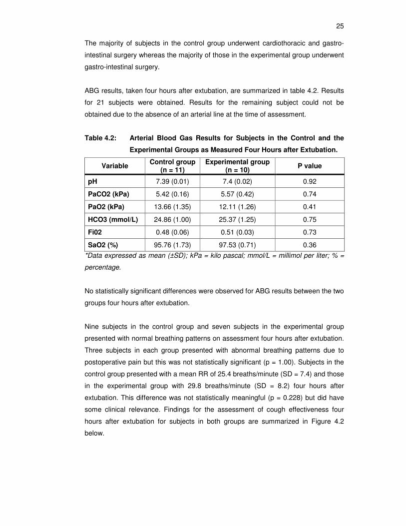
Table 4.1 : Characteristics of Participants …………………………………………............. 24 Table 4.2 : Arterial Blood Gas Results for Subjects in the Control and the Experimental
Groups as Measured Four Hours after Extubation …………......................... 25
LIST OF FIGURES
Page
Figure 4.1 : Admission Diagnoses of Subjects ……………………………………………… 24
Figure 4.2 : Cough Effectiveness Four Hours after Extubation ……………………………. 26
Figure 4.3 : Auscultation Findings Four Hours after Extubation …………………………… 26
vii
LIST OF ABBREVIATIONS
% : percentage
ABG : arterial blood gas
ACBT : active cycle of breathing technique
ARDS : acute respiratory distress syndrome
ARF : acute respiratory failure
AVR : aortic valve replacement
CABG : coronary artery bypass graft
CMV : conventional mechanical ventilation
CO : cardiac output
CO2 : carbon dioxide
COPD : chronic obstructive pulmonary disease
CPAP : continuous positive airway pressure
CPT : chest physiotherapy
CT : cardiothoracic
CVS : cardiovascular
FET : forced expiration technique
FiO2 : fractional concentration of oxygen in inspired gas
FRC : functional residual capacity
GIT : gastointestinal
GCS : glascow coma scale
HCO3 : bicarbonate
HR : heart rate
ICP : intracranial pressure
ICU : intensive care unit
kPa : kilo pascal
M : medical
MAP : mean airway pressure
MH : manual hyperinflation
mmol/Ρ : millimol per liter
MV : mechanical ventilation
MVR : mitral valve replacement
n : sample size
N : neuro
NaCl : sodium chloride
viii
NIPPV : non-invasive positive pressure ventilation
O2 : oxygen
PaCO2 : partial pressure of carbon dioxide in arterial blood
PaO2 : partial pressure of oxygen in arterial blood
PaO2/FIO2 : ratio of partial pressure of oxygen in arterial blood to fractional concentration of oxygen in inspired gas
PEEP : positive end expiratory pressure
PEFR : peak expiratory flow rate
PD : postural drainage
pH : hydrogen ion concentration
PPV : positive pressure ventilation
PS : pressure support
RCT : randomized controlled trial
RR : respiratory rate
SaO2 : arterial oxygen saturation
SD : standard deviation
SIMV : synchronized intermittent mandatory ventilation
TIPS : therapist implemented patient-specific
VAP : ventilator-associated pneumonia
V/Q : ventilation/perfusion ratio
WOB : work of breathing
1
CHAPTER 1
INTRODUCTION
1.1 BACKGROUND
The Intensive Care Unit (ICU) was established during the 1950s. Mechanical
ventilation (MV) was rapidly developed to manage patients postoperatively and those
with poliomyelitis (Norrenberg and Vincent 2002). Respiratory care was intensified
during the epidemic of poliomyelitis in Copenhagen in 1952 (Lassen 1953). Critically ill
patients are commonly managed in ICU, however it is not only limited to post-operative
care. The ICU “has become a concentration not only of critically ill patients and
advanced technology, but also of expert personnel with specialist training” (Norrenberg
and Vincent 2000). Despite the benefits of ventilatory support, secretion clearance
may be adversely affected by inadequate humidification, suctioning and certain
medication administered to patients (Judson and Sahn 1994). Patients in ICU are
traditionally managed by members of a multidisciplinary team. These members may
vary from unit to unit, but mainly consist of medical doctors, nursing staff,
physiotherapists and dieticians. The role of the physiotherapist in ICU may vary
depending on factors such as staffing levels and expertise (Stiller 2000). Other support
services needed in ICU are radiographic and laboratory services (Task Force 1991).
Two studies were identified that were conducted to determine the role of
physiotherapists working in ICU in Europe and South Africa. Questionnaires were
sent out to various hospitals targeting physiotherapists working in ICU. The response
rate was 22% in the European study (Norrenberg and Vincent 2000) compared to
60% response rate in the South African study (Van Aswegen and Potterton 2005). In
comparison, there was no statistical difference between the European and South
African studies in relation to the use of respiratory therapy (98% vs. 98%),
mobilization (100% vs. 98%) and positioning (90% vs. 95%) by physiotherapists
respectively. There was a significant difference between the European and South
African studies in the role of physiotherapy with regard to extubation from MV (25%
vs. 65%) and suctioning (70% vs. 98%). This study was conducted in an academic
hospital where physiotherapists performed chest physiotherapy treatment (CPT),
positioning, mobilization, exercises (both actively and passively) and assisted with
suctioning and extubation of patients in the ICU.
2
Acute respiratory failure (ARF) may be managed with the application of positive
pressure ventilation either invasively by using conventional mechanical ventilation
(CMV) or non-invasively by using non-invasive positive pressure ventilation (NIPPV)
delivered through a mask. Studies have been conducted to compare the two
methods of application of positive pressure ventilation (PPV) to improve gas
exchange (Conti et al 2002; Honrubia et al 2005). In both studies some patients
allocated to the NIPPV group were managed successfully and avoided intubation
(48% vs. 50% respectively) meanwhile 100% of patients in the CMV group were
intubated. NIPPV was shown to be the preferred treatment method as CMV is
normally associated with prolonged MV and the risk of the development of
complications. Wysocki and colleagues (1993) further investigated the indication for
NIPPV in patients with ARF, who were all managed with NIPPV. The results showed
47% success and 53% failure rate, but all had improvement in gas exchange after
one hour application of NIPPV. The results also showed that carbon dioxide (CO2)
retention was a better indication of NIPPV than severe hypoxemia.
This study will only concentrate on MV as a management of ARF in the ICU. The
guidelines for ventilator management of such patients include ventilatory support,
airway management (which entails intubation and secretion clearance) and
extubation (Task Force 1991). The aims of CPT in ICU are to promote more efficient
breathing patterns, prevent accumulation of secretions, improve the mobilization and
removal of secretions, and improve the distribution of ventilation as well as
cardiopulmonary endurance (Scanlan 1990). These aims can be achieved by using
invasive or non-invasive techniques. Airway clearance techniques such as manual
techniques (percussion and vibration), postural drainage (PD), active cycle of
breathing technique (ACBT) and coughing are non-invasive methods and suctioning
is an invasive method employed to achieve these aims (Piccuito 2005).
Stiller and colleagues (1990) conducted a study to compare multimodality CPT with
simple CPT in patients with acute lobar atelectasis. Intensive six-hour treatment
sessions of multimodality CPT (positioning, vibration, manual hyperinflation (MH) and
suction) led to significant and faster resolution of atelectasis as seen on chest
roentgenogram after one treatment intervention compared to patients in the simple
CPT (MH and suctioning) group. Wong’s case study (2000) showed that the 24-hour
availability of physiotherapy treatment may resolve atelectasis and assist in avoiding
intubation. Ntoumenopoulos and colleagues (2002) conducted a study on intubated
patients to investigate the effectiveness of CPT, which is commonly aimed at
3
secretion clearance, on the prevalence of ventilator-associated pneumonia (VAP).
The intervention group received CPT that included positioning, suctioning and chest
vibrations and was independently associated with a reduction in VAP. These studies
highlight the important role that physiotherapists play in the ICU.
However there is controversy about the efficacy of certain CPT techniques in some
patient populations. A study was done to evaluate the effectiveness of physiotherapy
for the management of pulmonary complications in mechanically ventilated trauma
patients (Ntoumenopoulos, Gild and Cooper 1998). The patients were randomly
allocated to either the physiotherapy group (A) or nursing care group (B). Patients in
both groups received routine nursing care (two-hourly turn and suctioning) and
musculoskeletal (passive and active exercises) treatment. Patients in group A had
additional physiotherapy treatment (MH and PD) twice a day. The sample size was
small, with 22 patients in group (A) and 24 in group (B) and four and eight patients
respectively were excluded from this study when they developed pneumonia. As
long-term ventilation is associated with development of pneumonia, the results of
patients who were ventilated for seven days or more were investigated. Days of MV
and ICU stay were the same in both groups and some patients developed
nosocomial pneumonia. The results were not statistically significant hence
physiotherapy treatment was not associated with a reduction in the incidence of
nosocomial pneumonia in trauma patients (Ntoumenopoulos, Gild and Cooper 1998).
Stiller and colleagues (1994) found that prophylactic CPT did not prevent
postoperative pulmonary complications in adults after coronary artery surgery.
Alexander, Weingarten and Mohsenifar (1996) conducted a randomized study to
investigate whether chest physiotherapy was provided to appropriate patients. The
results showed that more than 50% of CPT treatments were ordered for
inappropriate indications. The delivery of CPT was then limited to patients with
appropriate indications such as atelectasis and those who produced copious
secretions. The results of this study were the cost-effectiveness of appropriate
physiotherapy referrals without increasing mortality rate or length of stay of patients.
Evidence-based practice is mandatory for all service providers in ICU, including
physiotherapists, considering the cost associated with management of ICU patients
(Stiller 2000). CPT may prolong the duration of MV in critically ill patients ventilated
for more than 48-hours (Templeton and Palazzo 2007) and may be associated with
side effects, such as bronchospasm or short term hypoxemia (Selsby 1989) hence
evidence-based practice is encouraged.
4
1.2 STATEMENT AND SIGNIFICANCE OF THE PROBLEM
Ongoing assessment of the patient in ICU is essential to ensure optimal care.
Discontinuation of ventilatory support is indicated once the patient meets the criteria
for extubation. Prior to extubation all patients receive CPT and some of these
patients still fail to sustain sufficient respiratory function and to protect their airway
after extubation, hence require re-intubation. On the other hand, some of the self-
extubated patients, who did not receive CPT treatment prior to extubation, manage
well and never require re-intubation. Current practice in our ICU is that patients
receive routine CPT prior to extubation and are monitored closely for signs of
respiratory failure thereafter. With cost saving and evidence-based practice in mind,
the researcher will set out to investigate whether the omission of a single CPT
treatment immediately prior to extubation influences the outcome of the patient after
extubation.
1.3 RESEARCH QUESTION
Does the omission of one CPT treatment immediately prior to extubation influence
the outcome of the patient as assessed four hours after extubation?
1.4 THE AIM OF THE STUDY
To assess whether the omission of one CPT treatment immediately prior to
extubation influences the outcome of the patient four hours after extubation.
1.5 THE OBJECTIVES OF THE STUDY
� To compare arterial blood gas (ABG) results and oxygen saturation (SaO2) four
hours after extubation between the control and experimental groups.
� To compare the difference in breathing pattern four hours after extubation between
the control and experimental groups.
� To compare the difference in peak expiratory flow rate (PEFR) four hours after
extubation between the control and experimental groups.
� To compare the difference in respiratory rate (RR) four hours after extubation
between the control and experimental groups.
� To compare the difference in cough effectiveness four hours after extubation
between the control and experimental groups.
5
� To compare the difference in the rate of re-intubation four hours after extubation
between the control and experimental groups.
1.6 THE TYPE OF STUDY DONE
A randomized control trial (RCT) was conducted.
1.7 SUMMARY AND CONCLUSION
The aim of this pilot study was to determine the need for CPT in extubated patients as
a result of omission of one CPT treatment prior to extubation. In the next chapter, a
summary of the literature pertaining to the indications for admission in ICU as well as
airway management and extubation will be presented. The researcher will also review
the physiotherapy treatment techniques used in ICU and highlight the evidence for
these techniques.
6
CHAPTER 2
LITERATURE REVIEW
2.1 THE HISTORY OF CHEST PHYSIOTHERAPY
Physiotherapy practices existed in the ancient times and various components of
physiotherapy were gradually discovered. Techniques of massage and gymnastics
were practiced for health promotion in the 1580s and later breathing exercises were
recognized to promote health and also to improve voice production for singers
(Innocenti 1995). In 1894, the society of Masseuses was formed and published a book
which also addressed respiratory movements that aimed at promoting more rapid
oxygenation of blood. In 1898 Quicke recommended patients to be positioned in
prone lying with the head lower than the feet and assume the position for half an hour
or more to assist with secretion clearance (Quicke, cited in Innocenti 1995, p682) This
method was described as intermittent use of posture (Innocenti 1995). Ewart (1901)
described a continuous postural method for patients diagnosed with bronchiectasis
and chronic bronchial affections and recommended it over intermittent use of posture.
Some patients with tubercular cavities were instructed to lean over the edge of the bed
with the aim of emptying secretions. Nelson (1934) described various positions to
optimize PD.
Physical and breathing exercises were described by MacMahon (1915) for soldiers
suffering from trauma of the lung, pleura and diaphragm during the war. Localized
breathing was designed to expand lung tissue, to assist with discharge of pus, to break
down adhesions, to restore movement and shape of chest wall. During the 1940s
physiotherapy practices such as breathing exercises, PD, postural correction,
mobilizing exercises and the use of inhalers for patients with chest and heart
conditions continued to progress. CPT was demonstrated to be effective in preventing
post-operative pulmonary complications (Palmer and Sellick 1953). This study was
conducted on 180 post-operative patients who were allocated to either the
experimental group (received PD with percussion and vibration and inhalation before
and after surgery) or the control group (received breathing exercises only before and
after surgery). Nine percent of the patients in the experimental group developed
atelectasis postoperatively compared to 43% in the control group. The results
suggested that inhalation combined with the physiotherapy regime was necessary for
the prevention of atelectasis.
7
Respiratory care was intensified during the epidemic of poliomyelitis in Copenhagen in
1952 (Lassen 1953). Tracheotomy was performed in about 250 cases during the
epidemic of poliomyelitis due to invariable stagnation of secretions in the upper airway
which lead to inadequate ventilation. During this time physiotherapists become
accepted and recognized as integral members of the ICU team (Lassen 1953) in the
management of patients during and after their critical illness. As acutely ill patients
were managed in ICU, the role of physiotherapists was also recognized and
summarized by Holderness and Cooper (1968). The role of physiotherapy was
described as a) maintenance or restoration of full expansion, b) maintenance of full
range of movement of all joints, c) prevention of the accumulation of secretions d)
prevention of circulatory complications, e) prevention of muscle atrophy and lastly
prevention of the development of pressure sores.
Norrenberg and Vincent (2000) conducted a study to determine the role of
physiotherapists in European ICUs. They found that physiotherapists performed
respiratory therapy, mobilization and positioning in almost 100% of ICUs. A similar
study was conducted in South Africa to determine the role of physiotherapists working
in ICU in both the government and private sectors of health care (Van Aswegen and
Potterton 2005). This study revealed that physiotherapists working in the government
sector were mostly involved with manual techniques (100%), suctioning (100%),
extubation (65%), and positioning (94%). The respondents in the private sector were
also involved in the same components of physiotherapy and rated 100%, 98%, 65%
and 92% respectively. Van Aswegen and Potterton (2005) also compared their results
with those of Norrenberg and Vincent (2000). In the European study the suctioning
(70%) and extubation (25%) practices were different compared to the South African
studies (98% and 65%) respectively and were statistically significant.
This section provided a short summary of the development of physiotherapy and the
profession’s subsequent involvement in the treatment of patients in ICU. One of the
reasons for admitting patients to ICU is for management of respiratory failure and will
be discussed further.
2.2 RESPIRATORY FAILURE
Alveolar gas exchange and ventilation may be affected by a wide diversity of
conditions that may result in one of two types of respiratory failure. ‘Respiratory
failure is failure of the respiratory system to provide adequate gas exchange for
metabolic requirements’ (Hough 2001). The mechanism of respiratory failure may
8
manifest in either failure of gas exchange (Type I) or failure of ventilation (Type II)
(Hough 2001). Type I (hypoxemic) respiratory failure is caused by abnormalities of
oxygenation [low partial pressure of oxygen in arterial blood (PaO2)] (Scanlan 1990).
‘Hypoxemic respiratory failure is caused by failure of respiratory system’ (Hough
2001). Classical examples of Type I respiratory failure are pneumonia, asthma and
pulmonary oedema (Oh 2003) and are associated with ventilation-perfusion (V/Q)
mismatch, diffusion defect, right-to-left shunt, and alveolar hypoventilation (Hess and
Kacmarek 2002).
Type II (hypercapnic) respiratory failure results from inadequate ventilation (Scanlan
1990) and is associated with an elevated partial pressure of carbon dioxide in arterial
blood (PaCO2) (Hess and Kacmarek 2002). ‘Hypercapnic respiratory failure is
caused by failure of the respiratory pump and can be acute or chronic’ (Hough 2001).
Type II respiratory failure may occur in low PaO2 and high PaCO2 conditions such as
chronic bronchitis, head injury and chest wall injuries (Oh 2003).
2.3 MANAGEMENT OF RESPIRATORY FAILURE
The decision to intubate and mechanically ventilate a patient is based on his/her
work of breathing (WOB), oxygenation and effectiveness of ventilation (Pierce 1995).
MV is indicated for patients who are unable to oxygenate adequately, ventilate
adequately or both (Hough 2001). Type I respiratory failure can either be managed
with oxygen (O2) therapy for less severe cases of hypoxemia (Turner and Tasker
2002), continuous positive airway pressure (CPAP) or with NIPPV for moderate
hypoxaemia. In cases where the patient is severely ill and presents with severe
hypoxemia, for example in acute respiratory distress syndrome (ARDS), pneumonia
or chronic obstructive pulmonary disease (COPD), intubation and MV is considered
(Hess and Kacmarek 2002; Pierce 1995). The aims of ventilatory support are to
correct hypoxemia, to decrease the WOB and to improve alveolar ventilation
(Williams-Colon and Thalken 1990). The correction of hypoxemia may be achieved
by regulating the fractional concentration of oxygen in inspired gas (FiO2) and
applying positive end-expiratory pressure (PEEP), with the aim of maintaining SaO2
of at least 90% (Pierce 1995). PEEP maintains the pressure in the patient’s lungs
above the atmospheric pressure at the end of expiration to prevent atelectasis and
also recruits atelectatic alveoli (Pierce 1995; Scanlan 1990). Patients in ICU need
continuous monitoring and support (Hough 2001) hence abnormal blood gas results
warrants changes in ventilator settings in order to maintain adequate oxygenation
and ventilation.
9
Respiratory acidosis is characterized by PaCO2 > 45mmHg and hydrogen ion
concentration (pH) < 7.35. Respiratory alkalosis is characterized by pH > 7.45 and
PaCO2 < 35mmHg (Piece 1995). Type II respiratory failure is associated with
respiratory acidosis and hypoxaemia (Hough 2001). Respiratory acidosis is corrected
by adjusting the tidal volume and RR settings on the ventilator (Pierce 1995) to blow
out PaCO2. The researcher will discuss conventional mechanical ventilation further
as it is applicable to the methodology used in this study.
CMV refers to an invasive method of administering ventilatory support. An artificial
airway (endotracheal tube) is inserted into the trachea, (through a nasal or oral route)
or directly in the trachea (tracheostomy tube) (Simmons 1990). The indications for
intubation are to (a) facilitate the removal of secretions, (b) relieve airway obstruction,
(c) protect the lower airway from aspiration and (d) facilitate the application of PPV
(Simmons 1990). The process of MV is completely different from the normal
mechanism of respiration. When a spontaneous inspiration is initiated, muscular effort
is exerted by contraction of the diaphragm and external intercostals muscles, creating
a negative pressure in the lungs which leads to the movement of air from the
atmosphere into the lungs (Pierce 1995). With MV, inspiration will take place due to
the generation of positive pressure by an artificial process from a lower to a higher
positive pressure (alveolar and intrapleural) (Pierce 1995).
The treating doctor decides on the desired mode of MV to ensure optimal interaction
between the patient and the ventilator to achieve adequate ventilatory support and gas
exchange. The patient’s pathological condition is considered when choosing a
particular mode of ventilation (Scanlan 1990) and also depends on the WOB the
patient is able to perform. The mode of ventilation chosen for the subjects in this study
was based on the reason for admission to ICU. Subjects in this study were managed
with either synchronized intermittent mandatory ventilation (SIMV) alone or in
conjunction with pressure support (PS) or with volume control mode. With SIMV mode
the patient is guaranteed a preset amount of tidal volume and number of breaths, and
the patient may initiate breaths in between the mandatory breaths (Pierce 1995).
When the SIMV mode is used in conjunction with PS, only the spontaneous breaths
are augmented by the delivery of a preset amount of inspiratory positive pressure
(Pierce 1995). With volume control, also known as continuous mandatory ventilation,
the patient is guaranteed a preset amount of tidal volume and number of breaths per
minute and the ventilator performs all the WOB without any spontaneous efforts from
10
the patient (Pierce 1995). Patients are gradually weaned off ventilatory support once
spontaneous breathes are detected. This will be elaborated upon further in the section
on weaning and extubation. Tracheostomy is indicated when prolonged need for
intubation arises for example the management of patients who sustained severe
injuries such as blunt chest trauma or head injury (Simmons 1990). However,
tracheostomy will not be discussed any further as it is not relevant to this study.
2.4 COMPLICATIONS THAT ARISE DUE TO MECHANICAL VENTILATION
Patients who receive MV might develop haemodynamic instability due to the
mechanism of positive pressure ventilation (Conti et al 2002). PPV impedes venous
return to the heart due to the positive intrapleural pressure that it creates and in turn
decreases cardiac output (CO) which results in a reduction of renal, hepatic and
splanchnic blood flow (Hough 2001; Pierce 1995). ‘Haemodynamic compromise is
also likely to occur if mean airway pressure (MAP) is high; inspiratory time prolonged
or mean expiratory pressure raised as with PEEP’ (Hough 2001). MV is also
associated with alveolar over distention which is due to an increased MAP, large tidal
volume and the use of PEEP. This may result in compression of adjacent pulmonary
capillaries and regional hypoperfusion (Pierce 1995) that could lead to altered
oxygenation.
A second complication of intubation and MV is laryngeal and/or tracheal injury due to
the presence of the artificial airway. Laryngeal injuries include glottic stenosis,
oedema and vocal cord paralysis (Hess and Kacmarek 2002).Tracheal injuries
(tracheal stenosis and tracheomalacia) are caused by high endotracheal cuff
pressures that compress the tracheal mucosa (Hess and Kacmarek 2002). This
complication may be avoided by adhering to protocols for cuff inflation. The
movement of the tube, especially with self extubation and excessive neck flexion and
extension, may cause abrasion and could be avoided by securing the endotracheal
tube firmly to the nose or mouth (Plevak and Ward 1997). Malposition of the tube
(either high or low), mouth or lip pressure sore development, laryngospasm, glottic
oedema, tracheobronchial fistula, tracheomalacia and tracheal stenosis are
additional complications that may arise (Pierce 1995).
Another potential complication of MV is atelectasis. In any position assumed by the
patient, ventilation-perfusion (V/Q) mismatch occurs. The non-dependent areas of
the lungs are preferentially ventilated by the application of PPV (Woodard and Jones
2002) because the diaphragm becomes inactive due to sedation. The prolonged
11
application of high levels of FiO2 displaces alveolar and blood nitrogen and may
result in atelectasis (Pierce 1995). Absorption atelectasis may also occur due to low
tidal volume ventilation with high FiO2 (Hough 2001). ‘Spontaneous breathing draws
ventilation down to the dependent lung regions, causing a downward ventilation
gradient’ (Hough 2001). With PPV this gradient is reversed, unless if the mode of
ventilation encourages spontaneous breathing. The reversed ventilation gradient
leads to the dependent areas receiving the least ventilation and result in progressive
atelectasis (Hough 2001).
The presence of an artificial airway may impair the effectiveness of a cough and
affect the mucociliary activity (Hess and Kacmarek 2002). Impaired mucociliary
activity may be due to inadequate humidification, underlying lung pathology, high
FiO2, drugs (such as narcotics) and airway trauma due to suctioning (Hess and
Kacmarek 2002). The patient’s cough may be ineffective due to the depression of
the neurological status, a decreased functional residual capacity (FRC) due to
abnormal glottic function and result in accumulation of secretions (Judson and Sahn
1994; Hess and Kacmarek 2002; Pierce 1995). An inability to cough effectively,
(either due to respiratory muscle weakness or pain) may also be observed in patients
breathing spontaneously in ICU after extubation and therefore physiotherapists
should be familiar with assistance techniques and cough stimulation (Ciesla 1996).
Contamination of the lower airway may occur due to intubation, which bypasses the
normal filtering mechanism of the upper airway. Artigas and colleagues (2001)
conducted a prospective cohort study to determine risk factors for the development of
nosocomial pneumonia in critically ill trauma patients. The study sample was a group
of 103 patients admitted with varying conditions and 22.3% of them developed
nosocomial pneumonia. The group with nosocomial pneumonia had a significant
increase in days spend on MV and longer ICU stay. The most important risk factor
identified by the authors for development of nosocomial pneumonia was prolonged
MV, especially with PEEP. The assumption was that PEEP is normally used to treat
patients with severe ARF and was therefore associated with prolonged MV. Other
independent risk factors for nosocomial pneumonia were the presence of a
nasogastric tube as well as continuous enteral feeding due to the risk of aspiration.
The authors did mention that the main problem they experienced was the
identification of pneumonia, which could have impacted on the results. Fagon and
colleagues (1993) stated that pneumonias occurring in ventilated patients are
12
associated with high mortality as compared to patients who had underlying disease
(excluding pneumonia) alone.
2.5 THE ROLE OF PHYSIOTHERAPISTS IN ICU
The physiotherapist is regarded as an integral member of the multidisciplinary team
that is responsible for the management of patients in ICU in most hospitals in
developed countries (Stiller 2000). The most common intervention practiced by
physiotherapists in ICU is chest physiotherapy (Norrenberg and Vincent 2000; Stiller
2000; Van Aswegen and Potterton 2005). CPT includes various techniques which
will be discussed briefly and supported by the literature.
2.5.1 Manual Techniques
Physiotherapists use various manual techniques such as percussions, vibrations and
shaking to treat cardiopulmonary complications. Shaking and vibrations are done
during the expiratory phase of the respiratory cycle and are used to aid the removal
of secretions from the tracheobronchial tree (Pryor et al 2002; Scanlan 1990). The
physiotherapist places her hand on the chest wall and once the patient takes a deep
breath, rapid vibratory motion is applied to the chest wall on expiration (Scanlan
1990). Chest percussions are done throughout the respiratory cycle. The indication
of percussions is to assist with the mobilization of tenacious secretions from the
tracheobronchial tree (Pryor et al 2002; Scanlan 1990). Gallon (1991) conducted a
study on nine bronchiectatic patients to determine the effect of manual chest
percussion. The results showed a significant production of secretions when
percussion was done in conjunction with other physiotherapy modalities. These
modalities may vary from one study to the other but mainly include PD or modified
positioning, suctioning, MH, and ACBT as observed in other studies
(Ntoumenopoulos et al 2002; Stiller et al 1996; Wong 2000). Multimodality
physiotherapy treatment was demonstrated to be more effective than single modality
physiotherapy treatment (Stiller et al 1990) or breathing exercises alone (Palmer and
Sellick 1953) in resolving acute lobar atelectasis.
2.5.2 Positioning
Routine positioning is done by nurses and physiotherapists with the aim of reducing
adverse effects of bed mobility such as bedsores and pulmonary complications
(Dean 2002). Positioning is often used to facilitate the mobilization of secretions from
specific lung segments with the assistance of gravitational forces (known as PD); to
improve the distribution of ventilation (dependent positioning) and to relieve dyspnea
13
(relaxation technique) (Scanlan 1990). Alternate side lying is the most common
position used in the ICU to assist with secretion clearance (Dean 2002). In fact, Stiller
and colleagues (1990) showed that side lying with the affected lung uppermost in a
head down tilt position was most effective in the management of acute atelectasis. A
follow up study also showed that modified PD or PD in addition to MH and suctioning
hourly brought about the resolution of atelectasis after one treatment session (Stiller
et al 1996).
Berney and colleagues (2004) found that the addition of a head down tilt position to
physiotherapy treatment in the ICU enhanced secretion production. Patients with
predominantly unilateral lung disease may have improved oxygenation when nursed
in lateral side lying with the good lung down (Pierce 1995). Positioning intubated and
mechanically ventilated patients in a semi recumbent position has been shown to
minimize the aspiration of gastric contents to the lower airway (Torres et al 1992).
2.5.3 Suction
Suction is defined as an invasive airway clearance technique to aspirate material
from the upper airway mechanically (Plevak and Ward 1997). Suctioning is indicated
when an individual is unable to expectorate secretions due to an ineffective cough
(Pryor et al 2002), which may be the result of loss of consciousness and/ or the
inability to protect the airway (Plevak and Ward 1997). The process of suctioning
should be timed with interventions carried out in ICU such as change in the patient’s
position and during or after physiotherapy because of the greater prevalence of
upper airway secretions (Ciesla 1996) following these procedures.
2.5.4 Manual Hyperinflation (MH)
This technique refers to the delivery of a volume of gas greater than tidal volume to
the patient’s lungs via an endotracheal tube or tracheostomy or through a face mask
(Barker and Eales 1994; Risley and Jones 2003). Physiotherapists use MH for
recruitment of atelectatic lung segments (Hodgson, Carroll and Denehy 1999; Van
Aswegen and Eales 2004). In addition, MH is commonly used to (a) aid the removal
of secretions, (b) improve static lung compliance, and (c) improve oxygenation
(Berney and Denehy 2002; Hodgson et al 2000; Van Aswegen and Eales 2004). The
use of MH has been reported to enhance secretion clearance compared to side lying
alone (Hodgson et al 2000).
14
2.5.5 Active Cycle of Breathing Technique (ACBT)
This modality is commonly used to facilitate the mobilization and removal of
secretions from the airways. ACBT is a cycle of thoracic expansion exercises,
breathing control and FET for use in patients with excessive airway secretions (Pryor
et al 2002). Atelectasis was shown to be effectively managed with ACBT. Wong
(2000) conducted a case report on a patient with a medical history of COPD, who
developed type I respiratory failure after explorative laparatomy. The patient received
physiotherapy treatment comprising of ACBT, huffing and coughing (to facilitate
expectoration of secretions), mobilization and exercises. The patient had dyspnea
and couldn’t cope with manual techniques. This physiotherapy regime was
successful and intubation and MV was avoided.
2.6 THE PHYSIOTHERAPY MANAGEMENT OF COMPLICATIONS THAT ARISE DUE
TO MECHANICAL VENTILATION
Intubated and mechanically ventilated patients are at risk of the development of
complications such as accumulation of secretions, atelectasis and pneumonia
(Konrad et al 1994). The techniques used by physiotherapists to manage these
complications will be discussed further.
2.6.1 Accumulation of Secretions
As discussed previously, intubated patients in ICU frequently have impaired mucus
transport and an impaired cough mechanism, which lead to accumulation of
secretions and the development of pneumonia (Konrad et al 1994; Pierce 1995).
Several studies have been conducted to investigate the efficacy of CPT in various
hospital settings.
Ntoumenopoulos and colleagues (2002) conducted a study to investigate the
effectiveness of CPT in intubated and mechanically ventilated patients with the aim of
enhancing airway clearance. Patients were allocated to either an intervention group
(n = 24) or a control group (n = 36) in a non-randomized pattern after meeting the
inclusion criteria such as being mechanically ventilated for at least 48 hours. The
intervention group had CPT comprising of vibrations, modified or gravitational
postural drainage, and suctioning rendered twice a day, while the control group had
cardiopulmonary assessment and musculoskeletal physiotherapy. The intervention
group had a significant reduction in the frequency of VAP compared to the control
15
group (8% vs. 39% respectively). There were no statistical differences in ICU length
of stay or mortality.
2.6.2 Atelectasis
As mentioned, atelectasis of the dependent lung regions is common in intubated and
mechanically ventilated patients and certain components of CPT were shown to be
effective in the management of atelectasis. Patients in ICU frequently have impaired
mucous transport which is associated with the retention of secretions and pneumonia
(Konrad et al 1994). Artificial airway (endotracheal tube or tracheostomy) also
compromises both mucociliary function and cough effectiveness (Plevak and Ward
1997). A study conducted by Khamiees and colleagues (2001) showed that
excessive endotracheal secretions, especially in the absence of a good cough, could
lead to bronchial plugging, atelectasis, and/or aspiration pneumonitis, all of which can
cause respiratory failure.
Stiller and colleagues (1990) conducted a study to investigate whether multimodality
physiotherapy treatment was more effective than a single modality physiotherapy
regime in treating patients with acute lobar atelectasis. Patients were allocated to
either the experimental or control group in an alternate fashion. Seven patients were
allocated to the experimental group and had vibrations, MH (or deep breathing
exercise) positioning (with the affected lung uppermost) and suctioning (or coughing)
as their treatment regime. In the control group seven patients had MH (or deep
breathing exercises) and suctioning (or coughing) alone. Treatment was rendered
hourly for six hours in both groups. A significant resolution of atelectasis was
observed in the experimental group subjects compared to the control group after one
treatment intervention. The results showed that the addition of positioning and
vibrations added value to the efficacy of a simple chest treatment (MH and suctioning
alone). Stiller and colleagues (1996) conducted a follow up study comparing five
physiotherapy regimes. The results showed that patients who received modified PD
with or without vibrations (group 2 or 3), or PD (group 4) in addition to MH and
suctioning hourly had a better resolution of atelectasis after an hourly treatment
session for six, 24 and even 48 hours later than those who had hourly MH and
suctioning alone for six hours (group 1). In both studies the small samples sizes
could have influenced the results obtained.
Van Aswegen and Eales (2004) described a case study on one patient who was
assaulted and sustained severe facial injury and presented with low Glasgow Coma
16
Scale (GCS). The patient’s condition deteriorated as a right lung collapse developed.
The patient presented with Type II respiratory failure which warranted intubation and
MV to protect the airway. The patient was then transferred to ICU for further
management and CPT was commenced, which consisted of modified PD (left side
lying with the head elevated due to facial oedema), percussion and vibrations, MH
and suctioning. The patient had significant improvement in oxygenation after the first
treatment session and resolution of atelectasis within 24-hours.
2.6.3 Decreased Lung Compliance
Prolonged atelectasis may result in pulmonary infection and hypoxemia. The
consequence of prolonged atelectasis may be a reduction in pulmonary compliance
making ventilation difficult (Berney and Denehy 2002).
Berney and colleagues (2004) conducted a study on 20 patients who were intubated
and mechanically ventilated. The purpose of the study was to investigate the effect of
a head-down tilt during physiotherapy treatment on sputum production. Patients
were randomly allocated to receive MH treatment in both supine and head-down tilt
(350- 450 elevation) positions during two different sessions on one day. Static lung
compliance was measured before and after physiotherapy treatment. Compliance
improved significantly after physiotherapy treatment that included MH in both
positions. The study also showed a significant improvement (25%) in sputum
production with the addition of a head-down tilt rather than supine position.
MH was shown to be effective in improving static lung compliance in critically ill
patients (Hodgson et al 2000). The study was conducted on 18 mechanically
ventilated patients who acted as their own controls. Patients were randomly allocated
to receive either MH (consisted of side lying position, suctioning and MH) or side
lying (positioning and suctioning) treatment first and had two sessions per day. The
MH group had a significant increase in secretion clearance and total static
compliance, which was still present 20 minutes after treatment. Mean arterial
pressure and heart rate (HR) were not statistically different between the two
treatment sessions.
WEANING AND EXTUBATION
Weaning of the intubated patient involves the gradual reduction and removal of
ventilatory support and restoration of the patient’s spontaneous breaths (Halliday
2004: Turner and Tasker 2002). The process of weaning commences once the
17
patient is alert and cooperative, an improvement in clinical condition is observed and
the patient has the ability to clear bronchial secretions adequately (Turner and
Tasker 2002). The weaning process requires ongoing assessment and planning by
all members of the critical care team (Henneman et al 2001) and should be tailored
to the needs of each patient (Scanlan 1990). Readiness to wean is defined as normal
blood electrolyte levels and body temperature, resolution of respiratory failure (Hess
and Kacmarek 2002), haemodynamic stability, the ability to initiate adequate
respiratory drive (maintain normal PaCO2) and adequate nutritional support (to
prevent muscle weakness and failure) (Woodard and Jones 2002).
To date, several studies have been conducted to determine the best technique for
weaning patients from MV. Saura and colleagues (1996) conducted a study to
analyze the effect of the implementation of a weaning protocol on clinical outcomes.
The weaning protocol group was retrospectively compared to the historical group of
patients. Patients in the protocol group (n = 51) had a significant shorter duration of
MV and ICU stay compared to the historical control group (n = 50). Kollef and
colleagues (1997) conducted a study comparing two weaning methods from MV.
Protocol directed weaning from MV (performed by nurses and respiratory therapists)
was shown to be a safe technique and led to extubation more rapidly than physician
directed weaning. Consequently, the protocol-directed weaning group had a
significantly reduced duration of MV and more successful weaning than the physician
–directed weaning group. Scheinhorn and colleagues (2001) compared the outcomes
of patients on a therapist implemented patient-specific (TIPS) weaning protocol to
patients on a physician-directed weaning protocol in a retrospective study design
(conducted two years previously). The average duration of weaning from MV was
significantly reduced in the TIPS group compared to the physician-directed group.
Results from the three above mentioned studies would suggest that a
multidisciplinary approach to weaning is effective and reduces the risk of
complications associated with this procedure.
Weaning is performed by the gradual reduction of mandatory breaths performed by
the ventilator (e.g. SIMV) and thus allowing the patient to initiate more spontaneous
breaths while receiving some PS from the ventilator (Woodard and Jones 2002). PS
is reduced once the patient is able to maintain adequate oxygenation and maintain
normal levels of PaCO2. Weaning is progressed to either CPAP or T-piece (Woodard
and Jones 2002). CPAP weaning is the application of positive pressure to
spontaneous breaths during inspiration and expiration. The aim of CPAP is to
18
prevent alveolar collapse, improve FRC and enhance oxygenation (Pierce 1995).
During T-piece weaning the patient is disconnected from the ventilator and performs
all WOB spontaneously while receiving humidified oxygen (Pierce 1995). Once the
patient is able to maintain oxygenation and a normal breathing pattern on CPAP or a
T-piece, the patient is ready for extubation (Pierce 1995).
During the weaning period the patient should be monitored for signs of respiratory
muscle fatigue such as tachypnoea and respiratory paradoxus (abnormal breathing
pattern) (Hess 1997). The length of the weaning process varies and is dependent on
the individual’s ability to cope with the procedure. The weaning process may be short
in uncomplicated patients for example in controlled extubation post operatively. The
process may be prolonged in a long-term ventilated patient and is initiated by
reducing sedation and using positioning for optimal diaphragmatic excursion
(Woodard and Jones 2002).
Extubation is the process of removing the endotracheal tube once the patient can
protect his/her airway and the underlying problems have been resolved. Successful
extubation requires a patient to be able to mobilize and expectorate their secretions
and to protect their own airway after removal of the artificial airway (Hess and
Kacmarek 2002). Early successful extubation will reduce the risk of complications
due to prolonged MV.
Extubation may fail due to various reasons such as the inability to cough, nosocomial
pneumonia, atelectasis and bronchospasm as observed in Saura’s study (1996).
Extubation failure prolongs ICU stay, the duration of MV which leads to the need for
tracheostomy and it is associated with higher hospital mortality (Epstein 2002).
Extubation failure is synergistically enhanced in the presence of abundant
endotracheal secretions and poor cough strength (Khamiees et al 2001). Health care
workers also overlook the influence of individual tolerance and fatigue on successful
weaning and extubation. Re-intubation is a prolonged means of airway protection
and/or ventilatory support, not a predictor of mortality (Daley, Garcia-Perez and Ross
1996). Demling and colleagues (1988) compared the incidence of extubation failure
of patients managed in general surgical ICU vs. trauma/burns unit by using
standardized criteria. Patients in the trauma unit were young and in good health, and
had 3% failure rate. The reason for extubation failure was that patients couldn’t
protect the airway and clear secretions. Patients in the general surgical ICU had
4.4% failure rate and the reason was the need for further ventilatory support. The
19
mortality rate was 10% in the trauma unit compared to 40% in the general surgical
ICU which was dependent on the underlying disease process. In an attempt to
reduce these problems thorough patient assessment and collaborative teamwork is
emphasized in ICU worldwide.
2.8. CONCLUSION OF LITERATURE REVIEW
The literature presented in this chapter portrayed the management of respiratory
failure through intubation and MV, complications that arise due to MV and the role of
physiotherapy in the management of these complications as well as the process of
weaning and extubation. However there is a dearth of literature on the effectiveness
of CPT immediately prior to extubation and how the inclusion or omission thereof
might influence patient outcome. The next chapter of this report will describe the
methodology that was followed in order to conduct the RCT.
20
CHAPTER 3
RESEARCH METHODOLOGY
The methodology discussed in this chapter is based on the findings of the literature review
discussed in Chapter 2. The study design, sample population, hypotheses tested, data
collection procedure and instruments used are discussed in detail. The main methods used
for data analysis are given. Ethical considerations are addressed towards the end of this
chapter.
3.1 STUDY DESIGN
A randomized control trial was done.
3.2 RESEARCH METHOD
Quantitative data was collected. Respiratory function was assessed (RR, breathing
pattern, cough effectiveness, PEFR). Gas exchange was assessed (ABG analysis)
as well as lung sounds (auscultation).
3.3 VARIABLES
Independent variable: Omission of one CPT session prior to extubation.
Dependent variables:
� Respiratory function (RR, breathing pattern, cough effectiveness and PEFR).
Normal values used were according to those published by Middleton S and
Middleton PG (2002, p. 11). The effectiveness of the cough was a subjective
assessment and was only assessed by the researcher to reduce bias.
� Breath sounds (auscultation)
� Gas exchange [pH, PaCO2, partial pressure of oxygen in arterial blood (PaO2),
bicarbonate (HCO3), FiO2, SaO2] Normal values used were according to those
published by Middleton S and Middleton PG (2002, p 21).
3.4 HYPOTHESES
� The omission of one CPT treatment immediately prior to extubation leads to
higher indication for CPT in patients when assessed four hours post extubation.
21
� The null hypothesis: There is no difference in the indication for CPT four hours
after extubation between the groups.
3.5 SAMPLE SELECTION
The study sample consisted of all patients admitted to Dr George Mukhari Hospital
ICU, intubated and mechanically ventilated. Patients had to meet set criteria prior to
extubation: (1) HR < 140 beats /min, (2) RR < 35 breaths /min, (3) PaO2/FIO2 ratio of >
200, (4) awake and oriented, (5) not requiring vasoactive or inotropic agents, (6) PEEP
< 5cm of water.
3.6 INCLUSION AND EXCLUSION CRITERIA
The inclusion criteria was (1) any patient older than 18 years irrespective of their
condition, (2) mechanically ventilated and intubated for < 5 days, (3) eligible for
extubation on or before day 5. Patients excluded from the study were those who
were mechanically ventilated for more than 5 days.
3.7 DATA COLLECTION
The researcher and research assistant (qualified physiotherapist with ten years
experience) attended twice daily ward rounds in the ICU to identify potential subjects
for the study. All patients received CPT treatment twice daily throughout their ICU
stay. CPT comprised percussion, vibrations, alternate side lying, deep breathing
exercises, and coughing while suctioning.
Once the patient was deemed ready for extubation, the sister in charge of the patient
informed the researcher telephonically. The researcher explained the study to the
patient or relative using the subject information sheet (see appendix I) and obtained
informed consent from either the patient or relative prior to enrolment in the study.
The patient’s haemodynamic status and ABG results were recorded by the
researcher on the outcome measurement sheet (see appendix III). The researcher
then informed the research assistant that the patient was ready for extubation.
Patients who met the inclusion criteria were randomly allocated to either
experimental or control group by the research assistant using an Excel generated
randomization list (see appendix IV). A consecutive sampling technique was used.
The sister in charge of the patient and the research assistant had to prepare the
patient for extubation. The patient’s baseline haemodynamic status was monitored
throughout the treatment session. The research assistant explained the extubation
22
procedure and the benefits of suctioning to the patient, and pre-oxygenated the
patient. Treatment and extubation of the patient was carried out by the research
assistant. Patients allocated to the control group received CPT, as described above,
and suctioning prior to extubation. Patients in the experimental group were only
suctioned and extubated. All patients were placed in a semi fowler’s position just
before extubation to eliminate the possibility of aspiration. The patients were
instructed to take a deep breath, the endotracheal tube cuff was deflated and pulled
out and the patient was instructed to cough. Once the patient was extubated, he/she
was nebulized with a bronchodilator (Atrovent and Berotec) to prevent post
extubation bronchospasm and thereafter administered with the prescribed humidified
oxygen therapy.
Hourly observations and ABG were done by the sister in charge of the patient to
ensure that the patient was coping and was haemodynamically stable. Four hours
post extubation, the researcher recorded the patient‘s haemodynamic status and
ABG results on the outcome measurement sheet (see appendix III). Physiotherapy
chest assessment (which included RR, respiratory pattern, PEFR, cough and
auscultation) and treatment was carried out by the researcher following any abnormal
findings which warranted CPT. The presence of secretions was addressed with
mobilization and loosening of secretions through nebulization therapy with 0.9%
sodium chloride (NaCl) solution, modified PD, ACBT and manual chest therapy and
was complimented by coughing and/or suctioning to remove secretions.
3.8 DATA ANALYSES
Demographic information described by continuous parameters such as age, and
length of MV was summarized using means and standard deviations (SD). A two
sample t-test was performed on the above parameters. Demographic information
described by categorical parameters such as breathing pattern, cough effectiveness
and auscultation findings were summarized using frequencies, percentages and
cross tables. Groups were compared with respect to the categorical parameters
using the Fischer’s exact test. Quantitative information (RR) was expressed as
means and SD. A two-sample t-test with unequal variances and a non-parametric
Mann-Whitney test were used to compare the ABG and RR results between the
groups. The STATA 8 statistical software package was used for all statistical
analyses and throughout testing was done at the 0.05 level of significance. A sample
size of 35 patients per group would give 80% power to detect a change from 0.25 to
0.05 in the extubation failure rate between the experimental and the control groups.
23
3.9 ETHICAL CONSIDERATIONS
Ethical clearance was obtained from the University of the Witwatersrand Committee
for Research on Human Subjects (clearance number M050210 – see appendix II).
Permission was obtained from the Chief Executive Officer at Dr George Mukhari
Hospital to conduct the study.
Written consent was obtained from all subjects who participated in the study.
Confidentiality was maintained by coding all data that was captured on the Outcome
Measurement Sheets and the database on the computer. Subjects were allowed to
withdraw from this study at any time without compromise of regular treatment.
The results obtained through the above mentioned methodological process are
described in chapter 4.
24
CHAPTER 4
4.1 RESULTS
This chapter describes the results obtained from the RCT that was discussed in the
previous chapter.
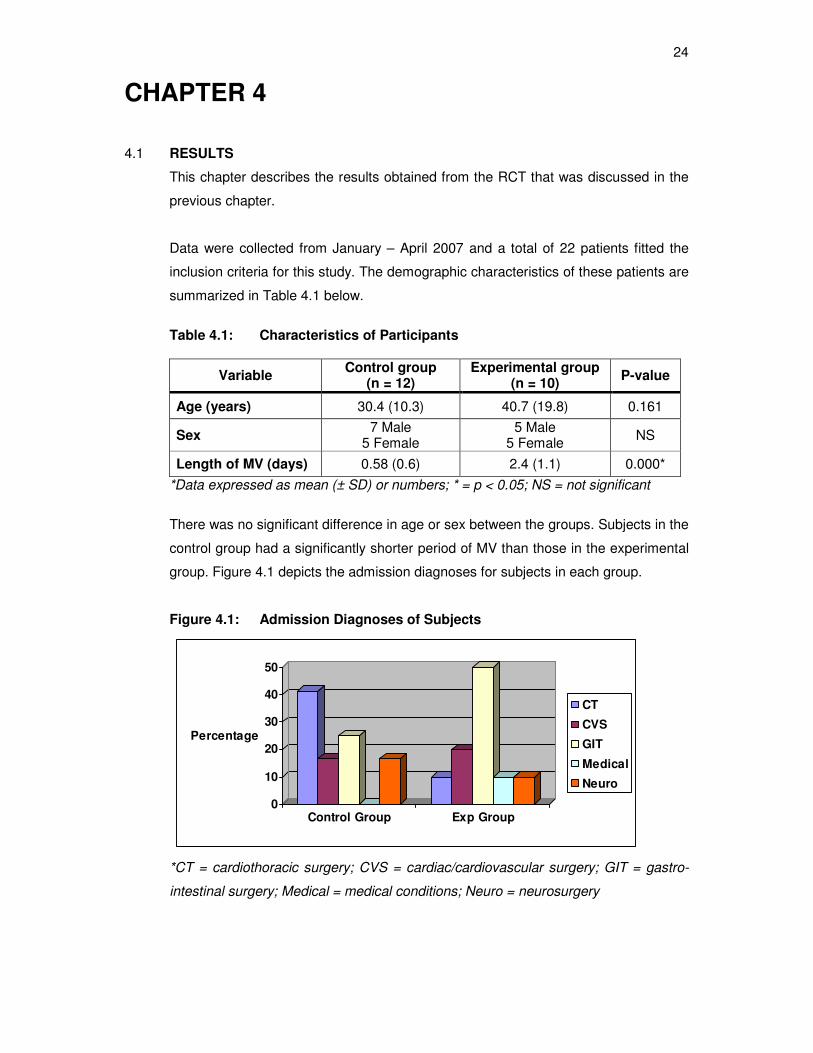
Data were collected from January – April 2007 and a total of 22 patients fitted the
inclusion criteria for this study. The demographic characteristics of these patients are
summarized in Table 4.1 below.
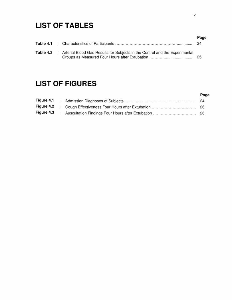
Table 4.1: Characteristics of Participants
Variable Control group
(n = 12) Experimental group
(n = 10) P-value
Age (years) 30.4 (10.3) 40.7 (19.8) 0.161
Sex 7 Male
5 Female 5 Male
5 Female NS
Length of MV (days) 0.58 (0.6) 2.4 (1.1) 0.000*
*Data expressed as mean (± SD) or numbers; * = p < 0.05; NS = not significant
There was no significant difference in age or sex between the groups. Subjects in the
control group had a significantly shorter period of MV than those in the experimental
group. Figure 4.1 depicts the admission diagnoses for subjects in each group.
Figure 4.1: Admission Diagnoses of Subjects
0
10
20
30
40
50
Percentage
Control Group Exp Group
CT
CVS
GIT
Medical
Neuro
*CT = cardiothoracic surgery; CVS = cardiac/cardiovascular surgery; GIT = gastro-
intestinal surgery; Medical = medical conditions; Neuro = neurosurgery
25
The majority of subjects in the control group underwent cardiothoracic and gastro-
intestinal surgery whereas the majority of those in the experimental group underwent
gastro-intestinal surgery.
ABG results, taken four hours after extubation, are summarized in table 4.2. Results
for 21 subjects were obtained. Results for the remaining subject could not be
obtained due to the absence of an arterial line at the time of assessment.
Table 4.2: Arterial Blood Gas Results for Subjects in the Control and the
Experimental Groups as Measured Four Hours after Extubation.
Variable Control group
(n = 11) Experimental group
(n = 10) P value
pH 7.39 (0.01) 7.4 (0.02) 0.92
PaCO2 (kPa) 5.42 (0.16) 5.57 (0.42) 0.74
PaO2 (kPa) 13.66 (1.35) 12.11 (1.26) 0.41
HCO3 (mmol/L) 24.86 (1.00) 25.37 (1.25) 0.75
Fi02 0.48 (0.06) 0.51 (0.03) 0.73
SaO2 (%) 95.76 (1.73) 97.53 (0.71) 0.36
*Data expressed as mean (±SD); kPa = kilo pascal; mmol/L = millimol per liter; % =
percentage.
No statistically significant differences were observed for ABG results between the two
groups four hours after extubation.
Nine subjects in the control group and seven subjects in the experimental group
presented with normal breathing patterns on assessment four hours after extubation.
Three subjects in each group presented with abnormal breathing patterns due to
postoperative pain but this was not statistically significant (p = 1.00). Subjects in the
control group presented with a mean RR of 25.4 breaths/minute (SD = 7.4) and those
in the experimental group with 29.8 breaths/minute (SD = 8.2) four hours after
extubation. This difference was not statistically meaningful (p = 0.228) but did have
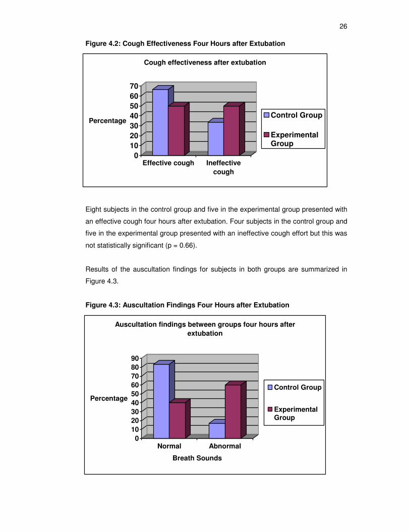
some clinical relevance. Findings for the assessment of cough effectiveness four
hours after extubation for subjects in both groups are summarized in Figure 4.2
below.
26
Figure 4.2: Cough Effectiveness Four Hours after Extubation
0
10
20
30
40
50
60
70
Percentage
Effective cough Ineffective
cough
Cough effectiveness after extubation
Control Group
ExperimentalGroup
Eight subjects in the control group and five in the experimental group presented with
an effective cough four hours after extubation. Four subjects in the control group and
five in the experimental group presented with an ineffective cough effort but this was
not statistically significant (p = 0.66).
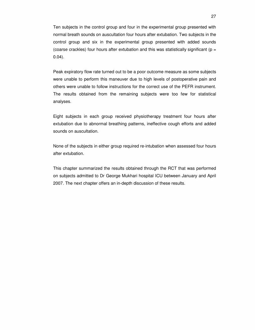
Results of the auscultation findings for subjects in both groups are summarized in
Figure 4.3.
Figure 4.3: Auscultation Findings Four Hours after Extubation
0
10
20
30
40
50
60
70
80
90
Percentage
Normal Abnormal
Breath Sounds
Auscultation findings between groups four hours after
extubation
Control Group
ExperimentalGroup
27
Ten subjects in the control group and four in the experimental group presented with
normal breath sounds on auscultation four hours after extubation. Two subjects in the
control group and six in the experimental group presented with added sounds
(coarse crackles) four hours after extubation and this was statistically significant (p =
0.04).
Peak expiratory flow rate turned out to be a poor outcome measure as some subjects
were unable to perform this maneuver due to high levels of postoperative pain and
others were unable to follow instructions for the correct use of the PEFR instrument.
The results obtained from the remaining subjects were too few for statistical
analyses.
Eight subjects in each group received physiotherapy treatment four hours after
extubation due to abnormal breathing patterns, ineffective cough efforts and added
sounds on auscultation.
None of the subjects in either group required re-intubation when assessed four hours
after extubation.
This chapter summarized the results obtained through the RCT that was performed
on subjects admitted to Dr George Mukhari hospital ICU between January and April
2007. The next chapter offers an in-depth discussion of these results.
28
CHAPTER 5
DISCUSSION
The main findings of this pilot study were that the omission of one CPT treatment
prior to extubation led subjects in the experimental group to present with a higher
mean RR and significantly more abnormal breath sounds on auscultation four hours
after extubation compared to those in the control group. However, an equal amount
of subjects in both groups required CPT treatment four hours after extubation.
Patients normally present with complications following surgery. Hough (2001)
described some respiratory complications after surgery as atelectasis, hypoxaemia
and chest infection. Chest infection occurs due to intubation and impaired mucociliary
activity due to anesthesia (Hough 2001). Pain leads to inhibition of breathing and
immobility and may affect the patient’s ability to cough, reduce FRC and result in
atelectasis (Hough 2001). Abdominal surgery has less direct effect on respiration
(Hough 2001) but the distention of the abdomen leads to reduction of tidal volume
due to diaphragmatic splinting. The incision through the rectus abdominis muscle will
decrease the patient’s ability to cough effectively resulting in secretion retention
postoperatively. Following thoracic surgery [aortic valve replacement (AVR), mitral
valve replacement (MVR), coronary artery bypass graft (CABG)] patients may
present with cardiovascular instability, which may restrict bed mobility. The CABG
may result in impaired cerebral perfusion and cause disorientation (Hough 2001).
The pain associated with thoracic surgery occurs as a result of the incision of the
muscles and resection of the rib causing restricted chest and shoulder movement
and eventually decreased tidal volume. Incision through the sternum contributes to
postoperative pain experienced and also leads to reduced cough effectiveness
(Jeyansingham et al 1998).
The physiotherapist is regarded as an integral member of the multidisciplinary team
that is responsible for the management of patients in ICU in most hospitals in
developed countries (Stiller 2000). The general principles of physiotherapy
management of a postoperative patient are to maintain adequate lung ventilation, to
promote reinflation of areas of atelectasis, to aid with removal of bronchial secretions,
to ensure correct posture, to assist in bed mobility and ensure early ambulation
(Ridley and Heinl-Green 2002). If the patient is unable to mobilize, either due to pain
or surgical procedure, the patient should be positioned comfortably in bed (Hough
29
2001). Patients may be positioned in supported crook lying or side lying, which ease
the load of abdominal contents on the diaphragm (Jenkins and Tucker 2002).
Optimum lung volumes may be achieved by assuming an upright position as it
increases FRC, therefore high sitting, sitting out of bed and ambulation may also be
encouraged (Jenkins and Tucker 2002). Mobilization of secretions may be enhanced
by various techniques such as PD, manual techniques, supported coughing and
huffing. The patient should be taught wound support. In the absence of a strong
productive cough, the patient may be suctioned to ensure secretion clearance.
Adequate pain management is imperative prior to airway clearance (Jenkins and
Tucker 2002). The patient should be advised on upper limb exercises and posture
correction (Ridley and Heinl-Green 2002) to avoid patients leaning towards the
incision side. These physiotherapy management principles do not vary much in the
treatment of patients with various surgical incisions.
Chest physiotherapy is an accepted treatment method in the ICU as well as on the
wards as evidence in the literature showed that it increases pulmonary volumes, is
effective in the clearance of excessive pulmonary secretions and re-inflates
atelectatic lung segments. Evidence also suggests that CPT leads to improvements
in oxygenation, lung compliance and CO2 clearance and reduces the incidence of
VAP (Hodgson et al 2000; Ntoumenopoulos et al 2002; Van Aswegen and Eales
2004).
Results from the current pilot study showed that subjects in the experimental group
had a significantly longer period of intubation than those in the control group. This
might have been due to the difference in admission diagnoses between these
subjects. The majority of subjects in the control group underwent elective surgery for
AVR, MVR or for CABG. These subjects were admitted to ICU for controlled
extubation, did not require prolonged MV and did not present with many respiratory
problems. Some subjects in the control group suffered from empyema and had
decortications and other had gastro-intestinal surgery. Subjects in the experimental
group mostly suffered from abdominal gunshot wounds or bowel obstruction.
Penetrating abdominal injuries carry a high risk of infection (Feliciano 2004) and
multiple organ injury (Adesanya et al 2000). Therefore these subjects often require
longer periods of MV and sedation. The abovementioned differences could have
attributed to the difference in MV time observed between the groups.
30
The longer period of intubation and MV in the experimental group might have led to
the development of respiratory muscle atrophy. Dean (2006) stated that disuse
atrophy starts at cellular level after only 4 hours of bed rest. These subjects also
presented with a significantly higher RR than those in the control group that could be
explained by the above mentioned respiratory muscle weakness. However, most of
these subjects underwent laparotomy incisions. Postoperative pain could also have
contributed to higher observed RR in the experimental group.
Subjects in the experimental group had significantly more added sounds on
auscultation four hours after extubation than those in the control group. Various
authors stated that MV led to decreased FRC due to lower volumes in dependent
lung segments (Dean 2006; Woodard and Jones 2002). Endotracheal or
tracheostomy tube also compromises both mucociliary function and cough
effectiveness (Plevak and Ward 1997). As mentioned in the previous chapter, some
patients were unable to do PEFR due to high levels of pain experienced
postoperatively. Pain could lead to poor cough effort that would cause secretion
retention especially in subjects with laparotomy incisions. Thus the presence of more
added sounds on auscultation for subjects in the experimental group could be
ascribed to low lung volumes and the poor cough efforts observed after extubation.
However, despite the difference in results observed between the two groups, an
equal amount of subjects from both groups presented with indications for CPT
treatment four hours after extubation. The researcher concluded from this
observation that the omission of one CPT treatment immediately prior to extubation
might not have influenced the subjects’ outcome much in the experimental group.
The presentation of these subjects after extubation was more likely to be due to the
longer period of MV and the effect of postoperative pain on respiratory function.
The researcher was unable to identify any studies that investigated the effectiveness
of CPT treatment immediately prior to extubation in adult subjects. Several
researchers investigated the effect of CPT in the paediatric population after
extubation (Al-Alalyan, Dyer and Khan 1996; Bagley et al 2005; Bloomfield et al
1998; Flenady and Gray 2002; Halliday 2004). Results showed that CPT did not
reduce the incidence of post-extubation lobar collapse but did reduce the need for re-
intubation when rendered one- to two-hourly after extubation. The researcher was
able to identify two studies that investigated the effectiveness of CPT treatment on
adult subjects after extubation. Papadopoulos and colleagues (2005) investigated the
31
effect of CPT immediately after extubation on a group of ICU patients with various
diagnoses. A RCT was done. The experimental group received physiotherapy in the
form of limb exercises, deep breathing, chest percussions and vibrations, assisted
coughing and FET. The control group received advice. The authors found that
subjects in the experimental group had significant improvement in vital capacity as
well as maximum negative inspiratory pressure. The authors concluded that CPT
could play an important role in the success of weaning and extubation. Porta and
colleagues (2005) investigated the effect of upper limb exercise training, in addition
to conventional physiotherapy treatment, after extubation on the outcome of patients
who received MV < 4 days in a respiratory ICU. A RCT was done. They found that
subjects who performed the exercises had greater exercise capacity and endurance
than their counterparts in the control group. They concluded that upper limb
exercises enhanced the effect of conventional physiotherapy in these subjects after
extubation. These studies highlight the important role that physiotherapy plays in the
period after extubation to prevent complications that may lead to re-intubation.
None of the subjects in this pilot study required re-intubation. However, many subjects
in both groups required CPT treatment when assessed four hours after extubation.
Based on these results the null hypothesis is accepted which stated that there is no
difference in the indication for CPT four hours after extubation between the groups
even if one CPT session is omitted immediately prior to extubation.
32
CHAPTER 6
LIMITATIONS AND RECOMMENDATIONS
The researcher identified the following limitations while conducting this study:
� The major limitation of this pilot study was the small sample size. The interpretation of
the results of this study should be done with caution as a Type II error could have
occurred during data analysis.
� Secondly, some patients were unable to follow instructions for the correct use of the
PEFR instrument due to high levels of postoperative pain; hence there were too few
results to be statistically analyzed
The researcher therefore recommends the following:
� A follow-up study (using a similar methodology) should be conducted over a longer
time period to investigate the impact of the omission of one physiotherapy treatment
immediately prior to extubation on the outcome of patients four hours later.
� PEFR measurement should be omitted as outcome measure in future studies as it
was not feasible for use in the early period after surgery due to postoperative pain
experienced by patients.
� The level of postoperative pain experienced by subjects should be objectively
measured if a follow-up study was to be conducted.
33
CHAPTER 7
CONCLUSION
The aim of this pilot study was to determine whether the omission of one CPT
treatment immediately prior to extubation influenced the outcome of patients four
hours after extubation. A RCT was conducted and quantitative data were collected
and analyzed to answer the research question. The most significant finding of this
pilot study was that there was no difference in the indication for CPT between the
groups when assessed four hours after extubation.
This pilot study on the influence of the omission of one CPT session immediately
prior to extubation on the outcome of subjects four hours after extubation (in those
ventilated less than five days) was the first of its kind to be conducted in Pretoria and
in South Africa. Various limitations were identified during the data collection phase
and interpretation of these results should be done with caution.
In conclusion the omission of one CPT treatment immediately prior to extubation did
not have a negative impact on the outcome of patients in the experimental group.
However there does seem to be an important role for physiotherapists to play in the
ICU after patients are extubated as many subjects in both groups were in need of
CPT treatment four hours after extubation. Physiotherapy intervention is thus very
important in order to prevent complications that may lead to re-intubation and
prolonged ICU stay.
34
REFERENCES
Adesanya AA, Da Rocha-Afodu JT, Ekanem EE and Afolabi IR 2000, ‘Factors affecting
mortality and morbidity in patients with abdominal gunshot wounds’, Injury, vol. 31, p. 397 –
404.
Al-Alalyan S, Dyer D and Khan B, 1996, ‘Chest physiotherapy and post atelectasis in
infants’, Pediatric Pulmonology, vol.21, no 4, p 227-230.
Alexandra EA, Weingarten S and Mohsenifer Z 1996, ‘Clinical strategies to reduce utilization
of chest physiotherapy without compromising patient care’, Chest vol. 110, p 430-432.
Artigas AT, Dronda SB, Valles EC, Marco J, Uson MCV, Figueras P, Suarez FJ and
Hernandez A 2001, ‘Risk factors for nosocomial pneumonia in critically ill trauma patients’,
Critical Care Medicine vol. 29, no. 2, p 304-309.
Bagley CE, Gray PH, Tudehope DI, Flenady V, Shearman AD and Lamont A 2005, ‘Routine
neonatal postextubation chest physiotherapy: A randomized controlled trial’, Journal of
Paediatrics and Child Health vol. 41, no. 11, p 592-597.
Barker M and Eales CJ 1994, ‘Ambubagging as a therapeutic technique: Clinical guidelines’,
Physiotherapy vol. 50, no. 2, p 26-27.
Berney S and Denehy L 2002, ‘A comparison of the effects of manual and ventilator
hyperinflation on static lung compliance and sputum production in intubated and ventilated
intensive care patients’, Physiotherapy Research International vol. 7, p. 100-108.
Berney S, Denehy L and Pretto J 2004, ‘Head-down tilt and manual hyperinflation enhance
sputum clearance in patients who are intubated and ventilated’, Australian Journal of
Physiotherapy vol. 50, p 9-14.
Bloomfield FH, Teele RL, Voss M, Knight DB and Harding JE 1998, ‘The role of neonatal
chest physiotherapy in preventing postextubation atelectasis’, Journal of Pediatrics, vol. 133,
no. 2, p. 269 – 271.
35
Ciesla ND 1996, ‘Chest physical therapy for patients in the intensive care unit’, Physical
Therapy, vol. 76, no 6, p 609-625.
Conti G, Antonelli M, Navalesi P, Rocco M, Bufi M, Spadetta G and Meduri GU 2002,
‘Noninvasive vs conventional mechanical ventilation in patients with chronic obstructive
pulmonary disease after failure of medical treatment in the ward: a randomized trial’,
Intensive Care Medicine vol. 28, p 1701-1707.
Daley BD, Garcia-Perez F and Ross SE 1996, ‘Reintubation as an outcome predictor in
trauma patients’, Chest vol. 110, p 1577-1580.
Dean E 2002, ‘Effects of positioning and mobilization’ In: Pryor JA & Prasad SA (eds)
Physiotherapy for respiratory and cardiac problems: adults and paediatrics, 3rd edition, p143-
159, Edinburgh, Churchill Livingstone Publishers
Dean E 2006. In: Frownfelter D, Dean E (eds) Cardiovascular and Pulmonary Physical
Therapy: Evidence and Practice, 4th edition, p 263 – 306: 307-324. Missouri, Mosby.
Demling RH, Read T and Lind LJ, Flanagan HL 1988, ‘Incidence of morbidity of extubation
failure in surgical intensive care unit patients’, Critical Care Medicine vol. 16, no 6, p 573-
577.
Epstein SK 2002, ‘Decision to extubate’, Intensive Care Medicine vol. 28, p 535 – 546.
Ewart W, 1901, ‘The treatment of bronchiectasis and of chronic bronchial affections by
posture and by respiratory exercises’, Lancet, p 70-72
Fagon J, Chastre J, Hance AJ, Montavers P, Novara A and Gibert C 1993, ‘Nosocomial
pneumonia in ventilated patients: a cohort study evaluating attributable mortality and hospital
stay’, American Journal of Medicine vol. 94, p 281-288.
Feliciano DV 2004, ‘Abdominal vascular injury’, In: EE Moore, DV Feliciano, KL Mattox
(eds), Trauma, 5th edn, McGraw-Hill Professional, New York, p. 755 – 778.
Flenady VJ and Gray PH 2002, ‘Chest physiotherapy for preventing morbidity in babies
being extubated from mechanical ventilation’, Cochrane Database of Systematic Reviews
vol. 2, CD000283.
36
Gallon A, 1991. ‘Evaluation of chest percussion in the treatment of patients with copious
sputum production’, Respiratory Medicine vol. 85, p 45-51.
Halliday HL 2004, ‘What interventions facilitate weaning from the ventilator? A review of the
evidence from systematic reviews’, Paediatric Respiratory Reviews, vol. 5, supplement A, p.
S347 – S352.
Henneman E, Dracup K, Ganz T, Molayeme O and Cooper C 2001, ‘Effect of a collaborative
weaning plan on patient outcome in the critical care setting’, Critical Care Medicine vol. 29,
no. 2, p 297-303.
Hess DR 1997, ‘Mechanical ventilation of adult patients: Initiation, management and
weaning’ In: Burton G, Hodgkin J & Ward JJ and associate editors Hess DR, Pilbeam SP,
Tietsort JA: Respiratory Care: A guide to clinical practice, 4th edn, p 611-641.Philadephia,
Lippincott-Raven Publishers.
Hess DR and Kacmarek RM 2002. In: Essentials of mechanical ventilation, 2nd edition, p
113-120; 295-306; 307-315. McGraw-Hill Publisher
Hodgson C, Carroll S and Denehy L 1999, ‘A survey of manual hyperinflation in Australian
hospitals’, Australian Journal of Physiotherapy, vol. 45, p 185-193.
Hodgson C, Denehy L, Ntoumenopoulos G, Santamaria J and Carroll S 2000, ‘An
investigation of the early effects of manual lung hyperinflation in critically ill patients’,
Anesthesia and Intensive Care vol. 28, p 255-261.
Holderness AE and Cooper RJ 1968, ‘The role of physiotherapists in the Intensive Care
Unit’, Physiotherapy, vol. 54, no. 3, p 90-93
Honrubia T, Lopez FJG, Franco N, Mas M, Guevara M, Daguerre M, Alia I, Algora A and
Galdos P 2005, ‘Noninvasive versus conventional mechanical ventilation in acute respiratory
failure: A multicentre, randomized controlled trial’, Chest vol. 128, p 3916-3924.
Hough A 2001, Physiotherapy in respiratory care: An evidence-based approach to
respiratory and cardiac management, 3rd edn, p 96-118: 119-146: 184-210: 248-286: 317-
342: 343-360: Cheltenham, Nelson Thornes.
37
Innocenti D 1995, ‘An overview of the development of breathing exercises into the specialty
of physiotherapy for heart and lung condition’, Physiotherapy, vol. 81, no. 7, p 681-93
Jenkins S and Tucker B 2002. Patients’problems, management and outcomes, In Pryor JA,
Prasad SA (eds) Physiotherapy for respiratory and cardiac problems: Adults and Paediatrics,
3rd edition, p 243-80, Edinburgh, Churchill Livingstone
Jeyansingham K, Kanagasabay R, Morritt G, Smith B, Smith M and Treasure T 1998, ‘The
Cardiothoracic Unit’, In: Smith M, Ball V (eds) Cardiovascular/Respiratory Physiotherapy, p
119 – 144. Italy, Mosby International.
Judson MA and Sahn SA 1994, ‘Mobilzation of secretions in ICU patients’, Respiratory Care
vol. 39, no 3, p 213-226.
Khamiees M, Raju P, DeGirolamo A, Amoateng-Adjepong Y and Manthous CA 2001,
‘Predictors of extubation outcome in patients who have successfully completed a
spontaneous breathing trial’, Chest vol. 120, p 1262-1270.
Kollef MH, Shapiro SD, Silver P, St. John RE, Prentice D, Sauer S, Ahrens TS, Shannon W
and Baker-Clinkscale D 1997, ‘A randomized, controlled trial of protocol-directed versus
physician-directed weaning from mechanical ventilation’, Critical Care Medicine vol. 25, no.
4, p 567-574.
Konrad F, Schreiber T, Brecht-Kraus D and Georgieff M 1994, ‘Mucociliary transport in ICU
patients’, Chest vol. 105, no 1 p 237-41.
Lassen HCA 1953, ‘A preliminary report on the 1952 epidemic of poliomyelitis in
Copenhagen with special reference to treatment of acute respiratory insufficiency’, Lancet
vol. 1, p 37-41
MacMahon C, 1915,’Breathing and physical exercises for use in cases of wounds in the
pleura, lung and diaphragm’. Lancet vol. 2, p 769-770.
Middleton S and Middleton PG 2002,’Assessment and investigations of patients’ problems’
In: Pryor JA, Prasad SA (eds) Physiotherapy for respiratory and cardiac problems: Adults
and Paediatrics, 3rd edition, p 3-25, Edinburgh, Churchill Livingstone.
38
Nelson HP 1934, ‘Postural drainage of the lung’, British Medical Journal, vol. 2, p 251-255
Norrenberg M and Vincent JL 2000, ‘A profile of European intensive care unit
physiotherapists’, Intensive Care Medicine vol. 26, p 988-94.
Norrenberg M and Vincent JL 2002, ‘Physiotherapy in ICU: Useful or Useless?’ European
Society of Anaesthesiologists. http://www.euroanesthesia.org. Accessed 18 July 2006.
Ntoumenopoulos G, Gild A and Cooper DJ 1998, ‘The effect of manual lung hyperinflation
and postural drainage on pulmonary complications in mechanically ventilated trauma
patients’, Anaesthesia and Intensive Care vol. 26, p 492-496
Ntoumenopoulos G, Presneill JJ, McElholum M and Cade JF 2002, ‘Chest physiotherapy for
the prevention of ventilator-associated pneumonia’, Intensive Care Medicine, vol. 28, p 850-
856
Oh TE 2003, ‘Oxygen therapy’, In: Berstern AD, Soni N, Oh TE (eds) Oh’s Intensive Care
Manual, 5th edn, p 275-282. Edinburgh, Butterworth Heinemann
Palmer KNV and Sellick BA 1953, ‘The prevention of postoperative pulmonary atelectasis’
Lancet p 164-8.
Papadopoulos ES, Kyprianou TH and Nanas S 2005, ‘Chest physiotherapy is effective in the
management of intensive care unit patients immediately after extubation’, Critical Care vol.
9, supplement 1 P128.
Piccuito CM 2005, ‘Airway clearance techniques’, In: Kacmarek RM, Dimas S, Mack CW
(eds) The essentials of respiratory care, 4th edition, p 639-658. Elsevier Mosby
Pierce L.N.B, 1995. In: Pierce L.N.B Guide to Mechanical Ventilation and Intensive
Respiratory Care, 1st edition, p 24-56: 92-121: 147- 174: 175-205: 206-242, Philadephia,
W.B Saunders
Plevak DJ and Ward JJ, 1997’ Airway management’ In Burton G, Hodgkin J & Ward JJ, and
associate editors Hess DR, Pilbeam SP, Tietsort JA: Respiratory care: A guide to clinical
practice, 4th edn, p 555-609. Philadephia, Lippincott-Raven Publishers
39
Porta R, Vitacca M, Gile LS, Clini E, Bianchi L, Zanotti E and Ambrosino N 2005, ‘Supported
arm training in patients recently weaned from mechanical ventilation’, Chest vol. 128, no. 4,
p 2511 – 20
Pryor JA, Webber BA, Bethune D, Enright S, Howarth A, Potter HM and Tagg L
2002,’Physiotherapy techniques’ In: Pryor JA, Prasad SA (eds) Physiotherapy for respiratory
and cardiac problems: Adults and Paediatrics, 3rd edition, p161-242, Edinburgh, Churchill
Livingstone.
Ridley SC and Heinl-Green A, 2002. ’Surgery for adults’ In: Pryor JA, Prasad SA (eds)
Physiotherapy for respiratory and cardiac problems: Adults and Paediatrics, 3rd edition,
p377-423, Edinburgh, Churchill Livingstone.
Risley JG and Jones MO 2003, ‘Physiotherapy in intensive care’ In: Bersten AD, Soni N, Oh
T (eds) Intensive Care Manual, 5th edn, p33-40. Edinburgh, Butterworth Heinemann.
Saura P, Blanch L, Mestre J, Valles J, Artigas A and Fernandez R 1996, ‘Clinical
consequences of the implementation of a weaning protocol’, Intensive Care Medicine vol.
22, p 1052-6.
Scanlan CL 1990, In: Scanlan C.L, Spearman C, Sheldon R.L. Egan’s Fundamentals of
Respiratory Care, 5th edition, p 177-202: 655-680: 683-699: 740-779: Missouri, Mosby
Company
Scheinhorn DJ, Chao DC, Stearn-Hassenpflug M and Wallace WA 2001, ‘Outcome in post-
ICU mechanical ventilation: A therapist-implemented weaning protocol’, Chest vol. 119, no
1p 236-42.
Selsby DS 1989, ‘Chest physiotherapy: May be harmful in some patients’, British Medical
Journal vol. 298, p 541-2.
Simmons KF 1990, ‘Airway care’ In: Scanlan CL, Spearman C, Sheldon RL (eds) Egan’s
Fundamentals of Respiratory Care, 5th edn, p 483-512. Missouri, Mosby Company.
Stiller K, Geake T, Taylor J, Grant R and Hall B 1990, ‘Acute lobar atelectasis: a comparison
of two chest physiotherapy regimes’, Chest vol. 98, p 1336-40.
40
Stiller K, Montarello J, Wallace M, Daff M, Grant R, Jenkins S, Hall B, Yates H 1994.
‘Efficacy of breathing and coughing exercises in the prevention of pulmonary complications
after coronary artery surgery’. Chest: 105: p 741-47.
Stiller K, Jenkins S, Grant R, Geake T, Taylor J and Hall B 1996, ‘Acute lobar atelectasis: a
comparison of five physiotherapy regimes’, Physiotherapy Theory and Practice vol. 12, p
197-209.
Stiller K 2000, ‘Physiotherapy in intensive care: Towards an evidence-based practice’, Chest
vol. 118, no 6, p 1801-13.
Task Force on Guidelines: Society of Critical Care Medicine 1991, ‘Guidelines for standards
of care for patients with acute respiratory failure on mechanical ventilatory support’, Critical
Care Medicine vol. 19, p 275-278.
Templeton M and Palazzo MGA 2007, ‘Chest physiotherapy prolongs duration of ventilation
in the critically ill ventilated for more than 48 hours’, Intensive Care Medicine, vol. 33, p.
1938 – 45.
Torres A, Serra-Batlles J, Ros E, Piera C, de la Bellacasa JP, Cobos A, Lomena F and
Rodriguez-Roisin R 1992, ‘Pulmonary aspiration of gastric contents in patients receiving
mechanical ventilation: The effect of body positioning’, Annals of Internal Medicine vol. 116,
p 540-3.
Turner JS and Tasker RC, 2002, ‘Mechanical support’ In: Pryor JA, Prasad SA (eds)
Physiotherapy for respiratory and cardiac problems: Adults and Paediatrics, 3rd edition, p
301-322. Edinburgh, Churchill Livingstone.
Van Aswegen H and Eales CJ 2004, ‘The effectiveness of manual hyperinflation during the
physiotherapy management of acute atelectasis: A case report’, South African Journal of
Physiotherapy vol. 60, no. 1, p 29-32.
Van Aswegen H and Potterton J 2005, ‘A pilot survey of the current scope of practice of
South African physiotherapists in intensive care units’, South African Journal of
Physiotherapy vol. 61, no. 1, p 17-21.
41
Williams-Colon S and Thalken F 1990, ‘Management and monitoring of the patient in
respiratory failure’ In: Scanlan CL, Spearman C, Sheldon RL (eds) Egan’s Fundamentals of
Respiratory Care, 5th edn, p 780-835. Missouri, Mosby Company.
Wong WP 2000, ‘Physical therapy for a patient in acute respiratory failure’, Physical Therapy
vol. 80, no. 7, p 662-70.
Woodard F and Jones M 2002, ‘Intensive care for the critically ill adult’ In: Pryor JA, Prasad
SA (eds) Physiotherapy for respiratory and cardiac problems: Adults and Paediatrics, 3rd
edn, p 347-375. Edinburgh, Churchill Livingstone.
Wysocki M, Tric L, Wolff MA, Gertner J, Millet H and Herman B 1993, ‘Noninvasive pressure
support ventilation in patients with acute respiratory failure’, Chest vol. 103, no. 3, p 907-13.
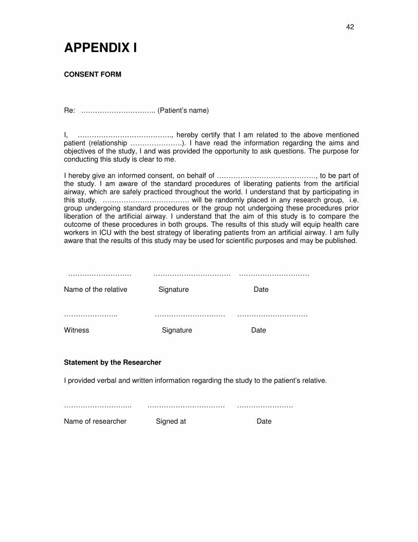
42
APPENDIX I
CONSENT FORM
Re: ………………………….. (Patient’s name) I, …………………………………., hereby certify that I am related to the above mentioned patient (relationship ………………….). I have read the information regarding the aims and objectives of the study, I and was provided the opportunity to ask questions. The purpose for conducting this study is clear to me. I hereby give an informed consent, on behalf of ……………………………………, to be part of the study. I am aware of the standard procedures of liberating patients from the artificial airway, which are safely practiced throughout the world. I understand that by participating in this study, ………………………………. will be randomly placed in any research group, i.e. group undergoing standard procedures or the group not undergoing these procedures prior liberation of the artificial airway. I understand that the aim of this study is to compare the outcome of these procedures in both groups. The results of this study will equip health care workers in ICU with the best strategy of liberating patients from an artificial airway. I am fully aware that the results of this study may be used for scientific purposes and may be published. ……………………… …………………………… ………………………… Name of the relative Signature Date ………………….. ………………………… ………………………… Witness Signature Date
Statement by the Researcher
I provided verbal and written information regarding the study to the patient’s relative. ……………………….. …………………………… …………………… Name of researcher Signed at Date
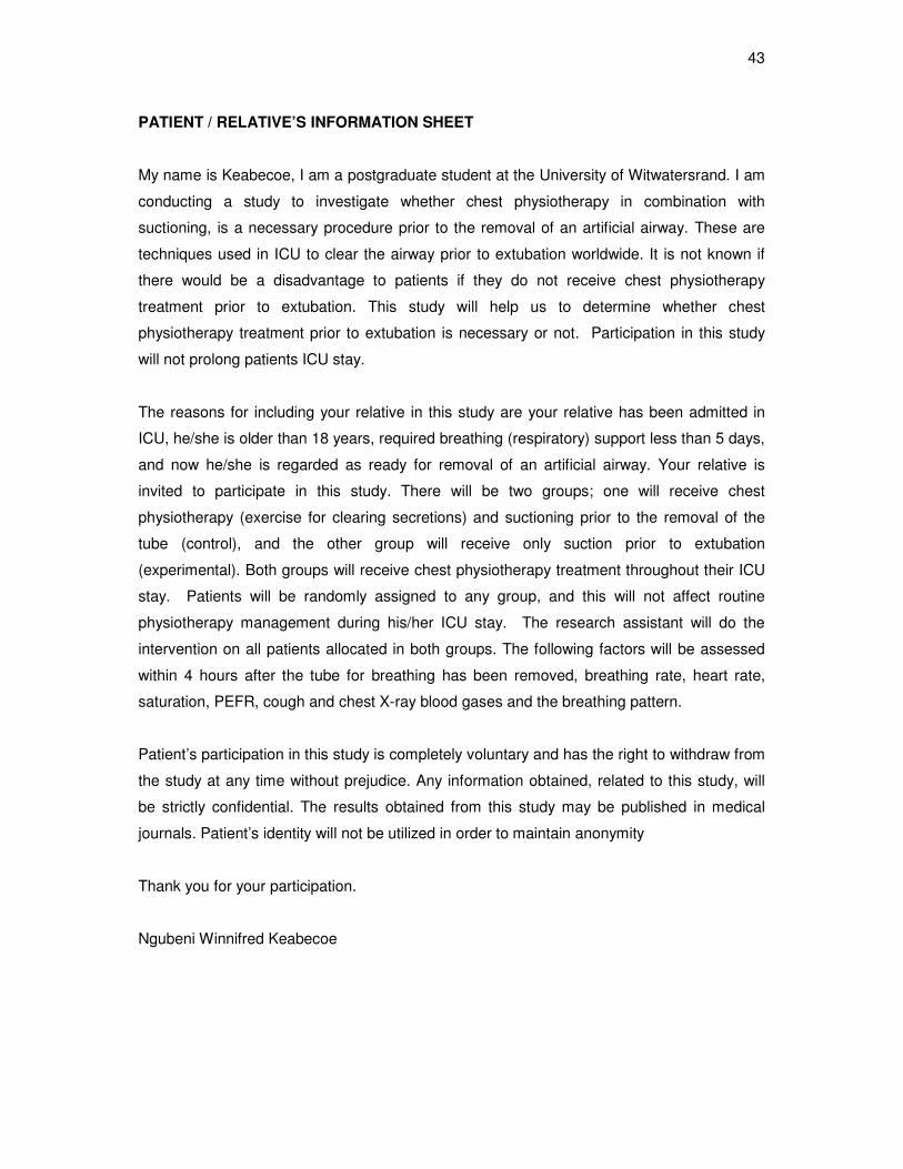
43
PATIENT / RELATIVE’S INFORMATION SHEET
My name is Keabecoe, I am a postgraduate student at the University of Witwatersrand. I am
conducting a study to investigate whether chest physiotherapy in combination with
suctioning, is a necessary procedure prior to the removal of an artificial airway. These are
techniques used in ICU to clear the airway prior to extubation worldwide. It is not known if
there would be a disadvantage to patients if they do not receive chest physiotherapy
treatment prior to extubation. This study will help us to determine whether chest
physiotherapy treatment prior to extubation is necessary or not. Participation in this study
will not prolong patients ICU stay.
The reasons for including your relative in this study are your relative has been admitted in
ICU, he/she is older than 18 years, required breathing (respiratory) support less than 5 days,
and now he/she is regarded as ready for removal of an artificial airway. Your relative is
invited to participate in this study. There will be two groups; one will receive chest
physiotherapy (exercise for clearing secretions) and suctioning prior to the removal of the
tube (control), and the other group will receive only suction prior to extubation
(experimental). Both groups will receive chest physiotherapy treatment throughout their ICU
stay. Patients will be randomly assigned to any group, and this will not affect routine
physiotherapy management during his/her ICU stay. The research assistant will do the
intervention on all patients allocated in both groups. The following factors will be assessed
within 4 hours after the tube for breathing has been removed, breathing rate, heart rate,
saturation, PEFR, cough and chest X-ray blood gases and the breathing pattern.
Patient’s participation in this study is completely voluntary and has the right to withdraw from
the study at any time without prejudice. Any information obtained, related to this study, will
be strictly confidential. The results obtained from this study may be published in medical
journals. Patient’s identity will not be utilized in order to maintain anonymity
Thank you for your participation.
Ngubeni Winnifred Keabecoe
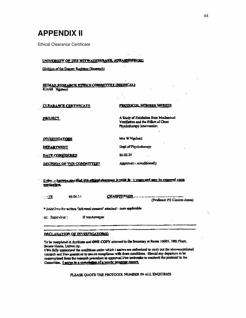
44
APPENDIX II
Ethical Clearance Certificate
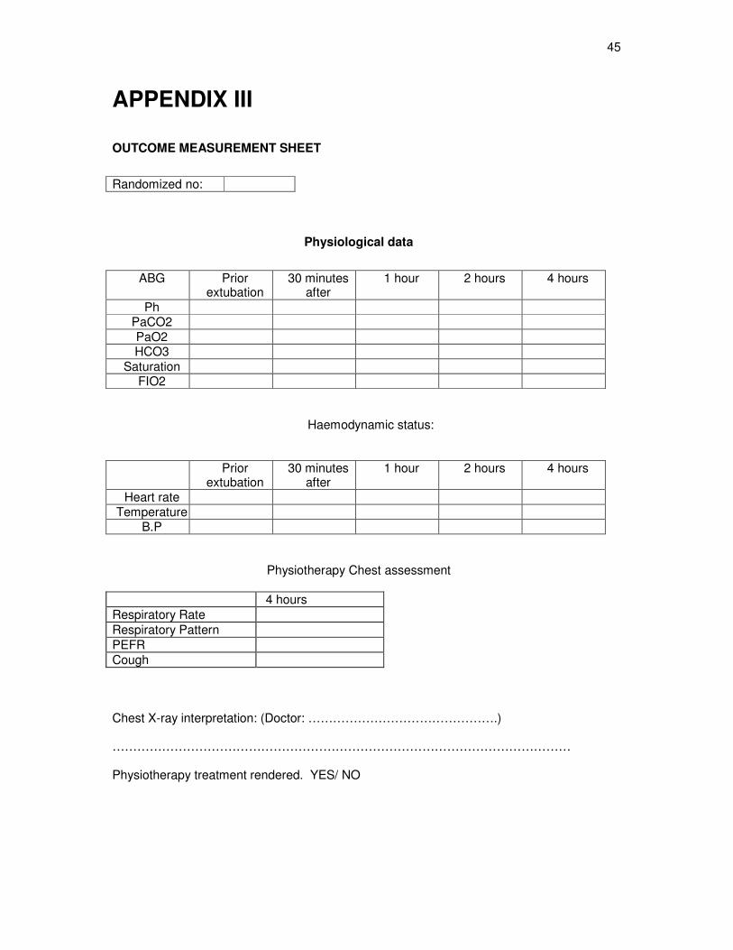
45
APPENDIX III
OUTCOME MEASUREMENT SHEET
Randomized no:
Physiological data
ABG Prior extubation
30 minutes after
1 hour 2 hours 4 hours
Ph PaCO2
PaO2 HCO3
Saturation FIO2
Haemodynamic status:
Prior
extubation 30 minutes
after 1 hour 2 hours 4 hours
Heart rate Temperature
B.P
Physiotherapy Chest assessment
1 h 4 hours
Respiratory Rate
Respiratory Pattern
PEFR
Cough
Chest X-ray interpretation: (Doctor: ……………………………………….)
………………………………………………………………………………………………… Physiotherapy treatment rendered. YES/ NO
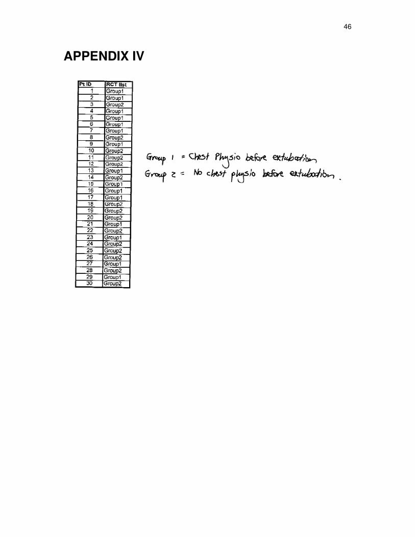
46
APPENDIX IV