A PARTIAL SKELETON OF CHINIQUODON … 2 - Oliveira et al.pdf · A PARTIAL SKELETON OF CHINIQUODON...
10
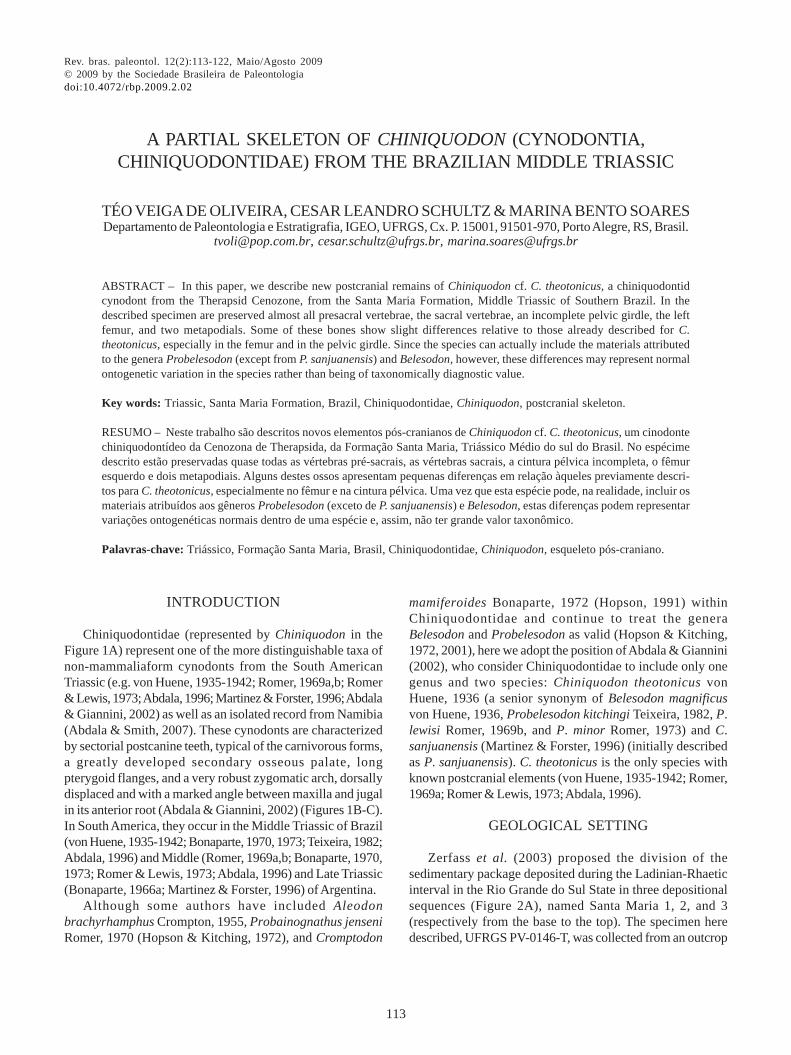
Rev. bras. paleontol. 12(2):113-122, Maio/Agosto 2009 © 2009 by the Sociedade Brasileira de Paleontologia doi:10.4072/rbp.2009.2.02 PROVAS 113 A PARTIAL SKELETON OF CHINIQUODON (CYNODONTIA, CHINIQUODONTIDAE) FROM THE BRAZILIAN MIDDLE TRIASSIC ABSTRACT – In this paper, we describe new postcranial remains of Chiniquodon cf. C. theotonicus, a chiniquodontid cynodont from the Therapsid Cenozone, from the Santa Maria Formation, Middle Triassic of Southern Brazil. In the described specimen are preserved almost all presacral vertebrae, the sacral vertebrae, an incomplete pelvic girdle, the left femur, and two metapodials. Some of these bones show slight differences relative to those already described for C. theotonicus, especially in the femur and in the pelvic girdle. Since the species can actually include the materials attributed to the genera Probelesodon (except from P. sanjuanensis) and Belesodon, however, these differences may represent normal ontogenetic variation in the species rather than being of taxonomically diagnostic value. Key words: Triassic, Santa Maria Formation, Brazil, Chiniquodontidae, Chiniquodon, postcranial skeleton. RESUMO – Neste trabalho são descritos novos elementos pós-cranianos de Chiniquodon cf. C. theotonicus, um cinodonte chiniquodontídeo da Cenozona de Therapsida, da Formação Santa Maria, Triássico Médio do sul do Brasil. No espécime descrito estão preservadas quase todas as vértebras pré-sacrais, as vértebras sacrais, a cintura pélvica incompleta, o fêmur esquerdo e dois metapodiais. Alguns destes ossos apresentam pequenas diferenças em relação àqueles previamente descri- tos para C. theotonicus, especialmente no fêmur e na cintura pélvica. Uma vez que esta espécie pode, na realidade, incluir os materiais atribuídos aos gêneros Probelesodon (exceto de P. sanjuanensis) e Belesodon, estas diferenças podem representar variações ontogenéticas normais dentro de uma espécie e, assim, não ter grande valor taxonômico. Palavras-chave: Triássico, Formação Santa Maria, Brasil, Chiniquodontidae, Chiniquodon, esqueleto pós-craniano. TÉO VEIGA DE OLIVEIRA, CESAR LEANDRO SCHULTZ & MARINA BENTO SOARES Departamento de Paleontologia e Estratigrafia, IGEO, UFRGS, Cx. P. 15001, 91501-970, Porto Alegre, RS, Brasil. [email protected], [email protected], [email protected] INTRODUCTION Chiniquodontidae (represented by Chiniquodon in the Figure 1A) represent one of the more distinguishable taxa of non-mammaliaform cynodonts from the South American Triassic (e.g. von Huene, 1935-1942; Romer, 1969a,b; Romer & Lewis, 1973; Abdala, 1996; Martinez & Forster, 1996; Abdala & Giannini, 2002) as well as an isolated record from Namibia (Abdala & Smith, 2007). These cynodonts are characterized by sectorial postcanine teeth, typical of the carnivorous forms, a greatly developed secondary osseous palate, long pterygoid flanges, and a very robust zygomatic arch, dorsally displaced and with a marked angle between maxilla and jugal in its anterior root (Abdala & Giannini, 2002) (Figures 1B-C). In South America, they occur in the Middle Triassic of Brazil (von Huene, 1935-1942; Bonaparte, 1970, 1973; Teixeira, 1982; Abdala, 1996) and Middle (Romer, 1969a,b; Bonaparte, 1970, 1973; Romer & Lewis, 1973; Abdala, 1996) and Late Triassic (Bonaparte, 1966a; Martinez & Forster, 1996) of Argentina. Although some authors have included Aleodon brachyrhamphus Crompton, 1955, Probainognathus jenseni Romer, 1970 (Hopson & Kitching, 1972), and Cromptodon mamiferoides Bonaparte, 1972 (Hopson, 1991) within Chiniquodontidae and continue to treat the genera Belesodon and Probelesodon as valid (Hopson & Kitching, 1972, 2001), here we adopt the position of Abdala & Giannini (2002), who consider Chiniquodontidae to include only one genus and two species: Chiniquodon theotonicus von Huene, 1936 (a senior synonym of Belesodon magnificus von Huene, 1936, Probelesodon kitchingi Teixeira, 1982, P. lewisi Romer, 1969b, and P. minor Romer, 1973) and C. sanjuanensis (Martinez & Forster, 1996) (initially described as P. sanjuanensis). C. theotonicus is the only species with known postcranial elements (von Huene, 1935-1942; Romer, 1969a; Romer & Lewis, 1973; Abdala, 1996). GEOLOGICAL SETTING Zerfass et al. (2003) proposed the division of the sedimentary package deposited during the Ladinian-Rhaetic interval in the Rio Grande do Sul State in three depositional sequences (Figure 2A), named Santa Maria 1, 2, and 3 (respectively from the base to the top). The specimen here described, UFRGS PV-0146-T, was collected from an outcrop
Transcript of A PARTIAL SKELETON OF CHINIQUODON … 2 - Oliveira et al.pdf · A PARTIAL SKELETON OF CHINIQUODON...
Rev. bras. paleontol. 12(2):113-122, Maio/Agosto 2009© 2009 by the Sociedade Brasileira de Paleontologiadoi:10.4072/rbp.2009.2.02
PROVAS
113
A PARTIAL SKELETON OF CHINIQUODON (CYNODONTIA,CHINIQUODONTIDAE) FROM THE BRAZILIAN MIDDLE TRIASSIC
ABSTRACT – In this paper, we describe new postcranial remains of Chiniquodon cf. C. theotonicus, a chiniquodontidcynodont from the Therapsid Cenozone, from the Santa Maria Formation, Middle Triassic of Southern Brazil. In thedescribed specimen are preserved almost all presacral vertebrae, the sacral vertebrae, an incomplete pelvic girdle, the leftfemur, and two metapodials. Some of these bones show slight differences relative to those already described for C.theotonicus, especially in the femur and in the pelvic girdle. Since the species can actually include the materials attributedto the genera Probelesodon (except from P. sanjuanensis) and Belesodon, however, these differences may represent normalontogenetic variation in the species rather than being of taxonomically diagnostic value.
Key words: Triassic, Santa Maria Formation, Brazil, Chiniquodontidae, Chiniquodon, postcranial skeleton.
RESUMO – Neste trabalho são descritos novos elementos pós-cranianos de Chiniquodon cf. C. theotonicus, um cinodontechiniquodontídeo da Cenozona de Therapsida, da Formação Santa Maria, Triássico Médio do sul do Brasil. No espécimedescrito estão preservadas quase todas as vértebras pré-sacrais, as vértebras sacrais, a cintura pélvica incompleta, o fêmuresquerdo e dois metapodiais. Alguns destes ossos apresentam pequenas diferenças em relação àqueles previamente descri-tos para C. theotonicus, especialmente no fêmur e na cintura pélvica. Uma vez que esta espécie pode, na realidade, incluir osmateriais atribuídos aos gêneros Probelesodon (exceto de P. sanjuanensis) e Belesodon, estas diferenças podem representarvariações ontogenéticas normais dentro de uma espécie e, assim, não ter grande valor taxonômico.
Palavras-chave: Triássico, Formação Santa Maria, Brasil, Chiniquodontidae, Chiniquodon, esqueleto pós-craniano.
TÉO VEIGA DE OLIVEIRA, CESAR LEANDRO SCHULTZ & MARINA BENTO SOARESDepartamento de Paleontologia e Estratigrafia, IGEO, UFRGS, Cx. P. 15001, 91501-970, Porto Alegre, RS, Brasil.
[email protected], [email protected], [email protected]
INTRODUCTION
Chiniquodontidae (represented by Chiniquodon in theFigure 1A) represent one of the more distinguishable taxa ofnon-mammaliaform cynodonts from the South AmericanTriassic (e.g. von Huene, 1935-1942; Romer, 1969a,b; Romer& Lewis, 1973; Abdala, 1996; Martinez & Forster, 1996; Abdala& Giannini, 2002) as well as an isolated record from Namibia(Abdala & Smith, 2007). These cynodonts are characterizedby sectorial postcanine teeth, typical of the carnivorous forms,a greatly developed secondary osseous palate, longpterygoid flanges, and a very robust zygomatic arch, dorsallydisplaced and with a marked angle between maxilla and jugalin its anterior root (Abdala & Giannini, 2002) (Figures 1B-C).In South America, they occur in the Middle Triassic of Brazil(von Huene, 1935-1942; Bonaparte, 1970, 1973; Teixeira, 1982;Abdala, 1996) and Middle (Romer, 1969a,b; Bonaparte, 1970,1973; Romer & Lewis, 1973; Abdala, 1996) and Late Triassic(Bonaparte, 1966a; Martinez & Forster, 1996) of Argentina.
Although some authors have included Aleodonbrachyrhamphus Crompton, 1955, Probainognathus jenseniRomer, 1970 (Hopson & Kitching, 1972), and Cromptodon
mamiferoides Bonaparte, 1972 (Hopson, 1991) withinChiniquodontidae and continue to treat the generaBelesodon and Probelesodon as valid (Hopson & Kitching,1972, 2001), here we adopt the position of Abdala & Giannini(2002), who consider Chiniquodontidae to include only onegenus and two species: Chiniquodon theotonicus vonHuene, 1936 (a senior synonym of Belesodon magnificusvon Huene, 1936, Probelesodon kitchingi Teixeira, 1982, P.lewisi Romer, 1969b, and P. minor Romer, 1973) and C.sanjuanensis (Martinez & Forster, 1996) (initially describedas P. sanjuanensis). C. theotonicus is the only species withknown postcranial elements (von Huene, 1935-1942; Romer,1969a; Romer & Lewis, 1973; Abdala, 1996).
GEOLOGICAL SETTING
Zerfass et al. (2003) proposed the division of thesedimentary package deposited during the Ladinian-Rhaeticinterval in the Rio Grande do Sul State in three depositionalsequences (Figure 2A), named Santa Maria 1, 2, and 3(respectively from the base to the top). The specimen heredescribed, UFRGS PV-0146-T, was collected from an outcrop

REVISTA BRASILEIRA DE PALEONTOLOGIA, 12(2), 2009114
PROVAS
Figure 1. The Chiniquodontidae. A, simplified phylogeny of the Cynodontia, where the Chiniquodontidae are represented by Chiniquodon(modified from Abdala, 2007); B-C, general skull morphology of a chiniquodontid, in lateral and palatal views, respectively (modified fromRomer, 1969b), with the main characters of the family labeled. Abbreviations are presented in Material section.
belonging to the basal portion of the Sequence Santa Maria1 (Ladinian), composed of massive or finely laminated reddishmudstones and some carbonatic concretionary levels,corresponding to a floodplain facies (Machado, 2004; Rubert& Schultz, 2004). This stratigraphic level bears thecharacteristic fossils of the Therapsid Cenozone, with thepredominance of the dicynodont Dinodontosaurus (see vonHuene, 1935-1942), followed by cynodonts (mainlyMassetognathus) (Barberena, 1974, 1981b; Teixeira, 1987) and“rauisuchians” as Prestosuchus (see von Huene, 1935-1942).Other components of the fauna include rare rhynchosaurs(Schultz & Langer, 2000), proterochampsids (cf. ChanaresuchusDornelles, 1995) and procolophonids (Candelaria Cisneros etal., 2004). Among cynodonts, in addition to Chiniquodontheotonicus and Massetognathus ochagaviae Barberena, 1981b,
the traversodontids Traversodon stahleckeri von Huene, 1935-1942 (see Barberena, 1981a) and Protuberum cabralensisReichel, Schultz & Soares, 2009 and the small probainognathianProtheriodon estudianti Bonaparte, Soares & Schultz, 2006 arepresent in the Therapsid Cenozone.
The final portion of the Sequence Santa Maria 1 (still inLadinian) shows a distinct fossiliferous assemblage namedTraversodontid Biozone (Abdala et al., 2001) (Figure 2B).The Therapsid Cenozone is correlated to the Los ChañaresFormation of Argentina (Rubert, 2003; Rubert & Schultz, 2004),from where were also recovered dicynodonts (e.g.Dinodontosaurus; see Cox, 1968) and the cynodontsMassetognathus (Romer, 1967; Jenkins, 1970; Abdala &Giannini, 2000) and Chiniquodon (Bonaparte, 1966a; Romer,1969b, 1973). Thus, it is possible to assign the Brazilian

115OLIVEIRA ET AL. – CHINIQUODON (CYNODONTIA, CHINIQUODONTIDAE) FROM SOUTHERN BRAZIL
PROVAS
Therapsid Cenozone an Early to Middle Chañarense age(Rubert & Schultz, 2004) (Figure 2B).
MATERIAL
The specimen UFRGS PV-0146-T was collected in themunicipality of Candelária, Rio Grande do Sul State, Brazil(Figure 3), approximately 200 km northwest of Porto Alegre,and consists of a cranium and mandible, twenty-six presacralvertebrae (probably lacking just the atlas), four sacralvertebrae, the incomplete right branch of the pelvic girdlearticulated to the corresponding sacral ribs, the disarticulatedleft acetabular portion of the pelvic girdle, the left femur, twosmall rounded bones probably representing carpals and/ortarsals, two metapodials, and a series of fragmented elementsincluding ribs and several undetermined bones.Anatomical abbreviations. ac, acetabulum; asi, articularsurface for the intercentra; ax, axis; cf, cranium fragment; cp,coronoid process of dentary; d, dentary; e, epipterygoid; f,frontal; fh, femoral head; gt, greater trochanter; itf,intertrochanteric fossa; j, jugal; l, lacrimal; lc, lateral condyleof femur; lt, lesser trochanter; m, maxilla; mc, medial condyleof femur; mf, mandible fragment; n, nasal; nf, notochordalfossa; o+ob, opisthotics and occipital bones; p, parietal; pb,post-dentary bones; pbs+pr+pt, parabasisphenoid, prooticsand pterygoids; pf, pterygoid flange; pfs, paracanine fossa;pl, palatine; pm, premaxilla; po, postorbital; pof, popliteal fos-sa; pr, prootic; prf, prefrontal; ps, patellar sulcus; psv, presacralvertebra; q+qj, quadrate and quadratojugal; sm, septomaxilla;sq, squamosal; sv, sacral vertebra.Institutional abbreviations. UFRGS PV-T, Universidade Fe-
deral do Rio Grande do Sul, Paleontologia de Vertebrados-Triássico, Porto Alegre, Rio Grande do Sul, Brasil.
SYSTEMATIC PALEONTOLOGY
Cynodontia Owen, 1861Eucynodontia Kemp, 1982
Chiniquodontidae von Huene, 1936Chiniquodon von Huene, 1936
Chiniquodon cf. C. theotonicus von Huene, 1936(Figures 4-8)
DescriptionThe specimen UFRGS PV-0146-T is assigned to the genus
Chiniquodon due the presence of a greatly posteriorlydeveloped secondary osseous palate and the marked anglebetween the anteroventral margin of the zygomatic arch and themaxillary zygomatic root (Figures 4A-C). In addition to thesefeatures, the snout is relatively short and stout and the zygomaticarch is very deep (Abdala & Giannini, 2002). Unfortunately thecrowns of the postcanine teeth are broken and theirpresumable sectorial morphology could not be observed.Vertebrae. Here we assume that Chiniquodon cf. C.theotonicus had twenty-seven presacral vertebrae, a frequentcondition among the non-mammaliaform cynodonts (Romer,1956; Romer & Lewis, 1973). The axis, the third, and the fourthvertebrae are articulated, but isolated from the remainingcolumn. The fifth to twenty-sixth presacrals and the foursacrals are articulated. Several rib fragments are associatedto the corresponding vertebrae.
In cynodonts, the limit between cervical and dorsal
Figure 2. Stratigraphic context of Brazilian Triassic. A, sequence stratigraphy showing the Therapsid Cenozone (coloured) (modified fromZerfass et al., 2003); B, comparative bio- and lithostratigraphy between Brazilian and Argentine Triassic [modified from Rubert & Schultz(2004) and Schultz & Soares (2006)].

REVISTA BRASILEIRA DE PALEONTOLOGIA, 12(2), 2009116
PROVAS
vertebrae is marked by the loss of intercentra, but as nointercentra are preserved in this specimen this distinctioncannot be made. On the other hand, the distinction betweenthoracic and lumbar regions in the non-mammaliaformcynodonts is recognized by differences in the morphologyof the ribs associated to the vertebrae. In several taxa [like inThrinaxodon, Cynognathus, and others (see Jenkins, 1971)],the vertebrae are named thoracic when the diaphysis of thecorresponding ribs runs beyond the costal plate or lumbarwhen the most terminal element of the rib is that plate (Brink,1955; Jenkins, 1971). UFRGS PV-0146-T exhibits no costalspecializations, so it is not possible to distinguish thoracicand lumbar vertebrae in this specimen. Thus, all the vertebraehere described will be named “presacral vertebrae”.
The atlas was not preserved and the axis centrum isdamaged (Figure 5). The centra of the following presacralvertebrae are very similar, approximately cylindrical, withsalient anterior and posterior rims (Figures 5-7). The anterioror posterior faces are exposed in very few vertebral centra,but in those where it is possible a relatively deep notochordalfossa can be observed (Figure 5B) and very probably all thecentra were amphicoelous, except that of the atlas. In the
anterior portion of the ventral surface of the sixth to eighthpresacral vertebrae there is a salience that could representthe attachment site for the corresponding intercentrum (‘asi’in Figure 7), although none of these elements were recovered.
In general, the presacral neural spines are similar exceptfor the axis, which exhibits a blade-like morphology with broadlateral surfaces and anteroposterior elongation (Figure 5A).The remaining neural spines are more slender andanteroposteriorly narrower (Figures 6-7). In the thirteenthpresacral vertebra the neural spine becomes backwardly inclinedabout 25-30° and this inclination is accentuated in the twenty-first presacral, where the neural spine is inclined about 40-45°.Starting at this point, the neural spines become less inclined andin the twenty-sixth vertebra it is almost vertical (Figures 6-7).
The articular facets of the zygapophyses in the more an-terior vertebrae are inclined at an angle about 45°; towardsthe sacrum the surface of prezygapophyses splits in twoareas, one dorsally oriented and other medially directed (see‘psv20’ in the Figure 6). Consequently, the postzygapophysesalso split their articular facet in one surface ventrally directedand another one more lateral.
The transverse processes of the presacral vertebrae are
Figure 3. Geographic location of the study area. A, Rio Grande do Sul State location in South America with outcropping unities (Mesozoicrocks in solid grey); B, road map with Candelária town pointed.

117OLIVEIRA ET AL. – CHINIQUODON (CYNODONTIA, CHINIQUODONTIDAE) FROM SOUTHERN BRAZIL
PROVAS
short and stout (Figures 6-7) becoming gradually shorter upto the more posterior vertebrae. In the more anterior vertebraethese structures are backwardly directed about 30°, becomingless inclined in the posterior region of the column. In most ofthe presacral vertebrae it is possible to visualize theparapophyseal notches in the anterior and posterior bordersof the vertebral centra. However, in some of the middle trunkvertebrae there is continuity between the diapophysis (inthe transverse process) and the lateral surface of the anteriorportion of the centrum, resulting in a single area to ribattachment, as a synapophysis. The synapophyses areanteriorly restricted by a descending anterior flange of thetransverse process [like the condition described e.g. by Kemp(1980a) in Luangwa, although this author had not named thecorresponding structure as synapophysis, by Reichel et al.(2009) in the more posterior presacral vertebrae ofProtuberum].
There are four preserved sacral vertebrae, with the
respective ribs associated to the right ilium (Figure 6). Thefourth sacral rib is associated with the posterior end of iliacblade, which suggest that only four sacral vertebrae werepresent in this specimen, a relatively common number amongnon-mammaliaform cynodonts (e.g. Bonaparte, 1963; Romer,1956). The sacral zygapophyses are firmly articulated to eachother, maybe even fused, and probably the movements inthis region were very restricted or nonexistent and the distalends of sacral ribs were probably also synostosed. The centraof the sacral vertebrae do not differ significantly from thoseof presacral vertebrae and probably are also amphicoelous.The neural spines are quite vertical (Figure 6) and thetransverse processes cannot be distinguished from the ribs.Ribs. There are not complete presacral ribs preserved, onlysome fragments attached to the vertebrae (Figures 6-7). Theonly ribs with remarkable features are the sacrals, with theirobvious adaptations to ilium attachment (Figure 6). The firsttwo ribs have an articular surface for the ilium approximately
Figure 4. Skull of Chiniquodon cf. C. theotonicus (UFRGS PV-0146-T). A-C, cranium in dorsal, palatal and left lateral views, respectively;D, mandible in left lateral view. Abbreviations are presented in Material section. Scale bars = 5 cm.

REVISTA BRASILEIRA DE PALEONTOLOGIA, 12(2), 2009118
PROVAS
quadrangular and facing dorsolaterally while in the third onethis surface is more rectangular and faces more laterally; thesame occurs in the fourth rib, although it is dorsoventrallynarrower. Thus, the whole articular surface for ilium changesfrom an inclined orientation, in the first two sacral ribs, to avertical one, more typical of non-mammaliaform cynodonts,in the two posterior ribs.Pelvic girdle. The right branch of the pelvic girdle is thebetter preserved (Figure 7), while the left one had preservedonly the acetabular portion and a little part of the ischium.The acetabulum is almost circular. The puboischiadic plate ispoorly preserved, so the condition of the obturator foramenis not clear. The iliac blade seems very concave, but this isclearly a taphonomic distortion, once the column is curved atthe sacral region and the ilium was pressed against thevertebrae. The preacetabular portion is more developed thanthe postacetabular one, although this latter is still great. Theilium bears an almost ventrally directed acetabular facet, witha supracetabular buttress dorsoanteriorly placed relative tothe acetabulum. The ischium has an anterolaterally directedacetabular facet edged by a postacetabular buttress, althoughsmaller than the supracetabular one. In cross-section theischium is triangular, with an ischiatic crest dividing the late-ral surface of the bone in two well defined areas, one dorsaland other ventrally directed. The better preserved portion ofthe pubis is the acetabular one, with a dorsoposteriorlydirected acetabular facet.Femur. In proximal view, the femoral head and the greatertrochanter are continuous (Figure 8). The head is medially
Figure 5. Presacral vertebrae 2 (axis) to 4 (UFRGS PV-0146-T), inright lateral (A) and caudal (B) (only fourth vertebra detailed) views.Abbreviations are presented in Material section. Scale bar = 5 cm.
Figure 6. Presacral vertebrae 5 to 26, sacral vertebrae 1 to 4 and right pelvic bones (UFRGS PV-0146-T) in dorsal (presacral 5 to neuralarch of presacral 20) and left lateral (the remaining bones) views. Abbreviations are presented in Material section. Scale bar = 10 cm.

119OLIVEIRA ET AL. – CHINIQUODON (CYNODONTIA, CHINIQUODONTIDAE) FROM SOUTHERN BRAZIL
PROVAS
and slightly dorsally displaced. Posteroventrally, there is awell developed intertrochanteric fossa; medially to this fos-sa is the lesser trocanter. Ventrodistally, there is a shallowpopliteal fossa. The medial condyle is more posteroventrallyprojected than the posterior one. The condylar articularsurface forms an angle about 45° relative to the long axis offemur, a generic adaptation for a semi-upright posture in thehind limbs (Jenkins, 1971).
DISCUSSION
Vertebrae. In the more anterior presacral vertebrae, as in thesixth one, are visible two small longitudinal ventral keels.Most other non-mammaliaform cynodonts [e.g. Thrinaxodon,Galesaurus and Cynognathus (Jenkins, 1971)], have no keelin this region, while Oligokyphus (Kühne, 1956) has a singlekeel, as occurs in the seventh presacral vertebrae of thespecimen here described. These anterior vertebrae do notdiffer from those of the middle and posterior regions of thetrunk, without the anteroposterior shortening observed inmore advanced cynodonts, such as Therioherpeton andProzostrodon (Bonaparte & Barberena, 2001) andmammaliaforms (Kielan-Jaworowska et al., 2004).
None of the presacral vertebrae are expanded at the apexof their neural spines, unlike the condition in, e.g. inExaeretodon frenguellii Cabrera, 1943 (see Bonaparte, 1963)and Kayentatherium (Lewis, 1986; Sues & Jenkins, 2006).Besides, the specimen UFRGS PV-0146-T [as well asProcynosuchus (Kemp, 1980b), Exaeretodon (Bonaparte,1963; Oliveira, 2006; Oliveira et al., 2007) andProbainognathus (Abdala, 1996)] also lack the anapophyses
assigned for cynodonts as Thrinaxodon (Jenkins, 1971),Diademodon (Brink, 1955), and Oligokyphus (Kühne, 1956).The presacral vertebrae do not differ significantly from thosedescribed by von Huene (1935-1942) for C. theotonicus.
As mentioned above, the four sacral vertebrae of thespecimen UFRGS PV-0146-T represent a frequent conditionamong the non-mammaliaform cynodonts (Romer, 1956;Kielan-Jaworowska et al., 2004), like occur e.g. in Diademodon(Gow & Grine, 1979), Therioherpeton (Bonaparte & Barberena,2001), Kayentatherium (Lewis, 1986) and Oligokyphus(Kühne, 1956). In spite of this, Romer & Lewis (1973) claimsthat Chiniquodon has actually five sacral vertebrae, whichcould be a result of the incorporation of a lumbar or a caudalvertebra to the sacrum (Bonaparte, 1963; Oliveira et al., 2006),probably in senile individuals. On the other hand, von Huene(1935-1942) attributed to Chiniquodon only three sacralvertebrae.
There are no significant differences among the sacralneural spines relative to those of the posterior presacralvertebrae, unlike the described e.g. for Thrinaxodon andCynognathus (Jenkins, 1971), Massetognathus (Jenkins,1970), and Exaeretodon (Bonaparte, 1963; Oliveira, 2006; Oli-veira et al., 2007), which have shorter and anteroposteriornarrower sacral spines.Ribs. In the preserved presacral ribs of the specimen UFRGSPV-0146-T there is no costal specialization, like in all knownprobainognathian (Hopson & Kitching, 2001). On the otherhand, several species of non-probainognathian cynodonts(but not all of them) show expanded ribs [e.g. Galesaurus(Parrington, 1934), Thrinaxodon (Jenkins, 1971), Diademodon(Brink, 1955), Cynognathus (Broom, 1914), as well as some
Figure 7. Presacral vertebrae 5 to 26, sacral vertebrae 1 to 4 and right pelvic bones (UFRGS PV-0146-T) in ventral (presacral 5 to neuralarch of presacral 20) and right lateral (the remaining bones) views. Abbreviations are presented in Material section. Scale bar = 10 cm.

REVISTA BRASILEIRA DE PALEONTOLOGIA, 12(2), 2009120
PROVAS
traversodontids (Bonaparte, 1966b, 1970; Jenkins, 1970;Kemp, 1980a)].
In the sacral ribs the most remarkable feature is the changeof the dorsolateral inclination of the articular surface for theilium in the anterior ribs to a more lateral orientation in thetwo more posterior ribs. This morphology is similar to thatobserved in Exaeretodon (Bonaparte, 1963; Oliveira, 2006;Oliveira et al., 2007) and is explained by the correspondingmorphology of the medial face of the iliac blade, which ismedioventrally directed in its anterior half and shifts mediallyin the posterior region.Pelvic girdle. The specimen UFRGS PV-0146-T shows apelvic morphology similar to Cynognathus and Diademodon(Jenkins, 1971), although its ilium has a slightly greaterpreacetabular portion than the African taxa. The pelvic girdleof Chiniquodon presented by von Huene (1935-1942) has aslightly more elongated postacetabular region. This conditiondiffers drastically from that observed in severaltraversodontids, e.g. Exaeretodon (Bonaparte, 1963) andMenadon (Kammerer et al., 2008), where the postacetabulararea of the iliac blade is greatly reduced or in some moreadvanced cynodonts, such as Prozostrodon (Bonaparte &Barberena, 2001), where this region is completely absent.
Femur. The femur shows similarities with Cynognathus andDiademodon (Jenkins, 1971), like the continuity between thehead and the greater trochanter, the relatively deep ventralintertrochanteric fossa, the presence of a smooth crest arisingin the lesser trocanter that runs up to the lateral condyle, anda more ventrally projected anterior condyle. The first of thesecharacters also appears in several cynodonts, such asExaeretodon (Bonaparte, 1963) and Therioherpeton(Bonaparte & Barberena, 2001). The referred crest wasassigned also for Exaeretodon (Bonaparte, 1963) andMassetognathus (Jenkins, 1970).
The femur UFRGS PV-0146-T is more advanced than thoseof e.g. Procynosuchus (Kemp, 1980b), Thrinaxodon (Jenkins,1971), and Massetognathus (Jenkins, 1970), i.e., isproportionally more slender and long and has its head moremedially displaced. On the other hand, it is considerably moreprimitive relative to the advanced pattern observed in e.g.Oligokyphus (Kühne, 1956) and Morganucodon (Jenkins &Parrington, 1976), where the femur is even more slender andthe head is very spherical and separated from the greattrocanter by a notch. The femur is similar to those describedby von Huene (1935-1942) for other specimens ofChiniquodon.
Figure 8. Left femur (UFRGS PV-0146-T) in cranial (A), caudal (B), lateral (C), medial (D), proximal (E) and distal (F) views. Abbreviationsare presented in Material section. Scale bar = 5 cm.

121OLIVEIRA ET AL. – CHINIQUODON (CYNODONTIA, CHINIQUODONTIDAE) FROM SOUTHERN BRAZIL
PROVAS
CONCLUSIONS
Some authors had described postcranial remains of thegenera Belesodon (von Huene, 1935-1942), Probelesodon(Romer & Lewis, 1973) and Chiniquodon (von Huene, 1935-1942; Romer, 1969a) where was possible to visualize somedifferences between these taxa. However, after the criticalevaluation of the taxonomic status of the species included inthese genera, presented by Abdala & Giannini (2002), andaccepting the assumption that these three correspondactually to a single genus, Chiniquodon, and representingdistinct ontogenetic stages of a single species, C.theotonicus, those postcranial differences must be re-evaluated. Two hypotheses could be presented: thedifferences could be consequence of individual (intraspecific)variation, once there are very few specimens to establish aconsistent morphological pattern to the species, or theyactually reflect distinct ontogenetic stages represented inthe fossil record. The specimen UFRGS PV-0146-T show somedifferences relative to the postcranial remains alreadydescribed as slight modifications in the pelvic and femoralmorphology, which can be attributed to the “normal” rangeof variation of the species.
ACKNOWLEDGMENTS
We thanks to CAPES (Coordenação de Aperfeiçoamentode Pessoal de Nível Superior) and CNPq (Conselho Nacionalde Desenvolvimento Científico e Tecnológico) for fundingthis study. We also thank two anonymous referees whoreviewed an early draft of this paper.
REFERENCES
Abdala, F. 1996. Los Chiniquodontoideos (Synapsida, Cynodontia)Sudamericanos. Universidad Nacional de Tucumán, PhDThesis, 381 p.
Abdala, F. 2007. Redescription of Platycraniellus elegans(Therapsida, Cynodontia) from the Lower Triassic of SouthAfrica, and the cladistic relationships of eutheriodonts.Palaeontology, 50(3):591-618.
Abdala, F. & Giannini, N.P. 2000. Gomphodont cynodonts of theChañares Formation: the analysis of an ontogenetic sequence.Journal of Vertebrate Paleontology, 20(3):501-506.
Abdala, F. & Giannini, N.P. 2002. Chiniquodontid cynodonts:systematic and morphometric considerations. Palaeontology,45(6):1151-1170.
Abdala, F. & Smith, R.M.H. 2007. Gondwanan Middle Triassiccynodonts from Namibia. Palaeontologia Africana, 42:117.
Barberena, M.C. 1974. Contribuição ao Conhecimento dosCinodontes Gonfodontes (Cynodontia, Tritylodontoidea) doBrasil. Universidade Federal do Rio Grande do Sul, Tese deLivre Docência, 194 p.
Barberena, M.C. 1981a. Novos materiais de Traversodon stahleckerida Formação Santa Maria (Triássico do Rio Grande do Sul).Pesquisas, 14:149-162.
Barberena, M.C. 1981b. Uma nova espécie de Massetognathus(Massetognathus ochagaviae, sp.nov.) da Formação SantaMaria, Triássico do Rio Grande do Sul. Pesquisas, 14:181-195.
Bonaparte, J.F. 1963. Descripción del esqueleto postcraneano deExaeretodon. Acta Geológica Lilloana, 4:5-52.
Bonaparte, J.F. 1966a. Chiniquodon Huene (Therapsida -
Cynodontia) en el Triásico de Ischigualasto, Argentina. ActaGeologica Lilloana, 8:5-31.
Bonaparte, J.F. 1966b. Una nueva “fauna” Triásica de Argentina(Therapsida: Cynodontia Dicynodontia). Consideracionesfilogenéticas y paleobiogeográficas. Ameghiniana, 4(8):243-296.
Bonaparte, J.F. 1970. Annotated list of the South American Triassictetrapods. In: GONDWANA SYMPOSIUM, 2, 1970.Proceedings and Papers, Pretoria, International Union ofGeological Sciences, p. 665-682.
Bonaparte, J.F. 1972. Cromptodon mamiferoides gen. et sp. nov.,Galesauridae de la Formación Río Mendoza, Mendoza, Argen-tina. (Therapsida - Cynodontia). Ameghniana, 9(4):343-353.
Bonaparte, J.F. 1973. Edades/réptil para el Triásico de Argentina yBrasil. In: CONGRESO GEOLÓGICO ARGENTINO, 5, 1973.Actas, vol. 3, Buenos Aires, Asociación Geológica Argentina, p.93-129.
Bonaparte, J.F. & Barberena, M.C. 2001. On two advancedcarnivorous cynodonts from the late Triassic of Southern Brazil.Bulletin of the Museum of Comparative Zoology, 156(1):59-80.
Bonaparte, J.F.; Soares, M.B. & Schultz, C.L. 2006. A new non-mammalian cynodont from the Middle Triassic of SouthernBrazil and its implications for the ancestry of mammals. TheTriassic-Jurassic Terrestrial Transition, New Mexico Museumof Natural History and Science Bulletin, 37:599-607.
Brink, A.S. 1955. A study on the skeleton of Diademodon.Palaeontologia Africana, 3:3-39.
Broom, R. 1914. On the theriodonts in the Albany Museum.Records of the Albany Museum, 1(2):82-87.
Cabrera, A. 1943. El primer hallazgo de terápsidos en la Argentina.Notas del Museo de La Plata, 8:317-333.
Cisneros, J.C.; Damiani, R.; Schultz, C.; Rosa, A.; Schwanke, C.;Neto, L.W. & Aurelio, P.L.P. 2004. A procolophonid reptilewith temporal fenestration from the Middle Triassic of Brazil.Proceedings of the Royal Society of London, B, 271:1541-1546.
Cox, C.B. 1968. The Chañares (Argentina) Triassic reptile fauna.IV. The dicynodont fauna. Breviora, 295:1-27.
Crompton, A.W. 1955. On some Triassic cynodont fromTanganyika. Proceedings of the Zoological Society of London,125:617-669.
Dornelles, J.E.F. 1995. Um tecodonte proterosuchídeo(Chanaresuchus sp.) do Triássico do Rio Grande do Sul. Co-municações do Museu de Ciências e Tecnologia UBEA/PUCRS(Série Ciências da Terra), 1:63-68.
Gow, C.E. & Grine, F.E. 1979. An articulated skeleton of a smallindividual of Diademodon (Therapsida; Cynodontia).Palaeontologia Africana, 22:29-34.
Hopson, J.A. 1991. Systematics of the nonmammalian Synapsidaand implications for patterns of evolution in Synapsida. In: H.-D. Schultze & L. Trueb (eds.) Origin of the higher groups oftetrapods: controversy and consensus, Comstock PublishingAssociates, p. 635-693.
Hopson, J.A. & Kitching, J.W. 1972. A revised classification ofcynodonts (Reptilia: Therapsida). Palaeontologia Africana,14:71-85.
Hopson, J.A. & Kitching, J.W. 2001. A probainognathian cynodontfrom South Africa and the phylogeny of nonmammaliancynodonts. Bulletin Museum of Comparative Zoology,156(1):5-35.
Huene, F.F. von. 1935-1942. Die Fossilen Reptilien desSudamerikanischen Gondwanalandes: Ergebnisse derSauriergrabuiigen in Südbrazilien 1928/1929. Munich,Beck’sche Verlagsbuchhandlung, 332 p.
Huene, F.F. von. 1936. Lieferung 2. Cynodontia. In: F.F. von Huene(ed.) Die fossilen Reptilien des SüdamerikanischenGondwanalandes. Ergebnisse der Sauriergrabungen in

REVISTA BRASILEIRA DE PALEONTOLOGIA, 12(2), 2009122
PROVAS
Südbrasilien 1928/29. Beck’sche Verlagsbuchhandlung, p. 83-160.
Jenkins, F.A. Jr. 1970. The Chañares (Argentina) Triassic reptilefauna VII. The postcranial skeleton of the traversodontidMassetognathus pascuali (Therapsida, Cynodontia). Breviora,352:1-28.
Jenkins, F.A. Jr. 1971. The Postcranial Skeleton of AfricanCynodonts: Problems in the Early Evolution of the MammalianPostcranial Skeleton. Peabody Museum of Natural History,216 p. (Bulletin 36).
Jenkins, F.A. Jr. & Parrington, F.R. 1976. The postcranial skeletonsof the Triassic mammals Eozostrodon, Megazostrodon andErythrotherium. Philosophical Transactions of the Royal Societyof London, Series B, Biological Sciences, 273:387-431.
Kammerer, C.F.; Flynn, J.J.; Ranivoharimanana, L. & Wyss, A.2008. New material of Menadon besairiei (Cynodontia:Traversodontidae) from the Triassic of Madagascar. Journal ofVertebrate Paleontology, 28(2):445-462.
Kemp, T.S. 1980a. Aspects of the structure and functional anatomyof the Middle Triassic cynodont Luangwa. Journal of Zoology,191:193-239.
Kemp, T.S. 1980b. The primitive cynodont Procynosuchus:structure, function and evolution of the postcranial skeleton.Philosophical Transactions of the Royal Society of London,288:217-258.
Kemp, T.S. 1982. Mammal-like reptiles and the origin of mammals.New York, Academic Press, 362 p.
Kielan-Jaworowska, Z.; Cifelli, R.L. & Luo, Z.-X. 2004. Mammalsfrom the age of dinosaurs: origins, evolution and structure.New York, Columbia University Press, 630 p.
Kühne, W.G. 1956. The Liassic Therapsid Oligokyphus. London,British Museum (Natural History), 149 p.
Lewis, G.E. 1986. Nearctylodon broomi the first neartic tritylodont.In: N. Hotton III; P.D. MacLean; J.J. Roth & C. Roth (eds.)The Ecology and Biology of Mammal-like Reptiles. SmithsonianInstitution Press, p. 295-303.
Machado, C.B. 2004. Concentrações fossilíferas controladas pelonível freático: um modelo tafonômico para reconstruçõespaleoambientais. Programa de Pós-graduação em Geociências,Universidade Federal do Rio Grande do Sul, Dissertação deMestrado, 73 p.
Martinez, R.N. & Forster, C.A. 1996. The skull of Probelesodonsanjuanensis, sp. nov., from the Late Triassic IschigualastoFormation of Argentina. Journal of Vertebrate Paleontology,16(2):285-291.
Oliveira, T.V. 2006. Descrição osteológica de materiais pós-cranianos de dois cinodontes não-mamalianos do Meso/Neotriássico (Formação Santa Maria, Bacia do Paraná) doRio Grande do Sul, Brasil. Programa de Pós-graduação emGeociências Universidade Federal do Rio Grande do Sul, Dis-sertação de Mestrado, 137 p.
Oliveira, T.V.; Schultz, C.L. & Soares, M.B. 2006. Discusión sobrela ocurrencia de homoplasias en el esqueleto postcraneano decinodontes no mamalianos (Therapsida, Cynodontia).Ameghiniana, 43(4-Suplemento):57R-58R.
Oliveira, T.V.; Schultz, C.L. & Soares, M.B. 2007. O esqueletopós-craniano de Exaeretodon riograndensis Abdala et al.(Cynodontia, Traversodontidae), Triássico do Brasil. RevistaBrasileira de Paleontologia, 10(2):79-94.
Owen, R. 1861. Palaeontology, or a systematic summary of extinctanimals and their geologicalrRelations. Edinburgh, Adam andCharles Black, 463 p.
Parrington, F.R. 1934. On the cynodont genus Galesaurus, with anote on the functional significance of the changes in the evolutionof the theriodont skull. Annals and Magazine of Natural History,10:38-67.
Reichel, M.; Schultz, C.L. & Soares, M.B. 2009. A newtraversodontid (Therapsida, Eucynodontia) from the MiddleTriassic Santa Maria Formation of Rio Grande do Sul, Brazil.Palaeontology, 52(1):229-250.
Romer, A.S. 1956. Osteology of the Reptiles. Chicago, The Universityof Chicago Press, 772 p.
Romer, A.S. 1967. The Chañares (Argentina) Triassic Reptile Fauna.III. Two New Gomphodonts, Massetognathus pascuali and M.teruggii. Breviora, 264:1-25.
Romer, A.S. 1969a. The Brazilian Triassic cynodont reptilesBelesodon and Chiniquodon. Breviora, 332:1-16.
Romer, A.S. 1969b. The Chañares (Argentina) Triassic reptile faunaV. A new chiniquodontid cynodont, Probelesodon lewisi -cynodont ancestry. Breviora, 333:1-24.
Romer, A.S. 1970. The Chañares (Argentina) Triassic reptile fauna.VI. A chiniquodontid cynodont with an incipient squamosal-dentary jaw articulation. Breviora, 334:1-18.
Romer, A.S. 1973. The Chañares (Argentina) Triassic reptile fauna.XVIII. Probelesodon minor, a new species of carnivorouscynodont; family Probainognathidae nov. Breviora, 401:1-4.
Romer, A.S. & Lewis, A.D. 1973. The Chañares (Argentina) Triassicreptile fauna XIX. Postcranial materials of the cynodontsProbelesodon and Probainognathus. Breviora, 407:1-26.
Rubert, R.R. 2003. Possibilidades de estabelecimento de um novohorizonte de correlação para o Triássico sul-rio-grandense.Programa de Pós-Graduação em Geociências, UniversidadeFederal do Rio Grande do Sul, Dissertação de Mestrado, 107 p.
Rubert, R.R. & Schultz, C.L. 2004. Um novo horizonte de correla-ção para o Triássico Superior do Rio Grande do Sul. Pesquisasem Geociências, 31(1):71-88.
Schultz, C.L. & Langer, M.C. 2000. Rincossauros - herbívoroscosmopolitas do Triássico. In: M. Holz & L.F. De Ros (eds.)Paleontologia do Rio Grande do Sul, CIGO/UFRGS, p. 246-272.
Schultz, C.L. & Soares, M.B. 2006. Proposta de nova denomina-ção para a Cenozona de Ictidosauria, do Triássico Superior(Formação Caturrita) do Rio Grande do Sul. In: CONGRESSOBRASILEIRO DE PALEONTOLOGIA DE VERTEBRADOS,5, 2006. Boletim de Resumos, Santa Maria, UFSM, p. 41.
Sues, H.D. & Jenkins, F.A., Jr. 2006. The postcranial skeleton ofKayentatherium wellesi from the Lower Jurassic KayentaFormation of Arizona and phylogenetic significance ofpostcranial features in tritylodontid cynodonts. In: M.T.Carrano; T.J. Gaudin; R.W. Blob & J.R. Wible (eds.) AmniotePaleobiology: perspectives on the evolution of mammals, birds,and reptiles, The University of Chicago Press, p. 114-152.
Teixeira, A.M.S. 1982. Um novo cinodonte carnívoro (Probelesodonkitchingi sp. nov.) do Triássico do Rio Grande do Sul, Brasil.Comunicações do Museu de Ciências da PUCRGS, 24:1-31.
Teixeira, A.M.S. 1987. Novas observações osteológicas etaxonômicas sobre Massetognathus ochagaviae Barberena, 1981(Reptilia, Therapsida, Cynodontia). Paula-Coutiana, 1:39-49.
Zerfass, H.; Lavina, E.L.; Schultz, C.L.; Garcia, A.J.V.; Faccini,U.F. & Chemale, F., Jr. 2003. Sequence stratigraphy of conti-nental Triassic strata of Southernmost Brazil: a contribution toSouthwestern Gondwana palaeogeography and paleoclimate.Sedimentary Geology, 161:85-105.
Received in April, 2009; accepted in June, 2009.