A Novel Rhabdovirus Associated with Acute Hemorrhagic ... · A Novel Rhabdovirus Associated with...
14
A Novel Rhabdovirus Associated with Acute Hemorrhagic Fever in Central Africa Gilda Grard 1,2. , Joseph N. Fair 3. , Deanna Lee 4,5. , Elizabeth Slikas 6 , Imke Steffen 6 , Jean- Jacques Muyembe 7 , Taylor Sittler 4,5 , Narayanan Veeraraghavan 4,5 , J. Graham Ruby 8,9 , Chunlin Wang 10 , Maria Makuwa 7 , Prime Mulembakani 7 , Robert B. Tesh 11 , Jonna Mazet 12 , Anne W. Rimoin 13 , Travis Taylor 3 , Bradley S. Schneider 3 , Graham Simmons 6 , Eric Delwart 6 , Nathan D. Wolfe 3 , Charles Y. Chiu 4,5,14 *, Eric M. Leroy 1,2 * 1 Viral Emergent Diseases unit, Centre International de Recherches Me ´ dicales de Franceville, Franceville, Gabon, 2 MIVEGEC, UMR (IRD 224 - CNRS 5290 - UM1 - UM2), Institut de Recherche pour le De ´veloppement, Montpellier, France, 3 Global Viral Forecasting, Incorporated, San Francisco, California, United States of America, 4 Department of Laboratory Medicine, University of California, San Francisco, California, United States of America, 5 UCSF-Abbott Viral Diagnostics and Discovery Center, San Francisco, California, United States of America, 6 Blood Systems Research Institute, San Francisco, California, United States of America, 7 Institut National de Recherche Biome ´dicale, Kinshasa, Democratic Republic of the Congo, 8 Howard Hughes Medical Institute, Chevy Chase, Maryland, United States of America, 9 Department of Biochemistry, University of California, San Francisco, California, United States of America, 10 Department of Biochemistry, Stanford University, Stanford, California, United States of America, 11 Department of Pathology, University of Texas Medical Branch, Galveston, Texas, United States of America, 12 Department of Epidemiology, University of California at Davis, Davis, California, United States of America, 13 Department of Epidemiology, University of California at Los Angeles, Los Angeles, California, United States of America, 14 Department of Medicine, Division of Infectious Diseases, University of California, San Francisco, San Francisco, California, United States of America Abstract Deep sequencing was used to discover a novel rhabdovirus (Bas-Congo virus, or BASV) associated with a 2009 outbreak of 3 human cases of acute hemorrhagic fever in Mangala village, Democratic Republic of Congo (DRC), Africa. The cases, presenting over a 3-week period, were characterized by abrupt disease onset, high fever, mucosal hemorrhage, and, in two patients, death within 3 days. BASV was detected in an acute serum sample from the lone survivor at a concentration of 1.09 6 10 6 RNA copies/mL, and 98.2% of the genome was subsequently de novo assembled from ,140 million sequence reads. Phylogenetic analysis revealed that BASV is highly divergent and shares less than 34% amino acid identity with any other rhabdovirus. High convalescent neutralizing antibody titers of .1:1000 were detected in the survivor and an asymptomatic nurse directly caring for him, both of whom were health care workers, suggesting the potential for human- to-human transmission of BASV. The natural animal reservoir host or arthropod vector and precise mode of transmission for the virus remain unclear. BASV is an emerging human pathogen associated with acute hemorrhagic fever in Africa. Citation: Grard G, Fair JN, Lee D, Slikas E, Steffen I, et al. (2012) A Novel Rhabdovirus Associated with Acute Hemorrhagic Fever in Central Africa. PLoS Pathog 8(9): e1002924. doi:10.1371/journal.ppat.1002924 Editor: David Wang, Washington University, United States of America Received May 23, 2012; Accepted August 8, 2012; Published September 27, 2012 Copyright: ß 2012 Grard et al. This is an open-access article distributed under the terms of the Creative Commons Attribution License, which permits unrestricted use, distribution, and reproduction in any medium, provided the original author and source are credited. Funding: CIRMF is supported by the government of Gabon, Total-Fina-Elf Gabon, and the Ministe `re des Affaires Etrange `res et Europe ´ennes de la France. This work is also partially financed by Global Viral Forecasting, graciously supported by the U.S. Department of Defense Armed Forces Health Surveillance Center, Division of Global Emerging Infections, Surveillance Operations (AFHSC GEIS), the Henry M. Jackson Foundation for the Advancement of Military Medicine, the Defense Threat Reduction Agency Cooperative Biological Engagement Program (DTRA-CBEP), Google.org, the Skoll Foundation, and the U.S. Agency for International Development (USAID) Emerging Pandemic Threats Program, PREDICT project, under the terms of Cooperative Agreement Number GHN-A-OO-09- 00010-00. RBT is supported by NIH contract HHSN272201000040I/HHSN27200004/DO4. ED is supported by R01-HL083254 and R01-HL105770 and by the Blood Systems Research Institute. CYC is supported by NIH grants R56-AI089532 and R01-HL105704, as well as an Abbott Viral Discovery Award. The funders had no role in study design, data collection and analysis, decision to publish, or preparation of the manuscript. The contents of the manuscript are the responsibility of the authors and do not necessarily reflect the views of the United States Government. Competing Interests: The authors have filed a patent application related to BASV. This does not alter the authors’ adherence to all PLOS Pathogens policies on sharing data and materials. * E-mail: [email protected] (EML); [email protected] (CYC) . These authors contributed equally to this work. Introduction Viral hemorrhagic fever (VHF) encompasses a group of diseases characterized by fever, malaise, bleeding abnormalities, and circulatory shock [1,2,3]. Quality research on these infections is hindered by the fact that they are sporadic and often occur in geographically remote and politically unstable regions of the developing world. Most VHF diseases are associated with a short incubation period (2–21 days), abrupt onset, rapid clinical course, and high mortality, placing VHF agents amongst the most virulent human pathogens [4]. All known VHFs are zoonoses, and to date have been attributed to only four families of enveloped, single- stranded RNA viruses – Arenaviridae, Bunyaviridae, Filoviridae and Flaviviridae. Viruses from these families have caused major deadly outbreaks on the African continent (Fig. 1). Lassa fever virus (Arenaviridae) causes an estimated 500,000 cases each year in West Africa [5]. Crimean-Congo hemorrhagic fever (CCHF) and Rift Valley Fever viruses (Bunyaviridae) are associated with outbreaks in PLOS Pathogens | www.plospathogens.org 1 September 2012 | Volume 8 | Issue 9 | e1002924
Transcript of A Novel Rhabdovirus Associated with Acute Hemorrhagic ... · A Novel Rhabdovirus Associated with...

A Novel Rhabdovirus Associated with AcuteHemorrhagic Fever in Central AfricaGilda Grard1,2., Joseph N. Fair3., Deanna Lee4,5., Elizabeth Slikas6, Imke Steffen6, Jean-
Jacques Muyembe7, Taylor Sittler4,5, Narayanan Veeraraghavan4,5, J. Graham Ruby8,9, Chunlin Wang10,
Maria Makuwa7, Prime Mulembakani7, Robert B. Tesh11, Jonna Mazet12, Anne W. Rimoin13,
Travis Taylor3, Bradley S. Schneider3, Graham Simmons6, Eric Delwart6, Nathan D. Wolfe3,
Charles Y. Chiu4,5,14*, Eric M. Leroy1,2*
1 Viral Emergent Diseases unit, Centre International de Recherches Medicales de Franceville, Franceville, Gabon, 2 MIVEGEC, UMR (IRD 224 - CNRS 5290 - UM1 - UM2),
Institut de Recherche pour le Developpement, Montpellier, France, 3 Global Viral Forecasting, Incorporated, San Francisco, California, United States of America,
4 Department of Laboratory Medicine, University of California, San Francisco, California, United States of America, 5 UCSF-Abbott Viral Diagnostics and Discovery Center,
San Francisco, California, United States of America, 6 Blood Systems Research Institute, San Francisco, California, United States of America, 7 Institut National de Recherche
Biomedicale, Kinshasa, Democratic Republic of the Congo, 8 Howard Hughes Medical Institute, Chevy Chase, Maryland, United States of America, 9 Department of
Biochemistry, University of California, San Francisco, California, United States of America, 10 Department of Biochemistry, Stanford University, Stanford, California, United
States of America, 11 Department of Pathology, University of Texas Medical Branch, Galveston, Texas, United States of America, 12 Department of Epidemiology,
University of California at Davis, Davis, California, United States of America, 13 Department of Epidemiology, University of California at Los Angeles, Los Angeles, California,
United States of America, 14 Department of Medicine, Division of Infectious Diseases, University of California, San Francisco, San Francisco, California, United States of
America
Abstract
Deep sequencing was used to discover a novel rhabdovirus (Bas-Congo virus, or BASV) associated with a 2009 outbreak of 3human cases of acute hemorrhagic fever in Mangala village, Democratic Republic of Congo (DRC), Africa. The cases,presenting over a 3-week period, were characterized by abrupt disease onset, high fever, mucosal hemorrhage, and, in twopatients, death within 3 days. BASV was detected in an acute serum sample from the lone survivor at a concentration of1.096106 RNA copies/mL, and 98.2% of the genome was subsequently de novo assembled from ,140 million sequencereads. Phylogenetic analysis revealed that BASV is highly divergent and shares less than 34% amino acid identity with anyother rhabdovirus. High convalescent neutralizing antibody titers of .1:1000 were detected in the survivor and anasymptomatic nurse directly caring for him, both of whom were health care workers, suggesting the potential for human-to-human transmission of BASV. The natural animal reservoir host or arthropod vector and precise mode of transmission forthe virus remain unclear. BASV is an emerging human pathogen associated with acute hemorrhagic fever in Africa.
Citation: Grard G, Fair JN, Lee D, Slikas E, Steffen I, et al. (2012) A Novel Rhabdovirus Associated with Acute Hemorrhagic Fever in Central Africa. PLoS Pathog 8(9):e1002924. doi:10.1371/journal.ppat.1002924
Editor: David Wang, Washington University, United States of America
Received May 23, 2012; Accepted August 8, 2012; Published September 27, 2012
Copyright: � 2012 Grard et al. This is an open-access article distributed under the terms of the Creative Commons Attribution License, which permitsunrestricted use, distribution, and reproduction in any medium, provided the original author and source are credited.
Funding: CIRMF is supported by the government of Gabon, Total-Fina-Elf Gabon, and the Ministere des Affaires Etrangeres et Europeennes de la France. Thiswork is also partially financed by Global Viral Forecasting, graciously supported by the U.S. Department of Defense Armed Forces Health Surveillance Center,Division of Global Emerging Infections, Surveillance Operations (AFHSC GEIS), the Henry M. Jackson Foundation for the Advancement of Military Medicine, theDefense Threat Reduction Agency Cooperative Biological Engagement Program (DTRA-CBEP), Google.org, the Skoll Foundation, and the U.S. Agency forInternational Development (USAID) Emerging Pandemic Threats Program, PREDICT project, under the terms of Cooperative Agreement Number GHN-A-OO-09-00010-00. RBT is supported by NIH contract HHSN272201000040I/HHSN27200004/DO4. ED is supported by R01-HL083254 and R01-HL105770 and by the BloodSystems Research Institute. CYC is supported by NIH grants R56-AI089532 and R01-HL105704, as well as an Abbott Viral Discovery Award. The funders had no rolein study design, data collection and analysis, decision to publish, or preparation of the manuscript. The contents of the manuscript are the responsibility of theauthors and do not necessarily reflect the views of the United States Government.
Competing Interests: The authors have filed a patent application related to BASV. This does not alter the authors’ adherence to all PLOS Pathogens policies onsharing data and materials.
* E-mail: [email protected] (EML); [email protected] (CYC)
. These authors contributed equally to this work.
Introduction
Viral hemorrhagic fever (VHF) encompasses a group of diseases
characterized by fever, malaise, bleeding abnormalities, and
circulatory shock [1,2,3]. Quality research on these infections is
hindered by the fact that they are sporadic and often occur in
geographically remote and politically unstable regions of the
developing world. Most VHF diseases are associated with a short
incubation period (2–21 days), abrupt onset, rapid clinical course,
and high mortality, placing VHF agents amongst the most virulent
human pathogens [4]. All known VHFs are zoonoses, and to date
have been attributed to only four families of enveloped, single-
stranded RNA viruses – Arenaviridae, Bunyaviridae, Filoviridae and
Flaviviridae. Viruses from these families have caused major deadly
outbreaks on the African continent (Fig. 1). Lassa fever virus
(Arenaviridae) causes an estimated 500,000 cases each year in West
Africa [5]. Crimean-Congo hemorrhagic fever (CCHF) and Rift
Valley Fever viruses (Bunyaviridae) are associated with outbreaks in
PLOS Pathogens | www.plospathogens.org 1 September 2012 | Volume 8 | Issue 9 | e1002924

West, South and East Africa [6]. Ebola and Marburg viruses
(Filoviridae) have caused several sporadic human outbreaks with
high mortality (50–90%) in Central Africa, where they have also
decimated local great ape populations [7]. Yellow fever and
dengue viruses (Flaviviridae) are widely distributed throughout Sub-
Saharan Africa where they cause both endemic and sporadic
epidemic diseases in human populations [8].
Rhabdoviruses are members of the family Rhabdoviridae and
order Mononegavirales and are enveloped viruses with single-
stranded, negative-sense RNA genomes [9]. Their genomes
encode at least five core proteins in the following order: 39-
nucleoprotein (N), phosphoprotein (P), matrix protein (M),
glycoprotein (G) and large protein, or RNA-dependent RNA
polymerase (L)-59 (N-P-M-G-L). Rhabdoviruses are currently
divided into six genera, with the two genera Ephemerovirus and
Vesiculovirus, together with about 130 unclassified viruses, forming
the dimarhabdovirus supergroup (‘‘dipteran mammal-associated
rhabdovirus’’) [10]. Notably, although rhabdoviruses span all
continents and exhibit a wide host range, infecting plants,
invertebrates, vertebrate animals, and humans, relatively few are
known to cause human infections. Rabies virus (RABV) and
related viruses from the Lyssavirus genus and Chandipura virus
(CHPV) from the Vesiculovirus genus are known to cause acute
encephalitis syndromes [11,12]. Other viruses from the genus
Vesiculovirus cause vesicular stomatitis (mucosal ulcers in the mouth)
and ‘‘flu-like’’ syndromes in both cattle and humans [13].
Unbiased next-generation or ‘‘deep’’ DNA sequencing is an
emerging method for the surveillance and discovery of pathogens
in clinical samples [14]. Unlike polymerase chain reaction (PCR),
deep sequencing does not rely on the use of target-specific primers.
Thus, the technique is particularly useful for the identification of
novel pathogens with high sequence divergence that would elude
detection by conventional PCR assays. Deep sequencing has been
used previously to discover a new hemorrhagic fever-associated
arenavirus from southern Africa, Lujo virus [15], as well as a new
polyomavirus in human Merkel cell carcinoma [16]. With the
depth of sequence data now routinely extending to .100 million
reads, de novo genome assembly of novel viruses directly from
primary clinical samples is feasible, as demonstrated by assembly
of the 2009 pandemic influenza H1N1 virus genome from a single
patient’s nasal swab without the use of a reference sequence [17].
Here we report the critical role of deep sequencing in the discovery
of a novel rhabdovirus associated with a small outbreak of
fulminant hemorrhagic fever in the remote village of Mangala,
Bas-Congo province, Democratic Republic of Congo (DRC),
between May 25 and June 14, 2009.
Results
Case Reports from an Acute Hemorrhagic Fever OutbreakPatient 1. The first case was a 15-year-old boy who presented
to the health center in Mangala village (Boma Bungu Health
Zone) on May 25, 2009 with malaise, epistaxis (nose bleeding),
conjunctival injection, gingival bleeding, hematemesis (vomiting
with blood), and watery diarrhea with blood (Table 1). No fever or
respiratory symptoms were noted. Hemorrhagic symptoms initial-
ly appeared on May 24, and the patient died 2 days later from
sudden circulatory collapse. The patient lived in the Tshela
neighborhood of Mangala village and attended the local public
school. All close contacts were monitored for 21 days, and none
developed any signs of illness.
Patient 2. The second case was a 13-year-old girl. She
attended the same public school as Patient 1 but was in a different
class. She also lived in the Tshela neighborhood of Mangala
village, about 50 meters from Patient 19s house. They knew each
other but had no known face-to-face contact during the previous
weeks. This patient presented to the health center on June 5, 2009
with headache, fever, abdominal pain, epistaxis, conjunctival
injection, mouth bleeding, hematemesis, and diarrhea with blood.
She was examined by a nurse and received acetaminophen and
dipyrone for fever and quinine for possible malaria. Symptoms
appeared on June 4, and the patient died suddenly on June 7,
three days after onset. None of her close contacts developed
symptoms during the 21 days of monitoring after her death.
Patient 3. The third case was a male nurse aged 32 years
working in the health center visited by Patients 1 and 2. His
disease appeared suddenly on June 13, 2009 with epistaxis, ocular
and oral hemorrhage, hematemesis, and diarrhea with blood. Two
days after the onset of hemorrhagic symptoms, he developed fever,
anorexia, headache, fatigue, and abdominal pain. He was
transferred to the regional general hospital of Boma (Fig. 1), a
city of about 200,000 inhabitants, where a serum sample was
obtained on June 15, just prior to treatment with fluid
resuscitation, blood transfusion, and empiric antibiotics. Labora-
tory tests for malaria, tuberculosis, dengue, and bacterial sepsis
were negative, and the patient recovered spontaneously a few days
later. All persons in Mangala and Boma who had contact with
Patient 3 were monitored for 21 days, and none became ill. Patient
3, like the two other patients, lived in the Tshela neighborhood of
Mangala village, about 50 meters from Patients 1 and 2.
Importantly, patient 3 was directly involved in the care of Patients
1 and 2 when they presented to the health center with
hemorrhagic symptoms.
No disease outbreaks had been reported in the past in Boma
Bungu Health Zone with the exception of a cholera diarrheal
outbreak in 2006, and, notably, no cases of hemorrhagic disease
had previously been reported. In addition, although DRC is a
country endemic for filovirus infection (Fig. 1), no outbreaks of
Ebola or Marburg fever have ever been described in Bas-Congo
province. No animal die-offs or other unusual events in association
with these cases were noted.
Author Summary
We used deep sequencing, a method for generatingmillions of DNA sequence reads from clinical samples, todiscover a novel rhabdovirus (Bas-Congo virus, or BASV)associated with a 2009 outbreak of 3 human cases of acutehemorrhagic fever in Mangala village, Democratic Republicof Congo (DRC), Africa. The cases, presenting over a 3-week period, were characterized by abrupt disease onset,high fever, bloody vomiting and diarrhea, and, in twopatients, death within 3 days. BASV was present in theblood of the lone survivor at a concentration of over amillion copies per milliliter. The genome of BASV,assembled from over 140 million sequence reads, revealsthat it is very different from any other rhabdovirus. Thelone survivor and a nurse caring for him (with nosymptoms), both health care workers, were found to havehigh levels of antibodies to BASV, indicating that they bothhad been infected by the virus. Although the source of thevirus remains unclear, our study findings suggest thatBASV may be spread by human-to-human contact and isan emerging pathogen associated with acute hemorrhagicfever in Africa.
Novel Rhabdovirus in Acute Hemorrhagic Fever
PLOS Pathogens | www.plospathogens.org 2 September 2012 | Volume 8 | Issue 9 | e1002924

Initial Sample Collection and Diagnostic TestingA cluster of three human cases of typical acute hemorrhagic
fever occurred between May 25 and June 13, 2009 in Mangala
village, located in a remote tropical forest region in Central Africa.
Cases were characterized by abrupt disease onset, high fever of
.39uC when present, overt hemorrhagic symptoms with epistaxis,
conjunctival injection, mouth and gastrointestinal bleeding,
followed by death within 3 days of symptom onset in two patients
(Table 1). The first patient, who died ,48 hours after presenta-
tion, exhibited hemorrhagic symptoms without a documented
fever, and only the third adult patient recovered from his illness.
All three patients lived within a 2500-m2 area in the same
neighborhood of Mangala, a remote village in Bas-Congo
province of DRC (Fig. 1). The first two patients died rapidly in
Mangala village, and no blood samples were collected. A blood
sample was collected from the third surviving patient two days
after symptom onset and sent to Centre International de
Recherches Medicales de Franceville (CIRMF) for etiological
diagnosis. The sample tested negative by TaqMan real-time PCR
assays for all viruses known to cause acute hemorrhagic fever in
Africa (data not shown).
Discovery and Genome Assembly of the BASVRhabdovirus
To identify a potential causative pathogen in the third surviving
patient with unknown hemorrhagic fever, RNA extracts from the
serum sample were analyzed using unbiased deep sequencing
(Fig. 2). The initial Roche 454 pyrosequencing library yielded a
total of 4,537 sequence reads, of which only a single 220 bp read
(0.022%) aligned with any annotated viral protein sequence in
GenBank. The translation product showed similarity to a segment
of the L protein, or RNA-dependent RNA polymerase, from
Tibrogargan and Coastal Plains rhabdoviruses, with 41% identity
to Coastal Plains virus (GenBank ADG86364; BLASTx E-score of
Figure 1. Map of Africa showing countries that are affected by viral hemorrhagic fever (VHF) outbreaks. Ebola VHF is pictured inorange, Marburg VHF in green, Crimean-Congo HF in violet, Lujo VHF in pink, and Lassa VHF in blue. Yellow fever and dengue VHF, which exhibit awide geographic distribution throughout Sub-Saharan Africa, are not shown. Mangala village, located in the Bas-Congo province in DRC, isrepresented by a red star.doi:10.1371/journal.ppat.1002924.g001
Novel Rhabdovirus in Acute Hemorrhagic Fever
PLOS Pathogens | www.plospathogens.org 3 September 2012 | Volume 8 | Issue 9 | e1002924

261026). This finding suggested the presence of a novel, highly
divergent rhabdovirus in the patient’s serum. Attempts to extend
the initial sequence by primer walking or PCR using rhabdovirus
consensus primers failed due to limited sample availability; thus,
we resorted to ultra-deep sequencing on an Illumina HiSeq 2000.
Out of the 140,164,344 reads generated from Illumina sequencing,
4,063 reads (0.0029%) had nucleotide or protein homology to
rhabdoviruses with an E-score of ,1025. These reads were used as
‘‘seeds’’ for iterative de novo assembly, resulting in construction of
an estimated 98.2% of the genome of the novel rhabdovirus. We
provisionally named this rhabdovirus BASV, or Bas-Congo virus,
referring to the province from which the outbreak originated.
The coverage of BASV achieved by deep sequencing was at
least 10-fold across nearly the entire genome and included 29,894
reads out of ,140 million (0.021%) (Fig. 2). The viral load in the
patient’s serum was 1.096106 RNA copies/mL by quantitative
RT-PCR. The only moderately high titer is consistent with the fact
that the sampled patient was a survivor of BASV infection and
would thus be anticipated to have relatively lower viral titers in the
blood, as also seen for survivors of Ebola virus infection [18].
Cultivation of the patient’s serum in Vero, BHK, LLC-MK2
(rhesus monkey kidney), CCL-106 (rabbit kidney) and C6/36
(Aedes albopictus mosquito) cell cultures failed to show cytopathic
effect, and serial quantitative BASV RT-PCR assays on primary
and passaged cell culture supernatants turned negative. Subse-
quent electron microscopy of inoculated cell cultures was negative
for viral particles. In addition, no illnesses or deaths occurred in
suckling mice inoculated intracerebrally with the BASV-positive
serum and observed over 14 days.
Phylogenetic Analysis of BASV and Comparison withother Rhabdoviruses
Phylogenetic trees reveal that BASV belongs to the dimarhabdo-
viridae supergroup and is distantly related to members of the
Tibrogargan group and the Ephemerovirus genus, although it
clusters separately from other rhabdoviruses in an independent
deeply rooted branch (Figs. 3 and 4; Fig. S1). Comparative
analysis of the concatenated BASV proteins with representative
dimarhabdoviruses reveals very low overall amino acid pairwise
identity of 25.0 to 33.7%, depending on the virus (Fig. 5). Notably,
BASV diverges significantly from either of the two main
recognized human pathogens among rhabdoviruses, rabies virus
or Chandipura virus.
The sequence divergence of BASV relative to other rhabdovi-
ruses is also correlated with differences in genome structure (Fig. 5).
The prototype genome organization of rhabdoviruses, found in
lyssaviruses, is N-P-M-G-L. However, molecular analysis of novel
rhabdoviruses has often revealed more complex genomes, with up
to 10 additional open reading frames (ORF) located within an
existing gene or interposed between the five core genes [19,20,21].
Rhabdoviruses from the Tibrogargan group (TIBV and CPV)
share a distinctive genome structure with three additional genes,
two between M and G (U1 and U2) and one between G and L
(U3) [22]. Interestingly, BASV also has these three additional
genes (U1–U3), confirming the phylogenetic relationship and
overall structural similarity to the Tibrogargan group viruses.
Based on their size, the U3 proteins of TIBV, CPV, and
presumably BASV are candidate viroporins [22]. BASV is more
distant structurally and phylogenetically from the Ephemero and
Hart Park Group rhabdoviruses (Figs. 3 and 4), which do not
contain U1 or U2 genes, but rather an additional two or three
genes between G and L (including a putative U3 viroporin in
BEFV referred to as the alpha-1 protein) (Fig. 5, asterisk). Moussa
virus (MOUV), another rhabdovirus recently discovered in Africa
(Fig. 4), does not contain any accessory genes but instead, shares
the prototype N-P-M-G-L rhabdovirus structure [23].
Table 1. Demographics of and clinical symptoms developed in the three patients suspected to be infected by Bas-Congo virus(BASV).
Patient 1 Patient 2 Patient 3
Sex Male Female Male
Age 15 13 32
Village Mangala Mangala Mangala
Neighborhood Tshela Tshela Tshela
Occupation Schoolboy Schoolgirl Nurse
Disease onset May 24 June 4 June 13
Time until death 2 days 3 days survived
Fever (T.396C) No Yes Yes
Weakness No No Yes
Malaise Yes No No
Headache No Yes Yes
Abdominal pain No Yes Yes
Epistaxis (nose bleeding) Yes Yes Yes
Ocular hemorrhage/conjunctival injection (eye bleeding) Yes Yes Yes
Oral hemorrhage (mouth bleeding) Yes Yes Yes
Hemorrhagic vomiting Yes Yes Yes
Hemorrhagic diarrhea Yes Yes Yes
doi:10.1371/journal.ppat.1002924.t001
Novel Rhabdovirus in Acute Hemorrhagic Fever
PLOS Pathogens | www.plospathogens.org 4 September 2012 | Volume 8 | Issue 9 | e1002924

BASV Serological Testing of the Case Patient and CloseContacts
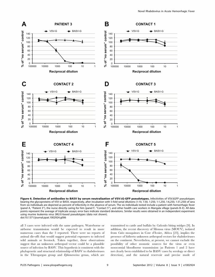
To confirm that BASV is infectious to humans, convalescent
sera were collected in early 2012 from surviving Patient 3 as well
as five additional health care workers from Mangala identified as
close contacts and tested in a blinded fashion for the presence of
neutralizing antibodies to BASV (Fig. 6). Two of the six sera tested
strongly positive with 50% protective doses between 1:1,000 and
1:5,000 (Figs. 6A and 6F). Moreover, the observed neutralization
was highly specific for BASV-G, since no neutralization was
observed with pseudoviruses harboring the vesicular stomatitis virus
glycoprotein (VSV-G). One of the neutralizing sera had been
collected from surviving Patient 3 (Fig. 6A, ‘‘Patient 3’’), whereas the
other serum sample, containing even higher titers, corresponded to
an asymptomatic nurse directly caring for Patient 3 during his
period of acute hemorrhagic illness (Fig. 6F, ‘‘Contact 5’’).
Specifically, Contact 5 was the primary health care provider to
Patient 3 at the health center and during his transfer to the general
hospital at Boma. All 6 individuals, including Patient 3, tested
negative for BASV viremia by specific RT-PCR (data not shown).
Epidemiological Screening for BASV in the DRCBASV was not detected by PCR in 43 serum samples from
other unknown cases or outbreaks of hemorrhagic fever reported
in the DRC from 2008–2010 (Fig. 7A, pink). Five of these 43
samples originated from the Bas-Congo outside of Mangala village
and the Boma Bungu Health Zone. In total, the unknown
hemorrhagic cases/outbreaks spanned 9 of the 11 provinces in the
DRC, and all 43 samples also tested negative by PCR for the
known hemorrhagic fever viruses circulating in Africa (data not
shown). Fifty plasma samples collected from randomly selected
blood donors in the Kasai-Oriental province of DRC (Fig. 7A,
star; Table S2) were also screened and found to be negative for
BASV-neutralizing antibodies (Fig. 7B).
Figure 2. Deep sequencing and whole-genome de novo assembly of BASV. After initial discovery of BASV from a single 454 pyrosequencingread, 98.2% of the BASV genome was assembled de novo from .140 million paired-end Illumina reads. The horizontal lines (red) depict regions of thegenome successfully assembled at the end of each cycle. PCR and Sanger sequencing were performed to confirm the assembly and genomicorganization of BASV (green lines).doi:10.1371/journal.ppat.1002924.g002
Novel Rhabdovirus in Acute Hemorrhagic Fever
PLOS Pathogens | www.plospathogens.org 5 September 2012 | Volume 8 | Issue 9 | e1002924

Discussion
Among more than 160 species of rhabdoviruses identified to
date, fewer than 10 have been isolated from humans [24]. In
addition, while human infection by rhabdoviruses has previously
been associated with encephalitis, vesicular stomatitis, or ‘‘flu-like’’
illness, the discovery of BASV is the first time that a member of the
Rhabdovirus family has been associated with hemorrhagic fever in
humans with a fulminant disease course and high fatality rate. To
our knowledge, this is also the first successful demonstration of
Figure 3. Phylogenetic analysis of the L proteins of BASV and other viruses in the order Mononegavirales. The host from which eachvirus was isolated is represented by a specific color. To generate the Mononegavirales (Rhabdoviridae, Filoviridae and Paramyxoviridae) phylogenytrees, all complete sequences of the large (L) protein, or RNA-dependent RNA polymerase (2000–2300 amino acids in length) were downloaded fromGenBank. Abbreviations and accession numbers used for the phylogenetic analysis are provided in Methods.doi:10.1371/journal.ppat.1002924.g003
Novel Rhabdovirus in Acute Hemorrhagic Fever
PLOS Pathogens | www.plospathogens.org 6 September 2012 | Volume 8 | Issue 9 | e1002924

de novo assembly of a novel, highly divergent viral genome in the
absence of a reference sequence and directly from a primary
clinical sample by unbiased deep sequencing.
Several lines of evidence implicate BASV in the hemorrhagic fever
outbreak among the 3 patients in Mangala. First, this virus was the
only credible viral pathogen detected in the blood of the lone
survivor during his acute hemorrhagic illness by exhaustive deep
sequencing of over 140 million reads. Analysis of the Illumina deep
sequencing reads for the presence of other viral pathogens yielded
only endogenous flora or confirmed laboratory contaminants (Table
S1 and Fig. S2). Some enteric pathogens, such as E. coli O157:H7,
Campylobacter, Shigella, and Salmonella, are diagnosed through
fecal laboratory testing and not blood, and have been associated with
hemorrhagic diarrhea [25]. However, these outbreaks are typically
foodborne and associated with larger clusters and much greater
numbers of clinical cases than reported here [26,27,28]. Further-
more, enteric diarrheal cases rarely present with systemic symptoms
such as fever or generalized mucosal hemorrhage, with bleeding
most often limited to the gastrointestinal tract, and overall mortality
rates are generally low [26]. Thus, the clinical syndrome observed in
3 patients with hemorrhagic fever in the DRC, a region endemic for
viral hemorrhagic fevers, is much more consistent with infection by a
VHF disease agent. BASV is a plausible hemorrhagic fever
candidate because it is a novel, highly divergent infectious virus,
thus of unknown pathogenicity, and was detected at a titer of .1
million copies/mL in blood from an acutely ill individual. In
addition, there is ample precedent for hemorrhagic disease from
rhabdoviruses, as members of the genus Novirhabdovirus cause severe
hemorrhagic septicemia in fresh and saltwater fish worldwide [29]
(Fig. 4). The detection of BASV seropositivity in an asymptomatic
close contact (Fig. 6) is not surprising given that up to 80% of patients
infected with Lassa virus do not exhibit any hemorrhagic fever
symptoms [30,31].
Prior to the BASV outbreak, no hemorrhagic disease cases had
been reported in Boma Bungu Health Zone. BASV was also not
detected in 43 serum samples from unknown, filovirus-negative
cases or outbreaks of hemorrhagic fever from 2008–2010 spanning
9 of the 11 provinces in the DRC (Fig. 7A). In addition, a
serosurvey of 50 random blood donors from Kasai-Oriental
province in central DRC was negative for prior exposure to BASV
Figure 4. Phylogenetic analysis of the L proteins of BASV and other rhabdoviruses. The geographic distribution for each virus or group ofviruses is indicated with a specific icon, while diseases associated with infection by certain rhabdoviruses are indicated by specific colors.Abbreviations and accession numbers used for the phylogenetic analysis are provided in Methods.doi:10.1371/journal.ppat.1002924.g004
Novel Rhabdovirus in Acute Hemorrhagic Fever
PLOS Pathogens | www.plospathogens.org 7 September 2012 | Volume 8 | Issue 9 | e1002924

(Fig. 7B). Taken together, these data suggest that the virus may
have emerged recently and locally from Boma Bungu in Bas-
Congo, DRC.
We were unable to isolate BASV despite culturing the RNA-
positive serum in a number of cell cultures and inoculation into
suckling mice. One explanation for these negative findings may be
that the virus inoculation titers of ,50 mL were insufficient,
although this is surprising given the concentration of .1 million
copies per mL of BASV in blood from the lone survivor. A more
likely explanation is viral inactivation resulting from the lack of
adequate cold chain facilities in remote Boma Bungu. Viral RNA
can often still be detected by RT-PCR in sera that is culture-
negative [32]. In support of this premise, we have observed that
the BASV-G/VSVDG-GFP pseudotyped virus efficiently infects
and replicates in a variety of insect and mammalian (including
human) cell lines (Steffen, et al., manuscript in preparation). In the
absence of a positive culture, a ‘‘reverse genetics’’ approach to
produce recombinant BASV particles, if successful, would greatly
facilitate further study of the virus, as established previously for
other rhabdoviruses such as VSV [33].
Based on our findings, some speculations on the origin of and
routes of transmission for BASV can be made. All 3 patients
became ill with acute hemorrhagic fever over a 3-week period
within the same 2500-m2 area of Mangala village, suggesting that
Figure 5. Schematic representation of the genome organization of BASV and its protein similarity plot compared to representativerhabdoviruses. The similarity plots are generated by aligning the concatenated rhabdovirus proteins and calculating scanning amino acid pairwiseidentities using a window size of 50 bp. The horizontal bar under each similarity plot shows the percent identity of the rhabdovirus protein relative toits corresponding protein in BASV. Genes coding for the 5 core rhabdovirus proteins are shown in green, while the accessory U1, U2, or U3 genes areshown in blue. Black bars correspond to accessory proteins which are not present in the genome. Note that BEFV contains 3 genes between G and L;only the alignment between the alpha-1 protein of BEFV and the U3 protein of BASV is shown (asterisk). The x-axis refers to the nucleotide positionalong the ,12 kb genome of BASV.doi:10.1371/journal.ppat.1002924.g005
Novel Rhabdovirus in Acute Hemorrhagic Fever
PLOS Pathogens | www.plospathogens.org 8 September 2012 | Volume 8 | Issue 9 | e1002924
all 3 cases were infected with the same pathogen. Waterborne or
airborne transmission would be expected to result in more
numerous cases than the 3 reported. There were no reports of
animal die-offs that would suggest potential exposures to infected
wild animals or livestock. Taken together, these observations
suggest that an unknown arthropod vector could be a plausible
source of infection by BASV. This hypothesis is consistent with the
phylogenetic and structural relationship of BASV to rhabdoviruses
in the Tibrogargan group and Ephemerovirus genus, which are
transmitted to cattle and buffalo by Culicoides biting midges [9]. In
addition, the recent discovery of Moussa virus (MOUV), isolated
from Culex mosquitoes in Cote d’Ivoire, Africa [23], implies the
presence of hitherto unknown arthropod vectors for rhabdoviruses
on the continent. Nevertheless, at present, we cannot exclude the
possibility of other zoonotic sources for the virus or even
nosocomial bloodborne transmission (as Patients 1 and 2 have
not clearly been established to be BASV cases by serology or direct
detection), and the natural reservoir and precise mode of
Figure 6. Detection of antibodies to BASV by serum neutralization of VSVDG-GFP pseudotypes. Infectivities of VSVDGFP pseudotypesbearing the glycoproteins of VSV or BASV, respectively, after incubation with 5-fold serial dilutions (1:10, 1:50, 1:250, 1:1,250, 1:6,250, 1:31,250) of serafrom six individuals are depicted as percent of infectivity in the absence of serum. The six individuals tested include a patient with hemorrhagic fever(panel A, ‘‘Patient 3’’), the nurse directly caring for him (panel F, ‘‘Contact 5’’), and other health care workers in Mangala village (panels B–E). All datapoints represent the average of triplicate assays; error bars indicate standard deviations. Similar results were obtained in an independent experimentusing murine leukemia virus (MLV)-based pseudotypes (data not shown).doi:10.1371/journal.ppat.1002924.g006
Novel Rhabdovirus in Acute Hemorrhagic Fever
PLOS Pathogens | www.plospathogens.org 9 September 2012 | Volume 8 | Issue 9 | e1002924

Figure 7. BASV Screening in DRC, Africa. (A) All 43 serum samples corresponding to unknown hemorrhagic fever cases or outbreaks in 2008–2010 from 9 provinces in DRC (pink) tested negative for BASV by PCR. (B) Sera from 50 donors in Kasai-Oriental province, DRC (Panel A, star) weretested for BASV-neutralizing antibodies. Sera at 1:50 (dark blue) or 1:500 dilution (light blue) were tested. Serum from the surviving Patient 3 wasincluded as a positive control (grey shaded area). Data points represent an average of duplicate assays.doi:10.1371/journal.ppat.1002924.g007
Novel Rhabdovirus in Acute Hemorrhagic Fever
PLOS Pathogens | www.plospathogens.org 10 September 2012 | Volume 8 | Issue 9 | e1002924

transmission for BASV remain unknown. A community-based
serosurvey in Boma Bungu and an investigation to track down
potential arthropod or mammalian (e.g. rodents and bats) sources
for BASV are currently underway.
Although we cannot exclude the possibility of independent
arthropod-borne transmission events, our epidemiologic and
serologic data do suggest the potential for limited human-to-
human transmission of BASV. Patient 3, a nurse, had directly
taken care of Patients 1 and 2 at the health center, and another
nurse (Contact 5), who had taken care of Patient 3 (but not
Patients 1 or 2) had serologic evidence of asymptomatic BASV
infection. for BASV transmission during the hemorrhagic fever
outbreak in which the initial infection of two children in Mangala
(Patients 1 and 2) was followed by successive human-to-human
transmission events involving two healthcare workers (Patient 3
and Contact 5) (Fig. 8). This pattern of transmission from the
community to health care workers is also commonly seen in
association with outbreaks of Ebola and Crimean-Congo hemor-
rhagic fever [6,34].
While rhabdoviruses are distributed worldwide, some authors
have suggested that the Rhabdoviridae family probably originated
from tropical regions of the Old or New World [9]. The discovery
of BASV in Central Africa suggests that additional rhabdoviruses
of clinical and public health importance likely await identification,
especially in these poorly investigated geographic regions. Active
epidemiological investigation and disease surveillance will be
needed to fully ascertain the clinical and public health significance
of BASV infection in humans, as well as to prepare for potentially
larger human outbreaks from this newly discovered pathogen.
Methods
Ethics StatementWritten informed consent for publication of their case reports
was obtained from the sole survivor of the hemorrhagic fever
outbreak and the parents of the two deceased children. Written
informed consent was obtained from the surviving patient and 5 of
his close contacts for analysis of the serum samples reported in this
study. Samples were analyzed under protocols approved by the
institutional review boards of University of California, San
Francisco, the University of Texas Medical Branch, and the
National Institute of Biomedical Research (INRB) and CIRMF in
Gabon, and the Institutional Animal Care and Use Committee
(IACUC) of the University of Texas Medical Branch.
Diagnostic SamplesNo diagnostic samples were available from Patient 1 or Patient
2. Blood was collected in a red top serum tube from Patient 3 on
June 16, during the acute phase, three days after hemorrhagic
onset. The sample was transported at 4uC to the BSL-4 facility at
CIRMF. Serum was obtained by centrifugation at 2300 rpm for
10 min. No other acute samples from Patient 3 were available. In
January of 2012 (,2.5 years after the outbreak), convalescent sera
were collected from Patient 3 and close contacts (other workers at
the health center) for BASV neutralization testing. Forty-three
Figure 8. Proposed model for BASV transmission during the hemorrhagic fever outbreak in Mangala. Patients presenting withsymptoms of acute hemorrhagic fever are depicted in red. Dashed red lines represent potential routes of BASV transmission. Contacts 1 through 5 arehealth care workers at the local health center in Mangala village. Abbreviations: HCW, health care worker; y/o, year-old; Ab, antibody.doi:10.1371/journal.ppat.1002924.g008
Novel Rhabdovirus in Acute Hemorrhagic Fever
PLOS Pathogens | www.plospathogens.org 11 September 2012 | Volume 8 | Issue 9 | e1002924

serum samples from other unknown hemorrhagic fever cases or
outbreaks representing 9 of 11 provinces in the DRC were
available for BASV PCR testing (Fig. 7A). Fifty available plasma
samples from random blood donors (median age 27.5 years; age
range 1–76 years) in Kasai Oriental province, DRC, were also
tested for antibodies to BASV (Fig. 7A and B; Table S2).
Nucleic Acid Extraction and Viral PCR TestingRNA was extracted from 140 ml of serum using the QIAamp
viral RNA mini kit (Qiagen). Taqman real-time reverse-transcrip-
tion-PCR (RT-PCR) testing for known hemorrhagic fever viruses
was performed using primers and probes specific for Marburg
virus (MARV), all four species of Ebola virus (Zaire, ZEBOV; Sudan,
SEBOV; Cote d’Ivoire, CIEBOV, and Bundibugyo, BEBOV),
Crimean-Congo hemorrhagic fever virus (CCHFV), Yellow fever
virus (YFV), Dengue virus (DENV), Rift Valley fever virus (RVFV)
and Chikungunya virus (CHIKV) (available upon request).
Discovery of the BASV Rhabdovirus by 454Pyrosequencing
200 mL of serum sample were inactivated in 1 mL of TRIzol
(Invitrogen), and nucleic acid extraction and purification were
performed according to the manufacturer’s instructions. Roche
454 pyrosequencing using randomly amplified cDNA libraries was
performed as described previously [35]. Viral sequences were
identified using BLASTn or BLASTx by comparison to the
GenBank nonredundant nucleotide or protein database, respec-
tively (E-score cutoff = 1025).
De novo Assembly of the BASV Genome by IlluminaSequencing
To recover additional BASV sequence, two sets of cDNA
libraries were prepared from DNase-treated extracted RNA using
a random PCR amplification method as described previously [36],
or random hexamer priming according to the manufacturer’s
protocol (Illumina). The libraries were then pooled and sequenced
on two lanes of an Illumina HiSeq 2000. Raw Illumina sequences
consisting of 100 base pair (bp) paired-end reads were filtered to
exclude low-complexity, homopolymeric, and low-quality se-
quences, and directly compared using BLASTn or BLASTx
alignments to a library consisting of all rhabdovirus sequences in
GenBank. The initial read obtained by 454 pyrosequencing as well
as other reads aligning to rhabdoviruses were then inputted as
‘‘seeds’’ into the PRICE de novo assembler [37] (Fig. 2), with a
criterion of at least 85% identity over 25-bp to merge two
fragments. De novo assembly of the BASV genome was performed
iteratively using PRICE and the Geneious software package
(Biomatters) [38]. The near-complete whole genome sequence of
the novel rhabdovirus (,98.2% based on protein homology to
other rhabdoviruses) was determined to at least 36redundancy by
de novo assembly as well as PCR and Sanger sequencing of low-
coverage regions. Sanger sequencing was also performed to verify
the accuracy of the assembly and confirm the genomic organiza-
tion of BASV (Fig. 2).
Deep Sequencing Analysis of the BASV Serum Sample forOther Pathogens
Rapid classification of the ,140 million 100-bp paired-end
Illumina reads was performed using a modified cloud computing-
based computational analysis pipeline [17] (Veeraraghavan,
Sittler, and Chiu, manuscript in preparation). Briefly, reads
corresponding to human sequences were taxonomically classified
using SOAP and BLAT software [39,40]. Other reads were then
identified using BLASTn or BLASTx by comparison to GenBank-
derived reference databases (E-score cutoff = 1025).
PCR Quantitation of BASV BurdenTo estimate the viral load in the patient’s serum, we first
designed a set of specific PCR primers for detection of BASV
targeting the L protein, BASV-F (59- CGCTGATGGTTTTT-
GACATGGAAGTCC-39)/BASV-R (59-TAAACTTCCTCTC-
TCCTCTAG-39), for use in a SYBR-Green real-time quantitative
RT-PCR assay. A standard curve for the assay was constructed as
described previously [36]. The viral load in the patient’s serum
was determined by comparison to the standard curve.
Structural Features and Phylogenetic AnalysisPredicted open reading frames (ORFs) in the BASV genome
were identified with Geneious [38]. Multiple sequence (Figs. 3 and
4; Fig. S1) and pairwise (Fig. 5) alignments of BASV proteins
relative to corresponding proteins from other rhabdoviruses were
calculated using MAFFT (v6.0) with the E-INS-i option and at
default settings [41]. To generate the phylogeny trees, all
rhabdoviruses in GenBank were included as well as representative
members of other families within the order Mononegavirales.
Bayesian tree topologies were assessed with MrBayes V.32
software (20,000 sampled trees; 5,000 trees discarded as burn-in)
[42]. Convergence was confirmed by the PSRF statistic in
MrBayes, as well as by visual inspection of individual traces using
TRACER from the BEAST software package [43]. Trees were
visualized after midpoint rooting with FigTree V1.31 [43].
Virus Cultivation in Cell Cultures or Suckling MiceInitial attempts were made to culture the virus using a total of
200 mL of BASV-positive serum inoculated onto confluent
monolayers of Vero E6 and C6/36 (Aedes albopictus mosquito) cells
in 6-well plastic tissue culture plates at 37uC and 28uC,
respectively, in a 5% CO2 environment as previously described
[44]. From 20–50 mL of serum were used to inoculate the cells,
which were examined daily for cytopathic effect (CPE) at days 5, 7,
and 14. Supernatants were harvested and two additional blind
passages were performed, each passage followed by 14 days of
observation for CPE. Cell culture supernatants were also
monitored for evidence of viral replication by quantitative RT-
PCR.
Using the remaining 100 uL of BASV-positive serum, further
attempts were made to culture the virus in 5 cell lines and in suckling
mice. The serum sample was split in half and diluted 1:20 or 1:10 in
phosphate-buffered saline with 20% fetal bovine serum (FBS) to
allow sufficient volume to inoculate cell cultures or mice,
respectively. The first diluted sample was inoculated intracerebrally
into a litter (n = 12) of 1 day old mice. Pups were observed daily for
14 days for lethality or signs of clinical illness. The second diluted
sample was inoculated into 12.5 cm2 tissue culture flasks of Vero,
BHK, LLC-MK2 (rhesus monkey kidney), CCL-106 (rabbit kidney)
and C6/36 cells. Vertebrate cells were held at 37uC for 14 days and
observed for evidence of CPE. Mosquito cells were maintained at
28uC for 10 days. Since no CPE was observed in any of the cultures,
cells were subsequently fixed for transmission electron microscopy
to see if viral particles could be visualized [45].
Construction of VSVDG-GFP Pseudotypes and BASVSerum Neutralization Testing
A pseudotype system based on a vesicular stomatitis virus (VSV)
construct carrying a reporter gene for green fluorescent protein
(VSVDG-GFP) and bearing the predicted synthesized BASV
Novel Rhabdovirus in Acute Hemorrhagic Fever
PLOS Pathogens | www.plospathogens.org 12 September 2012 | Volume 8 | Issue 9 | e1002924

glycoprotein (BASV-G) was used to generate a serum neutraliza-
tion assay for BASV. Briefly, the predicted BASV glycoprotein
(BASV-G) was synthesized (Genscript) and subcloned into the
pCAGGS expression plasmid. Human embryonic kidney 293T
cells were seeded (DMEM + 10% FBS + penicillin/streptomycin +Glutamax (Gibco) + non-essential amino acids (Gibco)) in 10 cm
culture dishes 24 hours prior to transfection. Cells were transfected
with 20 mg BASV-G, VSV-G, or empty pCAGGS DNA per dish
following a calcium phosphate transfection protocol [46]. The
culture medium was replaced 15 hours post-transfection and cells
were stimulated with 6.2 mM valproic acid for 4 hours before the
medium was replaced again. At 36 hours post-transfection the
transfected cells were infected with VSVDG-GFP/VSV-G pseu-
dotypes at a multiplicity of 0.1–0.3. The inoculum was removed
after 4 hours and replaced by fresh culture medium. At 24 hours
post-infection, infectious supernatants were harvested, filtered
through 0.45 mm filters, and concentrated 10-fold by centrifuga-
tion through a 100-kDA filter (Millipore). Concentrated viruses
were aliquoted and stored at 280uC.
For serum neutralization testing, human hepatoma Huh-7
cells were seeded (DMEM +10% FBS + penicillin/streptomycin
+ Glutamax (Gibco) + non-essential amino acids (Gibco)) in 48-
well plates 24 hours prior to infection. Per well 10 ml of
pseudovirus harboring either BASV-G or VSV-G (adjusted to
obtain 25–50% infection of target cells) was mixed with 10 ml of
the respective serum dilution and incubated for 45 minutes at
37uC. Subsequently, the mix was added to the target cells
(performed in triplicate) and cells were incubated for 24 hours at
37uC. The infected cells were detached with trypsin and washed
with PBS before fixing with 2% paraformaldehyde for 1 hour at
room temperature. GFP expression in infected cells was
quantified by flow cytometry using a LSR II (BD Biosciences)
and the collected data was analyzed with FlowJo software
(TreeStar).
Abbreviations and Nucleotide Sequence AccessionNumbers
The annotated, nearly complete sequence of BASV has been
submitted to GenBank (accession number JX297815). Deep
sequencing reads have been submitted to the NCBI Sequence
Read Archive (accession number SRA056894). Accession num-
bers used for the phylogenetic analyses in Figs 3, 4, and S1 are
listed as follows, in alphabetical order: ABLV, Australian bat
lyssavirus (NP_478343); ARAV, Aravan virus (ABV03822),
BEFV, Bovine ephemeral fever virus (NP_065409); BYSMV,
Barley yellow striate mosaic virus (BYSMV); CDV, Canine
distemper virus (AAR32274); CHPV, Chandipura virus
(ADO63669); CPV, Coastal Plains virus (ADG86364); COCV,
Cocal virus (ACB47438); DURV, Durham virus (ADB88761);
DUVV, Duvenhage virus (ABZ81216); EBLV1, European bat
lyssavirus 1 (ABZ81166), EBLV2, European bat lyssavirus 2
(ABZ81191); EBOV, Ebola virus (AAG40171, AAA79970,
BAB69010); EVEX, Eel virus European X virus (CBH20130);
FDLV, Fer-de-lance virus (NP_899661); FLAV, Flanders virus
(AAN73288); HeV, Hendra virus (NP_047113); HIRRV, Hirame
rhabdovirus (ACO87999); HMPV, Human metapneumovirus
(L_HMPVC); HPIV-1, Human parainfluenza virus type 1 (AA
A69579); HPIV-2, Human parainfluenza virus type 2 (CAA
40788); HPIV-3, Human parainfluenza virus type 3 (AAA46854);
HPIV-4, Human parainfluenza virus type 4 (BAJ11747); INHV,
Infectious hematopoietic necrosis virus (NP_042681); IRKV, Irkut
virus (ABV03823); ISFV, Isfahan virus (CAH17548); KHUV,
Khujand virus (ABV03824); LBV, Lagos bat virus (ABZ81171);
LNYV, Lettuce necrotic yellows virus (YP_425092); MARAV,
Maraba virus (AEI52253); MARV, Marburg virus
(YP_001531159); MeV, Measles virus (AF266288); MMV, Maize
mosaic virus (YP_052855); MOKV, Mokala virus (ABZ81206);
MOUV, Moussa virus (ACZ81407); MUV, Mumps virus (AF
201473); NCMV, Northern cereal mosaic virus (NP_597914);
NDV, Newcastle disease virus (ADH10207); NGAV, Ngaingan
virus (YP_003518294); NiV, Nipah virus (AAY43917); OVRV,
Oak Vale rhabdovirus (AEJ07657); PFRV, Pike fry rhabdovirus
(ACP28002); RABV, Rabies virus (NP_056797); RSV, Respira-
tory syncytial virus (NP_056866); RYSV, Rice yellow stunt
rhabdovirus (NP_620502); SIGMAV, Sigma virus (ACU65444);
SCRV, Siniperca chuatsi rhabdovirus (YP_802942); SHRV,
Snakehead virus (AAD56771); SMRV, Scophthalmus maximus
rhadovirus (ADU05406); SVCV, Spring viremia of carp virus
(NP_116748); SYNV, Sonchus yellow net virus (NP_044286);
TIBV, Tibrogargan virus (ADG86355); TUPV, Tupaia virus
(AAX47602); TVCV, Tomato vein clearing virus (YP_224083);
VHSV, Viral hemorrhagic septicemia virus (NP_049550); VSIV,
Vesicular stomatitis virus, Indiana (NP_041716); VSNJV, Vesicular
stomatitis virus, New Jersey (P16379); WCBV, West Caucasian bat
virus (ABV03821); WONGV, Wongabel virus (YP_002333280).
Supporting Information
Figure S1 Phylogenetic analysis of the N, P, M, and Gproteins of BASV and other rhabdoviruses. Each phyloge-
netic tree is rooted by using the corresponding protein from
human parainfluenza virus type 1 (HPIV-1), a paramyxovirus, as
an outgroup. Abbreviations and accession numbers used for the
phylogenetic analysis are provided in Methods.
(TIF)
Figure S2 Confirmation of laboratory contamination byrotavirus and absence of rotavirus in BASV serum byspecific PCR. An RT-PCR assay for detection of Group A
rotaviruses was performed using primers NSP3F (59-AC-
CATCTWCACRTRACCCTCTATGAG-39) and NSP3R (59-
GGTCACATAACGCCCCTATAGC-39), which generate an
87-bp amplicon (Freeman, et al., (2008) J Med Virol 80: 1489–
1496). PCR conditions for the assay were 30 min at 50uC, 15 min
at 95uC for the reverse transcription step followed by 40 cycles of
95uC, 30 s/55uC, 30 s/72uC, 30 s and 72uC/7 min for the final
extension. PCR products are visualized by gel electrophoresis,
using a 2% agarose gel and 1 kB ladder. Rotavirus is readily
detected in extracted RNA from a stool sample taken from an
ongoing study of viral diarrhea in the laboratory (lane 1), but not
in two separate aliquots of extracted nucleic acid from the BASV
serum sample (lanes 2 and 3).
(TIF)
Table S1 Viral reads in the deep sequencing datacorresponding to the BASV-positive serum sample.
(DOCX)
Table S2 Demographics of 50 blood donors from Kasai-Oriental province, DRC, randomly selected for BASVantibody screening.
(DOCX)
Acknowledgments
The authors thank the national and international teams involved in the
control of suspected hemorrhagic fever cases that occurred in 2009 in
Democratic Republic of the Congo (DRC). The national teams are
members of the DRC Ministry of Health and the National Institute of
Biomedical Research (INRB). The international teams are epidemiological
and medical experts of the World Health Organization (WHO) and the
Novel Rhabdovirus in Acute Hemorrhagic Fever
PLOS Pathogens | www.plospathogens.org 13 September 2012 | Volume 8 | Issue 9 | e1002924
NGO ‘Medecins Sans Frontieres’. We thank all those involved in sample
collection and case reporting, especially Etienne Mukendi at the Cellule de
Surveillance Epidemiologique, Bas-Congo, DRC. We are also grateful to A
Delicat and P Engandja from Centre International de Recherches de
Franceville (CIRMF), Gabon, Matthew LeBreton at Global Viral
Forecasting, Incorporated, Nicole A. Hoff at University of California,
Los Angeles, Thomas Geisbert at University of Texas Medical Branch, as
well as Samia Naccache, Rick Hsu and Yasamin Mohammadi at
University of California, San Francisco (UCSF), for technical assistance
during this work.
Author Contributions
Conceived and designed the experiments: GG JNF DL GS ED NDW
CYC EML. Performed the experiments: GG DL ES IS RBT. Analyzed the
data: GG JNF DL J-JM NV MM PM GS ED NDW CYC EML.
Contributed reagents/materials/analysis tools: TS JGR CW RBT JM
AWR TT BSS GS ED NDW CYC EML. Wrote the paper: GG JNF DL
IS RBT GS ED NDW CYC EML. Obtained consents from patients and
their families: PM.
References
1. Bray M (2005) Pathogenesis of viral hemorrhagic fever. Curr Opin Immunol 17:
399–403.
2. Carrion R, Jr., Patterson JL (2011) Vaccines against viral hemorrhagic fevers:
non-human primate models. Hum Vaccin 7: 667–673.
3. Geisbert TW, Jahrling PB (2004) Exotic emerging viral diseases: progress and
challenges. Nat Med 10: S110–121.
4. Borio L, Inglesby T, Peters CJ, Schmaljohn AL, Hughes JM, et al. (2002)
Hemorrhagic fever viruses as biological weapons: medical and public health
management. JAMA 287: 2391–2405.
5. Fichet-Calvet E, Rogers DJ (2009) Risk maps of Lassa fever in West Africa.
PLoS Negl Trop Dis 3: e388.
6. Ergonul O (2006) Crimean-Congo haemorrhagic fever. Lancet Infect Dis 6:
203–214.
7. Leroy EM, Gonzalez JP, Baize S (2011) Ebola and Marburg haemorrhagic fever
viruses: major scientific advances, but a relatively minor public health threat for
Africa. Clin Microbiol Infect 17: 964–976.
8. Monath TP (2007) Dengue and yellow fever–challenges for the development and
use of vaccines. N Engl J Med 357: 2222–2225.
9. Kuzmin IV, Novella IS, Dietzgen RG, Padhi A, Rupprecht CE (2009) The
rhabdoviruses: biodiversity, phylogenetics, and evolution. Infect Genet Evol 9:
541–553.
10. Fu ZF (2005) Genetic comparison of the rhabdoviruses from animals and plants.
Curr Top Microbiol Immunol 292: 1–24.
11. Gurav YK, Tandale BV, Jadi RS, Gunjikar RS, Tikute SS, et al. (2010)
Chandipura virus encephalitis outbreak among children in Nagpur division,
Maharashtra, 2007. Indian J Med Res 132: 395–399.
12. Warrell MJ, Warrell DA (2004) Rabies and other lyssavirus diseases. Lancet 363:
959–969.
13. Rodriguez LL (2002) Emergence and re-emergence of vesicular stomatitis in the
United States. Virus Res 85: 211–219.
14. Tang P, Chiu C (2010) Metagenomics for the discovery of novel human viruses.
Future Microbiol 5: 177–189.
15. Briese T, Paweska JT, McMullan LK, Hutchison SK, Street C, et al. (2009)
Genetic detection and characterization of Lujo virus, a new hemorrhagic fever-
associated arenavirus from southern Africa. PLoS Pathog 5: e1000455.
16. Feng H, Shuda M, Chang Y, Moore PS (2008) Clonal integration of a
polyomavirus in human Merkel cell carcinoma. Science 319: 1096–1100.
17. Greninger AL, Chen EC, Sittler T, Scheinerman A, Roubinian N, et al. (2010)
A metagenomic analysis of pandemic influenza A (2009 H1N1) infection in
patients from North America. PLoS One 5: e13381.
18. Towner JS, Rollin PE, Bausch DG, Sanchez A, Crary SM, et al. (2004) Rapid
diagnosis of Ebola hemorrhagic fever by reverse transcription-PCR in an
outbreak setting and assessment of patient viral load as a predictor of outcome.
J Virol 78: 4330–4341.
19. Allison AB, Palacios G, Travassos da Rosa A, Popov VL, Lu L, et al. (2011)
Characterization of Durham virus, a novel rhabdovirus that encodes both a C
and SH protein. Virus Res 155: 112–122.
20. Gubala AJ, Proll DF, Barnard RT, Cowled CJ, Crameri SG, et al. (2008)
Genomic characterisation of Wongabel virus reveals novel genes within the
Rhabdoviridae. Virology 376: 13–23.
21. Springfeld C, Darai G, Cattaneo R (2005) Characterization of the Tupaia
rhabdovirus genome reveals a long open reading frame overlapping with P and a
novel gene encoding a small hydrophobic protein. J Virol 79: 6781–6790.
22. Gubala A, Davis S, Weir R, Melville L, Cowled C, et al. (2011) Tibrogargan and
Coastal Plains rhabdoviruses: genomic characterization, evolution of novel genes
and seroprevalence in Australian livestock. J Gen Virol 92: 2160–2170.
23. Quan PL, Junglen S, Tashmukhamedova A, Conlan S, Hutchison SK, et al.
(2010) Moussa virus: a new member of the Rhabdoviridae family isolated from
Culex decens mosquitoes in Cote d’Ivoire. Virus Res 147: 17–24.
24. Fu ZF (2005) The world of rhabdoviruses. Berlin; New York: Springer. vi, 208 p.
25. Brooks JT, Shapiro RL, Kumar L, Wells JG, Phillips-Howard PA, et al. (2003)Epidemiology of sporadic bloody diarrhea in rural Western Kenya. Am J Trop
Med Hyg 68: 671–677.26. (2011) Vital signs: incidence and trends of infection with pathogens transmitted
commonly through food–foodborne diseases active surveillance network, 10 U.S.
sites, 1996–2010. MMWR Morb Mortal Wkly Rep 60: 749–755.27. Daniels NA, MacKinnon L, Rowe SM, Bean NH, Griffin PM, et al. (2002)
Foodborne disease outbreaks in United States schools. Pediatr Infect Dis J 21:623–628.
28. Rangel JM, Sparling PH, Crowe C, Griffin PM, Swerdlow DL (2005)Epidemiology of Escherichia coli O157:H7 outbreaks, United States, 1982–
2002. Emerg Infect Dis 11: 603–609.
29. Hoffmann B, Beer M, Schutze H, Mettenleiter TC (2005) Fish rhabdoviruses:molecular epidemiology and evolution. Curr Top Microbiol Immunol 292: 81–
117.30. (2004) Imported Lassa fever–New Jersey, 2004. MMWR Morb Mortal Wkly
Rep 53: 894–897.
31. Leroy EM, Baize S, Volchkov VE, Fisher-Hoch SP, Georges-Courbot MC, et al.(2000) Human asymptomatic Ebola infection and strong inflammatory response.
Lancet 355: 2210–2215.32. Levi JE, Tateno AF, Machado AF, Ramalho DC, de Souza VA, et al. (2007)
Evaluation of a commercial real-time PCR kit for detection of dengue virus insamples collected during an outbreak in Goiania, Central Brazil, in 2005. J Clin
Microbiol 45: 1893–1897.
33. Stanifer ML, Cureton DK, Whelan SP (2011) A recombinant vesicularstomatitis virus bearing a lethal mutation in the glycoprotein gene uncovers a
second site suppressor that restores fusion. J Virol 85: 8105–8115.34. Guimard Y, Bwaka MA, Colebunders R, Calain P, Massamba M, et al. (1999)
Organization of patient care during the Ebola hemorrhagic fever epidemic in
Kikwit, Democratic Republic of the Congo, 1995. J Infect Dis 179 Suppl 1:S268–273.
35. Victoria JG, Kapoor A, Li L, Blinkova O, Slikas B, et al. (2009) Metagenomicanalyses of viruses in stool samples from children with acute flaccid paralysis.
J Virol 83: 4642–4651.36. Chen EC, Yagi S, Kelly KR, Mendoza SP, Maninger N, et al. (2011) Cross-
species transmission of a novel adenovirus associated with a fulminant
pneumonia outbreak in a new world monkey colony. PLoS Pathog 7: e1002155.37. Earl D, Bradnam K, St John J, Darling A, Lin D, et al. (2011) Assemblathon 1: a
competitive assessment of de novo short read assembly methods. Genome Res21: 2224–2241.
38. Drummond A, Ashton B, Cheung M, Heled J, Kearse M, et al. (2010) Geneious
v5.3.4. Geneious website. Available from http://www.geneious.com. Accessed2012 Aug 16.
39. Kent WJ (2002) BLAT–the BLAST-like alignment tool. Genome Res 12: 656–664.
40. Li R, Yu C, Li Y, Lam TW, Yiu SM, et al. (2009) SOAP2: an improved ultrafasttool for short read alignment. Bioinformatics 25: 1966–1967.
41. Katoh K, Kuma K, Toh H, Miyata T (2005) MAFFT version 5: improvement in
accuracy of multiple sequence alignment. Nucleic Acids Res 33: 511–518.42. Ronquist F, Teslenko M, van der Mark P, Ayres DL, Darling A, et al. (2012)
MrBayes 3.2: Efficient Bayesian Phylogenetic Inference and Model ChoiceAcross a Large Model Space. Syst Biol 61: 539–42.
43. Drummond AJ, Rambaut A (2007) BEAST: Bayesian evolutionary analysis by
sampling trees. BMC Evol Biol 7: 214.44. Grard G, Biek R, Muyembe Tamfum JJ, Fair J, Wolfe N, et al. (2011)
Emergence of divergent zaire ebola virus strains in democratic republic of thecongo in 2007 and 2008. J Infect Dis 204 Suppl 3: S776–784.
45. Mihindukulasuriya KA, Nguyen NL, Wu G, Huang HV, da Rosa AP, et al.
(2009) Nyamanini and midway viruses define a novel taxon of RNA viruses inthe order Mononegavirales. J Virol 83: 5109–5116.
46. Kingston RE, Chen CA, Rose JK (2003) Calcium phosphate transfection. CurrProtoc Mol Biol Chapter 9: Unit 9 1.
Novel Rhabdovirus in Acute Hemorrhagic Fever
PLOS Pathogens | www.plospathogens.org 14 September 2012 | Volume 8 | Issue 9 | e1002924