A novel immuno-polymerase chain reaction protocol
32
A novel immuno-polymerase chain reaction protocol incorporating a highly purified streptavidin-DNA conjugate Author Shan, Jianguo, Toye, Philip Published 2009 Journal Title Journal of Immunoassay & Immunochemistry DOI https://doi.org/10.1080/15321810903084764 Copyright Statement © 2009 Taylor & Francis. This is an electronic version of an article published in Journal of Immunoassay and Immunochemistry Volume 30, Issue 3, 2009, 322-337. Journal of Immunoassay and Immunochemistry is available online at: http://www.informaworld.com with the open URL of your article. Downloaded from http://hdl.handle.net/10072/34201 Griffith Research Online https://research-repository.griffith.edu.au
Transcript of A novel immuno-polymerase chain reaction protocol

A novel immuno-polymerase chain reaction protocolincorporating a highly purified streptavidin-DNA conjugate
Author
Shan, Jianguo, Toye, Philip
Published
2009
Journal Title
Journal of Immunoassay & Immunochemistry
DOI
https://doi.org/10.1080/15321810903084764
Copyright Statement
© 2009 Taylor & Francis. This is an electronic version of an article published in Journalof Immunoassay and Immunochemistry Volume 30, Issue 3, 2009, 322-337. Journal ofImmunoassay and Immunochemistry is available online at: http://www.informaworld.com withthe open URL of your article.
Downloaded from
http://hdl.handle.net/10072/34201
Griffith Research Online
https://research-repository.griffith.edu.au

A novel immuno-polymerase chain reaction protocol incorporating a
highly purified streptavidin-DNA conjugate.
Jianguo Shana, b , Philip Toye a,c,*
aAGEN Biomedical Limited, Acacia Ridge, Queensland, AUSTRALIA
b Current address: Queensland Institute of Medical Research, Herston Road,
Herston, QLD 4006, AUSTRALIA
c Current address: The International Livestock Research Institute, P.O. Box
30709, Nairobi, KENYA, 00100.
*Corresponding author. Tel.: +254 20 422 3463; fax: +254 20 422 3001; e-
mail: [email protected]
2
Abstract
We have developed and tested an immuno-PCR format which includes a
highly purified STV-DNA conjugate. The protocol comprises standard ELISA
methodology and washing buffers together with a real-time PCR read-out
system. The conjugate was employed in both indirect and capture assay
formats. The procedure involves a 90-min coating step, two (indirect) or three
(capture) immunoassay steps followed by elution and real-time PCR. Thus,
the assay can be completed in a single day using standard laboratory
equipment. We show that the immuno-PCR is reproducible, with a larger
dynamic range and a sensitivity several orders of magnitude greater than the
corresponding conventional ELISA. The performance of this immuno-PCR
was similar to that with other systems, in terms of minimum concentration of
analyte detected (of the order 50 pg/ml) and of increase in sensitivity and
dynamic range over conventional ELISA. These characteristics should
contribute to the adoption of immuno-PCR by research and clinical
laboratories.
Keywords: Immuno-PCR, STV, biotin, monoclonal antibody, D-dimer, canine
heartworm.
3
1. Introduction
The detection and quantitation of analytes such as hormones, toxins,
cytokines and pathogens is a major activity of biomedical research and clinical
laboratories. The technologies used for this purpose often rely on the ability
of detector molecules or ligands to bind very specifically to the analyte under
investigation. The ligand is then detected by a more generic read-out system.
One very familiar example of this technology is ELISA, in which the analyte-
bound ligand, usually an antibody, is detected and quantitated through an
enzyme which induces a measurable colour change in an appropriate
substrate. The enzyme may be conjugated directly to the antibody, or linked
indirectly through another ligand such as anti-immunoglobulin or protein A.
The sensitivity of these assays depends on several interrelated factors,
including the avidity of the ligand, the amount of non-specific binding of the
ligand to unrelated components in the sample or to the assay platform, and
the number of detector molecules required to produce a detectable change in
the substrate. Several ‘amplification’ systems have been introduced to
enhance the signal, including the use of indirect ELISA to bind several
secondary reporter molecules to each ligand, and nickel enhanced reactions
(Bobrow et al. 1989)
The ability of the polymerase chain reaction (PCR) to amplify a few DNA
molecules to easily detectable levels offers an alternative means to increase
the sensitivity of traditional ELISA technology. This concept, termed immuno-
PCR, was first by exploited by Sano et al. (1992) and has been modified by
several other workers to follow a standard ELISA protocol, except that the

4
read-out system involves detection by PCR amplification of a DNA template
linked to the ligand, usually a monoclonal antibody. The advent of real-time
PCR assay technology has increased the sensitivity and ease of use of
immuno-PCR, and allowed accurate quantification of the analyte (Sims et al.
2000). The linking of the DNA template to the antibody has been achieved
through direct conjugation (Sano et al., 1992 , Lind and Kubista, 2005) and
indirectly through, for example, secondary antibodies and STV (STV) – biotin
bridges (Adler et al., 2003, Niemeyer et al., 2003). The advantage of the
indirect systems is their generic utility for different ligand-analyte combinations.
We have developed a novel immuno-PCR reagent system which aims to
decrease the number of components in a generic protocol and to reduce the
presence of unconjugated by-products. The system employs conjugation of
thiol-modified DNA template to STV and purification through size exclusion
and affinity chromatography. When used in detection assays for two antigens,
D-dimer and canine heartworm antigen (CHA) routinely assayed in human
and veterinary clinical laboratories respectively, we show that the sensitivity of
the immuno-PCR is up to 10,000 fold that of conventional ELISA, with an
estimated detection level of 50 fg. This was achieved with standard ELISA
washing protocols. The system employs a real-time PCR using an
intercalating dye, rather than a template-specific probe, which further
increases the generic utility the system.

5
2. Materials and methods
2.1 Monoclonal antibodies and antigens
The monoclonal antibodies (mAb) and antigens were supplied by Agen
Biomedical Ltd. and are those used in their commercial diagnostic assays.
MAb3B6 and 1D2 are specific for the human thrombosis breakdown product
D-dimer, while mAb4D2 and DI16 recognize an antigen from the canine
heartworm Dirofilaria immitis,
which is present in the sera of infected animals.
Both antigens are detected in routine diagnostic assays in clinical laboratories.
The concentration of the D-dimer was estimated by absorbance at 280 nm,
using an extinction co-efficient E1cm1% of 17.8 (Masci et al., 1985).
2.2 Synthesis of 5’ sulfhydryl-modified reporter DNA
A 602-base pair DNA template was synthesized using PCR from mouse
leukaemia virus genomic DNA isolated from the mouse myeloma cell line NS0
(Nikbakht et al., 1987). The DNA sequence can be found at GenBank
accession no. gb|AC114666.31|, bp 94745 – 94144. The forward and reverse
primer sequences (primers 1 and 2, Fig. 2a) are 5’ (thiol -S-S-)-5’-
CCCCACCATCAGGCTTAG-3’ and 5’-GGCTTTATTGGGAGCACGG-3’,
respectively (Sigma Genosys, Sydney). The primers were chosen such that
an NheI site is 3 bp downstream of the forward primer binding site, which
allows for release of most of the template DNA by NheI digestion. The PCR
was conducted in a total volume of 25 μl, comprising 1.0 U Taq DNA

6
polymerase (‘JumpStart’, Sigma Genosys, Sydney), 1 X supplied PCR buffer,
200 μM dNTPs, 1.0 mM of MgCl2, 40 nM each primer and 1 ng of NS0
genomic DNA. Amplification conditions were: denaturing at 95 °C for 15 s,
annealing at 57 °C for 30 s, and elongation at 72 °C for 15 s, for 40 cycles.
The PCR products were analysed by electrophoresis through a 2% agarose
gel to confirm the presence of the appropriately sized fragment using the
fluorescent dye ‘SYBR-Safe™’ (Invitrogen, USA.). The PCR product was
purified (‘QIAquick PCR purification kit’, QIAGEN, USA.) and the 5’-sulfhydryl
DNA was eluted into distilled water. The DNA concentration was determined
by absorbance at 260 nm.
2.3 Modification of STV with sulfosuccinimidyl-4-(N-maleimidomethyl)
cyclohexane-1-carboxylate (sulfo-SMCC)
Sulfo-SMCC (Pierce, USA) was dissolved in distilled water at 5.0 mg/ml and
20 μL of sulfo-SMCC solution was mixed and reacted with 100 uL of 10
mg/mL STV (Sigma) in 50 mM sodium phosphate buffer (pH7.0) at room
temperature (RT) for 2 h. The STV-SMCC was passed though a ‘HiTrap’
desalting column (Amersham Biosciences, USA), equilibrated with 50 mM
phosphate buffer (pH7.0), to remove unreacted sulfo-SMCC (Fig. 1a). The
concentration of STV-SMCC was determined by absorbance at 280 nm.
2.4 Conjugation of DNA to STV

7
The purified STV-SMCC (10 μg) was reacted with 20 μg 5’-sulfhydryl DNA in
50 mM phosphate buffer containing 5 mM EDTA and 10 mM Tris [2-
carboxyethyl] phosphine hydrochloride (TCEP.HCl, Pierce USA) for 4 h at RT.
The reaction mix was loaded onto a 2 mL Iminobiotin column (Pierce, USA),
which was equilibrated with binding buffer (50 mM ammonium carbonate
buffer with 0.5 M NaCl, pH11.0). After washing with 10 mL of binding buffer to
remove unreacted DNA and proteins, the STV-DNA conjugate and unreacted
STV were eluted with 0.1 M acetic acid as shown in Fig. 1b.
To recover the STV-DNA conjugate free of unreacted STV, the sample was
loaded onto a size exclusion HPLC column (Phenomenex Bio-SEC-3000
USA) equilibrated with 0.1 M phosphate buffer containing 200 mM NaCl,
pH6.5. Control STV was passed through the column which showed that the
elution time of unreacted STV was greater than 9 min, Fig 1c). The STV-DNA
conjugate was eluted from 5.5 - 6.2 min.
2.5 Biotinylation of mAb.
The mAb solution, 2 ml of 5 mg/ml in 50 mM phosphate buffer, pH7.0, was
mixed with 1 mg sulfo-NHS-LC-biotin (Pierce, USA). After incubation at RT
for 3 h, the biotinylated mAb was purified by size-exclusion chromatography
(P6DG column, Bio-Rad, USA).
2.7 Immuno-PCR assays.

8
The indirect and capture immuno-PCR assays were performed in 96-well
microtitre plates (‘MaxiSorp’, Nunc) and are represented schematically in Figs.
2b and 2c. For indirect assays, the respective antigens were coated onto the
plates in ten-fold serial dilutions in 50 μL coating buffer (50mM Na2CO3 buffer,
pH9.66) and incubated at RT for 90 min. The plates were washed three times
with phosphate buffered saline (PBS) containing 0.5% Tween-20 (PBS-T).
The biotinylated mAb (50 μL at 1μg/mL) was added to the plates and
incubated for 60 min at RT, following which the plates were washed three
times with PBS-T. Purified STV-DNA conjugate (50μL at 10ng/ml) was added
to the wells and incubated for 60 min at RT. The plates were washed a
further six times with PBS-T and the bound DNA was released by adding
elution buffer (10 μL comprising 1U NheI, 1 μL 10X restriction enzyme buffer,
1 μL BSA at 1 mg/mL and 8 μL water) for 60 min at 37 ºC. The restriction
enzyme and buffer were supplied by New England Biolabs (Mass., USA).
A 1μL sample of each well was removed and assayed by quantitative PCR as
follows. The reaction was conducted in a total volume of 25 μL as described
above (section 2.2) except that the forward primer sequence (primer 3, Fig. 1
a) is 5’- ATAGAGGTGCACAGTGCTCTGGC-3’, and 0.5X Sybr Green™
(Invitrogen Molecular Probes, USA) was included in the reaction mix.
Amplification conditions were as above. The reactions were performed in a
‘Rotor-Gene’ thermal cycler (Corbett Research, Sydney) and the supplier’s
software was used to estimate the Ct values. The cut–off point for the Ct
values was determined by the software. Melt curve analysis was performed
immediately after the PCR and consisted of an initial denaturation at 95 ºC for
60 sec, hybridisation at 55 ºC for 60 sec and at 50 ºC for 120 sec, followed by

9
a thermal gradient from 50 ºC to 95 ºC, holding for 5 sec at 1 ºC intervals.
Fluorescence emission data were collected at each interval during the thermal
gradient. Software supplied with the thermal cycler allowed automatic
conversion of the melt curve data to the first derivative (dF/dT) of the
fluorescence signals against temperature to produce positive peaks.
For capture assays, the plates were coated with mAb3B6 (D-dimer) or mAb
4D2 (CHA) in 50ul at 1 μg/mL in phosphate buffer (pH7.0) and incubated for
90 min at RT. The protocols were similar to the indirect assay described
above, with modifications to reduce background binding as described in the
text in sections 3.3 and 3.5. The rest of the assay was as described above.
2.8 Conventional ELISA
Corresponding ELISAs were performed as above except that STV conjugated
to horseradish peroxidase (Dako, Denmark) was used rather than the STV-
DNA. The conjugate (50 uL of 1 in 500 dilution in PBS-T with 2%BSA) was
added to the plates at RT for 60 min and the plates were washed three times
with PBS-T. The substrate 3,3’,5,5’-tetramethylbenzidine (TMB) was added
and the plates were incubated for 20 min at RT. The reaction was stopped by
adding an equal volume of 1M H2SO4. The absorbance at 450nm was
determined by an automatic plate reader.

10
3. Results
3.1. Generation of STV – DNA conjugate
The DNA and STV components were prepared for conjugation by separately
generating a thiol-S-S-DNA template and maleimide-modified STV. The
602bp DNA template from murine leukaemia virus was produced by PCR
using a thiol-S-S linked primer obtained commercially. The PCR product was
separated from unused primers and other reaction components by a standard
PCR ‘clean-up’ kit. The presence and size of the product were confirmed by
agarose gel electrophoresis (data not shown).
The maleimide-modified STV was prepared as described in the Materials and
Methods using sulfo-SMCC. Unused sulfo-SMCC was separated from the
maleimide-modified STV and unreacted STV by ion exchange
chromatography (‘HiTrap’, Fig. 1a).
The two components were conjugated by incubation in the presence of the
reducing reagent TCEP.HCl. Affinity chromatography using a 2-iminobiotin
column was used to separate the STV-DNA and unreacted STV from other
reaction components (Fig. 1b). The STV-DNA conjugate was separated from
unconjugated STV by size exclusion HPLC (Fig. 1c). The concentration of the
final conjugate was estimated by HABA reagent (Pierce, USA).

11
3.2. Indirect D-dimer assay
We compared the sensitivity of the immuno-PCR using the STV – DNA
conjugate with conventional ELISA in an indirect format (Fig. 2b) for the
human thrombosis breakdown product, D-dimer. The D-dimer was coated
directly onto the plates in ten-fold serial dilutions and probed with biotinylated
MAb3B6. The secondary reagents were the STV-DNA conjugate and STV-
HRP for immuno-PCR and conventional ELISA, respectively, followed by
detection as described. The results (Fig. 3 a) indicate that the sensitivity of
the immuno-PCR was estimated to be at least 1pg/mL (50 fg per well),
compared to a sensitivity of about 1ng/mL (50 pg per well) by conventional
ELISA. This represents at least a 1000-fold difference in sensitivity. The
dynamic range of the immuno-PCR assay extended from 10 ng / mL to less
than 1 pg / mL, or at least five orders of magnitude, compared with two to
three orders of magnitude for the ELISA.
3.3 Capture D-dimer assay
A capture format, in which the analyte is ‘captured’ by a ligand attached to the
microtitre plate, followed by detection with a second ligand, are commonly
employed in diagnostic assays. This format reduces the variability of
attachment of the analyte, especially when the analyte is added in a complex
biological fluid such as serum.
We therefore compared the sensitivity of immuno-PCR using STV-DNA
conjugate in a capture assay format to the conventional capture ELISA. Initial

12
experiments showed a higher background, and decreased sensitivity,
compared to the indirect assay format. Various changes to the basic protocol
described in section 2.7 were assessed, with the following resulting in the best
sensitivity:
- after coating, the wells were blocked with 2% BSA in PBS for 60
mins at RT
- the biotinylated MAb1D2 was added in 2% fat-free milk in PBS
- the STV-DNA conjugate was added in 0.1% BSA in PBS
The results, shown in Fig. 3b, indicate that the sensitivity of the immuno-PCR
is at least 1 pg/mL (50 fg per well), compared to a sensitivity of 1ng/mL (50pg
per well) by conventional ELISA. This represents at least a 1000-fold
increase in sensitivity and is similar to the results obtained with the indirect
assay. However, we did observe a smaller difference in Ct values with the
less concentrated samples compared with those obtained in the indirect
format.
3.4 Reproducibility
The reproducibility of both the indirect and capture D-dimer immuno-PCR
assays was assessed by comparing the Ct values obtained for triplicate
samples of D-dimer. The results, which are summarized in Table 1, show
highly reproducible Ct values at all levels of detection. The discernible
difference in Ct values between the minimum D-dimer concentration of 1
pg/mL and 0 pg/mL suggests that the actual detection limit for both assays is
less than 1 pg/mL. The greater difference in Ct values for 1 pg/mL and 0

13
pg/mL obtained for the indirect assay compared to the capture format
suggests that there was less non-specific binding in the former assay.
3.5 CHA capture assay
To confirm the performance of the STV-DNA conjugate in another system, we
used the conjugate in a capture immuno-PCR for CHA.
The immuno-PCR was compared with the conventional capture ELISA used
to detect heartworm antigen in clinical samples.
The initial experiments contained a modification to the basic protocol, in that
all reagents, except the capture MAb, were added in 5% BSA in PBS-T,
together with a blocking step of 5% BSA in PBS-T for 60 mins at RT after
addition of the capture MAb. The results (Fig. 3c) showed that the sensitivity
was similar to conventional ELISA. We suspected that non-specific binding of
the STV-DNA complex was contributing to the Ct value obtained with negative
control (0 ng/mL) and thus the low ∆Ct values at the low concentrations of
antigen. We addressed this by adding human genomic DNA (1 ng / mL) to
the STV-DNA complex.
The results obtained with this protocol were similar to those obtained for D-
dimer. The sensitivity of the conventional ELISA was between 0.1 to 1 ng /
mL, whereas the capture immuno-PCR was at least 0.1 pg / ml, which
represents a 1,000- to 10,000-fold increase in sensitivity.
3.6 Nature of the reporter PCR product.

14
The use of a DNA-intercalating dye such as Sybr-Green can be problematic if
there are non-specific reaction products from the PCR, e.g. from primer-dimer
formation. However, we observed that the fluorescence levels recorded from
negative control samples were usually below the level of the cut-off point for
Ct estimation, suggesting minimal formation of primer-dimers or other non-
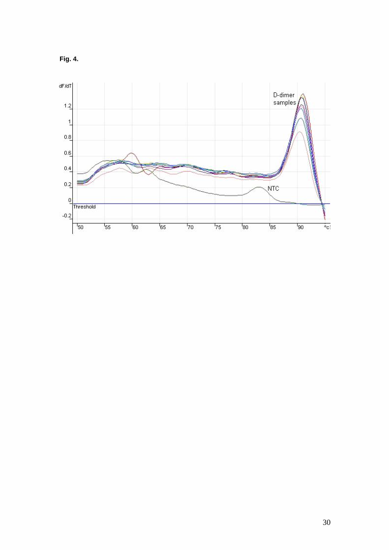
specific reaction products (data not shown). We also performed a melt-curve
analysis following PCR. The results of the analysis for the indirect D-dimer
assay are shown in Figure 4, but similar results were observed for all assays.
There is a small peak at about 83 ºC for the sample removed from a well to
which no STV-DNA conjugate had been added (no template control),
compared to the large peaks observed at about 91 ºC to 92 ºC for the sample
eluates. (The threshold shown in Figure is set at 0 and is not related to the
cut-off point used to determine Ct values.) The results suggest that a single
product was produced during the PCR reaction with reporter template present,
whereas a much smaller amount of a lower-sized product, possibly primer-
dimer, was produced in the absence of template DNA.

15
4. Discussion
The results presented here demonstrate the utility of a highly purified STV-
DNA conjugate in an immuno-PCR format in detecting and quantitating two
different analytes in both capture and indirect formats. The performance of
this immuno-PCR was similar to that with other systems, in terms of minimum
concentration of analyte detected (of the order 50 pg/ml) and of increase in
sensitivity and dynamic range over conventional ELISA.
The level of detection of immuno-PCR has not approached that of PCR which
can detect a few DNA molecules (e.g., Hobson-Peters et al., 2007), for two
main reasons. The first is the non-specific attachment of assay components
to each other (e.g. capture to detector antibody), to the analyte or to the assay
substrate, such as the microtitre well. This is readily observed in our
experiments as the difference in Ct values obtained from sample wells which
contain no analyte (background binding) and those to which no template DNA
was added (non-specific DNA amplification). In our case, this difference was
generally around seven cycles (data not shown), which is greater than the
difference in Ct values obtained from samples containing 10 or 0.01 ng/ml D-
dimer, or four orders of magnitude (Fig. 3a). To try to overcome this,
immuno-PCR protocols use various protein or DNA blocking reagents to
decrease non-specific adsorption, together with extensive washing steps to
remove unwanted components. Extensive washing has the disadvantage of
removing some authentic detection signal, the extent of which depends on the
number and stringency of washing steps and the binding affinity of the analyte
– ligand and subsequent detection reagents.

16
The second factor which decreases the sensitivity of immuno-PCR assays is
the presence of unconjugated by-products from the synthesis of conjugated
detection reagents. These reagents can increase the background noise or
block the available sites of attachment for the corresponding conjugated
reagents. Hence, purification of the conjugate away from unreacted reagents
is essential for increasing the signal-to-noise ratio.
Amplification of the signal in traditional ELISA is achieved by the use of
secondary reagents in the indirect ELISA format, in which more than one
reporter molecule is bound to the ligand. One example is the use of
conjugated anti-immunoglobulin antibody. These secondary reagents have
the additional advantage of genericity, such that a single conjugate can be
used for an array of analyte – ligand types. However, they can also contribute
to decreased sensitivity through non-specific binding, as discussed above.
Thus, the predominant disadvantages of the use of secondary reagents is the
increase in the number of washing steps and the time taken to perform an
assay, the latter being of especial importance in clinical laboratories. It can be
seen that the use of secondary reagents in immuno-PCR reflects a balance
between the advantages in signal amplification and genericity, against the
increase in background signal due to non-specific attachment of reagents,
more washing steps and time taken to perform the reaction.
Various methods have been employed to link the detector ligand to the DNA
template. A recent study which compared three types of assay format

17
concluded that detection of analyte with a mAb directly conjugated to the DNA
template was more sensitive than use of secondary detection methods
compromising biotinylated mAb and DNA template linked by a STV bridge in
an indirect or capture format (Lind et al., 2005). As mentioned above, one
disadvantage of this format is the use of a non-generic reagent produced by
several types of chemical modification and three purification steps. The direct
conjugation of DNA to protein is difficult to control, although recent reports
using intein-mediated, expressed protein ligation should decrease the
variability of production of DNA-MAb conjugates (Lovrinovic et al., 2005,
Burbulis et al., 2005).
We have examined an alternative system in which the number of assay
components is kept to a minimum whilst employing a generic detection
conjugate of STV linked directly to the reporter DNA. The key step in the
generation of the STV-DNA was the purification of the conjugate by
iminobiotin affinity chromatography. Although the generation of the STV-DNA
conjugate involved three chemical reactions and purification steps, this is a
generic reagent and a single production run in large quantity can be used for
many types of assays. The non-generic (assay specific) mAb was biotinylated
by a simple, two-step conjugation and purification procedure which facilitates
the application of this protocol to different analyte-antibody combinations.
A disadvantage of the system we have developed is that it cannot be applied
to the simultaneous detection of several analytes, due to the generic nature of
the STV-DNA conjugate and the use of an intercalating dye in the real-time
PCR.

18
We assessed the performance of the STV-DNA conjugate in both indirect and
capture formats. Although the indirect assay involves fewer steps, it can
suffer from variability in the attachment of analytes to the surface of the
microtitre plate. This is especially so when the analyte is present in a complex
biological matrix such as serum, where other components compete for
attachment to the well surface. The use of a second ligand, often a MAb, to
capture the analyte aims to decrease this variability. For commercial assays,
it also offers the advantage of obtaining plates to which the capture ligand has
been attached, thus reducing the time involved in coating the plate. The
disadvantage compared to the indirect format is the presence of the capture
ligand as another biological entity for potential non-specific binding of the
detector molecules. We observed much lower delta Ct values at the lower
antigen concentrations in the capture assays (Figure 3b and c), presumably
due to non-specific interaction with the capture ligand. The use of other more
inert ligands, such as aptamers, may resolve this problem.

19
Acknowledgements
The authors are very grateful to Jody Hobson-Peters for her expert technical
assistance in establishing the PCR methodology and to Dr. Mike Gerometta
for his insightful advice on the D-dimer and canine heartworm antibodies and
antigens.

20
References
Adler, M., Wacker, R., Niemeyer, C.M., 2003. A real-time immuno-PCR for
routine ultrasensitive quantification of proteins. Biochem. Biophys. Res.
Commun. 308, 240.
Bobrow, M.N., Harris, T.D., Shaughnessy, K.J., Litt, G.J., 1989. Catalyzed
reporter deposition, a novel method of signal amplification. Application to
immunoassays. J. Immunol. Methods 125, 279.
Burbulis, I., Yamaguchi, K., Gordon, A., Carlson, R., Brent, R., 2005. Using
protein-DNA chimeras to detect and count small numbers of molecules.
Nature Methods 2, 31.
Hobson-Peters, J., O’Loughlin, P., Toye, P., 2007. Development of an
internally controlled homogeneous PCR assay for the simultaneous
detection and discrimination of herpes simplex virus types 1 and 2 and
varicella-zoster virus. Mol. Cell. Probes 21, 24.
Masci, P.P., Whitaker, A.N., Winzor, D.J., 1985. A simple chromatographic
procedure for the purification of the D dimer fragment from crosslinked
fibrin. Anal. Biochem. 147,128.
Lind, K., Kubista, M., 2005. Development and evaluation of three real-time
immuno-PCR assemblages for quantification of PSA. J. Immunol.
Methods 304, 107.
21
Lovrinovic, M., Spengler, M., Deutsch, C., Niemeyer, C.M., 2005. Synthesis of
covalent DNA-protein conjugates by expressed protein ligation. Mol.
Biosyst. 1, 64.
Niemeyer, C.M., Wacker, R., Adler, M., 2003. Combination of DNA-directed
immobilization and immuno-PCR: very sensitive antigen detection by
means of self-assembled DNA-protein conjugates. Nucleic Acids Res. 31,
90.
Nikbakht, K.N., Boone, L.R., Glover, P.L., Myer, F. E., Yang, W.K., 1987.
Characterization of a molecular clone of RFM/Un mouse chromosomal
DNA that contains a full-length endogenous murine leukaemia virus-
related proviral genome. J. Gen. Virol. 68, 683.
Sano, T., Smith, C.L., Cantor, C.R., 1992. Immuno-PCR: very sensitive
antigen detection by means of specific antibody-DNA conjugates. Science
258, 120.
Sims, P.W., Vasser, M., Wong, W.L., Williams, P.M., Meng, Y.G., 2000.
Immunopolymerase chain reaction using real-time polymerase chain
reaction for detection. Anal. Biochem. 281, 230.

22
Figure 1.
a. Purification of maleimide-modified STV using a desalting column by ion
exchange chromatography. The sample was passed through a ‘HiTrap’
desalting column equilibrated with 50 mM phosphate buffer pH 7.0 at flow rate
5.0 mL/min. STV-SMCC conjugates were collected from 14 to 26 seconds and
unreacted sulpho-SMCC was eluted after 34 seconds as shown.
b. Purification of unconjugated STV and DNA-conjugated STV by affinity
chromatography. Samples were loaded onto a 2.0 mL 2-iminobiotin column
equilibrated with binding buffer (50 mM ammonium acetate with 0.5 M NaCl,
pH 11.0). The column was washed with 5 mL of binding buffer at 1.0 mL/min
to remove unbound proteins and DNA (peak 1). The STV and STV-DNA were
eluted with 0.1 M acetic acid (peak 2).
c. Separation of DNA-STV conjugate and unreacted STV by HPLC. Samples
were loaded on a size exclusion HPLC column (Phenomenex, Bio-SEC-3000
U.S.A.) equilibrated with 0.1 M phosphate buffer, pH 6.5, containing 200mM
NaCl. The STV-DNA (upper profile) was eluted between 5.5 and 6.2 min and
unreacted STV (upper profile) was eluted after 9 min. As a control, STV was
loaded onto HPLC to indicate the elution time of STV (lower profile).

23
Fig. 1a.
Fig. 1b.
Fig. 1c.

24
Figure 2.
a. Schematic representation of the DNA template. Primers 1 and 2 were
used to generate the template for attachment to STV. DNA for the read-
out PCR was released from the bound conjugate by digestion with NheI
and amplified with primers 3 and 2.
b and c. Schematic representation of the indirect (b) and capture (c) immuno-PCRs.

25
Fig. 2a.
Fig. 2b.
Fig. 2c.

26
Figure 3.
a. Comparison of indirect D-dimer immuno-PCR (■) and conventional ELISA
(♦).
b. Comparison of capture D-dimer immuno-PCR (■) and conventional ELISA
(♦).
c. Comparison of a capture CHA immuno-PCR with (■) and without (▲)
genomic DNA blocker, and conventional ELISA (♦).
In all figures, the left axis represents the difference in Ct value obtained with
the negative control (0 ng antigen) and the value obtained with the respective
antigen concentration. The right axis represents the absorbance at 450nm
obtained in conventional ELISA.

27
Indirect D-dimer
0
2
4
6
8
10
12
0 0.001 0.01 0.1 1 10 100 1000
D-Dimer (ng/ml)
Δ C
t
0
0.5
1
1.5
2
2.5
A40
5 nm
Capture D-dimer
0
1
2
3
4
5
6
7
8
9
0 0.001 0.01 0.1 1 10 100 1000
D-Dimer (ng/ml)
Δ C
t
0
0.5
1
1.5
2
2.5
A40
5 nm
Fig. 3a.
Fig. 3b.

28
Capture CHA
0
1
2
3
4
5
6
7
8
9
0 0.001 0.01 0.1 1 10 100 1000
CHA (ng/ml)
Δ C
t
0
0.5
1
1.5
2
2.5
3
3.5
4
A40
5 nm
Fig. 3c.

29
Figure 4.
Melt curve analysis of the PCR products generated during indirect D-dimer
assay. The samples eluted from wells to which STV-DNA had been added
showed melt curve peaks of about 91 ºC to 92 ºC, whereas that from the well
to which no STV-DNA had been added (NTC - no template control) exhibited
a much smaller peak at about 83 ºC. The fluorescence data are plotted as the
first derivative (dF/dT) of the fluorescence signal (vertical axis) against
temperature (horizontal axis).
30
Fig. 4.

31
Table 1. Reproducibility of indirect (a) and capture (b) immuno-PCR for D-
dimer.
a.
D-dimer (pg / ml) 100 10 1 0 Replicate 1 14.47a 15.85 19.53 23.14 Replicate 2 14.11 16.18 20.62 23.58 Replicate 3 14.02 16.19 19.79 23.26 Mean 14.2 16.07 19.98 23.33 SD 0.24 0.19 0.57 0.23
b.
D-dimer (pg / ml) 100 10 1 0 Replicate 1 21.37 a 22.56 23.54 24.46 Replicate 2 21.49 22.58 23.54 24.51 Replicate 3 n/d 22.66 23.61 n/d Mean 21.43 22.60 23.56 24.49 SD 0.085 0.053 0.040 0.035