A Novel Gene Containing a Trinucleotide Repeat That Is Expanded
13
Cell, Vol. 72, 971-963. March 26, 1993, Copyright 0 1993 by Cell Press A Novel Gene Containing a Trinucleotide Repeat That Is Expanded and Unstable on Huntington’s Disease Chromosomes The Huntington’s Disease Collaborative Research Group* Summary The Huntington’s disease (HD) gene has been mapped in 4~16.3 but has eluded identification. We have used haplotype analysis of linkage disequilibrium to spot- light a small segment of 4~16.3 as the likely location of the defect. A new gene, IT15, isolated using cloned trapped exons from the target area contains a poly- morphic trinucleotide repeat that is expanded and unstable on HD chromosomes. A (CAG), repeat longer than the normal range was observed on HD chromo- somes from all 75 disease families examined, com- prising a variety of ethnic backgrounds and 4~16.3 haplotypes. The GAG), repeat appears to be located within the coding sequence of a predicted -346 kd protein that is widely expressed but unrelated to any known gene. Thus, the HD mutation involves an unstable DNA segment, similar to those described in fragile X syndrome, spino-bulbar muscular atrophy, and myotonic dystrophy, acting in the context of a novel 4~16.3 gene to produce a dominant phenotype. ‘The Huntington’s Disease Collaborative Research Group comprises: Group 1: Marcy E. MacDonald,’ Christine M. Ambrose,’ Mabel P. Duyao,’ Richard H. Myers,2 Carol Lin,’ Lakshmi Srinidhi,’ Glenn Barnes,’ Sherry1 A. Taylor,’ Marianne James,’ Nicolet Groat,’ Heather MacFarlane,’ Barbara Jenkins,’ Mary Anne Anderson,’ Nancy S. Wexler,3 and James F. Gusella’ t ‘Molecular Neurogenetics Unit Massachusetts General Hospital and Department of Genetics Harvard Medical School Boston, Massachusetts 02114 ‘Department of Neurology Boston University Medical School Boston, Massachusetts 02118 3Hereditary Disease Foundation 1427 7th Street, Suite 2 Santa Monica, California 90401 Group 2: Gillian P. Bates, Sarah Baxendale, Holger Hummerich, Susan Kirby, Mike North, Sandra Youngman, Richard Mott, Gunther Zehetner, Zdenek Sedlacek, Annemarie Poustka, Anna-Maria Frischauf, and Hans Lehrach Genome Analysis Laboratory Imperial Cancer Research Fund Lincoln’s Inn Fields London, WCPA 3PX, England Group 3: Alan J. Buckler,’ Deanna Church,’ Lynn Doucette-Stamm,’ Michael C. O’Donovan,’ Introduction Huntington’s disease (HD) is a progressive neurodegener- ative disorder characterized by motor disturbance, cogni- tive loss, and psychiatric manifestations (Martin and Gu- sella, 1986). It is inherited in an autosomal dominant fashion and affects - 1 in 10,000 individuals in most popu- lations of European origin (Harper et al., 1991). The hall- mark of HD is a distinctive choreic movement disorder that typically has a subtle, insidious onset in the fourth to fifth decade of life and gradually worsens over a course of 10 to 20 years until death. Occasionally, HD is ex- pressed in juveniles, typically manifesting with more se- vere symptoms including rigidity and a more rapid course. Juvenile onset of HD is associated with a preponderance of paternal transmission of the disease allele. The neuro- pathology of HD also displays a distinctive pattern, with selective loss of neurons that is most severe in the caudate and putamen. The biochemical basis for neuronal death in HD has not yet been explained, and there is conse- quently no treatment effective in delaying or preventing the onset and progression of this devastating disorder. The genetic defect causing HD was assigned to chromo- some 4 in 1983 in one of the first successful linkage analy- ses using polymorphic DNA markers in humans (Gusella Laura Riba-Ramirer,’ Manish Shah,’ Vincent P. Stanton,’ Scott A. Strobel,2 Karen M. Draths,* Jennifer L. Wales,2 Peter Dervan,2 and David E. Housman’ ‘Center for Cancer Research Massachusetts Institute of Technology Cambridge, Massachusetts 02139 ‘Division of Chemistry and Chemical Engineering California Institute of Technology Pasadena, California 91125 Group 4: Michael Altherr, Rita Shiang, Leslie Thompson, Thomas Fielder, and John J. Wasmuth Department of Biological Chemistry University of California Irvine, California 92717 Group 5: Danilo Tagle, John Valdes, Lawrence Elmer, Marc Allard, Lucia Castilla, Manju Swaroop, Kris Blanchard, and Francis S. Collins Department of Internal Medicine and Human Genetics and The Howard Hughes Medical Institute University of Michigan Ann Arbor, Michigan 48109 Group 6: Russell Snell, Tracey Holloway, Kathleen Gillespie, Nicole Datson, Duncan Shaw, and Peter S. Harper Institute of Medical Genetics University of Wales College of Medicine Cardiff, CF4 4XN, Wales tCorrespondence should be addressed to James F. Gusella.
Transcript of A Novel Gene Containing a Trinucleotide Repeat That Is Expanded

Cell, Vol. 72, 971-963. March 26, 1993, Copyright 0 1993 by Cell Press
A Novel Gene Containing a Trinucleotide Repeat That Is Expanded and Unstable on Huntington’s Disease Chromosomes
The Huntington’s Disease Collaborative Research Group*
Summary
The Huntington’s disease (HD) gene has been mapped in 4~16.3 but has eluded identification. We have used haplotype analysis of linkage disequilibrium to spot- light a small segment of 4~16.3 as the likely location of the defect. A new gene, IT15, isolated using cloned trapped exons from the target area contains a poly- morphic trinucleotide repeat that is expanded and unstable on HD chromosomes. A (CAG), repeat longer than the normal range was observed on HD chromo- somes from all 75 disease families examined, com- prising a variety of ethnic backgrounds and 4~16.3 haplotypes. The GAG), repeat appears to be located within the coding sequence of a predicted -346 kd protein that is widely expressed but unrelated to any known gene. Thus, the HD mutation involves an unstable DNA segment, similar to those described in fragile X syndrome, spino-bulbar muscular atrophy, and myotonic dystrophy, acting in the context of a novel 4~16.3 gene to produce a dominant phenotype.
‘The Huntington’s Disease Collaborative Research Group comprises: Group 1: Marcy E. MacDonald,’ Christine M. Ambrose,’ Mabel P. Duyao,’ Richard H. Myers,2 Carol Lin,’ Lakshmi Srinidhi,’ Glenn Barnes,’ Sherry1 A. Taylor,’ Marianne James,’ Nicolet Groat,’ Heather MacFarlane,’ Barbara Jenkins,’ Mary Anne Anderson,’ Nancy S. Wexler,3 and James F. Gusella’t ‘Molecular Neurogenetics Unit Massachusetts General Hospital and Department of Genetics Harvard Medical School Boston, Massachusetts 02114 ‘Department of Neurology Boston University Medical School Boston, Massachusetts 02118 3Hereditary Disease Foundation 1427 7th Street, Suite 2 Santa Monica, California 90401
Group 2: Gillian P. Bates, Sarah Baxendale, Holger Hummerich, Susan Kirby, Mike North, Sandra Youngman, Richard Mott, Gunther Zehetner, Zdenek Sedlacek, Annemarie Poustka, Anna-Maria Frischauf, and Hans Lehrach Genome Analysis Laboratory Imperial Cancer Research Fund Lincoln’s Inn Fields London, WCPA 3PX, England
Group 3: Alan J. Buckler,’ Deanna Church,’ Lynn Doucette-Stamm,’ Michael C. O ’Donovan,’
Introduction
Huntington’s disease (HD) is a progressive neurodegener- ative disorder characterized by motor disturbance, cogni- tive loss, and psychiatric manifestations (Martin and Gu- sella, 1986). It is inherited in an autosomal dominant fashion and affects - 1 in 10,000 individuals in most popu- lations of European origin (Harper et al., 1991). The hall- mark of HD is a distinctive choreic movement disorder that typically has a subtle, insidious onset in the fourth to fifth decade of life and gradually worsens over a course of 10 to 20 years until death. Occasionally, HD is ex- pressed in juveniles, typically manifesting with more se- vere symptoms including rigidity and a more rapid course. Juvenile onset of HD is associated with a preponderance of paternal transmission of the disease allele. The neuro- pathology of HD also displays a distinctive pattern, with selective loss of neurons that is most severe in the caudate and putamen. The biochemical basis for neuronal death in HD has not yet been explained, and there is conse- quently no treatment effective in delaying or preventing the onset and progression of this devastating disorder.
The genetic defect causing HD was assigned to chromo- some 4 in 1983 in one of the first successful linkage analy- ses using polymorphic DNA markers in humans (Gusella
Laura Riba-Ramirer,’ Manish Shah,’ Vincent P. Stanton,’ Scott A. Strobel,2 Karen M. Draths,* Jennifer L. Wales,2 Peter Dervan,2 and David E. Housman’ ‘Center for Cancer Research Massachusetts Institute of Technology Cambridge, Massachusetts 02139 ‘Division of Chemistry and Chemical Engineering California Institute of Technology Pasadena, California 91125
Group 4: Michael Altherr, Rita Shiang, Leslie Thompson, Thomas Fielder, and John J. Wasmuth Department of Biological Chemistry University of California Irvine, California 92717
Group 5: Danilo Tagle, John Valdes, Lawrence Elmer, Marc Allard, Lucia Castilla, Manju Swaroop, Kris Blanchard, and Francis S. Collins Department of Internal Medicine and Human Genetics and The Howard Hughes Medical Institute University of Michigan Ann Arbor, Michigan 48109
Group 6: Russell Snell, Tracey Holloway, Kathleen Gillespie, Nicole Datson, Duncan Shaw, and Peter S. Harper Institute of Medical Genetics University of Wales College of Medicine Cardiff, CF4 4XN, Wales
tCorrespondence should be addressed to James F. Gusella.

Cell 972
D D DD D D 500 kb ii : 44 4
ss s i A 1 19 1
8”
I I
Figure 1. Long-Range Restriction Map of the HD Candidate Region
A partial long-range restriction map of 4~16.3 is shown (adapted from Lin et al. 119911). The HD candidate region determined by recombi- nation events is depicted by hatched bars be- tween D4SiO and D4S98. The portion of the HD candidate region implicated as the site of the defect by linkage disequilibrium haplotype analysis (MacDonald et al., 1992) is shown as a closed bar. Below the schematic map, the region from D4S780 to D4S182 is expanded to show the cosmid contig (averaging 40 kb per cosmid). The genomic coverage and, where known, the transcriptional orientation (arrows,
__ LlalFl L11386 L83D3 ~ LlSlSlO LIIOFS
5’ to 3’) of the IT1 5, IT1 1, IT1 OC3, and ADDA - __
- SJ58 genes is also shown. Locus names above the
L13488 __ __ L22BSB y24 ~ __ L4OD10 __ L88F,
map denote selected polymorphic markers that SJSBW __ A12
LlC2 L142C5 have been used in HD families. The positions of 045727 and D4S95, which form the core of
4 1 n - haplotype in the region of maximum disequilib- IT15 IT11 ITlOC3 ADDA rium, are also shown in the cosmid contig. Re-
striction sites are given for Notl (N), Mlul (M), and Nrul (R). Sites displaying complete digestion are shown in boldface, while sites subject to frequent incomplete digestion are shown as lighter symbols. Brackets around the N symbols indicate the presence of additional clustered Notl sites.
et al., 1983). Since that time, we have pursued a location cloning approach to isolating and characterizing the HD gene based on progressively refining its localization (Gu- sella, 1989, 1991). Among other work, this has involved the generation of new genetic markers in the region by a number of techniques (Pohl et al., 1988; Whaley et al., 1991; MacDonald et al., 1989a), the establishment of ge- netic (MacDonald et al., 1989b; Allitto et al., 1991) and physical maps of the implicated regions (Bucan et al., 1990; Bates et al., 1991; Doucette-Stamm et al., 1991; Altherr et al., 1992), the cloning of the 4p telomere of an /-/D chromosome in a yeast artificial chromosome clone (Bates et al., 1990; Youngman et al., 1992), the establish- ment of yeast artificial chromosome (Bates et al., 1992) and cosmid (S. B. et al., unpublished data) contigs of the candidate region, as well as the analysis and characteriza- tion of a number of candidate genes from the region (Thompson et al., 1991; Taylor et al., 1992; Ambroseet al., 1992; M. P. D. et al., submitted). Analysis of recombination events in HD kindreds has identified a candidate region of 2.2 Mb, between D4SlO and D4S98 in 4~16.3, as the most likely position of the HD gene (MacDonald et al., 1989b; Bateset al., 1991; Snell et al., 1992). Investigations of linkage disequilibrium between HD and DNA markers in 4~16.3 (Snell et al., 1989; Theilman et al., 1989) have suggested that multiple mutations have occurred to cause the disorder (MacDonald et al., 1991). However, haplotype analysis using multiallele markers has indicated that at least one-third of HD chromosomes are ancestrally related (MacDonald et al., 1992). The haplotype shared by these HD chromosomes indicates that a 500 kb segment be- tween D4S780 and D4S782 is the most likely site of the genetic defect.
Targeting this 500 kb region for saturation with gene
transcripts, we have used exon amplification as a rapid method for obtaining candidate coding sequences (Buck- ler et al., 1991). This strategy has previously identified three genes: the a-adducin gene (ADDA) (Taylor et al., 1992) and a putative novel transporter gene (ITlOC3) in the distal portion of this segment (M. P. D. et al., submit- ted), and a novel G protein-coupled receptor kinase gene (IT1 1) in the central portion (Ambrose et al., 1992). How- ever, no defects implicating any of these genes as the HD locus have been found. We have now applied the exon amplification approach to the proximal portion of the 500 kb segment. We have identified a large gene, IT15 span- ning - 210 kb, that encodes a previously undescribed pro- tein of - 348 kd. The IT1 5 reading frame contains a poly- morphic (CAG), trinucleotide repeat with at least 17 alleles in the normal population, varying from 11 to 34 CAG cop- ies. On HD chromosomes, the length of the trinucleotide repeat is substantially increased, to a range of 42 to over 66 copies, and shows an apparent correlation with age of onset, the longest segments being detected in juvenile HD cases. The instability in the length of the repeat is reminiscent of similar trinucleotide repeats in the fragile X syndrome and in myotonic dystrophy (Suthers et al., 1992). The presence of an unstable, expandable trinucleo- tide repeat on HD chromosomes in the region of strongest linkage disequilibrium with the disorder suggests that this alteration underlies the dominant phenotype of HD and that IT15 encodes the HD gene.
Results
Application of Exon Amplification to Obtain Trapped, Cloned Exons The HD candidate region defined by discrete recombina-

Huntington’s Disease Gene 973
12 3
- 28s
- 18s
Figure 2. Northern Blot Analysis of the IT15 Transcript
Results of the hybridization of IT15A to a Northern blot of RNA from normal (lane 1) and HD homozygous (lanes 2 and 3) lymphoblasts are shown. A single RNA of - 11 kb was detected in all three samples, with slight apparent variations being due to unequal RNA concentra- tions. The HD homozygotes are independent, deriving from an Ameri- can family (lane 2) and the large Venezuelan family (lane 3), respec- tively. The Venezuelan HD chromosome has a 4~16.3 haplotype of 5 2 2 defined by a (GT). polymorphism at D4S727and variable number tandem repeat and Taql restriction fragment length polymorphisms at D4S95. The American homozygote carries the most common 4~16.3 haplotype found on HD chromosomes: 2 11 1 (MacDonald et al., 1992).
tion events in well-characterized families spans 2.2 Mb between D4SlO and D4S98 as shown in Figure 1. The 500 kb segment between D4SlBO and D4S182 displays the strongest linkage disequilibrium with HD, with about one-third of disease chromosomes sharing a common haplotype, anchored by multiallele polymorphisms at 048127 and D4S95 (MacDonald et al., 1992). We have isolated 64 overlapping cosmids, spanning - 480 kb from D4S180 to a location between D4S95 and D4S182, by a combination of information from yeast artificial chromo- some (Baxendale et al., 1991) and cosmid probe hybridita-
tion to high density filter grids of a chromosome 4-specific library, as well as of additional libraries covering this re- gion. Sixteen of these cosmids providing the complete contig are shown in Figure 1. We have previously used exon amplification to identify ADDA, ITlOC3 (a novel puta- tive transporter gene), and IT1 1 (a novel G protein-cou- pled receptor kinase gene) in the region distal to 048727 (Figure 1).
We have now applied the exon amplification technique to cosmids from the region of the contig proximal to 048727. This procedure produces trapped exon clones, which can represent single exonsor multiple exons spliced together, and is an efficient method for obtaining probes for screening cDNA libraries. Individual cosmids were pro- cessed, yielding nine exon clones in the region from cos- mids L13489 to L181BlO.
Identification of the IT15 Gene Two nonoverlapping cDNAs were initially isolated using exon probes. IT15A was obtained by screening a trans- formed adult retinal cell cDNA library with exon clone DL118F5-U. IT16A was isolated by screening an adult frontal cortex cDNA library with a pool of three exon clones, DL83D3-8, DL83D3-1, and DL228B6-3. By North- ern blot analysis, we discovered that IT15A and IT16A both detected an - 10-l 1 kb transcript, suggesting that they derive from the same mRNA. Figure 2 shows an ex- ample of a Northern blot containing RNA from lymphoblas- toid cell lines representing a normal individual and two independent homozygotes for HD chromosomes of differ- ent haplotypes. The same - 1 O-l 1 kb transcript was also detected in RNA from a variety of human tissues (liver, spleen, kidney, muscle, and various regions of adult brain).
IT15A and IT16A were used to “walk” in a number of human tissue cDNA libraries in order to obtain the full- length transcript. Figure 3 shows a representation of five cDNA clones that define the IT1 5 transcript, under a sche-
IT15
composite
*c*** * * * * Figure 3. Schematic of cDNAClones Defining -- An the IT15 Transcript
Five cDNAs are represented under a sche-
IT16C - matic of the composite IT1 5 sequence. The thin line corresponds to untranslated regions. The thick line corresponds to codmg sequence, as-
IT16A - suming initiation of translation at the first Met codon in the open reading frame. Stars mark
IT166 the positions of ;he following exon clones 5’to 3’: DL8303-8, DL83D31, DL228f36-3, DL228B6-5, DL228B6-13, DL69F7-3, DL178H4-6, DL118F5-
IT156 An U, and DL134BSlJ4. The composite sequence was derived as follows. From 22 bases 3’ to
IT15A the putative initiator Met ATG, the sequence was compiled from the cDNA clones and exons
shown. There are 9 bases of sequence intervening between the 3’end of IT168 and the 5’end of IT158. These were identified by PCR amplification of first-strand cDNA and sequencing of the PCR product. At the 5’ end of the composite sequence, the cDNA clone IT16C terminates 27 bases upstream of the (CAG),. However, when IT16C was identified, we had already generated genomic sequence surrounding the (CAG), in an attempt to generate new polymorphisms. This sequence matched the IT16C sequence and extended it 337 bases upstream, including the apparent Met initiation codon.

Cdl 974
1 TTGCTtTtTGAGGtAWCCTGCGGCCGCACGGtCGGGC,GG,TCCCTGGCCAGCCAT,GGCAWGTCCCCACGCTAGGGC,GTCM,CA,GC,CGCCGGCGlGGCCCCGCCTCCGCCGG
12, CGCGGCCCCGCClCCGCCGGCCClCG,ClGG~CGCUCGtCCGGCCGClCAGGllClGCl lTTACCTGCGGCCCAGAGCCCCAllC
24; ATTGCCCCGGTGCTGAGCGGCGCCGC~GTCGGCCC~GGCC~CCGGG~C~GCCG~GCCGGGCGG~~CCGCCATGGC~CCC~G~GCT~T~GGCCTTC~GTCCC~C~G WATLEKLYKAFESLK
361 TCClTCCACCAGCAGCAGCAGCAGCAGCAGCAGCAGCAGCAGCAGCAGCAGCAGCAGCAGCAGCAGCAGCMCAGCCGCCACCGCCGCCGCCGCCGCCGCCGCClCCTCAGCTTCCTCAG 16 s f Q pp p P pa Q Q p a p P Q p 0 pa P P PP P P P P P P P P P P P P P L P P
48, CCGCCGCCGCAGGCACAGCCGCTGCTGCC,CAGCCGCAGCCGCCCCCGCCGCCGCCCCCGCCGCCACCCGGCCCGGC~G~GGCT~GGAGCCGCTGCACCGACC~G~GMCT~TCA 56 P P P PA P P L L P P P Q P P P P P P P P P PG PA" A E E P L H R P Y K E L S
60, GCTACCMGAAIGACCGTGTWTCA,TG,C,WCMTATGT~CATAGTGGCACAGTCTG,CACIM,lC,CCAGM,,TCA~CT,C,GGGCATCGC,A,G~C,TTTTCTG 96 A, K YD R" N H C L 1 ICE N I" A a S" P N S P E F P K L L G I A II E L F L
72, C,G,GCAG,W,GACCCAGAGTCA~TG,CAGW,GG,GGC,CICUJTGCC,CMC*MGT~A,C~GC,, ,~,GWT,CTU,CT,CCMGGT,ACAGC~CGAGC~CTATUGGAA 136 L C SD D A E SD" R WV A D E C L Y I(" I KA L N D SW L P R L a L E L Y I( E
841 ATTMAUGU~GG,GCCCCTCGWGTT,GCGTGC,GCCC,GTGWGGTTTGCTWGCTGGCTCACCTGGT~CGGCCTCA~TGCAGGCC~~ACCTGGTGUCC~~CT~CCG~GCC~G 176 I I: Y N G A P R S L R A A L Y R I A E LA H L" II P P Y C R P Y L" N L L P C L
96, ACTCGMCMGCMWGICCCGUWUTCIGTCAGTCCAG~~CC,TGGCTGCAGC,G,TCCC~~,~TGGCTTCT,TTGGCMTTTTGC~TGAC~TGA~TTMGG,,TTG,T~G 216 1 R,S I: I P E E S YQ ETL A A A" P K I HA S f G W F A" D U E I K" L L I:
m31 G~~TT~A,A~~~CC,~G,C~GC,CCCCCACCAT,CGG~GGA~AGCGG~,G~,CAGCAGT~~GCAT~,G~~AG~A~,~MGMGGACACMTATTTCTATAGTTGG~T*CT~T 256 A F ,A II L I: S S S P 1 IR R 1 A A GSA" S I C 0 H S R R T 0 Y F" S Y L L N
l20, G,GCTCT,AGGCTTACTCGl,CCTGTCGAGWTWICACTCCACTCTGCT~TTCT,GGCGTGC,GCTCACCC,CAGG,A,TTGt,GCCCTTGCTGCAGCAGCAGtTCUGGACACMGC 296" L L G L L" P" E D E H s T L L I L G Y L L, L R" L" P L L P Q P" K D T s
132, CTG~GGCAGCT,CGGAGT~CMG~G~,GGMGTCTCTCCTTCTGCAGAGCAGC,,GTCCAGG,l lATGMCT~CG,TACA,CAlACACAGCACCMGACCAC~,G,TGTG 336 L KG S F G" 1 RI: E W E" s P S A E P L" P" I E L T L H HlP H P D H WV V
144, ACCGGAGCCCTGGAGC,GTTGCAGCAGCTC,TCAGMCCCCTCCACCCGAGCT,CTGCIVUCCClGACCGCAGTCGGGGGCAT,GGGCAGCTCACCGCTGC,lGGAGGAGTC,GGTGGC 376 1 GA L E L L P Q L F R T P P P E L L 9 T L T A" G G It 0 LTA A K E E S G G
156, CGUGCCGTAG,GGGAC,ATTG,GCMCTTATAGCTGGAGGGGGTTCCTCA,GCAGCCCTG,CC,TTCMG~C~GGC~G,GCTC~,AGGAGMGAAGMGCCT,GGAGGAT 416 R S R SG ST" E LIA G GG S s C s P V L SR KP K GK" L L GEE E A L ED
,681 GACTCTGUTCWW,CGGA,GTCAGCACCTC,GCC,TMCAGCCTCAG,GMGGA,WGATCAGTGGAGAGC,GGC,GC,TCT,CAGGGGTTTCCACTCCAGGGTCAGCAGGTCATGAC 456 D S E S R s D" S s s A L T A s" K D E I S GE LA A S S G" ST P G SAG H D
l.S.0, ATCATCACAGAlCAGCCACGGTCACAGCACACACTGCAGGCGGACTCACTGGA,CTGGCCAGCTGT~C,,~CMGCTCTGCCACT~TGGG~TGAG~GGATATCT,GAGCCACAGC 496 I IT E Q P R S P H T L P AD S L D L As CDL T s s AT D G D E E D I L s H S
192, TCCAGCCAGGlCAGCGCCGTCCCAlCT~CCCTGCCATGGACCTGMTGATGGGACCCAGGCC,CGTCGCCCATCAGC~CAGCTCCCAGACCACCACCGMGGGCCTGAlTCAGCTG,T 536 S S P" S A" P s D PA II 0 L W D G, Q A s s P I S D S S P Tl, E G P D S A"
2041 ACCCC,TCAWCAGT~CTCU,TG,GT~AGACGGTACCGACMCCAGTA,,TGGGCCTGCAGATTGGACAGCCCCAGGA,GMGA,~GG~GCCACAGGTAT,CTTCCTGATG~GCC 576, P SDS S E I" L 0 G T 0 II Q I L G L P I GQ P P 0 E 0 E E A T G I L P D E A
2161 TCGCAGGCCT,CAGGMClC,,CCA,GGCCClTCMCAGGCACA,, ,A,T~CATGAGTCAC,GCAGGCAGCCTlCT~CAGCAGTGT,G*TA/UT,TGTGTTGAMGA,GMGCT 616 SE A F R" S SW ALP P A H L L K Y M S" C P P P S D S S "0 K F Y L R D E A
228, AC,CAACCGGG,GA,CM~MUCUGCCTTGCC,,GCCGCA,C~GG,GACATTGGACAGTCCACTGA,GATGACTC~GCACCTCT,GTCCATTCTGTCCGCCTTT,ATCTGC,,CGT,TTTG 656 1 E P GOP E II K P C RIK G 0 IG Q S T D D 0 SAP L" H S V R L L S A S F L
240, CTMCAGGGGGAMAAJ ,GTGC,GGTTCCGGACAGGCA,GTGAGGG,CAGCGTG~GGCCC,GGCCCTCAGCTGlGTGGGAGCAGCTG,GGCCC,CCACCCGG~TC,TTCTTCAGC~ 696 L 1 G G I: NV L" P 0 R 0 Y R Y S Y K A L A L S C" G A A Y A L H P E S F F SK
252, C,CTA,AUIGT,CCTC,,GACACCACGGMTACCCTGAGGMCAG,A,GTC,CAGACATC,T~CTACATCGAlCA,GGAGACCCACAGGTTCGAGGAGCCAC,GCCATTClC,GTGGG 736 L" K "P L D T T E I P E E P I" S 0 I L II" IO H G 0 P Q Y R GA T A I L C G
264, ACCCTCATCTGC,CCATCCTCAGCAGGTCCCGCT,CCACGTGGGAGATTG~TGGGCACCA,~A~CCCTCACAGG~TACATTTTCTTTGGCGGAT~GCA,TCC~,,GCTGCGG~ 776 1 L IC S,L S R S R F H" G D Y II G 1 I R 1 L 1 G N T F S LAD C I P L L R K
2761 ACAC,GMGGATGAGTCTTCTG,TACTTGCMG,,AGC,,GTACAGCTGTGAGGMC,GTG,CATGAGTC,C,GCAGCAGCAGCTACAG,GAGTTAGGACTGCAGCTGATCATCGATG,G 816 1 L I: D E S S" T C K L A CT A" R II C" II S L C S S S 'I S E L G L P L I I D"
288, CTGACTC,GAGGMCAGT,CCTA,TGGC,GGTGAGGACAGAGCTTC,GG~UCCCTTGCAGAGATTGACTTCAGGCTGGTGAGCTTTTTGGAGGCIUIUGCAGUCTTACACAGAGGG 856 L 1 L RN S S" Y L" R 1 E L L ET L A E I D F R L" S F L E A K A E Y L H R G
3001 GCTCATCA,TATACAGGGCllTT~C,GCMGMCGAGTGCTCM,MTG,lGlCATCCAl, lGCTTGGAGAlGMGACCCCAGGGTGCGACATG,TGCCGCAGCA,CACl~lTAGG 896 AH H ",G L L K L DE A" L W WV" I H L L G D E 0 P I(" R H Y A A A S L I R
3121 CTTG,CCCAMGCTGTTTlATMITtTGTGACC~GGACMGCT~,CCAGTAGTGGCCG,GGCM~~TC~GCAGTGT,,ACCTG~CTTCTCA,GCATGAGACGCAGCC,CCA,C, 936 L" P K L f Y K C D P G 0 AD P"" A Y A R D P S S" I L K L L h, H E T 0 P P S
3241 CATlTCTCCG,CAGCACMTMCCAGMTA,ATA~GGC,ATMCCTAClACCMGCATMCA~CGltAClA,GG~T~CCTTlCIUGAGlTATTGCAGCAGTlTCTCATGMCTA 976 H F S" ST,, RI" R E 'I W L L P S 1 T D VT M E W II L S R Y I A A" S H E L
336, ATCACATCMCCACCAGAGCACTCACA,TTGGATGC~GTGMGC,TTG,GTCT,C,,TCCACTGCC,TCCCAGTT,GCATTTGGAGTTTAGGTTGGCACTGTGGAG,GCCTCCACTGAGT 1016 I, S 11 R A L 1 F G C C E A L CL L ST A F P" C I YS L GY H C G" P P L S
3481 GC~T~AWTGAG,C,AG~GAG~TGTA~~GTTGGGA,GG~~A~MTGATTC,GACC~TG~T~TCGTCAGCTTGGTT~~CATTGGA,C~~T~AGCCCAT~~GA,G~,TTGATT,,GGCC 1056 AS 0 ES R K S C 1" GM A 1 II IL 1 L L S S A !, F P L D L S A" Q D A L,L A
3601 GGUJ~C,,GCT,GCAGCCAGTGC~CCC~,C,C,~~G,,CATGGGCCTCTWVIGUGU~GCCMCCCAGCAGCCACCP~P~G~GGAGGTCTGGCCAGC~~TGGGGWC~GGGCC 1096 G N L LA AS A P K S L I S S Y A S E E E A W P A A T K P E E" Y P A L G D R A
3841 MCCCCCC,,C,C,MGTCCCATCCWC~GGG~GGAG~GMCCAG~G~CMGCATCTG,ACCG,,GAGTCCCMGUUGG~AGTGAGGCCAGTGCAG~T,CTAGA~~TCT 1176 N P P SLS PIR R KG K E K E P G E GA S" P L S P K KG S E A SAA s A Q s
3961 GATACC~CAGGTCCTGT~ACMCMGT~TCCTCA~CAC,GGGGAG~,TC~A,~A,CT,CCT,CA,ACCTCAGACTGCA~~TG~CCTG~GCTACACACGC~~~~AC~GGTCACG 1216 D 1 S G P VT, S KS S S L G S F I H L P S I L R L" D" L K A," A ,, 'I K" T
4081 CTGGATCT,CAGMCAGCACGGMM~TTTG[~AGGGTTG~GGGTT,C~CCGCTCAGCCT~GGATGTTCTTTCTCAGATAC,AGAGCTGGCCACAC,GCAG~CATTGGGMG~G,G,TG~~GAT~ 1256 L 0 L Q N ST E K F G G‘L R S A L D" L S PTL E L A T La D I G K c" E E,
4201 C,AGW~ACCT~TCC,GC,TTAGTCGA~CCMT~TGGCMCTGTT,GTG~,CMCM,,G,TGMGACTCTC,TTGGCAC~CTTGGCCTCCCAG,T,GATGGCT,ATCTT~~ 1296 L G I L I( S C F S REP Y I( A TV C" Q Q L L KTL F Gl" L A S a F D G L S s
4321 IACCCCAGCI*GTCACMGGCC~GCACAGCGCCTTGGC,CCTCCAG,G,GAGGCCAGGCTTGTACCAC,ACTGCTTCATGGCCCCGTACACCCAC,TCACCCAGG~~~,~G~TGACGC~ 1336 N P S KS 0 G R A 9 R L G S S S" I P G L I HI C F II A P I," F T PA LA 0 A
4441 AGCC*GAGG~C*T~G~~CAGG~~~A~C~~~~~~~~~~~~GGGATGGTTT~*~*~C*CCA~~~GTCT~CCCA~TTGMGAC~CCTCACGAGTGTCAC~G~CC~*~CA 1376 S L R W W "‘I A E P E W 0,s G Y F D" L P K" ST P L K T w L, S" T K w A A
4561 GATMGMTGC,ATTCA~MTCACATTCGTTTGTTTG~CC,CTTGTTAT~GCT,,~CAGTACAC~C,ACMCATGTGTGCAGTTACAG~G~AGGT,TTAGATT,GC,GGCG 1416 D Y N AI" II" IR L F E P L "IK A L K P ITT T T C" P L P K Q" L D L LA
4681 CAGCTGG,,CAG,TACGGGTTMTTACTGTC,TCTG~~,CAGATCAGGTGT,TATTGGC,TTGTATTG~CAGTT,~TACA,T~G,GGGCCAGT,CAGGG~T~AGAGG~~TC 1456 0 L V Q L R Y U" C L L 0 SD Q" F I G F" L K P F E TIE" G P F R E SE A,
4801 A,TCC*MCA~C,,TTTC~TCT,GGTA,TAC~A~C~TATGMCGCTATCA,TC~CA~A,CATTGGMTTCCT~TCATTCAGCTCTGTGATGGCA,CA,GG~~AGTGGMGGAAG 1496 I P "IF I F L" L L S" E R 'I H S K Q I ,G,P K, I P L c D G 111 A S G R K
4921 GCTGTGACACA~GCCA,ACCGGCTCTGCAGCCCATAG,CCACGACCTCT,~G~ATTUGAGGMCW,PJUGCTGATGCAGG~AAGAGCTTGAAACCCAPIAAAGAGG~GGTGGTGTCA 1536 A" 1" AIP A L P PI"" D L F" L R G T W KA D A G K E L ET0 K E "y" s
5041 ATG,TA~TGA~A~T~AT~~AGTA~~AT~AGG,GTTGGAGATGT,~ATTCTTG,CC,GCAG~AGTGCCACAAGGAGAA,G~GAC~G,GG~GCGAC,G,CTCGACAGA,AG~TGA~ATC 1576 II LLR L,G Y H Q Y L E )I F I L" L P Q C H K E NE 0 KY K R L S A 9 ,A D,
5161 ATCCTCCCMTG,TAGCC~CAGCAGATGCACAT~GACTCTCATGA*GCCCTTGGAGTGTT~,ACA,TATTTGAGATTTTGGCCCCTT~CTCCCT~~GTCCGGTAGACA,G~TTT,A 1616 f L PM L AK P P II HID S" E A L G" L ",L F E,,. A P S s L A P" D M L L
5281 CGWGTATGTTCGTCAC~CC~CAC~TGGCGTCCG~GAGCAC~GT~C~C~G~GGATA~CGG~TTCTGGCCATT~TGAGGG~TCT~TTTCCCAGTCAACT~~~~~~TT~~~~TT 1656 R S I4 F" 1 P W T ,!A S Y S T Y Q L !,IS GlL AIL R" L,S a S T E D,",

Huntington’s Disease Gene 975
5401 1696
5521 1736
5641 1776
5761 1816
5881 1856
6001 1896
6121 1936
6241 1976
6361 2016
64.31 2056
6601 2096
6721 2136
6961 2216
7081 2256
7201 2296
732, 2336
7441 2376
7561 2416
7681 2456
7801 2496
7921 2536
8041 2576
8161 2616
8281 2656
8401 2696
8521 2736
%L
8761 2816
8881 2856
9001 2896
9121 2936
9241 2976
9361 3016
9481 3056
9601 3096
9721 3136
9841
9961
lOOS1
10201
10321
Figure 4. Composite Sequence of IT15 The composite DNA sequence of IT15 is shown. The predicted protein product is shown below the DNA sequence, based on the assumption that translation begins at the first in-frame methionine of the long open reading frame.

Cdl 976
matic of the composite sequence derived as described in the figure legend. Figure 3 also displays the locations on the composite sequence of the nine trapped exon clones.
The composite sequence of IT15 containing the entire predicted coding sequence, spans 10,366 bases, includ- ing a tail of 18 A’s as shown in Figure 4. An open reading frame of 9432 bases begins with a potential initiator methi- onine codon at base 316, located in the context of an optimal translation initiation sequence. An in-frame stop codon is located 240 bases upstream of this site. The pro- tein product of IT15 is predicted to be a 348 kd protein containing 3144 amino acids. Although we have chosen the first Met codon in the long open reading frame as the probable initiator codon, we cannot exclude the possibility that translation does not actually begin at a more 3’ Met codon, producing a smaller protein.
Polymorphic Variation of the (CAG). Trinucleotide Repeat Near its 5’ end, the IT1 5 sequence contains 21 copies of the triplet CAG, encoding glutamine (Figure 5). When this sequence was compared with our collection of genomic sequences surrounding simple sequence repeats in 4~16.3, we found that normal cosmid L191 Fl had 18 cop- ies of the triplet, indicating that the (CAG), repeat is poly- morphic (Figure 5). We chose primers from the genomic sequence flanking the repeat to establish a polymerase chain reaction (PCR) assayforthisvariation. In the normal population, this simple sequence repeat polymorphism displays at least 17 discrete alleles, ranging from about 11 to 34 repeat units (Table 1). Ninety-eight percent of the 173 normal chromosomes tested contained repeat lengths between 11 and 24 repeats. Two chromosomes were de- tected in the 25-30 repeat range and 2 normal chromo- somes had 33 and 34 repeats, respectively. The overall heterozygosity on normal chromosomes was 80%. We presume, based on sequence analysis of three clones, that the variation is based entirely on the (CAG),, but we cannot exclude the potential for variation of the smaller downstream (CCG),, which is also included in the PCR product.
Instability of the Trinucleotide Repeat on HD Chromosomes Sequence analysis of cosmid GUS72-2130, derived from a chromosome with the major HD haplotype (see below), revealed 48 copies of the trinucleotide repeat, far more than the number of copies in the largest normal allele (Fig- ure 5). When the PCR assay was applied to HD chromo- somes, a pattern strikingly different from the normal varia- tion was observed. HD heterozygotes contained one discrete allelic product in the normal size range and one PCR product of much larger size, suggesting that the (CAG), repeat on HD chromosomes is expanded relative to normal chromosomes.
Figure 6 shows the patterns observed when we per- formed the PCR assay on lymphoblast DNA from a se- lected nuclear family in a large Venezuelan HD kindred. In this family, DNA marker analysis has shown previously that the HD chromosome was transmitted from the father
1 AGCT
I,
NORMAL COSMID
(cAG),,
2 AG CT
NORMAL cDNA
(CAG),,
3 AGCT
CO~~ID
(cAG),,
Figure 5. DNA Sequence Analysis of the (CAG), Repeat
DNA sequence shown in panels 1,2, and 3 demonstrates the variation in the (CAG), repeat detected in normal cosmid L191Fl (1) cDNA IT16C (2). and HD cosmid GUS72-2130. Panels 1 and 3 were gener- ated by direct sequencing of cosrnid subclones using the primer 5’-GGCGGGAGACCGCCATGGCG-3’. Panel 2 was generated using the pBSKll T7 primer 5’-AATACGACTCACTATAG-3’.
Table 1. Comparison of HD and Normal Repeat Length
Range of Allele Normal
Sizes (Number Chromosomes
~___ of Repeats) Number Frequency
>46 0 0 42-47 0 0 30-41 2 0.01 25-30 2 0.01 924 169 0.96
Total 173 1.00
HD Chromosomes
Number Frequency
44 0.59 30 0.41
0 0 0 0 0 0
74 1.0

Huntington’s Disease Gene 977
12 3 4 5 6 7 8 9 10111213
AE
48
16
Figure 6. PCR Analysis of the (CAG), Repeat in a Venezuelan HD Sibship with Some Offspring Displaying Juvenile Onset Results of PCR analysis of a sibship in the Venezuelan HD pedigree are shown. Affected individuals are represented by closed symbols. Progeny are shown as triangles, and the birth order of some individuals has been changed for confidentiality. ANl, AN2, and AN3 mark the positions of the allelic products from normal chromosomes. AE marks the range of PCR products from the HD chromosome. The intensity of background constant bands, which represent a useful reference for comparison of the above PCR products, varies with slight differences in PCR conditions. The PCR products from cosmids L191Fl and GUS72-2130 are loaded in lanes 12 and 13 and have 18 and 48 CAG repeats, respectively.
(lane 2) to seven children (lanes 3, 5, 6, 7, 8, 10, and 11). The three normal chromosomes present in this mating yielded a PCR product in the normal size range (ANI, AN2, and AN3) that was inherited in a Mendelian fashion. The HD chromosome in the father yielded a diffuse, fuzzy PCR product slightly smaller than the 48 repeat product of our non-Venezuelan HD cosmid. Except for the DNA in lane 5, which did not PCR amplify, and in lane 11, which displayed only a single normal allele, each of the affected children’s DNAs yielded a PCR product of a different size (AE), indicating instability of the HD chromosome (CAG), repeat. Lane 6 contained an HD-specific product slightly smaller than or equal to that of the father’s DNA. Lanes 3, 7, 10, and 8, respectively, contained HD-specific PCR products of progressively larger size. The absence of an HD-specific PCR product in lane 11 suggested that this child’s DNA possessed a (CAG), repeat that was too long to amplify efficiently. This was verified by Southern blot analysis in which the expanded HD allele was easily de- tected and estimated to contain up to 100 copies of the repeat. Notably, this child had juvenile onset of HD at the very early age of 2 years. The onset of HD in the father was when he was in his early 40s typical of most adult HD patients in this population. The onset ages of the children represented by lanes 3, 7, 10, and 8 were 26, 25, 14, and 11 years, respectively, suggesting a rough correlation between age at onset of HD and the length of the (CAG), repeat on the HD chromosome. In keeping with this trend, the offspring represented in lane 6 with the fewest repeats has reached adulthood without showing symptoms of the disorder.
Figure 7 shows PCR analysis for a second sibship from
AE
i
AN1 -
AN2-
_I^ “-clr
/AN1 /AN2
Figure 7. PCR Analysis of the (CAG). Repeat in a Venezuelan HD Sibship with Offspring Homozygous for the Same HD Haplotype
Results of PCR analysis o‘a sibship from the Venezuelan IHD pedigree in which both parents are affected by HD are shown. Progeny are shown as triangles and birth order has been altered for confidentiality. No HD diagnostic information is given to preserve the blind status of investigators in the Venezuelan Collaborative Group. AN1 and AN2 mark the positions of the allelic products from normal parental chromosomes. AE marks the range of PCR products from the HD chromosome. The PCR products from cosmids LiQlFl and GUS72-2130 are loaded in lanes 29 and 30 and have 18 and 48 CAG repeats, respectively.

Cdl 978
the Venezuelan pedigree, in which both parents are HD heterozygotes carrying the same HD chromosome based on DNA marker studies. Several of the offspring are /-ID homozygotes (lanes 6 and 7, 10 and 11, 13 and 14, 17 and 18, 23 and 24) as reported previously (Wexler et al., 1987). Each parent’s DNA contained 1 allele in the normal range (AN1 and AN2), which was transmitted in a Mende- lian fashion. The /-/D-specific products (AE) from the DNA of both parents and children were all much larger than the normal allelic products and also showed extensive varia- tion in mean size. We have not provided a neurologic diag- nosisfor theoffspring in this pedigree to maintain the blind status of investigators involved in the ongoing Venezuela HD Project, although age of onset again appears to paral- lel repeat length. Paired samples under many of the indi- vidual symbols represent independent lymphoblast lines initiated at least 1 year apart, The variance between paired samples was not as great as between the different individ- uals, suggesting that the major differences in size of the PCR products resulted from meiotic transmission. Of spe- cial note is the result obtained in lanes 13 and 14. This HD homozygote’s DNA yielded one PCR product larger and one smaller than the HD-specific PCR products of both parents.
To date, we have tested 75 independent HD families, representing all different haplotypes reported by MacDon- ald et al. (1992) and a wide range of ethnic backgrounds. In all 75 cases, a PCR product larger than the normal size range was produced from the HD chromosome. The sizes of the HD-specific products ranged from 42 repeat copies to more than 66 copies, with a few individuals failing to yield a product because of the extreme length of the re- peat. In these cases, Southern blot analysis revealed an increase in the length of an EcoRl fragment, with the largest allele approximating 100 copies of the repeat. Fig- ure 8 shows the variation detected in members of an Amer- ican family of Irish ancestry in which the major HD haplo- type is segregating. Cosmid GUS72-2130 was cloned from the HD homozygous individual whose DNA was amplified in lane 2. As was observed in the Venezuelan HD pedigree (Figures 6 and 7), which segregates the disorder with a different 4~16.3 haplotype, the HD-specific PCR products for this family display considerable size variation.
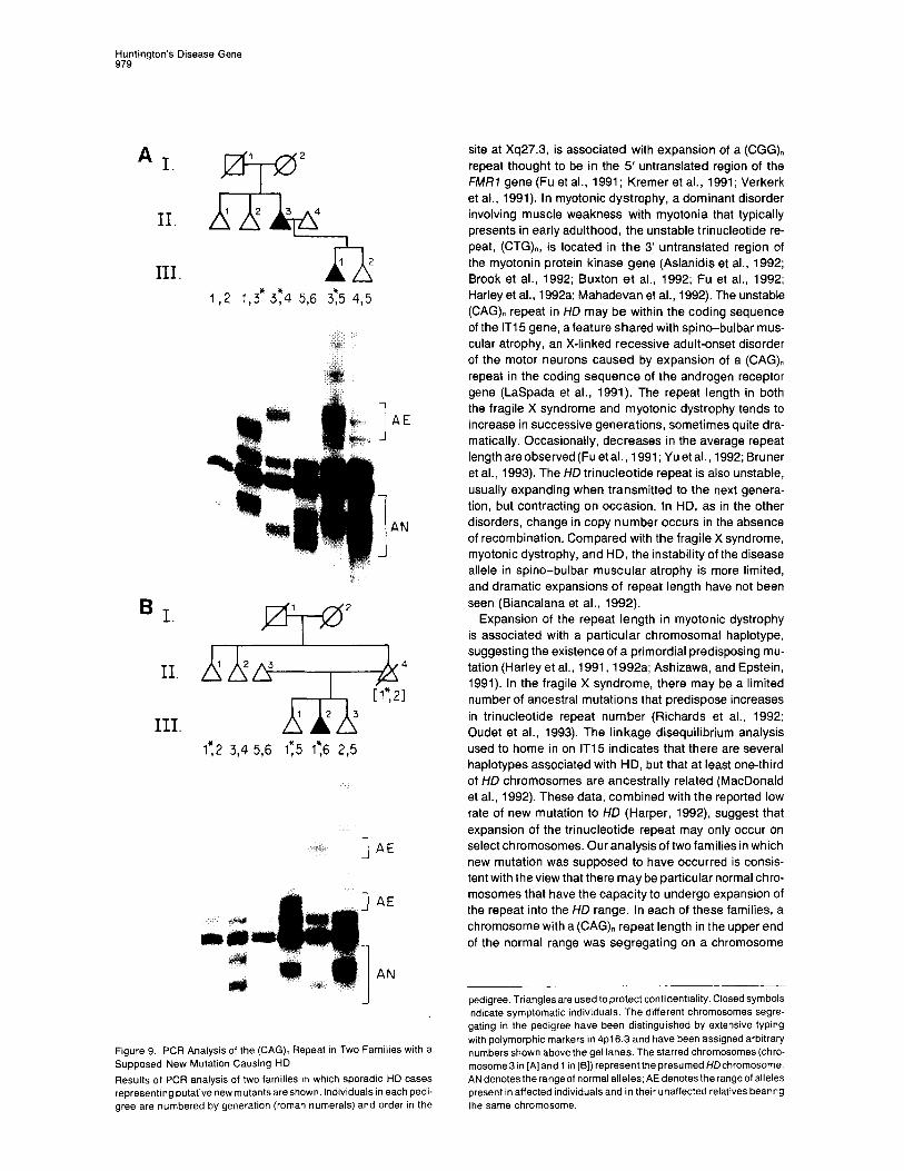
New Mutations to ND? The mutation rate in HD has been reported to be very low. To test whether the expansion of the (CAG), repeat is the mechanism by which new HD mutations occur, we have examined two pedigrees with sporadic cases of HD in which intensive searching failed to reveal a family history of the disorder. In thesecases, wegathered pedigree infor- mation sufficient to identify the same chromosomes in both the affected individual and unaffected relatives. Fig- ure 9 shows the results of PCR analysis of the (CAG), repeat in these families. The chromosomes in each family were assigned an arbitrary number based on typing for a large number of restriction fragment length polymorphism and simple sequence repeat markers in 4~16.3 defining distinct haplotypes; the presumed HD chromosome is starred.
1 2 3 4 5 6 7 8 9 10
I; L _I, . L
AN
:
es -18
Figure 8. PCR Analysis of the (CAG), Repeat in Membersof an Ameri- can Family with an Individual Homozygous for the Major HCI Haplotype Results of PCR analysis of members of an American family segregat- ing the major HD haplotype. AN marks the range. of normal alleles; AE marks the range of HD alleles. Lanes 1, 3,4, 5,7, and 8 represent PCR products from related HD heterozygotes. Lane 2 contains the PCR products from a member of the family homozygous for the same HD chromosome. Lane 6 contains PCR products from a normal individ- ual. Pedigree relationships and affected status are not presented to preserve confidentiality. The PCR products from cosmids L191 Fl and GUS72-2130 (which was derived from the individual represented in lane 2) are loaded in lanes 9 and 10 and have 18 and 48 CAG repeats, respectively.
In family 1, HD first appeared in individual 11-3, who trans- mitted the disorder, along with chromosome 3*, to Ill-l. This same chromosome was present in 11-2, an elderly unaffected individual. PCR analysis revealed that chromo- some 3* from II-2 produced a PCR product at the extreme high end of the normal range (- 36 CAG copies). However, the (CAG), repeat on the same chromosome in II-3 and Ill-l had undergone sequential expansions to -44 and -46 copies, respectively. A similar result was obtained in family 2, where the presumed new HD mutant Ill-2 had a considerably expanded repeat relative to the same chro- mosome in II-1 and Ill-1 (- 49 versus - 33 CAG copies). In both families 1 and 2, the ultimate HD chromosome displays the marker haplotype characteristic of one-third of all HD chromosomes, suggesting that this haplotype may be predisposed to undergoing repeat expansion.
Discussion
The discovery of an expanded, unstable trinucleotide re- peat on HD chromosomes suggests that the long-sought HD gene has at last been uncovered and that the disorder constitutes an example of a mutational mechanism that may prove quite common in human genetic disease. Elon- gation of a trinucleotide repeat sequence has been impli- cated previously as the cause of three quite different human disorders, the fragile X syndrome, myotonic dystrophy, and spino-bulbar muscular atrophy. Our initial observations of repeat expansion in HD indicate that this phenomenon shares features with each of these disorders.
In the fragile X syndrome, expression of a constellation of symptoms, including mental retardation and a fragile
Huntington’s Disease Gene 979
A I.
II.
III.
B I.
II.
III.
1 2
x
1 2 3 4
I I
A 1 2
I,2 1,3* 374 5,6 3:5 4,5
@ -IQ’ 1 2 3 9 4
x21 1 2 3
1:2 3,4 5,6 1:5 I:6 2,5
1 AE
1 AE
I
AN
Figure 9. PCR Analysis of the (CAG), Repeat in Two Families with a Supposed New Mutation Causing HD Results of PCR analysis of two families in whrch sporadic HD cases representingputativenewmutantsareshown. Individualsin each pedi- gree are numbered by generation (roman numerals) and order rn the
site at Xq27.3, is associated with expansion of a (CGG), repeat thought to be in the 5’ untranslated region of the FMR7 gene (Fu et al., 1991; Kremer et al., 1991; Verkerk et al., 1991). In myotonic dystrophy, a dominant disorder involving muscle weakness with myotonia that typically presents in early adulthood, the unstable trinucleotide re- peat, (CTG),, is located in the 3’ untranslated region of the myotonin protein kinase gene (Aslanidis et al., 1992; Brook et al., 1992; Buxton et al., 1992; Fu et al., 1992; Harley et al., 1992a; Mahadevan et al., 1992). The unstable (CAG), repeat in HD may be within the coding sequence of the IT15 gene, a feature shared with spino-bulbar mus- cular atrophy, an X-linked recessive adult-onset disorder of the motor neurons caused by expansion of a (CAG), repeat in the coding sequence of the androgen receptor gene (LaSpada et al., 1991). The repeat length in both the fragile X syndrome and myotonic dystrophy tends to increase in successive generations, sometimes quite dra- matically. Occasionally, decreases in the average repeat lengthareobserved(Fu etal., 1991; Yuetal., 1992; Bruner et al., 1993). The HD trinucleotide repeat is also unstable, usually expanding when transmitted to the next genera- tion, but contracting on occasion. In HD, as in the other disorders, change in copy number occurs in the absence of recombination. Compared with the fragile X syndrome, myotonic dystrophy, and HD, the instability of the disease allele in spino-bulbar muscular atrophy is more limited, and dramatic expansions of repeat length have not been seen (Biancalana et al., 1992).
Expansion of the repeat length in myotonic dystrophy is associated with a particular chromosomal haplotype, suggesting the existence of a primordial predisposing mu- tation (Harley et al., 1991, 1992a; Ashizawa, and Epstein, 1991). In the fragile X syndrome, there may be a limited number of ancestral mutations that predispose increases in trinucleotide repeat number (Richards et al., 1992; Oudet et al., 1993). The linkage disequilibrium analysis used to home in on IT15 indicates that there are several haplotypes associated with HD, but that at least one-third of HD chromosomes are ancestrally related (MacDonald et al., 1992). These data, combined with the reported low rate of new mutation to HD (Harper, 1992), suggest that expansion of the trinucleotide repeat may only occur on select chromosomes. Our analysis of two families in which new mutation was supposed to have occurred is consis- tent with the view that there may be particular normal chro- mosomes that have the capacity to undergo expansion of the repeat into the HD range. In each of these families, a chromosome with a (CAG), repeat length in the upper end of the normal range was segregating on a chromosome
pedigree. Triangles are used to protect confidentiality. Closed symbols indicate symptomatic individuals. The different chromosomes segre- gating in the pedigree have been distinguished by extensive typing with polymorphic markers in 4~16.3 and have been assigned arbitrary numbers shown above the gel lanes. The starred chromosomes (chro- mosome3 in [A] and 1 in 161) represent the presumed HDchromosome. AN denotes the range of normal alleles; AE denotes the range of alleles present in affected individuals and in their unaffected relatives bearing the same chromosome.

Cell 980
whose 4~16.3 haplotype matched the most common hap- lotype seen on HD chromosomes, and the clinical appear- ance of HD in these two cases was associated with expan- sion of the trinucleotide repeat.
The recent application of haplotype analysis to explore the linkage disequilibrium on HD chromosomes pointed to a portion of a 2.2 Mb candidate region defined by the majority of recombination events described in HD pedi- grees (MacDonald et al., 1992). Previously, the search for the gene was confounded by three matings in which the genetic inheritance pattern was inconsistent with the re- mainder of the family (MacDonald et al., 1989b: Pritchard et al., 1992). These matings produced apparently affected HD individuals despite the inheritance of only normal al- leles for markers throughout 4~16.3, effectively excluding inheritance of the HD chromosome present in the rest of the pedigree. Using our PCR assay, we have tested each of these families and find that, like other HD kindreds, an expanded allele generally segregated with HD in affected individuals of all three pedigrees. However, an expanded allele was not present in those specific individuals with the inconsistent 4~16.3 genotypes. Instead, these individuals displayed the normal alleles expected, based on analysis of other markers in 4~16.3. It is conceivable that these inconsistent individuals do not, in fact, have HD, but some other disorder. Alternatively, they might represent genetic mosaics in which the HD allele is more heavily represented and/or more expanded in brain tissue than in the lymphoblast DNA used for genotyping.
It can be expected that the capacity to monitor directly the size of the trinucleotide repeat in individuals “at risk” for HD will revolutionize preclinical testing for the disorder, eliminating the need for complicated linkage analyses, fa- cilitating genetic counseling, and extending the applicabil- ity of presymptomatic and prenatal diagnosis to at risk individuals with no living affected relatives. We consider it of the utmost importance that the current internationally accepted guidelines and counseling protocols for testing those at risk continue to be observed, and that samples from unaffected relatives should not be tested inadver- tently or without full consent. In our limited initial series of patients, there is an apparent correlation between repeat length and age of onset of the disease, reminiscent of that reported in myotonic dystrophy (Harley et al., 1992b; Tsilfidis et al., 1992). The largest HD trinucleotide repeat segments were found in juvenile onset cases, where there is a known preponderance of male transmission (Merrit et al., 1969). More detailed studies will be required to es- tablish whether expansion of the repeat occurs preferen- tially in transmission from males. It will also be essential to perform acareful analysis of the extent, if any, of overlap between the range of repeat lengths in normal and HD individuals, to evaluate fully the relationship between age of onset and repeat length, and to examine the possibility of somaticvariation in repeat length due to mitotic instabil- ity. These studies must be completed before the (CAG), size is used to provide prognostic information to at risk HD individuals.
The expression of fragile X syndrome is associated with direct inactivation of the FMRl gene (Pieretti et al., 1991;
DeBoulle et al., 1993). The recessive inheritance pattern of spino-bulbar muscular atrophy suggests that in this disorder an inactive gene product is produced. In myotonic dystrophy, the manner in which repeat expansion leads to the dominant disease phenotype is unknown. There are numerous possibilities for the mechanism of pathogenesis of the expanded trinucleotide repeat in HD. Since Wolf- Hirschhorn patients hemizygous for 4~16.3 do not display features of HD and IT15 mRNA is present in HD homozy- gotes, the expanded trinucleotide repeat does not cause simple inactivation of the gene containing it. The observa- tion that the phenotype of HD is completely dominant, since homozygotes for the disease allele do not differ clini- cally from heterozygotes, has suggested that HD results from again-of-function mutation, in which eitherthe mRNA product or the protein product of the disease allele would have some new property or would be expressed inappro- priately (Wexler et al., 1987; Myers et al., 1989). If the expanded trinucleotide repeat were translated, the conse- quences on the protein product would be dramatic, in- creasing the length of the poly-glutamine stretch near the N-terminus. It is possible, however, that despite the pres- ence of an upstream Met codon, the normal translational start occurs3’to the (CAG), repeat and there is no poly-glu- tamine stretch in the protein product. In this case, the repeat would be in the 5’ untranslated region and might be expected to have its dominant effect at the mRNA level. The presence of an expanded repeat might directly alter regulation, localization, stability, or translatability of the mRNA containing it, and could indirectly affect its counter- part from the normal allele in HD heterozygotes. Other conceivable scenarios are that the presence of an ex- panded repeat might alter the effective translation start site for the HD transcript, thereby truncating the protein, or alter the transcription start site for the IT15 gene, dis- rupting control of mRNA expression. Finally, although the repeat is located within the IT15 transcript, the possibility that it leads to HD by virtue of an action on the expression of an adjacent gene cannot be excluded.
Despite this final caveat, we believe it most likely that the trinucleotide repeat expansion causes HD by its effect, either at the mRNA or protein level, on the expression and/or structure of the protein product of the IT15 gene, which we have named huntingtin. Outside of the region of the triplet repeat, the IT15 DNA sequence detected no significant similarity to any previously reported gene in the GenBank data base. Except for the stretches of glutamine and proline near the N-terminus, the amino acid sequence displayed no similarity to known proteins, providing no conspicuous clues to huntingtin’s function. The poly-glu- tamine and poly-proline regions near the N-terminus indi- cate similarity to a large number of proteins that also con- tain long stretches of these amino acids. It is difficult to assess the significance of such similarities, although it is notable that many of these similarities are to DNA-binding proteins and that huntingtin does have a single leucine zipper motif at residue 1443. Huntingtin appears to be widely expressed, yet cell death in HD is confined to spe- cific neurons in particular regions of the brain. Thus, with the mystery of the genetic basis of HD apparently solved,

Huntington’s Disease Gene 981
defining the normal function of the huntingtin protein and delineating the mechanism whereby increased trinucleo- tide repeat length leads to the characteristic neuropathol- ogy of HD represent the next challenges in the effort to understand and to treat this devastating disorder.
Experimental Procedures
HD Cell Lines Lymphoblast cell lines from HD families of varied ethnic backgrounds used for genetic linkage and disequilibrium studies (Conneally et al., 1989; MacDonald et al., 1992) have been established (Anderson and Gusella, 1984) in the Molecular Neurogenetics Unit, Massachusetts General Hospital, over the past 13 years. The Venezuelan HD pedigree IS an extended kindred of over 12,000 members, in which all affected individuals have inherited the HD gene from a common founder (Gu- sella et al., 1983, 1984; Wexler et al., 1987).
DNA and RNA Blotting DNA was prepared from cultured cells, and DNA blots were prepared and hybridized as described (Gusella et al., 1979, 1983). RNA was prepared and Northern blotting was performed as described by Taylor et al. (1992).
Construction of Cosmid Contig The initial construction of the cosmid contig was by chromosome walk- ing from cosmids L19 and BJ58 (Allitto et al., 1991; Lin et al., 1991). Two libraries were employed, a collection of Alu-positive cosmids from the reduced cell hybrid H39-EC10 (Whaley et al., 1991) and an arrayed flow-sorted chromosome 4 cosmid library (NM87545) provided by the Los Alamos National Laboratory. Walking was accomplished by hy- bridization of whole cosmid DNA, using suppression of repetitive and vector sequences, to robot-generated high density filter grids (Nizetic et al., 1991; Lehrach et al.. 1990). Cosmids LlC2, L89F7, L228B6, and L83D3 were first identified by hybridization of yeast artificial chro- mosome clone YGAP to the same arrayed library (Bates et al., 1992; Baxendale et al., 1991). HD cosmid GUS72-2130 was isolated by stan- dard screening of a GUS72 cosmid library using a single-copy probe. Cosmid overlaps were confirmed by a combination of clone to clone and clone to genomic hybridizations, single-copy probe hybridizations, and restriction mapping.
cDNA Isolation and Characterization Exon probes were isolated and cloned as described (Buckler et al., 1991). Exon probes and cDNAs were used to screen human ?ZAPll cDNA libraries constructed from adult frontal cortex, fetal brain, adeno- virus-transformed retinal cell line RCA, and liver RNA. cDNA clones, PCR products, and trapped exons were sequenced as described (Sanger et al., 1977). Direct cosmid sequencing was performed as described (McClatchey et al., 1992). Data base searches were per- formed using the BLAST network service of the National Center for Biotechnology Information (Altschul et al., 1990).
PCR Assay of the (CAG). Repeat Genomic primers flanking the (CAG), repeat are 5’-ATGAAGG- CCTTCGAGTCCCTCAAGTCCTTC3’and 5’-AAACTCACGGTCGGT- GCAGCGGCTCCTCAG-3’. PCR amplification was performed in a re- action volume of 25 ul using 50 ng of genomic DNA, 5 ug of each primer, 10 mM Tris (pH 8.3) 5 mM KCI, 2 mM MgClz, 200 uM (each) dNTPs, 10% dimethylsulfoxide, 0.1 U of Perfectmatch (Stratagene), 2.5 uici of [32P]dCTP (Amersham), and 1.25 U of Taq polymerase (Boehringer Mannheim). After heating to 94°C for 1.5 min, the reaction mix was cycled according to the following program: 40 cycles of 1 min at 94%, 1 min at 60°C, 2 min at 72’C. Five microliters of each PCR was diluted with an equal volume of 95% formamide loading dye and heat denatured for 2 min at 95%. The products were resolved on 5% denaturing polyacrylamide gels. The PCR product from this reaction using cosmid L191 Fl (CAGla) as the template was 247 bp. Allele sizes wereestimated relative toa DNAsequencing ladder, the PCR products from sequenced cosmids, and the invariant background bands often present on the gel. Estimates of allelic variation were obtained by typing unrelated individuals of largely Western European ancestry,
who were normal parents of affected HD individuals from various pedi- grees.
Acknowledgments
We thank the many investigators who have supplied blood samples from HD pedigrees, particularly the Venezuela Huntington’s Disease Collaborative Group, and the many investigators who have over the years participated in or contributed to this project. We are also ex- tremely grateful to the HD families themselves for supporting and par- ticipating in this research effort. We thank P. M. Conneally, G. Evans, M. Frangione, C. Gilliam, R. Horvitz, L. Moyzis, R. Mulligan, A. Novel- letto, A. Tobin, and L. Zipursky for helpful discussions. This work was supported by National Institutes of Health grants NS16367 (Hunting- ton’s Disease Center Without Walls), NS22031, and NS25631 and by grants from Bristol-Myers Squibb, Inc., the Hereditary Disease Foun- dation Collaborative Research Agreement, the Joan and William A. SchreyerlMerrill Lynch Fund to Cure Huntington’s Disease of the He- reditary Disease Foundation, the Huntington’s Disease Society of America, the Foundation for the Care and Cure of Huntington’s Dis- ease, the W. M. Keck Foundation, the William J. Matheson Foundation, the Bay Foundation, The Charles A. Dana Foundation, the Medical Research Council (England), the Welsh Office, and the Wellcome Trust. Fellowship support was provided to C. M. A. by the Andrew B. Cogan Fellowship of the Hereditary Disease Foundation. Other postdoctoral fellowships to various investigators were generously pro- vided by the Hereditary Disease Foundation, the Huntington’s Disease Society of America, and the Huntington’s Society of Canada.
Received February 26, 1993; revised March 4, 1993
References
Allitto, B. A., MacDonald, M. E., Bucan, M., Richards, J., Romano. D., Whaley, W. L., Falcone, B., lanazzi, J., Wexler, N. S., Wasmuth, J. J.. Collins, F. S., Lehrach, H., Haines, J. L., and Gusella, J. F. (1991). Increased recombination adjacent to the Huntington’s disease-linked D4S10 marker. Genomics 9, 104-l 12.
Altherr, M. R., Plummer, S.. Bates, G., MacDonald, M., Taylor, S., Lehrach, T. H., Frischauf, A. M., Gusella, J. F., Boehnke, M., and Wasmuth, J. J. (1992). Radiation hybrid map spanning the Huntington disease gene region of chromosome 4. Genomics 13, 1040-1046. Altschul, S. F., Gish, W., Miller, W., Myers, E. W., and Lipman, D. J. (1990). Basic local alignment search tool. J. Mol. Biol. 275, 403-410.
Ambrose, C., James, M., Barnes, G., Lin, C., Bates, G., Altherr, M., Duyao, M., Groot, N., Church, D., Wasmuth, J. J., Lehrach. H., Hous- man, D., Buckler, A., Gusella, J. F., and MacDonald, M. E. (1992). A novel G protein-coupled receptor kinase cloned from 4~16.3. Hum. Mol. Genet. 7, 697-703.
Anderson, M. A., and Gusella, J. F. (1984). Use of cyclosporin A in establishing Epstein-Barr virus-transformed human lymphoblastoid cell lines. In Vitro 20, 856-858.
Ashizawa, T., and Epstein, H. F. (1991). Ethnic distribution of myotonic dystrophy gene. Lancet 338, 842-643. Aslanidis, C., Jansen, G.. Amemiya, C., Shutler, G., Mahadevan, M., Tsifidia. C., Chen, C., Alleman, J., Wormskamp, N. G. M., Vooijs, M., Buxton, J., Johnson, K., Smeets, H. J. M., Lennon, G. G., Carrano, A. V., Korneluk, R. G., Wieringa, B., and de Jong, P. J. (1992). Cloning of the essential myotonic dystrophy region and mapping of the putative defect. Nature 355, 548-551.
Bates, G. P., MacDonald, M. E., Baxendale, S., Sedlacek, i!., Young- man, S., Romano, D.. Whaley, W. L., Allitto, B.A., Poustka, A., Gusella, J. F., and Lehrach, H. (1990). A YAC telomere clone spanning a possi- ble location of the Huntington’s disease gene. Am. J. Hum. Genet. 46, 782-775.
Bates, G. P., MacDonald, M. E., Baxendale, S., Youngman, S., Lin, C., Whaley, W. L.. Wasmuth, J. J.. Gusella, J. F., and Lehrach, H. (1991). Defined physical limits of the Huntington disease gene candi- date region. Am. J. Hum Genet. 49, 7-16.
Bates, G. P., Valdes, J., Hummerich, H., Baxendale, S., Le Paslier, D. L., Monaco, A. P., Tagle, D., MacDonald, M. E.. Altherr, M., Ross,

Cdl 962
M., Brownstein, B. H., Bentley, D., Wasmuth, J. J., Gusella, J. F., Cohen, D., Collins, F., and Lehrach, H. (1992). Characterization of a yeast artificial chromosome contig spanning the Huntington disease gene candidate region. Nature Genet. 7, 180-187. Baxendale, S., Bates, G. P., MacDonald, M. E., Gusella, J. F., and Lehrach, H. (1991). The direct screening of cosmid libraries with yeast artificial chromosomes. Nucl. Acids Res. 79, 6651,
Biancalana, V., Serville, F., Pommier, J., Julien, J., Hanauer, A., and Mandel, J. L. (1992). Moderate instability of the trinucleotide repeat in spino-bulbar muscular atrophy. Hum. Mol. Genet. 7, 255-258. Brook, J. D., McCurrach, M. E., Harley, H. G., Buckler, A. J., Church, D., Aburatani. H., Hunter, K., Stanton, V. P., Thirion, J.-P., Hudson, T., Sohn, R., Zemelman, B., Snell, R. G., Rundle, S. A., Crow, S., Davies, J., Shelbourne, P., Buxton, J., Jones, C., Juvonen, V., John- son, K., Harper, P. S., Shaw, D. J., and Housman, D. E. (1992). Molecu- lar basis of myotonic dystrophy: expansion of a trinucleotide (CTG) repeat at the 3’ end of a transcript encoding a protein kinase family member. Cell 68, 799-808. Bruner, H. G., Jansen, G., Nillesen, W., Nelen, M. R., DaDie, C. E. M., Howeller, C. J., van Oost, 9. A., Wieringa, B., Ropers, H. H., and Smeets, H. J. M. (1993). Reverse mutation in myotonic dystrophy. N. Engl. J. Med. 328, 476-480. Bucan, M., Zimmer, M., Whaley, W. L., Poustka, A., Youngman, S., Allitto, B.A., Ormondroyd, E., Smith, B., Pohl, T. M., MacDonald, M., Bates, G., Richards, J., Volinia, S., Gilliam, T. C., Sedlacek,Z., Collins, F. S., Wasmuth, J. J., Shaw, D. J., Gusella, J. F., Frischauf, A. M., and Lehrach, H. (1990). Physical maps of 4~16.3, the area expected to contain the Huntington’s disease mutation. Genomics 6, l-15. Buckler, A. J., Chang, D. D., Graw, S. L., Brook, J. D., Haber, D.A., Sharp, P. A., and Housman, D. E. (1991). Exon amplification: astrategy to isolate mammalian genes based on RNA splicing. Proc. Natl. Acad. Sci. USA 88,4005-4009.
Buxton, J., Shelbourne, P.. Davies, J., Jones, C., Van Tongeren, T., Aslanidis, C., de Jong, P., Jansen, G., Anvret, M., Riley, B., William- son, R., and Johnson, K. (1992). Detection of an unstable fragment of DNA specific to individuals with myotonic dystrophy. Nature 355, 547-548
Conneally, P. M., Haines, J. L., Tanzi, R. E., Wexler, N. S., Penchasza- deh, G. K., Harper, P. S., Folstein, S. E., Cassiman, J. J., Myers, R. H., Young, A. B., Hayden, M. R., Falek, A., Tolosa, E. S., Crespi, S., Di Maio, L., Holmgren, G., Anvret, M., Kanazawa, I., and Gusella, J. F. (1989). Huntington disease: no evidence for locus heterogeneity. Genomics 5304-308.
DeBoulle, K., Verkerk, A. J. M. H., Reyniers, E., Vits, L., Hendrickx, J., VanRoy, B., VanDenBos, F., deGraaff, E., Oostra, 9. A., and Wil- lems, P. J. (1993). A point mutation in the FMR-1 gene associated with fragile X mental retardation. Nature Genet. 3, 31-35.
Doucette-Stamm, L. A., Riba, L., Handelin, B., Difilippantonio, M., Ward, D. C., Wasmuth, J. J., Gusella, J. F., and Housman, D. E. (1991). Generation and characterization of Goss-Harris hybrids of human chromosome 4. Somat. Cell Mol. Genet. 77, 471-480. Fu, Y.-H., Kuhl, D. P. A., Pizzuti, A., Pieretti, M., Sutcliffa, J. S., Rich- ards, S., Verkerk, A. J. M. H., Holden, J. J. A., Fenwick, R. G., Jr., Warren, S. T., Oostra, 8. A., Nelson, D. L., and Caskey, C. T. (1991). Variation of the CGG repeat at the fragile X site results in genetic instability: resolution of the Sherman paradox, Cell 67, 1047-1068. Fu, Y.-H., Pizzuti, A., Fenwick, R. G., King, J., Jr., Rajnarayan, S., Dunne, P. W., Dubel, J., Nasser, G. A., Ashizawa, T., DeJong, P., Wieringa, B., Korneluk, R., Perryman, M. B., Epstein, H. F., and Caskey, C. T. (1992). An unstable triplet repeat in a gene related to myotonic muscular dystrophy. Science 255, 1256-1259. Gusella, J. F. (1989). Location cloning strategy for characterizing ge- netic defects in Huntington’s disease and Alzheimer’s disease. FASEB J. 3, 2038-2041. Gusella, J. F. (!991). Huntington’sdisease. Adv. Hum. Genet. 20, 125- 151.
Gusella, J. F., Varsanyi-Breiner, A., Kao, F. T.. Jones, C., Puck, T. T., Keys, C., Orkin, S., and Housman, D. E. (1979). Precise localiza- tion of the human @globin gene complex on chromosome 11. Proc. Natl. Acad. Sci. USA 76, 5239-5243.
Gusella, J. F., Wexler, N. S., Conneally, P. M., Naylor, S. L., Anderson, M. A., Tanzi, R. E., Watkins, P. C., Ottina, K., Wallace, M. R., Saka- guchi. A. Y., Young, A. B., Shoulson, I., Bonilla, E., and Martin, J. B. (1983). A polymorphic DNA marker genetically linked to Huntington’s disease. Nature 306, 234-238.
Gusella, J. F., Tanzi, R. E., Anderson, M. A., Hobbs, W., Gibbons, K., Raschtchian, R., Gilliam, T. C., Wallace, M. R., Wexler, N. S., and Conneally, P. M. (1984). DNA markers for nervous system diseases. Science 225, 1320-1326.
Harley. H. G., Brook, J. D., Floyd, J., Rundle, S. A., Crow, S., Walsh, K. V., Thibault, M. C., Harper, P. S., and Shaw, D. J. (1991). Detection of linkage disequilibrium between the myotonic dystrophy locus and a new polymorphic DNA marker. Am. J. Hum. Genet. 49, 68-75.
Harley, H. G., Brook, J. D., Rundle, S. A., Crow, S., Reardon, W., Buckler, A. J., Harper, P. S., and Housman, D. E. (1992a). Expansion of an unstable DNA region and phenotypicvariation in myotonicdystro- phy. Nature 355, 545-546.
Harley, H. G., Rundle, S. A., Reardon, W., Myring, J., Crow, S., Brook, J. D., Harper, P. S., and Shaw, D. J. (1992b). Unstable DNA sequence in myotonic dystrophy. Lancet 339, 1125-1128.
Harper, P. S. (1992). The epidemiology of Huntington’s disease. J. Med. Genet. 89, 365-376.
Harper, P. S., Morris, M. J., QuarrelI, O., Shaw, D. J., Tyler, A., and Youngman, S. (1991). Huntington’s Disease(Philadelphia: W. B. Saun- ders).
Kremer, E. J., Pritchard, M., Lynch, M., Yu, S., Holman, K., Baker, E., Warren, S. T., Schlessinger, D., Sutherland, G. R., and Richards, R. 1.(1991). Mappingof DNAinstabilityatthefragileX toatrinucleotide repeat sequence p(CCG)n. Science 252, 1711-1714.
LaSpada, A. R., Wilson, E. M., Lubahn, D. B., Harding, A. E., and Fishbeck, H. (1991). Androgen receptor gene mutations in X-linked spinal and bulbar muscular atrophy. Nature 352, 77-79. Lehrach, H., Drmanac, R.. Hoheisel, J., Larin, Z., Lennon, G., Nizetic, D., Monaco, A., Zehetner, G., and Poustka, A. (1990). Hybridisation fingerprinting in genome mapping and sequencing. In Genome Analy- sis: Genetic and Physical Mapping, Volume 1, K. E. Davies and S. M. Tighlman, eds. (Cold Spring Harbor, New York: Cold Spring Harbor Laboratory Press), pp. 39-81. Lin, C. S., Altherr, M., Bates, G., Whaley, W. L., Read, A. P., Harris, R., Lehrach, H., Wasmuth, J. J., Gusella, J. F., and MacDonald, M. E. (1991). New DNA markers in the Huntington’s disease gene candidate region. Somat. Cell Mol. Genet. 17, 481-488.
MacDonald, M. E., Cheng, S. V., Zimmer, M., Haines, J. L., Poustka, A. M.. Allitto. B. A., Smith, B., Whaley, W. L., Romano, D., Jagadeesh, J., Lehrach, H., Wasmuth, J. J., Frischauf, A. M., and Gusella, J. F. (1989a). Clustering of multi-allele DNA markers near the Huntington’s disease gene. J. Clin. Invest. 84, 1013-1016. MacDonald, M. E., Haines, J. L., Zimmer, M.,Cheng, S. V., Youngman, S., Whaley, W. L., Bucan, W. L., Allitto, B. A., Smith, B., Leavitt, J., Poustka, A. M., Harper, P., Lehrach, H., Wasmuth, J. J., Frischauf, A. M., and Gusella, J. F. (1989b). Recombination events suggest possi- ble locations for the Huntington’s disease gene. Neuron 3. 183-190.
MacDonald, M. E., Lin, C., Srinidhi, L.. Bates, G., Altherr, M., Whaley, W. L., Lehrach, H.. Wasmuth, J., and Gusella, J. F. (1991). Complex patterns of linkage disequilibrium in the Huntington disease region. Am. J. Hum. Genet. 49, 723-734.
MacDonald, M. E., Novelletto, A., Lin, C., Tagle, D., Barnes, G., Bates, G., Taylor, S., Allitto, B., Altherr, M., Myers, R., Lehrach, H., Collins, F. S., Wasmuth, J. J., Frontali, M., and Gusella, J. F. (1992). The Huntington’s disease candidate region exhibits many different haplo- types. Nature Genet. 7, 99-103.
Mahadevan, M.. Tsilfidis, C., Sabourin, L., Shutler, G., Amemiya, C., Jansen. G., Neville, C.. Narang, M., Barcelo, J., O’Hoy, K., Leblond, S., Earle-MacDonald, J., DeJong. P. J., Wieringa, B., and Korneluk, G. (1992). Myotonic dystrophy mutation: an unstable CTG repeat in the 3’ untranslated region of the gene. Science 255, 1253-1255.
Martin, J. B.. and Gusella, J. F. (1986). Huntington’s disease: patho- genesis and management. N. Engl. J. Med. 375, 1287-1276.
McClatchey, A. I., Lin, C. S., Wang, J., Hoffman, E. P., Rojas, C., and

Huntington’s Disease Gene 983
Gusella, J. F. (1992). The genomic structure of the human skeletal muscle sodium channel gene. Hum. Mol. Genet. 7, 521-527.
Merrit, A. D., Conneatly, P. M., Rahman, N. F., and Drew, A. L. (1969). Juvenile Huntington’s chorea. In Progress in Neurogenetics, A. Barbeau and J. Ft. Brunette, eds. (Amsterdam: Excerpta Medica), pp. 645-650.
Myers, R. H., Leavitt, J., Farrer, L. A., Jagadeesh, J., McFarlane, H., Mark, Ft. J., and Gusella, J. F. (1969). Homozygote for Huntington’s disease. Am. J. Hum. Genct. 45, 615-618.
Nizetic, D., Zehetner, G., Monaco, A., Gellen, L., Young, B. D., and Lehrach, H. (1991). Construction, arraying, and high density screening of large insert libraries of human chromosomes X and 21: their poten- tial use as reference libraries, Proc. Natl. Acad. Sci. USA 88, 3233- 3237.
Oudet, C., Mornet, E., Serre, J. L., Thomas, F., Lentes-Zengerling, S., Kretz, C., Deluchat, C., Tejada, I., Boue, J., Boue, A., and Mandel, J. L. (1993). Linkage disequilibrium between the fragile X mutation and twocloselylinkedCArepeatssuggeststhatfragile Xchromosomesare derived from a small number of founder chromosomes. Am. J. Hum. Genet. 52,297-304.
Pieretti, M., Zhang, F., Fu, Y.-H., Warren, S. T., Oostra, B. A., Caskey, C. T., and Nelson, D. L. (1991). Absence of expression of the FMR-7 gene in fragile X syndrome. Cell 66, 617-622.
Pohl, T. M., Zimmer, M., MacDonald, M. E., Smith, B., Bucan, M., Poustka, A., Volinia, S., Searle, S., Zehetner, G., Wasmuth, J. J., Gusella, J., Lehrach, H., and Frischauf, A. M. (1988). Construction of a Not1 linking library and isolation of new markers close to the Hunting- ton’s disease gene. Nucl. Acids Res. 16, 9185-9198. Pritchard, C., Zhu, N., Zuo, J., Bull, L., Pericak-Vance, M. A., Roses, A. D., Milatovich, A., Francke, U., Cox, D. R., and Myers, R. M. (1992). Recombination of 4~16 DNA markers in an unusual family with Hun- tington disease. Am. J. Hum. Genet. 50, 1218-1230.
Richards, R. I., Holman, K., Friend, K., Kremer, E., Hillen, D., Staples, A., Brown, W. T., Goonewardena, P., Tarleton, J., Schwartz, C., and Sutherland, G. R. (1992). Fragile X syndrome: evidence of founder chromosomes. Nature Genet. 1, 257-260. Sanger. T., Nicklen, S., and Coulson, A. R. (1977). DNA sequencing with chain-termination inhibitors, Proc. Natl. Acad. Sci. USA 74,5463- 5467.
Snell, R. G., Lazarou, L., Youngman, S., QuarrelI, 0. W. J., Wasmuth, J. J., Shaw, D. J., and Harper, P. S. (1989). Linkage disequilibrium in Huntington’s disease: an improved localization for the gene, J. Med. Genet. 26, 673-675.
Snell, R. G., Thompson, L. M., Tagle, D. A., Holloway, T. L., Barnes, G., Harley, H. G., Sandkuijl, L. A., MacDonald, M. E., Collins, F. S., Gusella, J. F., Harper, P. S., and Shaw, D. J. (1992). A recombination event that redefines the Huntington disease region. Am. J. Hum. Genet. 51, 357-362.
Suthers, G. K., Huson, S. M., and Davies, K. E. (1992). Instability versus predictability: the molecular diagnosis of myotonic dystrophy. J. Med. Genet. 29, 761-765.
Taylor, S. A. M., Snell, R. G., Buckler, A., Ambrose, C., Duyao, M., Church, D., Lin, C. S., Altherr, M., Bates, G. P., Groat, N., Barnes, G., Shaw, D. J., Lehrach, H., Wasmuth, J. J., Harper, P. S., Housman, D. E., MacDonald, M. E., and Gusella, J. F. (1992). Cloning of the a-adducin gene from the Huntington’s disease candidate region of chromosome 4 by exon amplification. Nature Genet. 2, 223-227.
Theilman, J., Kanani, S., Shiang, R., Robbins, C., Quarrell, O., Hug- gins, M., Hedrick, A., and Hayden, M. R. (1969). Non-random associa- tion between alleles detected at D4S95 and D4S98 and the Hunting- ton’s disease gene. J. Med. Genet. 26, 676-681.
Thompson, L. M., Plummer, S., Schalling, M., Altherr, M. R.. Gusella, J. F.. Housman, D. E.. and Wasmuth, J. J. (1991). A gene encoding a fibroblast growth factor receptor isolated from the Huntington disease gene region of human chromosome 4. Genomics 7 7, 1133-1142. Tsilfidis, C., McKenzie, A. E., Mettler, G., Barcelo. J., and Korneluk, R. G. (1992). Correlation between CTG trinucleotide repeat length and frequency of severe congenital myotonic dystrophy. Nature Genet. 7, 192-195.
Verkerk, A. J. M. H., Pieretti, M., Sutcliffe, J. S., Fu, Y.-H., Kuhl, D. P. A., Pizzuti, A., Reiner, 0.. Richards, S., Victoria, M. F., Zhang, R., Eussen, B. E., van Ommen, G.-J. B., Blonden. L. A. J., Riggins, G. J., Chastain, J. L., Kunst, C. B., Galjaard, H., Caskey, C. T., Nelson, D. L., Oostra, B. A., and Warren, S. T. (1991). Identification of a gene (FMR-1) containing a CGG repeat coincident with a breakpoint cluster region exhibiting length variation in fragile X syndrome. Cell 65, 905- 914.
Wexler, N. S., Young, A. B., Tanzi, R. E., Travers, H., Starosta-Ru- benstein, S., Penney, J. B., Snodgrass, S. R., Shoulson, I., Gomez, F., Ramos-Arroyo, M. A., Penchaszadeh, G., Moreno, R., Gibbons, K., Farymarz, A., Hobbs, W., Anderson, M. A., Bonilla, E., Conneally, P. M., and Gusella, J. F. (1987). Homozygotes for Huntington’s dis- ease. Nature 326, 194-197.
Whaley, W. L., Bates, G. P., Novelleto, A., Sedlacek, Z., Cheng, S.. Romano, D., Ormondroyd, E., Allitto, B. A., Lin, C., Youngman, S., Baxendale, S., Bucan, M., Altherr, M., Wasmuth, J., Wexler, N. S., Frontali, M., Frischauf, A. M., Lehrach, H., MacDonald, M. E., and Gusella, J. F. (1991). Mapping of cosmid clones in the Huntington’s disease region of chromosome 4. Somat. Cell Mol. Genet. 77, 83-91. Youngman, S., Bates, G. P., Williams, S., McClatchey, A. I., Baxen- dale, S., Sedlacek, Z., Altherr, M., Wasmuth, J. J., MacDonald, M. E., Gusella, J. F., and Lehrach, H. (1992). The telomeric 60 kb of chromosome arm 4p is homologous to telomeric regions on 13p, 15p, 21~. and 22~. Genomics 14, 350-356. Yu, S., Mulley, J., Loesch, D., Turner, G., Donnelly, A., Gedeon, A., Hillen, D., Kremer, E., Lynch, M., Pritchard, M., Sutherland, G. R., and Richards, R. I. (1992). Fragile-X syndrome: unique genetics of the heritable unstable element. Am. J. Hum, Genet. 50, 966-960.
GenBank Accession Number
The accession number for the sequence reported in this paper is L12392.