A novel CD4+ T-helper lymphocyte epitope in the VP3 protein of hepatitis A virus
8
Journal of Medical Virology 72:525–532 (2004) A Novel CD4 þ T-Helper Lymphocyte Epitope in the VP3 Protein of Hepatitis A Virus Glo ` ria Sa ´ nchez, Rosa M. Pinto ´ , and Albert Bosch* Enteric Virus Group, Department of Microbiology, University of Barcelona, Barcelona, Spain Prediction analysis of T H -cell epitopes in the VP3 capsid protein of the hepatitis A virus (HAV) revealed the occurrence of a putative T H epitope in the 102–121 region complying with all the algorithms tested. To confirm these predictions, spleen T lymphocytes obtained from BALB/c mice previously immunised with HAV, were stimu- lated in vitro with different concentrations of synthetic peptides 102–121 and 110–121 of VP3. The ability of these peptides to stimulate CD4 þ T- helper lymphocytes proliferation was evaluated by an immunological flow cytometry detection of bromodeoxyuridine incorporation. Using this method, it is concluded that the amino terminal part of the VP3 102–121 sequence contains a T cell epitope. J. Med. Virol. 72:525 – 532, 2004. ß 2004 Wiley-Liss, Inc. KEY WORDS: HAV; T epitopes; immune response; synthetic vaccines; proliferation assays; flow cytometry INTRODUCTION The induction of specific CD4 þ T-helper (T H ) lympho- cytes proliferation in response to antigens is of critical importance for an efficient antibody response. Antigens are processed by the antigen-presenting cells (APCs) into peptide fragments which are presented on the cell surface of APCs in association with MHC class II molecules. The interaction of the peptide bound to the MHC class II molecule with the T cell receptor (TCR) of T H lymphocytes stimulates its proliferation. These T H lymphocytes secrete cytokines, which mediate a variety of immune responses, including the activation of antigen specific B lymphocytes. The localisation of T H epitopes corresponding to the peptide fragments which bound to the MHC class II molecules in viruses is of great important in the design of synthetic vaccines. In picornaviruses, characterisation of T H epitopes has been reported for poliovirus [Leclerc et al., 1991; Kutubuddin et al., 1992; Mahon et al., 1992; Graham et al., 1993], human rhinovirus [Hastings et al., 1993], food-and-mouth disease virus (FMDV) [Rodrı ´guez et al., 1994], Mengo virus [Muir et al., 1994] and Theiler’s virus [Usherwood et al., 1995]. In the case of HAV, putative T H epitopes in the VP2 region [Powdrill and Johnston, 1991] and in the carboxy terminal region of VP1 [Harmon et al., 1993] have been described. Later on, two peptides derived from the VP1 region, 17–33 and 276–298, were demonstrated to stimulate T lymphocytes of several mouse haplotypes [Ivanov et al., 1994]. In the same study, peptides 45– 57 and 137–150 derived from the VP3 protein were predicted as T-epitopes by a combination of different computational methods, but only the peptide 137–150 was able to stimulate one of the mouse haplotypes tested. In a previous study [Bosch et al., 1998], a continuous B-cell epitope was described in the VP3 protein of HAV at position 110–121. In order to improve the immuno- logical response against a synthetic peptide containing this B-epitope, a longer peptide was subsequently syn- thesised corresponding to the sequence VP3 102–121. The anti-HAV titre achieved with the VP3(102–121) peptide was 100 times higher than the response obtained with the VP3(110 – 121), although the recogni- tion of both peptides by a human convalescent serum was similar [Pinto ´ et al., 1998]. Taken together, these results suggested the presence of a putative T-epitope in the VP3 102–121 sequence. In the present study, the ability of the peptide VP3(102 – 121) to induce prolifera- tive responses of T H lymphocytes from BALB/c mice primed with HAV was evaluated, in order to confirm the occurrence of a T H -epitope in this sequence. Grant sponsor: CICYT, Spain; Grant number: BIO99-0455; Grant sponsor: Generalitat de Catalunya; Grant number: 2001/ SGR/00098; Grant sponsor: Generalitat de Catalunya (Centre de Biotecnologia de Catalunya, CeRBA). *Correspondence to: Albert Bosch, Department of Microbiology, University of Barcelona, Av. Diagonal 645, 08028 Barcelona, Spain. E-mail: [email protected] Accepted 11 November 2003 DOI 10.1002/jmv.20035 Published online in Wiley InterScience (www.interscience.wiley.com) ß 2004 WILEY-LISS, INC.
-
Upload
gloria-sanchez -
Category
Documents
-
view
214 -
download
2
Transcript of A novel CD4+ T-helper lymphocyte epitope in the VP3 protein of hepatitis A virus

Journal of Medical Virology 72:525–532 (2004)
A Novel CD4þ T-Helper Lymphocyte Epitopein the VP3 Protein of Hepatitis A Virus
Gloria Sanchez, Rosa M. Pinto, and Albert Bosch*
Enteric Virus Group, Department of Microbiology, University of Barcelona, Barcelona, Spain
Prediction analysis of TH-cell epitopes in the VP3capsid protein of the hepatitis A virus (HAV)revealed the occurrence of a putative TH epitopein the 102–121 region complying with all thealgorithms tested. To confirm these predictions,spleen T lymphocytes obtained from BALB/c micepreviously immunised with HAV, were stimu-lated in vitro with different concentrations ofsynthetic peptides 102–121 and 110–121 of VP3.The ability of these peptides to stimulate CD4þ T-helper lymphocytes proliferation was evaluatedby an immunological flow cytometry detectionof bromodeoxyuridine incorporation. Using thismethod, it is concluded that the amino terminalpart of the VP3 102–121 sequence contains a T cellepitope. J. Med. Virol. 72:525–532, 2004.� 2004 Wiley-Liss, Inc.
KEY WORDS: HAV; T epitopes; immuneresponse; synthetic vaccines;proliferation assays; flowcytometry
INTRODUCTION
The induction of specific CD4þ T-helper (TH) lympho-cytes proliferation in response to antigens is of criticalimportance for an efficient antibody response.
Antigens are processed by the antigen-presentingcells (APCs) into peptide fragmentswhich are presentedon the cell surface of APCs in association with MHCclass II molecules. The interaction of the peptide boundto the MHC class II molecule with the T cell receptor(TCR) of TH lymphocytes stimulates its proliferation.These TH lymphocytes secrete cytokines, whichmediatea variety of immune responses, including the activationof antigen specific B lymphocytes. The localisation ofTH epitopes corresponding to the peptide fragmentswhich bound to the MHC class II molecules in viruses isof great important in the design of synthetic vaccines.
In picornaviruses, characterisation of TH epitopeshas been reported for poliovirus [Leclerc et al., 1991;Kutubuddin et al., 1992; Mahon et al., 1992; Grahamet al., 1993], human rhinovirus [Hastings et al., 1993],food-and-mouth disease virus (FMDV) [Rodrıguez et al.,
1994], Mengo virus [Muir et al., 1994] and Theiler’svirus [Usherwood et al., 1995].
In the case of HAV, putative TH epitopes in the VP2region [Powdrill and Johnston, 1991] and in the carboxyterminal region of VP1 [Harmon et al., 1993] have beendescribed. Later on, two peptides derived from the VP1region, 17–33 and 276–298, were demonstrated tostimulate T lymphocytes of several mouse haplotypes[Ivanov et al., 1994]. In the same study, peptides 45–57 and 137–150 derived from the VP3 protein werepredicted as T-epitopes by a combination of differentcomputational methods, but only the peptide 137–150was able to stimulate one of the mouse haplotypestested.
In a previous study [Bosch et al., 1998], a continuousB-cell epitope was described in the VP3 protein of HAVat position 110–121. In order to improve the immuno-logical response against a synthetic peptide containingthis B-epitope, a longer peptide was subsequently syn-thesised corresponding to the sequence VP3 102–121.The anti-HAV titre achieved with the VP3(102–121)peptide was 100 times higher than the responseobtained with the VP3(110–121), although the recogni-tion of both peptides by a human convalescent serumwas similar [Pinto et al., 1998]. Taken together, theseresults suggested the presence of a putative T-epitope inthe VP3 102–121 sequence. In the present study, theability of the peptide VP3(102–121) to induce prolifera-tive responses of TH lymphocytes from BALB/c miceprimedwithHAVwas evaluated, in order to confirm theoccurrence of a TH-epitope in this sequence.
Grant sponsor: CICYT, Spain; Grant number: BIO99-0455;Grant sponsor: Generalitat de Catalunya; Grant number: 2001/SGR/00098; Grant sponsor: Generalitat de Catalunya (Centre deBiotecnologia de Catalunya, CeRBA).
*Correspondence to: Albert Bosch, Department of Microbiology,University of Barcelona, Av. Diagonal 645, 08028 Barcelona,Spain. E-mail: [email protected]
Accepted 11 November 2003
DOI 10.1002/jmv.20035
Published online in Wiley InterScience(www.interscience.wiley.com)
� 2004 WILEY-LISS, INC.

MATERIALS AND METHODS
Viruses and Cells
The cytopathogenic HM-175 strain of HAV (courtesyof T. Cromeans, Centers for Disease Control, Atlanta,GA) [Cromeans et al., 1987] was propagated in FRhK-4cells. Concentrated viral stocks were obtained asdescribed previously [Bishop et al., 1994]. Briefly, at5–6 days post-infection cells from a T-175 cm2 flaskwere recovered by trypsin treatment, collected bycentrifugation, resuspended in 500 ml of NT buffer(0.1 M NaCl, 10 mM Tris-HCl, 1% NP40, pH 7.4) andincubated for 30 min. These lysed cell suspensions werecentrifuged at 1,700g for 5 min, and the superna-tants again centrifuged at 13,000g for 5 min. Virusesrecovered in the supernatants were submitted to threesonication cycles of 30 sec at 60 W in the presence of0.4% SDS. Five hundred microlitre of these NP-40stocks were layered onto a 5–45% sucrose gradientin TNMg buffer (20 mM Tris-HCl, 10 mM NaCland 50 mM MgCl2, pH 6.7) and spun at 205,000g for165 min.
Fractions containing infectious virus (150S) andempty particles (70S) were pooled and dialysed againstphosphate buffered saline (PBS) to remove sucrose.After 3 days of dialysis, the pools were concentratedby centrifugation through Centriplus filters Y-50(Millipore Corp., Bedford, MA) following the manufac-turer’s instructions.
Prediction of T Cell Epitopesin the VP3 Protein
The computer program Epiplot 1.0 [Menendez-Ariasand Rodrıguez, 1990] was used to examinate the VP3aminoacid sequence of HM-175 strain [Cohen et al.,1987] for prediction of TH epitopes. It is based on fourdifferent algoritms: theFauchere–Pliskahydrophilicityscale [Fauchere and Pliska, 1983] with a block lengthof 11 and an amphipathicity score threshold of 8; theoccurrence of Rothbard–Taylor motifs recognised byMHC class II molecules [Rothbard and Taylor, 1988];the search for sequences with analogy to ovalbuminpeptides which are known to bind to the murine MHCclass II (Ia) molecules [Sette et al., 1989]; and thehydrophobic strip-of-helix algorithm with three turnsper helix [Stille et al., 1987].
The amphipathicity plot of VP3 was predictedusing the WinPep program [Hennig, 1999] availableat http://www.ipw.agrl.ethz.ch/�lhennig/winpep.html.Helical wheel representations of the VP3 102–114 andVP3 110–121 sequences were also drawn with theWinPep program and the hydrophobic character ofresidues was calculated according to a hydrophobi-city scale described previously [Black andMould, 1991].
The consensus aminoacid secondary structure pre-diction was undertaken using the Network ProteinServer Analysis (http://npsa-pbil.ibcp.fr/cgi-bin/npsa_automat.pl?page¼/NPSA/npsa_seccons.html) [Combetet al., 2000].
Synthetic Peptides
Peptides corresponding to the VP3(102–121)(ASICQMFCFWRGDLVFDFQV) and VP3(110–121)(FWRGDLVFDFQV) sequences (kindly provided byDr. Isabel Haro from the Department of Peptides, CID,CSIC, Barcelona, Spain) were synthesised by thecontinuous-flow F-moc-polyamide solid-phase method[Stewart, 1983]. Peptides were purified to a puritygreater than 95% by semi-preparative HPLC.
Immunization of Mice
Three 6-week-old female BALB/c (H-2d) mice (HarlanInterfauna Iberica, Barcelona, Spain) were injectedsubcutaneously with 3.7 ng of virus particles, diluted1:2 in Freund’s complete adjuvant, in a final volume of0.2 ml. A booster dose in Freund’s incomplete adjuvantwas administered 2 weeks later. Three mice inoculatedwith 0.2 ml of PBS were used as negative controls.
Animals were handled and killed according to specificguidelines of the Animal Ethics and ExperimentationCommittee of the University of Barcelona. All experi-ments were repeated three times.
Lymphocyte Proliferation Assays
One week after the booster dose, spleens from threeimmunised mice were aseptically removed, pooled inRPMI 1640 medium containing 10% faecal calf serum(FCS) and mechanically homogenised. A single cell sus-pension was prepared by filtering through an 80-meshscreen (Sigma, St. Louis, MO).
T lymphocytes were purified using nylon wool fibrecolumns (Polysciences Europe, Eppelheim, Germany)following the manufacture’s instructions. Briefly, afterthe column incubation with RPMI medium, the cellsuspension was layered onto the column and wasincubated for 1 hr at 378C. Nonadherent T cells werethen collected byusing twowashes.Accessory cells, to beused as antigen-presenting cells, were obtained fromspleens of non-immunised mice and treated withmitomycin C [Coligan et al., 2001].
Proliferation assays were carried out in 96-well tissueculture plates seeded with 5�105 purified T lympho-cytes and 5� 104 antigen-presenting cells per well, in afinal volume of 200 ml. Different concentrations ofpeptide antigens (0.05, 0.5 and 5 mM) were added andcultures were incubated for 6 and 10 days. Each con-dition was assayed in triplicate cultures. In all experi-ments the non-specific mitogen concanavalin A (Con A)at a concentration of 10 mg/ml was used as a positivecontrol, and cultures receiving only culture mediumwere used as negative controls.
Cellular proliferationwasmeasured by the incorpora-tion of 5-bromo-2-deoxyuridine (BrdU) into DNA duringthe last 16 hr of culture until the harvesting of the cells.The BrdU was used at the experimentally determinedoptimal concentration of 60 mM.
Following the incorporation of BrdU, cells from threeequivalent wells were mixed, washed twice in PBS
526 Sanchez et al.
containing 2% of BSA and 0.1% of Tween 20 (washingPBS) and labelled using a phycoerythrin (PE)-coupledanti-CD4 antibody (Becton Dickinson, San Jose, CA) for30 min, at room temperature (RT). Cells were subse-quently rinsed twice inwashingPBS, resuspended in0.5ml of ice-cold 0.15 M NaCl, fixed by addition of 1.2 ml ofice-cold ethanol and held for 30 min on ice. Cells werethen washed and permeabilized in PBS-1% paraformal-dehyde-0.01% Tween 20 for 1 hr at RT. Cells werewashed again and incubated with 50 kunits U/ml ofDNase for 30 min at 378C. After further washing withPBS containing 10% BSA and 0.5% Tween 20, sampleswere incubated with 10 ml of an anti-BrdU-FITC anti-body (BectonDickinson) for 2.5hr at 378C,washedagainwith PBS and analysed by flow cytometry (BeckmanCoulter XL, Miami, FL).
Flow Cytometry Analysis
In order to select the intact cell population, theforward-angle and the side-angle light scatter wereused to select cell size and cell shape respectively.
Three different representationswere obtained (Fig. 2).Anhistogramrepresenting theFITCfluorescencewhichcorresponds to the BrdU labelling, an histogram re-presenting thePEfluorescencewhich corresponds to theCD4þ labelling and a dot-plot of the FITC fluorescenceplotted versus the PE fluorescence. A cursor D wasdefined to include proliferative cells incorporatingBrdU in its DNA, a cursor E was defined to selectCD4þ lymphocytes, and both cursorswere used to definethe quadrant R2 which includes the subpopulation ofproliferative CD4þ lymphocytes among the completecell population. All these cursors were established incomparison to negative control cell populations. CursorD was established as described previously [Abad et al.,1998]. Briefly, an arbitrary cursor B was drawn at theright end of the fluorescent curves of the negativecontrols, including 2% of the cell population. The meanfluorescence of cells within cursor B was calculated foreach of the negative controls, and a new cursor D was
established starting at a cut-off level corresponding tothe mean plus 2 standard deviations of these meanfluorescence figures of the negative controls. Cursor Ewas arbitrarily drawn so to start at the inflexion pointbetween the two curves representingCD4þ lymphocytesand other T lymphocytes.
The stimulation index (SI)was calculated dividing thepercentage of cells in the R2 quadrant of the peptide orCon A stimulated cultures by the percentage of cells inthe R2 quadrant of the medium stimulated cultures. Inprevious studies the stimulation response was consid-ered significant when SI was higher than 2, 2.5 or 3[Ivanov et al., 1994; Muir et al., 1994; Rodrıguez et al.,1994].
RESULTS AND DISCUSSION
Prediction of T Cell Epitopesin the VP3 Protein
A high percentage of TH-cell epitopes are locatedwithin amphipathic helices [Berzofsky, 1988], thereforedifferent informatic programs were used for predictionof these structures in the VP3 capsid protein.
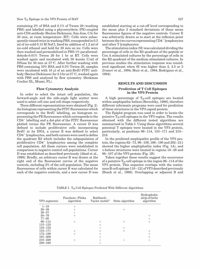
The Epiplot program was used in order to locate theputative TH-cell epitopes in the VP3 region. The resultsobtained with the different tested algorithms aresummarised in Table I. Using these algorithms severalpotential T epitopes were located in the VP3 protein,particularly, at positions 96–114, 153–171 and 210–218.
In the predicted amphipathic profile of the VP3 pro-tein, the regions 62–73, 96–109, 166–190 and 202–211showed the higher amphiphathic index (Fig. 1A), anda-helices structures were located in regions 18–28 and98–107 of the VP3 protein (Fig. 1B).
Taken together these results suggest the occurrenceof a putative TH-cell epitope in the region 96–114 of theVP3 protein. This sequence overlaps with the contin-uousB-cell epitope 110–121 ofVP3describedpreviously[Bosch et al., 1998]. Overlapping or adjacent B and
TABLE I. TH-Cell Epitopes Predicted With Different Algorithms
VP3 segmentsFauchere–Pliska
algorithmRothbard–
Taylor motifsa Sette algorithm
Hydrophobicstrip-of-helixalgorithm
1–19 þ20–38 þ þ39–57 þ58–76 þ þ77–95 þ96–114 þ þ þ þ
115–133 þ134–152 þ þ153–171 þ þ þ þ172–190 þ þ þ191–209 þ þ210–228 þ þ þ þ229–246 þ
New TH Epitope in the VP3 Protein of HAV 527

TH-cell epitopes have been observed in several picorna-viruses and it has been suggested a requirement ofspatial proximity for an efficient antibody production[Kutubuddin et al., 1992; Graham et al., 1993; Muiret al., 1994].
This potential TH-cell epitope in the VP3 94–114region of HAV aligns in part with an actual TH-cellepitope of FMDV [Usherwood and Nash, 1995] and
completely with a putative TH-cell epitope of Mengovirus [Muir et al., 1994], as can be seen in Figure 1C.In order to confirm the actual occurrence of this TH-cellepitope, two synthetic peptides including the T-B cellepitopes were synthesised. The synthetic peptideVP3(110–121) includes the B-cell epitope and only aminor part of the putative TH-cell epitope in spite ofits complete alignment with the actual FMDV TH-cell
Fig. 1. TH-cell epitope predictions of the hepatitis A virus (HAV)VP3protein. Amphipathicity profile (A). Consensus secondary structureprediction (B). The large black and the medium light gray boxesindicate consensus a-helixes and b-sheets respectively. VP3 partialsequence alignment of HAV strain HM-175, FMDV strain A10 andMengo virus [Luo et al., 1988] (C). The confirmed T epitopes areincluded in a rectangle and the putative TH-cell epitopes are under-
lined. Predicted secondary structure of the 102–121 VP3 sequencebased in a picornaviruses alignment [Luo et al., 1988] (D). Helicalwheel projections of the sequences 102–114 and 110–121 of VP3 (E).The hydrophobic character of the residues [Black and Mould, 1991] isdepicted as follows: plain for hydrophobic (F, I, W, Y, L, V, M, P, C, A),circled for neutral (G, T, S) and underlined for hydrophilic (K, Q, N, H,E, D, R).
528 Sanchez et al.

epitope (Fig. 1C). However, structurally this sequencedid not alignwith an a-helix butwith a b-sheet (Fig. 1D).In contrast, the synthetic peptide VP3(102–121)includes a major part of the predicted TH-cell epitope
and the B-cell epitope, and partially aligns with an a-helix (Fig. 1D).
The helical wheel representation of the peptidesequences corresponding to the theoretic TH-cell epitope
Fig. 2. Dot-plots and histograms of T lymphocytes cultured for 10 days after in vitro stimulation withdifferent products. T lymphocytes from mice primed with HAV particles were stimulated with culturemedium (A), concanavalin A (B) or synthetic peptide VP3(102–121) (C). T lymphocytes from miceinoculated with PBS were stimulated with the synthetic peptide VP3(102–121) (D). The numbers in thedot-plots show the percentage of cells within each quadrant.
New TH Epitope in the VP3 Protein of HAV 529

(102–114) and the B-cell epitope (110–121), demon-strated an amphipathic structure, with the hydrophobicresidues locatedonone sideand thehydrophilic residueson the opposite side of the wheel, only for the sequenceVP3 102–114 (Fig. 1E).
Proliferative Response of HAV-PrimedLymphocytes to Synthetic Peptides
The lymphoproliferative activity of the syntheticpeptides was assessed in spleen cells obtained fromBALB/c mice injected with 3.7 ng of purified HAVparticles or PBS (negative control). Lymphocytes werestimulated in vitro with 0.05, 0.5 and 5 mM of thesynthetic peptidesVP3(102–121) andVP3(110–121) for6 and 10 days. Lymphocyte proliferation was estimatedthrough the incorporation of BrdU in DNA, which wasmeasured by flow cytometry (Fig. 2). A cursor D wasdefined to include the proliferative cells, as can beobserved in the positive control (Fig. 2B) correspondingto Con A stimulated lymphocytes from HAV-primedmice, and negative controls (Fig. 2A,D) which corre-spond to culture medium stimulated lymphocytes fromHAV-primed mice and VP3(102–121) stimulated lym-phocytes frommice inoculated with PBS. Specific CD4þ
lymphocytes were detected by an immune flow cytome-try method based on the specific recognition of the CD4receptor. Again a cursor E was defined to separateCD4þ-expressing lymphocytes. Finally, the subpopula-tion of double labelled cells, quadrant R2 in the dotplots of Figure 2, was selected on the basis of the abovementioned cursors. Data from a representative experi-ment are shown in Figure 2, in which the percentage ofproliferative CD4þ lymphocytes at 10 days after stimu-lation in the R2 quadrant is 0.51% (Fig. 2A), 6.82%(Fig. 2B) and 4.14% (Fig. 2C) in culture medium, Con Aand VP3(102–121) stimulated lymphocyte culturesfrom HAV-primed mice, respectively, and 0.09%(Fig. 2D) inVP3(102–121) stimulated lymphocytes frommice inoculated with PBS.
The mean percentage of proliferative cells (Fig. 3A),proliferative CD4þ T lymphocytes (Fig. 3B) and CD4þ Tlymphocytes (Fig. 3C) were estimated from three sepa-rate experiments. These percentages are the result ofsubtracting the percentage of cells frommice inoculatedwith PBS to the percentage of cells from mice primedwith virus particles.
Interestingly, the percentage of total proliferativelymphocytes (Fig. 3A) and the percentage of specificproliferative CD4þ T lymphocytes (Fig. 3B) was equiva-lent in all the treatments, suggesting that CD4þ Tlymphocytes were responsible for most of the prolifera-tive responses.
Fig. 3. Mean percentage of proliferative T lymphocytes (A), pro-liferative CD4þ T lymphocytes (B) and CD4þ T lymphocytes (C) afterin vitro stimulation, of T lymphocytes obtained fromHAV-primedmice,with culture medium (control), concanavalin A (Con A) or VP3-derivedpeptides. The cell percentage in D (A) and E (C) cursors and R2quadrant (B) for each treatment was calculated by subtracting thepercentage of cells from mice primed with PBS to percentage of cellsfrom mice primed with virus particles.
530 Sanchez et al.

After 6 days of stimulationwith the peptideVP3(102–121), the percentage of proliferative CD4þ lymphocytesin the quadrant R2, was directly proportional to theconcentration of peptide and consequently the max-imum proliferation was achieved at the 5 mM concen-tration (Fig. 3B). In contrast, no proliferative CD4þ
lymphocyte response was observed with the syntheticpeptide VP3 110–121 (Fig. 3B and Table II).Con A wasused as a positive control, even though after 6 days ofculture the percentage of proliferative CD4þ lympho-cytes was not significantly different from that of thenegative controls (Fig. 3B and Table II).
The percentage of proliferative CD4þ lymphocytesafter 10 days of stimulation with both peptides washigher than that resulting after 6 days (Fig. 3Band Table II). At this same time (10 days post stimu-lation), Con A was able to induce CD4þ lymphocyteproliferation.
The SI, defined as the ratio between the percentageof proliferative CD4þ lymphocytes stimulated withthe antigen and the percentage of proliferative CD4þ
lymphocytes stimulated with culture medium, obtainedwith the different treatments are shown in Table II.Assuming a cut-off value of 3 [Muir et al., 1994], it isconcluded that at 6 days of stimulation only the peptideVP3(102–121) showed proliferation activity, while at10 days also the peptide VP3(110–121) and Con Ainduced proliferation, although at a lower level.
The occurrence of a TH-cell epitope is thereforeconfirmed in the sequence 102–114 of the VP3 proteinof HAV, which overlaps contiguously with a well-characterised B-cell epitope in the sequence 110–121[Bosch et al., 1998]. Although the putative TH-cellepitope is located at the sequence 96–114, the abilityof a peptide starting at position 102 to induce the proli-feration of primed CD4þ lymphocytes has been demon-strated in the present study. However, any potentialsynthetic vaccine should include an elongated carboxyend of the above mentioned B-epitope, since truncatedcarboxy peptides such as those corresponding to thesequences 110–117 and 110–119 are not recognised byconvalescent sera [Bosch et al., 1998] and carboxyelongated forms of this sequence are better recognisedby convalescent sera [Khudyakov et al., 1999].
A long-lasting anti-VP3 response has been observedin the sera of hepatitis A patients [Wang et al., 1996].The contribution of the VP3 TH-cell epitope described inthis study on this anti-VP3 response is unknown andrequires further clarification.
ACKNOWLEDGMENTS
We thank the technical expertise of the ServeisCientıfic-Tecnics of the University of Barcelona.
REFERENCES
Abad FX, Pinto RM, Bosch A. 1998. Flow cytometry detection ofinfectious rotavirus in environmental and clinical samples. ApplEnviron Microbiol 64:2392–2396.
Berzofsky JA. 1988. Immunodominance in T lymphocyte recognition.Immunol Lett 18:83–92.
Bishop NE, Hugo DL, Borovec SV, Anderson DA. 1994. Rapid andefficient purification of hepatitis A virus from cell culture. J VirolMethods 47:203–216.
BlackSD,MouldDR. 1991.Development of hydrophobicity parametersto analyze proteins which bear post- or co-translational modifica-tions. Anal Biochem 193:72–82.
Bosch A, Gonzalez-Dankaart JF, Haro I, Gajardo R, Perez JA,Pinto RM. 1998. A new continuous epitope of hepatitis A virus.J Med Virol 54:95–102.
Cohen JI, Ticehurst JR, Purcell RH, Buckler-White A, Baroudy BM.1987. Complete nucleotide sequence of wild-type hepatitis A virus:Comparison with different strains of hepatitis A virus and otherpicornaviruses. J Virol 61:50–59.
Coligan JE, Kruisbeek AM, Margulies DH, Shevach EM, Strober W,editors. 2001. Current protocols in immunology, vol. 4. New York:John Wiley & Sons, Inc.
Combet C, Blanchet C, Geourjon C, Deleage G. 2000. NSP@: Networkprotein synthesis analysis. Trends Biochem Sci 25:147–150.
Cromeans T, Sobsey MD, Fields HA. 1987. Development of a plaqueassay for a cytopathogenic, rapidly replicating isolate of a hepatitisA virus. J Med Virol 22:45–56.
Fauchere JL, Pliska V. 1983. Hydrophobic parameters of amino acidside chains from the partitioning of N-acetyl-amino acid amides.Eur J Med Chem 18:1090–1093.
Graham S, Wang ECY, Jenkins O, Borysiewicz LK. 1993. Analysisof the human T-cell response to picornaviruses: Identification ofT-cell epitopes close toB-cell epitopes inpoliovirus. JVirol 67:1627–1637.
Harmon SA, Broman B, Powdrill TF, Johnston JM, Ehrenfeld E,Summers DF. 1993. Localization of a priming epitope to the C-terminal portion of hepatitis A virusVP1. J InfectDis 167:990–992.
Hastings GZ, Francis MJ, Rowlands DJ, Chain BM. 1993. Epitopeanalysis of the T cell response to a complex antigen: Proliferativeresponses to human rhinovirus capsids. Eur J Immunol 23:2300–2305.
Hennig L. 1999. WinGene/WinPep: User-friendly software for theanalysis of aminoacid sequences. Biotechniques 26:1170–1172.
Ivanov VS, Kulik LN, Gabrielian AE, Tchikin LD, Kozhich AT, IvanovVT. 1994. Synthetic peptides in the determination of hepatitis Avirus T-cell epitopes. FEBS Lett 345:159–161.
Khudyakov YE, Lopareva EN, Jue DL, Fang S, Spelbring J,Krawczynski K, Margolis HS, Fields HA. 1999: Antigenic epitopesof the hepatitis A virus polyprotein. Virology 260:260–272.
Kutubuddin M, Simons J, Chow M. 1992. Identification of T-helperepitopes in the VP1 capsid protein of poliovirus. J Virol 66:3042–3047.
Leclerc C, DeriaudE,Mimic V, van derWerf S. 1991. Identification of aT-cell epitope adjacent to neutralization site 1 of poliovirus type 1.J Virol 65:711–718.
Luo M, Rossmann MG, Palmenberg AC. 1988. Prediction of three-dimensional models for foot-and-mouth disease virus and hepatitisA virus. Virol 166:503–514.
Mahon BP, Katrak K, Mills KHG. 1992. Antigenic sequences ofpoliovirus recognized by T cells: Serotype specific epitopes on VP1and VP3 and cross-reactive epitopes on VP4 defined by using CD4þ
T-cell clones. J Virol 66:7012–7020.
TABLE II. Stimulation Indexes of CD4þT LymphocytesProliferation
Peptide (mM)
Stimulation indexa
Day 6 Day 10
VP3 102–121 (0.05) 1.32 1.63VP3 102–121 (0.5) 2.03 13.65VP3 102–121 (5) 4.45 8.90VP3 110–121 (0.05) 0.91 0.53VP3 110–121 (0.5) 0.27 4.87VP3 110–121 (5) 0.47 3.57Con A 1.72 6.4
aThe stimulation index was defined as the mean of ratio between thepercentage of proliferative CD4þ T lymphocytes stimulated with theantigen divided by the percentage of proliferative CD4þT lymphocytesstimulated with culture medium.
New TH Epitope in the VP3 Protein of HAV 531

Menendez-Arias L, Rodrıguez R. 1990. A BASIC microcomputerprogram for prediction of B and T cell epitopes in proteins. ComputAppl Biosci 6:101–105.
Muir S, Kobasa D, Bittle J, Scraba D. 1994. Identification of Mengovirus T helper cell epitopes. J Gen Virol 75:2925–2936.
Pinto RM, Gonzalez-Dankaart JF, Sanchez G, Guix S, Gomara MJ,Garcıa M, Haro I, Bosch A. 1998. Immunogenicity of a syntheticpeptide bearing a new epitope of hepatitis A virus. FEBS Lett438:106–110.
Powdrill TF, Johnston JM. 1991. Immunologic priming with recombi-nant hepatitis A virus capsid proteins produced inEscherichia coli.J Virol 65:2686–2690.
RodrıguezA, Saiz JC,Novella IS, AndreuD, Sobrino F. 1994. Antigenicspecificity of porcine T cell response against foot-and-mouth diseasevirus structural proteins: Identification of T helper epitopes inVP1.Virology 205:24–33.
Rothbard JB, Taylor WR. 1988. A sequence pattern common to T cellepitopes. EMBO J 7:93–100.
Sette A, Buus S, Appella E, Smith JA, Chestnut R, Miles C, Colon SM,Grey HM. 1989. Prediction of major histocompatibility complexbinding regions of protein antigens by sequence pattern analysis.Proc Natl Acad Sci USA 86:3296–3300.
Stewart JM. 1983. Laboratory techniques in solid-phase peptidesynthesis. In: Stewart JM, Young JD, editors. Solid phase peptidesynthesis. Rockford: Pierce Chemical Co. pp 105–106.
Stille CJ, Thomas LJ, Reyes VE, Humphreys RE. 1987. Hydrophobicstrip-of-helix algorithm for selection of T cell-presented peptides.Mol Immunol 24:1021–1027.
Usherwood EJ, Nash AA. 1995. Lymphocyte recognition of picorna-viruses. J Gen Virol 76:499–508.
Usherwood EJ, Johnston ICD, Lovelidge LJ, Tonks P, Nash AA. 1995.Lymphocyte recognition elements on the VP1 protein of Theiler’svirus. Immunology 85:190–197.
WangCH, Tschen SY,HeinricyU,WeberM, FlehmigB. 1996. Immuneresponse to hepatitis A virus capsid proteins after infection. J ClinMicrobiol 34:707–713.
532 Sanchez et al.