A NEW SPECIES OF ARGULUS (BRANCHIURA) FROM A MARINE...
6
A NEW SPECIES OF ARGULUS (BRANCHIURA) FROM A MARINE FISH, PSAMMOPERCA WAIGIENSIS (CUVIER) BY M. DEVARAJ and K. M. S. AMEER HAMSA Central Marine Fisheries Research Institute, Regional Centre, Mandapam Camp, India A specimen of ArguluJ taken from the body surface of the marine perch Psammo- perca waigiensis (Cuvier) caught from the Palk Bay near Mandapam has been found to be a new species, and its description is given here. Species and subspecies of the genus Argulus Milller so far recorded from India are A. indieus Weber, A. gigan- teus Ramakrishna, A. bengalen.ri.r Ramakrishna, A. siameni-is Wilson, A. siamensis penin.rulari.r Ramakrishna and A. puthenvelien.ri.r Ramakrishna (see Ramakrishna, 1951, 1962 ) . The postembryonic development of A. puthenvelien.ri.r has been dealt with by Thomas (1961). Thomas & Devaraj (in press) have described two new species, namely A. fluviatili.r and A. cauveriensis collected from the river Cauvery. Argulus quadristriatus n. sp. (figs. 1-11) Material. - Holotype female, 9.10 mm long, from body surface of Psamnio- perca ivaigienJiJ ( Cuvier ) , Palk Bay, Mandapam fish landing centre, India, Sep- tember 4, 1973. The single specimen has been deposited in the Reference Collec- tion Museum of the Central Marine Fisheries Research Institute, Mandapam Camp, India, reg. no. CMFRI 183. Remarks. - After drawing the dorsal and ventral views of the whole animal, we removed the proboscis and the fourth pair of legs, as well as the antennule, antenna, eye and maxilliped of the left side of the body. These organs were mounted in glycerin-gelatin for detailed observations, and are deposited with the specimen designated as the holotype. Female. - The body (figs. 1, 2) is 9.10 mm long; the carapace is longer than wide, measuring 7.20 X 4.75 mm. The anterolateral sinuses are distinct. The cephalic area is 2.80 mm wide, broadly convex in front with an indistinct saucer- shaped depression in the mid-anterior region, and spined ventrally. The lateral lobes of the carapace are 6.25 mm long; they are rounded behind, reach to the anterior limit of the abdominal sinus and are spined on the anteroventral and lateroventral surfaces. The posterior sinus is more than four times as deep as its width in the middle; it reaches beyond the posterior margin of the third thoracic segment, leaving only the dorsomedian aspect of the fourth thoracic segment free. The longitudinal ribs of the dorsomedial pair lie fairly close together and are shaped
Transcript of A NEW SPECIES OF ARGULUS (BRANCHIURA) FROM A MARINE...

A NEW SPECIES OF ARGULUS (BRANCHIURA) FROM A MARINE FISH, PSAMMOPERCA WAIGIENSIS (CUVIER)
BY
M. DEVARAJ and K. M. S. AMEER HAMSA Central Marine Fisheries Research Institute, Regional Centre, Mandapam Camp, India
A specimen of ArguluJ taken from the body surface of the marine perch Psammo-
perca waigiensis (Cuvier) caught from the Palk Bay near Mandapam has been found
to be a new species, and its description is given here. Species and subspecies of the
genus Argulus Milller so far recorded from India are A. indieus Weber, A. gigan- teus Ramakrishna, A. bengalen.ri.r Ramakrishna, A. siameni-is Wilson, A. siamensis
penin.rulari.r Ramakrishna and A. puthenvelien.ri.r Ramakrishna (see Ramakrishna,
1951, 1962 ) . The postembryonic development of A. puthenvelien.ri.r has been dealt
with by Thomas (1961). Thomas & Devaraj (in press) have described two new
species, namely A. fluviatili.r and A. cauveriensis collected from the river Cauvery.
Argulus quadristriatus n. sp. (figs. 1-11) Material. - Holotype female, 9.10 mm long, from body surface of Psamnio-
perca ivaigienJiJ ( Cuvier ) , Palk Bay, Mandapam fish landing centre, India, Sep- tember 4, 1973. The single specimen has been deposited in the Reference Collec-
tion Museum of the Central Marine Fisheries Research Institute, Mandapam Camp, India, reg. no. CMFRI 183.
Remarks. - After drawing the dorsal and ventral views of the whole animal, we removed the proboscis and the fourth pair of legs, as well as the antennule,
antenna, eye and maxilliped of the left side of the body. These organs were
mounted in glycerin-gelatin for detailed observations, and are deposited with the
specimen designated as the holotype. Female. - The body (figs. 1, 2) is 9.10 mm long; the carapace is longer than
wide, measuring 7.20 X 4.75 mm. The anterolateral sinuses are distinct. The
cephalic area is 2.80 mm wide, broadly convex in front with an indistinct saucer-
shaped depression in the mid-anterior region, and spined ventrally. The lateral lobes of the carapace are 6.25 mm long; they are rounded behind, reach to the anterior limit of the abdominal sinus and are spined on the anteroventral and lateroventral surfaces. The posterior sinus is more than four times as deep as its width in the middle; it reaches beyond the posterior margin of the third thoracic
segment, leaving only the dorsomedian aspect of the fourth thoracic segment free. The longitudinal ribs of the dorsomedial pair lie fairly close together and are shaped

130
as an inverted bell: the basal halves are parallel, in the middle section they converge, while the anterior branches diverge beyond the level of the eye. From each dorso-
medial rib a pair of oppositely diverging sutures arises at the level of the median
eye, extending obliquely backward. The inner of these sutures meet at some distance
behind the median eye, along the longitudinal mid-line of the body. The horseshoe-
shaped suture, which separates the cephalic region from the posterior areas, is rather
short and wide. Two pairs of secondary sutures extend backward on either side of
the horseshoe-shaped suture, they are nearly straight and almost parallel to the outer
margin of the lateral lobes of the carapace. A pair of transverse sutures extends
laterally from either side of the horseshoe-shaped suture and joins the outer pair of secondary sutures. The inner pair of secondary sutures is connected by a trans-
verse suture just behind the posterior limit of the horseshoe-shaped suture. Only the dorsomedian pair of ribs, the horseshoe-shaped suture and one pair of secondary sutures are normally reported upon in Argulus. Probing the animal from the ventral
side, however, has clearly revealed the actual presence in the present specimen of
all the sutures described above. All sutures are marked by deep cinnamon coloured stripes (fig. 1). The four
longitudinal stripes on the two pairs of secondary sutures are most striking, and
the specific name "quadristriatus" refers to this character. A broad median longi- tudinal band of deep chocolate coloured coarse pigmentation shows on the surface
of the lateral lobes. The spaces between the longitudinal stripes and along the anterior region of the horseshoe-shaped suture are stippled with fine granules of
light chocolate coloured or reddish yellow pigmentation. Narrow white opaque regions are observed on either side of the longitudinal stripes and the transverse
stripe, around the compound eyes, and also between the dorsomedian ribs from
slightly behind the median eye to the level of the antennae. The cephalic area shows a pair of dark brown, funnel-shaped pigment zones, while translucent circular areas are present around the antennules.
The respiratory areas (fig. 2) are divided into a smaller kidney-shaped anterior and a larger oblong posterior region. The anterior respiratory areas are subequal, the left being slightly larger than its fellow on the right side. The respiratory areas are bordered by dark brown pigment bands. The abdomen is 2.90 X 2. 5 5 mm, it is truncate anteriorly. The abdominal lobes are nearly pointed with a very deep sinus reaching quite close to the base of the abdomen. Caudal rami have not been observed.
The antennules (fig. 3) are two-segmented. The basis has a stout slightly incurved spine. The coxa is provided with a stout incurved posterior spine, a blunt anterior process, and a large outward projecting antennular spine, which terminates in a strong recurved point. The spine on the basis, the posterior spine of the coxa and the antennular spine already show inside the new spines which become func- tional after ecdysis; these new spines do not lie strictly parallel to the outer integu- ment, except in the antennular spine. Such inside spines have been reported in the antennular spine for many species of Argulus (Thorell, 1865; Claus, 1875; Mar-
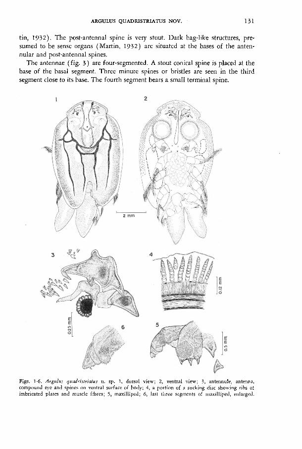
131
tin, 1932). The post-antennal spine is very stout. Dark bag-like structures, pre- sumed to be sense organs (Martin, 1932) are situated at the bases of the anten- nular and post-antennal spines.
The antennae (fig. 3) are four-segmented. A stout conical spine is placed at the
base of the basal segment. Three minute spines or bristles are seen in the third
segment close to its base. The fourth segment bears a small terminal spine.
Figs. 1-6. Argulu.r quadri,rtriatu,r n. sp. 1, dorsal view; 2, ventral view; 3, antennule, antenna, compound eye and spines on ventral surface of body; 4, a portion of a sucking disc showing ribs ot imbricated plates and muscle fibres; 5, maxilliped; 6, last three segments of maxilliped, enlarged.

132
The compound eyes (fig. 3) are placed almost in one line with the base of the
post-antennal spine. The median eye is well developed. The sucking discs are 0.78 mm in inside diameter. Each consists of 58 ribs, each
formed by 9 to 11 imbricate plates. The basal imbricate plate is of the same size
as the second. At the base of the ribs three concentric layers of muscle fibers can
be seen; in the outer of these layers the fibers are arranged radially, in the two
others the fibers are parallel to the outer margin of the disc (fig. 4). The proboscis (fig. 7) lies in the mid-ventral line of the body, it is flanked by
Figs. ?-11. Argulu.r quadristriatus n. sp. 7, proboscis; 8, distal part of proboscis showing internal structures, ventral view (left lateral lobe of lower lip displaced from its normal position by pressure, applied through cover slip); 9, mandible; 10, fourth pair of thoracic legs; 11, mature uterine eggs. bc, buccal cavity; bf, buccal fold; imb, inner mandibular bar; omb, outer mandibular bar; lb, labral
bar; llb, lateral lobe of lower lip; llp, lower lip; 1mb, lateral marginal bar; md, mandible.

133
the antennules, antennae, sucking discs and maxillipeds; its basal half is expanded, the distal part narrow, terminating in a stylet. The distal end of the buccal cavity
(fig. 8) is enclosed between the upper and lower lips. The upper and lateral edges of the buccal opening are formed by the upper lip and strengthened by a chitinous
labral bar and two lateral marginal bars. Anteriorly the marginal bars insert into
the wall of the proboscis and over some distance carry a series of transverse horny rods. The buccal folds are finely ciliated. A pair of inner and a pair of outer
mandibular bars render the main support to the proboscis. The anterior part of the
mandibles projects into the buccal cavity. The mandibles are concave interiorly and
convex exteriorly (figs. 8, 9); there are two prominent teeth on the convex side; the antero-interior border and an oblique narrow belt between the anterior teeth of
the convex side and the inner border are finely pectinated. The maxillipeds (figs. 5, 6) are five-segmented. The basal segment has three
stout posteromedial spines carrying inner spines, and a large, nearly circular
spinous pad flanked by an adjacent ill-defined one on its ventral side. The third
segment shows a ventral pad of spines and a row of similar spines along the inner
margin. The fifth segment is triangular in shape, with a blunt lobular end and two claws on the inner margin. There are two pairs of post-maxillar spines, which also
carry inner spines. The distal ends of the rami of the swimming legs reach beyond the outline of
the carapace. There are no flagella on any of the swimming legs. The basal lobe
of the left fourth leg is boot-shaped as in many species of Argulu.r. The basal lobe
and the third segment of the right fourth leg show abnormal outgrowths (fig. 10 ) , 1 which were so massive that the living animal was unable to move this leg while the other limbs were moving actively.
Mature uterine eggs (fig. 11 ) are thick-shelled and are waxy in consistency; they are four- to six-sided when attached to one another. They assume an oval shape
upon separation, and measure about 0.26-0.27 X 0.23-0.25 mm. The yolk is
arranged in spherules. Affinities. - Argulus quadristi-lilu.r may easily be distinguished from other
species of Argulus by a combination of the following characters: ( 1 ) the position of the smaller respiratory area is anterior to the larger one and (2) the ribs of the
suction cups are composed of 9 to 11 imbricated plates only. In Wilson's (1902) account, there are 22 species of Argulus including A. indicus with their respiratory areas arranged as in A. quadristriatus. Among the seven species of Argulus known to occur in India, A. giganteus and A. indieus have this type of respiratory areas.
In A. ku.ra fugu and A. scutiformis from Japanese fishes, Yamaguti & Yamasu
(1959) reported such an arrangement of the respiratory areas. Of the above cited
species, only A. 1nelanoJtictfiJ, A. pugettensis, A. niger) A. f loriden.ri.r and A.
giganteus have the arrangement of the respiratory areas combined with the fact that the ribs of the suction cups are exclusively formed of imbricated plates, as in the
present species. However, they differ from A. quadristriatus in the much greater number of these plates in each rib, viz., 30 in A. melanostictus, 25 to 26 in A.
giganteus and 15 to 20 in others.

134
ACKNOWLEDGEMENT
The authors are deeply indebted to Dr. R. V. Nair, Director, Central Marine Fisheries Research Institute, for critically going through the manuscript.
RÉSUMÉ
Une nouvelle espèce du genre Argulus Müller est décrite. Cette espèce, A. quadristriatus sp. nov., est caractérisée par la position de la plus petite aire respiratoire, antérieure à la plus grande, et par 9 à 11 plaques imbriquées sur les rayons des ventouses. Elle est étroitement apparentée à A. melanos- ticus, A. pugettensis, A. niger, A. floridensis et A. giganteus par la disposition des aires respiratoires et par les rayons des ventouses qui sont seulement composés de plaques imbriquées, lesquelles, cependant, sont bien moins nombreuses que chez les espèces ci-dessus.
REFERENCES
CLAUS, C., 1875. Ueber die Entwicklung, Organisation und systematische Stellung der Arguliden. Zeitschr. wiss. Zool., 25: 217-284.
MARTIN, M. F., 1932. On the morphology and classification of Argulus (Crustacea). Proc. zool. Soc. London, 1932: 771-806.
RAMAKRISHNA, G., 1951. Notes on Indian species of the genus Argulus Müller (Crustacea; Cope- poda) parasitic on fishes. Rec. Indian Mus., 49 (2): 207-216.
- , 1962. On a new species of Argulus Müller (Crustacea: Copepoda) from Kerala. Proc. all India Congr. Zool., 1 (2): 178, 179.
THOMAS, M. M., 1961. Observations on the habits and post-embryonic development of a parasitic branchiuran Argulus puthenveliensis Ramakrishna. Journ. mar. biol. Ass. India, 3: 75-86.
THOMAS, M. M. & M. DEVARAJ, in press. Two new species of Argulus Müller (Crustacea, Bran- chiura) from river Cauvery with a key to the Indian species. Journ. mar. biol. Ass. India, 17 (1).
THORELL, M. T., 1865. Om tvenne Europeiska Argulider; jemte anmärkningar om Argulidernas morfologi och systematiska ställning, samt en öfversigt af de för närvarande kända arterna af denna familj. Oefvers. Kongl. Vetensk.-Akad. Förhandl., 21 : 7-72.
WILSON, C. B., 1902. North American parasitic copepods of the family Argulidae, with a bibliog- raphy of the group and a systematic review of all known species. Proc. U. S. nation. Mus., 25 (1302): 635-742.
YAMAGUTI, S. & T. YAMASU, 1959. On two species of Argulus (Branchiura, Crustacea) from Japanese fishes. Biol. Journ. Okayama Univ., 5: 167-175.