A new method of kidney biopsy using low dose CT-guidance with
6
METHODOLOGY Open Access A new method of kidney biopsy using low dose CT-guidance with coaxial trocar and bard biopsy gun Xiao-ling Pi 1 , Zhen Tang 2* , Li-qian Fu 1 , Mei-hua Guo 1 , Mei-hua Shi 1 , Lan Chen 1 and Zheng-ying Wan 1 Abstract Background: To explore a new method of kidney biopsy with coaxial trocar and bard biopsy gun under low dose computed tomography (CT)-guidance and evaluate its accuracy, safety, and efficacy. Methods: Sixty patients underwent renal biopsy under CT-guidance. They were randomly divided into two groups: group I, low dose CT-guided (120 kV and 25 or 50 mAs) and group II, standard dose CT-guided (120 kV and 250 mAs). For group I, the coaxial trocar was accurately placed adjacent to the renal capsule of the lower pole, the needle core was removed, and samples were obtained with a bard biopsy gun. For group II, the coaxial trocar was not used. Total number of passes, mean biopsy diameter, mean glomeruli per specimen, mean operation time, mean scanning time, and mean radiation dose were noted. Dose-length product (DLP) was used to calculate the radiation doses. After 24 hours of the biopsy, ultrasound was repeated to identify any subcapsular hematoma. Results: Success rate of biopsy in group I was 100% while using low dose CT-guidance along with coaxial trocar renal. There was no statistic differences bewteen group I and II in the total number of passes, mean biopsy diameter, mean glomeruli per specimen and mean time of operation and CT scanning. The average DLP of group I was lower as compared to the value of group II (p <0.05). Conclusions: Kidney biopsy using coaxial trocar and bard biopsy gun under low dose CT was an accurate, simple and safe method for diagnosis and treatment of kidney diseases. It can be used for repeat and multiple biopsies, particularly suitable for obese and renal atrophy patients in whom the kidneys are difficult to image. Keywords: Kidney biopsy, Low dose CT scanning, Bard biopsy gun, Coaxial trocar Background Kidney diseases have been a silent killer with rising inci- dence worldwide and poor outcomes [1]. Early diagnosis and treatment can prevent the complications of decreased kidney function and reduce the risk of concomitant car- diovascular disease [2,3]. However, many of the kidney dis- eases are undiagnosed by non-invasive methods and renal pathological examination has indicated to establish defini- tive histopathological diagnosis in diffuse renal disease [4]. Renal biopsy is an immensely valuable tool to deter- mine the cause of the disease, predict the prognosis, and direct treatment which includes open biopsy (laparos- copically [5], transvenous [6] and percutaneous biopsy Ultrasonograpgy [7], CT [7,8], or magnetic resonance imaging (MRI) [9]. The only disadvantage with these methods could be the severe surgical trauma [10]. Ultrasound-guided percutaneous renal biopsy has trad- itionally been used to obtain biopsies but the mainten- ance of sterilization is very difficult with the use of coupling agents [11-13]. In patients with chronic renal failure and renal atrophy caused by glomerulosclerosis and interstitial fibrosis, the interface of renal cortex and medullary is unclear under ultrasound images [14]. Simi- larly, biopsy procedures could be unsuccessful in obese patients where imaging of the kidneys is difficult [15]. Thus, the puncture operation would become inconvenient leading to an inaccurate positioning. The diameter of bi- opsy tissues is proportional to the number of glomeruli in * Correspondence: [email protected] 2 Department of Radiology, Fengxian Center Hospital-Branch of Shanghai Sixth People’s Hospital, Jiaotong University, Shanghai 201400, China Full list of author information is available at the end of the article Biological Procedures Online © 2013 Pi et al.; licensee BioMed Central Ltd. This is an Open Access article distributed under the terms of the Creative Commons Attribution License (http://creativecommons.org/licenses/by/2.0), which permits unrestricted use, distribution, and reproduction in any medium, provided the original work is properly cited. Pi et al. Biological Procedures Online 2013, 15:1 http://www.biologicalproceduresonline.com/content/15/1/1
Transcript of A new method of kidney biopsy using low dose CT-guidance with

Biological ProceduresOnline
Pi et al. Biological Procedures Online 2013, 15:1http://www.biologicalproceduresonline.com/content/15/1/1
METHODOLOGY Open Access
A new method of kidney biopsy using lowdose CT-guidance with coaxial trocar andbard biopsy gunXiao-ling Pi1, Zhen Tang2*, Li-qian Fu1, Mei-hua Guo1, Mei-hua Shi1, Lan Chen1 and Zheng-ying Wan1
Abstract
Background: To explore a new method of kidney biopsy with coaxial trocar and bard biopsy gun under low dosecomputed tomography (CT)-guidance and evaluate its accuracy, safety, and efficacy.
Methods: Sixty patients underwent renal biopsy under CT-guidance. They were randomly divided into two groups:group I, low dose CT-guided (120 kV and 25 or 50 mAs) and group II, standard dose CT-guided (120 kV and 250mAs). For group I, the coaxial trocar was accurately placed adjacent to the renal capsule of the lower pole, theneedle core was removed, and samples were obtained with a bard biopsy gun. For group II, the coaxial trocar wasnot used. Total number of passes, mean biopsy diameter, mean glomeruli per specimen, mean operation time,mean scanning time, and mean radiation dose were noted. Dose-length product (DLP) was used to calculate theradiation doses. After 24 hours of the biopsy, ultrasound was repeated to identify any subcapsular hematoma.
Results: Success rate of biopsy in group I was 100% while using low dose CT-guidance along with coaxial trocarrenal. There was no statistic differences bewteen group I and II in the total number of passes, mean biopsydiameter, mean glomeruli per specimen and mean time of operation and CT scanning. The average DLP of group Iwas lower as compared to the value of group II (p <0.05).
Conclusions: Kidney biopsy using coaxial trocar and bard biopsy gun under low dose CT was an accurate, simpleand safe method for diagnosis and treatment of kidney diseases. It can be used for repeat and multiple biopsies,particularly suitable for obese and renal atrophy patients in whom the kidneys are difficult to image.
Keywords: Kidney biopsy, Low dose CT scanning, Bard biopsy gun, Coaxial trocar
BackgroundKidney diseases have been a silent killer with rising inci-dence worldwide and poor outcomes [1]. Early diagnosisand treatment can prevent the complications of decreasedkidney function and reduce the risk of concomitant car-diovascular disease [2,3]. However, many of the kidney dis-eases are undiagnosed by non-invasive methods and renalpathological examination has indicated to establish defini-tive histopathological diagnosis in diffuse renal disease [4].Renal biopsy is an immensely valuable tool to deter-
mine the cause of the disease, predict the prognosis, anddirect treatment which includes open biopsy (laparos-
* Correspondence: [email protected] of Radiology, Fengxian Center Hospital-Branch of ShanghaiSixth People’s Hospital, Jiaotong University, Shanghai 201400, ChinaFull list of author information is available at the end of the article
© 2013 Pi et al.; licensee BioMed Central Ltd. TCommons Attribution License (http://creativecreproduction in any medium, provided the or
copically [5], transvenous [6] and percutaneous biopsyUltrasonograpgy [7], CT [7,8], or magnetic resonanceimaging (MRI) [9]. The only disadvantage with thesemethods could be the severe surgical trauma [10].Ultrasound-guided percutaneous renal biopsy has trad-itionally been used to obtain biopsies but the mainten-ance of sterilization is very difficult with the use ofcoupling agents [11-13]. In patients with chronic renalfailure and renal atrophy caused by glomerulosclerosisand interstitial fibrosis, the interface of renal cortex andmedullary is unclear under ultrasound images [14]. Simi-larly, biopsy procedures could be unsuccessful in obesepatients where imaging of the kidneys is difficult [15].Thus, the puncture operation would become inconvenientleading to an inaccurate positioning. The diameter of bi-opsy tissues is proportional to the number of glomeruli in
his is an Open Access article distributed under the terms of the Creativeommons.org/licenses/by/2.0), which permits unrestricted use, distribution, andiginal work is properly cited.

Pi et al. Biological Procedures Online 2013, 15:1 Page 2 of 6http://www.biologicalproceduresonline.com/content/15/1/1
the renal cortex hence, renal cortex becomes an adequatesite for the biopsies [16]. This makes accurate positioningessential. Conventional CT-guided renal biopsy is ex-tremely accurate but high radiation poses a major prob-lem. Hence, CT-guided renal biopsy has been reserved formore difficult cases [4]. However, significant changes inthe biopsy design as well as use the minimum radiationdose to achieve a reasonable diagnostic image quality mayfurther enhance the accuracy, safety, and efficacy of CT-guided kidney biopsy. To understand the scope of CT-guided biopsy techniques the present study was conductedto investigate the utility of a low radiation, minimally inva-sive, and accurate biopsy method using a low dose CT-guided coaxial trocar and automatic biopsy gun.
Results and discussionThe cores of 13.5 mm (range 9–31 mm) from cortico-medullary junction were collected with a biopsy needlein compliance with the diagnostic requirements. Eachcore contained mean glomeruli per specimen 15.3 (range12–37) and 15.2 in the group-I and II, respectively. Themean operation and scanning time was 13 minutes(range 10–13 minutes) and 4.1 sec (range 3.935-5.268 sec), respectively in the group I. The mean ope-ration and scanning time was 13.5 minutes and 4.2 sec,respectively in the group II. There was no significant dif-ference between the two groups with the five main para-meters (p >0.05) (Table 1). The total average DLP ofgroup I (311.5 mGy × cm) was low as compared to thatof group II (1166.3 mGy × cm) and difference was statis-tically significant (p <0.05). The total average DLP ofgroup I was less than 1/3 of the group II DLP. The de-crease in the total average DLP for group I was achievedby modifying the tube current-time product (mAs).
ComplicationsRoutine urine for three consecutive times showed thatall of the patients experienced microhematuria after bi-opsy, there were no patients with gross hematuria. Twopatients in the group I (6.7%) and three patients in thegroup II (10%) had small sub capsular hematoma24 hours after biopsy, but all of them remained asymp-tomatic, without any medical treatment.
Table 1 Comparison of low-dose CT-guided and standard-dos
Total numberof passes
Mean biopsiesdiameter(mm)
Mean glomspecimen
low-dose group(30cases)
62 13.5 ± 0.8 15.3 ± 1.0
Standard dosegroup(30cases)
64 13.8 ± 0.7 15.2 ± 1.1
P value, student’st-test
>0.05 >0.05 >0.05
DiscussionsKidney biopsy under CT-guidance has been the goldstandard for diagnosis and treatment of glomerular andtubulo-interstitial disease [17,18]. Computed tomographyprovides detailed view of anatomical structures, whichhelps localization of even small lesions located in the kid-neys. It helps in accurate planning of the needle biopsy,avoiding inadvertent puncture of vascular structures [19].Computed tomography guided kidney biopsies canshorten the period of hospital stay and decrease the num-ber of operations as well as the treatment costs [20]. Asthere is a risk of high dose radiation exposed complica-tions, as well as multiple or repeated examinations arevery likely required for patients suffered of chronic kidneyfailures, methods for its protection have drawn increasingattention from researchers and clinicians. Although alter-native imaging technologies such as ultrasound do notpose risk of radiation, the difficulties of maintainingsterilization with coupling agents and having clear viewswith ultrasound imaging in patients with chronic renalfailure caused by glomerulosclerosis and interstitial fibro-sis restrict their usages in renal biopsy. In CT scanning,the minimum radiation dose should be as low as reason-ably achievable for maintenance of diagnostic quality[21,22]. In our study, we reduced the radiation dose by de-creasing tube current-time from 250 mAs to 80mAs,which was less than 1/3 of the standard dose of CT. Theaverage DLP for group I was 311.5 mGy × cm, which wasonly 26.7% of the standard dose of CT and was almostnegligible. Moreover, use of this method could result ingood diagnostic image quality for routine clinical diagnos-tic applications.Generally, CT-guided coaxial trocar renal biopsy is
easy to operate, ensures the sterility of entire visual field,and helps in reduction of the risk of infection. The num-ber of glomeruli in the cortex is critical for the diagnosisof kidney disease; generally a minimum of 5 to 10 intactglomeruli would satisfy the requirements [4,18]. Biopsytissues diameter is proportional to the number of glom-eruli in the renal cortex so the biopsies must be takenfrom renal cortex. Therefore, an accurate positioningunder CT-guidance is required. In our study, low dose CTscan is a bit weak, but it can clearly distinguish the kidneycortex and medulla. The mean glomeruli per specimen
e CT-guided renal biopsy
eruli per Mean operationtime (M)
Mean scanningtime (S)
Mean radiation dose(mGy.cm)
13.2 ± 2.0 4.1 ± 1.0 311.5 ± 9.8
13.5 ± 2.1 4.2 ± 1.1 1166.3 ± 10.4
>0.05 >0.05 <0.01

Figure 2 5.0 mm axial CT scan was taken through the lowerpole of the kidney to determine the puncture site (Standard-dose scanning).
Pi et al. Biological Procedures Online 2013, 15:1 Page 3 of 6http://www.biologicalproceduresonline.com/content/15/1/1
were 15.3, which could be successfully analyzed by lightmicroscopy, immunofluorescence, and electron micros-copy. The operation time was less (10 to 15 minutes perpatient; the median time was 13 min) compared with earl-ier reported studies [14,18]. Some of these studies reportedthat in order to reduce hemorrhage complications, 18-gauge biopsy needle and 3 cores were generally requiredfor pathologic diagnosis [14]. In our study, adequate tissuewas obtained using the 16-gauge biopsy needle and only 2cores were needed. The pathological diagnosis rate was100%, which was slightly higher than related previous stud-ies [4,18]. The biggest advantage of coaxial trocar renal bi-opsy under thin-layer scan was the accurate positioning,safe operation, and high successful rate. The majority ofthe studies reported the use of 10 mm axial CT scans [4].In our study 5 mm axial CT scans were used to providemore accurate positions. The location of puncture needlecould be determined by coaxial trocar before biopsy, whichis similar to having renal biopsy under direct vision. Thus,multiple biopsies could be possible with only one position-ing. This is convenient, efficient, and helps in reducing theradiation exposure.The biopsy gun provided high-quality specimen with lit-
tle bleeding and rapid action. The action of biopsy gunmay be slowed down if used alone, due to the resistanceof lumbar muscles in patients. This could result in low-quality specimen and increased chances of bleeding [4].Nonetheless, in coaxial trocar assisted biopsy, the bard bi-opsy gun passes through a trocar to minimize the fric-tional resistance between the biopsy needle and the trocar.
Figure 1 The U-shaped locator (arrow) was placed for skinpositioning (topography).
In conclusion, low dose CT-guided renal biopsy wasaccurate, safe, and effective. It had the advantages ofclearly displaying the regional anatomic location andstructure successfully with no or few complications.While this technology is beneficial to all sorts of patientswith chronic renal failure or renal atrophy as the inter-face of renal cortex and medulla is much clear in low-dosed CT scanning, it is especially useful to obtainenhanced images in patients who were obese or hadrenal atrophy.
MethodsPatients and medical historyWe studied 60 patients, who underwent renal biopsies atthe Department of Nephrology, Pudong New Area GongliHospital (Shanghai, China) from October 2009 to Decem-ber 2010. Prior to each procedure, the risks and benefitsof these biopsies were discussed, and informed consentwas obtained from each patient. This study was approvedby ethics committees of Shanghai Pudong New AreaGongli Hospital and Fengxian Center Hospital-Branch ofShanghai Sixth People’s Hospital. In this study 29 maleand 31 female patients were included. The median age ofpatients were 51 years (range 23–80 years) with a medicalhistory of nephritic syndrome (28 patients, 46.66%), acuterenal failure (5 patients, 8.33%), hypersensitivity nephropa-thy (4 patients, 6.66%), microscopic hematuria (2 patients,3.33%), interstitial nephritis (2 patients, 3.33%) and sixpatients each of diabetic nephropathy, chronic renal fail-ure with obesity, and proteinurea. Out of 28 nephroticsyndrome patients, one was with massive ascites and twowere obese. From six chronic renal failure patients, threehad kidney length diameter less than 9 cm.
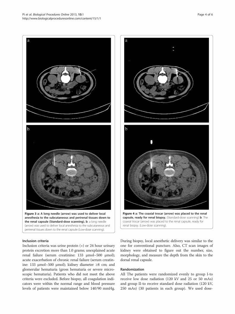
Figure 3 a: A long needle (arrow) was used to deliver localanesthesia to the subcutaneous and perirenal tissues down tothe renal capsule (Standard-dose scanning). b: a long needle(arrow) was used to deliver local anesthesia to the subcutaneous andperirenal tissues down to the renal capsule (Low-dose scanning).
Figure 4 a: The coaxial trocar (arrow) was placed to the renalcapsule, ready for renal biopsy. (Standard-dose scanning) b: Thecoaxial trocar (arrow) was placed to the renal capsule, ready forrenal biopsy. (Low-dose scanning).
Pi et al. Biological Procedures Online 2013, 15:1 Page 4 of 6http://www.biologicalproceduresonline.com/content/15/1/1
Inclusion criteriaInclusion criteria was urine protein (+) or 24 hour urinaryprotein excretion more than 1.0 grams; unexplained acuterenal failure (serum creatinine: 133 μmol~500 μmol);acute exacerbation of chronic renal failure (serum creatin-ine: 133 μmol~500 μmol); kidney diameter ≥8 cm; andglomerular hematuria (gross hematuria or severe micro-scopic hematuria). Patients who did not meet the abovecriteria were excluded. Before biopsy, all coagulation indi-cators were within the normal range and blood pressurelevels of patients were maintained below 140/90 mmHg.
During biopsy, local anesthetic delivery was similar to theone for conventional puncture. Also, CT scan images ofkidney were obtained to figure out the number, size,morphology, and measure the depth from the skin to thedorsal renal capsule.
RandomizationAll The patients were randomized evenly to group I-toreceive low dose radiation (120 kV and 25 or 50 mAs)and group II-to receive standard dose radiation (120 kV,250 mAs) (30 patients in each group). We used dose-

Pi et al. Biological Procedures Online 2013, 15:1 Page 5 of 6http://www.biologicalproceduresonline.com/content/15/1/1
length product (DLP) as a dose descriptor indicator. Itcharacterizes the total ionizing energy imparted to thereference phantom for a given examination and can beused to evaluate the radiation doses received by thepatients. All biopsies were performed under CT (PhilipsBrilliance 16 CT scanner) guidance using bard biopsy gun(16-gauge × 16 cm), self-made coaxial trocar (20-gaugetrocar, 16-gauge needle core), self-made U-shaped locator,and 7-gauge needle (6 cm). The renal biopsies were per-formed by a full-time nephrology faculty experienced inthe technique.After defecation/urination, the patient was instructed to
lie down in the appropriate position on the CT scannertable and a soft pillow was placed under the abdomen assupport. The patient was instructed not to move duringthe procedure. Under CT-guidance a U-shaped locatorwas placed at the puncture site of the lower pole and ap-proximately 6–7 cm parallel to the spine (Figure 1). Thepuncture site was fully covered and patients were askedto hold their breath, after which a 5.0 mm axial CT scanwas taken through the lower pole of the kidney (Figure 2).The laser scan line was used as an anchor tag. The crosspoint of the laser scan lines and the U-shaped locator waslocalized for biopsy and the puncture site was markedwith a marker pen. The puncture site was sterilized withcompound iodine solution, a sterile drape was placed overthe site, and local anesthesia was administered to the sub-cutaneous and perirenal tissues down to the renal capsuleusing 7-gauge needle. The distance from the skin to therenal cortex was measured and a second CT scan wasperformed (Figure 3). Later the 7-gauge needle was pulledout and a small incision on the puncture site was madewith a small sharp knife. The coaxial trocar was placed tothe renal capsule in the same path of the anesthetic nee-dle and rescanned (Figure 4). The style of the coaxial tro-car was removed and replaced with an activated bardbiopsy gun. The biopsy was then done immediately bytriggering the biopsy gun during suspended respiration.After biopsy, the biopsy gun was pulled out immedi-
ately; all the specimens inside the needle were removedand placed on ice-cold saline-soaked gauze for patho-logic examination. When the specimens were not suffi-cient to establish the diagnosis, repeat biopsies wereperformed using the same technique as the first. Theneedle was removed after the tissue sample was taken;gauze was placed on the site for 3 minutes to stop thebleeding and cores were sent for pathological analysis.After the biopsy, patients were asked to stay in the bedfor 24 hours. At 24 hours after the biopsy, ultrasoundwas repeated to identify any subcapsular hematoma.Total number of passes, mean biopsies diameter, meanglomeruli per specimen, mean operation time, meanscanning time, and mean radiation dose were recorded.Statistical significance between groups was assessed by
student’s t-test and p <0.05 was considered to be thresh-old for statistical significance.
Competing interestsThe authors declare that they have no competing interests.
Authors’ contributionsXLP conceived of the study and participated in operations of all patients. ZTwas Guarantor of integrity of the entire study and participated in its designand Data acquisition. LQF and MHG screened patients and participated inoperations of some patients. MHS, LC and ZYW participated preoperativeand postoperative observation. All authors read and approved the finalmanuscript.
AcknowledgementsWe extend our sincere thanks to Dr. Peter Pothula, BioQuest for his valuableeditorial guidance in the generation of this report.
FundingThe authors disclosed receipt of the following financial support for theresearch, authorship and/or publication of this article: Fengxian district ofShanghai grant (FK20111001).
Author details1Department of Nephrology, Pudong New Area Gongli Hospital, Shanghai200135, China. 2Department of Radiology, Fengxian Center Hospital-Branchof Shanghai Sixth People’s Hospital, Jiaotong University, Shanghai 201400,China.
Received: 9 October 2012 Accepted: 15 December 2012Published: 7 January 2013
References1. Levey AS, Andreoli SP, DuBose T, Provenzano R, Collins AJ: CKD: common,
harmful, and treatable–World Kidney Day 2007. Am J Kidney Dis 2007,49:175–179.
2. Remuzzi G, Ruggenenti P, Perico N: Chronic renal diseases: renoprotectivebenefits of renin-angiotensin system inhibition. Ann Intern Med 2002,136:604–615.
3. Levey AS, Atkins R, Coresh J, Cohen EP, Collins AJ, Eckardt KU, Nahas ME,Jaber BL, Jadoul M, Levin A, Powe NR, Rossert J, Wheeler DC, Lameire N,Eknoyan G: Chronic kidney disease as a global public health problem:approaches and initiatives - a position statement from Kidney DiseaseImproving Global Outcomes. Kidney Int 2007, 72:247–259.
4. Song JH, Cronan JJ: Percutaneous biopsy in diffuse renal disease:comparison of 18- and 14-gauge automated biopsy devices. J Vasc IntervRadiol 1998, 9:651–655.
5. Shetye KR, Kavoussi LR, Ramakumar S, Fugita OE, Jarrett TW: Laparoscopicrenal biopsy: a 9-year experience. BJU Int 2003, 91:817–820.
6. Mal F, Meyrier A, Callard P, Altman JJ, Kleinknecht D, Beaugrand M, FerrierJP: Transjugular renal biopsy. Lancet 1990, 335:1512–1513.
7. Richter F, Kasabian NG, Irwin RJ Jr, Watson RA, Lang EK: Accuracy ofdiagnosis by guided biopsy of renal mass lesions classifiedindeterminate by imaging studies. Urology 2000, 55:348–352.
8. Eshed I, Elias S, Sidi AA: Diagnostic value of CT-guided biopsy ofindeterminate renal masses. Clin Radiol 2004, 59:262–267.
9. Sharma KV, Venkatesan AM, Swerdlow D, DaSilva D, Beck A, Jain N, WoodBJ: Image-guided adrenal and renal biopsy. Tech Vasc Interv Radiol 2010,13:100–109.
10. Hojs R: Kidney biopsy and power Doppler imaging. Clin Nephrol 2004,62:351–354.
11. Reef V: Adult abdominal ultrasonography. In Equine diagnostic ultrasound.Edited by Reef VB. Philadelphia: W.B. Saunders Co; 1998:348–352.
12. Tucker R: Ultrasound-guided biopsy. In Equine diagnostic ultrasonography.Edited by Rantanen BW, McKinnon AO. Baltimore: Williams & Wilkens;1998:649–653.
13. Nass K, O’Neill WC: Bedside renal biopsy: ultrasound guidance by thenephrologist. Am J Kidney Dis 1999, 34:955–959.
14. Sateriale M, Cronan JJ, Savadler LD: A 5-year experience with 307 CT-guided renal biopsies: results and complications. J Vasc Interv Radiol 1991,2:401–407.

Pi et al. Biological Procedures Online 2013, 15:1 Page 6 of 6http://www.biologicalproceduresonline.com/content/15/1/1
15. Fine DM, Arepally A, Hofmann LV, Mankowitz SG, Atta MG: Diagnosticutility and safety of transjugular kidney biopsy in the obese patient.Nephrol Dial Transplant 2004, 19:1798–1802.
16. Wang HJ, Kjellstrand CM, Cockfield SM, Solez K: On the influence of samplesize on the prognostic accuracy and reproducibility of renal transplantbiopsy. Nephrol Dial Transplant 1998, 13:165–172.
17. Margaryan A, Perazella MA, Mahnensmith RL, Abu-Alfa AK: Experience withoutpatient computed tomographic-guided renal biopsy. Clin Nephrol2010, 74:440–445.
18. Kudryk BT, Martinez CR, Gunasekeran S, Ramirez G: CT-guided renal biopsyusing a coaxial technique and an automated biopsy gun. South Med J1995, 88:543–546.
19. Protopapas Z, Westcott JL: Transthoracic hilar and mediastinal biopsy.Radiol Clin North Am 2000, 38:281–291.
20. Maya ID, Allon M: Percutaneous renal biopsy: outpatient observationwithout hospitalization is safe. Semin Dial 2009, 22:458–461.
21. Davies HE, Wathen CG, Gleeson FV: The risks of radiation exposure relatedto diagnostic imaging and how to minimise them. BMJ 2011, 342:d947.
22. Sarti M, Brehmer WP, Gay SB: Low-dose techniques in CT-guidedinterventions. Radiographics 2012, 32:1109–1119. discussion 1119–1120.
doi:10.1186/1480-9222-15-1Cite this article as: Pi et al.: A new method of kidney biopsy using lowdose CT-guidance with coaxial trocar and bard biopsy gun. BiologicalProcedures Online 2013 15:1.
Submit your next manuscript to BioMed Centraland take full advantage of:
• Convenient online submission
• Thorough peer review
• No space constraints or color figure charges
• Immediate publication on acceptance
• Inclusion in PubMed, CAS, Scopus and Google Scholar
• Research which is freely available for redistribution
Submit your manuscript at www.biomedcentral.com/submit