A New Biocompatible and Antibacterial Phosphate Free … · A New Biocompatible and Antibacterial...
9
A New Biocompatible and Antibacterial Phosphate Free Glass-Ceramic for Medical Applications Bele ´n Cabal 1 , Luı ´s Alou 2 , Fabio Cafini 2 , Ramiro Couceiro 3 , David Sevillano 2 , Leticia Esteban-Tejeda 4 , Francisco Guitia ´n 3 , Ramo ´n Torrecillas 1,5 & Jose ´ S. Moya 4 1 Nanomaterials and Nanotechnology Research Center (CINN-CSIC) – Universidad de Oviedo (UO) – Principado de Asturias, Parque Tecnolo ´gico de Asturias, 33428, Llanera, Spain, 2 Microbiology Unit, Medicine Department, School of Medicine, Universidad Complutense, Avda. Complutense s/n, 28040 Madrid, Spain, 3 Instituto de Cera ´ mica de Galicia, Avda. Maestro Mateo, 15782 Santiago de Compostela, Spain, 4 Institute of Materials Science of Madrid (ICMM-CSIC), Cantoblanco, 28049, Madrid, Spain, 5 Moscow State University of Technology (STANKIN), Vadkovskij per. 1, Moscow, Moscow Oblast, Russian Federation. In the attempt to find valid alternatives to classic antibiotics and in view of current limitations in the efficacy of antimicrobial-coated or loaded biomaterials, this work is focused on the development of a new glass-ceramic with antibacterial performance together with safe biocompatibility. This bactericidal glass-ceramic composed of combeite and nepheline crystals in a residual glassy matrix has been obtained using an antimicrobial soda-lime glass as a precursor. Its inhibitory effects on bacterial growth and biofilm formation were proved against five biofilm-producing reference strains. The biocompatibility tests by using mesenchymal stem cells derived from human bone indicate an excellent biocompatibility. G lass-ceramics are defined as polycrystalline materials, with residual amorphous phase, obtained from glass melting and controlled crystallization. They offer numerous advantages compared with parent glasses such as improved thermo-physical properties, higher strength and wear resistance. In general, glass-ceramics show favorable chemical, thermal, dielectric and biological properties superior to metals and various polymers 1 . They are ideal materials for the design and manufacture of bioceramics with superior mech- anical properties. Most biomedical glass-ceramics are based on compositions similar to those of Hench’s bioactive glasses (BioglassH), having P 2 O 5 as a common ingredient in all of them 2 . In the last few decades the important clinical applications of glass-ceramic are related to the repair and reconstruction of skeletal system (bone, joints and teeth) owing to their biocompatibility with living tissues. One of the main problems associated with the clinical use of these biomaterials in orthopedic surgery is contamination by infectious microorganisms. In fact, bone and joint have been listed among the most frequent sites of infections for which antibacterials are prescribed 3 . Biomaterial-associated infections are a major problem in modern medicine. They have an enormous impact in terms of morbidity of the patients and costs to national health systems. Bacterial colonization and biofilm formation on the implanted device may lead to acute and chronic infection of the underlying bone and the adjacent soft tissues 4 . Bacteria start implant infection by adhering to biomaterials surfaces and/or interfaces producing biofilms, which represent a major reason for bacterial persistence. Moreover, the formation of a biofilm increases the resistance of the bacteria to antibiotics. The variety of bacterial species implicated in the etiology of implant infections commonly enlists Gram-positive bacteria of the genera Staphylococcus as well as Gram-negative microorganisms of the genera Pseudomonas, but often includes also other less frequent oppor- tunistic pathogens with varying degree of virulence 5 . The multifactorial nature of the variables influencing the development of biomaterial-associated infections makes the assessment of clinical efficacy of anti-infective biomaterials an extremely difficult task. Various strategies have been adopted to retard biofilm development on implant surfaces: (a) modification of the biomaterial surface to give anti-adhesive and bacteria repelling properties, (b) antibacterial coatings, (c) nanostructured materials, (d) molecules interfering with bacterial biofilm 6,7 . Once a mature bacteria biofilm has been established, conventional medical therapies based on systemic antibiotics are not efficacious, and implant removal often represents the only chance to eradicate the infection. OPEN SUBJECT AREAS: INFECTION BIOMEDICAL MATERIALS ANTIMICROBIAL RESISTANCE Received 26 November 2013 Accepted 6 June 2014 Published 25 June 2014 Correspondence and requests for materials should be addressed to B.C. ([email protected]) SCIENTIFIC REPORTS | 4 : 5440 | DOI: 10.1038/srep05440 1
Transcript of A New Biocompatible and Antibacterial Phosphate Free … · A New Biocompatible and Antibacterial...

A New Biocompatible and AntibacterialPhosphate Free Glass-Ceramic forMedical ApplicationsBelen Cabal1, Luıs Alou2, Fabio Cafini2, Ramiro Couceiro3, David Sevillano2, Leticia Esteban-Tejeda4,Francisco Guitian3, Ramon Torrecillas1,5 & Jose S. Moya4
1Nanomaterials and Nanotechnology Research Center (CINN-CSIC) – Universidad de Oviedo (UO) – Principado de Asturias,Parque Tecnologico de Asturias, 33428, Llanera, Spain, 2Microbiology Unit, Medicine Department, School of Medicine,Universidad Complutense, Avda. Complutense s/n, 28040 Madrid, Spain, 3Instituto de Ceramica de Galicia, Avda. MaestroMateo, 15782 Santiago de Compostela, Spain, 4Institute of Materials Science of Madrid (ICMM-CSIC), Cantoblanco, 28049,Madrid, Spain, 5Moscow State University of Technology (STANKIN), Vadkovskij per. 1, Moscow, Moscow Oblast, RussianFederation.
In the attempt to find valid alternatives to classic antibiotics and in view of current limitations in the efficacyof antimicrobial-coated or loaded biomaterials, this work is focused on the development of a newglass-ceramic with antibacterial performance together with safe biocompatibility. This bactericidalglass-ceramic composed of combeite and nepheline crystals in a residual glassy matrix has been obtainedusing an antimicrobial soda-lime glass as a precursor. Its inhibitory effects on bacterial growth and biofilmformation were proved against five biofilm-producing reference strains. The biocompatibility tests by usingmesenchymal stem cells derived from human bone indicate an excellent biocompatibility.
Glass-ceramics are defined as polycrystalline materials, with residual amorphous phase, obtained fromglass melting and controlled crystallization. They offer numerous advantages compared with parentglasses such as improved thermo-physical properties, higher strength and wear resistance. In general,
glass-ceramics show favorable chemical, thermal, dielectric and biological properties superior to metals andvarious polymers1. They are ideal materials for the design and manufacture of bioceramics with superior mech-anical properties. Most biomedical glass-ceramics are based on compositions similar to those of Hench’s bioactiveglasses (BioglassH), having P2O5 as a common ingredient in all of them2. In the last few decades the importantclinical applications of glass-ceramic are related to the repair and reconstruction of skeletal system (bone, jointsand teeth) owing to their biocompatibility with living tissues. One of the main problems associated with theclinical use of these biomaterials in orthopedic surgery is contamination by infectious microorganisms. In fact,bone and joint have been listed among the most frequent sites of infections for which antibacterials areprescribed3.
Biomaterial-associated infections are a major problem in modern medicine. They have an enormous impact interms of morbidity of the patients and costs to national health systems. Bacterial colonization and biofilmformation on the implanted device may lead to acute and chronic infection of the underlying bone and theadjacent soft tissues4. Bacteria start implant infection by adhering to biomaterials surfaces and/or interfacesproducing biofilms, which represent a major reason for bacterial persistence. Moreover, the formation of abiofilm increases the resistance of the bacteria to antibiotics. The variety of bacterial species implicated in theetiology of implant infections commonly enlists Gram-positive bacteria of the genera Staphylococcus as well asGram-negative microorganisms of the genera Pseudomonas, but often includes also other less frequent oppor-tunistic pathogens with varying degree of virulence5. The multifactorial nature of the variables influencing thedevelopment of biomaterial-associated infections makes the assessment of clinical efficacy of anti-infectivebiomaterials an extremely difficult task. Various strategies have been adopted to retard biofilm developmenton implant surfaces: (a) modification of the biomaterial surface to give anti-adhesive and bacteria repellingproperties, (b) antibacterial coatings, (c) nanostructured materials, (d) molecules interfering with bacterialbiofilm6,7.
Once a mature bacteria biofilm has been established, conventional medical therapies based on systemicantibiotics are not efficacious, and implant removal often represents the only chance to eradicate the infection.
OPEN
SUBJECT AREAS:INFECTION
BIOMEDICAL MATERIALS
ANTIMICROBIAL RESISTANCE
Received26 November 2013
Accepted6 June 2014
Published25 June 2014
Correspondence andrequests for materials
should be addressed toB.C. ([email protected])
SCIENTIFIC REPORTS | 4 : 5440 | DOI: 10.1038/srep05440 1
Antibiotic-loaded biomaterials are currently part of standard med-ical procedures for both prophylaxis and local treatment of implantinfection. Considering the large risk of antibiotic resistance assoc-iated with antibiotic loaded coatings, non-antibiotics agents in thecoating became very attractive alternatives4.
Pointing out the need for further research in the strategies to treatinfection, this work is focused on the development of an antibacterialglass-ceramic that could be used for medical applications. This glass-ceramic can be obtained from several soda-lime glasses belonging tothe SiO2-Na2O-CaO-B2O3 system with increasing content of cal-cium oxide. These family glasses were probed to be efficient antimi-crobials in a previous work8. There are many examples in theliterature of glass/glass-ceramic with bactericidal properties, but atleast to the author’s knowledge, there is no previous work in theliterature about the efficacy to inhibit biofilm formation by a glass-
ceramic which is not loaded with antibiotics or metallic nanoparti-cles (silver, copper).
Bactericidal activity is rarely highly specific and uniquely orientedtowards prokaryotic cells. Often it is associated to a certain degree ofcytotoxicity. However, several research works have proposed bio-compatible glasses and glass ceramic with antibacterial properties9,10.It is critical to find a correct balance between bactericidal effects andbiocompatibility properties. On this regard, a strong requirement isto ensure a proper stability with the high level of biological safety.Therefore, both the bactericidal activity and cytotoxicity of this glass-ceramic were evaluated in the present study.
ResultsCharacterization of the glass ceramic. Thermal properties of theprecursor glasses were investigated by differential thermal analysis
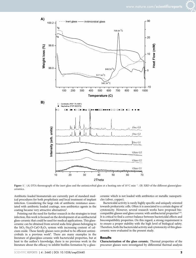
Figure 1 | (A) DTA thermograph of the inert glass and the antimicrobial glass at a heating rate of 10uC min21. (B) XRD of the different glasses/glass-
ceramics.
www.nature.com/scientificreports
SCIENTIFIC REPORTS | 4 : 5440 | DOI: 10.1038/srep05440 2

(DTA-Tg) (Fig. 1.A). No exothermic peaks are observed in the case ofthe inert glass (G1). On the contrary, thermograms of the antimicro-bial glasses exhibits two well defined crystallization exotherms ataround 645 and 700uC, suggesting that at least two crystallinephases are being formed when these glasses are reheated. The XRDanalyses allow us to identify the formed crystalline phases (Fig. 1.B).Before sintering, antimicrobial glasses are completely amorphous,present the characteristic broad peak at about 2h 5 25u, but thesintered materials exhibited a diffraction pattern characteristic of aglass-ceramic structure. The XRD study conducted on both anti-microbial glasses after heat treatment at 750uC showed that devitri-fication of these glasses lead to a glass-ceramic material composed ofcombeite (Na4Ca4(Si6O18), PDF 75-1687) and nepheline (Na6.65
Al6.24Si9.76O32, PDF 83-2372) dispersed in a glassy matrix. Analysisof the relative intensities of the peaks concluded that the same glass-ceramic can be obtained from both antimicrobial glasses. Therefore,the rest of the work was carried out using only the glass-ceramicobtained from the antimicrobial glass G3. In the case of the inertglass after thermal treatment, peaks of quartz (PDF 001-0649) areclearly visible.
SEM image (Fig. 2) illustrates the presence of dense arrangementof crystals and it also reveals the presence of hexagonal shaped androd likes crystals. Pores are also perceptible. The specific surface areas(SBET) of the glassy pellets were calculated from the N2 adsorptionisotherms using the multipoint Brunauer-Emmett-Teller (BET)technique. Both glassy pellets present similar SBET: 0.33 m2/g theglass-ceramic, and 0.31 m2/g the inert control glass. Higher averagesurface roughness values (Ra) was found for the glass-ceramic discs(0.97 6 0.09) than for the inert glass discs (0.04 6 0.03).
TEM/EDS provide more detailed microstructural characterizationof the glass-ceramic. In TEM image (Figure 3) both crystalline phasescan be clearly identified in close agreement with the XRD results(Fig. 1B). Nepheline crystals (Fig. 3, nu2) show larger sizes thancombeite crystals ((Fig. 3, nu1). The EDS analysis of crystal 1 showedthat the Ca/Si ratio reached values corresponding to combeite.Likewise, Al/Si ratio in crystal 2 is consistent with nepheline. TheCaO content in the residual glassy phase (Fig. 3, nu3) is similar to thatof the parent glass.
Bactericidal property of the glass ceramic. Bacterial growth. Totalcolony forming units of all studied strains from inert glass powder(control) achieved 108–109 CFU/mL in all study time period (0–72 h). Colony forming units from glass-ceramic cultures weresignificantly lower (p , 0.0001) than those from inert soda-limeglass cultures for all strains and at all study time period, except forS. aureus at 24 h (p 5 0.02), S. aureus at 48 h (p 5 0.004), E. coli at
24 h (p 5 0.004). Figure 4 shows the logarithm reduction (CFU/mL)observed in the glass-ceramic cultures respect to inert glass cultures.Glass-ceramic powder significantly reduced the number of bacterialcells of all ATCC strains with respect to control glass powder by99.99% at 48 and 72 h except for S. aureus where reductions of96.6% and 99.7% were observed, respectively. S. epidermidis, M.lutea and E. coli were the most affected strains achieving a 5 log10
reduction at 72 h.In order to evaluate the role of each crystalline phase in the bac-
tericidal capability, antibacterial tests were also carried out with thephases fully crystallized. As it is clearly seen (Figure 5) no bactericidalactivity against E. coli was obtained for nepheline, whereas in the caseof combeite a quite similar behavior to the glass-ceramic wasobserved.
Biofilm formation. Total colony forming units of biofilms formed inthe inert glass discs (control) achieved 106–108 CFU/mL for allstrains and at all study time period (0–72 h). Colony forming unitsfrom control discs were significantly higher (p , 0.0001) than thosefrom glass-ceramic discs for all ATCC strains. Figure 6 shows thelogarithm reduction (CFU/mL) observed in the glass-ceramic discsrespect to inert glass discs. Glass-ceramics significantly reduced theadherence of all ATCC strains with respect to inert glass by 99.99% at24 h, except for S. epidermidis where a reduction of 99.87% wasobserved. It is noteworthy the case of M. lutea strain, antibiofilmactivity increased over time achieving a 5–6 log10 reduction at 48and 72 h.
Biocompatibility of antibacterial glass ceramic. Cells performedgenerally better when cultured on the antibacterial glass-ceramicsubstrates than on the control surfaces. The values obtained for theMTT assay (Figure 7) marked an optimal and gradually increasingproliferation values for the glass-ceramic (p-value , 0.05, t-studenttest, 95% confidence level). Such results are in harmony with theapoptotic rates (Figure 8). A nominal apoptotic rate is alwaysfound in cell cultures even in cell friendly surfaces from cultureflasks, however, soda-lime glass used as control is showing agreater apoptotic rate than expected when compared to a morebiocompatible surface like the glass-ceramic.
Cells need to attach in order to proceed with the colonization ofany biomaterial surface, while attached, DNA concentration could beextracted and calculated in order to gain an idea of which surface ispresenting a higher cell concentration. The glass-ceramic showsagain (Figure 9) a good performance when compared to the inertglass (p-value , 0.05, t-student test, 95% confidence level), support-ing the previous observation for apoptosis and proliferation. Whencells are stained for a live/dead assay (Figure 10), glass-ceramic sub-strate shows a clear viability of hBMSCs.
DiscussionIn the present work, a new glass-ceramic composed of combeite andnepheline in a residual soda-lime glassy matrix was fabricated bysintering at 750uC – 1 h of an antimicrobial glassy frit. Powders(,30 mm) were conformed to discs by conventional axial pressing.
The obtained antibacterial results (Fig. 4–6) clearly show that thisglass-ceramic free of P2O5 not only is effective to diminish the growthof bacteria but also to inhibit adhesion and biofilm formation. It iswell known that surface roughness promote a bacterial adhesionprocess11. In this particular case, the bactericidal properties are notaffected although the glass ceramic is rougher than the inert glassused as control. The pH of the medium after the biocide test wasmeasured. There was a slight increase of the pH of the medium but itwas not significantly higher than the pH of the fermentation broth atthe end of the growth. By way of example, the pH of the medium afterthe bactericidal test against E. coli is: 8.71 for the culture, 8.87 for theinert glass and 8.97 for the glass-ceramic. Therefore, it was not pos-sible to conclude from these experiments that the antibacterial activ-
Figure 2 | Scanning electron micrograph of the glass-ceramic pellet. Rod
likes crystals are indicated by empty white circles.
www.nature.com/scientificreports
SCIENTIFIC REPORTS | 4 : 5440 | DOI: 10.1038/srep05440 3

Figure 3 | Transmission electron micrographs of the glass-ceramic pellet. Inset show the corresponding select area electron diffraction (SAED) patterns.
Identified combeite crystal phase (nu1), nepheline phase (nu2), residual glassy phase (nu3).
www.nature.com/scientificreports
SCIENTIFIC REPORTS | 4 : 5440 | DOI: 10.1038/srep05440 4

ity was due to a high pH per se. The antimicrobial activity of theparent glass arises from the capability to release calcium ions at theglass-particle interface, which leads to membrane depolarization andthe subsequent death of the cell, as well reported in previous work8. Itis well known that cellular calcium ions overload, or perturbation ofintracellular Ca21 compartmentalization, may cause cytotoxicity andresult in either apoptotic, necrotic or autophagic cell death12–14. Fromthose results we can infer that something similar happens with theglass-ceramic. Then role of calcium must be pointed out when thebiocide activity is evaluated with the fully crystallized phases. Onlythe phase contains calcium (i.e., combeite [Na2Ca2Si3O9]) showedantibacterial capability. Calcium is contained in both combeite crys-tals and the residual glassy phase of the glass-ceramic (Fig. 3).Therefore, both phases are somehow involved in its antibacterialcapability. Combeite is a complex structure constituted by piling of6-fold silica tetrahedral rings. Sodium atoms are placed in the centreof these rings. Alternated calcium and sodium atoms are located inthe open space between the rings columns of sodium15. Then, thesecalcium ions, as a consequence of its light bonding, can be easilyexchanged by H1 (or H3O1) ions from the solution in a similar
way than in the original glass structure. In this sense, we consideredthe operating mechanism must be very similar to that of the parentglass.
A similar situation has been previously observed by the authors16
in the case of a bioactive glass-ceramic made from sintered 45S5BioglassH, containing Na2Ca2Si3O9 as crystalline phase. Despite theamount of research and literature on antimicrobials, there is noprevious work, at least to the author’s knowledge, about the efficacyto inhibit biofilm formation by a glass-ceramic which is not loadedwith antibiotics or metallic nanoparticles (e.g. silver, copper).
An ideal material for the treatment of infection should not onlyposses a bactericidal property, preventing microbial adhesion andcolonization, but it must also have excellent biocompatibility. Thecytotoxicity of the glass-ceramic was assessed by the MTT assayusing the hBMSCS as a cell model. As shown in Fig. 7, the numberof viable cells on the antibacterial glass-ceramic is noticeably higherthan that on the inert glass after culturing the hBMSCs for 1, 2 and 5days. In addition, the enhanced cell adhesion and proliferation on thebactericidal glass-ceramic (Fig. 8–9) indicates no obvious adverseeffects on cell viability and proliferation. The results obtained in
Figure 4 | Antibacterial capability of the glass-ceramic powders respect to inert glass powder (control).
Figure 5 | Comparison of the bactericidal activity of the fully crystallized phases and the glass-ceramic against E.coli.
www.nature.com/scientificreports
SCIENTIFIC REPORTS | 4 : 5440 | DOI: 10.1038/srep05440 5

the present investigation open the possibility to fabricate a multi-functional biomaterial, which at the same time inhibits bacterialcolonization (i.e. biofilm formation) as well as effective integratingwith host tissues. This new glass-ceramic could found importantmedical applications such as coating (prosthesis, dental devices),scaffolds, etc.
On the other hand, this glass-ceramic acts as a delivery systemproviding sustained release of the ions which may affect and poten-tiate a more appropriate and rapid tissue response. The crystallinephase combeite (Na2Ca2Si3O9) has been formed and identified inother studies on sintered bioactive glasses17–19. It is known that thisphase is bioactive and biodegradable in amorphous calcium phos-phate upon immersion in simulated body fluid20,21. Certain composi-tions of the glass-ceramics have shown bioactivity, generally definedas the ability to elicit favorable cellular response. Phosphate contentis considered important for the bioactivity but it was proved thatP2O5 free CaO?SiO2 also showed bioactivity22. Base on this, it wouldbe expected that this new antibacterial free of P2O5 glass-ceramic is
also bioactive; however, even if the literature reports bioactivity prop-erties also for P2O5-free glass compositions, in vitro bioactivity testwill be/have to be performed for this new antimicrobial free of P2O5
glass-ceramic in order to verify its ability to create a chemical bondwith tissue.
The results obtained in this investigation show the feasibility ofthe production by a sinter-crystallisation process of an antibac-terial glass-ceramic composed of combeite and nepheline crystalsin a residual glass matrix. This glass-ceramic is significantly ableto inhibit bacterial growth, minimize bacterial adhesion and pre-vent biofilm formation. In addition, the in vitro biocompatibilityassays with human stem cells demonstrate that this glass-ceramichas an excellent biocompatibility. This antibacterial glass-ceramiccould find application in the production of anti-infective bioma-terials. Final form of this glass-ceramic could be as powders,granules, dense pieces, coatings and porous scaffolds, thus pro-viding a wide spectrum of alternatives for each specific clinicaluse.
Figure 6 | Antibiofilm activity of the glass-ceramic discs respect to inert glass discs (control).
Figure 7 | Cells proliferation results according to absorbance values on the samples after incubation for different times.
www.nature.com/scientificreports
SCIENTIFIC REPORTS | 4 : 5440 | DOI: 10.1038/srep05440 6

MethodsPreparation of the antibacterial glass-ceramic. In previous studies8,23 theantimicrobial capability of several soda-lime glasses belonging to the SiO2-Na2O-CaO-B2O3 system, with increasing content of calcium oxide, were probed againstdifferent microorganisms. Two of those glasses with a significant biocide activityagainst bacteria and yeast were selected as precursors of the antibacterial glass-ceramic. Other one, with null effect against microorganism was selected as control inthe bactericidal and biocompatibility tests.
Two antimicrobial soda-lime glasses (labeled as G2 and G3) were prepared bymelting appropriate mixtures of reagent grade SiO2 (Cuarzos Industriales S.A.,Santiago de Compostela), a-Al2O3 (Taimei Chemical Co. Ltd., Japan), H3BO3,Na2CO3, and CaCO3 (Sigma Aldrich). Their chemical compositions are shown intable 1. The starting materials were weighed, mixed and melted in a Pt crucible for 1 hat 850uC to favor decarbonation of samples, and subsequently for 1 h at 1400uC. Themelts were then quenched in water and grounded by ball milling to fine particulates,and sieved to obtain particle size , 30 mm. The study of the thermal behavior of theseglasses were performed with a thermogravimetric-differential thermal analyzer (TAInstruments, Q600). The heating rate was 10uC min21 up to a maximum temperatureof 1100uC in air. Based on the DTA data (Figure 1), the schedule of thermal treatmentwas designed to convert the glasses into glass-ceramics. Fractions of 1 g of powderwere compacted at 250 MPa by uniaxial pressing to obtain discs (10 mm in diameter
and 2 mm in height). Afterwards, the discs were sintered in air by heating at a rate of10uC min21 to 750uC and holding for 1 h.
Inert soda-lime glass (labeled as G1) with the chemical composition showed intable 1 was prepared in analogous procedure as the antimicrobial glasses. Fractions of1 g of powder were compacted at 250 MPa by uniaxial pressing to obtain discs(10 mm in diameter and 2 mm in height). Afterwards, discs (10 3 2 mm) weresintered in air by heating at a range of 10uC min21 to 700uC and holding for 1 h.
Fully crystallize phases were synthesized in order to evaluate the role of the crys-talline phases of the glass-ceramic in the antibacterial capability. Combeite wassynthesized by solid state reaction at 980uC from appropriate mixtures or reagentgrade SiO2 (Cuarzos Industriales S.A., Santiago de Compostela), Na2CO3 and CaCO3
(Sigma Aldrich). Nepheline was also synthesized according to the Na2O-Al2O3-SiO2
system, by solid state reaction at 900uC starting from a proper proportions of SiO2
(Cuarzos Industriales S.A., Santiago de Compostela), Na2CO3 and Al2O3 (SigmaAldrich).
Characterization of the antibacterial glass ceramic. The XRD analyses of glass-ceramic discs were carried out in a Bruker D8 diffractometer using CuKa radiationworking at 40 kV and 30 mA in a step-scanning mode from 4u to 70u with a stepwidth of 0.05u and a step time of 0.5 seconds. The morphology of the obtained glass-ceramic was studied by scanning electron microscopy (SEM) (Hitachi S-4300), and
Figure 8 | Activation of caspase 3 in hBMSCs.
Figure 9 | Cell adhesion measured by means of DNA concentration.
www.nature.com/scientificreports
SCIENTIFIC REPORTS | 4 : 5440 | DOI: 10.1038/srep05440 7
also characterized using transmission electron microscopy (TEM) (JEOL FXII) withan accelerating voltage of 200 kV. In order to estimate the surface morphology, thesurface of the discs were measured with a surface profilometer (Talysurf CLI 500,Taylor Hobson, Leicester, UK) that maps the surface by putting a stylus in mechanicalcontact with the sample. The stylus arm has 90u conisphere diamond styli with 2 mmnominal radius tip. The data sampling interval in X and Y was 0.5 mm and 2.5 mmrespectively. The resolution (Z) was 32 nm. The profilometer was used to determinethe three-dimensional topographic map and to calculate the roughness factor orspecific surface area (the ratio of the surface to the projected area). Three samples ofeach glass were scanned to evaluate the average surface roughness (Ra) of the surfacesat five different locations. Textural characterization was carried out by measuring theN2 adsorption isotherms at 2196uC in an automatic apparatus (TriStar II 3020,Micromeritics Instrument Corporation). The Barret-Emmett-Teller (BET) methodwas utilized to calculate the specific surface areas (SBET).
Evaluation of the bactericidal effect. Bacterial strains. Five biofilm-producingreference strains: Staphylococcus aureus ATCC 29213, Staphylococcus epidermidisATCC 35984, Pseudomonas aeruginosa ATCC 23389, Escherichia coli ATCC 25922and Micrococcus lutea ATCC 9341, were used in both antibacterial assays (biocideand biofilm formation assays). They are the most frequent pathogens implicated inthe etiology of biomaterials-associated infections24.
Biocide assays. A single colony from each microorganism was inoculated in LuriaBertani (LB) broth (Difco; BD Diagnostics, Sparks, MD, USA) and incubated over-night at 37uC. Each culture (10 mL) was diluted into fresh media (1 mL) and culturedat 37uC for 6 h; finally, fresh media (1 mL), with inert soda-lime glass (control) orglass-ceramic powders (75 mL of 200 mg/mL aqueous suspension), were inoculatedwith the above cultures (10 mL). Samples were taken at 0, 24, 48, and 72 hours todetermine the viable bacteria and expressed as colony forming units (CFU/mL). Allexperiments were performed in triplicate. The limit of detection was 5 3 101
CFU/mL. Antibacterial effectiveness is expressed as the logarithm reduction in viablecounts of the test bacteria (CFU/mL). It was calculated by subtracting the log10 colonycounts in the glass ceramic cultures from those in the inert glass cultures (control).The percentage was calculated as %IR 5 100 2 (100 3 In)/It, where %IR is per-centage of inoculum reduction, In is the bacterial count (CFU/mL) in the glass-ceramic cultures, and It is the inoculum (CFU/mL) in the control cultures.
Similarly, the antibacterial capability against E. coli of the fully crystallized phases(combeite and nepheline) was evaluated.
Biofilm formation assays. Colonies of ATCC strains from an overnight culture onColumbia sheep blood agar (Difco, BD Diagnostic Systems, Sparks, MD, USA) wereallowed to grow in LB medium at 37uC to a density of 0.5–1 3 108 CFU/mL asmeasured by a UV-Visible spectrophotometer (GBC, Model Cintra 101, Australia)and diluted 1/10 in fresh LB medium. Glass-ceramic and inert soda-lime glass discswere placed in 24-well culture plates and 300 mL of the inoculum suspension wereinoculated for two hours (adhesion phase). After the adhesion period, the mediumwas discarded (non-adherent cells were removed) and 300 mL of fresh LB mediumwere added in each well. The discs were incubated at 37uC for 72 h in a wet chamber.Samples were taken at 0, 24, 48, and 72 hours to determine the viable bacteria.
Viable counts were used to determine the quantity of viable adherent bacteria.After biofilm formation, discs were carefully washed three times with sterile saline
solution to remove unattached cells and inserted in new well containing 1 mL ofsterile saline solution. The discs were sonicated for 10 minutes in ultrasonic bath(FB15050; Thermo-Fischer Scientific) to disperse bacterial cells. Samples of 200 mLfrom sonicated biofilms were serially diluted in 0.9% saline solution and plated ontoColumbia sheep blood agar to determine the total number of viable cells andexpressed as CFU/mL. All experiments were performed in triplicate. The limit ofdetection was 2.5 3 102 CFU/mL. Preliminary experiments indicated that sonicationwas not associated with a loss of viability of the cells. Antibacterial effectiveness isexpressed as the logarithm reduction in viable counts of the test bacteria (CFU/mL). Itwas calculated by subtracting the log10 colony counts in the glass ceramic discs fromthose in the inert glass discs. The percentage was calculated as %IR 5 100 2 (100 3
In)/It, where %IR is percentage of inoculum reduction, In is the bacterial count (CFU/mL) in the glass-ceramic discs, and It is the inoculum (CFU/mL) in the control discs.
Statistical analysis of the tests carried out with prokaryotic cells. All statistical analyseswere performed by the unpaired Student’s t test. A P-value of ,0.05 was consideredstatistically significant.
Proliferation of hBMSCs. Cell culture. For investigating the cellular responses to thedeveloped glass-ceramic, mesenchymal stem cells derived from human bone marrowwere used (hBMSCs). hBMSCs were obtained by the Department of OrthopaedicSurgery (University of Santiago de Compostela Clinical Hospital) during routinaryhip prosthesis surgeries by ethically approved protocols. Bone marrow was stored inheparin coated tubes and taken to the cell culture laboratory for proceeding withmesenchymal stem cell isolation. A Ficoll protocol as described by Yeo et al.25 wasused. Briefly, a bone marrow as slowly poured on top of a Ficoll volume (151) in a15 mL Falcon tube. Rapidly after this step a centrifugation takes place (2000 rpm,30 min). Cells are pipetted onto fresh medium (D-MEM, 20%FCS, Gentamycin) andcultured for 2 weeks until a 90% confluence is reached. The total cell density used was25000 cells/cm2. Human bone marrow cells response was evaluated by determiningcell adhesion and cell proliferation.
Cell proliferation by MTT assays. The survival and proliferation of hBMSCs on glass-ceramic discs was examined with a MTT assay after 1, 2 and 5 days of culture. TheMTT assays were carried out as per manufacturer’s protocol (Sigma-Aldrich). Briefly,hBMSCs were seeded on the sample discs (d 5 10 mm) at a density of 25 3 103 cellsper well in a 24-well plate. At each time point, the medium were removed, MTTsolution was added, and cells were incubated overnight. After removal of the MTTsolution, the purple formazan crystals were dissolved in 100 mL of dimethyl sulph-oxide (DMSO) by shaking the plate for 15 min, and 50 mL solution of each well wasadded into a new 24-well plate. The OD value was quantified by measuring theabsorption at 570 nm using an automated plate reader (PerkinElmer).
Cellular adhesion. Viable cells should be adhered to the surface of the assayed bio-materials. An attachment assay gave us an insight of how many cells were attached onthe material surface. Briefly, cells were incubated for 24 hours to allow the attachmentto happen, after this first incubation, the culture media was removed to wash out anyfloating cell. Samples were gently washed with phosphate buffered saline (PBS),which helped to remove the non attached cells, right after this step, a 3.5% para-formaldehyde solution was added and incubated for 1 h. Finally, cells were stainedwith 0.1% toluidine blue for 3 h. The dye was washed out with destilled water and thecells lysed with 500 mL of 0.1% sodium dodecyl sulfate (SDS). The resulting aliquotwas assed with an optical density at 595 nm.
Plates were incubated at 37uC in 5% CO2 for the indicated time period. Wells wereflicked dry and were washed three times with PBS, and adherent cells were fixed in100 mL of 10% formalin for 15 minutes at RT. Plates were stained overnight with100 mL of toluidine blue in 10% formalin. Plates were washed three times withcopious amounts of distilled water before lysis in 2% SDS for 10 minutes. Opticaldensity (OD) was read at 600 nm with a microtiter plate reader.
Apoptosis: caspase activity assay. Caspase-3 activity was measured in supernatants by aQuantikine immunoassay (ELISA) active caspase-3 kit (R&D Systems, Minneapolis,MN). Briefly, cytoplasmic protein extract was incubated with biotin-ZVKD-fluoro-methylketone inhibitor, which covalently binds and makes detectable the activecaspase-3. A monoclonal antibody specific for active caspase-3 was precoated onto amicroplate, and active protease concentrations were assessed in cell lysates, accordingto the manufacturer’s instructions.
Confocal microscopy. live/dead staining. Calcein-AM and propidium iodide were usedto stain the hBMSCs cultured on the glass ceramic pellets. After 48 h of incubation thesamples were gently washed with D-MEM. The viable and non viable cells were
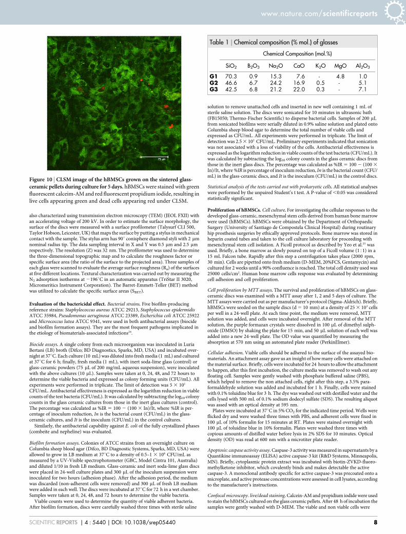
Figure 10 | CLSM image of the hBMSCs grown on the sintered glass-ceramic pellets during culture for 5 days. hBMSCs were stained with green
fluorescent calceim-AM and red fluorescent propidium iodide, resulting in
live cells appearing green and dead cells appearing red under CLSM.
Table 1 | Chemical composition (% mol.) of glasses
Chemical Composition (mol.%)
SiO2 B2O3 Na2O CaO K2O MgO Al2O3
G1 70.3 0.9 15.3 7.6 - 4.8 1.0G2 46.6 6.7 24.2 16.9 0.5 - 5.1G3 42.5 6.8 21.2 22.0 0.3 - 7.1
www.nature.com/scientificreports
SCIENTIFIC REPORTS | 4 : 5440 | DOI: 10.1038/srep05440 8

visualized by using a confocal laser scanning microscope (CLSM-SP2, Leica), theviable cells appear fluorescent green, whereas nonviable cells appear fluorescent red.
Statistical analysis of the tests carried out with eukaryotic cells. Mintab 16 (MinitabINC) was used for the statistical data analysis. T-student tests were carried out with ana 5 0.05.
1. Holand, W. & Beall, G. H. Glass-ceramic technology: Second Edition (John Wileyand Sons, New Jersey, 2012).
2. de Aza, P. N., de Aza, A. H., Pena, P. & de Aza, S. Bioactive glasses and glass-ceramics. B. Soc. Esp. Ceram. V 46, 45–55 (2007).
3. Campoccia, D., Montanaro, L., Speziale, P. & Arciola, C. R. Antibiotic-loadedbiomaterials and the risks for the spread of antibiotic resistance following theirprophylactic and therapeutic clinical use. Biomaterials 31, 6363–6377 (2010).
4. Goodman, S. B., Yao, Z., Keeney, M. & Yang, F. The future of biologic coatings fororthopaedic implants. Biomaterials 34, 3174–3183 (2013).
5. Arciola, C. R., An, Y. H., Campoccia, D., Donati, M. E. & Montanaro, L. Etiology ofimplant orthopedic infections: a survey on 1017 clinical isolates. Int. J. Artif.Organs. 28, 1091–1100 (2005).
6. Campoccia, D., Montanaro, L. & Arciola, C. R. A review of the biomaterialstechnologies for infection-resistant surfaces. Biomaterials 34, 8533–8554 (2013).
7. Simchi, A., Tamjid, E., Pishbin, F. & Boccaccini, A. R. Recent progress in inorganicand composite coatings with bactericidal capability for orthopaedic applications.Nanomed. Nanotechnol. 7, 22–39 (2011).
8. Moya, J. S., Esteban, L., Pecharroman, C., Mello-Castanho, S. R. H., da Silva, A. C.& Malpartida, F. Glass powders with a high content of calcium oxide: A steptowards a ‘‘green’’ universal biocide. Adv. Eng. Mater. 13, B256–B260 (2011).
9. Zhang, D. et al. Antibacterial effects and dissolution behavior of six bioactiveglasses. J. Biomed. Mater. Res. Part A 93, 475–483 (2010).
10. Gomes, C. H. et al. Assesment of antimicrobial effect of biosilicateH againstanaerobic, microaerophilic and facultative anaerobic microorganisms. J. Mater.Sci: Mater. Med. 22, 1439–1446 (2011).
11. Teughels, W., Van Assche, N., Sliepen, I. & Quirynen, M. Effect of materialcharacteristics and/or surface topography on biofilm development. Clin. OralImplan. Res. 17, 68–81 (2006).
12. Rizzuto, R., Stefani, D., Raffaello, A. & Mammucari, C. Mitochondria as sensorsand regulators of calcium signaling. Nat. Rev. Mol. Cell Bio. 13, 566–578 (2012).
13. Zhivotovsky, B. & Orrenius, S. Calcium and cell death mechanisms: A perspectivefrom the cell death community. Cell Calcium 50, 211–221 (2011).
14. Krebs, J. & Michalak, M. (Eds). Calcium: A matter of life or death. Elsevier 2007.15. Ohsato, H., Takeuchi, Y. & Maki, I. Structure of Na4Ca4(Si6O18). Acta Crystallogr.
Sect. C: Cryst. Struct. Commun. C42, 934–937 (1986).16. Cabal, B. et al. The development of bioactive glass-ceramic substrates with biocide
activity. Adv. Eng. Mater. 13, B462–B466 (2011).17. Rahaman, M. N. et al. Bioactive glass in tissue engineering. Acta Biomater. 7,
2355–2373 (2011).
18. Salman, S. M., Salama, S. N. & Abo-Mosallam, H. A. The role of strontium andpotassium on crystallization and bioactivity of Na2O-CaO-P2O5-SiO2 glasses.Ceram. Int. 38, 55–63 (2012).
19. Belluci, D., Cannillo, V. & Sola, A. An overview of the effects of thermal processingon bioactive glasses. Sci. Sinter. 42, 307–320 (2010).
20. Chen, Q. Z., Thompson, I. D. & Boccaccini, A. R. 45S5 BioglassH-derived glass-ceramic scaffolds for bone tissue engineering. Biomaterials 27, 2414–2425 (2006).
21. Du, R. & Chang, J. Preparation and characterization of bioactive sol-gel-derivedNa2Ca2Si3O9. J. Mater. Sci. Mater. M. 15, 1285–1289 (2004).
22. Ebisawa, Y. & Kokubo, T. Bioactivity of CaO SiO2-based glasses: in vitroevaluation. J. Mater. Sci. Mater. M. 1, 239–244 (1990).
23. Moya, J. S. et al. Mechanism of calcium lixiviation in soda-lime glasses with astrong biocide activity. Mater. Lett. 70, 113–115 (2012).
24. Moriarty, F. Z. & Sebastian, A. J. Biomaterials associated infection. [Busscher, H. J.(ed.)] (Springer, New York, 2013).
25. Yeo, C. et al. Ficoll-Paque versus Lymphoprep: A comparative study of twodensity gradient media for therapeutic bone marrow mononuclear cellpreparations. Regen. Med. 4, 689–696 (2009).
AcknowledgmentsThis work was supported by the Spanish Ministry of Economy and Competitiveness(MINECO) under the project MAT2012-38645, and by CSIC project ref. nu 201360E012. B.Cabal acknowledges financial support from JAE-Doc program (CSIC, cofounded by FSE).
Author contributionsConceived and designed the experiments: B.C., L.A., D.S., F.G., R.T. and J.S.M. Performedthe experiments: B.C., F.C., R.C. and L.E.-T. Analyzed the data: B.C., L.A., R.C., D.S., R.T.and J.S.M. Wrote the paper: B.C., L.A., R.C., D.S. and J.S.M. All authors reviewed themanuscript.
Additional informationCompeting financial interests: The authors declare no competing financial interests.
How to cite this article: Cabal, B. et al. A New Biocompatible and Antibacterial PhosphateFree Glass-Ceramic for Medical Applications. Sci. Rep. 4, 5440; DOI:10.1038/srep05440(2014).
This work is licensed under a Creative Commons Attribution-NonCommercial-NoDerivs 4.0 International License. The images or other third party material inthis article are included in the article’s Creative Commons license, unless indicatedotherwise in the credit line; if the material is not included under the CreativeCommons license, users will need to obtain permission from the license holderin order to reproduce the material. To view a copy of this license, visit http://creativecommons.org/licenses/by-nc-nd/4.0/
www.nature.com/scientificreports
SCIENTIFIC REPORTS | 4 : 5440 | DOI: 10.1038/srep05440 9