A mechanism for bacterial transformation of dimethylsulfide to...
14
warwick.ac.uk/lib-publications Original citation: Lidbury, Ian , Kröber, Eileen , Zhang, Zhidong, Zhu, Yijun , Murrell, J. Colin, Chen, Yin and Schäfer, Hendrik. (2016) A mechanism for bacterial transformations of DMS to DMSO : a missing link in the marine organic sulfur cycle. Environmental Microbiology . Permanent WRAP URL: http://wrap.warwick.ac.uk/78661 Copyright and reuse: The Warwick Research Archive Portal (WRAP) makes this work of researchers of the University of Warwick available open access under the following conditions. This article is made available under the Creative Commons Attribution 4.0 International license (CC BY 4.0) and may be reused according to the conditions of the license. For more details see: http://creativecommons.org/licenses/by/4.0/ A note on versions: The version presented in WRAP is the published version, or, version of record, and may be cited as it appears here. For more information, please contact the WRAP Team at: [email protected]
Transcript of A mechanism for bacterial transformation of dimethylsulfide to...

warwick.ac.uk/lib-publications
Original citation: Lidbury, Ian , Kröber, Eileen , Zhang, Zhidong, Zhu, Yijun , Murrell, J. Colin, Chen, Yin and Schäfer, Hendrik. (2016) A mechanism for bacterial transformations of DMS to DMSO : a missing link in the marine organic sulfur cycle. Environmental Microbiology . Permanent WRAP URL: http://wrap.warwick.ac.uk/78661 Copyright and reuse: The Warwick Research Archive Portal (WRAP) makes this work of researchers of the University of Warwick available open access under the following conditions. This article is made available under the Creative Commons Attribution 4.0 International license (CC BY 4.0) and may be reused according to the conditions of the license. For more details see: http://creativecommons.org/licenses/by/4.0/ A note on versions: The version presented in WRAP is the published version, or, version of record, and may be cited as it appears here. For more information, please contact the WRAP Team at: [email protected]

A mechanism for bacterial transformation ofdimethylsulfide to dimethylsulfoxide: a missing link inthe marine organic sulfur cycle
Ian Lidbury,1† Eileen Kr€ober,1† Zhidong Zhang,2
Yijun Zhu,1 J. Colin Murrell,3 Yin Chen1* and
Hendrik Sch€afer1**1School of Life Sciences, Gibbet Hill Campus,
University of Warwick, Coventry CV4 7AL, UK.2Institute of Microbiology, Xinjiang Academy of
Agriculture Sciences, Urumqi 830091, China.3School of Environmental Sciences, University of East
Anglia, Norwich Research Park, NR4 7TJ, Norwich, UK.
Summary
The volatile organosulfur compound, dimethylsulfide
(DMS), plays an important role in climate regulation
and global sulfur biogeochemical cycles. Microbial
oxidation of DMS to dimethylsulfoxide (DMSO) repre-
sents a major sink of DMS in surface seawater, yet
the underlying molecular mechanisms and key micro-
bial taxa involved are not known. Here, we reveal that
Ruegeria pomeroyi, a model marine heterotrophic
bacterium, can oxidize DMS to DMSO using trimethyl-
amine monooxygenase (Tmm). Purified Tmm
oxidizes DMS to DMSO at a 1:1 ratio. Mutagenesis of
the tmm gene in R. pomeroyi completely abolished
DMS oxidation and subsequent DMSO formation.
Expression of Tmm and DMS oxidation in R. pomer-
oyi is methylamine-dependent and regulated at the
post-transcriptional level. Considering that Tmm is
present in approximately 20% of bacterial cells inhab-
iting marine surface waters, particularly the marine
Roseobacter clade and the SAR11 clade, our obser-
vations contribute to a mechanistic understanding of
biological DMSO production in surface seawater.
Introduction
Approximately 300 Tg of the volatile organosulfur com-
pound, dimethylsulfide (DMS) is produced in the marine
environment annually (Curson et al., 2011), making the
oceans the primary contributor to DMS in the atmosphere
with an estimated flux of �20.7 Tg year21 (Watts, 2000).
DMS production occurs through transformation of the multi-
functional metabolite dimethylsulfoniopropionate (DMSP)
by a variety of algal and bacterial lyases (Curson et al.,
2011; Todd et al., 2012). Interest in the cycling of DMS
increased after the proposal that it can lead to the formation
of cloud condensation nuclei, increasing cloud albedo and
thereby acting as a global coolant (Charlson et al., 1987).
Therefore, any process contributing to the removal of DMS
from surface seawater will have a direct effect on the
attenuation of this flux, which may in turn have significant
impacts on the global sulfur cycle, specifically its deposition
to the terrestrial biome. DMS loss in surface seawater is
mediated through different pathways including photochemi-
cal oxidation (Brimblecombe and Shooter, 1986) and
biological consumption, which includes its assimilation into
biomass, dissimilation to CO2 and inorganic soluble sulfur
compounds, or oxidation to dimethylsulfoxide (DMSO)
(Sch€afer et al., 2010). Importantly, DMSP, DMS and DMSO
are frequently detected in the water column (Hatton et al.,
1998; Sim�o et al., 2000; Vila-Costa et al., 2008) and also
sea ice (Asher et al., 2011) at comparable values with con-
centrations of dissolved DMSO tending to increase towards
the end of phytoplankton blooms as cells undergo senes-
cence (Sim�o et al., 1998; Hatton and Wilson, 2007) when
DMSO concentrations can even exceed those of DMSP
(Hatton et al., 1998).
In surface seawater, biological consumption of DMS was
shown to be a major component of the global sink for
DMS, accounting for up to 99% of the DMS produced
(Kiene and Bates, 1990). Tracer studies using 35S-DMS
suggest that only a small fraction of DMS (�2–7%) is
assimilated as a sulfur source by marine microorganisms
whilst the majority of DMS is transformed into either
DMSO or sulfate (Zubkov et al., 2001; Vila-Costa et al.,
2006; del Valle et al., 2007a), potentially via other inorganic
sulfur compounds such as thiosulfate and tetrathionate
Received 24 February, 2016; accepted 19 April, 2016. For corre-spondence. *E-mail [email protected]; Tel. 00442476528976;Fax 144 (0)24 7652 2052. **E-mail [email protected]; Tel.00442476575052; Fax 144 (0)24 7652 2052. †These authors contrib-uted equally to this study.
VC 2016 The Authors Environmental Microbiology published by Society for Applied Microbiology and John Wiley & Sons LtdThis is an open access article under the terms of the Creative Commons Attribution License, which permits use, distribution andreproduction in any medium, provided the original work is properly cited.
Environmental Microbiology (2016) 00(00), 00–00 doi:10.1111/1462-2920.13354

which are known end-products of DMS catabolism in
marine DMS-degrading bacteria (De Zwart et al., 1996;
Boden et al., 2012). In marine surface waters, oxidation to
DMSO is the major fate of DMS, accounting for �70% and
50–70% of the total oxidized DMS in the Sargasso Sea
(del Valle et al., 2007a) and in the Ross Sea (del Valle
et al., 2009) respectively. Although it has previously been
suggested that DMSO production is carried out by phyto-
plankton (Sim�o et al., 1998; del Valle et al., 2007b), DMSO
production from DMS oxidation can be stimulated by the
availability of organic carbon suggesting a role of marine
heterotrophic bacteria in DMSO formation. The underpin-
ning molecular and biochemical mechanisms, however,
remain poorly understood (Gonz�alez et al., 1999; Green
et al., 2011; Hatton et al., 2012).
Marine surface waters are frequently dominated by two
groups of heterotrophic bacteria of the Alphaproteobacteria
phylum, the marine Roseobacter clade (MRC) and the
SAR11 clade (Morris et al., 2002; Buchan et al., 2005; Gie-
bel et al. 2011; Gilbert et al., 2012). The SAR11 clade are
oligotrophic microbes that are adapted to low nutrient envi-
ronments and are found throughout the world’s oceans,
dominating bacterial communities in the open ocean gyres
(Morris et al., 2002; Giovannoni et al., 2005). In contrast,
the MRC can account for up to 20% of bacterial cells in
coastal waters during periods of elevated primary produc-
tion (Gonz�ales et al., 2000; Buchan et al., 2005; Gilbert
et al., 2012). Combined, these two clades are major players
in the uptake and metabolism of phytoplankton-derived
organic carbon and nitrogen and reduced sulfur com-
pounds in marine surface waters (Miller and Belas, 2004;
Alonso and Pernthaler, 2006; Sowell et al., 2008; Sun et al.,
2011; Williams et al., 2012; Gifford et al., 2013; Ottesen
et al., 2013; Aylward et al., 2015; Lidbury et al., 2015a).
The abundance of MRC in surface seawater is often posi-
tively correlated with elevated levels of DMSP, DMS and
DMSO (Gonz�ales et al., 2000; Zubkov et al., 2001; Hatton
et al., 2012; Nelson et al., 2014; Cui et al., 2015).
We have previously shown that bacterial trimethylamine
monooxygenase (Tmm), which is responsible for the oxi-
dation of trimethylamine (TMA) into trimethylamine N-oxide
(TMAO), can also oxidize DMS at comparable rates to that
of TMA (Chen et al., 2011). Specifically, purified Tmm from
both the SAR11 clade and MRC can efficiently oxidize
DMS. The gene encoding Tmm occurs in �20% of bacte-
rial cells inhabiting the surface waters of the oceans (Chen
et al., 2011), and is particularly common in bacteria of the
MRC and SAR11 clade. In the Sargasso Sea, a region
where biological DMSO production occurs (del Valle et al.,
2007a), TMA was rapidly oxidized to CO2 (Sun et al.,
2011) and in the Antarctic Ocean, both concentrations of
TMAO and DMSO were significantly higher than either
TMA or DMS, suggesting that cooxidation of these com-
pounds was taking place at the time of sampling (Gibb and
Hatton, 2004). We, therefore, hypothesized that a signifi-
cant proportion of the observed DMSO formation in
surface seawater is due to the oxidation of DMS by Tmm-
containing heterotrophic bacteria. In this study, we
describe for the first time DMSO formation in the model
marine bacterium, Ruegeria pomeroyi DSS-3 (Gonz�ales
et al., 2003) and demonstrate that methylated amine-
dependent DMSO formation is a common trait of the MRC.
Results
Oxidation of DMS to DMSO by Ruegeria pomeroyi DSS-3
We previously showed that purified Tmm from selected
MRC and SAR11 bacteria can oxidize DMS with an affinity
similar to that of its native substrate, TMA (Chen et al.,
2011). In order to determine the product of DMS oxidation
by Tmm, we purified Tmm of R. pomeroyi from a recombi-
nant E. coli expressing Tmm and showed that DMS
oxidation resulted in the formation of DMSO at a 1:1 ratio
(Fig. 1A). Unlike TMAO, which can be further degraded in
this bacterium through a TMAO demethylase (Tdm,
E.C.4.1.2.32) (Lidbury et al., 2014, Zhu et al., 2014),
DMSO is not a substrate for Tdm (Fig. 1B), suggesting
that DMSO is the end-product of DMS co-oxidation by
Tmm (Fig. 1C).
We then investigated whether DMSO could be formed
from DMS oxidation by R. pomeroyi. It has previously been
reported that R. pomeroyi can oxidize DMS (Gonz�alez
et al., 1999), however, no significant DMS oxidation was
observed when it was grown on glucose and ammonium
as the sole carbon and nitrogen source, respectively, (Fig.
2A). Under these growth conditions, Tmm activity was not
detectable (Chen et al., 2011). TMA has previously been
shown to be both a nitrogen and energy, but not a carbon
source for R. pomeroyi (Chen et al., 2011; Lidbury et al.,
2014). In order to induce Tmm expression, we incubated a
R. pomeroyi starter culture supplemented with 0.25 mM
TMA in addition to glucose and ammonium (Chen et al.,
2011) and used this as the inoculum for induction experi-
ments. Subsequently, complete DMS oxidation occurred
within 42 h (Fig. 2B). All the DMS (0.8 mM) was trans-
formed into DMSO (0.81 6 0.03 mM) indicating that R.
pomeroyi does not further catabolize DMSO under these
growth conditions. To further confirm that DMS oxidation
was due to the activity of Tmm in vivo, we tested the tmm
knockout mutant (Dtmm::Gm) (Lidbury et al., 2015b),
which can no longer utilize TMA, under the same condi-
tions. As expected, no DMS oxidation was detected
throughout the growth cycle (Fig. 2C). The essential role of
Tmm in DMS oxidation to DMSO was further validated by
complementation of the tmm knockout mutant with either
tmm from R. pomeroyi or Roseovarius sp. 217 (Sch€afer
et al., 2005). Complementation of the Dtmm::Gm mutant
with either tmm homolog restored both growth on TMA as
2 I. Lidbury et al.
VC 2016 The Authors Environmental Microbiology published by Society for Applied Microbiology and John Wiley & Sons Ltd,Environmental Microbiology, 00, 00–00
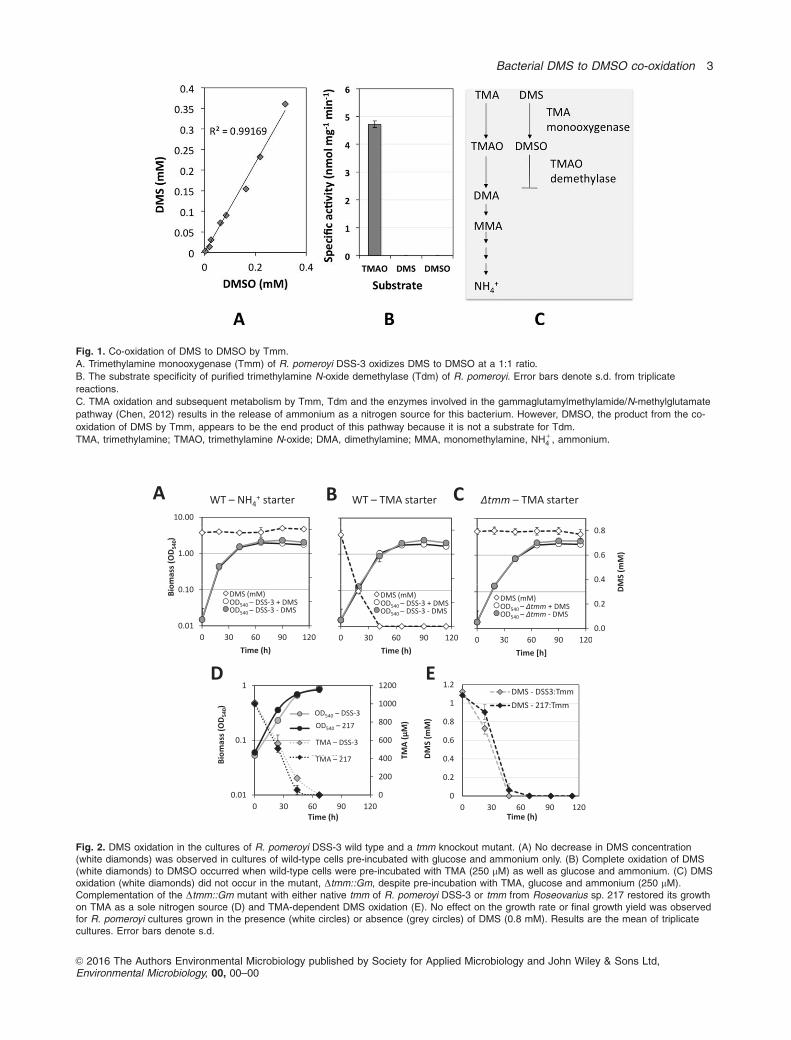
Fig. 1. Co-oxidation of DMS to DMSO by Tmm.
A. Trimethylamine monooxygenase (Tmm) of R. pomeroyi DSS-3 oxidizes DMS to DMSO at a 1:1 ratio.
B. The substrate specificity of purified trimethylamine N-oxide demethylase (Tdm) of R. pomeroyi. Error bars denote s.d. from triplicate
reactions.
C. TMA oxidation and subsequent metabolism by Tmm, Tdm and the enzymes involved in the gammaglutamylmethylamide/N-methylglutamate
pathway (Chen, 2012) results in the release of ammonium as a nitrogen source for this bacterium. However, DMSO, the product from the co-
oxidation of DMS by Tmm, appears to be the end product of this pathway because it is not a substrate for Tdm.
TMA, trimethylamine; TMAO, trimethylamine N-oxide; DMA, dimethylamine; MMA, monomethylamine, NH14 , ammonium.
Fig. 2. DMS oxidation in the cultures of R. pomeroyi DSS-3 wild type and a tmm knockout mutant. (A) No decrease in DMS concentration(white diamonds) was observed in cultures of wild-type cells pre-incubated with glucose and ammonium only. (B) Complete oxidation of DMS(white diamonds) to DMSO occurred when wild-type cells were pre-incubated with TMA (250 mM) as well as glucose and ammonium. (C) DMSoxidation (white diamonds) did not occur in the mutant, Dtmm::Gm, despite pre-incubation with TMA, glucose and ammonium (250 mM).Complementation of the Dtmm::Gm mutant with either native tmm of R. pomeroyi DSS-3 or tmm from Roseovarius sp. 217 restored its growthon TMA as a sole nitrogen source (D) and TMA-dependent DMS oxidation (E). No effect on the growth rate or final growth yield was observedfor R. pomeroyi cultures grown in the presence (white circles) or absence (grey circles) of DMS (0.8 mM). Results are the mean of triplicatecultures. Error bars denote s.d.
Bacterial DMS to DMSO co-oxidation 3
VC 2016 The Authors Environmental Microbiology published by Society for Applied Microbiology and John Wiley & Sons Ltd,Environmental Microbiology, 00, 00–00

a sole nitrogen source (Fig. 2D) and TMA-dependent DMS
oxidation (Fig. 2E). Collectively, our data confirm that DMS
can be oxidized to DMSO in Ruegeria pomeroyi through
Tmm, which can be induced by TMA.
TMA attenuates DMS production during growth of R.pomeroyi on DMSP
Next, we investigated whether R. pomeroyi can also pro-
duce DMSO during growth on DMSP. DMSP is an
important osmolyte and a precursor for DMS, which is
common in seawater (Kiene et al., 2000). R. pomeroyi was
isolated using DMSP as the sole carbon source from sea-
water collected off the coast of Georgia during an algal
bloom (Gonz�alez et al., 1999; 2003). R. pomeroyi can use
DMSP as the sole carbon source and it is known to pro-
duce DMS directly from the cleavage of DMSP using
various DMSP lyases (Curson et al., 2011; Todd et al.,
2012). When the wild type and the Dtmm mutant were
grown on ammonium and DMSP (5 mM) as the sole nitro-
gen and carbon sources, respectively, �1.4 mM of DMS
accumulated in the culture and was not subjected to fur-
ther degradation (Fig. 3A and B). To determine whether
TMA had an impact on the production and subsequent
conversion of DMS to DMSO, R. pomeroyi was grown on
DMSP (5 mM) as the sole carbon source with TMA
(0.25 mM) as the sole nitrogen source. The Dtmm mutant
failed to grow on TMA as the sole nitrogen source (Fig.
3B); consequently, only 0.2 mM DMS was produced (Fig.
3B). Given the fact that no DMS oxidation occurred when
this mutant was grown on glucose and ammonium after
pre-incubation with TMA (Fig. 2C) it was not surprising that
none of the DMS produced was converted to DMSO. Wild-
type TMA-grown cells showed a 27% reduction of maximal
DMS concentrations in the headspace during initial growth
(within 100 h) compared to that in the ammonium-grown
cultures (Fig. 3A). This was followed by near complete oxi-
dation of DMS to DMSO in TMA-grown cells (Fig. 3A).
These data therefore suggest that in the presence of TMA,
DMSO represents a major metabolic end product of
DMSP catabolism in R. pomeroyi.
Co-oxidation of DMS can also be induced by themetabolites of TMA degradation in R. pomeroyi
Considering that individual methylated amines species
[monomethylamine (MMA), dimethylamine (DMA), TMA or
TMAO] rarely exist in isolation in the marine environment
(Gibb et al., 1999; Gibb and Hatton, 2004), we investigated
whether Tmm activity and hence DMS oxidation occurred in
the presence of other methylated amines e.g. MMA, DMA or
TMAO. The wild-type and the Dtmm mutant of R. pomeroyi
were grown on methylated amines which are metabolites of
TMA degradation (TMAO, DMA and MMA) as a source of
nitrogen and DMS consumption was quantified. For the wild-
type cells, in addition to TMA, growth on TMAO and DMA as
a nitrogen source also led to the oxidation of DMS to DMSO
(Fig. 4A). As expected, the Dtmm mutant failed to oxidize
DMS despite growth on these metabolites of TMA metabo-
lism (Fig. 4B) further confirming that Tmm is solely
responsible for DMS oxidation in R. pomeroyi.
To investigate the transcriptional regulation of tmm, and
hence DMS oxidation to DMSO by Tmm, the sensitivity of
the promoter of tmm (Ptmm) in response to methylated com-
pounds was determined using a lacZ reporter assay (Todd
et al., 2012). A R. pomeroyi transconjugant, containing the
Ptmm-lacZ fusion plasmid, was grown overnight on a minimal
medium containing glucose and ammonium and
Fig. 3. DMS oxidation to DMSO in R. pomeroyi DSS-3 grown onDMSP as the main carbon substrate. Quantification of DMS(diamonds) during growth of R. pomeroyi (circles) on eitherammonium (grey) or TMA (white) as the sole nitrogen source(0.25 mM) and DMSP (5 mM) as the main carbon substrate. Eitherwild-type (A) or the mutant, Dtmm::Gm (B), cells were used forincubations. Results are the mean of triplicate cultures. Error barsdenote s.d.
4 I. Lidbury et al.
VC 2016 The Authors Environmental Microbiology published by Society for Applied Microbiology and John Wiley & Sons Ltd,Environmental Microbiology, 00, 00–00

supplemented with TMA, TMAO, DMA, MMA, DMS or
DMSO (0.5 mM) before assaying for the reporter, b–galacto-
sidase activity (Lidbury et al., 2014). Compared with controls
with NH14 , incubation with TMA and DMA led to the induction
of the Ptmm-lacZ fusion giving twofold and threefold levels of
b–galactosidase activity (Fig. 5A). Strikingly, DMS alone
resulted in a �7-fold induction of the Ptmm-lacZ fusion, how-
ever, R. pomeroyi does not oxidize DMS without methylated
amines in the defined medium (Fig. 4A). RT-PCR assays tar-
geting tmm confirmed that the presence of DMS does indeed
result in the upregulation of tmm transcription (Fig. 5C)
despite there being no detectable enzyme activity of Tmm
(Fig. 5B). No apparent difference in the expression of the
16S rRNA gene was detected between either the control cul-
tures or those cultures supplemented with either TMA or
DMS (Fig. 5C). Furthermore, no detectable Tmm activity was
observed in cells grown on TMAO as a nitrogen source, but
the presence of both TMAO and DMS resulted in the induc-
tion of Tmm activity (Fig. 5B). These results confirm that
TMA, DMA, as well as DMS are capable of inducing tmm
transcription. However this induction of tmm at the transcrip-
tional level does not necessarily result in a functional Tmm.
The transcriptional regulator TmoR is required for tmm
transcription, however production of a functional Tmm isregulated post-transcriptionally in R. pomeroyi
The lacZ reporter assays suggested that tmm transcription
in wild type cells of R. pomeroyi is induced by TMA and
other methylated amine intermediates, such as DMA. Tran-
scriptional regulation of tmm is predicted to be regulated
by a putative GntR repressor encoded by SPO1553, con-
taining a conserved pfam00392 domain, which is found in
GntR-like proteins. This putative GntR-like repressor (here-
after named as TmoR) was therefore targeted for
mutagenesis, generating the mutant, DtmoR::Gm. Results
from Ptmm-lacZ reporter assays in the DtmoR::Gm mutant
(Fig. 5A) demonstrate that tmm is constitutively expressed
in this mutant, confirming that TmoR functions as a
repressor for tmm. RT-PCR assays confirmed that tmm is
constitutively expressed even in the absence of methylated
amines or DMS (Fig. 5C). Despite constitutive expression
of tmm in the DtmoR::Gm mutant, this strain still could not
oxidize DMS in the absence of methylated amines (Fig.
5D), but could oxidize DMS in their presence. These
results, therefore, point to post-transcriptional regulation of
Tmm activity, and hence mediation of DMS oxidation to
DMSO by methylated amines.
To better understand which methylated amines are
responsible for post-transcriptional regulation of Tmm
activity, we performed DMS oxidation experiments using
the Dtdm::Gm mutant, which is unable to demethylate
TMAO to DMA (Lidbury et al., 2014). When grown under
similar conditions to the wild-type, i.e. the addition of TMA
to the starter culture, no DMS oxidation was observed
(Fig. 6A). When the same experiment was performed
using the tdm-complemented strain, which has a restored
ability to demethylate TMAO to DMA (Lidbury et al., 2014),
complete oxidation of DMS occurred (Fig. 6A). This sug-
gests that formation of a fully functional Tmm is probably
dependent on the presence of DMA. To test this hypothe-
sis, we grew the Dtdm mutant (using a starter culture
Fig. 4. Methylamine-dependent DMSoxidation in R. pomeroyi DSS-3.Quantification of DMS during growth ofR. pomeroyi wild type (A) or themutant Dtmm::Gm (B) on differentmethylated amines as a sole nitrogensource. Results are mean of triplicatecultures. Error bars denote s.d.
Bacterial DMS to DMSO co-oxidation 5
VC 2016 The Authors Environmental Microbiology published by Society for Applied Microbiology and John Wiley & Sons Ltd,Environmental Microbiology, 00, 00–00

supplemented with TMA to induce tmm) using ammonium
as a nitrogen source and supplemented it with TMA,
TMAO or DMA (0.5 mM). Indeed, the rate of DMS oxida-
tion was significantly greater in the presence of DMA
(1.38 mM d21) compared with that of cultures grown in the
presence of either TMA (0.32 mM d21) or TMAO (0.22 mM
d21) or ammonium alone (0 mM d21) (Fig. 6B), suggesting
that, out of the compounds tested in this study, DMA is
mainly responsible for post-transcriptional regulation of
Tmm activity.
Methylamine-dependent DMS co-oxidation is a common
trait of MRC bacteria
In order to investigate whether methylamine-dependent
DMS co-oxidation is unique in Ruegeria pomeroyi DSS-3,
we further tested a number of cultivated bacteria of the
MRC clade. The data presented in Table 1 showed that
several members of the MRC that are capable of utilizing
TMA, including Roseovarius sp. TM1035, Roseobacter
litoralis Och149, also displayed methylamine-dependent
DMS oxidation. No DMS oxidation occurred unless cul-
tures were supplemented with methylated amines such as
TMA and TMAO. Those isolates containing tmm also oxi-
dized DMS when grown in a complex medium (1/2 YTSS),
probably due to the presence of amines in this complex
medium. Furthermore, two new strains, Phaeobacter sp.
TMAL401 and Roseobacter sp. TMAL402, which were iso-
lated during this study on TMA as a sole carbon source
from coastal seawater off the coast of Plymouth also
showed methylamine-dependent DMS oxidation (Table 1).
Together these data suggest that methylamine-dependent
Fig. 5. Transcriptional regulation of tmm in R. pomeroyi.
A. lacZ reporter assays using the promoter of tmm (Ptmm) show that Ptmm is activated by DMS as well as TMA and that knocking out the
repressor gene (tmoR) leads to constitutive transcription of Ptmm.
B. No Tmm activity was detected when R. pomeroyi was exposed to DMS without methylated amines present.
C. Reverse transcription-PCR results show that both DMS and TMA led to the up-regulation of transcription of tmm (compared to the levels of
16S rRNA transcription) in the wild type strain whereas in the tmoR mutant, tmm transcription appears to be constitutive. Minus sign indicates
negative control without reverse transcriptase.
D. DMS consumption in the DtmoR mutant in the presence of various methylamines. Results presented in panels A, B and D are mean of
triplicate cultures. Error bars denote s.d.
6 I. Lidbury et al.
VC 2016 The Authors Environmental Microbiology published by Society for Applied Microbiology and John Wiley & Sons Ltd,Environmental Microbiology, 00, 00–00

co-oxidation of DMS may be a ubiquitous trait of TMA-
utilising heterotrophic bacteria.
Discussion
Here we present evidence that the oxidation of DMS by Tmm
is responsible for DMSO formation in the heterotrophic bacte-
rium, R. pomeroyi and other closely-related members of the
MRC that harbour the tmm gene. The MRC is frequently
associated with phytoplankton blooms and often represents a
major proportion of the active community involved in the turn-
over of algal-derived organic matter, including DMSP
(Gonz�alez et al., 2000; Vila et al., 2004; Buchan et al., 2005;
Alonso and Pernthaler, 2006). Our results reveal that in R.
pomeroyi the DMS produced by DMSP lyases (Curson et al.,
2011; Todd et al., 2012) during growth on DMSP can be fur-
ther oxidized to DMSO by Tmm in the presence of
methylated amines. We therefore speculate that this molecu-
lar mechanism of DMS oxidation likely explains the frequently
observed link between increased phytoplankton activity, dis-
solved organic matter including DMSP, abundance of MRC
cells and elevated concentrations of DMSO (Hatton et al.,
1998; 1999; 2012; Zubkov et al., 2001; 2004; Hatton and Wil-
son, 2007; Green et al., 2011). Further research measuring
the concentrations of methylated amines during periods of
elevated primary production and phytoplankton blooms is
required to better understand the role of Tmm-containing het-
erotrophs in the production of DMSO.
Table 1. A comparison of dimethylsulfide consumption among different members of the MRC when grown on different media.
Strain1 tmm2 NH134 TMA3 TMAO3 1=2 YTSS
Ruegeria pomeroyi DSS-3 wild type 1 2 1 1 a
Dtmm::Gm 2 2 2 1 2
Dtmm::Gm 1 DSS-3:tmm 1 2 1 NT NT
Dtmm::Gm 1 217:tmm 1 2 1 NT NT
DtmoR::Gm 1 2 1 1 NT
Dtdm::Gm 1 2 Reduced Reduced NT
Dtdm::Gm 1 DSS-3:tdm 1 2 1 1 NT
Roseovarius sp. 217 1 2 1 1 1
Roseovarius sp. TM1035 1 2 1 1 1
Roseovarius nubinhibens ISM 1 2 1 1 1
Roseobacter litoralis Och 149 1 2 1 1 1
Phaeobacter sp. TMAL401 1 a 1 1 1
Roseobacter sp. TMAL402 1 2 1 1 1
Roseobacter sp. MED193 2 2 2 2 2
a. Indicates variable DMS oxidation.1Incubations were performed in triplicate supplemented with 1 mM DMS.2Presence (1) or absence (2) of the tmm gene in the sequenced genome.3Defined medium supplemented with different nitrogen sources.NH1
4 , ammonium; TMA, trimethylamine; TMAO, trimethylamine N-oxide; 1=2 YTSS, half strength yeast extract-tryptone-sea salt medium.
Fig. 6. Methylated amine-dependent DMS oxidation inthe R. pomeroyi mutant,Dtdm::Gm. The Dtdm mutant(white) and the complementedDtdm mutant (grey) grown onglucose as a sole carbon andenergy source and ammoniumas a sole nitrogen source withTMA (0.25 mM) added to thestarter culture (A). DMSoxidation rates calculated forthe Dtdm mutant when grownon glucose and ammonium andsupplemented with either TMA,TMAO or DMA (B). Results aremean of triplicate cultures.Error bars denote s.d.
Bacterial DMS to DMSO co-oxidation 7
VC 2016 The Authors Environmental Microbiology published by Society for Applied Microbiology and John Wiley & Sons Ltd,Environmental Microbiology, 00, 00–00

It was previously shown that tmm is abundant in meta-
genomes of marine surface waters, due to the occurrence
of members of the MRC and the SAR11 clade (Chen
et al., 2011). Whether or not SAR11 clade bacteria can
perform methylated amine-dependent DMS oxidation
remains to be established, however purified recombinant
Tmm from strains of the SAR11 clade can oxidize DMS in
a similar manner to that of Tmm from MRC (Chen et al.,
2011). It is also known that Candidatus Pelagibacter ubi-
que HTCC1062, a Tmm-containing representative of the
SAR11 clade, can oxidize TMA to stimulate ATP produc-
tion (Sun et al., 2011). Furthermore, addition of TMA to
natural Sargasso Sea water resulted in its complete oxida-
tion (Sun et al., 2011). In oligotrophic waters, such as the
Sargasso Sea, SAR11 clade bacteria represent the
numerically dominant proportion of the heterotrophic com-
munity (Morris et al., 2002, Sj€ostedt et al., 2014) and
peptides mapping to Tmm from SAR11 have been
detected in this region (Sowell et al., 2008). Therefore, this
clade may be largely responsible for the organic carbon-
stimulated increase in DMSO production (�70–90%) from
DMS oxidation in the Sargasso Sea (Vila-Costa et al.,
2006).
As Tmm can oxidize DMS to DMSO, understanding the
regulation of this enzyme is vital in order to determine the
factors that control the loss of DMS loss from marine sur-
face waters. Our data reveal that Tmm expression is
controlled at both the transcriptional (through the
repressor, TmoR) and post-transcriptional level because
the TmoR knockout mutant did not have a methylamine-
independent DMS oxidation phenotype (Fig. 5D). Post-
transcriptional regulation in bacteria can be mediated by
the occurrence of cis-acting antisense RNA molecules
(asRNA) which can modulate translation of a given target
gene through a variety of mechanisms (Georg and Hess,
2011; Sesto et al., 2013). In the cyanobacterial strains,
Synechocystis sp. PCC6803 and Prochlorococcus sp.
MED4, asRNA can help promote translation through inter-
actions with the primary gene transcript (Stazic et al.,
2011; Sakurai et al., 2012). Interestingly, the diazotrophic
cyanobacterium, Trichodesmium erythraeum IMS101, has
a tmm homolog (53.6% identity to that of R. pomeroyi) and
an associated asRNA molecule (Pfreundt et al., 2014) and
we also detected a putative promoter (Pastmm) located on
the antisense strand of tmm within the coding region
(Fig. 7). We therefore propose a model for Tmm regulation
in R. pomeroyi (Fig. 7), which includes the presence of an
asRNA molecule that helps to stabilize the primary tran-
script of tmm, thus protecting it from degradation by
RNases. Based on our experimental data, we hypothesize
that transcription of the asRNA molecule (astmm) is
induced by DMA through an as yet unknown regulator. In
support of this model, RT-PCR assays targeting the anti-
sense strand of tmm revealed the presence of a potential
asRNA molecule that is upregulated in the presence of
TMA (Supporting Information Fig. S1). Although other
posttranscriptional and posttranslational regulation mecha-
nisms of Tmm activity may also occur in this bacterium,
such as protein modification and bacterial riboswitches
(Bastet et al., 2011), further research regarding the possi-
ble involvement of asRNA on Tmm activity is certainly
warranted to better understand the regulation of this eco-
logically important enzyme.
DMS oxidation to DMSO can enhance chemoorganohe-
terotrophic growth in some marine bacteria (Boden et al.,
2011; Green et al., 2011), but the molecular mechanism
for the increase in growth yield in these isolates is
unknown. The phototrophic bacterium Rhodovulum sulfi-
dophilum can utilize DMS to DMSO (performed by DMS
dehydrogenase) as an electron donating reaction for pho-
toautotrophic growth (McDevitt et al., 2002), however
oxidation of DMS by Tmm does not result in any net gain
of electrons or reducing equivalents. Indeed, Tmm actually
consumes NADPH for the conversion of TMA to TMAO
and DMS to DMSO (Chen et al., 2011). This begs the
question ‘why do these bacteria possess an enzyme that
shares a similar high affinity for two differing substrates?’
The FAD-binding site of Tmm and closely related flavin-
containing monooxygenases (FMOs) has been likened to
a ‘cocked gun’ often present in a reduced state within the
cell, awaiting a suitable substrate (Krueger and Williams,
2005). Bacterial and mammalian FMOs produce both
superoxide anion radicals and hydrogen peroxide through
spontaneous NADPH oxidation in the absence of these
substrates (Williams et al., 1985; Krueger and Williams,
2005; Alfieri et al., 2008). Indeed, in FMO purified from
rabbit liver, up to 41% of the total NADPH oxidized resulted
in hydrogen peroxide production (Tynes et al., 1986). Co-
oxidation of DMS by Tmm may therefore have a role in the
prevention of free radical formation and subsequent oxida-
tive stress when TMA stocks are depleted. In addition,
DMSO can accumulate to higher concentration within the
cell compared with DMS (Sim�o et al., 2000) and does
indeed act as a highly effective antioxidant in a number of
algal species during times of physiological stress (Lee and
De Mora, 1999; Sunda et al., 2002; Riseman and DiTullio,
2004; Bucciarelli et al., 2013).
In conclusion, we show that marine heterotrophic bacte-
ria of the MRC can rapidly oxidize DMS to DMSO using
the enzyme Tmm. The widespread occurrence of this
enzyme in key microbial taxa that are ubiquitous in the
marine environment represents a significant molecular
mechanism for the oxidation of this volatile compound to
DMSO in the marine surface waters. We speculate that
this methylamine-dependent co-oxidation pathway of DMS
may be a significant route for DMSO production in the
oceans.
8 I. Lidbury et al.
VC 2016 The Authors Environmental Microbiology published by Society for Applied Microbiology and John Wiley & Sons Ltd,Environmental Microbiology, 00, 00–00

Experimental procedures
Cultivation of Ruegeria pomeroyi DSS-3 and other MRC
isolates
A complete list of R. pomeroyi DSS-3 strains used in this study
can be found in Tables 1 and Supporting Information S1. All bac-
terial strains were maintained on 2216 marine broth (Difco) agar
plates (1.5% w/v). For growth experiments, R. pomeroyi was
grown at 308C in 125-ml serum vials in triplicate using the defined
marine ammonium mineral salts (MAMS) medium using a 5% (v/
v) inoculum. Vitamins were added as described previously
(Chen, 2012). Glucose (10 mM) or DMSP (5 mM) was used as
the main carbon substrate and either ammonium or TMA as a
sole nitrogen source (0.5 mM). Starter cultures were grown in
glucose and ammonium in the presence or absence (1/2) of
TMA (0.5 mM). Other MRC isolates were grown in triplicate at
either 308C or 188C in 125-ml serum vials with rubber stoppers in
MAMS using a 5% inoculum (v/v). Ammonium (NH14 ), TMA,
TMAO, DMA or MMA (0.5 mM) was used as the sole N source.
Succinate (5–10 mM) was used as the sole carbon source 1/2
DMS (0.5 mM). Two MRC isolates (Phaeobacter sp. TMAL401,
Roseobacter sp. TMAL402) isolated from seawater collected
from the L4 sampling station, off the coast of Plymouth (Devon,
UK) in June 2012, were also used in this study. The seawater
samples were enriched using a defined medium as described
previously (Chen, 2012) with TMA (0.5 mM) as the sole carbon
source. These isolates were obtained by plating out liquid enrich-
ments in increasing dilutions onto MAMS agar plates with TMA
(3 mM) as the sole carbon source.
Quantification of methylated amines, DMS, DMSP and
DMSO
Quantification of methylated amines and ammonium was
achieved by cation-exchange ion chromatography with a Met-
rosep C4/250-mm separation column and a conductivity
detector (Metrohm) as described previously (Lidbury et al.,
2014). The eluent used for separation was prepared as follows
(103 stock solution): double distilled water (ddH2O) up to 1 L,
nitric acid (1.4 M), 500 ml acetone, 350 ml 2, 4-
pyridinedicarboxylic acid monohydrate. Eluent stock solution
was stored at 48C and diluted with ddH2O prior to use.
Fig. 7. Proposed model of transcriptional and post-transcriptional regulation of trimethylamine monooxygenase (Tmm) in R. pomeroyi.
A. The transcriptional regulator TmoR acts as a repressor of tmm. In the presence of TMA and DMS, TmoR is released from the promoter
(Ptmm), allowing transcription of tmm. A promoter on the antisense strand of tmm (Pastmm) is predicted. Transcription of the antisense
regulatory RNA (asRNA) is directly regulated by DMA.
B. The role of the putative asRNA is likely to stabilize tmm transcript by preventing its degradation by RNases. In the absence of methylated
amines, DMS leads to the transcription of tmm, but not asRNA, resulting in the degradation of tmm prior to translation. However, in the
presence of methylated amines and DMS, both tmm and astmm are transcribed which subsequently form a duplex that protects tmm transcript
from nuclease degradation. TmoXWV encodes an ABC-type transporter for TMAO. SPO1552 is annotated as an unknown periplasmic protein.
SPO1554 encodes a putative ammonium transporter.
Bacterial DMS to DMSO co-oxidation 9
VC 2016 The Authors Environmental Microbiology published by Society for Applied Microbiology and John Wiley & Sons Ltd,Environmental Microbiology, 00, 00–00

R. pomeroyi cultures incubated with DMS were grown in
125-ml serum vials and sealed with rubber stoppers. Quantifi-
cation of DMS in headspace gas was measured by injecting
100 ll of a headspace gas sample into a Shimadzu GC-2010
Plus gas chromatograph (Shimadzu Corporation, Columbia,
USA) fitted with a 30 m 3 0.32 mm ID SHIM-1 3 3.0 mm capil-
lary dimethylpolysiloxane column (Shimadzu Corporation,
Columbia, USA.). Helium was used as the carrier gas (column
flow rate, 2 ml min21) and the column temperature was
1808C. A flame photometric detector was used to detect DMS.
DMS concentrations were calculated by regression analysis
based on a five-point calibration with standard DMS solutions
in MAMS.
DMSP concentrations in the cultures were measured as
DMS following alkaline hydrolysis as reported previously
(Miller and Belas, 2004). An aliquot of the culture sample was
added to a 25-ml serum vial with the addition of the same vol-
ume of either 5 M NaOH or distilled water. Solutions of pure
DMSP at 0.1–1 mM dissolved in distilled water were prepared
along with each experiment. After overnight incubation, DMS
resulting from alkaline hydrolysis of DMSP was measured as
described above. DMSO was determined according to a minor
modification of the method of Jonkers (Jonkers et al., 1996):
DMSO was measured as DMS after reduction with acidified
stannous chloride (20 g SnCl2 in 100 ml 37% HCl, v/v) for 90
min at 558C.
Activity assay of DMS oxidation to DMSO by Tmm,DMSO demethylation by Tdm and TMA oxidation to
TMAO by Tmm
E. coli BLR (DE3) expressing either Tmm from R. pomeroyi
DSS-3 or Tdm from R. pomeroyi was used for activity assays
(Chen et al., 2011; Lidbury et al., 2014). E. coli cells were
grown at 378C to an OD540 of 0.6, and isopropyl b-D-1-
thiogalactopyranoside (IPTG) was then added to a final con-
centration of 0.2 mM. Cells were broken by passing three
times through a French pressure cell (American Instrument) at
110MPa. Cell debris was removed by centrifugation at (21 000
x g) for 15 min. Overexpressed Tmm was purified using a His-
tag protein purification kit (Novagen) as described in the man-
ufacturer’s protocol (Merck KGaA, Darmstadt, Germany).
Tmm activity was measured by following the decrease in
absorbance at 340 nm of NADPH (Sigma-Aldrich). Enzyme
assays were performed in triplicate at 228C. A 2-ml mixture
contained 0.26 mg purified enzyme, 10 mM PIPES (pH 7.6),
and 0.25 mM NADPH. The reaction was initiated by adding
the substrate, and the decrease in NADPH and DMS was
measured via UV-visible spectrophotometer and gas chroma-
tography respectively. Enzyme assays were carried out with
varying concentrations of DMS (0.05 mM, 0.1 mM, 0.15 mM,
0.2 mM, 0.25 mM, 0.3 mM, 0.35 mM, 0.4 mM). DMSO con-
centrations were then determined as described above. Tdm
activity assays were performed as previously described (Zhu
et al., 2014).
Construction of the mutant, DtmoR::Gm
To construct a tmoR mutant, a region towards the 50 end (with
PstI and XbaI sites engineered in) and a region towards the 30
end (with a HindIII and XbaI engineered in) of the target gene
(Spo1553) was amplified. The two regions, along with a genta-
micin gene cassette inserted at an XbaI site between the two
regions, were cloned into the cloning vector, pGEM-T (Prom-
ega). Primers used for PCR amplification are listed in
Supporting Information Table S2. The entire construct was
ligated into the suicide vector pK18mobsacB, harbouring a
kanamycin resistance cassette, at sites PstI and HindIII. The
resulting plasmid was transformed into E. coli S17-1 via elec-
troporation and mobilized into R. pomeroyi via conjugation,
using 1=2 YTSS as the medium (DSMZ). Transconjugants were
selected for on the sea salts minimal medium with gentamicin
(10 mg ml21) and MMA (3 mM) as a sole nitrogen source.
Double crossover mutants were selected by their sensitivity to
kanamycin (80 mg ml21) and homologous recombination was
confirmed by PCR and subsequent DNA sequencing.
Construction of the Ptmm:lacZ fusion report probe andquantification of b-galactosidase activity
The 250 bp 50 untranslated region of tmm (SPO1551) of R.
pomeroyi was cloned into the reporter probe plasmid,
pBIO1878 (Todd et al., 2012) using the restriction sites KpnI
and PstI. The plasmid was mobilized into R. pomeroyi wild
type and the DtmoR::Gm mutant by conjugation with E. coli
S17-1. A mixed cell suspension was plated onto minimal
medium plates containing MMA as the sole nitrogen source
plus spectinomycin (175 mg ml21) to select against E. coli
S17-1. Activity assay for b-galactosidase was performed as
described in the Supporting Information (Lidbury et al., 2014).
Complementation of the mutant, Dtmm::Gm, with tmm ofR. pomeroyi or Roseovarius sp. 217
The Dtmm::Gm mutant was constructed previously (Lidbury
et al., 2014). To complement this mutant, the promoter of tmm
from R. pomeroyi DSS-3 was amplified with an XbaI and an
NdeI site engineered into the 50 and 30 end, respectively, and
sub-cloned into pGEM-T. The promoter sequence was then
released via enzymatic digestion and inserted into the
pET28a plasmid, containing the tmm from R. pomeroyi or
Roseovarius sp. 217 (Chen et al., 2011). The combined con-
struct was released from pET28a and inserted into the broad-
host range plasmid, pBBR1MCS-km at sites XbaI/EcoRI. The
resulting plasmids were transformed via electroporation into
E. coli S17-1 and then mobilized into the R. pomeroyi
Dtmm::Gm mutant via conjugation. Transconjugants were
selected for using kanamycin (80 lg ml21) as described previ-
ously (Lidbury et al., 2014). The Dtdm::Gm mutant and the
complemented tdm mutant were constructed previously (Lid-
bury et al., 2014).
Extraction and amplification of nucleic acids
All DNA extractions were performed using the FastDNATM
SPIN Kit for Soil (MP Biomedicals, LLC, CA, USA) according
to the manufacturer’s instructions. For cultivated bacterial iso-
lates, 1–5 ml of liquid culture was centrifuged (8000x g for 5
min) to generate a cell pellet. For RNA work, all glassware,
water and solutions were treated with diethylpyrocarbonate
10 I. Lidbury et al.
VC 2016 The Authors Environmental Microbiology published by Society for Applied Microbiology and John Wiley & Sons Ltd,Environmental Microbiology, 00, 00–00

(DEPC) (or prepared with DEPC-treated water where appro-
priate) by shaking overnight at 378C in a 0.1% (v/v) solution
prior to autoclaving. Total RNA was isolated from R. pomeroyi
using the hot acid-phenol method of Gilbert and colleagues.
(Gilbert et al., 2000). The quality of the RNA was analysed by
running 1–5 ml on a 1% (w/v) TBE-agarose gel. DNA was
removed by one treatment using RNase-free DNase (Prom-
ega) and the DNA-free RNA was purified using an RNeasy
spin column (Qiagen, Crawley, UK) following the manufac-
turer’s instructions (RNA-clean up protocol). Removal of DNA
was confirmed by the absence of a PCR product of the 16S
rRNA gene, using the primer set 341F/518R (Supporting Infor-
mation Table S2) in reactions using 1 ll or 4 ll (150–200 ng of
RNA equivalent) of RNA template undergoing 35 cycles of
PCR. In addition, minus (2) reverse transcriptase (RT) con-
trols were performed in parallel to cDNA library preparation to
ensure no DNA contamination was affecting the results from
RT-PCR. PCR amplification of tmm cDNA was performed
using the primers tmm_RTF: 50-CCGGCTACAAG-
CATTTCTTC-30/tmm_RTR: 50-GATGTCTTCGCCCTTGTGTT-
30. 150 ng of cDNA was used as a template. Conditions for
PCR amplification were as follows: 3 min denaturation step at
958C, followed by 30 cycles of, 958C for 1 min, an annealing
step (45–608C dependent on primers) of 30 s, an elongation
step at 728C for an appropriate length of time (30 s for every
500 bp), followed by a final elongation step at 728C for 5 min.
Acknowledgements
We thank the Natural Environment Research Council of the
UK for supporting this work through PhD studentships to IL
and EK and research grants to HS and YC (NE/L006448/1,
NE/H008918/1, NE/H016236/1). We are also grateful for a
DAAD studentship to EK, a Warwick Chancellor’s Interna-
tional Scholarship to YZ and a China Scholarship Council
award to ZZ.
References
Alfieri, A., Malito, E., Orru, R., Fraaije, M.W., and Mattevi, A.
(2008) Revealing the moonlighting role of NADP in the
structure of a flavin-containing monooxygenase. Proc Natl
Acad Sci 105: 6572–6577.Alonso, C., and Pernthaler, J. (2006) Roseobacter and SAR11
dominate microbial glucose uptake in coastal North Sea
waters. Environ Microbiol 8: 2022–2030.Asher, E.C., Dacey, J.W.H., Mills, M.M., Arrigo, K.R., and
Tortell, P.D. (2011) High concentrations and turnover rates
of DMS, DMSP and DMSO in Antarctic sea ice. Geophys
Res Lett 38: 1–5.Aylward, F.O., Eppley, J.M., Smith, J.M., Chavez, F.P., Scholin,
C.A., and DeLong, E.F. (2015) Microbial community tran-
scriptional networks are conserved in three domains at
ocean basin scales. Proc Natl Acad Sci 112: 5443–5448.Bastet, L., Dub�e, A., Mass�e, E., and Lafontaine, D.A. (2011)
New insights into riboswitch regulation mechanisms. Mol
Microbiol 80: 1148–1154.Brimblecombe, P., and Shooter, D. (1986) Photo-oxidation
of dimethylsulphide in aqueous solution. Mar Chem 19:
343–353.
Boden, R., Murrell, J.C., and Sch€afer, H. (2011) Dimethylsul-
fide is an energy source for the heterotrophic marine bacte-
rium Sagittula stellata. FEMS Microbiol Lett 322: 188–193.Boden, R., Kelly, D.P., Murrell, J.C., and Sch€afer, H. (2012)
Oxidation of dimethylsulfide to tetrathionate by Methylo-
phaga thiooxidans sp. nov.: a new link in the sulfur cycle.
Environ Microbiol 12: 2688–2699.
Bucciarelli, E., Ridame, C., Sunda, W.G., Dimier-Hugueney, C.,
Cheize, M., and Belviso, S. (2013) Increased intracellular
concentrations of DMSP and DMSO in iron-limited oceanic
phytoplankton Thalassiosira oceanica and Trichodesmium
erythraeum. Limnol Oceanogr 58: 1667–1679.Buchan, A., Gonz�alez, J.M., and Moran, M.A. (2005) Over-
view of the marine Roseobacter lineage. Appl Environ
Microbiol 71: 5665–5677.Charlson, R.J., Lovelock, J.E., Andreae, M.O., and Warren,
S.G. (1987) Oceanic phytoplankton, atmospheric sulphur,
cloud albedo and climate. Nature 326: 655–661.Chen, Y. (2012) Comparative genomics of methylated amine
utilization by marine Roseobacter clade bacteria and devel-
opment of functional gene markers (tmm, gmaS). Environ
Microbiol 14: 2308–2322.
Chen, Y., Patel, N.A., Crombie, A., Scrivens, J.H., and Murrell,
J.C. (2011) Bacterial flavin-containing monooxygenase is
trimethylamine monooxygenase. Proc Natl Acad Sci 108:
17791–17796.Cui, Y., Suzuki, S., Omori, Y., Wong, S.-K., Ijichi, M., Kaneko,
R., et al. (2015) Abundance and distribution of dimethylsul-
foniopropionate degradation genes and the corresponding
bacterial community structure at dimethyl sulfide hot spots
in the tropical and subtropical Pacific Ocean. Appl Environ
Microbiol 81: 4184–4194.Curson, A.R.J., Todd, J.D., Sullivan. M.J., and Johnston,
A.W.B. (2011) Catabolism of dimethylsulphoniopropionate:
microorganisms, enzymes and genes. Nat Rev Micro 9:
849–859.del Valle, D.A., Kieber, D.J., and Kiene, R.P. (2007a) Depth-
dependent fate of biologically-consumed dimethylsulfide in
the Sargasso Sea. Mar Chem 103: 197–208.del Valle, D.A., Kieber, D.J., Bisgrove, J., and Kiene, R.P.
(2007b) Light-stimulated production of dissolved DMSO by
a particle-associated process in the Ross Sea, Antarctica.
Limnol Oceanogr 52: 2456–2466.del Valle, D.A., Kieber, D.J., Toole, D.A., Bisgrove, J., and
Kiene, R.P. (2009) Dissolved DMSO production via biologi-
cal and photochemical oxidation of dissolved DMS in the
Ross Sea, Antarctica. Deep Sea Res Part 1 Oceanogr Res
Pap 56: 166–177.
De Zwart, J.M.M., Nelisse, P.N., and Kuenen, J.G. (1996) Iso-
lation and characterization of Methylophaga sulfidovorans
sp. nov.: an obligately methylotrophic, aerobic, demethylsul-
fide oxidizing bacterium from a microbial mat. Microbial
Ecol 20: 261–270.Georg, J., and Hess, W.R. (2011) cis-Antisense RNA, another
level of gene regulation in bacteria. Microbiol Mol Biol Rev
75: 286–300.Gibb, S.W., and Hatton, A.D. (2004) The occurrence and dis-
tribution of trimethylamine-N-oxide in Antarctic coastal
waters. Mar Chem 91: 65–75.Gibb, S.W., Mantoura, R.F.C., Liss, P.S., and Barlow, R.G.
(1999) Distributions and biogeochemistries of methylamines
Bacterial DMS to DMSO co-oxidation 11
VC 2016 The Authors Environmental Microbiology published by Society for Applied Microbiology and John Wiley & Sons Ltd,Environmental Microbiology, 00, 00–00

and ammonium in the Arabian Sea. Deep Sea Res Part 2
Top Stud Oceanogr 46: 593–615.Giebel, H.-A., Kalhoefer, D., Lemke, A., Thole, S., Gahl-Janssen,
R., Simon, M., et al. (2011) Distribution of Roseobacter RCA
and SAR11 lineages in the North Sea and characteristics of
an abundant RCA isolate. ISME J 5: 8–19.Gifford, S.M., Sharma, S., Booth, M., and Moran, M.A. (2013)
Expression patterns reveal niche diversification in a marine
microbial assemblage. ISME J 7: 281–298.Gilbert, B., McDonald, I.R., Finch, R., Stafford, G.P., Nielsen,
A.K., and Murrell, J.C. (2000) Molecular analysis of the pmo
(particulate methane monooxygenase) operons from two
type II methanotrophs. Appl Environ Microbiol 66: 966–975.Gilbert, J.A., Steele, J.A., Caporaso, J.G., Steinbruck, L.,
Reeders, J., Temperton. B., et al. (2012) Defining seasonal
marine microbial community dynamics. ISME J 6: 298–308.Giovannoni, S.J., Tripp, H.J., Givan, S., Podar, M., Vergin,
K.L., Baptista, D., et al. (2005) Genome streamlining in a
cosmopolitan oceanic bacterium. Science 309: 1242–1245.Gonz�alez, J.M., Kiene, R.P., and Moran, M.A. (1999) Transfor-
mation of sulfur compounds by an abundant lineage of
marine bacteria in the a-subclass of the class Proteobacte-
ria. Appl Environ Microbiol 65: 3810–3819.Gonz�alez, J.M., Sim�o, R., Massana, R., Covert, J.S.,
Casamayor, E.O., Pedr�os-Ali�o, C., et al. (2000) Bacterial com-
munity structure associated with a dimethylsulfoniopropionate-
producing North Atlantic algal bloom. Appl Environ Microbiol
66: 4237–4246.
Gonz�alez, J.M., Covert, J.S., Whitman, W.B., Hendriksen,
J.R., Mayer, F., Scharf, B., et al. (2003) Silicibacter
pomeroyi sp. nov. and Roseovarius nubinhibens sp. nov.,
dimethylsulfoniopropionate-demethylating bacteria from marine
environments. Int J Syst Evol Microbiol 53: 1261–1269.Green, D.H., Shenoy, D.M., Hart, M.C., and Hatton, A.D.
(2011) Coupling of dimethylsulfide oxidation to biomass pro-
duction by a marine Flavobacterium. Appl Environ Microbiol
77: 3137–3140.Hatton, A., and Wilson, S. (2007) Particulate dimethylsulphox-
ide and dimethylsulphoniopropionate in phytoplankton cul-
tures and Scottish coastal waters. Aquat Sci 69: 330–340.Hatton, A.D., Malin, G., and Liss, P.S. (1999) Distribution of
biogenic sulphur compounds during and just after the
southwest monsoon in the Arabian Sea. Deep Sea Res
Part 2 Top Stud Oceanogr 46: 617–632.Hatton, A.D., Turner, S.M., Malin, G., and Liss, P.S. (1998)
Dimethylsulphoxide and other biogenic sulphur compounds
in the Galapagos Plume. Deep Sea Res Part 2 Top Stud
Oceanogr 45: 1043–1053.Hatton, A., Shenoy, D., Hart, M., Mogg, A., and Green, D.
(2012) Metabolism of DMSP, DMS and DMSO by the culti-
vable bacterial community associated with the DMSP-
producing dinoflagellate Scrippsiella trochoidea. Biogeo-
chemistry 110: 131–146.Jonkers, H.M., Maarel van der M.J.E.C., van Gemerden, H., and
Hansen, T.A. (1996) Demethylsulfoxide reduction by marine
sulfate-reducing bacteria. Microbiol Ecol 136: 283–287.Kiene, R.P., and Bates, T.S. (1990) Biological removal of
dimethyl sulphide from sea water. Nature 345: 702–705.Kiene, R.P., Linn, L.J., and Bruton, J.A. (2000) New and
important roles for DMSP in marine microbial communities.
J Sea Res 43: 209–224.
Krueger, S.K., and Williams, D.E. (2005) Mammalian flavin-
containing monooxygenases: structure/function, genetic
polymorphisms and role in drug metabolism. Pharmacol
Therapeut 106: 357–387.Lee, P.A., and De Mora, S.J. (1999) Intracellular dimethylsulf-
oxide (DMSO) in unicellular marine algae: speculations on
its origin and possible biological role. J Phycol 35: 8–18.Lidbury, I., Murrell, J.C., and Chen, Y. (2014) Trimethylamine
N-oxide metabolism by abundant marine heterotrophic bac-
teria. Proc Natl Acad Sci 111: 2710–2715.Lidbury, I., Kimberley, G., Scanlan, D.J., Murrell, J.C., and
Chen, Y. (2015a) Comparative genomics and mutagenesis
analyses of choline metabolism in the marine Roseobacter
clade. Environ Microbiol 17: 5048–5062.
Lidbury, I., Murrell, J.C., and Chen, Y. (2015b) Trimethylamine
and trimethylamine N-oxide are supplementary energy sour-
ces for a marine heterotrophic bacterium: implications for
marine carbon and nitrogen cycling. ISME J. 9: 760–769.McDevitt, C.A., Hugenholtz, P., Hanson, G.R., and McEwan,
A.G. (2002) Molecular analysis of dimethyl sulphide
dehydrogenase from Rhodovulum sulfidophilum: its place in
the dimethyl sulphoxide reductase family of microbial
molybdopterin-containing enzymes. Mol Microbiol 44:
1575–1587.Miller, T.R., and Belas, R. (2004). Dimethylsulfoniopropionate
metabolism by Pfiesteria-associated Roseobacter spp. Appl
Environ Microbiol 70: 3383–3391.Morris, R.M., Rappe, M.S., Cannon, S.A., Vergin, K.L.,
Siebold, W.A., Carlson, C.A., et al. (2002) SAR11 clade
dominates ocean surface bacterioplankton communities.
Nature 420: 806–810.
Nelson, C.E., Carlson, C.A., Ewart, C.S., and Halewood, E.R.
(2014) Community differentiation and population enrich-
ment of Sargasso Sea bacterioplankton in the euphotic
zone of a mesoscale mode-water eddy. Environ Microbiol
16: 871–887.Ottesen, E.A., Young, C.R., Eppley, J.M., Ryan, J.P., Chavez,
F.P., Scholin, C.A., et al. (2013) Pattern and synchrony of
gene expression among sympatric marine microbial popula-
tions. Proc Natl Acad Sci 110: E488–497.Pfreundt, U., Kopf, M., Belkin, N., Berman-Frank, I., and
Hess, W.R. (2014) The primary transcriptome of the marine
diazotroph Trichodesmium erythraeum IMS101. Sci Rep 4.
doi: 10.1038/srep06187.Riseman, S.F., and DiTullio, G.R. (2004) Particulate dimethyl-
sulfoniopropionate and dimethylsulfoxide in relation to iron
availability and algal community structure in the Peru
upwelling system. Can J Fish Aquat Sci 61: 721–735.Sakurai, I., Stazic, D., Eisenhut, M., Vuorio, E., Steglich, C.,
Hess, W.R., and Aro, E.-M. (2012) Positive regulation of
psbA gene expression by cis-encoded antisense RNAs
in Synechocystis sp. PCC 6803. Plant Physiol 160:
1000–1010.Sch€afer, H., McDonald, I.R., Nightingale, P.D., and Murrell,
J.C. (2005) Evidence for the presence of a CmuA methyl-
transferase pathway in novel marine methyl halide oxidising
bacteria. Environ Microbiol 7: 839–852.Sch€afer, H., Myronova, N., and Boden, R. (2010) Microbial
degradation of dimethylsulphide and related C1-sulphur
compounds: organisms and pathways controlling fluxes of
sulphur in the biosphere. J Exp Bot 61: 315–334.
12 I. Lidbury et al.
VC 2016 The Authors Environmental Microbiology published by Society for Applied Microbiology and John Wiley & Sons Ltd,Environmental Microbiology, 00, 00–00

Sesto, N., Wurtzel, O., Archambaud, C., Sorek, R., and Cossart,P. (2013) The excludon: a new concept in bacterial antisenseRNA-mediated gene regulation. Nat Rev Micro 11: 75–82.
Sim�o, R., Hatton, A., Gillian Malin, G., and Liss, P. (1998) Par-ticulate dimethyl sulphoxide in seawater: production bymicroplankton. Mar Ecol Prog Ser 167: 291–296.
Sim�o, R., Pedr�os-Ali�o, C., Malin, G., and Grimalt, J.O. (2000)Biological turnover of DMS, DMSP and DMSO in contrast-
ing open-sea waters. Mar Ecol Prog Ser 203: 1–11.Sj€ostedt, J., Martiny, J.B.H., Munk, P., and Riemann, L. (2014)
Abundance of broad bacterial taxa in the Sargasso Seaexplained by environmental conditions but not water mass.Appl Environ Microb 80: 2786–2795.
Sowell, S.M., Wilhelm, L.J., Norbeck, A.D., Lipton, M.S.,Nicora, C.D., Barofsky, D.F., et al. (2008). Transport func-tions dominate the SAR11 metaproteome at low-nutrientextremes in the Sargasso Sea. ISME J 3: 93–105.
Stazic, D., Lindell, D., and Steglich, C. (2011) Antisense RNA
protects mRNA from RNase E degradation by RNA–RNAduplex formation during phage infection. Nucleic Acids Res39: 4890–4899.
Sun, J., Steindler, L., Thrash, J.C., Halsey, K.H., and Smith,
D.P., Carter, A.E., et al. (2011) One carbon metabolism inSAR11 pelagic marine bacteria. PLoS One 6: e23973.
Sunda, W., Kieber, D.J., Kiene, R.P., and Huntsman, S. (2002)An antioxidant function for DMSP and DMS in marine algae.Nature 418: 317–320.
Todd, J.D., Kirkwood, M., Newton-Payne, S., and Johnston,A.W.B. (2012) DddW, a third DMSP lyase in a model Rose-obacter marine bacterium, Ruegeria pomeroyi DSS-3.ISME J 6: 223–226.
Tynes, R.E., Sabourin, P.J., Hodgson, E., and Philpot, R.M.
(1986) Formation of hydrogen peroxide and N-hydroxylatedamines catalyzed by pulmonary flavin-containing monooxy-genases in the presence of primary alkylamines. Arch Bio-chem Biophys 251: 654–664.
Vila, M., Sim�o, R., Kiene, R.P., Pinhassi, J., Gonz�alez, J.M.,
Moran, M.A., et al. (2004) Use of microautoradiography com-bined with fluorescence in situ hybridization to determinedimethylsulfoniopropionate incorporation by marine bacterio-plankton taxa. Appl Environ Microbiol 70: 4648–4657.
Vila-Costa, M., Kiene, R.P., and Sim�o, R. (2008) Seasonalvariability of the dynamics of dimethylated sulfur com-pounds in a coastal northwest Mediterranean site. LimnolOceanogr 53: 198–211.
Vila-Costa, M., del Valle, D.A., Gonz�alez, J.M., Slezak, D.,
Kiene, R.P., Sanchez, O., et al. (2006). Phylogenetic identifi-cation and metabolism of marine dimethylsulfide-consumingbacteria. Environ Microbiol 8: 2189–2200.
Watts, S.F. (2000) The mass budgets of carbonyl sulfide,
dimethyl sulfide, carbon disulfide and hydrogen sulfide.
Atmos Environ 34: 761–779.Williams, D.E., Hale, S.E., Muerhoff, A.S., and Masters, B.S.
(1985) Rabbit lung flavin-containing monooxygenase. Purifi-
cation, characterization, and induction during pregnancy.
Mol Pharmacol 28: 381–390.Williams, T.J., Long, E., Evans, F., DeMaere, M.Z.,
Lauro, F.M., Raftery, M.J., et al. (2012) A metaproteomic
assessment of winter and summer bacterioplankton from
Antarctic Peninsula coastal surface waters. ISME J 6:
1883–1900.Zhu, Y., Jameson, E., Parslow, R.A., Lidbury, I., Fu, T.,
Dafforn, T.R. et al. (2014) Identification and characterisation
of trimethylmaine N-oxide (TMAO) demthylase and TMAO
permease in Methylocella silverstris BL2. Environ Microbiol
16: 3318–3330.
Zubkov, M.V., Fuchs, B.M., Archer, S.D., Kiene, R.P., Amann,
R., and Burkhill, P.H. (2001) Linking the composition of bac-
terioplankton to rapid turnover of dissolved dimethylsulpho-
niopropionate in an algal bloom in the North Sea. Environ
Microbiol 3: 304–311.Zubkov, M., Linn, L.J., Amann, R., and Kiene, R.P. (2004)
Temporal patterns of biological dimethylsulfide (DMS) con-
sumption during laboratory-induced phytoplankton bloom
cycles. Mar Ecol Prog Ser 271: 77–86.
Supporting information
Additional supporting information may be found in the online
version of this article at the publisher’s web-site:
Quantification of b-galactosidase activity
Table S1. A list of R. pomeroyi DSS-3 strains and plasmids
used in this study
Table S2. Primers used in this study. Bases underlined rep-
resent the restriction sites engineered in for enzymatic
digestion.Fig. S1. RT-PCR assays targeting the antisense strand of
tmm of R. pomeroyi cells (duplicate cultures, 1 and 2)
grown on either ammonium (NH14 ) or trimethylamine (TMA)
as the sole nitrogen source respectively. The presence of
an antisense RNA was observed in TMA-grown cultures.
Controls were performed for RT-PCR where no reverse
transcriptase was added (-) in the reverse transcription
reactions. Positive and negative PCR controls were also set
up where genomic DNA of R. pomeroyi or ddH2O was used
as the template, respectively.
Bacterial DMS to DMSO co-oxidation 13
VC 2016 The Authors Environmental Microbiology published by Society for Applied Microbiology and John Wiley & Sons Ltd,Environmental Microbiology, 00, 00–00


![Interplay between daily rhythmic serum-mediated bacterial killing … · 75 bacterial killing activity, a vital defence mechanism in a number of fish species [2-7]. 76 In recent years,](https://static.fdocuments.net/doc/165x107/5e9f2be754af0809d53d20a2/interplay-between-daily-rhythmic-serum-mediated-bacterial-killing-75-bacterial-killing.jpg)