A Look at Posture and Scoliosis- Flexibility and Function...
86
Transcript of A Look at Posture and Scoliosis- Flexibility and Function...

A Look at Posture and Scoliosis- Flexibility and Function with the Special Needs Population Michelle Lindsey PT, CPT, MBA
• To comply with professional boards/associations standards, I declare that I do not have any financial relationship in any amount, occurring in the last 12 months with a commercial interest whose products or services are discussed in my presentation.
CROSS COUNTRY EDUCATION WWW.CROSSCOUNTRYEDUCATION.COM


A Look at Posture and Scoliosis- Flexibility and Function with the Special Needs Population

ObjectivesDiscuss ways in which scoliosis and poor posture affects
our muscles, range of motion, and functional abilities
Identify major components of spinal structure by defining spinal terminology related to scoliosis when writing evaluations and learn how scoliosis is screened for and measured in order to consider the best possible therapeutic treatment plan
Identify how to facilitate better body alignment, body awareness, and improved function with proper cues and positioning

How does It FEEL to be out of Normal Anatomical Alignment and unable to perform functional
activities?
• How Does It FEEL to be in pain and unable to communicate that you need help moving and getting rid of the pain?
• Is a tonal issue really just a postural issue?
• Are you seeing the world differently than most by the way you are holding your body?
• Are poor postural habits related to some sensory imbalance, i.e.. vision, hearing, etc.?

Runners VS. the Clients We See

The Systems Model
• This model is what most therapists, clinicians/families would benefit from
• It allows us to look deeper into the cause of the problem without immediately making an assumption or judgment

Systems Model
Olfaction
Visceral
Emotion
Motivation/declarative memory
General Adaptation Syndrome (GAS)
Other
Fear Anger Rage Violence
(FARV)
Fear
Anger
Rage
Violence
MusculoskeletalCNS Components:
sensory, processing and programming
output
Visceral Support Systems Lungs, Heart, circulatory,
etc.Environmental
context & restraints
Motor Programs (Plasticity & constraints
degrees of freedom)
Sensory
Perceptual Memory S.T./Intermit./L.T.
Retrieval new/old
Learning Types and Styles
(Jurgen Jora, 1991)

Systems Model and Posture
• Visual impairments and postural changes• Psychological problems and postural
changes• Auditory impairments and posture• Physical problems and posture i.e. heart,
coldness• Fear or Anger and postural changes

There is a complex functional relationship of the muscles and the joints
Poor posturing is linked to the following:• Painful conditions of the extremities• Balance disorders• Poor endurance• Decreased Eyesight• Behavioral changes • Headaches• TMJ

THE SPINE• Vertebrae- 7 cervical, 12 thoracic, 5 lumbar• Disks-vertebrae in the spinal column are separated from each other by small cushions of cartilage
known as intervertebral disks-shock absorbers• Inside each disk is the nucleus pulposus surrounded by tough fibrous ring called the annulus
fibrosis - 80% of disk is water• The disks rely on nearby blood vessels to keep them nourished• Processes- bony projections located on each vertebrae in the spine-the spinous and transverse
processes attach to muscles in back allows spine to twist or bend• Zygapophysial joints or z joints- form the joints between vertebrae themselves • Spinal canal- encloses the spinal cord (central trunk of nerves that connects the brain with the rest
of the body)• Spinal nerves exit through holes in these bony vertebrae known as intrervetebral foramen• Problems with spine –herniated discs, facet joint arthritis, spinal stenosis
About Aadam- Scoliosis
Lehmann- www.scoliosis-assoc.org

New Research
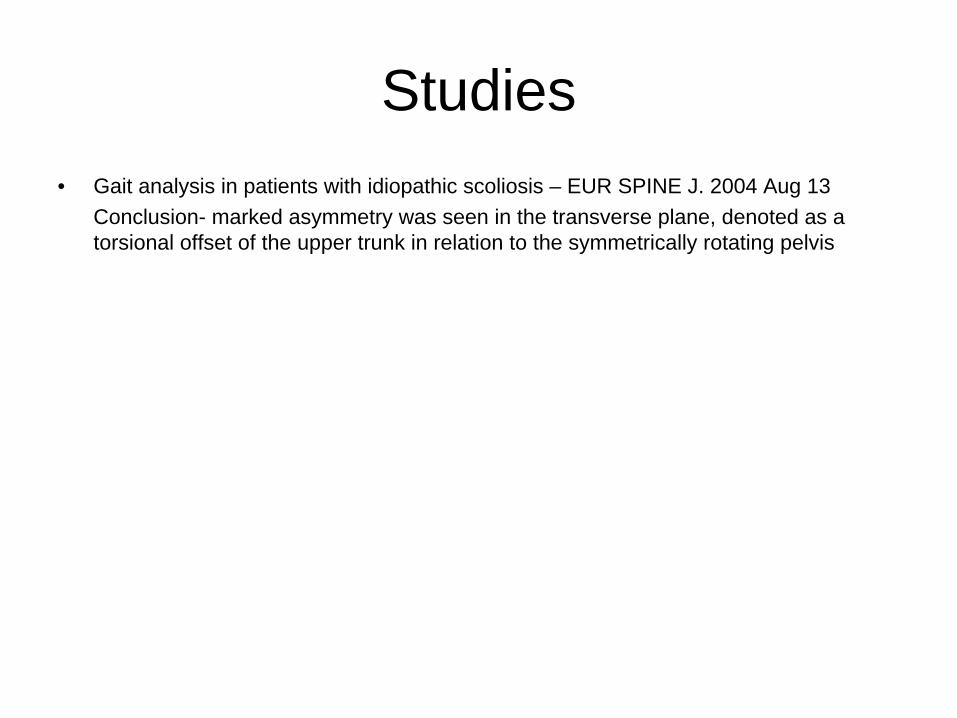
Studies• Gait analysis in patients with idiopathic scoliosis – EUR SPINE J. 2004 Aug 13
Conclusion- marked asymmetry was seen in the transverse plane, denoted as a torsional offset of the upper trunk in relation to the symmetrically rotating pelvis
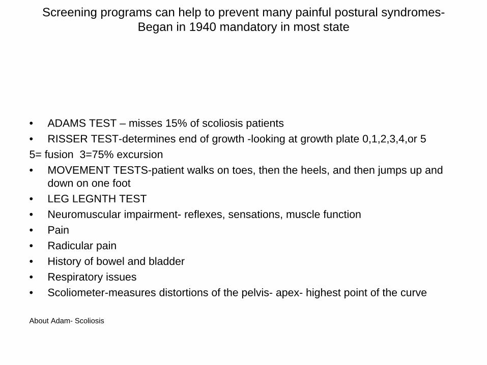
Screening programs can help to prevent many painful postural syndromes- Began in 1940 mandatory in most state
• ADAMS TEST – misses 15% of scoliosis patients• RISSER TEST-determines end of growth -looking at growth plate 0,1,2,3,4,or 55= fusion 3=75% excursion• MOVEMENT TESTS-patient walks on toes, then the heels, and then jumps up and
down on one foot• LEG LEGNTH TEST• Neuromuscular impairment- reflexes, sensations, muscle function• Pain• Radicular pain• History of bowel and bladder• Respiratory issues• Scoliometer-measures distortions of the pelvis- apex- highest point of the curve
About Adam- Scoliosis

Clinical evaluation
• Horizontal plane- rotation of shoulders, rotation of thorax, rotation of pelvis
• Tilt in pelvis• Frontal Plane-tilt of the
shoulders, tilt of the thorax and tilt of the pelvis
• Prominence- levels and values• Pain• Scapula deformity• Clinical Flat Back• Rib asymmetry• Heel liftsSpincor System-Training manual
• Look for :• A tilted head that does not line up with
shoulders• A protruding shoulder blade• An uneven neckline• Leaning more to one side than the other• One hip or shoulder that is higher than the
other

Decision to Treat or Wait• Monitor the curve if less than 20 degrees• Curves greater than 25 degrees or those that progress while being monitored may
require treatment• The older the child the less likely it is the curve will progress• Girls have higher risk than boys• Thoracic curves progress more than thoracolumbar• Children in poor health may suffer more from stressful scoliosis than other children
About Adam- Scoliosis

Radiographic examinationX-rays- in standing-gravity of spine- PA and lateral views
reveals degree and severity of curve, determines whether skeletal growth has reached maturity, differentiates between structural and nonstructural
MRI’s – identify spinal cord and brain stem abnormalities, helpful in planning fusion levels, will show degenerative disc disease
Ero and Blessey- Adult Scoliosis: Evaluation and Treatment
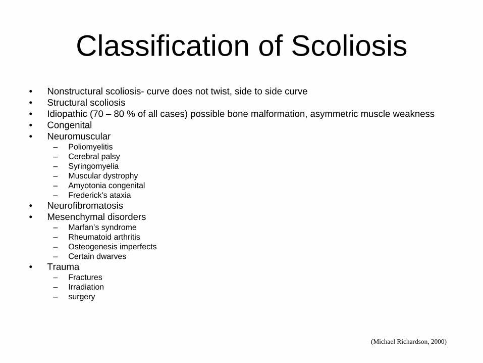
Classification of Scoliosis• Nonstructural scoliosis- curve does not twist, side to side curve• Structural scoliosis• Idiopathic (70 – 80 % of all cases) possible bone malformation, asymmetric muscle weakness • Congenital• Neuromuscular
– Poliomyelitis– Cerebral palsy– Syringomyelia– Muscular dystrophy– Amyotonia congenital– Frederick's ataxia
• Neurofibromatosis• Mesenchymal disorders
– Marfan’s syndrome– Rheumatoid arthritis– Osteogenesis imperfects– Certain dwarves
• Trauma– Fractures– Irradiation– surgery
(Michael Richardson, 2000)

Congenital Scoliosis
• Caused by inborn spinal deformities that may result in absent fused vertebrae
• Kidney problems-especially one kidney usually coincide with problem
• Evident age 2 or at ages 8-13 as the spine begins to grow quickly
• Early surgical treatment before age 5 may be important• Bracing is rarely used for either type of congenital curves
Schommer- National Scoliosis Foundation About Adam- Scoliosis

Nonstructural Scoliosis
• Unequal leg length• Muscle spasms
About Adam-Scoliosis
Idiopathic Scoliosis
Classified based on age presentation
• Juvenile: Up to years old• Adolescent: Ten years old through teen years• Infantile: Up to 3 years old
About Adam- Scoliosis
Idiopathic Scoliosis-Possible Causes
• Dietary issues-calcium is lacking causing softening of the bones
• Nerve and muscle abnormalities• Central mechanisms of the ear• Fluid around the spinal cord flows asymmetrically
(Cedars-Sinai Institute for Spinal Disorders- Los Angeles)
• Abnormalities in collagen-high level of enzymes in the disks, enzymes repair and remodel collagen
• High archesAbout Adam- ScoliosisPashman-Scoliosis-Frequently Asked Questions

Scoliosis Facts• 80% of cases are idiopathic • Most of the curves are right thoracic between T-4 and T-12• The incidence of adult scoliosis is estimated to be between 4% to 8% • Scoliosis in adults may be a result of adolescent idiopathic scoliosis or arise in adult life secondary
to osteoporosis, osteomalacia, spinal stenosis and degenerative changes
• Spirometric pulmonary tests are usually unaffected in the idiopathic scoliosis patients until the curve exceeds 60 to 65 degrees and the mortality is unaffected until the curve exceeds 90 to 100 degrees
• Respiratory distress is greater in the neuromuscular group of patients • Pain is the most common reason for patients to seek medical treatment• Affects 2-3 percent of population• Commonly diagnosed in children 10-15 years, 10 percent have some degree of scoliosis, less
than 1 percent actually need treatment • Location of structural curve is defined by the location of apical vertebra• May be evident in Young athletes- 2-24 percent (loosening of joints, delay in puberty onset,
uneven load on spine, stresses on growing spine)Schommer- National Scoliosis FoundationEro and Blessey- Adult Scoliosis: Evaluation and TreatmentAbout Adam-Scoliosis

ScoliosisScoliosis• Lateral curvature of the spine• Etiology, severity, age of onset, and progression vary• Usually develops in childhood• Can cause structural abnormalities of the pelvis,
vertebrae, and thoracic cage• Can occur in the cervical, thoracic or lumbar regions of
the spine• If untreated and undetected, can cause severe
deformity, drastically affecting appearance, and possible shorten life expectancy
• Early identification and treatment are key to prevention of severe deformity
(Carolyn Kisner, 1990)
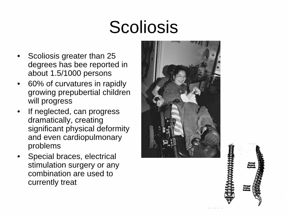
Scoliosis • Scoliosis greater than 25
degrees has bee reported in about 1.5/1000 persons
• 60% of curvatures in rapidly growing prepubertial children will progress
• If neglected, can progress dramatically, creating significant physical deformity and even cardiopulmonary problems
• Special braces, electrical stimulation surgery or any combination are used to currently treat

Structural Scoliosis• Irreversible lateral curvature of the
spine with fixed rotation of the vertebrae
– Vertebral bodies rotate toward the convex side of the curve
– Spinous processes rotate away from the convex side
– Curve increases, amount of rotation increases
• A rib hump occurs on the convex side of the curve caused by the rotation of the vertebrae and the rib cage
– Compression of the ribs on the concave side
– Separation of the ribs on the convex side
– Prominence of the ribs and scapula posteriorly on the convex side

As Scoliosis Progresses• The vertebrae and
spinous processes around the major curve rotate toward the concavity of the curve
• On concave side, the ribs are close together – on convex side the ribs are widely spread

Severity of Scoliosis
• Severity of the lateral curve determines the rotation of the vertebrae more severe = greater rotation
• Severity of the curve, the greater the impact and secondary changes in the cardiopulmonary systems – Decreased vital capacity and total lung
capacity– Hypertrophy of the right ventricle and atrium
from pulmonary hypertension

Facts About Scoliosis• *Think – severe scoliosis can lead to
contractures and severe body deformity• Our body is a connective chain of muscles • With scoliosis, the spine will rotate causing the
muscles to shorten and tighten • This will in turn change posture and body
alignment causing other muscles in other parts of the body to shorten or get tight
• Legs may be windswept – always classify windswept to where knees are pointing (ex. knees are pointing to the right, state legs are windswept right)

Nonstructural (functional) Scoliosis
• Correction of the lateral curve is possible by– Forward or side bending– Positional changes and alignment– Muscle contraction
• Characterized by– Asymmetric shoulder level– Prominence of the scapula on the side of the
convexity– Protrusion of the hip on one side– Pelvic obliquity– Increased lumbar lordosis

Classification of Scoliosis• S-Curve – most common curve seen in
idiopathic scoliosis– Right thoracic left lumbar curve– Structural changes in the vertebrae of the major curve
• Description of curves – direction of the curve identified by the convexity (ex. right thoracic scoliosis = convexity of the curve on client’s right concavity on the client’s left)
• Apex of curve – vertebra that is the greatest distance from the midline of the spine = apical vertebra

Classification of Scoliosis
• The Cobb method of measurement of scoliosis.
• Line drawn perpendicular to the upper margin of the vertebra that inclines most toward the concavity
Classification of Severity of the Curvature
– Mild scoliosis• Curves of less than 20 degrees
– Moderate scoliosis• Curves from 20 to 40 or 50 degrees• Associated with early structural changes in the vertebrae and rib
cage
– Severe scoliosis• Curves of 40 to 50 degrees or greater• Involves significant rotational deformity of the vertebrae and ribs• Curves of 40 degrees or more can cause pain and degenerative
joint disease (DJD) of the spine• Curves of 60 to 70 degrees or more can cause significant
cardiopulmonary changes and decreased life expectancy
(Carolyn Kiser, 1990)

Treatment of Scoliosis• Correction of curves greater
that 40 or 50 degrees are usually corrected by surgical intervention
• Electrical stimulation of the trunk muscles on the convex side of the curve have recently become treatments that are used for mild and moderate scoliosis
• Bracing, traction, and exercise are other methods that have shown to benefit individuals with scoliosis
(Carolyn Kisner, 19900)

BRACING and Research• Clinical research presented during the last
ten years shows• 1. Bracing is the only proven non-surgical
method of potentially successful treatment of adolescent idiopathic scoliosis
• 2. Bracing is most effective for curves between 20 and 40 degrees in growing children with Risser signs of 0, 1, 2 or 3
• 3. Bracing should apply forces to the spine such that significant curve correction (30 %) occurs in the brace
• 4. Brace wearing time should be approximately 20 hours plus per day for maximum benefit
• 5. The brace program should continue with intensity until growth has ceased as indicated by no further height increase and a Risser 4 status
Winter- www.scoliosis-assoc.org
• Any current statements about the effects of brace compliance on outcome of treatment are purely speculative
• For optimal performance, bracing needs to be started early( >25 degrees and progressive) and must reduce the curves and maintain curve reduction (> 50 percent) throughout the duration of wear
• Role of bracing for idiopathic scoliosis is to arrest curve progression and yield a post brace curve that is of magnitude that will not progress as an adult
Gavin-www.scoliosis-assoc.org

SOSORTSociety of the International Society on Scoliosis
Orthopedic and rehabilitation Treatment
• The studies reveal that among participating SOSORT specialists there continues to be a strongly held and conflicting if not a contentious opinion regarding brace design and treatment.
• All agree that bracing should unload the growth plates of the apical vertebral bodies on the concavity
• Many clinicians seem to fit braces empirically rather than using “ curve – specific” biomechanical 3D models • Rigo, Weiss, Grivas,Maruyama,Kotwicki, and SOSORT members-
SOSORT Consensus paper on Brace Action: TLSO Biomechanics of Correction

WHAT BRACES ARE ON THE MARKET
The Milwaukee Brace- first modern brace designed for scoliosis -1975-Wisconsin
• -cervico-thoraco-lumbo-sacral-orthosis• Prescribed for curves high in the spine• Everything works together to keep the body
straight and to prevent progression of the curve while the patient is growing
TLSO Braces- low profile –made of plastic and are contoured to conform to patient’s bodyBoston Brace- Boston-1970
works by applying three-point pressure to the curve to prevent its progression
Forces lumbar area to flex, which pushes in the abdomen and flattens the posterior lumbar curve- pads place pressure on the curve and relief voids are located opposite the areas of pressure
• Charleston Bending Brace- 1979- worn at night –is molded to conform to the patient’s body while he or she is bent towards the convexity of the curve- over correcting the curve during 8 hours the brace is worn- recommended for curves of 20-35 degrees with the apex of the curve below the level of the shoulder blade-studies show no evidence of improved compliance the potential for a patient to wear a part time brace, especially while sleeping, rather than the usual full- time 22-23 hour regimen
• SPINCOR-next slide
Bracing for Adolescent Idiopathic scoliosis-National Scoliosis Foundation-www.scoliosis.org
Types of Braces for Scoliosis- www.iscoliosis.com

SPINECOR Brace
• First and only truly dynamic brace, which provides a progressive correction of idiopathic scoliosis
• Preserves normal body movement and growth and allows normal activities of daily living
• Can be worn under clothes• Shown to be effective at 2 year follow up with 450
plus patient pool- 7% of those patients maintained their stabilization of correction, which far exceeds rigid bracing
• X-rays done every six months on clients• uses adjustable bands and a cotton vest that
allows flexibility
Correct Scoliosis- Spinecor-www.correctscoliosis.comS li i li t li i i li t
• Designed by pediatric orthopedic surgeons- in Quebec Canada over the last 20 years
• Shown to be as effective or if not more as rigid braces in curves between 20 and 50 degrees
• Fitted on anyone over 5 years old• FDA approved and is covered for reimbursement by
most insurance companies• Don’t need to be referred to orthotist can see
chiropractor for fitting• Used for adults to help improve posture and
decrease pain • “Due to the elastic nature of the brace, there is a
struggle between the patients interpretation of normal posture and where the brace wants to keep the patients correct posture. This struggle promotes activation of the muscular system to keep it strong, it promotes neuromuscular re-education of the patient’s posture and it stresses the osseous system helping to stop the progression of bone deformity. This allows the patient to finish with a neuromuscular skeletal system that is retained and strong to allow the patient to maintain the correction”

Drawbacks of TLSO Braces• Don’t provide a lot of flexibility• Hot to wear• Hard to Hide under clothing• Produce atrophy of spinal
muscles • Result in the spine returning
back to the pre-treatment state and beyond
Correct Scoliosis- SpineCOR- www.correctscoliosis.comAbout Adam- Scoliosis
• Flattens ribs• Doesn’t help well with
breathing- reduce lung capacity by 20 percent
• Cause, mild, temporary changes in kidney function

SURGERY
• Based on Medical Criteria includes the degree of curvature, the skeletal maturity of the patient, and the progression of the curvature
Pashman-Scoliosis Frequently Asked Questions
• Can be corrected to 40% of the original size
• Surgical goal: Stabilize the spine and produce a fused spine that leaves the patient balanced
• For Adults- To stop progression and to improve quality of live without pain

Surgical Approaches
• Posterior Approach• Opening back of patient• Harrington Rod Surgery• Advantages- fewer complications, good
correction, fusion rates are good• Disadvantages- crankshaft phenomenon
• About Adam-Scoliosis
• Anterior Approach• Performs operation through chest wall• Incision in the chest, deflates the lung, and
removes a rib to reach the spine
• Advantage-low risk for lower-back injury• Disadvantage- Poor lung function• Hardware problems

Surgical Treatment in Adult Scoliosis
Surgery recommended for the following:
1. Curvatures over 50 degrees with persistent pain
2. Curvatures over 60 degrees- surgery is almost always recommended
3. Progressive mid and low back curve or low back curve with persistent pain
4. Reduced heart and lung function- not severe lung function or heart failure
• Prefer to operate on adults under 50 years
• Adults at higher risk than children for nerve damage, complications like pneumonia, infection, poor wound healing, and persistent pain
ScoliosisTreatment for Adult Scoliosis- Health Guide- New York Times-2009

Spinal Instrumentation
• Serves three purposes:1. Provides a stable, rigid
column that encourages bones to fuse after spinal- fusion surgery
2. Redirects stresses over wider area
3. Restores spine to its proper alignment
4. Davidson-Spinal Instrumentation-www.healthatoz.com
• Harrington Rods-simplest instrumentation to install-achieves 50 percent correction-loss of 10-25percent of correction over time
• Cotrel-Dubousset instrumentation is the most complex and risky
• Patients after surgery remain in body casts for about six months, and then wear a brace for another three to six months while the bone fusion solidifies
• Bone is removed from the hip and placed along side the area to be fused
• Crankshaft phenomenon- is a continued growth in the anterior front of the spine after a posterior fusion is performed in a young growing patient
Crankshaft Phenomena-www.scoliosisassociates.comAbout Adam-Scoliosis
Complications of all Procedures
• Bleeding• Postoperative pain• Infection• Nerve damage• Lung Function• Disk Degeneration• Gallstones• Intestinal obstructionAbout Adam- Scoliosis

Spine Problems in Previously Treated Scoliosis Patients
• Spinal Fusion Disease• Disk Degeneration and low back
pain• Height loss- growth takes place in
long bones• Lumbar flatback• Rotational trunk shift
About Adam- Scoliosis
Therapeutic Treatment Plan
• GOALS
Greater FUNCTION Independence
SafetyLife ExpectancyDiminished PainBetter Breathing

SCHROTH METHOD• In a typical scoliotic curve- back musculature pulls lower ribs
so that the lumbar abdominal region rotates laterally, downwards , and backwards
• Scrhoth method aims to reverse all the abdominal curvatures- addressing all three planes-sagittal, frontal, and transverse- “three dimensional” therapy
• Addresses the patients’ pelvis position, spinal elongation, and on derotation in all three planes
• Focuses on strengthening exercises tailored to the individual patient, breathing exercises, focuses on vertebral derotation, and increasing the patient’s vital capacity
• Deviations of the trunk to the side or backwards can only develop if the corresponding supportive muscles give way and become elongated
• Treatment must improve posture so that the body can regain its original vertical axis- This can happen by developing and training the corresponding muscle groups responsible for upright posture
• To restore muscular balance, those muscles that have grown longer must be shortened and those that have become shorter must be lengthened
• 1930 developed by Katherine Schroth
• Schroth increases vital capacity
• Reduces abnormal curves by over 30% and increases lung capacity
• Shevchuk-The Schroth Method

Physical Therapy
• Decrease Pain• Improve the patients’
strength, flexibility, and Fitness
• Education on posture- practicing in front of mirror
• PT usually begins 3 months after surgery or when the fusion starts to consolidate
• After 12 months if fusion heals normal activities can start up agin
• When the spine is curved, other parts of the body compensate in order to keep the head straight arid the eyes- exercises should work on locating areas that are compromised and compensating and build up these muscles
Brady-www.scoliosis-assoc.org

The brain is calling to the muscles to move – similar to a telephone conversation between the brain and muscles.
All of a sudden there is a disconnection whether the cord was cut or a signal dropped.
Since this disconnection has occurred, the muscles aren’t able to move – however, with certain advances in medicine and unique techniques – we can learn to help the muscles and brain reconnect – visual, verbal, and tactile cuing will be of the utmost importance in this process of regeneration.
The Brain Analogy
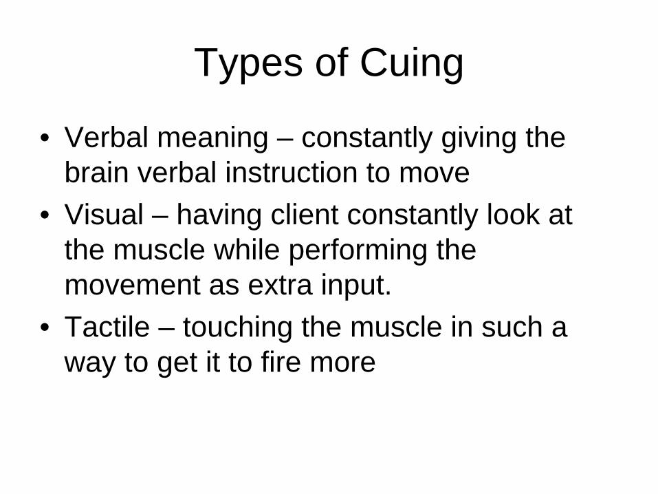
Types of Cuing
• Verbal meaning – constantly giving the brain verbal instruction to move
• Visual – having client constantly look at the muscle while performing the movement as extra input.
• Tactile – touching the muscle in such a way to get it to fire more

Types of Cuing

Handling the Client• Our bodies reflect postural
habits, physical demands, and injuries
• Our bodies adjust to problems• Improper body alignment can
cause progressive damage to muscles, bones, joints, and nerves
• Problems that are neglected can affect more body parts and increase pain
• To provide effective therapeutic intervention, body symmetry and functional mechanics must be considered
(Michele Nicosia, 2004)

Handling the Client
• Key points– Voice – low / soft /
gentle– Hands – soft / gentle– Therapists have
natural tendency toward heavy- handedness
(Candice Strack, 2000)

Palpation
• Similar to principles designed by Cranial Sacral therapy
• Lighter forces produce better results when using Cranial Sacral Therapy
• Palpation – the art of using touch to examine the body– Explore structures beneath the skin– Body fluids can be sensed– Motion of one bone in relation to another
(Candice Strack, 2000)

Palpation • Heavy palpatory force results in tightening of muscles and initiates
pain reflexes and body defends against palpator’s hands more Provides more information about defense mechanisms than underlying condition
• Nonintrusive palpation permits examination without evoking resistance– Allows the client and therapist to experience “melding”– Allows the therapist to absorb information through the practitioner’s
hand– Therapist accept any information that is received– Accept your experience as true– Goal of palpation – unobtrusive – Therapist must use lightest force possible in for palpation and treatment
(Candice Strack, 2000)

Handling the Client
• When placing hands on client palpation should be similar to– The force needed to
raise a nickel with one finger
– The force used when you comfortably place pressure on closed eye-lids
(Candice Strack, 2000)

PositioningPositioning• Thorough evaluations start with the feet
and end with the head considering alignment and body mechanics
• Pelvic obliquities produce knee and hip stresses– Functional scoliosis– Paraspinal muscle imbalance– Stress headaches
(Michele Nicosia, 2004)

Positioning
– Weak abdominal muscles, pelvic obliquities, and sacroiliac dysfunctions contribute to multiple painful conditions such as osteoarthritis
– Dysfunctional mechanics cause wear and tear and improper alignment
– Prolonged sitting ->forward head and shoulders, tight cervical thoracic muscles
(Michele Nicosia, 2004)

Types of Equipment

Positioning
• Information needed prior to equipment use– Diagnosis– Orthopedic information– Muscle tone– Abnormal patterns– Contractures or potential deformities– Asymmetry– Sensory abnormalities– Purpose of position or equipment– Consistency of treatment goals with purpose
of position or equipment (Donna Cech, 1993)

Guidelines for Positioning• Guideline for position
1. Posture is number 1 – always make sure head, shoulders, hips, knees, and feet are in correct alignment – should be able to draw a straight line from these points
2. If the client has scoliosis, one side shortened, position in sideline to opposite side of tight side *Example – right side tight – position in left sideline to open right side – work on opening curve, manual stretching
3. Position and allow client to experience a functional activity while in this position
4. Use pillows, sandbags, etc. to get body in proper alignment

Guidelines for Positioning5. Make sure arms are in front of body and hands are weight bearing6. Try to get tight hands to open using towel rolls or pipe cleaners7. Break up tonal patterns *Example – increased extensor tone in legs –
position supine with legs over bolster8. Incorporate range of motion, acupressure, and sensory integration while
positioned 9. Positions can include
• Supine• Prone• Sideline• Standing• Supine – knees flexed over bolster• Hands/knees over bolster• New positioning equipment available• Now made
– Adaptable – Can clean– Comfortable– Many parts to create different positions depending on clients disability

Guidelines for Positioning10 Also think functional movement
– Positioning equipment– Scooter board– Rolling doughnut– Rocking chair
11 No more than 45 – 60 minutes in one position. Check client regularly – too long will lead to fatigue/pressure build up

Benefits of Good Positioning
12 Will improve breathing– Benefits of breathing
• Releases stress/tension – relaxed breathing – diaphragmic breathing
• Builds energy and endurance• Contributes to emotional mastery• Prevents and heals physical problems• Contributes to graceful aging• Manage pain• Enhance mental concentration and physical performance• *Breathing is the path to the most essential of human
experience leading to love
(Gary Hendricks, 1995)

Positioning

Benefits of Good Positioning• Each position provides
the same sort of benefit *Example – SUPINESUPINE – work on midline orientation

SUPINE

Benefits of Good Positioning
• Side-lying– If client has tight right side
because of scoliosis, position them in left side- lying position to open right side

Benefits of Good Positioning
• Prone benefits– Promote shoulder
girdle stability – Trunk extension– Good head control– Can stretch
hamstrings– Weight bearing
through arms– Able to extend neck

Handling the Client
• Standing - will discuss benefits later

Benefits of Good Positioning• Sitting – fitting a wheelchair
– Entire length of the femur should be supported up to ½ inch behind the knee
– Seat belt should be at a 45° angle to the chair – promotes hip flexion and a slight anterior tilt
– Feet must be supported and the ankles kept in a neutral or a dorsiflexion position
– Arms should have opportunity for support and weight bearing provided by lap tray
– Position as upright and symmetrical as possible– Chair tilt may be added to help with poor head control– *Always make sure client is seated correctly prior to
making any other adjustments in wheelchair – always use lap tray

HOW TO KEEP THESE POSITIONING PROGRAMS RUNNING SMOOTHLY
• Visual Aids• Take pictures before and after correction• Use the number and color system• Make charts to record data and keep data
posted on wall• Continual Staff training

Incorporating positioning into assessment and treatment
• Make sure you always place client prior to working with them in good anatomical alignment
• Remember positioning prior to function• Watch and record if you change position
will greater functionality occur

CONCLUSION

Bibliography1. Adler, Lezlie. “Integrating NDT SI and Motor Control Perspectives in
Pediatrics.” Educational Resources, Inc. Phoenix, 2000. 2. Becker, Kirsten. “To Stand or Not to Stand – The Correct Stander can
Improve a Patient’s Physical and Psychological Well-being.” Rehab Management. March 2005. 28-30.
3. Berkus, Rusty. Life is a Gift. California: Red Rose Press, 1982.4. Brown, Jackson. Live and Learn and Pass it on. Tennessee: Rutledge
Hill Press, 19955. Byers, Dwight. Better Health with Foot Reflexology: The Original Ingham
Method. Florida: Ingham Publishing, Inc., 1993.6. Cech, Donna. “Pediatric PT.” Finch University of Health Sciences,
Chicago Medical School. Illinois, 1993.7. Carroll, Lee and Tober, Jan. The Indigo Children. California: Hay
House, Inc., 1999.8. Department of Economic Security, Arizona. Early Intervention Program
Standards of Practice Training – “ Child Development and Developmental Risk and Variation.” Arizona, 2002-2003.

Bibliography9. The Diagram Group. The Brain: A User’s Manual. New York: Berkley
Books, 1983.10 Doman, Glenn. What to do About Your Brain – injured child. New York:
Avery Publishing Group, 1994.11 Guilett, Sharon. “More that a Chair.” The Interdisciplinary Journal of
Rehabilitation 17.2(2004).12 Guyton, Arthur. Human Physiology and Mechanisms of Disease.
Philadelphia: W.B. Saunders Company, 1992.13 Hendricks, Gary, PHD. Conscious Breathing. New York. 1995.14 Horan, Paula. Empowerment Through Reikji. The Path to Personal and
Global Transformation. Wisconsin: Lotus Light Publications, 1992.15 Jora, Jurgen. “Foot Reflexology: A Visual Guide for Self Treatment.”
New York: St. Martin’s Press, 1991.16 Kisner, Carolyn and Coltz, Lynn. Therapeutic Exercise. Philadelphia: F.
A. Davis, 1990.17 McCormick, Sheila. “Sensory Integrations Sensors.” Phoenix, 20 Nov.
2002.

Bibliography18 Napolean, Christina. Doctor of Naprapathy. Phoenix, AZ.19 Nicosia, Michele B. “The Pain Fighters.” The Interdisciplinary Journal of
Rehabilitation 17.2 (2004).20 Paleg, Ginny. “Gateway to Gait.” The Interdisciplinary Journal of
Rehabilitation 17.2 (2004).21 Paleg, Ginny. “ on Mobility – Proof Positive – Clinicians Should
Advocate for Access to Clinically Proven Durable Medical Devices.” Advance Editions for Directors in Rehabilitation 15 (2006). 21.
22 Paramananda, Swani. Book of Daily Thought and Prayers. Massachusetts, 1977.
23 Purtilo, Ruth. Health Professional and Patient Interaction. Philadelphia: W. B. Saunders Company, 1990.
24 Richardson, Michael. Scoliosis. University of Washington Department of Radiology (2000). 1-9.
25 Roman, Christine. “ Environmental Considerations Workshop Material. West Virginia.

Bibliography26 Rosen, Lauren. “Stand Up for Your Patients – With the Help of Standers,
Patients Can Improve Functions, Reduce Spasticity, and Clear the Way for ADL.” Physical Therapy Products. Sept (2003). 28-29.
27 Ryan, Jo. Inspiring Lessons about Life. London: Tangent Publications, 2004.
28 Strack, Candice. “CranioSacral Therapy I Course.” Upledger Institute, Inc. Phoenix. 15 June 2000.
29 Tappert, Susan. “Therapeutic Exercise II.” Finch University of Health Sciences, Chicago Medical School, Illinois, 1993.
30 Tarpan, Frances. Healing Massage Techniques. New Jersey: Appleton and Lange, 1988.
31 Umphred, Darcy. An Integrated Approach to Adult Neurological Deficits wit Head Injured Tonal Clients. Pacific Coast Services. Illinois, 3-5 May 1996.
32 Umphred, Darcy. “Neurological Rehabilitation.” St. Louis: The C. V. Mosby Company, 1990.
33 Website Reference – www.pacificrehabinc.com. “Gait Trainers.” September 2004.

A Look at Posture and Scoliosis - Flexibility and Function with the Special Needs Population
By Michelle Lindsey
BIBLIOGRAPHY
1. Aruin, AS, The effect of asymmetry of posture on anticipatory postural adjustments, Neuroscience Letters, Volume 401, Issues 1-2, 19 June 2006, Pages 150-153
2. About: A.D.A.M. Healthcare Center – Scoliosis. http://adam.about.com/reports/0000683. Becker, Kirsten. To Stand or not to Stand. The correct stander can improve a Patient's
Physical and Psychological Well-Being, Rehab Management. March 2005. 28-30.4. Bruyneel, AV, Chavet, P, Bollini,G, Allard, P, Berton, E, Mesure, S, Lateral steps
reveal adaptive biomechanical strategies in adolescent idiopathic scoliosis, Annales de Réadaptation et de Médecine Physique, Volume 51, Issue 8, November 2008, Pages 630-641
5. Burwell, RG, Cole, AA. Pathogenesis of idiopathic scoliosis. The Nottingham concept. www.ncbi.nlm.nih.gov/pubmed/1456018?ordinalpos=1&itool=EntrezSystem2.PEntr
6. Burwell, RG, Cole, AA. Gait analysis in patients with idiopathic scoliosis. www.ncbi.nlm.nih.gov/pubmed/1456018?ordinalpos=1&itool=EntrezSystem2.PEntr
7. Correct Scoliosis.Gilbert, Arizona – an accredited Spinecor Center.

www.correctscoliosis.com/health_pro.php8. Crankshaft Phenomena. Scoliosis and Spine Associates. 212 East 69th St. New York,
NY 10021 Tel: (212) 737-5540 Fax (212) 737-1369 www.scoliosisassociates.com/subject.php?pn=crankshaft-phenomena
9. Ero, Sunday U., MD, Blessey Peter, MD. Adult Scoliosis: Evaluation and Treatment. www.dcmsonline.org/jax-medicine/1999journals/june1999/scoliosis.htm
10. Fang, Lin, Parthasarathy, S, Taylor, SJ, Pucci, D, Hendrix, RW, Makhsous, M, Effect of Different Sitting Postures on Lung Capacity, Expiratory Flow, and Lumbar Lordosis, Archives of Physical Medicine and Rehabilitation, Volume 87, Issue 4, April 2006, Pages 504-509 http://cat.inist.fr/aModele=afficheN&cpsidt=17713118
11. Guillett, Sharon. More than a chair. The Interdisciplinary Journal of Rehabilitation 17.2 (2004).
12. Janovsky, Julie. Watch their backs: Signs of scoliosis in adolescence are clear if parents keep an eye out. Scottsdale Tribune east Valley Life D1. April3, 2007. [email protected]
13. Janssen, Beth. Physical therapy options for scoliosis. www.scoliosisrehabschrothusa.com.
14. Lateral steps revealadaptive biochemical strategies in adolescent idiopathic scoliosis. www.ncbi.nlm.nih.gov/pubmed/18597882
15. LeBauer, Aaron, Brtalik, Robert, Stowe, Kathrine, The effect of myofascial release (MFR) on an adult with idiopathic scoliosis, Journal of Bodywork and Movement Therapies, Volume 12, Issue 4, October 2008, Pages 356-363
16. Medtronic. Bone Morphogenetic Proteins. January 23, 2007. www.infusebonegraft.com/omp_bmp.html

17. Muntoni, F, Bushby, K, Manzur, AY, Muscular Dystrophy Campaign Funded Workshop on Management of Scoliosis in Duchenne Muscular Dystrophy 24 January 2005, London, UK, Neuromuscular Disorders, Volume 16, Issue 3, March 2006, Pages 210-219
18. National Scoliosis Foundation. Bracing for Adolescent Idiopathic Scoliosis.1-800- NSF-MYBACK (673-6922). [email protected] www.scoliosis.org/resources/medicalupdates/adolescentbracing.php
19. Nicosia, Michele 13. The Pain Fighters. The Interdisciplinary Journal of Rehabilitation 17.2 (2004).
20. Paleg, Ginny. Gateway to Gait. The Interdisciplinary Journal of Rehabilitation 17.2 (2004).
21. Paleg, Ginny. On Mobility - Proof Positive - Clinicians should advocate for Access to Clinically Proven Durable Medical Devices. Advance Edition for Directors in Rehabilitation 15 (2006). 21
22. Pivonka Family Chiropractic. 1355 South Higley Rd., Suite #102, Higley, AZ 85236. Phone (480) 892-0022, Fax (480) 892 5509.Interview.
23. Preuss, R, Fung, J, Musculature and biomechanics of the trunk in the maintenance of upright posture, Journal of Electromyography and Kinesiology, Volume 18, Issue 5, October 2008, Pages 815-828
24. Rosen Lauren. Stand Up for your Patients – With the help of standers, Patients can improve Functions, reduce Spasticity and clear the way for AOL. Physical Therapy Products. Sep (2003). 28-29
25. Scheck, Anne. Back on Track. Engeneering Treatment for Scoliosis. Today in PT. May 14, 2007. Pg 45-47. [email protected]

26. Scoliosis Association, INC. An international support and information organization. 800-800-0669 www.scoliosis-assoc.org
27. Scoliosis. http://adam.about.com/reports/Scoliosis.htm28. Scoliosis Frequently Asked Questions. www.espine.com/scoliosis-faq.htm29. Scoliosis Research Society (SRS). Thoracic Insufficiency Syndrome. VEPTR
Expansion Thoracoplasty for Thoracic Insufficiency. www.srs.org/professionals/education/tis/veptr.php
30. Scoliosis Specialists. West:888-5058850 Midwest:800-516-6272 Southeast:877-297- 0901 Northeast:800-516-6272. www.scoliosisspecialsts.com/
31. Schroth Method. Exercises for Scoliosis.www.schrothmethod.com/about.html www.schrothmethod.com/studies.html
32. Sosort consensus paper on brace action: TLSO biomechanics of correction (investigating the rationale for force vector selection). www.pubmedcentral.nih.gov/articlerender.fcgi?tool=pubmed&pubmedid=16857045
33. Spinal Instrumentation. www.healthatoz.com/healthatoz/Atoz/common/standard/transform.jsp?requestURI=/
34. The New York Times Health Guide, January 30, 2009. Treatment for Adult Scoliosis. http://health.nytimes.com/health/guides/disease/scoliosis/treatment-for-adult- scoliosis.html
35. Types of Braces for Scoliosis. www.iscoliosis.com/articles.html36. Vergara, M, Page, A, Sancho, JL, Analysis of lumbar flexion in sitting posture:
Location of lumbar vertebrae with relation to easily identifiable skin marks, International Journal of Industrial Ergonomics, Volume 36, Issue 11, November 2006, Pages 937-942
37. Wang, Jeffrey C., MD. UCLA Comprihensive Spine Center. BMP Improves Spinal Fusion Results. www.spineuniverse.com/displayarticle.php/article1711.html
38. Winter, Robert B., MD. Orthopedic Spine Surgeon, St. Paul, Minnesota. www.scoliosis-assoc.org Non-Surgical Management.
39. Wong, MS, Cheng, CY, Ng, BKW, Lam, TP, Sin, SW, Lee-Shum, LF, Chow, HK, Tam, YP, The effect of rigid versus flexible spinal orthosis on the gait pattern of patients with adolescent idiopathic scoliosis, Gait & Posture, Volume 27, Issue 2, February 2008, Pages 189-195


Michelle Lindsey, PT, MBA, CPT is a physical therapist and the owner of Rising Star Therapy Specialist, LLC. Michelle Lindsey received her undergraduate degree in
Kinesiology at Indiana University. She then attended The Finch University of Health Sciences: Chicago Medical School in Chicago, Illinois where she received her
bachelor’s degree in Physical Therapy. She also received her Masters in Business Administration Health Care Management at University of Phoenix. Michelle has been
practicing physical therapy for twelve years. Michelle also has experience in many facets of the fitness profession. She was a competitive gold medal figure skater for 16 years, an elite marathon runner where she was in the 1997 United States Maccabiah Open Track and Field Team, a triathlete, a professional speed skater and has also
competed as a figure model in many competitions. She has received a special recognition award from the United States Tennis Association in 2001 for coaching and directing a team for the Special Olympics. Additionally Michelle has delivered many in- services to school; day programs, group homes, and state operate services on the topic
she will be speaking about.
Michelle views her clients as unique individuals with varied skills and abilities. Her goal is to encourage them to develop to their fullest potential in a comfortable,
compassionate, and respectful environment that promotes and maintains their health, fitness, and quality of life. By putting faith in her client’s abilities, she strives to help them
believe in themselves, inspires them to extend beyond their best efforts, and teaches them how to remain motivated and focused. She stresses these and other key lessons
to ensure that her clients have a lifetime of success and happiness.


![The Open Orthopaedics Journal - benthamopen.com · Fig. (2). Scoliosis brace examples: past to present: From left, Abbott 1910, Milwaukee Brace, ... balanced posture [5]. ... group](https://static.fdocuments.net/doc/165x107/5b8a042a7f8b9aa81a8da80f/the-open-orthopaedics-journal-fig-2-scoliosis-brace-examples-past-to.jpg)