A level Biology Homework Book Cell Structure and Division
47
Page 1 of 47 St Mary’s Science Department A level Biology Homework Book Cell Structure and Division Name: ________________________ Class: ________________________ Teacher : ________________________ Question Date given Date due Mark 1 2 3 4 5 6 7 8 9 10 11 12 13 14 15 16 Total Score Grade
Transcript of A level Biology Homework Book Cell Structure and Division

Page 1 of 47
St Mary’s Science Department
A level Biology
Homework Book
Cell Structure and Division
Name: ________________________
Class: ________________________
Teacher : ________________________
Question Date given Date due Mark
1
2
3
4
5
6
7
8
9
10
11
12
13
14
15
16
Total Score Grade

Page 2 of 47
Q1. A scientist examined the structure of mustard plant leaves. He viewed temporary mounts of leaf tissues with an optical microscope. The figure below shows a drawing of typical results.
(a) Describe how temporary mounts are made.
___________________________________________________________________
___________________________________________________________________
_____________________________________________________________________________________________
___________________________________________________________________
(2)
(b) Calculate the distance in micrometres between G and H on the leaf.
Answer = ____________________ µm
(2)
(c) Describe how the scientist could have used the temporary mounts of leaves to determine the mean number of chloroplasts in mesophyll cells of a leaf.
___________________________________________________________________
___________________________________________________________________
___________________________________________________________________
(3)
(Total 7 marks)
Page 3 of 47
Feedback Mark (out of) Score
Skill Strength Could improve
Knowledge
Understanding
Evaluation
Application
Calculation
Data analysis
How science works
To improve
Student response

Page 4 of 47
Q2. A bacterium is shown in the diagram.
(a) Calculate the magnification of the image.
Magnification = _________________
(1)
(b) Complete the table to show the features of a bacterium and a virus.
Put a tick (✔) in the box if the feature is shown.
Surface Bacterium Virus
Cell-surface membrane
Nucleus
Cytoplasm
Capsid
(2)
(c) DNA and RNA can be found in bacteria.
Give two ways in which the nucleotides in DNA are different from the nucleotides in RNA.
1. _________________________________________________________________
2. _________________________________________________________________

Page 5 of 47
(2)
(Total 5 marks)
Feedback Mark (out of) Score
Skill Strength Could improve
Knowledge
Understanding
Evaluation
Application
Calculation
Data analysis
How science works
To improve
Student response

Page 6 of 47
Q3. Read the following passage.
In a human, there are over 200 different types of cell clearly distinguishable from each other. What is more, many of these types include a number of different varieties. White blood cells, Wfor example, include lymphocytes and granulocytes.
5
Although different animal cells have many features in common, each type has adaptations. associated with its function in the organism. As an example, most cells contain the same organelles, but the number may differ from one type of cell to another. Muscle cells contain many mitochondria, while enzyme-secreting cells from salivary glands have particularly large amounts of rough endoplasmic reticulum.
10
The number of a particular kind of organelle may change during the life of the cell. An example of this change is provided by cells in the tail of a tadpole. As a tadpole matures into a frog, its tail is gradually absorbed until it disappears completely. Absorption is associated with an increase in the number of lysosomes in the cells of the tail.
Use information from the passage and your own knowledge to answer the following questions.
(a) Explain the link between.
(i) mitochondria and muscle cells (lines 6 - 7);
______________________________________________________________
______________________________________________________________
______________________________________________________________
______________________________________________________________
______________________________________________________________
(3)
(ii) rough endoplasmic reticulum and enzyme-secreting cells from salivary glands (lines 7 - 8).
______________________________________________________________
______________________________________________________________
______________________________________________________________
______________________________________________________________
(2)
(b) Use information in the passage to explain how a tadpole’s tail is absorbed as a tadpole changes into a frog.
___________________________________________________________________
___________________________________________________________________
______________________________________________________________________________________________________________________________________
(2)

Page 7 of 47
(c) Starting with some lettuce leaves, describe how you would obtain a sample of undamaged chloroplasts. Use your knowledge of cell fractionation and ultracentrifugation to answer this question.
___________________________________________________________________
___________________________________________________________________
___________________________________________________________________
___________________________________________________________________
___________________________________________________________________
___________________________________________________________________
___________________________________________________________________
___________________________________________________________________
___________________________________________________________________
___________________________________________________________________
(6)
(Total 13 marks)
Feedback Mark (out of) Score
Skill Strength Could improve
Knowledge
Understanding
Evaluation
Application
Calculation
Data analysis
How science works
To improve
Student response

Page 8 of 47
Q4. A stomach ulcer is caused by damage to the cells of the stomach lining. People with stomach ulcers often have the bacterium Helicobacter pylori in their stomachs.
A group of scientists was interested in trying to determine how infection by H. pylori results in the formation of stomach ulcers.
The scientists grew different strains of H. pylori in liquid culture.
The table below shows the substances released by each of these strains.
H. pylori strain Substances released by the H. pylori cells
Toxin Enzyme that neutralises acid
A
B
C
The scientists centrifuged the cultures of each strain to obtain cell-free liquids. They added each liquid to a culture of human cells. They then recorded the amount of damage to the human cells.
Their results are shown below. The error bars show ± 1 standard deviation.
(a) Describe and explain how centrifuging the culture allowed the scientists to obtain a cell-free liquid.
___________________________________________________________________
___________________________________________________________________
___________________________________________________________________

Page 9 of 47
___________________________________________________________________
___________________________________________________________________
___________________________________________________________________
[Extra space] _______________________________________________________
___________________________________________________________________
___________________________________________________________________
(3)
(b) The scientists measured cell damage by measuring the activity of lysosomes. Give one function of lysosomes.
___________________________________________________________________
___________________________________________________________________
___________________________________________________________________
(1)
(c) H. pylori cells produce an enzyme that neutralises acid. Suggest one advantage to the H. pylori of producing this enzyme.
___________________________________________________________________
___________________________________________________________________
___________________________________________________________________
___________________________________________________________________
___________________________________________________________________
(2)
(d) What do these data suggest about the damage caused to human cells by the toxin and by the enzyme that neutralises acid? Explain your answer.
___________________________________________________________________
___________________________________________________________________
___________________________________________________________________
___________________________________________________________________
___________________________________________________________________
___________________________________________________________________
[Extra space] _______________________________________________________
___________________________________________________________________
___________________________________________________________________

Page 10 of 47
(3)
(e) The scientists carried out a further investigation. They treated the liquid from strain A with a protein-digesting enzyme before adding it to a culture of human cells. No cell damage was recorded. Suggest why there was no damage to the cells.
___________________________________________________________________
___________________________________________________________________
___________________________________________________________________
___________________________________________________________________
___________________________________________________________________
___________________________________________________________________
[Extra space] _______________________________________________________
___________________________________________________________________
___________________________________________________________________
(3)
(Total 12 marks)
Feedback Mark (out of) Score
Skill Strength Could improve
Knowledge
Understanding
Evaluation
Application
Calculation
Data analysis
How science works
To improve
Student response

Page 11 of 47
Q5. The diagram shows a eukaryotic cell.
(a) Complete the table by giving the letter labelling the organelle that matches the function.
Function of organelle Letter
Protein synthesis
Modifies protein (for example, adds carbohydrate to protein)
Aerobic respiration
(3)
(b) Use the scale bar in the diagram above to calculate the magnification of the drawing. Show your working.
Answer = ____________________
(2)
(Total 5 marks)

Page 12 of 47
Feedback Mark (out of) Score
Skill Strength Could improve
Knowledge
Understanding
Evaluation
Application
Calculation
Data analysis
How science works
To improve
Student response

Page 13 of 47
Q6. The photograph shows part of the cytoplasm of a cell.
(a) (i) Organelle X is a mitochondrion.
What is the function of this organelle?
______________________________________________________________
______________________________________________________________
(1)
(ii) Name organelle Y.
______________________________________________________________
(1)
(b) This photograph was taken using a transmission electron microscope. The structure of the organelles visible in the photograph could not have been seen using an optical(light) microscope. Explain why.
___________________________________________________________________
___________________________________________________________________
___________________________________________________________________
___________________________________________________________________
___________________________________________________________________
(2)
(Total 4 marks)

Page 14 of 47
Feedback Mark (out of) Score
Skill Strength Could improve
Knowledge
Understanding
Evaluation
Application
Calculation
Data analysis
How science works
To improve
Student response

Page 15 of 47
Q7. An amoeba is a single-celled, eukaryotic organism. Scientists used a transmission electron microscope to study an amoeba. The diagram shows its structure.
(a) (i) Name organelle Y.
______________________________________________________________
(1)
(ii) Name two other structures in the diagram which show that the amoeba is a eukaryotic cell.
1. ____________________________________________________________
2. ____________________________________________________________
(2)
(b) What is the function of organelle Z?
___________________________________________________________________
___________________________________________________________________
___________________________________________________________________
(1)
(c) The scientists used a transmission electron microscope to study the structure of the amoeba. Explain why.
(2)
(Total 6 marks)

Page 16 of 47
Feedback Mark (out of) Score
Skill Strength Could improve
Knowledge
Understanding
Evaluation
Application
Calculation
Data analysis
How science works
To improve
Student response
Page 17 of 47
Q8. (a) Describe how you could use cell fractionation to isolate chloroplasts from leaf
tissue.
___________________________________________________________________
___________________________________________________________________
___________________________________________________________________
___________________________________________________________________
___________________________________________________________________
___________________________________________________________________
(Extra space) _______________________________________________________
___________________________________________________________________
(3)
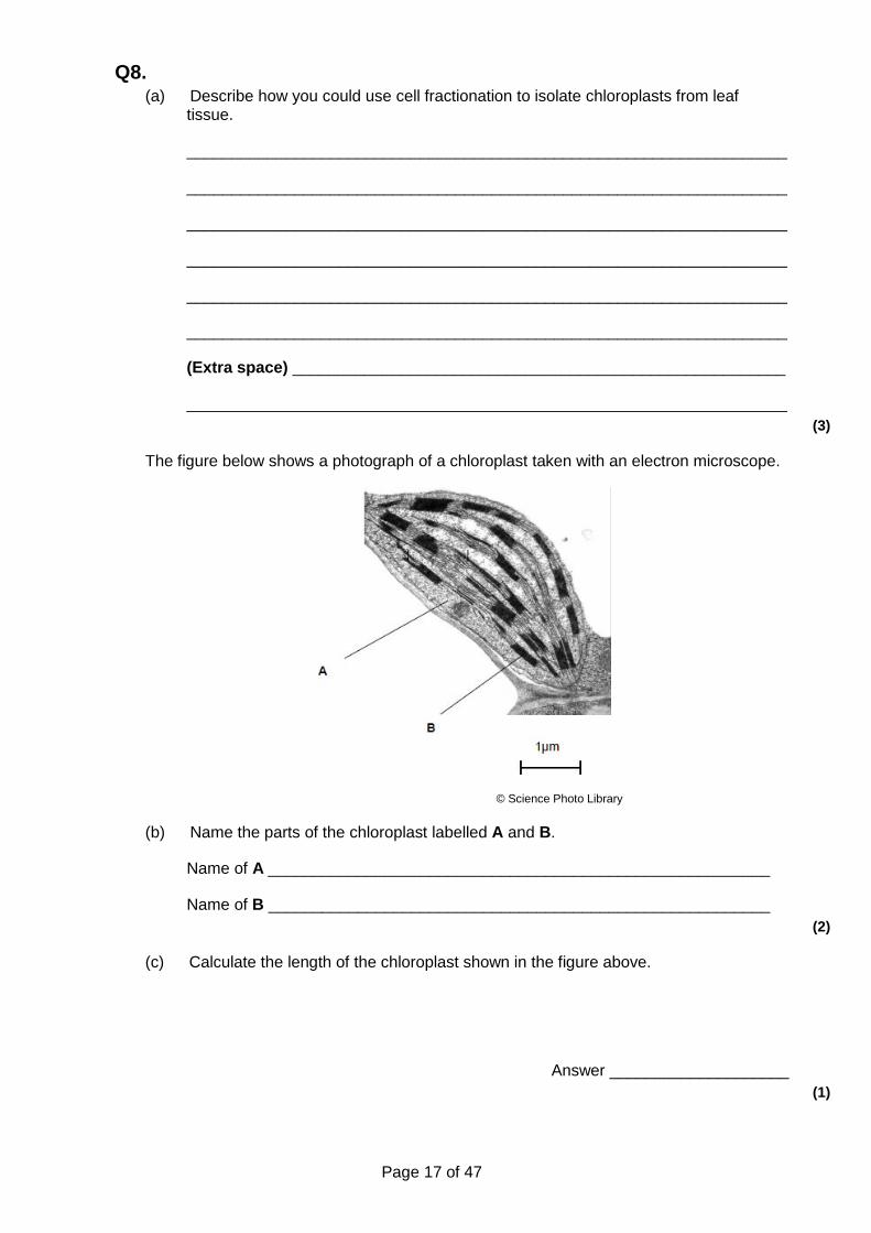
The figure below shows a photograph of a chloroplast taken with an electron microscope.
© Science Photo Library
(b) Name the parts of the chloroplast labelled A and B.
Name of A ________________________________________________________
Name of B ________________________________________________________
(2)
(c) Calculate the length of the chloroplast shown in the figure above.
Answer ____________________
(1)

Page 18 of 47
(d) Name two structures in a eukaryotic cell that cannot be identified using an optical microscope.
1. _________________________________________________________________
2. _________________________________________________________________
(1)
(Total 7 marks)
Feedback Mark (out of) Score
Skill Strength Could improve
Knowledge
Understanding
Evaluation
Application
Calculation
Data analysis
How science works
To improve
Student response

Page 19 of 47
Q9. Gorter and Grendel investigated the structure of the surface membrane of cells. They extracted the phospholipids from the surface membranes of red blood cells in 1 cm3 of blood and placed them in the apparatus shown in Figure 1.
Figure 1
The piston was pushed across the surface of the water until the phospholipid molecules were tightly packed into a single layer. The area covered by the phospholipid molecules was measured. This area was compared with the estimated surface area of the red blood cells from which phospholipids were extracted.
Gorter and Grendel obtained the data shown in the table.
Number of red blood cells per cm3 of blood 4.74 × 10⁹
Estimated mean surface area of one red blood cell 99.4 μm2
Surface area of membrane phospholipids extracted from 1cm3 of blood
0.92 m2
(a) Explain what these data suggest about the arrangement of phospholipids in the surface membranes of red blood cells. Support your explanation with suitable calculations.
Show your working.
___________________________________________________________________
___________________________________________________________________
___________________________________________________________________
___________________________________________________________________
___________________________________________________________________
___________________________________________________________________
(3)

Page 20 of 47
(b) Figure 2 shows a red blood cell and a white blood cell.
Red blood cell White blood cell
Figure 2
Explain why red blood cells were used in this investigation rather than white blood cells.
___________________________________________________________________
___________________________________________________________________
___________________________________________________________________
___________________________________________________________________
(2)
(Total 5 marks)
Feedback Mark (out of) Score
Skill Strength Could improve
Knowledge
Understanding
Evaluation
Application
Calculation
Data analysis
How science works
To improve
Student response

Page 21 of 47
Q10. The diagram shows a bacterium.
(a) Give the function of
(i) organelle X;
______________________________________________________________
(ii) organelle Y.
______________________________________________________________
(2)
(b) (i) Give two ways in which the structure of this bacterium is similar to the structure of a cell lining the human small intestine.
1. ____________________________________________________________
______________________________________________________________
2. ____________________________________________________________
______________________________________________________________
(2)
(ii) Give two ways in which the structure of this bacterium differs from the structure of a cell lining the human small intestine.
1. ____________________________________________________________
______________________________________________________________
2. ____________________________________________________________
______________________________________________________________
(2)
(Total 6 marks)

Page 22 of 47
Feedback Mark (out of) Score
Skill Strength Could improve
Knowledge
Understanding
Evaluation
Application
Calculation
Data analysis
How science works
To improve
Student response

Page 23 of 47
Q11. The electron micrograph shows part of a chloroplast.
(a) Name the parts labelled A and B and, for each, describe one role in the process of photosynthesis.
A Name ___________________________________________________________
Role ______________________________________________________________
B Name ___________________________________________________________
Role ______________________________________________________________
(4)
(b) (i) Name the main substance present in the part labelled C.
______________________________________________________________
(1)
(ii) How is this substance formed?
______________________________________________________________
______________________________________________________________
(1)
(Total 6 marks)

Page 24 of 47
Feedback Mark (out of) Score
Skill Strength Could improve
Knowledge
Understanding
Evaluation
Application
Calculation
Data analysis
How science works
To improve
Student response

Page 25 of 47
Q12. The diagram shows the structure of a plasma membrane.
(a) Name
protein A ___________________________________________________________
protein B ___________________________________________________________
molecule C _________________________________________________________
(3)
(b) Name two structures found in a prokaryotic cell that are not found in a human cell.
1. _________________________________________________________________
2. _________________________________________________________________
(2)
(Total 5 marks)

Page 26 of 47
Feedback Mark (out of) Score
Skill Strength Could improve
Knowledge
Understanding
Evaluation
Application
Calculation
Data analysis
How science works
To improve
Student response
Page 27 of 47
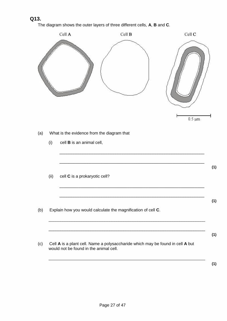
Q13. The diagram shows the outer layers of three different cells, A, B and C.
(a) What is the evidence from the diagram that
(i) cell B is an animal cell,
______________________________________________________________
______________________________________________________________
(1)
(ii) cell C is a prokaryotic cell?
______________________________________________________________
______________________________________________________________
(1)
(b) Explain how you would calculate the magnification of cell C.
___________________________________________________________________
___________________________________________________________________
(1)
(c) Cell A is a plant cell. Name a polysaccharide which may be found in cell A but would not be found in the animal cell.
___________________________________________________________________
(1)

Page 28 of 47
(d) Penicillin is an antibiotic. It prevents the formation of bacterial cell walls. As a result, bacterial cells that have been treated with penicillin swell and burst as water enters.
(i) Explain how water enters a bacterial cell.
______________________________________________________________
______________________________________________________________
______________________________________________________________
______________________________________________________________
(2)
(ii) Suggest why penicillin has no effect on plant cells.
______________________________________________________________
______________________________________________________________
(1)
(Total 7 marks)
Feedback Mark (out of) Score
Skill Strength Could improve
Knowledge
Understanding
Evaluation
Application
Calculation
Data analysis
How science works
To improve
Student response

Page 29 of 47
Q14. (a) Describe how DNA is replicated.
(6)
(b) The graph shows information about the movement of chromatids in a cell that has just started metaphase of mitosis.
(i) What was the duration of metaphase in this cell?
minutes
(1)
(ii) Use line X to calculate the duration of anaphase in this cell.
minutes
(1)
(iii) Complete line Y on the graph.
(2)
(c) A doctor investigated the number of cells in different stages of the cell cycle in two tissue samples, C and D. One tissue sample was taken from a cancerous tumour. The other was taken from non-cancerous tissue. The table shows his results.
Percentage of cells in each
stage of the cell cycle

Page 30 of 47
Stage of the cell cycle
Tissue sample C
Tissue sample D
Interphase 82 45
Prophase 4 16
Metaphase 5 18
Anaphase 5 12
Telophase 4 9
(i) In tissue sample C, one cell cycle took 24 hours. Use the data in the table to calculate the time in which these cells were in interphase during one cell cycle. Show your working.
Time cells in interphase ____________________ hours
(2)
(ii) Explain how the doctor could have recognised which cells were in interphase when looking at the tissue samples.
______________________________________________________________
______________________________________________________________
______________________________________________________________
(1)
(iii) Which tissue sample, C or D, was taken from a cancerous tumour? Use information in the table to explain your answer.
______________________________________________________________
______________________________________________________________
______________________________________________________________
______________________________________________________________
______________________________________________________________
(2)
(Total 15 marks)

Page 31 of 47
Feedback Mark (out of) Score
Skill Strength Could improve
Knowledge
Understanding
Evaluation
Application
Calculation
Data analysis
How science works
To improve
Student response

Page 32 of 47
Q15. Patau syndrome is a condition caused by a mutation affecting chromosome number. All the cells of the body will have this mutation.
Figure 1 shows the chromosomes from one of the cells of a female who has Patau syndrome.
(a) What is the effect of Patau syndrome on the chromosomes of this female?
___________________________________________________________________
___________________________________________________________________
(1)
(b) Describe how the change in chromosome number in Patau syndrome was produced.
___________________________________________________________________
___________________________________________________________________
___________________________________________________________________
___________________________________________________________________
___________________________________________________________________
(2)

Page 33 of 47
(c) Explain why all the cells of the body will have this mutation.
___________________________________________________________________
___________________________________________________________________
___________________________________________________________________
___________________________________________________________________
___________________________________________________________________
___________________________________________________________________
___________________________________________________________________
(2)
(d) Most children born with Patau syndrome die in the first 12 months, often due to defects of circulation of blood.
One of these defects is patent ductus arteriosus (PDA). This can result in some of the blood flowing between the aorta and the pulmonary artery. Figure 2 shows a healthy child’s heart and the heart of a child with PDA.
Suggest how the flow of some of the blood between the aorta and pulmonary artery could cause children to die in the first 12 months.
___________________________________________________________________
___________________________________________________________________
___________________________________________________________________
___________________________________________________________________
___________________________________________________________________
Page 34 of 47
___________________________________________________________________
___________________________________________________________________
___________________________________________________________________
(3)
(Total 8 marks)
Feedback Mark (out of) Score
Skill Strength Could improve
Knowledge
Understanding
Evaluation
Application
Calculation
Data analysis
How science works
To improve
Student response

Page 35 of 47
Q16. (a) The letters A, B, C, D and E represent stages in mitosis.
• A – anaphase • B – interphase • C – metaphase • D – prophase • E – telophase
Write one of the letters, A to E, in the box to complete the following statement.
Chromosomes line up on the equator of the mitotic spindle in
(1)
(b) Scientists looking for treatments for cancer are investigating the use of substances called kinesin inhibitors (KI). These inhibitors prevent successful mitosis. Some kinesin inhibitors cause the development of a monopolar spindle in mitosis.
The diagram below shows chromosomes attached to a normal mitotic spindle and to a monopolar mitotic spindle.
Suggest why the development of a monopolar mitotic spindle would prevent successful mitosis.
___________________________________________________________________
___________________________________________________________________
___________________________________________________________________
___________________________________________________________________
___________________________________________________________________
(2)

Page 36 of 47
(c) Scientists investigated the effect of different concentrations of a kinesin inhibitor (KI) on mitosis of human bone-cancer cells grown in a culture.
The following table shows the scientists’ results.
Concentration of kinesin inhibitor / nmol dm−3
Percentage of dividing human bone-cancer cells
showing a monopolar mitotic spindle
0 0
1 0
10 8
100 93
1000 100
10 000 100
A student who saw these results concluded that in any future trials of this kinesin inhibitor with people, a concentration of 100 nmol dm−3 would be most appropriate to use.
Do these data support the student’s conclusion? Give reasons for your answer.
___________________________________________________________________
___________________________________________________________________
___________________________________________________________________
___________________________________________________________________
___________________________________________________________________
___________________________________________________________________
___________________________________________________________________
___________________________________________________________________
___________________________________________________________________
___________________________________________________________________
___________________________________________________________________
___________________________________________________________________
___________________________________________________________________
___________________________________________________________________
(4)

Page 37 of 47
(d) At the start of their investigation, the scientists made a solution of kinesin inhibitor (KI) with a concentration of 10 000 nmol dm−3. They used this to make the other concentrations by a series of dilutions with water.
Describe how they made 100 cm3 of 1000 nmol dm−3 solution of kinesin inhibitor.
___________________________________________________________________
___________________________________________________________________
___________________________________________________________________
___________________________________________________________________
___________________________________________________________________
(2)
(Total 9 marks)
Feedback Mark (out of) Score
Skill Strength Could improve
Knowledge
Understanding
Evaluation
Application
Calculation
Data analysis
How science works
To improve
Student response

Page 38 of 47
Mark schemes
Q1. (a) 1. Thin slice/section;
2. Put on slide in water / solution / stain; 3. Add cover slip;
Accept: ‘between two slides’ Max 2
(b) 200 (μm);;
OR 1. Divide image length by key length eg 64/16 = 4; 2. Multiply by 50 eg 4 × 50;
Accept for 2 marks answers in the range of 185-217 (μm)
Max 1 mark for responses not within the range
Accept: measurements in the ranges 63-65mm and 15-17mm
2
(c) 1. Select large number of cells / select cells at random;
Accept: > 3 for “large number”
Accept: many fields of view for ‘large number of cells’
Accept: all cells in field of view 2. Count number of chloroplasts; 3. Divide number of chloroplasts by number of cells;
Ignore: ‘calculate the mean’ 3
[7]
Q2. (a) × 20 000
Accept range from 18 000 to 22 000 1
(b)
✔
✔
✔
1 mark for each correct column 2
(c) 1. DNA contains thymine and RNA contains uracil;
2. DNA contains deoxyribose and RNA contains ribose. 2
[5]

Page 39 of 47
Q3. (a) (i) Mitochondria site of respiration;
Production of ATP / release of energy; For contraction;
Do not award credit for making or producing energy. 3
(ii) Enzymes are proteins; Proteins synthesised / made on ribosomes;
2
(b) Lysosomes produce / contain enzymes; Which break down / hydrolyse proteins / substances / cells of tail;
2
(c) 1. Chop up (accept any reference to crude breaking up); 2. Cold; 3. Buffer solution; 4. Isotonic / same water potential; 5. Filter and centrifuge filtrate; 6. Centrifuge supernatant; 7. At higher speed; 8. Chloroplasts in (second) pellet;
max 6
[13]
Q4. (a) 1. Large / dense / heavy cells;
2. Form pellet / move to bottom of tube (when centrifuged); 3. Liquid / supernatant can be removed.
Must refer to whole cells. 3
(b) Break down cells / cell parts / toxins.
Idea of ‘break down / digestion’ needed, not just damage 1
(c) 1. To stop / reduce them being damaged / destroyed / killed;
Reject (to stop) bacteria being denatured.
2. By stomach acid.
Must be in context of stomach. 2
(d) 1. More cell damage when both present / A;
2. Some cell damage when either there on their own / some cell damage in B and C;
MP1 and MP2 − figures given from the graph are insufficient.
3. Standard deviation does not overlap for A with B and C so difference is real;
MP3 and MP4 both aspects needed to gain mark.
4. Standard deviations do overlap between B and C so no real

Page 40 of 47
difference.
MP3 and MP4 accept reference to significance / chance for ‘real difference’
3 max
(e) 1. Enzyme (a protein) is broken down (so no enzyme activity);
Accept hydrolyse / digested for ‘broken down’.
2. No toxin (as a result of protein-digesting enzyme activity);
Must be in the correct context.
3. (So) toxin is protein.
This must be stated, not inferred from use of ‘protein−digesting enzyme’.
3
[12]
Q5.
(a)
Protein synthesis L;
Modifies protein H;
Aerobic respiration
N;
3
(b) 1800−2200;
1.8, 2.0 or 2.2 in working or answer = 1 mark.
Ignore units in answer.
1 mark for an incorrect answer in which student clearly divides measured length by actual length (of scale).
Accept I / A or I / O for 1 mark but ignore triangle.
Accept approx 60mm divided by 30μm for 1 mark 2
[5]
Q6. (a) (i) (Aerobic) respiration;
Accept ATP production / energy release
Reject anaerobic respiration
Reject energy production 1
(ii) Golgi (apparatus / body);
Ignore smooth ER 1
(b) (‘It’ = Optical microscope)
Ignore reference to magnification

Page 41 of 47
1. Has low resolution / not high enough resolution;
Accept converse relating to EM
2. (Because) wavelength of light not short enough / too long;
Accept larger wavelength
Accept statements that microscopes have a wavelength 2
[4]
Q7. (a) (i) Golgi (apparatus / body);
1
(ii) 1. Nucleus;
Accept: nucleolus / nuclear envelope / nuclear membranes
2. Mitochondrion;
Accept cristae / mitochondrial membranes
3. Endoplasmic reticulum / ER;
Ignore reference to rough / smooth
4. Lysosome;
Reject lysozyme 2 max
(b) (Aerobic) respiration / ATP production / provide energy;
Accept Krebs cycle / electron transport.
Ignore 'produces energy'
Reject anaerobic respiration
Ignore what energy is used for 1
(c) 1. High / better resolution;
2. Shorter wavelength;
3. To see internal structures / organelles / named organelles;
Accept ultrastructure 2 max
[6]
Q8. (a) 1. How to break open cells and remove debris;
2. Solution is cold / isotonic / buffered; 3. Second pellet is chloroplast.
3
(b) 1. A stroma; 2. B granum.
Accept thylakoid 2
Page 42 of 47
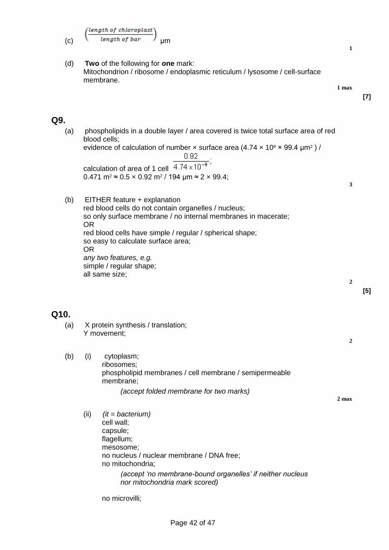
(c) μm 1
(d) Two of the following for one mark: Mitochondrion / ribosome / endoplasmic reticulum / lysosome / cell-surface membrane.
1 max
[7]
Q9. (a) phospholipids in a double layer / area covered is twice total surface area of red
blood cells; evidence of calculation of number × surface area (4.74 × 109 × 99.4 μm2 ) /
calculation of area of 1 cell 0.471 m2 ≈ 0.5 × 0.92 m2 / 194 μm ≈ 2 × 99.4;
3
(b) EITHER feature + explanation red blood cells do not contain organelles / nucleus; so only surface membrane / no internal membranes in macerate; OR red blood cells have simple / regular / spherical shape; so easy to calculate surface area; OR any two features, e.g. simple / regular shape; all same size;
2
[5]
Q10. (a) X protein synthesis / translation;
Y movement; 2
(b) (i) cytoplasm; ribosomes; phospholipid membranes / cell membrane / semipermeable membrane;
(accept folded membrane for two marks) 2 max
(ii) (it = bacterium) cell wall; capsule; flagellum; mesosome; no nucleus / nuclear membrane / DNA free; no mitochondria;
(accept ‘no membrane-bound organelles’ if neither nucleus nor mitochondria mark scored)
no microvilli;

Page 43 of 47
no Golgi; no ER; 70S / smaller ribosomes;
2 max
[6]
Q11. (a) A – granum / thylakoid;
chlorophyll molecules to trap light / light absorbing pigments / light dependent reaction / part of light dependent reaction;
2
B – stroma; (contains enzymes for) carbon dioxide fixation / light-independent reaction / part of light-independent reaction; (allow ribosome role of protein in photosynthesis)
2
(b) (i) C – starch; 1
(ii) from glucose in a condensation / polymerisation reaction / many glucose molecules joined together;
1
[6]
Q12. (a) A ‒ receptor /extrinsic (protein);
Accept glycoprotein/antigen
B ‒ transmembrane/intrinsic/channel/carrier (protein);
Accept hydrophobic tail
C ‒ phospholipid;
Ignore ref. to bilayer 3
(b) Cell wall;
Accept smaller/70S ribosome(s)
Capsule/slime layer;
Accept DNA without histone
(Bacterial) flagellum;
Reject capsid
Circular DNA/chromosome;
Plasmid;
Mesosome; 2 max
[5]

Page 44 of 47
Q13. (a) (i) no cell wall / only has (plasma) membrane;
1
(ii) has capsule / slime layer; 1
(b) correct approach which makes use of scalebar; ignore reference to units. 1
(c) cellulose / starch / amylose / amylopectin; 1
(d) (i) water potential lower / more negative in cell; (water enters by) osmosis;
2
(ii) plant cell wall made of a different substance / cellulose / penicillin does not affect cellulose;
1
[7]
Q14. (a) 1. Strands separate / H-bonds break;
1. Q Neutral: strands split
1. Accept: strands unzip
2. DNA helicase (involved);
3. Both strands / each strand act(s) as (a) template(s);
4. (Free) nucleotides attach;
4. Neutral: bases attach
4. Accept: nucleotides attracted
5. Complementary / specific base pairing / AT and GC;
6. DNA polymerase joins nucleotides (on new strand);
6. Reject: if wrong function of DNA polymerase
7. H-bonds reform;
8. Semi-conservative replication / new DNA molecules contain one old strand and one new strand;
8. Reject: if wrong context e.g. new DNA molecules contain half of each original strand
6 max
(b) (i) 18;
Do not accept 17.5 1
(ii) 10; 1
(iii) 1. Horizontal until 18 minutes;

Page 45 of 47
Allow + / - one small box
2. (Then) decreases as straight line to 0 μm at 28 minutes;
2. Allow lines that start from the wrong place, ending at 0 at 28 minutes
2
(c) (i) Two marks for correct answer of 19.68 or 19.7;;
Accept 19hrs 41mins
One mark for incorrect answers in which candidate clearly multiplies by 0.82;
Allow one mark for incorrect answers that clearly show 82% of 24 (hours)
2
(ii) 1. No visible chromosomes / chromatids / visible nucleus; 1
(iii) D (no mark)
1. Lower % (of cells) in interphase / higher % (of cells) in mitosis / named stage of mitosis;
1. Accept: ‘less’ or ‘more’ instead of ‘%’
1. Do not accept: higher % (of cells) in each / all stage(s)
2. (So) more cells dividing / cells are dividing quicker;
2. Accept: uncontrolled cell division
2. Do not award if Tissue C is chosen 2
[15]
Q15. (a) Three of chromosome 13 / an extra chromosome 13;
Accept trisomy 13
Accept circle around three chromosomes or any other correct indication on Figure 1
Do not allow references to any other chromosomes.
Do not accept chromatids for chromosomes. 1
(b) 1. In meiosis; 2. Homologous chromosomes / sister chromatids do not separate;
2. Accept non-disjunction 2 max
(c) 1. Mutation / extra chromosome in gamete / egg / sperm (that formed zygote);
2. All cells derived (from a single cell / zygote) by mitosis; OR 3. All cells derived from a single cell / zygote by mitosis; 4. Mitosis produces genetically identical cells / a clone;
Mark points 1 and 2 OR 3 and 4
4. Accept: have same DNA / same alleles

Page 46 of 47
2
(d) 1. (Some) oxygenated blood (from the aorta) flows into pulmonary artery; OR Less oxygenated blood flows out through aorta; OR Lower blood pressure in aorta;
2. Less oxygen delivered to cells / tissues / organs / named organ / via named blood vessel;
3. So less / not enough oxygen for aerobic respiration (in cell / tissue / organ);
4. Tissue / organ doesn’t grow / develop properly (causing death); OR Tissue dies / organ stops working (causing death);
1. Accept mixing of deoxygenated with oxygenated blood in pulmonary artery
2. Do not accept “no oxygen”
3. Do not accept “produce energy” 3 max
[8]
Q16. (a) C
Auto mark 1
(b) 1. No separation of chromatids/chromosomes/centromeres;
Accept anaphase prevented
Accept nondisjunction
Reject homologous pairs 2. Chromatids/chromosomes all go to one pole/end/sides
of cell/not pulled to opposite poles; 3. Doubles chromosome number in cell/one daughter cell
gets no chromosomes or chromatids;
Accept DNA for chromosomes
Accept ploidy
Ignore references to ‘genetic information’
Ignore simple descriptions of what normally happens in mitosis
2 max
(c) 1. (No, because) at 100 there are still some (7%) cancer cells dividing/undergoing mitosis;
Accept idea that all division stops only at 1000 2. So, cancer not destroyed/may continue to
grow/spread/form tumours;
Must refer to cancer spreading not cells dividing 3. Best concentration may be between 100 and
1000/need trials between 100 and 1000; 4. This research in culture, don’t know effect of KI on
people;
Reject ‘not tested on humans’
Reject ‘done in animals’

Page 47 of 47
5. (Yes, because) above 100 produces little increase in % of cells not dividing/undergoing mitosis/at 100, most (93%) cancer cells unable to divide/dead;
Must clearly link lack of monopolar mitotic spindles with cell division
6. Above 100 may be harmful (to body);
Accept ‘above 100/high concentrations produce harmful side effects/named effects'
7. Higher concentrations more expensive; 8. (Above 100) will have more effect on (rapidly dividing)
cancer cells;
Must relate to 100 4 max
(d) 1. 10 cm3 of 10 000 nmol dm−3/ (original) solution; 2. 90 cm3 of water;
If ratio correct but make wrong volume e.g. 1 litre, award 1 mark
2
[9]