Laparoscopic Surgeon in Bangalore | Laparoscopic Surgery in India
A41
International Journal of Contemporary Medical Research International Journal of Contemporary Medicine Surgery and Radiology Volume 6 | Issue 1 | January-March 2021
ISSN (Online): 2565-4810; (Print): 2565-4802 | ICV 2019: 98.48 |
A Laparoscopic Comparative Study of Right and Left Adrenalectomy – A Case of Non-Identical Twins: Two Case Reports with Review of LiteratureYash Rohatgi1, Prakash Chandra Shetty2, Abhijit Joshi3
1Second Year Resident, Department of General Surgery, Dr. L.H.Hiranandani Hospital, Powai, 2Department of Urology, Dr. L.H. Hiranandani Hospital, Powai, 3Department of General, G I & Endo-Laparoscopic Surgery, Dr.L.H.Hiranandani Hospital, Powai, India
Corresponding author: Dr. Yash Rohatgi, Second Year Resident, Department of General Surgery, Dr. L.H.Hiranandani Hospital, Powai, Mumbai, India
DOI: http://dx.doi.org/10.21276/ijcmsr.2021.6.1.9
How to cite this article: Yash Rohatgi, Prakash Chandra Shetty, Abhijit Joshi. A laparoscopic comparative study of right and left adrenalectomy – a case of non-identical twins: two case reports with review of literature. International Journal of Contemporary Medicine Surgery and Radiology. 2021;6(1):A41-A47.
INTRODUCTION The adrenal glands were first described by Bartholomaeus in 1552, as glands lying on the kidney. The left adrenal gland is semilunar in shape whereas the right gland is triangular. In 1805, Cuvier described the anatomy of the adrenals as cortex and medulla. These glands are chiefly responsible for releasing hormones in response to fear, flight and fright through the synthesis of corticosteroids like cortisol and catecholamines like nephrine and metanephrines. These glands also play an important role of regulating blood plasma osmolarity through the action of aldosterone on the kidneys.Surgery of the supra-renal glands is considered difficult due to their deep retroperitoneal location and close proximity to important anatomical structures. The world’s first planned open trans-peritoneal adrenalectomy was performed in 1914 by Sargent. Mayo performed the first flank approach adrenalectomy for phaeochromocytoma in 1927. Since its inception in 1992(Gagner), laparoscopic adrenalectomy was rapidly and widely accepted due to the difficult anatomical location of the gland and the relative ease of the laparoscopic versus open approach, in its surgical excision. Due to this and the other obvious benefits like lesser requirement of analgesia, shortened hospital stay, faster recovery, better cosmesis
and the fact that it has significantly reduced mortality and morbidity associated with the open technique by minimizing intra and post operative significant hemorrhage; laparoscopic adrenalectomy has swiftly climbed the pedestal of being the gold standard for adrenal excisions, for most pathologies.
CASE REPORTSCase -1: A 35 year old male with no comorbidities presented to the surgery out patient department with complaints of non-specific and vague abdominal pain since the last two months, not associated with any other symptoms.On physical examination, his pulse rate was 86 beats per minute, respiratory rate 18 breaths per min, blood pressure was 130/80 mmHg and he was afebrile. A per abdominal examination revealed mild tenderness in the right lumbar region. His laboratory reports revealed : hemoglobin - 14 gm/dl, WBC - 9000/microlitre, platelets - 2.2 lakhs/microlitre, sodium – 136 mEq/ L, potassium – 4mmol/L and creatinine – 0.8ml/dl. A contrast enhanced computed tomography(CECT) scan of the abdomen revealed a large non contrast enhancing right adrenal mass measuring 9 centimeters in diameter, most likely to be a myelolipoma (Fig 1A). The patient was then worked up to rule out functional adrenal tumours. He was subjected to the low
A B S T R A C T
Introduction: The different anatomies of the right and left adrenal glands make them a textbook case of non-identical twins. The divergent course of the main blood vessel namely the adrenal vein as well as the widely contrasting anatomy of the surrounding organs on the right versus the left sides practically make right and left adrenalectomy what they are - two different operations with the same name !Case report: In this paper, we endeavor to make a side by side pictorial comparison of laparoscopic right and left adrenalectomy vis-à-vis their pre-operative radiology and the important steps of their laparoscopic resection, the most important being the dissection, identification, skeletonization and control of the right and left adrenal veins. Also we summarise a comprehensive review of literature that attempts to cover all the dimensions of minimal invasive adrenalectomy. Conclusion: Laparoscopic adrenalectomy is now considered the gold standard for treatment of most adrenal gland pathologies.
Keywords: A Laparoscopic Comparative Study, Right and Left Adrenalectomy, Non-Identical Twin
Case RepoRt

Rohatgi, et al. A Laparoscopic Comparative Study of Right and Left Adrenalectomy
A42
International Journal of Contemporary Medical Research International Journal of Contemporary Medicine Surgery and Radiology Volume 6 | Issue 1 | January-March 2021
ISSN (Online): 2565-4810; (Print): 2565-4802 | ICV 2019: 98.48 |
Figure-1: CECT appearances: A – right side : Coronal view showing non enhancing right adrenal myelolipoma, B – left side : Coronal view depicting an enhancing left adrenal mass
Figure-2: Initial steps of dissection: A - right side : division of posterior parietal peritoneum just below the liver at the superior aspect of the mass, B – left side : medial reflection of descending colon from over the the left Gerota’s fascia
Figure-3: Dissection & control of the adrenal vein : A&B-right side:skeletonised & clipped short, stubby right adrenal vein draining directly into the IVC, C&D-left side: clipped longer right adrenal vein draining into the renal vein about to be divided
Figure-4: further steps of lap. right adrenalectomy : A)division of the right triangular ligament of liver, B)entry into the space between IVC and the right adrenal gland, C)dissection inferior to the right tumour at the superior pole of right kidney, D)lateral dissection, E)superior dissection
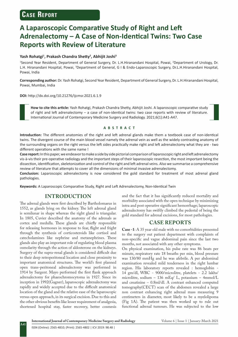
Figure-5: further steps of lap. left adrenalectomy – A)inferior dissection at the superior pole of left kidney, B)lateral dissection, C)medial dissection, D)superior dissection
Figure-6: Excised adrenalectomy specimens : A) right myelolipoma, B) left non functioning pheochromocytoma
dose dexamethasone suppression test, which showed a serum cortisol value of 1.2 mcg/dl (1.8 mcg/dl is the recommended cut off value for diagnosing Cushing’s syndrome). The plasma fractionated metanephrine level was 42 pg/ml, serum DHEA (Di-hydro- epi-androsterone) levels were 6 ng/ml and the urinary cortisol level was 15 mcg/ 24 hours.Thus a diagnosis of a non functioning incidentaloma - myelolipoma, was made and the patient underwent a laparoscopic transabdominal right adrenalectomy by flank approach. The histopathology report confirmed a myelolipoma. The postoperative recovery was uneventful and
Rohatgi, et al. A Laparoscopic Comparative Study of Right and Left Adrenalectomy
A43
International Journal of Contemporary Medical Research International Journal of Contemporary Medicine Surgery and Radiology Volume 6 | Issue 1 | January-March 2021
ISSN (Online): 2565-4810; (Print): 2565-4802 | ICV 2019: 98.48 |
Sr.
No.
Auth
or/s
[Ref
. No.
]Jo
urna
l(yr.
Of
publ
icati
on)
No.
of p
atien
tsTy
peof st
udy
Met
hods
/Re
sults
Conc
lusi
ons
1.St
ephe
n E
Burp
ee,
Greg
g H
Joss
art,
Mic
hel G
agne
r14
Surg
ical
Tre
atm
ent:
Evid
ence
-Bas
ed a
nd
Prob
lem
-Orie
nted
: Bo
ok(2
001)
*Ser
ies o
f lap
adr
.: 10
52pt
.(108
2 pr
oced
ures
)*L
ap v
s Ope
n se
ries :
La
p-22
8pts
.O
pen-
242p
ts./
276
proc
edur
es*T
rans
vs
Retr
oper
itone
-al s
erie
s :
Tran
sper
itone
-al :
11
8pts
.Re
trop
erito
neal
: 77
3pts
.
Book
cha
pter
:Co
mpa
rativ
e st
udy
of
mul
tiple
serie
s (co
mpa
-ris
on o
f ind
icati
ons,
open
vs l
ap &
tran
s vs
retr
oper
iton-
--eal
ap
proa
ches
)
*Var
ious
con
cern
ed p
ublis
hed
serie
s stu
died
& su
mm
arise
d on
th
e ba
sis o
f the
ir ou
tcom
es*s
tudi
es b
ased
on
op. ti
me,
leng
th
of st
ay, b
lood
loss
, con
vers
ions
, ot
her c
ompl
icati
ons,
mal
e vs
fe
mal
e pt
s., i
ndic
ation
s etc
.
*Lap
adr
safe
& e
ffecti
ve*L
ater
al tr
ansp
erito
neal
app
roac
h m
ore
com
mon
than
retr
oper
itone
al*b
lood
loss
& le
ngth
of s
tay
less
in
lap
than
ope
n*o
vera
ll co
mpl
icati
ons l
esse
r in
lap
*out
com
es si
mila
r bet
wee
n tr
ans &
re
trop
erito
neal
serie
s*P
oste
rior r
etro
perit
onea
l ap
proa
ch c
an a
void
adh
esio
ns fr
m
prev
ious
abd
omin
al su
rger
ies &
sa
ves ti
me
in B
/L a
dren
alec
tom
ies
as p
t. do
es n
ot h
ave
to b
e re
-po
sition
ed*H
owev
er it
has
a st
eepe
r & lo
nger
le
arni
ng c
urve
due
to u
nfam
iliar
ity
of a
nato
my
& re
stric
ted
wor
king
sp
ace
*ope
n ad
r. ha
s a ro
le in
ver
y la
rge
tum
ors &
obv
ious
mal
igna
ncy
*In
larg
er tu
mor
s, tr
ansp
erito
neal
ap
proa
ch is
cle
arly
supe
rior t
o re
tro.
2.J M
ares
caux
, D M
utter
, M
H W
heel
er18
Surg
ical
end
osco
py(1
996)
27Ca
se se
ries
*18w
omen
& 9
men
*mea
n ag
e-50
.8y
*12r
t & 1
5lt g
land
s rem
oved
*tra
nspe
rit. fl
ank
appr
oach
-26
retr
oper
it. fl
ank
appr
oach
-1*m
edia
n gl
and
size-
2cm
s*a
vg. o
p. T
ime-
140m
ins
*avg
. hos
p. S
tay-
4.6d
*Lap
adr
. is s
afe
and
offer
s fas
t re
cove
ry a
nd sh
ort i
n-ho
spita
l sta
y *L
apar
osco
pic
adre
nale
ctom
y co
mbi
nes t
he a
dvan
tage
s of b
oth
the
conv
entio
nal a
nter
ior a
nd
post
erio
r app
roac
h
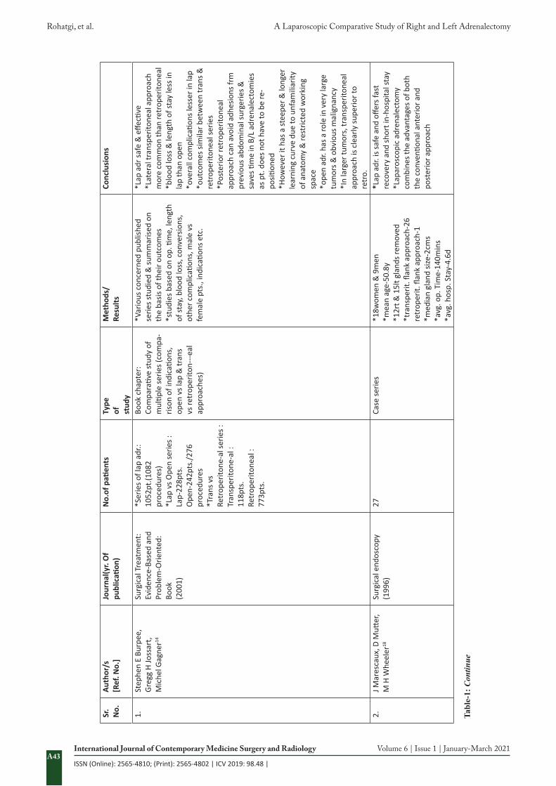
Tabl
e-1:
Con
tinue

Rohatgi, et al. A Laparoscopic Comparative Study of Right and Left Adrenalectomy
A44
International Journal of Contemporary Medical Research International Journal of Contemporary Medicine Surgery and Radiology Volume 6 | Issue 1 | January-March 2021
ISSN (Online): 2565-4810; (Print): 2565-4802 | ICV 2019: 98.48 |
3.M
Gag
ner,
A Po
mp,
B
T He
nifo
rd, D
Pha
rand
, an
d A
Lacr
oix15
Anna
ls of
surg
ery
(199
7)10
0Cl
inic
al tr
ial
*88p
ts, m
ean
age-
46y
*55p
ts. h
ad p
rev.
abdo
. sur
gery
*mea
n op
. tim
e-12
3min
s*m
ean
bld
loss
-70m
l*c
onve
rsio
ns to
ope
n-3
*avg
. Len
gth
of st
ay-2
.4 d
*dur
ing
follo
w u
p 2
had
reno
vasc
ular
hyp
erte
nsio
n &
non
e ha
d re
curr
ence
of h
orm
onal
exc
ess
*dur
ing
phae
o. E
xcisi
on 5
6%
had
hype
rten
sion
& 5
2% h
ad
hypo
tens
ion
*Pha
eos.
(25)
Aldo
ster
onom
as(2
1)N
on-fu
nc. a
deno
mas
(20)
Corti
sol p
rodu
cing
ade
nom
as(1
3)Cu
shin
g’s d
iseas
e(8)
Oth
ers(
13)
*Lap
adr
. is s
afe,
effe
ctive
, de
crea
ses h
ospi
tal s
tay
and
wou
nd
com
plic
ation
s *
Prio
r abd
omin
al su
rger
y is
not a
con
trai
ndic
ation
*P
heoc
hrom
ocyt
omas
can
be
rese
cted
safe
ly la
paro
scop
ical
ly
desp
ite b
lood
pre
ssur
e va
riatio
ns*
Veno
us th
rom
bosis
pro
phyl
axis
is m
anda
tory
*The
lap
appr
oach
is th
e pr
oced
ure
of c
hoic
e fo
r adr
enal
ecto
my
exce
pt
in th
e ca
se o
f inv
asiv
e ca
rcin
oma
or
mas
ses >
15
cm
4.M
atthe
w J.
Mel
lon,
Am
anjo
t Set
hi, a
nd C
hand
ru P.
Sun
dara
m16
Indi
an jo
urna
l of
urol
ogy
(200
8)
Not
app
licab
leN
arra
tive
revi
ew a
rticl
eIn
dica
tions
, con
trai
ndic
ation
s,in
str-
umen
tatio
n &
pt.p
ositi
onin
g di
scus
sed
in rt
.& lt
.adr
. vis-
a-vi
s lat
eral
tran
sper
itone
al, l
at.
retr
oper
itone
osco
pic,
pos
terio
r re
trop
erito
neos
copi
c ap
proa
ches
. Al
so la
p B/
L ad
r, la
p pa
rtial
adr
to
uche
d up
on &
com
plic
ation
s su
mm
arise
d
*Lap
adr
. is a
safe
and
effe
ctive
te
chni
que
for t
he su
rgic
al re
mov
al
of a
dren
al m
asse
s *
Clea
r adv
anta
ges o
ver o
pen
rese
ction
5.U
mbe
rto
Mae
stro
ni,
Stef
ania
Fer
retti
, F
ranc
esco
Zig
lioli,
Dav
ide
Cam
poba
sso,
Da
rio C
eras
i, Pi
etro
Co
rtel
lini7
Uro
logi
a(2
011)
6Ca
se se
ries
*Adr
mas
ses b
etw
een
7-15
cms
*Mea
n op
erati
ng ti
me-
120m
ins
*Mea
n bl
ood
loss
-50c
cs*D
ischa
rge
on P
OD3
*No
post
op. c
ompl
icati
on*S
afe
and
effec
tive
also
in th
e ca
se
of g
iant
mas
ses
*Pre
oper
ative
dia
gnos
is ha
s a
pred
omin
ant r
ole
to d
eter
min
e th
e co
ntra
indi
catio
n of
this
tech
niqu
e (in
vasiv
e ad
rena
l car
cino
ma)
6.J J
Sob
le, I
S G
ill13
Uro
logy
(199
8)39
(42
proc
edur
es)
Clin
ical
tria
l(n
eedl
esco
pi-c
ad
rena
lect
om-y
)
*3 c
onve
rted
to c
onve
ntion
al la
p. &
1
to o
pen
*avg
.op.
time-
132m
ins
*avg
.bld
.loss
-67c
cs
*Ini
tial e
xper
ienc
e-pr
omisi
ng*I
n se
lect
pts
.-saf
e &
feas
ible
*Fur
ther
eva
luati
on &
im
prov
emen
t in
2mm
in
stru
men
tatio
n &
opti
cal
tech
nolo
gy re
quire
d
Tabl
e-1:
Con
tinue
Rohatgi, et al. A Laparoscopic Comparative Study of Right and Left Adrenalectomy
A45
International Journal of Contemporary Medical Research International Journal of Contemporary Medicine Surgery and Radiology Volume 6 | Issue 1 | January-March 2021
ISSN (Online): 2565-4810; (Print): 2565-4802 | ICV 2019: 98.48 |
7.M
K W
alz, K
Pei
tgen
, M
V W
alz,
R H
oerm
ann,
B
Salle
r, R
M G
iebl
er, F
Jo
cken
höve
l, T
Phili
pp,
C E
Broe
lsch,
F W
Eig
ler,
K M
ann11
Wor
ld jo
urna
l of
surg
ery
(200
1)
130(
142
proc
ed-u
res)
Case
serie
s-pr
ospe
ctive
re
view
*tum
or si
ze ra
nge
0.5-
7cm
s*P
artia
l adr
. in
39 p
ts.
*con
vers
ion
to o
pen
in 5
pts.
&7p
ro.
*Op.
time
101+
/-39
min
s*b
lood
loss
54+
/-72
ml
*low
requ
irem
ent o
f ana
lges
ics
*mea
n ho
sp. S
tay
– 3
days
Post
erio
r ret
rope
riton
eosc
opic
ad
rena
lect
omy
is a
safe
met
hod
&
has b
ecom
e a
stan
dard
pro
cedu
re
in e
ndoc
rine
surg
ery
8.I S
Gill
, A M
Mer
aney
, J
C Th
omas
, G T
Sun
g, A
C
Nov
ick,
I Li
eber
man
20
The
jour
nal o
f uro
logy
(200
1)3
Case
repo
rts
*All
3 pt
s had
sign
ifica
nt p
rior
abdo
min
al sc
arrin
g aft
er
eith
er p
artia
l or t
otal
radi
cal
neph
rect
omy,
ther
eby
prec
ludi
ng
effici
ent t
rans
abdo
min
al
lapa
rosc
opic
acc
ess
*Dou
ble
lum
en e
ndot
rach
eal
intu
batio
n*4
por
t tra
ns-t
hora
cic
appr
oach
*Dia
phra
gm in
cise
d un
der r
eal ti
me
lap
USG
gui
danc
e*a
fter e
xcisi
on o
f gla
nd, d
ia. s
utur
e cl
osed
*mea
n op
.tim
e-4.
5hrs
*mea
n bl
d lo
ss-2
34cc
s*h
osp.
stay
-2da
ys
In se
lect
pati
ents
with
sign
ifica
nt
conc
omita
nt in
tra-
perit
onea
l and
re
trop
erito
neal
scar
ring
from
prio
r m
ajor
abd
omin
al o
r ren
al su
rger
y, la
paro
scop
ic a
dren
alec
tom
y ca
n be
safe
ly p
erfo
rmed
with
the
tran
s-th
orac
ic tr
ans-
diap
hrag
mati
c ap
proa
ch
Tabl
e-1:
Sum
mar
y of
revi
ew o
f lite
ratu
re c
once
rnin
g th
e va
rious
dim
ensio
ns o
f min
imal
inva
sive
adre
nale
ctom
y
he was discharged on post operative day(POD) 3.
Case -2 : A 41 year old male presented to the surgery out patient department with complaints of left sided pain in abdomen since 6 months which was dull aching in nature, not associated with nausea and vomiting. On clinical examination, his pulse rate was 82 beats per minute, respiratory rate was 16 breaths per min and blood pressure was 120/70 mmHg. He was afebrile. A per abdomen examination revealed mild left loin tenderness. He did not have a palpable abdominal lump and had no clinical evidence of free fluid in the abdomen. His blood investigations revealed a hemoglobin of 14.4 gm/dl, WBC - 7300/microlitre, platelets - 2.4 lakhs/microlitre, sodium – 140 mEq/ L, potassium – 4.2 mmol/L and creatinine – 0.7 ml/ dl. A CECT scan of the abdomen revealed a large contrast enhancing left sided adrenal mass measuring 8.6 cm in diameter which was partly cystic in appearance(Fig 1B). The endocrine workup revealed a serum cortisol value of 1.0 mcg/dl on low dose dexamethasone suppression test, plasma fractionated metanephrine level was 36 pg/ml, serum electrolytes were within normal range, serum DHEA level was 3 ng/ml and the urinary cortisol level was 11 mcg/ 24 hours; thereby suggesting that it was a non functioning adrenal mass. He underwent a laparoscopic transabdominal left adrenalectomy by flank approach. The histopathology report revealed a phaeochromocytoma. Thus the patient turned out to be a rare case of non-functioning phaeochromocytoma. His post operative recovery was uneventful and he was discharged on POD 5.
DISCUSSIONThe adrenal glands are situated on supero-medial poles of each kidney in the retroperitoneum. Right adrenal gland is triangular and the left adrenal is a crescent shaped structure. The adrenal glands are located in front of the 12th rib on the right, in front of the 11th and 12th ribs on the left and on the lateral edge of the vertebral column at the level of 12th thoracic vertebra. Each adrenal gland is composed of a cortex and medulla, both of which secrete hormones. The adrenal cortex is divided into three parts secreting different hormones, namely:- 1) Zona glomerulosa - mineralocorticoids, Zona fasiculata - glucocorticoids and Zona reticularis - androgens. The adrenal medulla secretes catecholamines like epinephrine and norepinephrine in response to stress.Each gland is supplied by three groups of vessels : 1) superior adrenal arteries derived from the

Rohatgi, et al. A Laparoscopic Comparative Study of Right and Left Adrenalectomy
A46
International Journal of Contemporary Medical Research International Journal of Contemporary Medicine Surgery and Radiology Volume 6 | Issue 1 | January-March 2021
ISSN (Online): 2565-4810; (Print): 2565-4802 | ICV 2019: 98.48 |
inferior phrenic artery, 2) the middle adrenal arteries derived from the aorta and the 3) inferior adrenal arteries are derived from the renal artery. Each adrenal is drained by a single adrenal vein. The right adrenal vein is usually short (5mm in length) and drains directly into the IVC, while the left adrenal vein is longer (30 mm in length) and empties into the left renal vein after joining the inferior phrenic vein.Indications of adrenalectomy are : incidentaloma more than 4 cms in size, aldosteronoma, pheochromocytoma, myelolipoma, adrenal cyst, Cushing’s adenoma etc. For non-functional adrenal tumors, the indication for surgery is the risk of malignancy-according to the lesion size. If the tumor is less than 4 cm, the risk of malignant conversion is approximately 2%. For lesions ranging from 4–6 cm, the risk of malignancy is around 6%, while for lesions larger than 6 cm, the risk of malignancy is increased significantly to 25%.1 The differential diagnosis of a supra-renal gland mass comprises of adrenal adenoma, adrenal cyst, adrenal myelolipoma, pheochromocytoma, adrenal cancer, metastatic cancer, hyperplasia, incidentalomas and tuberculosis. All the patients with adrenal mass should undergo all the hormonal, biochemical and localizing investigations. The goal of initial workup is to differentiate the benign lesion from the metastatic disease and also to distinguish between a non functioning tumor from the hyper- functioning lesion. The biochemical and hormonal tests advocated are – a)Dexamethasone Suppression Test: the sensitivity and specificity of this test ranges from 90- 100% in establishing a diagnosis of Cushing’s syndrome. The cut- off values range from 50-138 nmol/L to diagnose adrenal disorder, b)24 hour urinary catecholamines and vanilmandelic acid levels is best utilized for diagnosing a case of pheochromocytoma, with sensitivity and specificity being 95%.2 But the best test for diagnosing pheochromocytoma is the more recent addition to the screening modality known as the Fractionated plasma metanephrine levels, c)Plasma aldosterone and plasma renin levels should also be assessed in hypertensive patients presenting with adrenal lesions to see for Conn’s disease & d)Sex hormone levels like dehydroepiandrosterone should also be evaluated to rule out rare adreno-cortical cancer producing symptoms of virilisaton and feminization.The imaging modalities most commonly and effectively used for identifying different types of adrenal masses are a) Contrast enhanced computed tomography(CECT) - most of the adrenal lesions like the cyst, myelolipomas, adenomas, carcinoma and hemorrhage can be identified using this modality. Differentiation is made between adenoma and carcinoma on the basis of Hounsfield units. Lesions with a low attenuation value of less than 10 units are considered to be adenomas and lesions above 18 units are typically considered carcinomatous.3,4 However CECT scans cannot distinguish between functioning and non-functioning lesions, b)Magnetic resonance imaging(MRI) - it is the investigation of choice for diagnosing adrenal mass. MRI is costlier in comparison to CECT but avoids exposure to ionizing radiation. Adenomas usually have low signal enhancement on T2 weighted images whereas pheochromocytomas have a bright (Electric bulb sign) signal intensity on T2 weighted images. Recently, chemical shift MRI has been
used to distinguish between benign and malignant lesions. Benign lesions have a high lipid content and typically show a loss of signal intensity i.e they darken on the chemical-shift whereas malignant lesions show no loss of signal intensity, c)Adrenal scintigraphy - This is another method used to differentiate between benign and malignant lesions. This is done using metaiodobenzylguanidine(MIBG).5 MIBG scan is especially used for pheochromocytomas that take up MIBG in adrenal vesicles. A normal adrenal gland has very few adrenal vesicles to pick up MIBG and hence cannot utilize it to produce an image. However a pheochromocytoma, has extra vesicles and will take up enough MIBG to produce an image on scintigraphy. Hence, MIBG scintigraphy is useful in localizing recurrent tumors or hormone secreting pheochromocytomas & d)Positron emission tomography(PET) - the differentiation of malignant and benign adrenal tumors can also be facilitated with the help of PET scan. The 18-FDG PET is based on the increased uptake of glucose by the high metabolic activity of lesions.6 The use of PET is best reserved for cases in which CT imaging and clinical symptoms are inconclusive.The decision for surgical management depends on several factors including size, functionality, malignant potential and overall clinical status of the patient. Management of incidentalomas between 4 and 6 cm is more controversial. The risk of malignancy in this size range is estimated to be only 6% and the risk of adrenal carcinoma is less than 2% in lesions <4 cm in size. Whereas, the chances of malignant conversion in lesions >6 cm is significantly higher, approximately 25%.1,17 Malignant adrenal gland lesions were initially considered to be a contraindication for laparoscopic excision but studies have suggested that adrenal lesions upto 14 cm without local invasion can be removed laparoscopically.7 Based on several studies, Grumbach et al.1 concluded that adrenal lesions more than 6 cm should be considered malignant unless proven otherwise. Adrenal surgeries can be performed, either by the conventional open method or by minimally invasive (laparoscopic) technique. Since it’s introduction in 1992 by Gagner, laparoscopic adrenalectomy has become the gold standard.8,19 Now, open surgery is only reserved for invasive and large adrenal masses. Prior to 1980 all adrenalectomies were performed by trans- abdominal open approach. The benefits of laparoscopic approach are decreased hospital stay, faster recovery time, improved patient satisfaction and better cosmetic results. It has also significantly lowered the mortality and morbidity associated with the open procedure due to reduced intra and post operative haemorrhage.9 The laparoscopic adrenalectomy can be performed using either transperitoneal or the retroperitoneal approach. Laparoscopic transperitoneal adrenalectomy can be performed with patient in either supine or contralateral lateral decubitus positions. Retroperitoneoscopic adrenalectomy can be performed in either prone or lateral positions. The transperitoneal approach has an easier learning curve as the surgeon has more working space and hence suitable for large tumours(>6 cm) as well as obese patients. However in this approach mobilisation of other organs is required thereby increasing chances of injury. Also it involves longer duration of the surgery and increased risk of incisional hernias. Since its inception in 1995 by Miyake

Rohatgi, et al. A Laparoscopic Comparative Study of Right and Left Adrenalectomy
A47
International Journal of Contemporary Medical Research International Journal of Contemporary Medicine Surgery and Radiology Volume 6 | Issue 1 | January-March 2021
ISSN (Online): 2565-4810; (Print): 2565-4802 | ICV 2019: 98.48 |
et al, posterior retroperitoneal adrenalectomy (PRA) has been utilised frequently.10 This technique directly approaches the adrenal gland through the retroperitoneal space, without breaching the peritoneum thereby resulting in a shorter operative time, less blood loss, less postoperative pain, and shorter hospital stay. Disadvantages of this technique are long and arduous learning curve, unsuitability for large tumors and obese patients due to limited working space.11 Retroperitoneoscopic adrenalectomy in prone position has the added benefit in bilateral adrenalectomy cases of not having to change patient position intraoperatively. Robotic adrenalectomy was first performed in the year 2000 by Horgan and Vanuno.The surgical procedure of robotic adrenalectomy is similar to the laparoscopic approach, such as the patient position, port sites, CO2 insufflation for creating pneumo -peritoneum and specimen retrieval. Robotic surgery is now considered as a standard surgical method for adrenal lesions since it is safe, feasible and an effective technique. It also provides a three dimensional perception, with precise camera control and improved moving capacity of the robotic arms.12 Needlescopic adrenalectomy- Laparoscopic adrenalectomy with needlescopic instruments (2mm in size) can be performed in most patients with adrenal lesions less than 5 cm. Pheochromocytomas can also be managed but with a longer operative time.13
CONCLUSIONLaparoscopic adrenalectomy is now considered the gold standard for treatment of most adrenal gland pathologies. This is because it’s magnified view provides better visualization of this deeply situated gland thereby facilitating improved precise dissection and reducing the morbidity and mortality associated with the open surgery. Apart from the conventional advantages, this technique has significantly reduced the incidence of intra and post-operative complications, wound complications, incisional hernias and respiratory problems.
REFERENCES1. Arnold DT, Reed JB, Burt K. Evaluation and
management of the incidental adrenal mass. Proc (Bayl Univ Med Cent). 2003; 16(1): 7-12.
2. Boyle JG, Davidson DF, Perry CG, connell JM. Comparison of diagnostic accuracy of urinary free metanephrines, vanillyl mandelic Acid and catecholamines and plasma catecholamines for diagnosis of pheochromocytoma. J Clinic Endocrinol Metab. 2007; 92(3): 4602-4608.
3. Bovio S, Cataldi A, Reimondo G, Sperone P, novella S, Berruti A, et al. Prevalence of adrenal incidentaloma in a contemporary computerized tomography series. J Endocrinol Invest. 2006; 29(6): 298-302.
4. Blake MA, Kalra MK, Sweeney AT, Lucey BC, Maher MM, Sahani DV, et al. Distinguishing benign from malignant adrenal masses; multi-detector row CT protocol with 10-minutes delay. Radiology. 2006; 238(2): 578-585.
5. Rubello D, Bui C, Casara D, Gross MD, Fig LM, Shapiro B. Functional scintigraphy of the adrenal gland. Eur J Endocrinol. 2002; 147(5): 13-28.
6. Groussin L, Bonardel G, Silvera S, Tissier F, Coste
J, Abiven G, et al. 18-Fluorodeoxyglucose positron emission tomography for the diagnosis of adrenocortical tumor: a prospective study in 77 operated patients. J Clin Endocrinol Metab. 2009; 94(4): 1713-1722.
7. Maestroni U, Ferretti S, Ziglioli F, Campobasso D, Cerasi D, Cortellini P. Laparoscopic adrenalectomy for giant masses. Urologia 2011;78 (Suppl 18):S54-58.
8. Smith CD, Webwe CJ, Amerson JR. Laparoscopic adrenalectomy: new gold standard. World J Surg. 1999; 23(1): 389-396.
9. Imai T, Kikumori T, Ohiwa M, Mase T, Funahashi H. A case-controlled study of laparoscopic compared with open adrenalectomy. Am J Surg. 1999; 178(5): 50-54.
10. Duh QY, Siperstein AE, Clark OH, et al. Laparoscopic adrenalectomy. Comparison of the lateral and posterior approaches. Arch Surg 1996;131(6):870–875
11. Walz MK, Peitgen K, Walz MV, Hoermann R, Saller B, Giebler RM, Jockenhovel F, Philipp T, Broelsch CE, Eigler FW, Mann K. Posterior retroperitoneoscopic adrenalectomy: lessons learned within five years. World J Surg 2001;25(1): 728–734
12. Brunaud L, Ayav A, Zarnegar R, et al. Prospective evaluation of 100 robotic-assisted unilateral adrenalectomies. Surgery 2008;144:995-1001.
13. Gill IS, Soble JJ, Sung GT, Winfield HN, BravoEL,Novick AC. Needlescopic adrenalectomy–the initial series: comparison with conventional laparoscopic adrenalectomy. Urology 1998;5:180–186.
14. G.H. Jossart et all Surgery of the adrenal glands Endocrinol Metab Clin North Am 2001;29 (1):57-68
15. Gagner M, Pomp A, Heniford BT, Pharand D,Lacroix A Laparoscopic adrenalectomy; lessons learned from 100 consecutive procedures.Annals of Surgery, 01 Sep 1997, 226(3):238-46;
16. Mellon MJ, Sethi A, Sundaram CP. Laparoscopic adrenalectomy: Surgical techniques. Indian J Urol. 2008;24(4):583-589.
17. Grumbach MM, Biller MK, Braunstein GD, Campbell KK, Carney JA, Godley PA, et al. Management of the clinically inapparent adrenal mass (‘incidentaloma’). Ann Intern Med. 2003; 138(4): 424-429
18. J Marescaux, D Mutter, M H Wheeler .Laparoscopic right and left adrenalectomies Surgical procedures.
19. Gagner M. Laparoscopic adrenalectomy. Surg Clin North Am 1996;76(1):523–537.
20. Gill IS, Meraney AM, Thomas JC, Sung GT, Novick AC, Lieberman I. Thoracoscopic transdiaphragmatic adrenalectomy: The initial experience. J Urol. 2001;165(2):1875–81
Source of Support: Nil; Conflict of Interest: None
Submitted: 22-12-2020; Accepted: 24-01-2021; Published online: 26-02-2021