A-l?70 COLORADO STATE UNIV FORT COLLINS DEPT OF … · The mechanism of most of the work was to...
74
A-l?70 COLORADO STATE UNIV FORT COLLINS DEPT OF PHYSIOLOGY --ETC F/A A/20 DIETARY PRDTECTION AGAINST PULMONARY OXYGEN POISONING. 1W DEC 80 C L SCHAYTE, M M MATHIAS N00GOIA76C-0437 UNCLASSIFIED NL
Transcript of A-l?70 COLORADO STATE UNIV FORT COLLINS DEPT OF … · The mechanism of most of the work was to...

A-l?70 COLORADO STATE UNIV FORT COLLINS DEPT OF PHYSIOLOGY --ETC F/A A/20
DIETARY PRDTECTION AGAINST PULMONARY OXYGEN POISONING. 1WDEC 80 C L SCHAYTE, M M MATHIAS N00GOIA76C-0437
UNCLASSIFIED NL

FINAL TECHNICAL REPGT.
DIETARY PROTECTION AGAINST PULMONARY, XYGEN POISONING0
Contract No N 6 0 4-76.C- 373) Office of Naval Rese5bh.
ODates: 1 April 1974 - 31 December 1980
Principal Investigator: j Christopher L./Schatte T .Department of Physiololjy and BiophysicsColorado State UniversityFort Collins, Colorado 80523
(303) 491-5768
Co-Investigator: Melvin M. Mathias, Ph.D.Department of Food Science and NutritionColorado State UniversityFort Collins, Colorado 80523(303) 491-5234
C)*
-AJ
DISRIUTION S'rA A~FK
Akpproved for public ips;-* a I \Distlbadton Un' ~'avd i L40 gV
81 3 24 016

TABLE OF CONTENTS
Page
Introduction . ............ . . .. . .. . . . . . . . . . . . . . I
Oxygen Toxicity in Mice . .................. .... 4Hypoxia ..... ....................... . . . . . . . . 5Age and sex ................ . . . . . ........ 8
Antioxidant Supplementation ...... ..................... 11Vitamin E and Selenium, Dietary ..... .................. 12Vitamin E and Selenium, Parenteral ......... . ...... 16
Pulmonary Antioxidant Enzymes ....... ..................... .17Sex ..... ............. . ... .................... o17Hypoxia.... ......... ............................. .18Dietary Fat ....... ............................. 18Vitamin E and Selenium ........ ....................... .20
Dietary Fat and Prostaqlandins o.......... . ......... 28Dietary Fat and Mortality ... ..................... 28Prostaglandin Synthesis ................ .......... 29Prostaglandin Metabolism ......... ...................... 38Review ................. .................... 49Dietary Fat and Prostaglandin Metabolism ...... .............. 64Vitamin E. .......... .............................. .64Selenium ........ .............................. 65
Miscellany ... .............................. ....... 66Selenium and Prostaglandin Synthesis ...................... .66Aspirin and Oxygen Toxicity ...... ....................... 67
Summary ......... ................................ 68
References .................. . ............. 69
Presentations. ............... . . . . ................ 70
Acceson P,.NTIS CF&
L 7
13vJDkt i f r ij_ .
_Dist

ABSTRACT
This project was designed to determine any influence of dietary composition
on susceptibility to pulmonary oxygen toxicity in rats and mice. Of a variety
of vitamins given in supranormal doses, only vitamin E proved efficacious in
delaying the onset of toxic symptoms in rats exposed to pure oxygen at one
atmosphere absolute. Dietary supplementation with the trace element selenium
appeared to be beneficial in some experiments but not others. Alteration of
amount and type of dietary fat-influenced mortality of rats at IATA oxygen.
The mechanism by which polyunsaturated fats in the diet might change suscep-
tibility to oxygen toxicity was not elucidated, but a possible relationship
with pulmonary prostaglandin metabolism is suggested.
AKey Words: diet, pulmonary oxygen toxicity, rodents, vitamin E, selenium,
polyunsaturated fatty acids, prstaglandins.
'7!"Sl

INTRODUCTION
The goal of this project was to develop a dietary regimen optimal for
attenuation of oxygen poisoning. It was predicated on the fact that cells
contain natural antioxidant systems which have evolved to combat overoxidation
during normal metabolism in an oxygen-containing environment. Examples include
Vitamin E as an integral part of all membranes, the enzyme glutathione peroxidase,
some forms of which contain the trace metal selenium, the enzyme superoxide
dismutase, the hexose monophosphate pathway of glycolysis which produces
reducing equivalents and the sulphydryl-containing substances glutathione as
well as sulfur-containing amino acids. The relationship of most known anti-
oxidants is shown in Figure 1.
Because these are naturally-occurring systems, it was hypothesized that
their effectiveness in combatting overoxidation during severe oxidant stress,
e.g. hyperoxia, might be amplified by manipulating concentrations of key
precursors in the cell or inducing higher fizyme activity. In many cases, the
latter can be done by dietary means.
The mechanism of most of the work was to u-Te supranormal dietary con-
centrations of selected constituents in an attempt to increase cell concentrations,
as in the case of Vitamin E, or altering the content of a dietary constituent,
such as lipid amount or composition, in order to achieve greater resistance to
oxygen toxicity.
The major advantage of using physiologicel changs in diet is that favorab e
results allow testing in a range of species end the promise of use in man
without untoward side effects or threat to health. In all cases, dietary
alterations were restricted to physiologically reasonable changes which could
likely be applied with similar qualitative results in man.
-1 -

This report summarizes chronologically the work performed during this
contract by providing a brief introduction to the various experiments followed
by the pertinent data, usually in the form of the resulting publication.
-2

Figure 1: Schematic representation of the interrelationships among theknown major antioxidant, pathways ini mammalian cells.
RATID
PROSTAGLANDINS -wPROSTAGLANDIN IREE SUPEROXID 0W.0H
SYt4TIETAWE RAALS MISMUTASE 2020(ASPIRIN INHIBITS)
C T L S 20CATAASEH 20+0 2
UNSATURATED LIPIDS (VITAMIN E ORGANIC _---ELDMGINHIBITS) PEROXIDES L AMG
~-0XIDATIONGLUTATHIONE PEROXIDASE SLNU
CYSTEINEGSre GSH-.-W---- GLYCINE
IGLUTAMATE
GLUTATHIONE REDUCTASELIPID SYNTHESIS -or-"
NAOP NADPtHH
HEXOSE 4MONOPHOSPHATE GAMMA
SHUNT AMINOBUTYRIC
ACIISOCITRATE
C- KETOGLUTARATE SUCCIN ATEk KRES
PYRUVATE-* MALIC ENZYME -MALATE
_,. . . . - _ - If . .. . . . -. .. .. .. . .
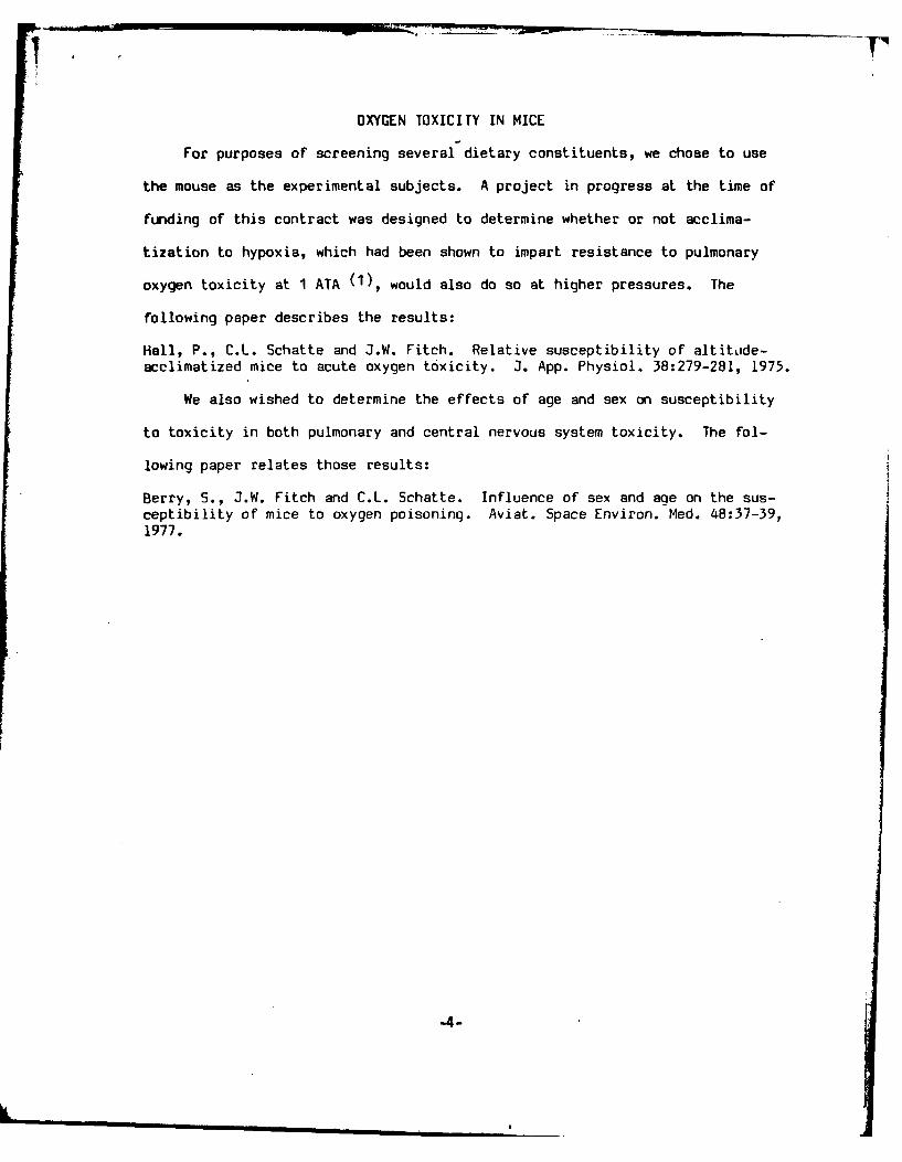
OXYGEN TOXICITY IN MICE
For purposes of screening several dietary constituents, we chose to use
the mouse as the experimental subjects. A project in progress at the time of
funding of this contract was designed to determine whether or not acclima-
tization to hypoxia, which had been shown to impart resistance to pulmonary
oxygen toxicity at 1 ATA (1), would also do so at higher pressures. The
following paper describes the results:
Hall, P., C.L. Schatte and J.W. Fitch. Relative susceptibility of altitode-acclimatized mice to acute oxygen toxicity. 3. App. Physiol. 38:279-281, 1975.
We also wished to determine the effects of age and sex on susceptibility
to toxicity in both pulmonary and central nervous system toxicity. The fol-
lowing paper relates those results:
Berry, S., j.W. Fitch and C.L. Schatte. Influence of sex and age on the sus-ceptibility of mice to oxygen poisoning. Aviat. Space Environ. Med. 48:37-39,1977.
-4-
IL m

J04.'RNAL of APPLur, P"%SIOLOcGv
VW1. 38K No,.2. Februal, 1975. Pn.s. U.S.A.
Relative susceptibility of alItitude- acclimatized
mice to acute oxygen toxicity
PETER HALL, CHRISTOPHER L. SCHATrE, AND JOHN IV. FITCHHypo-Hyperbaric P'acility- , lDepartrnant of Physiology and Biophysics,Coloyndo State University, Fort Collins, Colorado 80,52,3
HALL, PETER, CHRISTOPH4ER L. SCHATTE, AND JOHN WV FITCH. interrelationships between a range of acclimatization alt-Relative suscrptibility of altiiude-acclimatized mice to acute oxigen toxicitv. tudes, durations of acclittiatization, and OHP levels.J. Appi. Physiol. 38(2): 279-281. I 973.--The influence of by-
poxic acclimatization at. altitude,. of 0, 5,000, or 1 5,000 ft on. th e METHODS AND MATE~RIALSrelative susceptibility to acute oxygen poisoning was de!terminiedin 288 adult female mice. Ater acclimatization periods of 1. 2, A group of 300 feinale mnice (CF%V strain. Carworth4, or 8 wk, the mice were exposed to oxygen at high pressures Farmns) with a ican :ASL) weight of 19.34 =1 1.71 g %%a(OHP) of 4, 6, or 9 ATA and the times to convulsion and death raised to adulthood at sea level, randontly diMid! intorecorded. A factorial analysis of variance indicated that attituide three groups and placed inl three chambers at altitudes ofand OHP level had inverse, lug-linear effects onl both paratnett-rs. 0 1 t (PH = 760 niniHi, P0, 159 nisnHai, 3,ouW ft (PaThe duration of acclimatization progressively decreasedl the time -63 Po,=15,idto death. The onset of convuiWons and death Nas independent of -32 PI~ - 3j a Kd5,000) ft (PH 437, P~j. =89body weight. There wkere siznilcan, interaction,; on the rnea'ored in air All wecre itnilarly houwed aind led Purina laboratoryparameters between variouis combinations ofa~ltitude, Oil1 P level, chOWi. t )Ince day.1 te chaimobers \v.'ri bilyOpene,(dand duration of acclimatization. While alterations in the mce- to anibient prr~sure i W2 im-I) for (-I-%icinlg. The ranvestabolism of gamma-aminobot\ ric acid and highi-cnerg compount'; of chamtber (empteratttrcs 126 29*0 , relative hutiitiiesare common to both hypoxi-. and lnyperoxia, the most platii (1 9-56'. 1, onc 0 ) tl, -' i 10 ( -J 19', )were maintainedexplanation of the .results , to thle decrease in buffer 1
-,'i~e by., adjustment of vas do\% thr-ouigh the c-it imbers.induced by hvpoxic a:- umtnan.ation which might have caused AXt inter\ .ul- ol 1, 2, 4, antd 8 \vk, a group of 24 mnice
CO2 otetiaionof O! smp~ms.was removecd liutim vach cliattiber, weigzhed, and dividedinto three grotip, of eighlt aniumals. These aroups were ex-
hypoxia; acclimatization; o\;-qtn at high pressures; oxygen posed to (JIHP it 4, 1), or 9 ATA mI), 9ot, or 1.35 psiat inpoisoning; convulsions; deathahyebrccitbr ouie lOj)ltr ted ih
_______________________________________________ a 24-coinpartment %soodi and wirecloth c \nc hich allow\edan unobstructed \it-%% of all subject.. All oxygen rxposures
IN ADDITION to the .-aditional professions of caisson anid had eight mice fron eachi of the three altitude groups,underwater work, thi-rt is increasing exposure of humans thereby enabling a valid comparison between groups forto oxygen at high pre~sure (OHP) during thle treatment c~ery OHP test.of certain diseases, radiotherapy, and sport diving. A stib- Compression rate was 0.66 ATA mnin anid a flow wasstantial number of these people live at an altitude other maintained through the chatmber such that oxygen analh~esthan sea level and are tnl-refore acclimatized to aI reduced of the gas entering and leaving the chamnber differed by noinspired oxygen tension, more than 0.2'" (Servoimex paratmagnetic analvreri.
If hypoxic acclitnatizaflon prior to a hyperoxic episode Chamber temperatures varied between 24 and 260C dluriingalters the indlividual's susceptibility to thle symptoins of the exposures.oxygen toxicity, it would be necessary to properly adjust The time to convulsion for eachi subject wais recorded asexposure tinmes and ita~iinurn OHP levels to tmaintain the timec in minutes between reaching pressure and thesafe operating conditionis. Brauer et al. (1) have reported onset of full clonic spasm. Timec to death \w-as taken as thcthat rats acclimatized to an altitude of 17.400 ft (382 titsiHg) tutne in iminumtes betwveen reaching pressure and thle las;tsurvived the pulmonary dlamage attendant upon an OHJP visible respiratory Itovettient.level of 1.08 atmospheres absolute (ATA) inore than threetimes longer than sea-level controls. But at 7 ATA, the RESUL-Maltittude rats convulsed in iialf thle tinec of controls. The mean body weight, tilie to convulsion, and timec
The present study was itndertakcn to further describe the to death for each combination of acclimatization attittide,relationship between hypoxic aicliiutization anid t0- duration at altitude, and ()HP exposure is listed in Tiblerelative susceptibility to convulsions ztwl death resulti:Ig I. Thle data weeanalyzed usin i a ctutia I i taj% 1 *ifront exposure to Our. Specific eimphasis was placed on variance for each of themteiasiired paranieters; al stmmmnirvstatistically. quantifying the influmenct of and possible of thle statistical restults is shown in Table 2. The daia for

280 HALL, SCH*VTIE, AND FITCU
TABLE 1. Body weig~ht and times to convulsion and TABLE 2. Summarr of a factorial anal rsis ofdeath for each eight-mouse cell as a function v ariance for measurfd parametersof attitude of acclimati:(itwn, duration ___________
of acclimnali.atiun, and ox) gen presjurt Parater Source of Variation dt 161S Pt
Altitude, DuxAtion, 0: Pressure, Wt.~ Time to Time to Death, Body wt Altitude 2 106.54 0_005ft WL A TA * coo,,uO in. min min OHP 2 0.56
Duration at altitude 3 437.0-0 0.t050 1 4 22. 1 =1.6 135.2±t17.9 196.5+- 16.6 Alt X OHP 4 11.57 O.23
6 23.0=L1.0 12.0* 4.5 33.8±j- 6.4 Alt X Dur 6 14.35 0.019 24.0±+2.0 3.4j± .3 24.1±t2.8 OHP X Dur 6 .92
2 4 24.6:i=1.5 150.2:139.3 249. Izj 82.2 Alt X 01W X Dur 12 5.746 23.7+2.2 8.1±:t 3. 1 43.7=t 5.4 Error 252 3.669 23.3±*1.8 4.8=L 1.9 22.6±t: 2.1
4 4 27.8:±1.8 137. Ji 33.4 235.Y100O.9 Time to convulsion* Altitude 2 1.317 0.0056 24.7±L2.9 18.8±10b.6 72.8+± 16.1 OHP 2 64.944 0.0059 26.3±12.6 4. 1±i .6 20.1±=I 1.3 Duration at altitude :3 0.039
8 4 28.6±t3.7 179.0±37.4 215. o±l 40.0 Alt X OHP 4 0.333 (1.0056 26.9±t2.0 7.7±j 2.7 45.9±-4 4.4 Alt X Dur 6 0. 068 (Jul9 29.0±i1.7 3.b0± .8 22.2±i 2.6 OPXDm .5 .2
Alt X 0111' X Dur 12 0.080 0. W.53,000 1 4 *21.7:1l.5 124.7±-31.2 185.3±j 22.1 Error 252 0.023
6 21 .5±t1 .6 7.1±= 1.8 48.0±i 9.39 21 .8±-1.2 3.9=i .7 23.4±i- 2.9 Time to death* Altitude 2 0.124 0.005
2 4 21.2±12.0 165.8±i:42.8 277.3±i 28.2 OHP 2 23.573 0. (x56 21.8±L1.3 12.0±i 6.3 50.0±=k 11.9 Duration at altitu~de :3 0.021 0.0259 22.1±J2.1 3. 1±t .3 20.6±t 2.7 Alt X OHI' 4 0.023 0.005
4 4 25.0±1I.3 137.6±43.3 202.1±+ 34.4 Alt X Dur 6 0.026 M10036 26.9±12.0 8.7±1- 4.0 52.4±-L 4.6 OHP X Dtmr 6 0.030 0.0039 26.1±j:1. 6 3.2±1- .3 21.0±1 .5 Alt X OH? X Dur 12 0.009
e 4 27.6:f2,7 176.8±t33.8 223.3±+ 35.3 Error 252 0.0066 28.4 z-1. 8 9.2±1 6.6 48.3±j 11.8,1 28 .4±k2.3 3.8±:1 .0 22.0±1 1.3 * Mean square %alties based on logarithmic transformation of
the data. t Probability of Fisher ratio for those factors with15,000 1 4 21.5±1l.1 111.6±125.5 196.7±i 14.4 P < 0.05 or better.
6 21 .0j=1.6 4.8±1 1.6 43.9±i 8.79 21.0±1l.6 3.4±1 1.3 23.8±L 2.6
2 4 22,6±+1.5 34.3:E32.1 197.9±i 43.0 was found to have had no effect on any of V.other pa-6 22.6±107 3.4:t± 3.4 49.7±=i 11.4 rameters. This agrees with the pre\ iously r i orted tind-9 21.9±1.7 3.0.! .8 21.9±L 1.7
4 4 22.9±0.6 109.2±i44.9 181. 7±i 62. 0 ings (1, 5) that oxygen toxtcity is indcpeic. .- of body6 24.5±-1.2 4.5±k 1.2 4b. 7±i 5.3 weight within a species.9 23.8±=1.2 3.3j= .5 20.8±i 1.3 The logarithm of time to convulsion \ariec inversely
8 4 25.6±-1.5 120.9±L46.0 10b9. 7±=- 39.6 as a function of altitude and OHP level and the oithugonal6 26. 1±L1. 6 5.4±t 1.9 33.9-L 8.3 analysis indicated that these relastionships werre Prim~arily9__25._1:*-_1._9_2.9=k__1.0_19.8_±__1.8 log-linear with a smtall quadratic conipone'". Yne dura*-
Values are means ±L SD. tion of acclimatization at altitudue had no ( r,.ct. Statts-tically significant interactions of lesser itmg)ntude were
titmes to convulsion and death were analyzed using the noted between altitude and OHP level, altitud,- and dur-
common logarithmic transfortmations because a plot of tonaaliueOH lvladdrtontatrdadthese times versus OHP level revealed an exponential re- all three of these factors in combination.
lationship. Furthier, the means and stndard deviations o" Following a pattern similar to that for time tt, convul-
the 36 eight-mouse groups exhibited a constant ratio for '.n tlelirtmo itet et aidivrevaall three OH-P level1s, a condition for w\hich logarithmic function of altitude. 0l-ll level, and duration at altitude.
transformation of the data is t.alid and reconnuendvud as a A significant variation due' to duration at altitu~t' resulted
more powerful analysis (8). fromt a drop in timett to death at 8 wk; there \% a.. tso sig-
Body weight increased significantly throughout the ni fieant fluctuation at 1, 2. or 4 wk timse. Thle rtl tionssips
experiment but altitude inversely affected the rate of in- "l tietCet:oattd ndO Plvlwr ~hnacrease, a finding typical of hypoxic exposure (9). An with sinall quadratic components %%hile that for Curation
orthogonal breakdown of~ the sunss of squares indicated at altitude was a combination of linear, quadrtitic. and
that both the effects of altitude and duration at altitudes cubic functions. The log-linecar relationship of time to death
were linear futic ions. ln addition to these single factor is in agreement with previouvs reports (4) which show an
effects, there wcrc significant interactions between the expotsential or log-linear pattern for ice over a rangealtitude and duration of accrlimatization and betweets alti- of oxvgeni pre'ssures fromi 0,2 to 10 ATA.tude and OH P ie~el. Ho-wever, thsese interactions were There wrre ta tistirally Nigiiician. interactions of les'er
s~tatstcally of lesser magnitude tsan the single factor miagnitude betwevii .ltitmdt antd ( )H Ic lt l, .ttitude andC ffc ts. duration ast atltitttde, asnd ( ) if' let tI atnd duration at alti-
Byuse of correlation and regression tests, body weight tude.

HYPOXIC POTENTIATION OF O2 TOXICITY 281
DISCUSSION The most plausible explanation of the results relates toThe results indicate that acclimiatization to hypobaric' an influence oi hypoxia and hyperoxia on blood( and, pre-
hypoxia prior to exposure to oxygen at high~ pressure en- sumiably, tissue C( ) content. Hypoxic accliimatizationhanced the susceptibility of mice to convulsions and death. lowers blood C:O. tension and buner base (9). Oxygen atPredictably, the OHP level was the primary determinant high pressure elevates C( ), tension due to saturation ofof timtes to convulsion and deazth but altitude of acclilliati- hemoglobin with oxygen and CO21 retention concoimitantzation also had an inverse ellect of lesser magnitude on with increased density of the gas mixture (2). Elevationboth parameters. Duration of acclimatization at altitude of blood C0O1 potentiates oxygen poisoning while the ad-had no eflectE on time to convulsion but decrestsed time dition of \arious builers affords some protection (2'. Ifto death. the imiice acclimatized to altitude did have a reduced
Of particular note is the fact that times to convulsion buffer capacity, it iiav be inferred that exposure to OHPand death sho-wed a log-linear relationship to both altitude elevated blood CO, to the point that C:02, synergismi oc-and OHP lee.This suggests that the influen~ce of either curred, thereby shortening the tiiies to convulsion andfactor mnight he predicted for a given combination of alti- death.tude and tJHl' exposure. Whatever those moechanisims by. which hypoxia influences
The results confirm dic lindings of Braue r et al. (I) that OHP toxicity iia y be, our data and that of Brauer et al.rats acclimatized to 17,4001 ft (382 iniHv, Po2 = 80 mimHg) (I) suggest that there is an OHP level betwseen I and 4had a lower miean time to convulsion than sea-level con- ATA at which hypoxia ceases to protect aqainst oxygentrols during exposure to an OHI' level of 7 ATA. The mecan poisoning, but rather .-nhances it. It may be that oxygendecrease in convulsion tiime for the rats was 30'', which pressure at which the predomiinant, and ultimately fatal,quantitatively agrees with thme approximate 37.' decrease symptom of oxygen poisoning ceases to he pulmonaryin our msice maintained at 15.000 ft and exposed to 4, 6, deterioration and b~ecoimes a central nervous svstem lesion.or 9 ATA. Thus, it might be speculated that either a single Mecha-
The mechanism by which altitude acclimatization po- nism of hypoxia exist, w~hich p~rotects the lun-s but istentiates the effects of OHP cannot be discerned froii our detrimental to the CNS, or that moore than one amechanismdata but three possibilities are pertinent. First, the in- is operative and the level of OHP determines which onehibitory neurotransimitter gamma-amiunobutyric acid will predominate.(GABA) has been shown to decrease during exposure to The results have practical imoplications in hvperoxmcOHP (10) and inrease diuring hypoxia (11), although a therapy or any other circumstances in which a personsimulated altitude of 24,0030 ft wa's required to do so in acclimatized to h\ypoxia becomes exposed to oxxgen atrats. Since a relatively -amld devree of hypoxia was used in raised partial pressure. This is particularly importantthe present study an( :hen was detrimental rather than because animal species larger than mice or rats are moreprotective during OH17 c-ourv, we do not believe that a susceptible to oxygnpioig3 ota h~mxbcommon effect of1,lvpoxiL a,. I OHP on brain GABA levels affected by altitude to an even greater extent than theprovides a reasom,6le exl. amiatioii of the reults. mice used in this study. Persons living at altitude who
The second poss;ble explanation relates to an influence must be treated with hyperbaric oxygen for severe cardio-of hypoxia and 0l-P on brain high-energy compounds, respiratory dysfunction, radiotherapy of tumors, or in-typified by ATP. Snicrs et al. (7) have presented cv.i- fectiotis diseases may be predisposed to the toxic eihiects ofdence suggesting thn.(HP-induced convulsions result oxygen. This risk may be further potentiated if other factorsfromt a decrease in b):-,in ATP concentration below a which predispose to oxygen toxicity (exercise, hypercapnia,threshold level. While it is conceivable that hypoxia could high temperature, certain drugs) are also present. We there-reduce brain ATP levc-ls, even acute severe hypoxia (Pao, fore suggest that physicians or other persons involved with
= 2 - 4 mm~g ha Len sownto ot ignficntl aler hyperbaric oxygenation adjust exposure times to a giventhe adenine nucleotide levis in rat brains (6). It therefore lveofOP nIdoes not seeim likely that the hypoxic potentiation of oxygen lvlo H ontie basis of previous acclimati7ation totoxicity in this study resulted fromt a reduction of brain hpxahigh-energy compounds. Received for publication 15 July 1974.
REFERENCESI . BRAUER, R. W., D. F. PAxR~is, R, 0. WAY, P. C. PRATT, AND tton of brain energy metabolism in hvpoxia. Aaa Pis ac. Somd.
R. L. PIMSSOrTI. Protcctiar' by altitude accimaiaion against 88: 284-286, 1973.lung damage fromt expt~surc to o~xygcn at 8525 in Fig. J. App1. 7. SANDERS, D). P., R. S. KRAXMER, B. XWOODtt 51.t. AND XX. 1). CuiRRIEPhysiot. 28: 474-411, 197o1. Brain adensimne mriphosphaime: decreased cimucentramin pr.~-r
2. CLARR, J. %L., A-4o C. J. LASIBERTSEN. Pulmonary oxygen toxic- convulsions. Science 169: 206i-208, 1970).ity: a reiew. Pharrnscot. Rit. 23; 37-133, 1971. 8. SNEDECOR, G. WX., AND XW. G. CocaIRAN. St,tca/ Ilrh~,d (tith
3. CURRIE, WV. D., R. M. GIENJRAND A. P. SANDERS. CO.' ed.). Ames, Iowa: Iowa State Univ. Press. 1'4,7, 51 ; pp.parison of prutccti'.e atents againist hyperbairic oxygen in laige 9. VAN LIERE, E. .J., AND j.C. SL-ICENEY. Hvps,la. (:ltka,~o Ill..animtals. -~Pazre iled. 44:'O~~'( 1973 Univ. of Chiceo Press,I u.
4. GFRsu NN,. D. L.. G;Iwm kr F,~ AN 1. (:..CCssvtF. E~fteen k! ) 1 j0. Jon 01., AND) W. \I 'N. Gammim-ummi nohitric aivariou , i -ho nes on, s is .11a tiflmes of nie rxpi ned w~ dilercien eest h rt, frt 'j~ndt x'~i mIi ,pesrshigh o, -i ztm ensionis. Ams. J. Phvma. 192:54, 1 5771 19M1. l-csi h riio asep~ dt ~tt theipesr
5. Gt HFR 1. 1). L., R. (.IESMttsAN, AND XW. 0. l iNN. (t1rects of Can. .!. lisrhemn. Phi-wI. 41: 1067-13, 1%i1.fsiotme tmd X-irradiattin on xvit,em ptisotim itt mirle. Am. J1. 11. WOOD, J1. D_. W. J. WAISO% tsm NiI A. . 1)UC~rR. The effect ofPhy nd. 11 272-274, P15. hypoixia oni hr.,in o.t n aniojmir cid lesels. I Xun ~
t,. .Ewi, L. D)., U. Pfo.i iN, %.%I) B. K. Sirsjo. Irloineostatic regula- 15: (WO-4)08, 11.

Influence of Sex and Age on theSusceptibility of P.7Jicei to OxygenPoisoning
SUSAN BERRY, JOHN W. FITCH, and CHRISTOPHER L. SCHATTE
Department of Physiology and Biophysics, Colorado State Uni-versity, Fort Collins, Colorudo 80523.
BERRY, S., 1. W. Frrc~i. and 'C. L. SCHAI-TE. Influence of sex vulsions induced by exposure to 4.4 ATA oxygen in-and age on the stiscepithit v ot mice to oxy' gen puiotsnins. Aviat. creased with age up to 21 d, after which it plateaued.Space Ensiron. Med. 45' , 1 , 117'
We exposed 360 inice of equal we distribution to 2, 4. or 6 This study was undertaken to confirm the sex-relatedATA of pure o.)gcn at .20. 30. 1. 90. or 120 d of aize and re- response of mice to aicute oxygen poisoning, to describecorded the times iv death. Fcni~e mice suried long~er than the post-wcaning age-related response, and to ascertainmales at all ages and us:,ctI prc~sures tested, in effect more whether or not these factors may be interrelated.pronounced at 4 and 6 A I A t)\%r. All mice shossed a con-~ssent decrease in time to dca~th hetsseen 20 and 90 it ot age-,thereafter, the trend %4j% rvs,.r'cd. Ificre s'ere significant inier-actions betvieen seix and *Jx',zur pressure, and age and o\igen MATERIALS AND 'METHIODSpressure. The mechanismns (if these re'unisvs could not hediscerned. but the data mtigesfed that a post- pis ertalI increase Two CD-I mice (Carworth Farm) were transportedIn the produvtion of gonada.l steroids nia% not hase been the to and bred at 5,000 ft (632 mm Hez) to produce the
detrmiingfacor sit repec toIii± sx dffeencssubject animals. 0Oser four generations. 360 offspring- -. _______________ vere raised under uniform conditions of cagin-, temr-
perature, and HL~hting. The mice %%ere weaned at 20 d
T HE PROLIFERATION -4f rccearch on oxygen poi- of age, fed Puirina Laboratory Chow and water adT soning has made it :!.,r ,b ~t ?s incidence and seveni- libitin, and seeregated by sex.ty are influenced h% a Mrac' range oi factors. anioni! At 20), 30, 60, 9)0, or 120 d of age. 72 an~imals wecrewhich are a!ce and sex Thesc f..:iorS are Of Obvious randomlyv selected. weiched and divided into threeinterest, both to the invC'~izatot seekine mechanisms of group,; of 24 animals. eaich wiih equal sex distribution.oxygen poisoning and the ciki,-an face'd with the treat- for exposurc to 100c% oxxeen at 2. 4. or 6 ATA. Be:-ment Of Noun'- and adult 1-itients 'of both sexes using, cause no simncle veneration of mice produced a numb!erhyperbaric oxvizenation. sufficient to test all five age groups simultaneously, each
Nevertheless, the reportcd offeets of ace, sex, and the ceneration %iias allowed to miaure to a selecte'd ace:administration of gonadal s',etoids are neither quantita- first generation. 9(0 d: scond ocneration. 60 di firdlive nor conclusis e. Wood ')i noted that female rats tzeneration. 120 d fourth) generation. 20 and 30 j. Thetended to survive exposure -,o 75 psi. oxsgen longer pressures \kterc chosen wkith the anticipation o ci aitercflti-than males. Tro-, and Ford (5) reported that the ating atiy prc1,1,Lre-rcfatcd cifects of ac.e or sex. Exposuremean time to convtilsion for feniale rats \\ as significantly to 100%' ox , en rexultced prirnaril\ in pulmonary s> mri-longer zhan that for males %sben exposed to 75 psi. toms at 2 A LA. p rmtnrily in CNS seizures itt 6 A r.-\.oxseen. These sam(e authors found that adrenalcctomrv and both \%ith about equal incidence at 4 .- TA. All ex-abolished seizures in all fcrie.I and some miale rats. posures took place in a 1,000-I chamber fitted %%*;th aSubcutaneous administration kot estradiol to miale adren- 24-compartmecnt wood and wire-cloth cace. %shich al-alectornizell rats doubled their resistance to seizures. lowed obse:rsainon of indis idual animals. CompressionOn the other hand, guineca pigs of either sex injected rate was 0,66 ATA/min and gas flow through the
~sih corinicgon.i~Aotrnpin, progestins. or estroacrns, chamber was maintained at a rate sufficient to keepdemonstrated a (ulffiinating i:jema when exposed to 02,<0,5% . Chatmber temperatures ranged fronm 21-I 00'% oxygen at I AlIA and died sooner than untreated 241 C. All exJ)osures began bet%,een 0800 and 1000animals (2). hours, and ended %Ohen all animals \%ere dead. Time to
With respect to age. I ludlson atid Fcrdnian 3,') oh- &,Ithl sas 1taken as1 the timle in! minuLteS bckkoen reCaCli;oz)!served that newborn mice %%ecre substantially more sus- r ressure and the last %.isible respiratory 11os em1ent (errorceptib~e to pulmonary ox% gen poisoning- than aduilts _45 s at " ard (6 A J' and ± 4 miii at 2 ATA). lI e%shc:n expose:d to 30 psi. ox% -cri. lHowever. Sperling data %'ere inal%/,:d factorially lor variance. Fi'hier prob-(4) shosed that the sevecrity of hutg damae and con- ahiikty ,;ilues (it 5S or less 'scre considered significant.
A viation, Space. and rei vironnicnzgal %Ircdicinc Januorv, 1977 -

AGE, SEX, AND OXYGEN POISONING-BERRY ET AL.
TABLE 1. MEAN ± S.D. TIMES TO DEATH, IN MINUTES. OFMICE AS A FUNCTION OF AGE IN DAYS. SEX, ANDOXYGEN PRESSURE IN ATMOSPHERES AIISOLUTE. FACH . ,,I.
MEAN REPRESENTS 12 OBSERVATIONS. ____ eM00Mal
Age Pressure Female Male
20 2 1954 ", 169 1707 ±. Ii4 339 31 262 ± 606 111± 11 77± 25
30 2 1735± 72 1521± 804 318± 65 251± 566 83± 27 58± 6 700
60 2 1560:± 133 1284 ±2294 277 t 64 219± 47 06 78 24 54± 14 %
90 2 1391± 86 1250 136 %4 233± 53 184± 18 c6 51 ±t 8 45± 7
120 2 1482 ± 106 1371 -121 Z- a4 307± 55 188+ 47 6006 61 ±11 43± 8 %•
RESULTS AND DISCUSSION os . -
The mean times t: death of the various groups arelisted in Table I. There was a consistent decrease in timeto death of both sexes between 20 and 90 d of ace at allt iree oxygen pressurcs. Between 90 and 120 d of age,there was a reversal of this trend. Female animals had . ., n.Y.
consistently loncer times to death than maies, the dif- Fig. 1. GCand mean times to death in minutes for all oxxgenferences becoming , rcater with increasing oxygen pres- pressures of male and femalde mice as a function of age in days.sure. The reuative r- rccniace increase in mean time todeath of females ver meales was 14, 34, and 37% atOxygen pressures 3f 2. 4, and 6 ATA, respectively.
A summary ,)f z, 'actorial analvsis of variance of soning effected by chances in the leels of the gonadaltimes to death is shc.wn i0 Table II. The effects of both steroids would be expected to have been manifested atsex and age were highly significant as were interactions 60 d and beyond. Such a trend was not apparent. As in-between sex anJ pres.;ure, and age and pressure. There dicated in Table 1I, there was no interact:on betweenwas no effect of body wveight on time to death. age and sex, i.e., the elfect of sex did not chanze with
Our results \kL,' rot designed to provide information age. This is graphically depicted in Fig. 1. in which itconcerning the mcc;anisms by which sex and age might may be obsct ,ed that the difference in the mean timeaiier the response o oxygen poisoning. Hokkever, one to death at all oxygen pressures between males and fe-
feature concerning the possible role of the sex steroids males remained relatively constan from 20 to 120 d ofmay be pertinent. Vice attain reproductive capacity age.between 28 and 50 d )f age (I). at which time it would The reason for the increase in death times of bothbe expected that the production of estroens by females male and female mice between 90 and 120 d of age isand androgens by inales would be considerably hicher unknown. Because this trend was observed in both sexesthan that prior to piterty and fertility. If this were the and at all three oxygen pressures, w e believe it probablycase, any alteration ir. the susceptibility to oxygen poi- represents a legitimate age-related effect rather than ane aartifact.
With regard to the effect of age on the response tooxygen poisoning, we have extended the work of Sper-
TABLE 11. FACTORIAl. ANALYSIS OF VARIANCE OF TIMiES ling (4) and found that increasing age is associatedTO DEATH. with a decrease in tolerance to hyperbaric ox~gen up to
Sour: dF F - at least 90 d of age. To the researcher investigatingSex t 125 8 <,tbi mechanisms of oxygen poisoning. the inverse relation-Age 4 7636 <00001 ship between tolerance to oxven poisoning and agePressure 2 1103262 <1 C"0I succgsts biochemical aspects of CNS maturation as pos-ese x age 4 1 47 02 i)2t sible determinant factors. To the clinician treating pa-
S" x pressure 2 31 98 < 0 uo 0IAge x pre'v.ure 2 35 <0 0001 tients ranging from infants to adults, this relationship
Sex X ige X pressure R 1.79 0.07693 suggests that age may be one of several variables whichError 330 should be considered v.hen determining optimum oxygen
Total 39 ___exposure regimens. Our results, and th6se of Sperling
38 Aviation, Space, nrid Eiironniental e imcine • January, 1977

AGE' SEX, AND OXYGEN POISONING-BERRY ET AL.
(4), further indicate that the age-related response to 1236.hyperoxia can be sufficiently defined to allow the pre- 3. Hudson. L. H.. and R. R. Ferdman. 1966. Pulmonary vascular
changes in newborn mice following exposure to increaseddiction of an individual's relative susceptibility to a oxge tension~s under moderate hyperbaric conditions.given oxygen exposure intensity and duration based on ,ngiology 17 819-824. oye oiiyi ebrage as well as sex. 4.Sperling, D. R. P)70. Hyperbaric oye oiiyi ebr
and developing~ mice. J. App!. Phvsiol. 29:422-474.5. Troy, S. S., and D). H. Ford. 1972. Hormonal protection of
REFERENCES rats breathing oxygen at high pressure. Adra Neurol.1. Bonsn. . H. C.P. agg andG. . Sell 196. I Th 6. Scand. 48:231-242.
1. Bonsn, . H. C.P. agg andG. . Sell 196. I Th 6.Wood, J. D., W. 1. Watson. and N. E. Stacey. 1966. A com-Biology of the Laboratory Mouse. E. L. Green, ed.. 2nd. parative study of hyperbaric osygen-induced and drug-edition, McGraw-Hill Publishing Co.. Chapt. 11. induced convulsions with particular reference to gamma-
2. Bruno, P. D., and L. V. Shields. 1954. High oxygen and aminobuyric acid metabolism. J. Neurochem. 13:361-hyaline-like membranes. Arn. 1. Obst. Gynecol. 67:1224- 370.
Nerve division for ischemnic foot painPeripheral nerve division can provide pain relief in selected patients with non-
reconstructible ierterial occlusion, ischemic forefoot lesions, and unbearable footpain.
Under most circumstances, iimbs of patients with favorable segmental lesionsat any level in the extremity should be revascularized and if arterial reconstructionis impossible, amputation is the operation of choice and not necessarily anadmission of failure. Indications for nerve division, however, include severe andchronic pain, local gangrene, nonreconstructible arterial lesions, absence of overtinfection, prcdicted inasbility to heal a lesser amputation, and refusal of majoramputation~.
Contrairccatioits are active sepsis, pain localized to the heel, and predominanceof neuropat.ic nain
Although -Lae ,ro~ration produces anesthesia of the forefoot and paralysis ofthe intrinsic nbot muscles. pro)prioception is not lost, and ambulation offers nodifficult-,,. Local ischemic lesions remain dry and contained with conservative footcare. Patients with both diabetic neuropathvy and i-,chemia have delayed svmpto-matic reFcf that is not as complete as that of patients without neuropathyv.
Scrupuious hemostasis must be maintained during surgery. since the vasa vasorumor nervosvrn may be torn during nerve immobilization. The length of the nerveremoved betv.-een clips varies with the exposure and does not h-ave an observedeffect on ti.:! idequacy or extent of forefoot anesthesia. Delayed w~ound healingmay be a prt.-blem despite gentle tissue handling and adequate hcmostasis.
During a thrce-vear interval. 12 multisensory peripheral nerve divisions wereperformed iis !0 patients. Only two had to have subsequent limb amputationwhile seven p.±ticnts have been ambulatory with minimal foot pain for threemonths to two and one-half years.
JOSEPH ALPF-kT. N1D DONALD K. BRIF. NID. BRUCE 1. BRENER. MD. andVICTOR PARiONNET. MID. Newark Beth Israel Medical Center and Col!egc of Medicineand Dentistry of New Jcee, Newark. Peripheral nerve division for relentless ischemic foot
pain Ar/i Srg 1157-56, 176.Reprinted from MODERN MEDICINE OF 1976
Av'iation, Space, and Environmental Medicine J~anuary. 1977

DIETARY ANTIOXIDANT SUPPLEMENTATION
The initial experiments were designed to screen dietary compounds likely
to affect natural cellular antioxidant systems. In almost all experiments
performed under this contract, a semi-purified diet having a content similar to
that of the average American was used as the basal diet. Desired changes could
then be made for trace constituents without changing diet composition or
consistency.
Results of supplementation with vitamins E and K, selenium and the sulfur
amino acids are described in the following paper:
Schatte, C. and A. Swansinqer. Effect of dietary anti-oxidant supplementationon the susceptibility to oxygen toxicity in mice. Aviat. Space Environ. Med.47:147-150, 1976.
Additional experiments supplementing vitamins A, C and several congeners
of K were without effect.
The parenteral administration of vitamin E and Se to mice prior to
oxygen exposure showed that high blood and 'issue levels of E protected against
oxygen poisoning while Se did not (Figure 2). Ik also indicated that dietary
vitamin E probably did not get to the tissues in high amounts and that higher
levels than those used in the feeding experimants were needed to observe an
effect on oxygen poisoning.
-11-

R~pnint & Copyright 0 byAerospace Medical Association, Washington, DC
affect of Dietary "Antiloxidant"fSupplementation on the Susceptibilityto Oxygen Toxicity In Mice
CHRISTOPHER SCHATTE and ANITA SWANSINGER
Deportment of Physiology and Biophysics, Colorado StateUniversity, Fort Collins, Colorado 80523
SCHATTE, C.. and A. SWANSINGER. Effect of dietary' "anti- tral nervous system manifestations of oxygen poisoning,oxidant" supplementation on thic susceptihility to oxygen toxicity persons might be safely treated by means of diet prior to
is c.Ait pc nio. e.4()1710 961b study %sas undertaken to test chronic feeding of somie a hyperoxic episode. Toward this end, a study was de-normal dietary constituents on susceptibility to oxygen toxicity, signed to investigate on a preliminary basis four so-Eight-week-old onale CD-i mice were ted a semi-purified diet called antioxidanti (vitamins E and K, selenium, and thesimulating that of the averaite 4raerican male and supplemented sulfur amino acids methionine and cystine), the incorpo-with either vitamnin E. vitamin K, selenium, or the sulfur amino ration of Ashich would be exp~ected to increase duringacids snethionute and cystine. After 2, 4. 8, or 16 %eeks, groups crncsplmnain hchaenttxci hsoof mice %ere exposed to o-)gen at 1. 4, or 8 ATA and times to hocsupe ntinwchaeotoxcnpyi-respiratory distress, convulsion. and death recorded. Vitamin E logicai doses, and which might be prophylactic to theand amaino acid supplementation had no effect whereas vitamin K symptoms of oxygen poisoning.and selenium supplements increased time to death at I ATA.Only the effect of selenium wa~, statistically significant. All diets MATERIALS AND METHODSsignificantly increased the timre cf onset of the measured param-eters beginning after 4 %-eks, suggesting that one or more Eight-week-old male CD-i mice (Charles River Lab-constituents of the basal 1'0 affoided some protection against oratories) were used in two experiments. The mice wereoxygen toxicity. housed and fed under the same conditions in both stud-
ies. The basal diet used was designed to approximate____________________- -that of the average American male (3) and contained
by weight 19 c protein. 21%1' fat (saturated /unsaturatedHi lE INCREASING Incidence of exposure to oxygen ratio of 3:2), and 5 1%c carbohydrate. The composi-
T1 at high pressure daring diving and medical treatment rion is listed in Table 1.has spurred interest in tbe mechanisms of oxygen toxicity In the first experiment, one group of mice was givenand possible prophyl?. ",c therapy. Several agents have supplemental selenium as Na2Se03 (10 ppm VS. 0.1shown promise in atten'az:ting the effects of oxygen poi- ppmn for controls) and another ci-tocopheryl acetatesoning although many apiear to have undesirable side- (600 i.u./kg. diet vs. 60 i.u./kg. diet for controls). Ineffects which may re-_trilt their use (4,6,12,16,19). the second experiment, the two experimental groupsAmong these agents are some normal dietary con- were given vitamin K3 (menadione bisulfite, 100 mg/kgstituents which, when admii-.istered acutely, can prolong diet vs. 1 mg/kg diet for controls) or the sulfur aminothe onset of oxygen poisoning and which do not appearto have toxic side effects when administered at highdosages (9,11,13,15,22).TAL1.CMOIONFBSLDE.AL
The fact that certain normal dietary constituents canTA EI.OM STONOBSLDETAL,to xyge poiCONSTITUENTS LISTED IN g',kg DIET.
influence the susceptibilit; tooye osoning suggests_____________- __
that an organism's diet might be formulated so as to en- Lactailburin 190
hance protection against the symptoms of oxygen tox- Cornstarch 253icity. If increased cellular incorporation of these sub- Sucrose 250
Beet Tallow 127stances, as a result of chronic dietary supplementation, Safflower Oil 85were to decrease susceptibility to the pulmonary and cen Cellulose 25
Williams-Briggs salt mix, soVitamin mix' 20
This research was supported by Offi~ze of Naval Research 'Supplemented wkith Na Sc(), it, pjic 0.1 ppm Sr,,kg diet.contract N00014-67-A-02199.0022 with funds provided by the "Contains per 2 kg of mix miide up with curn~larch: sitam'n A icetate.Naval Medical Reseirch and Development Command. 40.0 U . 000E:d--oethrlet.c .0 L ea
The authors wish to thank Dr. C. R. Adams. Manager of Ani- dione hisulfile. 100 nmg; hiotin. 9l) mg; B,, in miantutol X 1000 10 S;m-A Nutrition, Dep.ir;mcnt of Agriculture aind Animal Health, Ca pintothenate, 2 g; tJivlinc C1. 41N) g; folaic, 200 mg;. PAIIA, 20 S;Roche Chemical Div~sion. Hofmann-LaRoche, Irnc., for directing inositol, 2.0 g: niacin, 3 g; p)riduslne IICI, 500 mg; riboflavin.assays for tissue tocopherols. 500 mg; ihiamin, 500 mg; ascorbit; acid, 10 g,
Aviation, Space, and Environmpenital Medicine -February, 1976

DIET & OXYGEN TOXICITY-SCHATTE & SWANSINGER
acids methionine and cystine (each at 0.6% vs. - 0.3%for controls). These levels of supplementation were se-lected as being sufficiently high to be effective yetphysiological........
At intervals of 2, 4, 8, or 16 weeks after beginning the .......diets, 24 mice were randomly picked from each dietaryI ..."
group, divided into three groups of eight each, and ex-posed to 100% oxygen at 1, 4, or 8 ATA. Thesepressures were selected with the intention of demon- jstrating any selective influence of the dietary supple-ments. It %as anticipated that supplements influenc-ing primarily the pulmonary symptoms might be mosteffective at I ATA and those having a more central ...
effect might protect against symptoms at 4 and 8 ATA.If a single mechanism of oxygen toxicity exists, it wasanticipated that proph% lactic %upplements would be effec- -___ -___tive at all three oxygen levels. . 0 ,
The exposures at.4 and 8 ATA were carried out in a1000-1 chamber containing a wood and wirecloth cage Fig. 1. Mean ± S.E.M. time to death of mice supplementedwith 24 separate compartments; all subjects could there- with selenium (Se) or a-tocopherylacetate (TCA) during ex-fore be observed individually. The I ATA exposures posure to 100% oxygen at 1 ATA. (n-8).
were made in a thermostated 300-/ chamber. The .mice were allowed to run free and had food and water Cu-ad lib. A 12:12-h light cycle was maintained during A TCA
the I ATA exposures.All exposures were carried out with eight mice from ,,
each of the two experimental groups and eight controls.Compression was at a rate of I ATA/min, carbon di- -.oxide was maintained at <0.5% as measured by gaschromatography, and temperature was held at 25-26C ' "
At 4 and 8 ATA, the times to respiratory distress(gasping), clonic convulsion, and death were recorded. - - -----......................The chamber was checked at 4-h intervals during the 31 -I ATA exposures and time to death recorded with anaccuracy of ± 2 h. ".
The data were analyzed for variance using a 2 X 3 X 4design for times to respiratory distress and convulsion ,_. . . .and a 3 x 3 x 4 design for time to death. Statement of ....... I'
statistical significance refers to a Fisher ratio ofp<0.05 or better. Fig. 2. Mean ±S.E.M. body weight of mi;-e supplemented
with selenium (Se) or a-tocopherylacetate (TCA) ior 2, 4, 8, or16 weeks. (n-24).
RESULTS
In the first experiment, there was no significant effectof diet or duration of feeding on any parameter measured ,,-.during exposure to 4 or 8 ATA. There was, however, asignificant effect of diet and duration of diet feeding ontime to death of mice exposed to OHP at 1 ATA.(Fig. 1). Se-supplemented mice had significantly pro-longed times to death when compared to controls. Al-though not statistically significant, animals fed a-toco- -"pherylacetate showed consistently shorter times to death j "than controls.
Fig. I shows that the influence of duration of feeding ,.manifest itself ,,rimarily as a substantial increase in timeto death after 4 weeks. While the duration of diet feedingdid not significontly alter parameters measured duringthe 4 and 8 Al A exposures, times to respiratory dis- .* , , 1*
tress, convulsion, and death exhibited a similar patternas time to death at 1 ATA. i.e., a substantial increase Fig. 3. Mean ± S.E.M. body weight of mice supplementedin the time to onset of these symptoms at 4 weeks. .with vitamin K. (K) or sulfur amino acids (SAA) for 2, 4, 3,
In the second experiment, identical to the first except or 16 weeks. (n-24).
Aviation, Space, and Environmental Medicine • February, 1976

DIET & OXYGEN TOXICITY-SCHATTE & SWANSINGER
vide protection against oxygen poisoning, and 4) oneor more components of the basal diet. other than the
-experimental variables, substantially increased survival1'.. "times after being fed for 4 weeks.
:1-.". Selenium: Selenium has an apparent "antioxidant" func-tion in the cell which is related to vitamin E function (51.
, - .. It has a known but not necessarily exclusive role as acomponent of glutathione peroxidase (GSH-Px) whichis thought to catalyze the destruction of lipid peroxidesby using them to oxidize glutathione (10). Levels of
* c--- GSH-Px have been shown to be directly dependent on0 "A Se levels in the diet (2). It may be speculated that, if
Se-treated mice in this study had elevated GSH-Px levels.the prolongation of time to death may have been aresult of an increased ability to cope with lipid peroxides
;, ,, , formed during oxygen exposure. Because selenium en-hances the effectiveness of the sulfur amino acids atleast in vitro (1), a more efficacious route of adminis-
Fig. 4. Mean - S.EM. time to d.ath of mice supplementedwith vitamin K, (K) or sulfur amino acids (SAA) during ex- tration might be as seleno-amino acids rather than asposure to 100% oxygen at I ATA. (n-8). selenite.
a-Tocopherylacetate (TCA): Vitamin E is a well-methionine known "antioxidant" which has been shown by several
testing the effects of the suur amino studies to attenuate the onset and severy of oxenand cystine, there was no significant effect of diet on any toxicity ( 13.15.20,22). But in most cases, supplementedparameter except body weight (Fig. 3). Both expert- were compared with deficient controls such thatmental groups gained weight at a faster rate than did nat scontrols. It may be seen in Fig. 4, however, that animals rather dramatic differences were obtained. In one stuar
supplemented with vitamin K3 had longer times to emploming acute supplementation of TCA above dietary
death during exposure to oxygen at 1. ATA than controls requirements. TCA had no effect on times to convulsionin or death in mice or lung damage in rats 1 12). However,
after 2, 4, and 16 wee'.,s: the unexplained decrease in chow-fed mice injected acutely with TCA have beentime to death at 8 wee' , precluded a statistical differ- shown by Mengel's group to be protected againstence between th-t K-treate J mic.- and controls. sonb ru ob rtce gishen e wasent-z -tret, c. ofuran tionrodiet feed hemolysis and seizures resulting from exposure to 100%There was a signif;:aiu eth 'ct of duration of diet feed- oxyeat4psa(32)
ing n ties o repirtorydistessat 4ATA tim to oxgyyen at 45 psia ( 13.22).ing on times to respiratornd distress at 4 ATA. time to In those studies, the vitamin was administered inconvulsion at 4 and 8 ATA. rnd time to death at , i, higher doses and over a shorter period of time than inand 8 ATA. The patts rn th.t duration of feeding had is the present work. It is possible that those animals hadrepresented in Fig. 4. As n the case of the first expert- relatively high blood levels but relatively low cellularment, there was a subs ,at~al increase in the parameters incorporation. It has been demonstrated that at leastat 4 and 8 weeks when e.cnpared to 2 weeks. Peculiar tote secnd 8wexerimwen th re to a largek. deca in al one agent prophylactic to oxygen poisoning, lithium, isthe second experiment. th-re %as a large decrease in all effective at peak blood levels but not at peak tissuevalues at 16 weeks, the L au.e of which is uncertain, levels (16). If such is the case for TCA, it might
Analyses of brain and i.rng for selenium, total to- explain the discrepancy between Mengel's results andcophelain the disrepnc betfhyery groups' reslt andgconcopherol, and total sulfhyery) groups are being com- ours. Whatever the explanation, our results indicate thatpleted and are not reportel here. Based on the data chronic supplementation of TCA at moderate doses doesavailable thus far, there aipears to be a general in- not enhance protection against the symptoms of oxygencrease with duration of feeding in the tissues levels of poisoning despite apparent increases in brain and lungsupplemented substances, ,.e increase being more pro- tissue concentrations.nounced in brain. That scl:nium did accumulate in the
Se-treated mice is suggested by the depressed rate of Methionine and Cvstine: These amino acids have beenweight gain in these animal' (Fig. 2), a fiading asso- shown to have an "E-sparing" effect (17), perhaps viaciated with marginal Se toxicity (8). their formation into reduced glutathione which is known
No analyses for vitamin K concentration were at- to protect against oxygen toxicity (4,19,21 ). Supplemen-tempted. tation of these amino acids in the diets of chicks canDISCUSSION increase tissue glutathione levels (14) and the pre-
liminary indications are that both protein and non-The principal findings of this study are: 1) supple- protein sulfhydryl grotips were increased in the brains
mental selenium can significantly increase time to death and lungs of treated mice in this study. If the increasedin mice exposed to 100% oxygen at 1 ATA, 2) vitamin non-protein sulfhydryl concentration reflected trueK3 gives some indication of a similar effect, 3) under glutathione concentration, the lack of any protective ef-the conditions of these experiments, supplemental fect is intuitively puzzling. However, it should be notedc-tocopherylacetate or sulfur amino acids do not pro- that previous reports concerning the prophylactic eflect"
A viation, Space, and Environmental Medicine February, 1976
- a S

DIET & OXYGEN TOXICITY-SCHA'I-E & SWANSINGER
of glutathione involved acute administration of the corn- REFERENCES
pound (4,19,21 ) such that blood, but perhaps not tissue, i. Caldwell. K. A., and A. L. Tappel. 1964. Reactions of seleno-and stLifoamino acids with hydroperoxides. Biochens. 3:
levels were elevated. If this was true. it may be specu- 1643-47.laied that glutathione, like lithium and perhaps vitamin 2. Chow, C. K., and A. L. Tappel. 1974. Response of gluta-E, may be more effective extracellularly than intracel- thione peroxidase to dietary selenium in rats. I. Nutr.lularly. An alternative possibility is that the rate of 104:444-451.cellular glutathione synthesis is relatively slow such that 3. Consumer and Food Economics Research Division. ARS,
endogenous pools are quickly consumed during oxygen USDA. 1969. Food intake and nutritive value of diets in#yen, women and children in the United States, Spring
exposure; an exogenous source administered prior to or 1965, U.S. Government Printing Office, Washington. DC.during oxygen exposure might prevent severe depletion 4. Currie, W. D., R. M. Gelein. and A. P. Saunders. 1973. Com-temporarily, thereby prolonging the breakdown of the parison of protective agents against hyperbaric oxygen insystem. Further, the ability of the glutathione system to large animals. Aeropace Med. 44:996-998.cope with cellular over-oxidation mav be lim ited y S. Diplock, A. T. 1974. A possible role for trace amounts of
p wselenium and vitamin E in the electron-transfer systemglutathione utilization rather than total amount of gluta- of rat liver microsomes. In Trace Element metabolism inthione such that quantitative increases of cellular gluta- Animals-2, ed. W. G. Hoekstra et al.. University Parkthione may not be too important ( 18 l. Press, Baltimore, pp. 147-160.
6. Faiman, M. D., R. 1. Nolan, and F. W. Ochme. 1974. EffectVitamin K: Like se!enium and the sulfur amino acids, of disulfiram on oxygen toxicity in beagle dogs. Aerospacevitamin K can replace vitamin E to some extent, sug- Med. 45:29-32.gesting that it has some antioxidant activity (17). Sup- 7. Griffiths, D. E. 1965. Oxidative phosphorylation. In Essays
esnin Biochemistry, vol. I. ed. P. N. Campbell and G. D.plenientation of both the water-soluble (Ko and oil- Greville, Academic Press, Loridon, pp. 91-120.soluble (K,) forms has been reported to increase sur- 8. Halverson, A. W. 1974. Growth and reproduction with ratsvival of mi, e following exposure to oxygen or irradia- fed selenite-Se. Ii Trace Element Metabolism in Animalslion (l1). -2, ed. W. G. Hoekstra et at.. University Park Press,
Raltimore, pp. 587-589.Our results suggest that chronic supplementation with 9. Hess, R. T., and D. B. Menzel. 1971. Effect of dietary anti-
moderate doses of K. has some protective effect against oxidant level and oxygen exposure on the fine structure ofthe pulmonary symptoms of ox\gen poisoning at I ATA. the proximal convoluted tubules. Aerospace M,'d. 42:646-The mechanism of action is unclear, although it prob- 649.ably act% in its postulated role as a redox cofactor in the 10. Hoekstra, W. G. 1974. Biochemical role of selenium. InTrace Element Metabolism in Animals-2, ed. W. G.electron transport ssten 170). Vitamin K has several Hoekstra el al., University Park Press, Baltimore pp.analogues with Naryin- nh';sicochemlcal properties; it is 61-77.possible that the mc, .- fticacious form has yet to be II. Horne, T. 1966. Chemical protection against the toxic actiontested, of hyperbaric oxygen. In Proc. 3rd Inter. Conf. Hyper.
Med., 1. W. Bro n and B. G. Cox. eds.. publ. 1404 NAS-Ejects of Durati, of Ft 'dite: Perhaps the most in- NRC, Washington, DC.triguing obsersatton of these two experiments was the 12. Jamieson. D.. and H. A. S. Van den Brenk. 1964, The effectsmarked ( 20 -5 0c: ) increas. in death times at I ATA of antioxidants on high pressure oxygen toxicity. Bioclhen.marked occurred aftrees,' fein g. imilar ut lAYharniacol. 13:159-164.which occurred after 4 weeks' feeding. Similar but less 13. Kann, H. E., C. E. Mengel. W. Smith. and B. Horton. 1964.pronounced pattern, ,cre observed in the second experi- Oxygen toxicity and vitamin E. Aerospace Med. 35:840-ment at 4 and 8 Al A Since all three diets effected the 844.same change, it is t.,:npting to speculate that one or 14. Kano, A. K., D. F. Hougham, and L. W. Charkey. 1968.
Effects of dietary dl-methionine on tissue levels of gluta-more components cf ti e basal diet, other than the ex- thione in hypothyroid chicks. J. Nir. 94:233-236.perimental variables, ethanced a critical reaction be- 15 Mino, Nf. 1973. Oxygen poisoning and vitamin E deficiency.tween 2 and 4 weeks feeding duration such that the ,. Nur. Sci. Virami.,al. 19:95-!04.onset of oxygen poisoning was prolonged. 16. Radoniski. M. W.. and W. J. Watson. 19"3. Effect of lithium
or acute oxygen toxicity and associated changes in branIt cannot be ascertained from the present data what gamma-aminobutyric acid. Aerospace Med. 44:387-392.
these components might be, but we have not observed 17. Roels. 0. A. 1967. Present knowledge of vitamin E. Nutr.such an increase in other experiments in which standard Rev. 25:33-37.Laboratory Chow was fLc The most dramatic difference 18. Rotruck, I. T., A. L. Pope, H. E. Ganther, and W. G.between the synthetic J.ets used here and Laboratory Hoekstra. 1972. Prevention of oxidative damage to rat
erythrocytes by dietary selenium. 1. Nrtr. 102:689-696.Chow is the fat content (21 " vs. ~ 4%, respectively). 19. Sanders, A. P., R. M. Gelein, R. S. Kramer, and W. D.We have previously shown (unpublished results) that the Currie. 1972. Protection against the chronic effects offatty acid content can influence susceptibility to oxygen hyperbaric oxygen toxicity bv succinate and reduced glu.convulsions and we are, therefore, pursuing the pos- tathione. Aerospace Med. 43:533-536.sibilitv that the fat contet t of the average American uir 20. Taylor. D. W. 1956. The effects of vitamin E and of methyl-ene blue on the manifestation of oxygen poisoning in themay influence susceptibility to oxygen poisoning. rat. 1. Physiol. 131:200-206.
Although normal dietary constituents may not prove 21. Taylor, D. W. 1958. Effects of tocopherols. methylene blueto be as efficacious ai some synthetic compounds which and glutathione or, 'he manifestations of oxygen poisoning
in vitamin E-deficient rats. J. Physiol. 140:37-47.may eventually come into use, it is clear that diet can 22. Zirkle, L. G.. C. E. Mengel, B. D. Horton, and E. J. Duffy.play a role in attenuating the onset and severity of 1965. Studies of oxygen toxicity in the central nervousoxygen poisoning. system. Aerospace Ah'd. 36:1027-1032.
Ariation, Space, and Environmental Medicine • February, 1976
-. .~....I.,..I
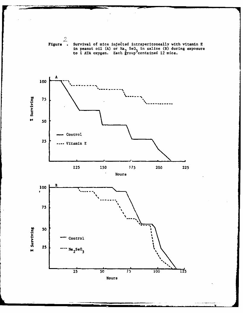
Figure .Survival of mice injeEted intraperitoneally with vitamin Ein peanut oil (A) or Na 2 SeO 3 in saline (B) during exposureto 1 ATA oxygen. Each group contained 12 mice.
A100
50
75
:'4
Cont50
25
25 50 75 00 1225
Hous

PULMONARY ANTIOXIDANT ENZYMES
We wished to evaluate vitamin E and selenium on pulmonary parameters
prior to mortality. It had been demonstrated that the activity of certain
antioxidant enzymes, e.g. glutathione peroxidanse, the hexose monophophate
shunt and superoxide dismutase, increased during oxygen exposure and thus might
be used as an index of oxidant stress(2). We therefore evaluated changes in
the activities of these enzymes as a function of treatments thought to protect
against oxygen poisoning--pre-treatment with hypoxia, sex and dietary supple-
mentation with vitamin E and selenium.
We switched to the rat as an experimental subject because of its larger
size for tissue sampling and better-defined dietary requirements.
Table 1 shows that the antioxidant enzymes are not different in male
vs. female rat lungs prior to or after 48 hours of hyperoxia.
Table 1. Mean + SD Lung Weiqhts (q) and Enzyme Activities (e.u./lO0 mgFresh--Tissue) in Rats (N = 4).
Sex Hours Lung Weights GSH-Px HMP SOD
Exposure
Male 0 1.53 + .17 .62 + .04 .23 + .02 1.22 + .11
24 1.66 + .05 .73 + .07 .26 + .02 . + .14
48 1.96 + .27 .51 + .07 .09 + .03 .- + .16
Female 0 1.28 + .10 .68 + .05 .18 + .02 1. .38
24 1 81 + .63 .70 + .06 .26 + .08 . + .25
48 1.99 + .29 .52 + .03 .07 + .02 .66 + .19
-17-

Table 2 shows that rats acclimatized to hypoxia prior to oxygen exposure
do not have different activities of the antioxidant enzymes.
Table 2: Mean + SD Lung Weights (g) and Enzyme activities (e.u./100 mg FreshTissue) for Normoxic (N) and Hypoxic (H) Rats Following 0, 24 or 48Hours of Oxygen Exposure (n = 4).
Treatment Lung Weight GSH-Px HMP SOD
N-O 1.40 + .17 0.51 + .03 0.31 + .07 0.67 + .12
N-24 1.49 + .13 1.18 + .12 0.28 + .01 0.61 + .17
N-48 1.82 + .44 0.43 + .10 0.32 + .10 0.54 + .10
H-O 1.98 + .11 0.51 .08 0.29 + .01 0.58 + .06
H-24 1.81 + .20 1.01 + .07 0.31 + .03 0.61 + .08
H-48 1.59 + .31 0.51 + .05 0.28 + .06 0.56 + .05
An attempt to induce elevated hexose riunt monophosphate activity in lungs
by the use of a low fat, fasting-refeeding technique was successful in liver but
not in lung (Table 3).
Table 3: Mean + SD Enzyme Activities Expiessed as Units per 100 mg FreshTissue (n = 5).
Enzyme Fat Content
Tissue 0166 1.2% 1.2%(F) 21.2%
HMP Liver 2.37 + 1.28 1.14 + .26 2.92 + 1.92 0.31 + .14
Lungs 0.32 + .08 0.36 + .04 0.37 + .03 0.32 + .07
-18-

Dietary selenium and vitamin E as apossible prophylactic to pulmonaiyoxygen poisoning
C. L. Schatte
INTRODUCTIONThe scope of oxygen therapy is limited largely by its potential toxicity. If thetoxicity could be ameliorated, the therapeutic use of oxygen might be extended.With the goal of delaying the onset of pulmonary oxygen poisoning duringcontinuous exposure at 1 ATA, I have experimented with the dietary manipulationof the natural antioxidant systems of pulmonary cells prior to oxygen exposure.This approach has the advantage of enhancing the cells' defence mechanisms byusing normally consumed substances. The extensive literature on the pharma-cological effects of many of these substances in humans implies that their potentialclinical use may be facilitated.
The cells' antioxidant defences apparently can detoxify metabolicallyproduced oxidants 2t their i,ormal rate of production, which is PO, dependent,(superoxide and peroxide radicals) but they cannot cope when the pO2 is increased.
The 'core' of the cells' antioxidant defence is probably the vitamin E-glutathione peroxidase system. Vitamin E (E) probably acts to preferentiallyprevent peroxidation of membrane polyunsaturated fatty acids. Lipid peroxideswhich do form are detoxified by a series of reactions, the most important of whichis catalysed by glutathione peroxidase (GSH-P,)
Several factors in the GSH-P, system are diet-dependent. One form ofGSH-P. requires selenium (Se), the level of which in the diet regulates theenzyme's activity.' Thus, a key antioxidant system enzyme can be induced tosupra-normal activity by elevating dietary Se. Glutathione (GSH) synthesisrequires cysteine, glycine and glutamate, the latter also serving as a source ofreduced phosphopyridine nucleotide (NADPH1 H) formation and as a precursor ofthe neurotransmitter gamma-aminobutyric acid which has been implicated in theaetiology of oxygen-induced convulsions.2 ,3
The activity of hexose monophosphate shunt (HMP) enzymes which helpcontrol the rate of NADPH H production-involved in the complex GSH-P.system-can be increased in liver by diet.',5
The importance of E-GSH-Px as an antioxidant system is illustrated bythe fact that GSH, GSH-PK IMP enzymes and GSH reductase have all beenshown to increase during adaptation to hyperoxia.6
Prof. 6th Wm. tog. on HYPERBARIC MED'CINE, 1977, pp. 84-91. Aberdeen University Press

_ -oom
PULMONARY OXYGEN TOXICITY 85
METHODS AND MATERIALS
The experiments wecre designed to determine prophylactic effects ofdietary supplementation 'ilth E and Se alone and in combination, on the onset ofpulmonary oxygen toxiciti. Weantling male Sprazue-IDa\Iev CD-i rats (CharlesRiver Company) w~ere ted a semii-purified diet (Table 1) similar to that of theaverage American 7 supplemented %%ith nothing, 6ooo ppm E (ioox<control)I PPM Se (30 X control) or both. After feeding periods of 4, 8 or 12 weeks, the ratswere exposed to pure oxygen at t ATA fo~r 24 or 48 hours. At the 4-wXeek testing,the oxygen exposures were 36 and 6o hours but unexpectedly high mortality at
HMP GSH-Px PROTEIN
U)cD35-Z
0.30- 600 VI0
'25- 500 Z
0 0
15- -300~
2 lo- 200
0Lo24 48 0 24148 -0 24 48 10
HOURS OF OXYGEN EXPOSURE
Fig. 1 Response of lung glutathione peroxidase (GSH-P,). hexosemanophasphate shunt enzymes (HMVP) and lung wash protein content(Protein) in rats exposed to pure oxygen at 1 ATA for 0, 24 or 48 hours.
4

r..
86 OXYGEN TOXICITY
6o hours (see Results) necessitated the 24- and 48-hour exposures at 8 and z2weeks.
After oxygen exposure the lungs were removed and lavaged. Lung washeswere analysed for protein content," presumably a good indicator of oxygen-induced lung damage.9 GSH-Px10 and the HIMP enzymes"' were measured on ahigh-speed supernatant made from homogenised lungs.
RESULTS
The results of an experiment demonstrating the progressive increase it theactivities of lung GSH-P., HMP and lavage protein content during uxygen
MEAN ± S.E.M. LUNG CYTOSOLIC GLUTATHIONE PEROXIDASE ACTIVITY (n=4)
4 WEEK +
8 WEEK T-
20-
I~ + _" 10
2 44 .248 248 2448
20 12 WEEK
1 24 4 48 24 4 8 _eBA BO Se E SeE
TREATMENTSFig. 2 Response of whole lung glutathione peroxidase (GSH-P,) activityof rats fed a basal diet and exposed to air (SA) or oxygen (60) or fedthe basal diet plus Selenium (Se), vitamin E (E) or both (SeE) andexposed to oxygen. Numbers in the upper left hand corner of each panelindicate duration of dietary pre-treatment. Unless otherwise indicatedin parentheses, each bar represents four observations.

PULMONARY OXYGEN TOXICITY 87
MEAN ± S.E M LUNG HEXOSE MONOPHOSPHATE SHUNTENZYMES ACTIVITY (n=4)
4-4 WEEK
2 - NO DATA
4- 8 WEEK
2 4-I
6- 12 WEEK -
4- T IT
2
2448 24148 12414 2414 24 i48
BA 80 Se E SeE
TREATMENTSFig. 3 Response of wnole lung hexose monophosphate shunt enzymes(HMP) to oxygen exposure. See Fig. 2 caption for synibol explanation.
exposure arc shown in Fig. t. Note that GSH--P, acnivitv and protein content
* ncrease substantiallv between 24 and 48 hours, a time duringz which noticeablehistological evidence of toxicity appears.
In diet experimnicts note the trend of mc.:.nred parameters between 24 and43 hours and assess the efficacy of a particular treatm'mtn bastd on w~hether or notit altered that trend. Mlany observed changes '%cre statistically significant; thesrnall number of observations precludes any dctiitiyc conclusions.

88 OXYGEN TOXICITY
MEAN t S.EM PROTEIN CONTENT OF LUNG WASH (n'4)
4 WEEK
2000-
1000 (I
z 8 WEEK
2000-
1000-
_ n F 44
BA 80 3 E SeETREA', KENTS
Fig. 4 Response of lung wash prc-)t~in c ontent to oxygen exposure.See Fig. 2 caption for explanation.
The results for wNhole lunmz GS I-Px ar( shiow\n in Uig. 2. The high nmortalitvafter 6o hours oxvtzen exposure during the 4'%weeK te sts prcluded param~etric trendanalysis. The pattern of' mortality e,,Ident \\ a4 consistent \Ith trends obwervedduring subsequent tests at 8 and i 2N weeks. The-re wtere no sur\ ivori in untreatedoxygen-exposed controls after 6o hour,,, of .1 rats aliv e in L-fed rats xith orwithout Se, and 3 Of 4 rats ali~e in the Sc-itupplemeitedl group. The higher

PULMONARY OXYGEN TOXICITY 89
GSfI-P. leels seen in both Se-fed groups suggested induction of the enzyme bySe during the pre-exposure diet treatment period.
At 8 weeks, there was uncharacteristically no increase in enzyme activitybetween 24 and 48 hours in the untreated, oxVgen-exposcd rats, a finding contraryto those of this and other experiments. Rats fed supplemental Se again showechigher GSH-Px acti% its' than controls but no increase betNucen 24 and 48 hoursoxygen exposure. E-fed rats had slightly below control activity at 24 hours and alarge increase in activity between 24 and 48 hours, suggesting little protectionagainst oxygen toxicity.
After 12 weeksof feeding, all three treatment groups were protected based cino increase in enzyme activity between 24 and 48 hours when compared to un-treated controls. Interestingly, the presumed Se-dependent stimulation of GSH-P,activity in Se-supplemented rats observed after 4 and 8 weeks was absent at 12
weeks. This may be significant because it suggests that the apparent protectionof Se may not have been linked to GSH-P, activity.
The data for the HMP enzymes (Fig. 3) were consistent with those forGSH-Px. Data for the 4 week tests were discarded for technical reasons. That for8 and 12 weeks indicated that Se-supplementcd rats were protected somewhatbased on no increase in enzyme actiity betwcen 24 and 48 hours. Vitamin Esupplementation apparently was lot eltective until 12 "weeks. As with GSfl-Px, Se-fed rats tended to have higher enzyme activity at 24 hours than controls.
Figure 4 shows the data for protein content of lung lavages. At 4 weeks,those rats still alive after 6o hours oxven exposure were clearly in worse conditionthan unexposed air controls. After 8 weeks, the Se- and the E-supplemented rat,were somewhat protected based on the increase in alveolar protein between 24 and48 hours; those fed both SC and E were not. After 12 weeks, all treatment groupstolerated oxygen exposure better than untreated controls.
DISCUSSION
The results suggest:
i. Dietary Se supplemenation for 4 or more weeks elevates SGH-F"activity of whole lung and appears to attenuate the severity of pulmonaryoxygen poisoning resulting from continuous exposure at i ATA: tl'cmode of action of Se is not knoNn, besides its involvement with GSH-P.it is also possible Se functions in electron transportia2. Dietary E supplementation offlcrs some protection against toxicity butrequires a feeding period of iz weeks and may not be as great as that ofSe. The mode of administration of E may he a factor since intra periton-eal injections exert a very dramatic protective etlect.3. There appears to be no synergistic eIect between E and Se with regardto protection against toxicity although E did appear to reduce GSII-P.activity in some cases.

90 OXYGEN TOXICITY
The use of Se and E as prophylatic agents is experimental and requires anaccurate description of dosages, duration of supplementation, and mechanism ofaction. However, their potential is enhanced by -he fact that they are natural tomammalian systems and their biological roles are, to some degree, understood.Pursuit of this potential can lead to their use clinically and may allow safe extensionof the currently accepted oxygen exposure limits.
ACKNOWLEDGMENT
This work was supported by contract Nooo14- 76-C-0 4 37 from theOffice of Naval Research with funds supplied by the Naval Research and Develop-ment Command.
REFERENCES
i Chow, C. K. and D. L. Tappel 1974 Response of glutathione peroxidase todietary selenium in rats. j. Vutr. 104: 444-51
2 Wood, J. D. and S. J. Pcesker 1974 Development of an expression whichrelates the excitable state of the brain to the level of GAD activit',, andGABA content, with particular reference to the action of hydrazine and itsderivatives. J. Neurochem. 23: 703-12
3 Wood, J. D. and S. J. Peesker 1975 The anti-convulsant action of GABA-elevating agents: a re-evaluation. J. .VNurochem. 25: 277-82
4 Tepperman, H. M. and J. Tepperman 1963 On the response of hepati"glucose-6-phosphate dehydrogenase activity to changes in diet compositionand food intake pattern. Adv. Eny. Reg. Vol. 1. G. Weber (ed.). AcademicPress
5 Johnson, B. C. and H. F. Sasson 1967 Studies on the induction of liverglucose-6-phosphate dehydrogenase in the rat. Adv. Eny. Reg. Vol. V.G. Weber (ed.). Academic Press
6 Kimball, R. E., K. Reddy, T. H. Peirce, L. W. Schwartz, M. G. Mustafa andC. E. Cross 1976 Oxygen toxicity: augmentation of antioxidant defensemechanisms in rat lung. .Am. J. Physiol. 230: 1425-31
7 Consumer and Food Economics Research Division, ARS, USDA 1969 Foodintake and nutritive value of diets of men, women and children in the U.S.,Spring 1965. Printing Office, Washington, D.C.
8 Lowry, 0. H., N. J. Rosebrough, A. L. Farr and R. J. Randall 1951 Proteinmeasurement with the Folin phenil reagent. J. biol. Chem. 193: 265-75
9 Niinikoski, J., T. Nikkan and 13. Kulonen to7I Pulmonary oxygen toxicity:composition of endobrozichial saline extracts of rats during exposure tooxygen. Aerospace Med. 42: 525-9
to Paglia, D. E. and W. N. Valentine i967 Studies on the quantitative andqualitative characterisation of erythrocyte glutathione peroxidase. 5. Lab.din. Aled. 70: 158-69

PULMONARY OXYGEN TOXICITY 91
ix Lohr, G. W. and H. D. Walker 1965 Glucose-6-phosphate dehydrogenase.In: Methods of Enzymatic Analysis. H. U. Bergmeyer (ed.). AcademicPress, New York
x2 Diplock, A. T. and J. A. Lucy 1973 The biochemical modes of action ofvitamin E and selenium: a hypothesis. FEBS Lett. 29: 205-Io

The effects of dietary supplementation with vitamin E and selenium
on the response of the antioxidant enzymes during hyperoxia is described in the
following paper:
Scatte, C.L. Dietary selenium and vitamin E as a possible prophylactic topulmonary oxygen poisoninq in Proc. VIth Internat. Cong. Hyperbaric Med., ed.G.E. Smith, Aberdeen University Press, 1979.
-19-

DIETARY FAT AND PULMONARY PROSTAGLANDINS
The results of an experiment in which dietary fat content was varied
suggested that lipid metabolism might influence susceptibility to oxygen
toxicity (Table 4).
Table 4: Mean + SD Enzyme Activity (e.u./lOO Uq Fresh Tissue) and Mortalityof Rats after 72 hours Exposure to Oxygen.
Fat Content GSH-Px HMP Mortality
Lab chow, 5% .28 + .05 .33 + .12 1/11
Synthetic, 5% .40 + .10 .27 + .03 3/10
Synthetic, 9.3% .63 + .12 .32 + .04 5/11
Synthetic, 21.2% .47 + .13 .31 + .04 10/11
Synthetic, 36.5% .56 + .13 .29 + .01 9/10
Because dietary fat content can influence the formation of prostaol 2dins,
substances having a range of physiological effects which might impact oxyg'en
toxicity, we carried out a series of experiments desiqned to measure chanoes in
lung prostaglandin levels during hyperoxia, the effect of dietary fat on those
levels and the effect of hyperoxia on metabolism of the prostaglandins.
The following papers described those experiments:
Meydani, S.N., M.M. Mathias and C.L. Schatte. Dietary fat type and ambientoxygen tension influence pulmonary prostaqlandin synthetic potential. Pris-taglandins and Medicine 1:241-249, 1978.
Vader, C.R., M.M. Mathias and C.L. Schatte. Pulmonary prostaqlandin metabolismduring normobaric hyperoxia. Prostaglandins and Medicine, in press, 1980.
Schattr. C.L. and M.M. Mathias. Pulmonary prostaglandin metabolism durinqnormobai'c hyperoxia. in Proc. VII Symp. Underwater Physiol., ed. C.W. Shilling,in press, 1981.
-28-

Prostaglandins and Medicine 1: 241-249, 1978.
DIETARY FAT TYPE AND AMBIENT OXYGEN TENSION INFLUENCE PULMONARYPROSTAGLANDIN SYNTHETIC POTENTIAL
S. N. Meydani, M. M. Mathias, C. L. Schatte. Department ofFQod Science and Nutrition and Department of Physiology andBiophysics, Colorado State University, Fozt Collins, Colorado80523. (reprint requests to CLS)
ABSTRACT
Chronic hyperoxia produces pathological changes in lung whichcan be fatal. With an interest in delineating dietary factorswhich might affect the pulmonary response to hyperoxia, we fedrats a semi-synthetic diet containing polyunsaturated fattyacids (PUFA) as either 5% or 78% of the fat complement. Tierats were exposed to pure oxygen at one atmosphere. Half theanimals in each diet group were injected with aspirin Cd ring thehyperoxic exposure. Radioimmunoassay of lung prostaglardins (PG)F 2a, E2 and E, were performed at 0, 24, 48 and 72 hours. ThEmajor findings were: (1) Feeding the high PUFA diet elevated lungPG synthetic potential tenfold over that of low PUFA-fed animals.There was no effect of diet on mortality. (2) Hyperoyia signi-ficantly increased F2a-synthetic potential wiring the first 24hours of hyperoxia and moderately increased the synthe ).c- po-tential of E 2 and El. (3) Aspirin significantly deprezcdsynthetic potential of all three PG prior to oxygen expos ure butits effect was overcome during hyperoxia. Aspirin-injected ratsshowed 80% mortality in oxygen vs. 50% for saline contrvli.
We concluded that dietary PUFA and hyperoxia alter PG syntheticpotential but its role in the pulmonary response to hypercxiaremains obscure.
INTRODUCTION
Chronic exposure to hyperoxia causes pulmonary edema andcongestion which can be fatal (1). It has been well establishedthat prostaglandins (PG) are intimately involved in lungfunction; PGF 2a has pulmonary vasoconsttictor activity whilePGE, is a vasodilator in most species studied (2) including rats(3). Our interest in identifying dietary factors which might
24

influence the pulmonary response to hyperoxia has led us tofocus on PG metabolism for the following reasons. PG synthesisis known to require oxygen and essential fatty acid:. It isconceivable that a net increase in vasoconstrictor PG activityduring exposure to hyperoxia might enhance the edema formation.Aspirin, a known inhibitor of PG synthesis, has been shown toexacerbate the toxic effects of hyperoxia (4). It has beendemonstrated that in vitro hyperoxygenation will accelerate PGproduction by rat renal-medulla (5) and that synthesis by atleast adipose tissue and platelets can be influenced by dietaryfat type (6,7). This study was conducted to investigate anychanges in pulmonary PG synthetic capacity following treatmentwith diets containing different amounts of polyunsaturatedfatty acids (PUFA) and during exposure to hyperoxia.
MATERIALS AND METHODS
One hundred-thirty-two weanling Sprague-Dawley-derived rats(Charles River, Wilmington, !A) were maintained so as to keepchronic respiratory disease -t a minimum. They were fed a basaldiet containing 19.0% casein, 0.4% dl-methionine, 25.0% corn-starch, 24.9% sucrose, 2.5% cellulose, 5.0% salt mix, 2.0%vitamin mix, and 21.2% fat. Half the animals were fed a dietcontaining primarily saturated fat (approximately 5% PUFA) asbeef tallow and the other half approximately 78% PUFA as saf-flower oil. The diet containing beef tallow as the fat sourceclosely approximated that consumed by the average American (8).The diets and tap water were provided ad libitum for one month,after which 18 rats from each dietar; group were exposed to airand the rest were exposed to oxygen at 1 atmosphere in controlledenvironment chambers (9). In our mc3Rl system oxygen exposureproduces discernible pulmonary biochem-cal changes after 24 hours,incipient edema and histological symptoms at 48 hours and up to100% mortality by 72 hours.
In order to gain insight into the prcsible effect of aspirin onPG synthetic capacity during hyperc: ia, half of the animals ineach dietary group were injected intraperitoneally with 50 mgaspirin/kg in saline at 1, 24 and .18 hours of oxygen exposure.This dose rate was determined, during a preliminary experiment,adequate to maintain a pulmonary concentration in the range of20-100pM for 16-20 hours. That concentration range providedup to 50% inhibition of PGF 2c, PGE 2 and PGE, synthesis by ratlung homogenate in vitro (unpublished results).
At 24, 48 and 60 hours, groups of animals were killed withchloroform. The left lung was homogerized in 10 volumes of42 mM aspirin in 0.1 M potassium phosphate buffer (pH 7.4) witha teflon-pestle, glass-vessel homogenizer within 2 minutes. PGwere probably synthesized during tissue manipulation so the PGcontent reflected the physiological potential of the tissue forPG biosynthesis. The homogenates were kept frozen for sun-sequent analyses for PGF 2 a, PGE 2 and PGEI using the radio--imunoassay described by Hwang et al (7) and McCosh, et al (10).
" 'U"MliM . ..

This method involved overnight precipitation of each prosta-glandin-specific antiserum with the rabbit anti-gamma globulin(Method II). The PGEI antiserum had a cross-reactivity of8+2% with PGE2 and the PGE 2 antiserum had a cross-reactivityof 6+1% with PGEI. The cross-reactivity with 6-keto-PFGa ofthe antisera for PGF 2a, PGE2 and PGEI was 2%, 0.5% and 1.0%,respectively. Parallelism was established and the recoveriesof PG were shown to be 90-100%.
The results were analyzed using a 2 (oxygen response for 24 and48 hours) x 2 (fat) x 2 (aspirin) factorial design analysis ofvariance. The controls and 60-hour results were analyzed byt-tests due to the unequal sample numbers. All data are pre-sented as mean + standard error.
RESULTS
Figures 1-3 illustrate the response of the measured PG todietary fat treatment, aspirin injection and continuous ex-posure to hyperoxia. We recognize that cross-reactivity of anti-bodies used for radioimmunoassay precludes accurate statements oftrue PG concentrations; all references to PG actually indicateconcentrations of F 2U-, E2- and El- like material in the assaysamples. The number of observations in each group for the sampletimes were: 0 hours-9; 24 hours-8; 48 hours-8; 60 hours-from 1-4.The variable sample number at 60 hours of oxygen exposure re-sulted from an unexpectedly high mortality at that time. Whiledietary fa- type had no effect, aspirin-injected rats experienced80% mortality between 48 and 60 hours vs. 50% for saline-injectedanimals.
The most -rominent effect on pulmonary PG synthetic potential wasthat of d*.tta-y fat type. Rats fed the high PUFA-safflower oildiet showad significantly (p<.001) higher levels of the three PGat all sample times than did those fed the low PUPA-beef tallowdiet. The relative magnitude of the difference in saline-injected, non-oxygen-exposed controls was 8-, 13- and 11-foldfor F 2c, E2 , and El, respectively.
Aspirin injection significantly (p<.001) decreased all three PGlevels in non-oxygen-exposed rats. The respective decreases forthe oil-fPc animals were 69% for F 2c, 52% for E2 and 58% for El,while those for tallow-fed animals were 48% for F2a, 25% forE2 and 29% for El. These findings correspond to our observationsin vitro that F2a production was mc~e sensitive to aspirin inhibi-ton--han either E2 or Ei. Of particular interest was the factthat raising the ambient oxygen tension from the approximately130 mmHg in air to >700 mmHg during hyperoxia attenuated thedepressant effect of aspirin injection on PG synthetic potential.Hyperoxygenation appeared to be a more potent stimulator of PGsynthetic potential than was aspirin a depressant.

Figure 1. Concentration of PGF2a-like material in rat lunghomogenate.
40-S~e T*$at. Fat & So n0 PUFA & Saline
MSot.Fot & Asp. rin PA&AIpi
S.
30-
d20-
LL-
0 24 is 72
HOURS OF OXYGEN EXPOSURE

Figure 2. Concentration of PGE 2-like material in rat lunghomogenate.
40" eSot. Fa & Saline 0 PUFA & Saline
lSat. Fot &Aspirin oPUFA&Aspifin
30
20
(L-
0!
24 4 " 7
HOURS OF OXYGEN EXPOSURE

Figure 3. Concentration of PGE 1-like material in rat lunghomogenate.
so-
40.
* Sat. Fat & Solihe
8 Sat. Fat & Aspirin0.30-
-. O PUFA &So'.neLJ
OPUFA&P-pirin
20-
10-
060 24 48 72
HOURS OF OMY1jEN EXPOSURE

That hyperoxia did significantly (p<.Ol) increase F2a b, t notE2 or E, during the first 24 hours' exposure of non-aajirin-injected rats fed the safflower oil diet was a third Iajorfinding of this study. It is consonant with the report thatthe rate of FPa synthesis by rat kidney medulla was acceleratedin vitro when gassed with 95% oxygen (5). PGF 2a in the oil-fedrats significantly (p<.05) decreased between 48 and 60 hours,perhaps in response to the hypoxemia known to occur prior todeath (11).
DISCUSSION
The results clearly demonstrate that elevated dietary PUFA andambient oxygen tension increase the PG synthetic potential ofrat whole lung. The oxygen effect was most prominent for F2aand was maximal after 24 hours' exposure. If PG are invclved inthe pulmonary symptoms which occur during chronic hyperoxia,the changes during the first 24 hours ma.' be critical. Few bio-chemical or histological changes are observed at that time (1).
Our data do not provide information concerning the in vitrophysiological responses to the change in synthetic potential 'Jutthree observations are pertinent. Administration of aspirin, aninhibitor of PG synthesis which is known to predispose patientsreceiving hyperoxic therapy to CNS symptoms (12), increasedmortality and decreased the zero time PG synthetic potentialsubstantially in all diet treatment groups. These two phenomenamay or may not be related but the fact that aspirin inhibitionof synthetic potential was overcome after 24 hours' exposureto oxygen points to the changes in PG synthetic potential OJrirgthis time as both substantial and probably important.
The second point is that, despite a 10-fold increase in syn-thetic potential of the oil-fed rats, there was no difference inmortality between the two diet groups. While this fact seeminclysuggests that PG synthetic potential is not important in thepathological response of the lung to oxygen, we believe tbitmay not be the case. We feel rather that substantial cha:.'esin the synthetic potential of substances with powerful vaso-activity such as prostaglandins probably constitute a respornseof the cell to the hyperoxic stress and that the relativelycrude parameter of mortality was not sufficiently sensitive toreflect more subtle differences between the two diet groups.Further experiments incorporating sensitive histological andfunctional parameters, particularly during the first 24 hours ofhyperoxia, would provide a more definitive assessment of anyeffect resulting from the diet-induced changes in PG syntheticpotential.
The third observation of interest is the fact that aspirin poten-tiation of oxygen toxicity may not be PG-related. While aspirinadministration increased mortality, there was no significantdifference in PG synthetic potential between the aspirin- andzaline-injected animals after 24 hours of oxygen exposure.

Either the aspirin effect does not involve PG or the lower PGsynthetic potential of aspirin-injected rats prior to andduring the first 24 hours of hyperoxia was critical.
It is possible that PG-related factors other than syntheticpotential might explain the results. Recently-discovered inter-mediates and other products such as thromboxane A 2 or PGI2 arephysiologically more potent than the PG we measured (31) and mayhave been responsible for our results. The rate of PG catabolismmay have been altered since PG dehydrogenase has been reportedto be inhibited by hyperoxia (14). Perhaps the rate of PGconversion to less potent substances is more crucial than syn-thetic potential or endogenous levels at any given time. Thelungs' sensitivity to hyperoxia may well be related to anyimpairment of its central role in PG catabolism.
Whatever the mechanisms involved, this study suggests that bothdietary fat type and ambient oxygen tension are factors pertinentto pulmonary prostaglandin synthesis. To the clinician utilizingdietary fat alterations or oxygen therapy as treatment, thepotential consequences of increased prostaglandin synthesis mustbe considered. To the basic scientist working with prostaglandinsynthesis, our results indicate that endogenous substances (PUFAand oxygen) may be controlling factors of the lung's PG syntheticpotential. We are further investigating these possibilities.
ACKNOWLEDGrENT
This work was supported by Office of Naval Research ContractN00014-76-C-0437 with funds supplied by the Naval Medical Re-seazch and Development Command.
LITERATURE CITED
2. Clark JM and Lambertsen CJ. Pulmonary oxygen toxicity: areview. Pharmacol. Rev. 23:38, 1971.
2. Ki-dowitz PJ, Joiner PD and Hyman AL. Physiological andpharmacological roles of prostaglandins. Ann. Rev. Pharmacol.15:285, 1975.
3. Weir EK, Reeves JT and Grover RF. PGEI inhibits the pulmo-nary vascular pressure response to hypoxia and PGF 2 a.Prostaglandins 10:623, 1975.
4. 3errill S, Jefferson D, Quick J and Mengel CE. Effect ofacetylsalicylic acid and ascorbic acid on oxygen toxicity.Aerospace Died. 42:436, 1971.
5. ! enser TV, Levitt MJ and Davis BB. Effect of oxygen andsolute on PGE and FGF production by rat kidney slices.'rostaglandins 13:145, 1977.
6. Christ EJ and Nugteren DH. The biosynthesis and possiblefunction of prostaglandins in adipose tissue. Biochem.Biophys. Acta. 218:293, 1970.

7. Hwang DH, Mathias MM t, Dupont J and Meyer DL. Lineoleateenrichment of dief and prostaglandin metabolism in rats.J. Nutr. 105:995, 1975.
8. Consumer and Food Economics Research Division, ARS, USDA.Food intake and nutritive value of diets in men, women andchildren in the United States, Spring 1965. U. S. GovernmentPrinting Office, Washington, D. C., 1966.
9. Jordan JP, Huston LJ, Simmons JB, Clarkson DP and Schatte CL.An environmental chamber system for prolonged metabolicstudies on small animals. Space Life Sci. 4:424, 1973.
10. McCosh EJ, Meyer DL, and Dupont JD. Radicimmunoassay of
PGEI, E2 and F2c in unextracted plasma, serum and myo-cardium. Prostaglandins 12:471, 1976.
11. Valimiki M. Arterial and tissue gas tensions in rats duringdevelopment of pulmonary oxygen poisoning. Aviat. SpaceEnviron. Med. 46:883, 1975.
12. Hart GB, Pierce EC, Strauss MB. The incidence of centralnervous system (CNS) oxygen toxicity in a clinical settinq.in Proceedings of the VIth International Conference onHyperbaric Medicine 1977. Aberdeen University Press,Aberdeen, 1978 (in press).
13. Samuelsson B. Introduction: New Trends in ProstaglaniinResearch. p.1 in Advances in Prostaglandin and ThromooxaneResearch. (B Samuelsson, R Paoletti eds) The Raven Press,New York, 1976.
14. Parkes DG and Elinq TE. The influence of environmentalagents on PG biosynthesis and metabolism in the lung.Biochem. J. 146:549, 1975.

PULMONARY PROSTAGLANDI NE TABDLISM- DURING
NOROBARIC HYPEROXIA
Connie R. Vader, Melvin 14. sathias and Christopher L. Schatte
Department of Physiolcqy and Biophysics
and
Departent of Food Science and Nutrition
Colorado State University
Fort Collins, Colorado 80523
(reprint requests to CLS)

ABSTRACT
Prostaglandin metabolism by rat lung tissue was measured followingexposures of 6, 24 and 48 hours to either pure oxygen or air at oneatmosphere. Tissue concentrations of PGE1 , PGE 2 and PGF 20 were notaltered by oxygen exposure. Prostaglandin synthetase activity decreasedbetween 24 and 48 hours but was not significantly different from controlat 48 hours. Combined prostaglandin dehydrogenase/reductase activitydecreased betveen 24 and 48 hours to 13% of control values and wassignificantly lover than in air at 48 hours. The plasma concentrationof 13,14 dihydro-15-keto PGF2a, a catabolite of PGF 2a, was significantlylo%:er in oxygen-exposed rats at 24 and 48 hours. '-e conclude thatendogenous pulmonary prostaglandin concentrations are maintained duringhyperoxia but that catabolism of pgostaglandins by the lungs may beimpaired.

INTRODUCIION
Exposure to oxygen at an ambient pressure of one atmosphere producespulmonary pathology characterized by congestion, edema, capillary endothelialcell necrosis and consolidation (1). While no relationship between prosta-glandins (PG, and pulmonary oxygen toxicity has been demonstrated, severalobservations ouggest such a possibility.
First, r:lecular oxygen is a requirement for PG synthesis. If it is arate-limitine reactant, increasing available oxygen might increase PGproduction. Consistant with this possibility, production of PGE and PGFseries was er-anced by hyperoxia in rat renal medullary slices (2) and lungtissue hornogeates (3).
Second, one of the mechanisms by which hyperoxia is thought to exertdeleterious effects on cells is through accelerated production of peroxides(4). Increased peroxide tone has been reported to enhance the activity ofPC synthetase (5).
Third, aspirin, an inhibitor of PG synthetase, exacerbated convulsionsinduced by ox×gen at high p. ?ssure (6) and increased mortality during oxygenexposure at one atmosphere 3). Indo7.ethacin, another inhibitor of PGsynthesis, partially attenuated the decrease in pulmonary vascular resistanceebser\ed durimo hyperoxia (7) but did not affect the mortality of ratsexposed to nor-obaric hyperoxia (8). The vasoactivity of selected PG hasbeen shown to vary as a function of oxygen tension (9).
Finolly, there is evidence that the endothelial cells of the pulmonaryvasculature ::e the first to show signs of necrosis resultino from oxygentoxicity (In . Vascular endothelial cells have also been identified as aprimary sit, o. sythesis of prostacyclin, or PGI 2, one of the mostpotent vasodi.atczs and anti-aggregating agents in the circulation (11).
These otservations present the possibility that aberrations in PGmetabolism d r-,ng oxygen exposure might be Pssociated, causally or otherwise,with the vzs>-lar pathology which produces the knowin sumptoms of oxygenpoisoning.
This st.nyo% ..as undertaken to profile plasma and lung tissue concentra-tions of selected PG and the activities of key enzymes during exposure tonormobaric h- eroxia.
METHODS
lie performed two similar experiments which differed primarily in dura-tion of exposu;e to hyperoxia. In both, chronic disease-free male Sprague-Dawley rats (Charles River Co.) v;eiohing approximately 180g w,:ere indi\iduallycaged in a room maintained at 220C and 40% relative humidity. Wlater anda semi-synthetic diet (3) were available ad libitum. The diet compositionwas similar to that consumed by the averaoe AmFerican, containing 40% ofcalories as fat with a polyunsaturated: saturated fatty acid ratio of0.7.

After 10 days of diet feeding, the animals were randomly divided into
groups for exposure either to air or pwre oxygen at one atmosphere. Expo-
sures were carried out in converted autoclaves with a volume of 300 liters.Ambient temperature was maintained at 25-270 C by means of a water jacket.
Carbon dioxide concentration was held at <0.5% by adjusting gas flow throughthe chamber. Animal waste dropped through the floor grid onto absorbentmixed with boric acid to reduce the buildup ammonia. Diet and water were
available ad libitum throughout the exposure.
Animals vwere sacrificed by decapitation after 0, 6 or 24 hours of
exposure in the first experiment and after 6, 24 or 48 hours exposure in the
second. About lOml of blood %:as collected in polypropylene cups containingfinal concentrations of 1 PM indomethacin and 10 mg LDTA to stop PG synthesis.
Blood was mixed during collection to prevent clotting and then Lept on
ice.
Lungs weie excised with a minimum of mechanical stress and processedfor determination of endogenous PG concentrations or the activities of PGsynthetase and combined PG dehydrogenase/reductase as follcw-s.
Plasma and Tissue Prostamlandins
Blood was spun at 60,000g for 20 minutes at 40 C to obtain clatelet-poor plasma, which :as then adjusted to pH 3.8 + 0.2 w-ith O.1M citrate.Tissues which had heen quick-Frozen in liquid nitrocen at sacrifice 6.,erehomogenized 1:10 w/v in 0.1M citrate-phosphate buffer, pH 3.8, containing
1 mH indomethacin.
All samples %,ere extracted twice with 5 volumes ethyl acetate for45 minutes on a horizontal shaker. Recoveries ranoed from 87-95%. The
extracts were combined and dried under nitrogen, reconstituted to 2.5m1
with buffer and radioi-,unoassayed for PGE1 , PGU 2 , PGF2a and 13,14 dihydro-.
15-keto PGF rPGF2), the major catabolite of PGF2.
Prostaolandin Svnthetase
Freshly-excised lung tissue was homogenized on ice 1:5 %-,/v in 0.25,"sucrose containing 1mM EDTA and 0.111 Tris buffer, pH 7.4. Homogenates %!erecentrifuged at 9000g for 20 minutes at 40 C, the pellet removed, then recen-
trifuged at 100,O00g for 60 minutes at 40C. The microscmal pellet w.as
removed, the supernate boiled for 5 minutes and the denatured protein
removed by centrifuging at 7000g for 15 minutes. Protein concentration w,.as
determined by the microbiuret method (12) and the microsomal suspension
adjusted to a protein concentration of 10mg/ml.
Enzymatic activity was neasured using a modification of the method ofParkes and Elino (13). The following mixture %:as incubated at 37°C for
two minutes: 1.C.ml 0.1M Tris buffer, pH 9.0, 100 '4 1.0m" CuSC-4 , 100 Vi
20m' dithiothreitol, 5C0 boiled supernate, 100 pl of 0.6 43 11 arachidonicacid and 100 il of the microomal suspension. The reaction ;.as stoppeo by
-j

adding 50 p1 1N HC1. Excess arachidonate was imrediately removed by extrac-tion with lOml hexane. Sample pH was adjusted to 7.0 with 50 pl 1N NaOH.The amount of product generated during-the incubation %.as determinedby radioimmunoassay for PGF2a. Blanks contained 100 pl buffer instead ofthe microsomal preparation.
Combined Prostaolandin Dehvdrooenase/Reductase (PGDH/R)
Freshly-excised lung tissue was homogenized on ice 1:10 w/v in 0.0921.1potassium phosphate buffer, pH 7.3, containing 4,rri [qC1 2 and 0.1mM dithio-threitol. The homooenates were centrifuged at 10,000g for 20 minutes, thepellet removed, and recentrifuoed at 78,0 00 g for 60 minutes at 40C.Protein concentration in the supernatant was determined by microbiuret andadjusted to 10mq/ml.
Enzyme activity was deterhined usino modifications of the methods ofParkes and Eling (14) and Lee and Levine (15). The follow;ing mixture wasincubated at 370C for &L0 minutes: Lnl pl 0.251M potassium phosphatebuffer, pH 8.0, 100 p 8ml1 NAD, 100 11 20 pg/ml PGF2? and 100 Pl supernatant.Following the incubation, 100 pl 32mw', NADH was added to inhibit PGDH andserve as a cofactor for the reductese. The incubation %,-as continued for 20minutes, then stopped by adjusting the pH to 3.0 .,ith 100 1 1.5', H1 andplacing the reaction tubes on ice. Prior to r~dioimunoassay for 13,11,dihydrc-15-1,eto PGF , sa.ple pil %;as acjusted to 7.0 ,,ith 100 A 1%' %aOH.Blanks contained 100 P1 buffer in lieu of the enzyme preparation.
Radioi77unoassav
Double antibody sequential precipita£ on radioimmunoassays (flethod I)were done under equilibrium conditions at 'i°C (16). Antisera for PGE andPGFX v'ps obtained from Research Products i'ternational, Elks GroveVillace, IL; for PGE 2, from Sioma Chemical Co.. St. Louis, HO; and for13,14-dihydro-15-"eto PGF24 fro J. D. Duport and .I. .1. Vathies, PeDartrrerit
of Foc Science and Nutrition, Coloradc State University. Cross reactivitiesare shown in Table 1. Recoveries of added P3 ranoed from 90-104% and paral-lelisr was established for all assays. .Iiterassay precision expressed as thcoefficient Of Variation (n=6) %12S 6.6'. for PGEI, 3.C'e% for PGE2, 7.8. forPGF t and 8X0? for mPGF 2X.

Table 1. Specificity of the prostaolandin aniisera used in the r.-dio-immunoassays.
Antisera
Prostaglandin PGEla PGE2 a PGF 2aa mPGF2 b
PGE 1 100.0 1.8 < 4.0 1.3PGE 2 4.3 100.0 < 3.0 0.2PGFla < 0.1 < 0.1 < 3.0 < 0.1PGF 2 L < 0.1 < 0.1 100.0 0.1PGF 9 Lb NAC NA < 3.3 NAmPGF 2 b < 0.1 NA 2.5 100.0,rRE"E79 < 0.1 0.1 NA 2.3mPGE 1b 0.1 < 0.1 NA 1.1Thromboxane B2 NA NA NA < 0.1Arachidonat, < 0.01 NA < 0.01 < 0.01
a Data obtained from supplier.b 13,14-dihydro-15-keto PG; data from 1-1. 1',athias.
c Not available.
RESULTS
In the first experiment, there v.rere no ",icnifzcant chances in luna tissuePG concentrations or the circulating ,etaboite -)t PGF 2, (Table 2). Lungtissue mPGF 2. levels tenoed to increase wit' )xyoen exposure v,h-ile PGF2adecreased but it is unclear whether or not t 4 ,ere related.
Table 2. Effect of air or oxygen on lung and plasma prostalandinconcentrations (Experiment I).
Hours of Lune Plasma
ExposurePGF 2 PGE1 PGE 2 mPGF 2 a mPGF 2a
(rg/10 mg wet wt.) (ng/'ml)
0air 6.5 + 5.8a 4.4 + 2.5 2.4 + 1.5 2.2 + 0.8 3.9 + 0.8
6air 3.1 + 2.2 5.2 + 1.8 2.3 + 1.4 3.5 + 0.5 12.8 + 1.1oxygen 3.8 + 3.4 3.6 + 1.8 1.1 + 0.3 3.4 + 2.6 9.5 _ 0.8
24air 3.6 + 1.9 6.7 + a,.8 1.6 + 0.9 3.4 + 0.6 4.7 + i.6oXyqen 1.4 + 0.7 2.5 + 0.8 1.0 + 0.6 2.P + 0.6 6.2 + 1.0
a -n + S.E.M.. n = C.

In the second experiment (Table 3), tissue PG concentrations wereagain unaffected by oxygen hut plasma -,PGF2x was significantly reduced inoxygen-exposed rats after 24 and 48 hours. This presented the possibilitythat inactivation of PGF2. miaht have become impaired. Consistent withthis result, Crutchley et al. (17) recently reported that mPGF2 releasefrom isolated-perfused guinea pig lungs was depressed following exposure toh\ oeroxia.
Table 3. Effect of air or oxygen on lung and plasma prostaglandinconcentrations (Experiment II).
Hours of Lunq Plasma
ExposurePGE 1 PGE 2 PGF 2( mPGF 20
(ng/100 mg wet wt.) (ng/ml)
6air 1.7 + 0.7a 0.55 + 0.20 7.2 + 2.6 NPb
oxygen 1.8 + 0.9 0.68 7 0.20 5.8 + 2.3 NA24
air 1.2 + 0.3 0.38 + 0.20 4.7 + 0.7 &.0 + 0.3oxygen 1.7 + 0.6 0.61 + 0.20 5.4 + 1.9 2.2 + 0.6c
48air 1.3 + 1.5 0.51 + 0.30 4.3 + 3 5 4.1 + 0.8oxyqen 0.9 + 0.3 0.52 + 0.20 4.1 7 0.9 2.6 _; 0 . 5 d
a lean + S.E.M., n = 6.b Not available.c p<.001.d p<.03.
The activity of PG synthetase declined significantly (p<.O01) bet,-en24 and 48 hours (Ficure 1) of oxygen exposure, but V.as not sionificanti.different from air control values at that tire. This is consistent v:,ththe findincs of Parkes and Elina (14) iio observed no difference in svrthe-tase activity in ouinea pig lung after a similar exposure. PGDH/R activ-tvalso decreased sicnificantly at 4P hours (p<.O01) to 13% of the activity inair controls. Our results are quantitatively similar to the 7050 decline inactivity of PGDH cbsered in guinea pig lungs after &8 hours of hyperoxia(14) and an 850 decrease seen in the same species after 72 hours (17).Thus, the potential for normal rates of synthesis but lesser rates ofdegradation existed in oxynen-exposed animals althouqh we cannot say thatthe actual rates of synthesis or catabolism ;.ere altered in vivo.
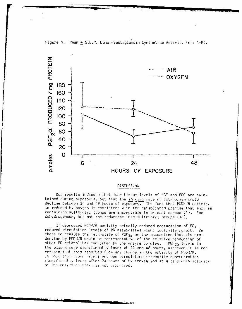
Figure 1. f.'ean + S.E.V'. Lung Prostaglandin 5ynthetase Activity (n 4-8).
z
AIRa. OXYGEN
o o IS160 -
) 40-120-
aL- 80o-d 6 0 -
tD40-
0
E 6 24 48HOURS OF EXPOSURE
DI SCUS jIj N
Our results indicate that lung tissu; levels of PGE and PGF are main-tained during hvperoxia, but that the io %ivo rate of cataholis, coulddecline between 2L4 and Zi8 hours of e,,posurs . The fact that PGH/R activitvis reduced by oxygen is consistent with the established premise that enzr'scontaining sulfhdryl croups are susceptible to oxidant damage (L). Thedehydrocenase, but not the reductase, has ,3ulfhydryl groups (1B).
If depressed PCOF/R activity actually reduced degradation of PG,reduced circulatino levels of P0 r etaholites miaht loqically result. VWechose to reasure the retalolite of PGF2ca an the assu:mption that its pro-ducLion by PGDH/R w:ould be representative of the relative production ofother PG r:?ta'mliLes converted by the enzyre corplex. mPGF 2a levels inthe plasma ..ere sirnificantly lo.'.er at 24 and 48 hours, althouah it is notcertain that this i'esulted from any chanqe in the activity of P"DI-l,"R.In onl. th. r;cu'_nd e ,:rri-(-nt .as circulating m,- taboljte concentrationsiCrific:4tylv Icv'r after 2d 'ours of hoperoxia and at a tire wt-en activityof lh, unzr- rr'!r, not r:prrssed.

Figure 2. I-lean + S.E.14 . Lung Co,-ibined Prostaglandin Dehydrogenase/ReductaseActivity (n 4-8).
0~300
E
0
Cj
0
E0
6 48HOURS OF E\P03URE
The timinq of these Changes in PC, r-tatholism are intriouin. in liqhtof the chronoloqv of known pathoinilcl E,' yptor-s associated with puli-,Dnarytoxicity. Olur r t -:r-jdel tolerates nor;::ha.-iv h\peroxia ...ell for 21, hours.Up until that tir-,e, no oi-ess syrpto-,s or histolooical L-.hr-ormalities areotbserved. The a,2tivities of key antioxid&ait enz\re,,s, such as alutathioneperoxidase, oh sc6ph hate deh\drocen--se and superoxide disrnutase, donot chsnce z-Dreci '-hlv (19) quctcFestinq th4L the cells are not yet respon-dino to cxi-iant st,-coss. By 48 hours, histological evidence of edema is2parent. There after, the animals' conditicn deteriorates rapidly.Respiratory distren-s, ar-orexia, weioht loss, intrapleural edIema and orosshiStolooiC;;l Path0o0Cy Occurs. Pulmonary dntioxidiant enzyme activitycfecreasos "18). 1'>rtalitv rangles from. 50-i0)JL% by 72 hours.
It th'._refcre s-2-7is likely that one or rore biochemical events occur-ring tpt~en 24' cnrt LB hours precedles, and f-erhopqs trinne"rs, the nnset ofo\vert to' icitv. t~clicve that an -Iterat ion in the r;Ptaholisl-, Lf PG,
p~-t cu~l a<rc-nein their c,'j~z.,Loiirn fray be urie of those chanqes.

Our data do not address the question of mechanism. But one possibilityis that an i!:alance of vasoconstrictor and vasodilator substances miohtalter pul,3nary ascular pressure and perhaps capillary integrity. Yam andRoberts (7) fo-,rd that hyperoxia caused a decreased pulmonary vascularresistance in n:onatal swine. The effect was attenuated by administration ofthe PG s.;thetase inhibitor, irndomethacin. If, for instance, catabolism ofdilator PK were selectively impaired more than that of constrictor PG, theresultina vasodilatation miaht produce or exacerbate the congestion andvascular stasis seen in the early stines of pulnionary toxicity. Prosta-
glandins are also involved in the integrity of cells through rediation ofionic transport arid calcium concentrations (20). If an alteration of PGmetabolism cor pro7ised the integritv of pulmonary capillary cells, edemaformation mioht be triggered or enhanced.
The field of prostanlandin pharmacology is currently ripe with newdiscoveries of more prostaQlandin and thrur.oxane £_ujhstarnces, r.any of whichmay be more irmortant than those measureo in this study. For exawrple,hyperoxia :as recently reported to enhance the release of Tx 2 insensitized isoiated-cerfused nuinea pig lungs (17); we have preliminaryevidence that svnthesis of this Dtent vicsoconstricter and platelet-acorecating a:ent is similarly v::reased by hyperoxia in vivo. '-e a repursuing the c,-estion of the rei tiur, ship between the cecrease in PCDH/Racti ity and the onset of rml,;Jcnary syrPtons as well as any pssilie rolenewly-disr overed prostaclandins and thro:.boxares nay have in pulmonaryoxygen toxicity.
ACK NO',JLEDCtA.ENT
This wao k wa.s supported by the Office of Naval Research, ContractN0001-76-C-5:17, with funds supplied by the Naval Medical Research andDeveloprent Cctmnd.
REFERENCES
1. Clark . !!, Lambertsen CJ: Pulmonary oxvoen toxicity: A review.Pharmacol. Rev. 23:38-133, 1971.
2. Zenser TV, Levitt HJ, Davis B8: Effect of oxven and solute on PGE andPGF prodiction by rat kidney slices. Prostaglandins 13:145-151,
1977.
3. 1,evdani Si',, "athias r:11, Schatte CL: Dietary fat type and abientoxygen teaision influence pulmonary prostaglandin synthetic potential.Prostagl-ndins and IMledicine 1:241-249, 1978.
4. Hauooard N: Cellular mechanisms of oxygen toxicity. Physiol. Rev.L8:311-373, 1968.
5. Hemler VE, Cook H'., Lands VEM: Prostaglandin biosynthesis can betriciered by lipide peroxides. Arch. Biochem. Biophvs. 193:340-3tt4,1979.
i

6. Serrill S, Jefferson D, Ouick J, Vennel CE: Effect of a:et% lslicvlicacid and ascorbic acid on oxygen toxicity. Aerospace f'ed. L2:436-436,1971.
7. Yam J, Roberts RJ: todification of alveolar hvperoxia-irnouced vaso-
dilatation by indomethacin. Prostaglandins 2:679-689, 1976.
8. Yam J, Roberts RJ: Pharmacological alteration of oxyoen-induced lung
toxicity. Tox. Appl. Pharmacol. 47:367-375, 1979.
9. Starling 19, Eliott FR: The effects of prostanlandins, ri-ostailandininhibitors and ox~oen on the closure of the ductus arteriosus, rjlrarvarteries and ur.bilical veins in vitro. Prostarilandins E:187-203,1974.
10. Adaimson IYR, Pc'.'den DH, V',vatt JP: Oxyoen poisoning in mice: Ultra-structural and sur-ctart studies durino exposure and reccvery. Arch.Pathol. 90:463-472, 1970.
11. Hloncada S, Vane JR: The role of prostacyclin in vascular tissue.Fed. Proc. 3:46-71, 1979.
12. Bailey JT: Tec!.rioues in Protein Chemistry. Amsterdam, ElsevierPublishino Co., 1.67.
13. Parkes DG, Eling TE: Characterization of prostaglanoin s~nthetase inguinea pig lun: isolation of a new prostaolardin derivative fromarachidonic acid. Biochem. 13:2598-2604, 1974.
14. Parkes DG, Eling TE: The influence of environmental aoents onprostaglandin bicsynthesis and metabolism in the luno. Piochem. .
146:549-556, 1975.
15. Lee SC, Levine L: Prostaqlandin retabolism II: identification oftwo 15-hydroxy prostaclandin dehydrooenase types. J. Piol. Chem.25n:548-556, 1975.
16. I'cCosh EJ, ",eyer DL, Dupont JU: Radioimmunoassay of prostanlandinsEl, E2, and Fa, in unextracted plasma, serum and myocardium.Prostaglandins 12:471-lE4, 1976.
17. Crutchley DJ, £ovd JA, Elino IE: Enhanced thro I.oxne 62 releasefrom challenord nuinea pia luna after oxygen exposure. Am. Rev.Res. Dis. 121:695-699, 1980.
1F. Hansen H: 15-hydroxyprnstaqlandin dehydrooenase: a review. Prcsta-
qlandins 12:647-677, 1976.
19. Schatte CL: Dietary seleni um and tocopherol as a possible prnph1act icto pulmonary ox\.n.. poinoninq. In: G. Smith ed., Proc. VI Interr at.Conf. on Hpurtar. 1,.-d., Abc-rdeen, Aberdeen Univ. Press, 1979.
2. "- rie FH, C.ll ', 1, ' r F.]: EI! iil;r .c-. iss of en[&-
rla'.din -ict ion. Thn. rXL . F.\iiol. 41:6 '- . , 6317Q.

PULMONARY PROSTAGLANDIN METABOLISM DURING
NtJRMOBARIC HYPEROXIA
Christopher L. Schatte and Melvin M. Mathias
Department of Physioloqy and Biophysics
and
Department of Food Science and Nutrition
Colorado State University
Fort Collins, Colorado 80523, USA

Schatte
INTRODUCTION
We have evaluated the effect of several dietary constituents on the inci-
dence and time course of pulmonary oxygen toxicity. The most strikinq alteration
of relative susceptibility to pulmonary toxicity was produced by varying
dietary fat content (Table I). Since two antioxidant enzymes known to increase
their activity as part of the cellular response to oxidant gases (1,2) were
un?.ered by the diets, we looked for some other factor which could have caused
thu differential response to hyperoxia. The ratio of polyunsaturated fatty
acids (PUFA) to saturated fatty acids, P/S ratio, was held constant as fat
content was increased in this experiment. We therefore wondered if the differ-
ential content of the PUFA, linoleic acid, a precursor of prostaglandins (PG)
and thromboxanes (Tx), might account for the differences in mortality.
Table I. Mean + SO pulmonary activities (e.u.iiOn0 n fresh tissue) of puimrlnryantioxidant enzymes in rats fed diets containinq various amountz cf rat
calories and mortality after 72 hours exposure to normobaric hypfroxla.
Fat Contenta GSH-Pxb G6PDHc Mortality
Lab chow, 5% .28 .33 1/1!Semi-synthetic, 5% .40 .27 3/10Semi-synthetic, 9% .63 .32 5/11Semi-synthetic, 21% .47 .31 10/11Semi-synthetic, 36% .56 .29 9/10
a Percent by weight. P/S ratio was 0.7 for all diets.
b Clutathione peroxidase.
C Glucose-6-phosphate dehydroqenase.

Schatte
We have subsequently performed experiments to investigate the effects
of dietary fat content on pulmonary PG synthesis, and any changes in PG metabo-
lism which might occur during hyperoxic exposure. Our intention is to develop
a dietary regimen optimum for tolerating a potentially toxic oxygen exposure.
METHODS AND RESULTS
Effect of PUFA on LunQ PG Profiles:
We demonstrated that endogdnous levels of PGE 1 , PGE 2 , PGF 2 and PG1 2 can
be altered in rat lung by varying P/S ratio of the dietary fat. Table II shows
an upward trend of PG concentrations as P/S ratio rises. However, marked
differences occurred only with the diet containing the hiqhest PUFA level. All
experiments reported in this paper utilized a semi-synthetic diet which approxi-
mates that of the average American male. It contained, by weight, 21% fat, 19%
balanced protein, 50% carbohydrate, 2.5% fiber and nutritionally adequate
amounts of vitamins and minerals. In all e'.jerimnts, weanling male Sprague-
Dawley rats (CDF-1 strain, Charles River Co.) ;wcre fed the diets for 2-4 weeks
prior to the experimental protocol. Prostaglaridin analyses were done by
radioimmunoassay as previously described (3).
Four observations are pertinent. First, the increase in PG concentrations
with increasing P/S ratio was probably due to increasinq amounts of linoleic
acid. By weight, beef tallow contains about 2.5%, soybean oil about 50% and
safflower oil about 75% linoleate. Second, tiE relative magnitude of the
concentrations of PG may have biological significance. PGF 2 , a pulmonary
-2-

Schatte
Table I. Endogenous prostaqlandin concentrations (ng/q fresh tissue) in ratlung as a function of P/S ratio.
Fat typea P/S ratio PGF 2 PGE 2 PGE 1 TxB 2b 6-keto-PGF1 b
Fat-free 0.00 21.3 3.4 22.7 9.2 215Beef tallow 0.05 26.9 6.8 28.3 9.3 253Beef tallow/
soybean oil 0.55 26.3 4.8 29.6 11.9 359Soybean oil 5.50 40.9 5.0 39.5 18.8 332Safflower oil 10.32 91 .1d 13 .6d 69 .8d 26 .2d 79 8d
a 21% of diet, by weight.
b spontaneous degradation product of TxA 2.
c spontaneous degradation product of PGI 2 ,
d significant (P<0.05 or better) effect of dietary fat.
vasconstrictor (4), and PGE 1 , a vasodilator f4), are in about equal concentra-
tion. It is interesting to speculate wheth - or not vascular resistance might
be influenced by a specific balance between tre two. If so, an imbalance in
the concentrations or activities of the two substances could play a role in the
vascular stasis and congestion which characterizes the toxic stage of hyperoxia.
Third, TxA 2 is a potent vasoconstrictor and platelet aggregator which has
been implicated in pulmonary hypertension (5). While its concentration is
relatively low, it increased with increasing dietary fat content. It is
possible that the correlation between fat content and mortality may have been
associated with pulmonary hypertension. Fourth, PG1 2 is one of the most

Schatte
potent vasodilators and inhibitors of platelet aggregation yet discovered (6).
Its concentration in the lung was ten times that of the other PG. The impli-
cations for oxygen poisoning of this high concentration are uncertain but bear
further scrutiny.
Effect of Hyperoxia and Aspirin on Lung PG Profiles
PG concentrations might reasonably be expected to be altered by hyperoxia,
and possibly play a role in the pulmonary response to it, because oxygen
is required for PG synthesis. Consistent with this, PG synthesis in vitro by
kidney slices was significantly enhanced by hyperoxyaenatinq the medium (7).
If PG profiles do change during hyperoxia, it might be possible to alter
the lungs' response to hyperoxia by inhibiting their synthesis. Aspirin, which
inhibits the rate-controlling synthetase enzyme, has been reported to exacerbate
the toxic centrpi nervous system effects attendant upon hyperbaric oxygen
exposure (8). But indomethacin, another synthetase inhibitor, did not alter
mortality rates of rats during normobaric hyperoxia (9).
We fed rats the semi-synthetic diet containing either 21% beef tallow or
safflower oil or three weeks and then exposed them to normobaric hyperoxia for
up to 60 hours (3). Half the animals in each diet group were injected intraperito-
neally with aspirin durinq each day of the oxygen exposure, at a dose determined
during prelin I experiments to provide 50% inhibition of PG synthesis.
Animals were sacr..ced at 0, 24, 48 and 60 hours and the lungs ana)yzed for
PGE1 , PGE 2 and PGF 2

Schatte
Figure 1 shows the results for PGF 2 which are representative of the
pattern seen for the other PG. The safflower oil diet stimulated PG synthesis
to levels about ten times those in the tallow-fed rats. Aspirin depressed
synthesis of all the PG prior to oxygen exposure in both diet groups. But
hyperoxia powerfully stimulated synthesis even in the presence of aspirin.
While diet had no effect on mortality at 60 hours, aspirin-injected rats had an
80% death rate versus 50% for saline controls at that time.
The fact that the safflower oil diet markedly altered PG synthesis but did
not alter mortality suggests that concentration changes of at least the PG
measured here do not appear to play a role in the toxic response. We now
believe that TxA 2 and PGI 2, for which we did not have assays at the time of
this experiment, may have been responsible for the effects observed rather than
the PGE and PGF series measured. Aspirin clearly can enhance the toxicity
although it iL not certain that its effect on PG synthesis is the mechanism.
Effect of Oxvonr. or- PG Synthesis and Degradation
We next wiihed to ascertain whether or not hyperoxia altered the activity
of PG synthetLi or degradative enzymes in rats. We had assumed that hyperoxia
increased PG cncentrations by providing substrate, 02. However, Parkes and
Eling (10) reported that oxygen exposure did not alter the activity of PG
synthetase but did decrease that of PG dehydrogenase in guinea pigs. They
suggested that drgradation of circulating PG by the lungs might be impaired
during prolonged nxygen exposdre. Studies usinq isolated-perfused lung prepara-
tions have shown that clearance of infused PG is indeed reduced in lungs

Schatte
exposed to hyperoxia (11,12). If degradation were impaired, PG levels might
increase without a change in rate of synthesis.
Rats were fed the semi-synthetic diet with a P/S ratio of 0.7, then exposed
to air or oxygen at atmospheric pressure for 0, 6, 24 or 48 hours. Lung concen
trations of PGE 2 , PGF 2 , TxB 2 , 6-keto-PGF1 and 13,14-dihydro-15-keto PGF 2
(mPGF2 ) the metabolite of PGF 2 were assayed. Plasma levels of mPGF 2 were
also measured as an indicator of clearance of PGF 2 from the blood by the lungs.
This metabolite assay was the only one available at the time of the experiment.
It was assumed that it would be representative of the clearance of other PG
from the blood. In addition, the activities of PG synthetase and i e PG
dehydrogenase/ reductase (PGDH/R) complex were measured in pulmonai tissue
as previously described (13).
Figure 2 s'.ows that synthetase activity was not altered ty oxygen exposure
(13), suggestir.. that synthetic capacity was not altered. PGDH,1 activity
decreased substaritially between 24 and 48 hours of oxygen expisure (Fiqure J).
In Table III, it may be seen that circulatinq levels of mPGF 2 declined pro-
gressively during hyperoxia and were significantly different from both zero
time and air-exposed controls at 48 hours. Thus, our in vivo results are
supportive of the previous findings in isolated lungs with regard to PG clear-
ance during hyperixia.
Concentratiois of PGE 2 and TxA 2 increased with oxygen exposure but were
not significantly different from air-exposed controls. The other measured PG
did not change during the exposures. The absence of systematic changes in
endoqenous PG concentration and the decine in circulating metabolite during
-6-

Schatte
hyperoxia confirm our previous results (13). These results suggest that
pulmonary PG synthesis during oxygen exposure is not markedly enhanced when the
rats are fed a mixed fat diet. They also suggest that an apparent change in PG
degradation does not significantly change endogenous pulmonary PG concentrations.
Table III. Plasma mPGF 2 (nq/ml) and pulmonary prostaglandin (nq/q fresh
tissue) concentrations of rats exposed to normobaric hyperoxia.
Plasma Lung
Hyperoxia 6-keto-
(hours) mPGF 2 PE 2 PGF 2 mPGF2 TxB 2a PGF 1b
0 14.5 5.4 40.6 14.5 6.7 74.8
6 9.8 7.5 29.7 17.6 9.1 113.624 10.1 11.7 48.1 30.7 10.1 133.0
48 6 .8c 12 .3c 42.9 27.9 12.5 c 110.0
a Spontanecus deoadation product of TxA 2.
b Spontaneous ,,ngradation product of PG1 2.
c SignificanL -. O.05 or better) effect of time.
DISCUSSION
PG Degradation and Oxynen Toxicity
We cannot yet say whether or not the decline in activity of PGDH/R and
levels of circuiat.inq mPGF 2 are causally-related to the development of
pulmonary toxicit,. But they coincide chronologically with the appearance of
visible symptoms of that toxicity. Our rat model tolerates hyperoxia well for
24 hours; no change in the activity of antioxidant enzymes (14) or histopathology
is evident at that time. Between 24 an# 48 hours, one or more biochemical
-7-I

Schatte
events occur to produce visible symptoms of dyspnea, pulmonary hemorrhage and
intrapleural edema at 48 hours. Mortality is close to 100% by 72 hours. The
declining activity of PGDH/R and an impaired ability to remove circulating PG
could be one of the critical biochemical events.
Possible Mechanisms of PG
Although PG involvement in pulmonary oxygen poisoning is not yet proved,
there are at least two ways in which they might play a role. All of the PG are
vasoactive and at least PGI 2 and TxA 2 influence platelet aggreqatibility
(15). it is possible that alterations in the vasoactivity of one or more PG
during hyperoxia contribute to the vascular conqestion and edema which charac-
terize toxicity.
A second possibility hinges on the fact that the integrity of the pul-
monary capill'.y endothelium is compromised. The vasodilator PGI 2 is
probably synthesizeo by these cells (16). And it is endothelial cells which
seem most suscup*.ble to hyperoxia, undergoing death and necrosis prior to the
onset of edemr (17). If PGI 2 synthesis were impaired as a result, changes in
pulmonary vaccular pressure might contribute to the edema. Alternatively, it
may be an aberration of PG production which initiates endothelial cell necrosis
since PG are involved in membrane transport and intracellular functions (18).
Clinical Implicacions of PG in Pulmonar), Oxygen Poisoning
If PG prove to play a part in the etiology of oxygen toxicity, manipula-
tion of their concentrations and activity by the use of diet and drugs might
allow reduction of the occurrence and severity of symptoms. Several drugs with
-8-

Schatte
demonstrated efficacy in altering PG synthesis are already used clinically to
treat platelet hyper-agqregability (19). Our results thus far suggest that PG
synthesis might also be altered by manipulation of dietary fat content.
Further definition of any role these important and ubiquitous substances might
have in preliminary toxicity includes the promise of useable clinical treatments
to reduce the detrimental effects of normobaric hyperoxia.

Schatte
ACKNOWLEDGfNT
This work was supported by Office of Naval Research Contract N00014-
76-C-0437 with funds supplied by the Naval Medical Research and Development
Command and SEA contract 616-15-171 with Funds supplied by the U.S. Depart-
ment of Agriculture.
Schat te
REFERENCES
1. Kimball RE, Reddy K, Peirce TH, Schwartz LW, 4istafa M4G, Cross, CE.Oxygen toxicity: augmentation of antioxidant defense mechanisms in ratlung. Am J Physiol 1976; 230:1425-1431.
2. Chow CK, lappel AL. Activities of pentose shtunt and qlycolytic enzymesin lungs of ozone-exposed rats. Arch Eniroii Health 1973; 26:205-208.
3. Meydani SM, Mathias MM, Schatte CL. Dietary fat type and ambient oxygentension influence pulmonry prostaqlandin synthetic potential. Prostaolandand Med 1978; 1:241-249.
4. Kadowitz PJ, Spannhake FW, Levin JL, Hyman AL. Differential actions ofthe prostaqlandins on the pulmonary vascular bed. In: Samuelsson B,Ramwell PW, Paoletti R, eds. Adv Prostaqlandin and lhromboxane Res.Neb York: Academic Press 1980; 7:731-743.
5. Frolich JC, Oqletree M, Peskor BA, Brigham KL. Pulmonary hypertensioncorrelated to pulmonary thromboxane synthesis. ibid pp. 745-750.
6. Moncada S, Vane JR. The role of prostacyclin in vascular tissue. FedProc 1979; 3P:6-71.
7. Zenser TV, 'evitt MJ, Davis BB. Effect of oxygen and solute on PGE andPGF produc:ion by rat kidney slices. Prostagland 1977; 13:145-151.
8. Serrill S, 7effe-son D, Quick J, Menqel CE. Effect of acetylsalicylicacid ai,; isorbic acid cn oxygen toxicity. Aerospace Med 1971; 42:436-a38.
9. Yam 3, Ro-erts RJ. Pharmacological alteration of oxygen-induced lungtoxicity. lox Appl Pharmacol 1979; 47:367-375.
10. Parkes DG, Fling TE. The influence of environmental agents on PG bio-synthesis and metabolism in the lung. Biochem J 1975; 146:549-556.
11. Chaudhari A, Sivarajah K, Warnock R, Feling TF, Anderson MW. Inhibition
of pulmonary prostaglandin metabolism by exposure of animals to oxygenor nitrogen cioxide. Biochem J 1979; 184:51-57.
12. Klein LS, Fisher AB, Soltoff S, Coburn RF. Effect of 02 exposure onpulmonary m-Labolism of prostaglandin E 2 . Ann Rev Res Dis 1978;118:622-625.
13. Vader CR, Mathias MM, Schatte CL. Pulmonary prostaqlandin metabolismduring normobaric hyperoxia. Pros agland Med 1980; submitted.
14. Schatte CL. Dietary selenium and vitamin E as a possible prophylacticto pulmonary oxygen poisoninq. In: Smith GR, ed. Proc VIth InternatCong Hyperbar Med. Aberdeen: Aberdeen University Press 1979; 84-91.

F - - - .___ ... -,-,. _
Schatte
15. Gorman RR. Modulation of human platelet function by prostacyclin and
thromboxane A2 . Fed Proc 1979; 38:83-88.
16. MacIntyre DE, Pearson JD, Gordon JL. Localization and stimulation ofprostaglandin production in vascular cells. Nature 1978; 271-549-551.
17. Kistler GS, Caldwell PRB, Weibel ER. Development of fine structuraldamage to alveolar and capillary lining cells in oxygen-poisoned ratlungs. J Cell Biol 1967; 33:605-628.
18. Kuehl FA. Prostaglandins, cyclic nucleotides and cell function.Prostagland 1974; 5:325-340.
19. Busch JW, Stanford N, Majesus PW. Inhibition of platelet prostaglandinsynthetase by oral aspirin. J Clin Invest 1978; 61:314-321.
kLi

ng mPGF 2a/mg fresh lung
I , I I I
00 I
-I'
CD
'0"/I
' '</\
{/) //
I~.H

pmoles PGF 2a PRODUCED /mg PROTEIN
cc~oOcnO
m
xI0I
;u 0o x
(I))
mmx /
OD//I
o /

In the course of these experiments, we found the decrease of pulmonary
prostaqlandin dehydroqenase/reductase activity to be a sensitive indicator of
biochemical toxicity, the most sensitive non-fatal parameter we have encountered.
We assessed the effects of dietary fat content on relative susceptibility using
it as a biochemical index of toxicity (Table 5)
% Dietary Fat % Decrease in Enzyme Activity after48 hcurs Hyperoxia (n = 8)
5.0 76
9.3 80
21.2 59
36.5 53
The general trend of a lesser decrease in enzyme activity with higher
dietary fat content contrasted with increaj,:d mortality as a function of fat
content (Table 4).
We used the same index to assess the efficacies of dietary supplementation
with vitamin E and selenium (Tables 6 and ').
Table 6: Percent reduction of enzyme activity following 48 hours exposure tohyperoxia (n = 8)
Dietary vitamin E (I.U.) % Decrease
0 96
60 85
60 (injected i.p.) 89
600 52
-64-

Table 7: Percent reduction of enzyme activity following 48 hours exposure to
hyperoxia (n = 8)
Dietary Selenium (ppb) ' Decrease
0 93
100 63
1000 88
These results suggest that supplementation with vitamin E at about 10 times
the normal 'evel can alter the symptoms of pulmonary toxicity, while supranormal
supplementation with selenium does not.
-65-
_________________,

MISCELLANY
The antioxidant enzyme qlutathione peroxidase plays a role in prosto-
glandin synthesis and we wondered whether differences in its activity could
change endoqenous prostaglandin concentrations. The results in Table 8 suggest
that its activity does not alter PG concentrations.
Table 8:
Tissue Se (ppb) GSH-Px (e.u./g) Prostaglandins (uq/oilO min.)
F2 E2 TxB 2* 6-keto-FI **
Lung 0 2 2.0 0.39 3.4 235
100 20 2.2 0.42 3.1 325
1000 23 1.7 0.30 3.2 223
Liver 0 15 1.2 0.42 0.08 0.28
100 96 1.0 0.34 0.11 0.34
1000 120 1.1 0.32 0.09 0.34
* Stable metabolite of thromboxand A2** Stable metabolite of PGI 2
We were also interested in the eF; ctc rf aspirin on susceptibility to oxygen
poisoning. It is commony believed that Psn'rin exacerbates the symptoms of
toxicity but we are aware of only one repoit demonstrating experimental evidence,
and that used unphysioloqically high doses(3Y. If prostaglandins play a significant
role in the etiology of oxygen toxicity, then synthesis-inhibition by aspirin
should affect the onset of symptoms. The rtsilts of subcutaneously-administered
aspirin on mortality in mice is shown in TabI3 9.
The results were equivocal. Controls at 1 ATA had lower mortality but about
the same death times as those given aspirin. Mortality at 2 and 4 ATA was
uniformly high for all qroups. At 2 ATA, mean death time increased ,vith dose
at 4 ATA. These results did not permit a conclusion as to the eFfec.s of
aspirin on development of oxygen poison'q.
-66-

Table 9: Percent mortality and mean + S.D. time to death (minutes) of miceinjected with aspirin prior to hyperoxic exposure (n 12).
Oxygen, ATA
Aspirin, mg/kg 1 2 4
0 66% 100% 100%6540 + 840 1356 + 132 128 + 59
10 830 900 100%6240 + 780 1344 + 108 151 + 91
30 83% 100% 100%6540 + 1320 1314 + 60 157 + 76
50 83% 90% 100%6780 + 2760 1296 + 108 213 + 77
-67-

SUMMARY
This project has investigated two-areas of dietary influence on sus-
ceptibility to oxygen poisoning about which conclusions can be drawn. First,
supplementation with supranormal levels of vitamins, trace metals or amino
acids which would seem to play a role in the antioxidant defenses of cells does
not provide appreciable protection against toxicity except in the case of
vitamin E. Although some of our evidence is conflicting, we conclude that
dietary intakes of vitamin E of at least 10 times the daily requirement can
impart some resistance to toxicity. The beneficial effect is small when
compared to non-dietary techniques, such as pre-acclimization to hyperoxia, but
is easy and apparently without untoward side effects. We can see no reason why
supranormal dietary supplementation should not be used in persons who will
undergo exposure to hyperoxia.
Second, there is some evidence that amount and possibly type of dietary
fat will influence susceptibility to oyidant toxicity. How such an effect is
mediated is not clear but synthesis and cctabolism of prostaglandins is a good
possibility. The lungs have one of the highast tissue concentrations of
prostaqlandins and is the major orga~i responsible for their clearance from the
blood. The well-documented deterioration in the ability to detoxify prosLa-
glandins with the onset of toxicity is a likely mechanism for some of the
cardiovascular-related events in the etiology of oxygen poisoning. Because a
role for prostaglandins in oxygen toxicity has not been demonstrated, we cannot
advocate prophylactic steps which might be taken. If such a role is eventually
proved, we have demonstrated that alteration of dietary fat content and com-
position can change tissue prostaglandin profiles. It might thus be feasible
to optimize resistance to oxygen poisoning by changing prostaglandin metabolism
via dietary treatment.
-68-

REFERENCES
1. Brauer, R.W., D.F. Parrish, R.O. Way, P.C. Pratt and R.l. Pressotti.Protection by altitude acclimatization against lung damage from exposureto oxygen at 825 mmHg. J. Appl. Physiol. 28:474-481, 1970.
2. Kimball, R.E., K. Reddy, T. Pierce, L.W. Schwsrtz, M.G. Mustafa andC.E. Cross. Oxygen toxicity: Augmentation of anti-oxidant defensemechanisms in rat lung. Am. J. Physiol. 230:1425-1431, 1976.
3. Serrill, S., D. Jefferson, J. Quick and C.E. Mengal. Effect of acetyl-salicylic acid and ascorbid acid or oxygen toxicity. Aerospace Med.42:436-38, 1971.
-69-

PRESENTATIONS
In addition to the publications cited in this report, the following pre-
sentations were also made with support from this contract:
Susceptibility of altitude-acclimatized mice to acute oxygen toxicity, Annual
Scientific Meeting, Undersea Medical Society, Washington, D.C., 1974.
Oxygen Poisoning Panel, Navy Biomedical Workshop, Panama City, Florida, 1976.
Dietary lipid and the possible role of prostaglandins in pulmonary oxygenpoisoning, Annual Meeting, Undersea Medical Society, Toronto, Canada, 1977.
Dietary selenium and tocopheral as a possible prophylactic to pulmonaryoxygen poisoning, VI International Conference on Hyperbaric Medicine,Aberdeen, Scotland, 1977.
Effect of dietary fat and aspirin on pulmonary oxygen toxicity, Annual FASEBMeeting, Chicago, 1977.
Response of rat lung prostaglandin metabolism to normobaric hyperoxia,Annual FASEB Meeting, Dallas, 1979.
Pulmonary prostaglandin metabolism during normobaric hyperoxia, VII Symposiumon U'derwater Physiology, Athens, Greece, 1980.
Dietary fat and oxygen poisoning, Nutrition Institute. Colorado StateUniversity, Fort Collins, Colorado, 1980.
-70-
)